Connect alveolar injury to shunt, Berlin and global definitions, protective ventilation, prone positioning, fluid decisions, and escalation in ARDS.
A patient with pancreatitis develops bilateral lung opacities and a rising oxygen requirement. The tempting response is to deliver larger breaths. The safer question is how much functioning lung remains to receive each breath. ARDS can make a normal-sized tidal volume too large for the available lung.
A leaking barrier creates two gas-exchange problems
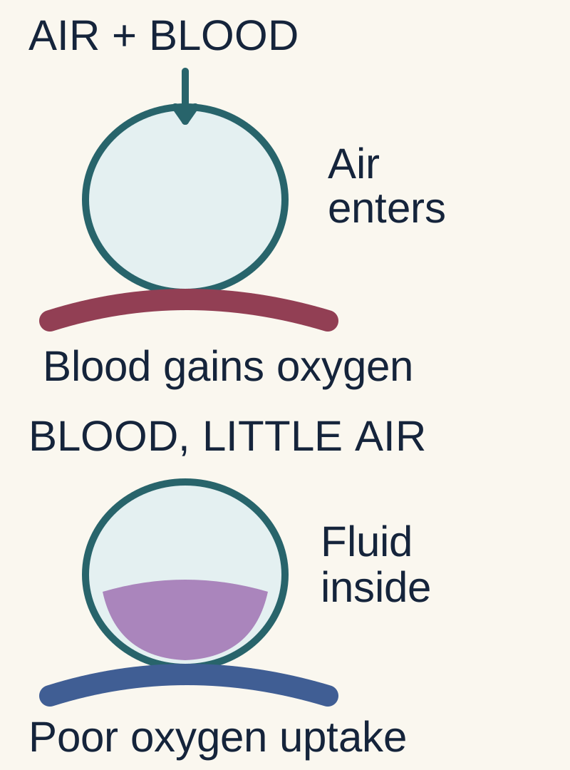
Ventilation means air delivery; perfusion means blood delivery. A shunt is blood passing a region without effective ventilation. Dead space is the opposite mismatch: air reaches a region with too little blood flow. Barrier injury can create both patterns in different parts of the same lung.
If blood still reaches a flooded air sac, what is missing?
Fresh air. Ventilation means air delivery; perfusion means blood delivery.
Can that blood collect oxygen from an air sac receiving no fresh air?
Very little. Blood bypasses effective gas exchange, creating a shunt.
What changes if air arrives but blood does not?
Ventilation is wasted. This is alveolar dead space, the opposite regional mismatch.
Acute respiratory distress syndrome is an acute inflammatory lung injury with pulmonary edema that is not primarily explained by hydrostatic pressure. Pneumonia and aspiration injure the lung directly. Sepsis outside the lung, pancreatitis, shock, major trauma, burns and transfusion can injure it through systemic inflammation. Pulmonary contusion, smoke exposure and drowning are additional direct settings. Infection and aspiration must be treated while respiratory support is organized. A syndrome name does not replace a search for its cause. [2]
Injury to capillary endothelium and alveolar epithelium permits protein-rich fluid to enter the airspaces. Neutrophil products and inflammatory mediators contribute to barrier damage. Loss of functional surfactant favors collapse. Gas is distributed unevenly through a lung containing flooded units, recruitable units and relatively preserved units. The preserved region receives a disproportionate share of the delivered tidal volume.
Flooded unit
Air delivery is absent or very low. Blood flow continues. Blood leaves without adequate oxygen uptake.
Shunt or very low V/Q
Vascularly obstructed unit
Air arrives. Blood flow is reduced by vascular injury or microthrombi. Ventilation contributes little to CO2 clearance.
Alveolar dead space or high V/Q
Read each column from air delivery to blood flow to consequence. Both patterns can coexist in ARDS. This is a conceptual comparison, not a diagnostic V/Q scan.
Blood perfusing unventilated alveoli produces an intrapulmonary shunt. Increasing inspired oxygen helps the ventilated lung but has limited effect on the completely unventilated portion. PEEP and prone positioning may improve recruitment and the distribution of ventilation. They do not guarantee that every opaque region is recruitable. Meanwhile, increased dead space makes CO2 clearance less efficient. Hypoxemia and hypercapnia therefore answer different physiological questions. [2][3]
Compliance is the change in volume divided by the change in pressure. A stiff respiratory system requires more pressure for a given tidal volume. That is why protective ventilation is needed even before oxygenation becomes catastrophic. Compliance can vary considerably among patients meeting ARDS criteria; a relatively compliant lung does not exclude the syndrome.
Case 6
Show answer and explanations for case 6
A. Lower oxygen affinity of circulating hemoglobin (Why this does not fit)
What would changing hemoglobin affinity alter?
Affinity changes alter oxygen binding at a given tension.
Which variable did the intervention actually change?
The observed intervention restores regional ventilation with unchanged perfusion, directly correcting a gas-delivery mismatch.
How should this distinction guide a similar patient?
Use the altered variable to identify the immediate mechanism.
Read the complete explanation
Affinity changes alter oxygen binding at a given tension. The observed intervention restores regional ventilation with unchanged perfusion, directly correcting a gas-delivery mismatch. Use the altered variable to identify the immediate mechanism.
B. Less blood leaving unventilated alveoli (Best answer)
What still reaches the flooded regions before recruitment?
Blood continues to perfuse them.
What missing component returns during recruitment?
Fresh air reaches the reopened airspaces.
What can that regional blood now do?
It can collect oxygen where it previously bypassed effective gas exchange.
What mismatch has decreased?
Less blood leaves unventilated units: shunt-like admixture falls.
Read the complete explanation
Shunted blood bypasses effective alveolar oxygen uptake. Reopening ventilation lets the same regional blood encounter fresh gas. Recruiting a perfused unit can reduce shunt-like admixture.
C. Less ventilation of unperfused alveoli (Why this does not fit)
What makes ventilation physiologically wasted?
Air reaches alveoli receiving little or no blood, so that ventilation contributes little to gas exchange.
Was blood absent from these regions before recruitment?
Blood was present in the affected regions before recruitment.
How should this distinction guide a similar patient?
Establish which component is missing before naming the mismatch.
Read the complete explanation
Reducing wasted ventilation can improve ventilatory efficiency. Blood was present in the affected regions before recruitment. Establish which component is missing before naming the mismatch.
D. Greater diffusion distance across the alveolar membrane (Why this does not fit)
What does a longer diffusion path do to transfer?
A longer diffusion path impedes gas transfer.
Did recruitment lengthen the barrier or restore air entry?
Improved air entry increases access to alveolar oxygen rather than increasing the diffusion barrier.
How should this distinction guide a similar patient?
Separate ventilation access from membrane diffusion distance.
Read the complete explanation
A longer diffusion path impedes gas transfer. Improved air entry increases access to alveolar oxygen rather than increasing the diffusion barrier. Separate ventilation access from membrane diffusion distance.
Takeaway: Recruiting a perfused unit can reduce shunt-like admixture.
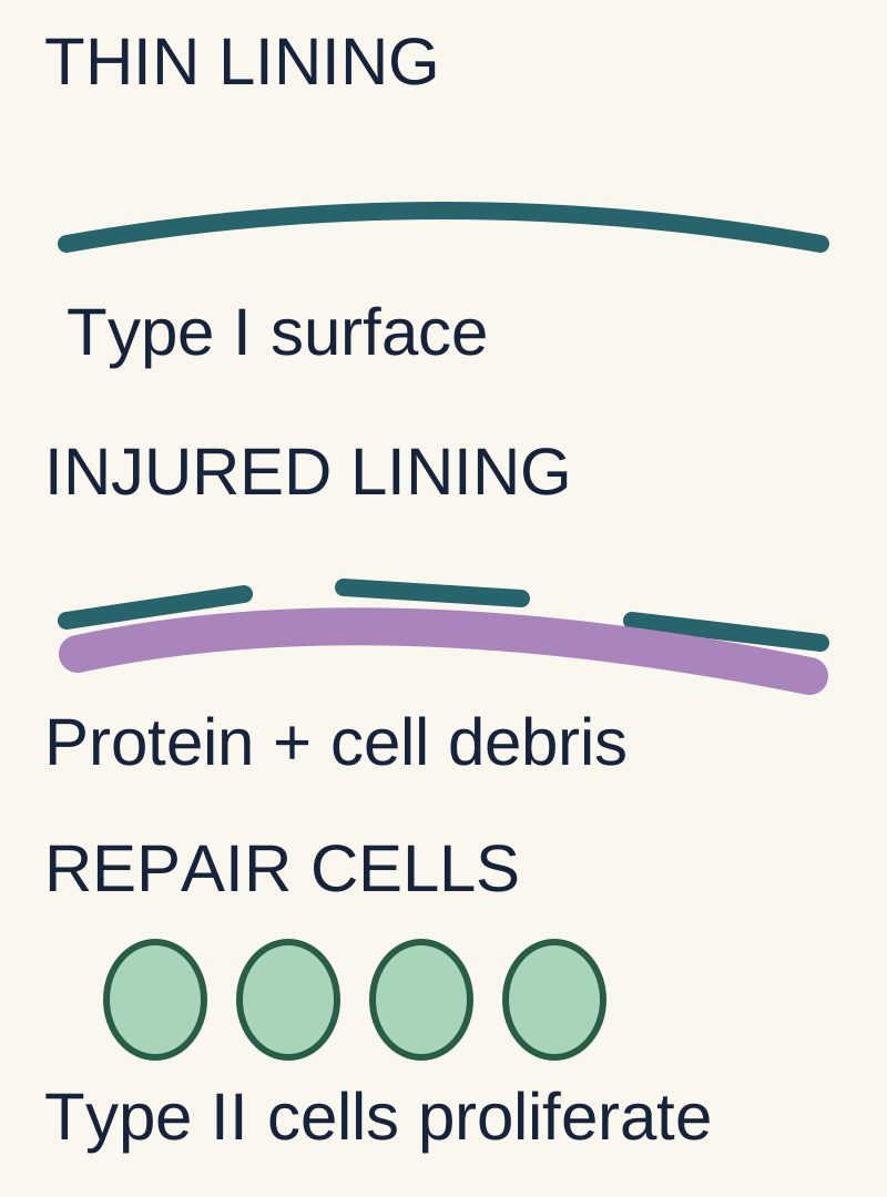
Hyaline membranes are protein-rich material and cellular debris lining injured airspaces. They support the diffuse alveolar damage pattern, without naming its cause. Type II pneumocytes make surfactant and can help replace injured epithelial cells. Repair and injury overlap; permanent fibrosis is not inevitable.
The thick purple material represents the hyaline-membrane injury pattern. The green cells represent epithelial repair. These processes can overlap; the drawing does not impose a fixed timeline.
Protein-rich material and cellular debris line an injured airspace. It supports diffuse alveolar damage, not one specific cause.
Which epithelial cell helps restore the lining?
Type II pneumocytes can proliferate, replenish epithelial cells and make surfactant.
Must repair pass through permanent fibrosis?
No. Injury and repair overlap; some lungs recover without a fibrotic phase.
Diffuse alveolar damage, or DAD, is the classic tissue pattern. Early exudative injury includes edema, epithelial injury and hyaline membranes composed of proteinaceous material and cellular debris. Hyaline membranes belong to the injury pattern; they do not identify a unique cause. Clinical ARDS does not require biopsy, and not every clinically defined case has DAD at autopsy. [8]
Early injury, often the first week Barrier leak, type I pneumocyte injury, protein-rich airspace material and surfactant dysfunction impair gas exchange.
Repair, often during the following weeks Type II pneumocytes proliferate and can replenish the epithelial lining. Edema clearance and resolution may restore function.
Persistent injury in some patients Fibroproliferation and fibrosis can impair compliance and prolong ventilator dependence. Pulmonary vascular disease can add right-heart strain.
These are overlapping processes, not three compulsory appointments on a calendar. Fibrosis can develop earlier than day 21, and recovery can occur without a fibrotic stage. A patient who remains hypoxemic also needs evaluation for infection, fluid accumulation, pneumothorax, embolism or another diagnosis rather than an automatic assumption of irreversible fibrosis. [2][8]
Establish the syndrome before calculating its severity
First establish acute onset, bilateral opacities, an appropriate edema mechanism and qualifying support. PaO2 is arterial oxygen tension in mmHg; FiO2 is the inspired oxygen fraction. P/F = PaO2 ÷ FiO2. Berlin categories are mild above 200 through 300, moderate above 100 through 200, and severe at or below 100.
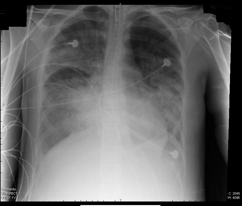
Enlarge the complete imageBoth lungs contain airspace opacities. In this ARDS image, opacities alone cannot distinguish inflammatory from hydrostatic edema. Use timing, support and cardiac assessment. Image: Original image shown
The timing, bilateral opacities, edema mechanism and required respiratory support must fit the definition.
For PaO2 60 and FiO2 0.60, what is P/F?
60 ÷ 0.60 = 100. FiO2 is a fraction, so 60% is entered as 0.60.
Which Berlin category includes exactly 100?
Severe. Moderate starts above 100; the endpoint is part of the distinction.
The Berlin definition combines onset within one week of an insult or new/worsening respiratory symptoms, bilateral opacities not fully explained by effusions, collapse or nodules, and respiratory failure not fully explained by cardiac failure or fluid overload. Oxygenation is assessed with positive pressure of at least 5 cm H2O. CPAP can qualify for mild Berlin ARDS. Moderate and severe Berlin categories can be assessed during invasive or noninvasive ventilation with PEEP of at least 5 cm H2O. [1]
P/F = PaO2 in mmHg divided by FiO2 as a fraction. For PaO2 60 on FiO2 0.60, P/F is 100, not 1.
Berlin oxygenation boundaries with the required support
Mild
Moderate
Severe
Mild200 < P/F ≤ 300
Moderate100 < P/F ≤ 200
SevereP/F ≤ 100
The endpoints matter. Exactly 200 is moderate; exactly 100 is severe. P/F 140 is moderate by Berlin, although it can fall within the oxygenation range used in prone-positioning trials. Record FiO2 and PEEP with the ratio because support settings affect the result. A low P/F alone does not diagnose ARDS. [1][5]
Assess the edema mechanism using the clinical course, fluid balance and cardiac evaluation. Normal left ventricular ejection fraction does not exclude heart failure with preserved ejection fraction. A low wedge pressure supports a nonhydrostatic process in context, but a mandatory wedge-pressure cutoff is absent from Berlin. ARDS and hydrostatic edema can coexist. BNP and bilateral opacities are not definitive separators. [1][2]
After transfusion, consider transfusion-related acute lung injury, or TRALI, when new hypoxemic pulmonary edema begins during or within six hours of transfusion and hydrostatic pressure does not principally explain it. Donor leukocyte antibodies can participate, but their detection is not required for the clinical diagnosis. Transfusion-associated circulatory overload, or TACO, is primarily hydrostatic. Timing alone cannot distinguish them, and an indeterminate or mixed category is possible. Stop the implicated transfusion, support oxygenation and notify the transfusion service for assessment. [13]
The 2024 global consensus expands recognition to nonintubated patients receiving HFNO at least 30 L/min or NIV/CPAP with at least 5 cm H2O end-expiratory pressure. Its nonintubated oxygenation criterion is P/F ≤300 or S/F ≤315, with SpO2 ≤97% when S/F is used. It also permits ultrasound by a trained operator and describes a resource-limited category without mandatory PEEP or flow thresholds. Keep these categories distinct from Berlin severity rather than silently applying invasive-ventilation labels to every oxygen device. [2]
Case 1
Show answer and explanations for case 1
A. Severe, then severe (Why this does not fit)
Where does the severe P/F range end?
Severe requires P/F at or below 100 at the time assessed.
Does the later ratio of 120 remain in that range?
The later value of 120 crosses the severe boundary.
How should this distinction guide a similar patient?
Reassess severity using current measurements and support.
Read the complete explanation
Severe requires P/F at or below 100 at the time assessed. The later value of 120 crosses the severe boundary. Reassess severity using current measurements and support.
B. Severe, then moderate (Best answer)
What is 60 divided by 0.60?
The initial P/F ratio is 100.
Which Berlin band includes exactly 100?
Severe includes the endpoint 100.
What is 72 divided by the unchanged 0.60?
The later P/F ratio is 120.
Where does 120 fall?
Above 100 through 200 is moderate: severe, then moderate.
What should you carry to the next assessment?
Recalculate the ratio on the current support; a category change does not mean ARDS has ended.
Read the complete explanation
Severe includes P/F at or below 100; moderate extends above 100 through 200. The ratio rises from 100 to 120 on qualifying support. Improved oxygenation can change the category without ending ARDS.
C. Moderate, then mild (Why this does not fit)
Where does the mild P/F range begin?
Mild requires P/F above 200 through 300.
Does the later ratio reach that range?
The later ratio is 120, which remains below the mild range.
How should this distinction guide a similar patient?
Calculate both ratios before assigning a change in category.
Read the complete explanation
Mild requires P/F above 200 through 300. The later ratio is 120, which remains below the mild range. Calculate both ratios before assigning a change in category.
D. Moderate, then moderate (Why this does not fit)
Does the moderate range include exactly 100?
Moderate includes P/F above 100 through 200.
Which initial calculation reaches that endpoint?
The initial ratio is exactly 100; that endpoint belongs to severe.
How should this distinction guide a similar patient?
The lower moderate boundary is exclusive.
Read the complete explanation
Moderate includes P/F above 100 through 200. The initial ratio is exactly 100; that endpoint belongs to severe. The lower moderate boundary is exclusive.
Takeaway: Improved oxygenation can change the category without ending ARDS.
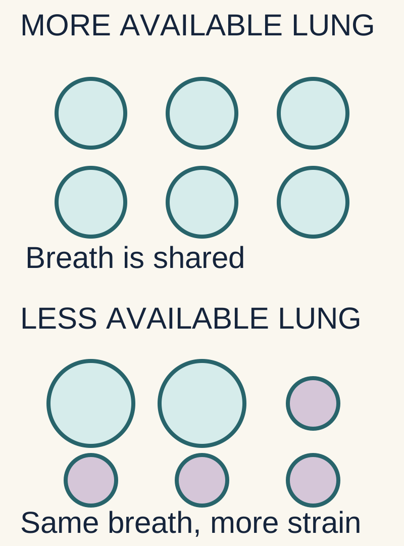
Flooded or collapsed regions accept little air, concentrating each breath in the remaining aerated lung. Predicted body weight estimates lung size from height and sex. Start near 6 mL/kg, then check plateau pressure, gas exchange and circulation; a starting calculation is not the final safety assessment.
The circles represent available airspaces, not measured lung volumes. Keeping the same total breath when less lung is available concentrates distension in the remaining aerated regions.
Why can an ordinary-sized breath overfill an injured lung?
Flooded or collapsed regions accept little air. The remaining aerated lung receives more of each breath.
Which weight estimates lung size for the initial volume?
Predicted body weight, derived from height and sex. Extra body mass does not proportionally enlarge the lungs.
Does starting at 6 mL/kg finish the safety check?
No. Reassess plateau pressure, gas exchange and circulation. The same volume can create different pressures in different lungs.
For an intubated adult, start near 6 mL/kg predicted body weight, calculated from height and sex rather than actual weight. A predicted weight of 70 kg gives 420 mL. Obesity does not proportionally enlarge the lungs. Current guidance supports a protective range of 4 to 8 mL/kg with plateau pressure below 30 cm H2O; the delivered volume may need to fall below 6 if pressure remains excessive. [3][4]
Measure plateau pressure during an end-inspiratory pause when flow is absent and the patient is not actively distorting the measurement. Peak pressure includes the resistive pressure needed to push gas through the tube and airways. A high peak with a much lower plateau suggests added airway resistance, such as secretions or bronchospasm. A high plateau raises concern about respiratory-system distension, though chest-wall mechanics also affect it.
Excess volume can overdistend the aerated lung, termed volutrauma. Excess distending pressure can contribute to barotrauma, including pneumothorax. Repetitive collapse and reopening contribute to atelectrauma. Mechanical injury can amplify inflammation, termed biotrauma. A mode name does not prevent these injuries. Pressure control still requires monitoring delivered tidal volume, and volume control still requires pressure monitoring. [4]
PEEP helps prevent end-expiratory collapse. In moderate or severe ARDS, ATS conditionally favors higher PEEP without prolonged recruitment maneuvers. Titration must account for oxygenation, mechanics and circulation. Excessive PEEP can overdistend open lung, reduce venous return and impair the right ventricle. New hypotension after a pressure change is a reason to reassess, not simply add more pressure. [3]
Permissive hypercapnia accepts some CO2 retention to avoid injurious ventilation. There is no universal PaCO2 ceiling of 60 mmHg or universally safe pH cutoff of 7.20. Consider pH trajectory, hemodynamics, respiratory rate, expiratory time and dead space. Intracranial hypertension is a major concern because CO2 dilates cerebral vessels; severe right-heart failure also warrants caution. A stable patient with modest acidemia does not need larger breaths simply to produce a normal gas. Severe or worsening acidemia requires active reassessment rather than unconditional acceptance. [3][4]
The original low-volume trial found mortality of 31.0% versus 39.8%, an absolute difference of 8.8 percentage points and a relative reduction of about 22%. Those describe different measures of benefit. A volume just above 6 mL/kg is not guaranteed to rupture an alveolus; risk depends on the lung and the pressures it experiences. [4]
Case 10
Show answer and explanations for case 10
A. 560 mL (Why this does not fit)
What dose per predicted kilogram gives 560 mL?
Eight mL/kg predicted weight lies at the upper end of a protective range used in some settings.
Is 8 mL/kg the usual initial target asked for?
The usual initial target asked for here is 6 mL/kg rather than 8.
How should this distinction guide a similar patient?
Distinguish the starting target from a range that may be individualized later.
Read the complete explanation
Eight mL/kg predicted weight lies at the upper end of a protective range used in some settings. The usual initial target asked for here is 6 mL/kg rather than 8. Distinguish the starting target from a range that may be individualized later.
B. 710 mL (Why this does not fit)
Which weight calculation gives approximately 710 mL?
Six mL/kg actual weight would give approximately this volume.
Does actual weight estimate the relevant lung size?
Extra body mass does not proportionally increase lung size, so actual weight is the wrong basis.
How should this distinction guide a similar patient?
Obesity changes body mass more than the lung-size estimate used for ventilation.
Read the complete explanation
Six mL/kg actual weight would give approximately this volume. Extra body mass does not proportionally increase lung size, so actual weight is the wrong basis. Obesity changes body mass more than the lung-size estimate used for ventilation.
C. 840 mL (Why this does not fit)
What dose per predicted kilogram gives 840 mL?
Twelve mL/kg predicted weight reflects a traditional larger-volume approach.
Does that match the protective starting strategy?
The protective strategy starts with a substantially smaller breath.
How should this distinction guide a similar patient?
Avoid confusing historical comparison volumes with the protective starting target.
Read the complete explanation
Twelve mL/kg predicted weight reflects a traditional larger-volume approach. The protective strategy starts with a substantially smaller breath. Avoid confusing historical comparison volumes with the protective starting target.
D. 420 mL (Best answer)
Which supplied weight estimates lung size for this calculation?
Predicted body weight:70 kg.
What starting volume follows from 6 mL/kg?
Seventy times six gives 420 mL.
Does that finish the ventilator prescription?
No. Obtain valid pressure and gas-exchange measurements and adjust the starting setting as needed.
Read the complete explanation
A usual starting point is 6 mL/kg predicted body weight. Seventy times six gives 420 mL; subsequent pressure and gas-exchange measurements guide adjustment. Use predicted lung size for the starting breath, then assess its effects.
Takeaway: Use predicted lung size for the starting breath, then assess its effects.
Combine effective support without waiting for collapse
Proning can distribute ventilation and lung stress more evenly, including better ventilation of perfused dorsal regions. An oxygenation response alone does not establish survival benefit. Fluid decisions depend on the phase of illness: restore perfusion during shock, then consider conservative fluid management when circulation permits.
Does proning work by draining all fluid out?
No. It redistributes ventilation and lung stress, often improving ventilation of well-perfused dorsal regions.
If oxygen rises after a treatment, has survival benefit been proved?
No. A physiological response and a patient-centered outcome are different kinds of evidence.
What comes before diuresis when shock persists?
Assess and restore perfusion. Conservative fluid management after shock is not an instruction to remove fluid during unresolved shock.
Prone positioning redistributes ventilation and lung stress more evenly, often improving ventilation of dorsal regions while perfusion remains substantial there. It is not simply fluid draining out of the lung. PROSEVA studied P/F below 150 with FiO2 at least 0.60 and PEEP at least 5, using sessions of at least 16 hours. This is an early treatment alongside protective ventilation for eligible severe hypoxemia, not a final maneuver after every drug has failed. Safe positioning requires a trained team, secure lines and airway, and attention to pressure injury and spinal stability. [5]
Systemic corticosteroids have a conditional ATS recommendation for intubated patients with ARDS. For nonintubated patients, use depends on an underlying cause with established steroid benefit; their role in other causes remains uncertain. The best drug, dose and duration are not settled for all causes. Assess timing, underlying infection and adverse effects; do not convert this into routine high-dose pulse therapy or wait for fibrosis to justify treatment. Starting steroids more than two weeks after onset has concerning older evidence and needs particular judgment. [3]
For early severe ARDS with persistent breath stacking despite appropriate analgesia, sedation and ventilator adjustment, a short neuromuscular-blocker course can help maintain protection. Paralysis does not provide analgesia or unconsciousness. ROSE compared routine early infusion with a usual-care strategy that included lighter sedation and found no mortality advantage. Selective use and daily reassessment are more accurate teaching than promising benefit to everyone. [3][7]
Fluid management has two phases. Restore perfusion during shock using individualized resuscitation and vasopressors when indicated. Once shock has resolved, avoid unnecessary fluid and consider diuresis with renal and perfusion monitoring. FACTT improved ventilator-free days with a conservative strategy but did not demonstrate a significant mortality reduction. Its protocol does not justify aggressive diuresis solely because a vasopressor-supported MAP looks acceptable. [6][12]
Contact an experienced ECMO center early when severe, potentially reversible failure persists despite appropriate conventional support. Selected patients may benefit from venovenous ECMO, which supports gas exchange but does not directly provide circulatory pumping support. Evaluation includes severity and duration of hypoxemia or acidosis, reversibility, bleeding risk and other organ failure. Referral need not wait for completion of an inflexible sequence of every possible intervention. [3]
Do not confuse temporary oxygenation improvement with survival benefit. Inhaled nitric oxide may serve as a selected bridge but has not established a routine survival benefit. Adult surfactant replacement has not reproduced the established role of surfactant in neonatal deficiency. Routine HFOV is avoided after trials including OSCILLATE showed harm. Prolonged high-pressure recruitment maneuvers are specifically discouraged. [9][10][11][3]
At the bedside, establish the cause and definition, protect volume and pressure, reassess perfusion, add prone positioning when appropriate, and evaluate adjuncts according to the patient's trajectory. A sudden deterioration requires a fresh search for tube obstruction, pneumothorax and circulatory failure before attributing everything to ARDS progression.
Apply the lesson
Case 2
Show answer and explanations for case 2
A. Unchanged oxygenation efficiency; both measurements are mild (Why this does not fit)
What proportional change would preserve P/F?
Similar proportional changes in PaO2 and FiO2 preserve P/F.
Are the PaO2 and FiO2 increases proportional?
A 12.5% rise in PaO2 accompanies a 50% rise in FiO2, lowering the ratio.
How should this distinction guide a similar patient?
Compare fractions, not the direction of PaO2 change alone.
Read the complete explanation
Similar proportional changes in PaO2 and FiO2 preserve P/F. A 12.5% rise in PaO2 accompanies a 50% rise in FiO2, lowering the ratio. Compare fractions, not the direction of PaO2 change alone.
B. Improved oxygenation efficiency; moderate becomes mild (Why this does not fit)
What P/F value is needed for mild ARDS?
Mild ARDS has P/F above 200.
Did PaO2 rise enough to keep pace with FiO2?
FiO2 increased proportionally more than PaO2; the ratio falls to 150.
How should this distinction guide a similar patient?
A higher PaO2 can conceal an increased oxygen requirement.
Read the complete explanation
Mild ARDS has P/F above 200. FiO2 increased proportionally more than PaO2; the ratio falls to 150. A higher PaO2 can conceal an increased oxygen requirement.
C. Worse oxygenation efficiency; both measurements are moderate (Best answer)
What is the first P/F ratio?
Eighty divided by 0.40 is 200.
What is the second ratio?
Ninety divided by 0.60 is 150.
Why can the ratio fall while PaO2 rises?
FiO2 increased proportionally more than PaO2.
Do these values cross a Berlin category boundary?
No. Both 150 and 200 are moderate; the oxygenation ratio worsened within that band.
What does this comparison avoid?
Do not infer better oxygenation efficiency from PaO2 alone or equate a support-dependent ratio change with proven intrinsic disease progression.
Read the complete explanation
P/F compares arterial oxygen tension with the delivered oxygen fraction. The ratio falls from 200 to 150; both fall above 100 through 200. Track support requirements as well as arterial oxygen tension.
D. Worse oxygenation efficiency; moderate becomes severe (Why this does not fit)
What ratio defines the severe boundary?
Severe Berlin ARDS includes P/F at or below 100.
Does the final ratio cross that boundary?
The ratio falls, but its final value is 150.
How should this distinction guide a similar patient?
Physiologic deterioration can occur within one severity band.
Read the complete explanation
Severe Berlin ARDS includes P/F at or below 100. The ratio falls, but its final value is 150. Physiologic deterioration can occur within one severity band.
Takeaway: Track support requirements as well as arterial oxygen tension.
What ratio would make the second assessment severe?
Severe ARDS requires P/F at or below 100.
What does 60 divided by 0.40 actually give?
The second ratio is 150 despite greater support.
How should this distinction guide a similar patient?
Increasing support does not by itself define the severe oxygenation band.
Read the complete explanation
Severe ARDS requires P/F at or below 100. The second ratio is 150 despite greater support. Increasing support does not by itself define the severe oxygenation band.
B. Moderate, then moderate (Why this does not fit)
What is the upper boundary of moderate ARDS?
Moderate requires P/F above 100 through 200.
Where does the first ratio of 250 fall?
The first ratio is 250, placing it in the mild band.
How should this distinction guide a similar patient?
Divide PaO2 by the fractional FiO2 at each assessment.
Read the complete explanation
Moderate requires P/F above 100 through 200. The first ratio is 250, placing it in the mild band. Divide PaO2 by the fractional FiO2 at each assessment.
C. Mild, then outside Berlin support criteria (Why this does not fit)
Can qualifying positive pressure be noninvasive?
Berlin categories require specified positive pressure, not necessarily an invasive tube.
What support and pressure are supplied at the second assessment?
The second assessment supplies NIV with PEEP 8.
How should this distinction guide a similar patient?
Check the pressure and mode requirements directly.
Read the complete explanation
Berlin categories require specified positive pressure, not necessarily an invasive tube. The second assessment supplies NIV with PEEP 8. Check the pressure and mode requirements directly.
D. Mild, then moderate (Best answer)
What is the first P/F ratio?
Seventy-five divided by 0.30 is 250.
Does CPAP 5 qualify for the mild band?
Yes. A ratio above 200 through 300 on qualifying CPAP meets mild oxygenation criteria.
What is the later ratio?
Sixty divided by 0.40 is 150.
Does noninvasive PEEP 8 permit the moderate classification?
Yes. This qualifying positive-pressure assessment is moderate; intubation is not the deciding requirement.
What two checks travel together?
Check the oxygenation interval and the support criteria of the named definition.
Read the complete explanation
Berlin permits qualifying positive-pressure support, including noninvasive support. Ratios are 250 on CPAP 5 and 150 on NIV PEEP 8. Classify oxygenation and verify support without using intubation as a shortcut.
Takeaway: Classify oxygenation and verify support without using intubation as a shortcut.
A. Elevated pulmonary capillary hydrostatic pressure (Best answer)
Does an EF of 60% exclude elevated LV filling pressure?
No. Preserved systolic EF can coexist with high filling pressure.
What does the Doppler assessment add?
It supports high left-sided filling pressure as a source of pulmonary hydrostatic pressure.
How does the rapid treatment response affect that explanation?
Improvement with afterload reduction and diuresis supports a dominant pressure-driven mechanism.
What is the transferable distinction?
Assess filling pressure and clinical course rather than using EF alone to exclude cardiogenic edema.
Read the complete explanation
High left-sided filling pressure drives fluid into the lungs. Preserved EF coexists with high filling pressure, and afterload reduction plus diuresis rapidly helps. Preserved EF does not exclude cardiogenic pulmonary edema.
B. Inflammatory alveolar-capillary barrier leak (Why this does not fit)
What kind of injury produces permeability edema?
Acute inflammatory injury can cause bilateral permeability edema.
Which observed trigger and response instead support a pressure mechanism?
The supplied trigger, high filling pressure and rapid cardiac-treatment response instead support hydrostatic edema.
How should this distinction guide a similar patient?
Bilateral opacities require assessment of edema mechanism.
Read the complete explanation
Acute inflammatory injury can cause bilateral permeability edema. The supplied trigger, high filling pressure and rapid cardiac-treatment response instead support hydrostatic edema. Bilateral opacities require assessment of edema mechanism.
C. Impaired lymphatic drainage from malignant obstruction (Why this does not fit)
What course might lymphatic obstruction produce?
Lymphatic obstruction can cause persistent interstitial edema.
How does this rapid treatment response change its plausibility?
Abrupt onset and rapid reversal with cardiac treatment fit a pressure-driven process better.
How should this distinction guide a similar patient?
Time course and reversibility help distinguish edema mechanisms.
Read the complete explanation
Lymphatic obstruction can cause persistent interstitial edema. Abrupt onset and rapid reversal with cardiac treatment fit a pressure-driven process better. Time course and reversibility help distinguish edema mechanisms.
D. Acute reduction of plasma oncotic pressure (Why this does not fit)
Which plasma-protein change can lower oncotic pressure and favor edema?
Marked hypoalbuminemia can favor edema formation.
What observed evidence makes that less explanatory in this episode?
No protein-loss process is supplied; the temporally linked hypertension and filling pressure explain this episode.
How should this distinction guide a similar patient?
Prefer the mechanism supported by the observed hemodynamics.
Read the complete explanation
Marked hypoalbuminemia can favor edema formation. No protein-loss process is supplied; the temporally linked hypertension and filling pressure explain this episode. Prefer the mechanism supported by the observed hemodynamics.
E. Acute pulmonary arterial outflow obstruction (Why this does not fit)
Which ventricle faces the primary load in PE?
PE can abruptly load the right ventricle and impair oxygenation.
Which filling pressure is actually elevated here?
The measured abnormality is elevated LV filling pressure with a cardiac-treatment response.
How should this distinction guide a similar patient?
Localize the pressure abnormality before attributing dyspnea to obstruction.
Read the complete explanation
PE can abruptly load the right ventricle and impair oxygenation. The measured abnormality is elevated LV filling pressure with a cardiac-treatment response. Localize the pressure abnormality before attributing dyspnea to obstruction.
Takeaway: Preserved EF does not exclude cardiogenic pulmonary edema.
Which oxygen measurement and support does mild Berlin grading use?
Mild Berlin grading uses PaO2/FiO2 on qualifying CPAP or PEEP.
Does this HFNO assessment supply them?
This patient has HFNO and an S/F measurement, not the required Berlin measurements and support.
How should this distinction guide a similar patient?
Match both oxygenation measure and support to the definition used.
Read the complete explanation
Mild Berlin grading uses PaO2/FiO2 on qualifying CPAP or PEEP. This patient has HFNO and an S/F measurement, not the required Berlin measurements and support. Match both oxygenation measure and support to the definition used.
B. Moderate Berlin ARDS (Why this does not fit)
Which oxygen measurement does moderate Berlin grading use?
Moderate Berlin grading uses an arterial P/F ratio on qualifying positive pressure.
What measure is actually available here?
S/F 188 cannot be substituted directly for a Berlin P/F value.
How should this distinction guide a similar patient?
Saturation and tension ratios have different thresholds.
Read the complete explanation
Moderate Berlin grading uses an arterial P/F ratio on qualifying positive pressure. S/F 188 cannot be substituted directly for a Berlin P/F value. Saturation and tension ratios have different thresholds.
C. Nonintubated global ARDS (Best answer)
Which saturation number goes into S/F?
Use 94, not 0.94, for the percentage saturation.
What is 94 divided by 0.50?
The S/F ratio is 188.
Does this assessment meet the global HFNO and saturation conditions?
Flow 40 is above 30 L/min; SpO2 is 94%, within the at-most 97% range for using S/F.
Which classification follows with the other stated criteria?
Nonintubated global ARDS is supported; the measurement is not a Berlin P/F severity grade.
What prevents mixing definitions?
Name the framework before applying its oxygenation measure and support requirements.
Read the complete explanation
The global pathway permits HFNO at least 30 L/min and S/F at most 315 when SpO2 is at most 97%. Flow is 40, saturation is 94%, and S/F is 188 with the other criteria satisfied. Name the definition before applying its oxygenation criteria.
D. Severe Berlin ARDS (Why this does not fit)
Which oxygenation and support criteria establish severe Berlin ARDS?
Severe Berlin ARDS requires P/F at or below 100 with qualifying positive pressure.
Which required Berlin information is missing?
Neither an arterial P/F nor qualifying positive-pressure support is supplied.
How should this distinction guide a similar patient?
A large oxygen requirement does not supply missing classification criteria.
Read the complete explanation
Severe Berlin ARDS requires P/F at or below 100 with qualifying positive pressure. Neither an arterial P/F nor qualifying positive-pressure support is supplied. A large oxygen requirement does not supply missing classification criteria.
Takeaway: Name the definition before applying its oxygenation criteria.
A. A larger fraction of each breath reaches insufficiently perfused alveoli (Best answer)
What can dilute mixed expired CO2?
A larger contribution from ventilated units exchanging little CO2 with blood.
What does rising arterial CO2 imply when production is unchanged?
Effective CO2-clearing ventilation has fallen.
How can that happen while total delivered ventilation stays constant?
More of each breath is spent in poorly perfused units: the dead-space fraction rises.
What should be compared in another ventilated patient?
Compare total delivered ventilation with the fraction that actually exchanges gas.
Read the complete explanation
Ventilated units receiving little blood contribute gas with little CO2. Their added contribution dilutes mixed expired CO2 while less effective ventilation allows arterial CO2 to rise. Compare effective gas-exchanging ventilation with total delivered ventilation.
B. An increase in pulmonary perfusion of previously unperfused ventilated units (Why this does not fit)
What happens when perfusion returns to a ventilated unit?
Restoring perfusion makes previously wasted ventilation available for CO2 elimination.
Would that predict the observed retention and expired-gas dilution?
That change would improve clearance rather than explain increasing arterial retention with lower expired CO2.
How should this distinction guide a similar patient?
Predict the direction of gas-exchange change when air and blood overlap more effectively.
Read the complete explanation
Restoring perfusion makes previously wasted ventilation available for CO2 elimination. That change would improve clearance rather than explain increasing arterial retention with lower expired CO2. Predict the direction of gas-exchange change when air and blood overlap more effectively.
C. A fall in total delivered minute ventilation with unchanged perfusion (Why this does not fit)
What delivery change accompanies global hypoventilation?
Global hypoventilation can raise arterial CO2 by reducing minute ventilation.
Did delivered minute ventilation fall here?
Delivered volume and rate remain unchanged, while the expired mixture contains less CO2.
How should this distinction guide a similar patient?
Look for a change in efficiency when the delivered minute ventilation has not fallen.
Read the complete explanation
Global hypoventilation can raise arterial CO2 by reducing minute ventilation. Delivered volume and rate remain unchanged, while the expired mixture contains less CO2. Look for a change in efficiency when the delivered minute ventilation has not fallen.
D. An increase in tissue CO2 production with unchanged gas matching (Why this does not fit)
How can increased metabolism raise arterial CO2?
Increased metabolism can raise arterial CO2 when ventilation cannot keep pace.
Which production and expired-gas findings argue against that account?
Metabolic state is unchanged, and a falling mixed-expired value points toward dilution by poorly exchanging gas.
How should this distinction guide a similar patient?
Assess production and gas-exchange efficiency separately.
Read the complete explanation
Increased metabolism can raise arterial CO2 when ventilation cannot keep pace. Metabolic state is unchanged, and a falling mixed-expired value points toward dilution by poorly exchanging gas. Assess production and gas-exchange efficiency separately.
Takeaway: Compare effective gas-exchanging ventilation with total delivered ventilation.
A. Usual interstitial pneumonia is present; chronic fibrosis explains the course (Why this does not fit)
What architecture and time course characterize UIP?
UIP is a chronic patchy fibrosing pattern with architectural remodeling.
Does an eight-day diffuse injury pattern fit that course?
An eight-day course with diffuse acute epithelial injury fits DAD more closely.
How should this distinction guide a similar patient?
Integrate time course with the dominant tissue pattern.
Read the complete explanation
UIP is a chronic patchy fibrosing pattern with architectural remodeling. An eight-day course with diffuse acute epithelial injury fits DAD more closely. Integrate time course with the dominant tissue pattern.
B. Organizing pneumonia is the dominant pattern; the cause remains open (Why this does not fit)
Which dominant tissue structure supports organizing pneumonia?
Organizing pneumonia features intra-alveolar fibroblastic plugs as a dominant finding.
Is that the dominant structure described?
The described dominant lesions are epithelial injury and hyaline membranes.
How should this distinction guide a similar patient?
Identify the actual histologic structures before assigning a pattern.
Read the complete explanation
Organizing pneumonia features intra-alveolar fibroblastic plugs as a dominant finding. The described dominant lesions are epithelial injury and hyaline membranes. Identify the actual histologic structures before assigning a pattern.
C. Bacterial pneumonia is established; the organism remains unidentified (Why this does not fit)
What tissue evidence would help establish bacterial pneumonia?
Suppurative inflammation or organisms can support an infectious pneumonia diagnosis.
Do hyaline membranes identify a pulmonary pathogen?
Hyaline membranes are not organism-specific and the sampled stains show no organisms.
How should this distinction guide a similar patient?
Tissue injury may fit sepsis without proving a pulmonary pathogen.
Read the complete explanation
Suppurative inflammation or organisms can support an infectious pneumonia diagnosis. Hyaline membranes are not organism-specific and the sampled stains show no organisms. Tissue injury may fit sepsis without proving a pulmonary pathogen.
D. Cardiogenic edema is the dominant pattern; LV disease is the likely cause (Why this does not fit)
What pressure mechanism produces cardiogenic edema?
What additional tissue injury makes simple hydrostatic edema less fitting?
Prominent diffuse epithelial injury with hyaline membranes supports an acute damage pattern instead.
How should this distinction guide a similar patient?
Edema alone and diffuse alveolar damage are not equivalent tissue descriptions.
Read the complete explanation
Hydrostatic edema follows elevated pulmonary capillary pressure. Prominent diffuse epithelial injury with hyaline membranes supports an acute damage pattern instead. Edema alone and diffuse alveolar damage are not equivalent tissue descriptions.
E. Diffuse alveolar damage is present; the initiating cause needs clinical correlation (Best answer)
What are the eosinophilic lining membranes in this acute injury?
Hyaline membranes composed of protein-rich material and cellular debris.
Which injury pattern do they support with diffuse epithelial damage?
Diffuse alveolar damage.
Does that morphology establish one initiating cause?
No. Sepsis is compatible, but the pattern is not cause-specific.
What is the next-case rule?
Combine tissue-pattern recognition with clinical evidence about the trigger.
Read the complete explanation
Hyaline membranes with diffuse epithelial injury are characteristic of DAD. Sepsis is a compatible trigger, but the morphology does not uniquely establish it. Use pathology to identify the injury pattern and clinical evidence to assess cause.
Takeaway: Use pathology to identify the injury pattern and clinical evidence to assess cause.
A. Collagen deposition and interstitial contraction (Why this does not fit)
Which cell population deposits interstitial collagen?
Activated fibroblasts produce extracellular matrix and remodel tissue.
Are the sampled cells stromal or epithelial?
The sampled cells are lamellar-body-containing epithelium, not stromal fibroblasts.
How should this distinction guide a similar patient?
Distinguish epithelial regeneration from fibrotic remodeling.
Read the complete explanation
Activated fibroblasts produce extracellular matrix and remodel tissue. The sampled cells are lamellar-body-containing epithelium, not stromal fibroblasts. Distinguish epithelial regeneration from fibrotic remodeling.
B. Surfactant replacement and epithelial repopulation (Best answer)
Which alveolar epithelial cell contains lamellar bodies?
The type II pneumocyte.
What product do those organelles connect it to?
Surfactant, which helps stabilize alveolar surfaces.
How does proliferation of this population support repair?
Type II cells can help repopulate injured alveolar epithelium.
Which two functions are preserved together?
Surfactant replacement and epithelial repopulation.
Read the complete explanation
Type II pneumocytes make surfactant and can replenish alveolar epithelium. Lamellar bodies and cuboidal proliferation identify this reparative cell population. Type II cell survival supports both surface stability and epithelial repair.
C. Mucus secretion and ciliary particle clearance (Why this does not fit)
Are these cells from that conducting-airway compartment?
Alveolar lamellar-body-containing cells are specialized for a different compartment.
How should this distinction guide a similar patient?
Match epithelial function to its anatomic location.
Read the complete explanation
Conducting-airway epithelium supports mucociliary clearance. Alveolar lamellar-body-containing cells are specialized for a different compartment. Match epithelial function to its anatomic location.
D. Endothelial barrier repair and vascular tone regulation (Why this does not fit)
Which side of the barrier is lined by endothelium?
Pulmonary endothelial cells regulate the blood-facing barrier and vascular signaling.
Where are these lamellar-body-containing cells located?
These cells line airspaces and contain surfactant-associated organelles.
How should this distinction guide a similar patient?
Localize the cell to the air or blood side of the barrier.
Read the complete explanation
Pulmonary endothelial cells regulate the blood-facing barrier and vascular signaling. These cells line airspaces and contain surfactant-associated organelles. Localize the cell to the air or blood side of the barrier.
E. Particle phagocytosis and antigen presentation (Why this does not fit)
Which alveolar cell chiefly phagocytoses particles?
Alveolar macrophages ingest particles and participate in immune signaling.
Do lamellar-body-containing epithelial cells match that lineage?
Cuboidal epithelial cells with lamellar bodies identify a different lineage.
How should this distinction guide a similar patient?
Use cell identity before assigning its dominant repair role.
Read the complete explanation
Alveolar macrophages ingest particles and participate in immune signaling. Cuboidal epithelial cells with lamellar bodies identify a different lineage. Use cell identity before assigning its dominant repair role.
Takeaway: Type II cell survival supports both surface stability and epithelial repair.
A. Perform bronchoscopy before changing ventilator settings (Why this does not fit)
What pressure pattern would suggest an obstructed airway?
An obstructed airway can cause elevated resistive pressure.
Do the patent tube and high plateau support that target?
The tube is patent and the abnormal measurement is plateau pressure.
How should this distinction guide a similar patient?
Confirm an airway-resistance problem before targeting airway clearance.
Read the complete explanation
An obstructed airway can cause elevated resistive pressure. The tube is patent and the abnormal measurement is plateau pressure. Confirm an airway-resistance problem before targeting airway clearance.
B. Raise PEEP by 5 while preserving tidal volume (Why this does not fit)
What benefit does useful PEEP recruitment aim to provide?
PEEP can recruit lung and improve oxygenation in selected patients.
Has recruitment benefit been demonstrated in this case?
Recruitment benefit is not demonstrated here, while plateau is already excessive.
How should this distinction guide a similar patient?
Titrate PEEP using recruitability and circulation rather than an isolated high plateau.
Read the complete explanation
PEEP can recruit lung and improve oxygenation in selected patients. Recruitment benefit is not demonstrated here, while plateau is already excessive. Titrate PEEP using recruitability and circulation rather than an isolated high plateau.
C. Increase inspiratory flow while preserving tidal volume (Why this does not fit)
Which pressure component changes with inspiratory flow?
Higher flow shortens inspiratory time and can alter peak pressure.
Would that directly lower a no-flow plateau measurement?
It does not reduce the static distending pressure measured during a no-flow hold.
How should this distinction guide a similar patient?
Treat plateau pressure as a separate variable from flow-dependent peak pressure.
Read the complete explanation
Higher flow shortens inspiratory time and can alter peak pressure. It does not reduce the static distending pressure measured during a no-flow hold. Treat plateau pressure as a separate variable from flow-dependent peak pressure.
D. Increase the rate to 35/min while preserving tidal volume (Why this does not fit)
What problem can increasing rate help correct?
A higher rate may offset CO2 retention during protective ventilation.
Is severe acidemia the immediate measured hazard?
The immediate abnormality is excessive plateau pressure, not severe acidemia.
How should this distinction guide a similar patient?
Correct the identified mechanical hazard before pursuing a normal PaCO2.
Read the complete explanation
A higher rate may offset CO2 retention during protective ventilation. The immediate abnormality is excessive plateau pressure, not severe acidemia. Correct the identified mechanical hazard before pursuing a normal PaCO2.
E. Reduce tidal volume and reassess pressure and pH (Best answer)
Which pressure was measured with no inspiratory flow?
Which proposed adjustment directly reduces the volume causing that pressure?
Reduce tidal volume within a protective strategy.
What must follow the adjustment?
Reassess plateau and pH so pressure protection remains balanced with gas exchange.
Read the complete explanation
Smaller breaths can reduce plateau pressure within a protective strategy. Plateau is above target and current pH leaves room for a monitored volume reduction. Reassess mechanics and gas exchange together after changing volume.
Takeaway: Reassess mechanics and gas exchange together after changing volume.
A. Inspect the circuit and pass a suction catheter through the tube (Best answer)
What changed in the peak–plateau difference?
The gap widened because peak rose while plateau stayed 25.
Which pressure component disappears during a passive no-flow hold?
The flow-dependent resistive component.
Where should the new obstruction search begin?
Inspect the circuit and evaluate tube and airway patency, including passage of a suction catheter.
What prevents the wrong adjustment?
Localize a pressure rise before treating it as worsening lung stiffness.
Read the complete explanation
Tube or airway resistance increases pressure during flow. The new pressure rise disappears during the inspiratory hold. A widening peak–plateau gap directs attention to the resistive pathway.
B. Reduce tidal volume to treat worsening respiratory-system compliance (Why this does not fit)
How does lower compliance affect plateau at fixed volume?
Lower compliance raises static pressure for a fixed volume.
Did the static pressure rise here?
Plateau is unchanged, making a new compliance loss less likely than increased resistance.
How should this distinction guide a similar patient?
Compare static and dynamic pressures before attributing a peak rise to lung stiffness.
Read the complete explanation
Lower compliance raises static pressure for a fixed volume. Plateau is unchanged, making a new compliance loss less likely than increased resistance. Compare static and dynamic pressures before attributing a peak rise to lung stiffness.
C. Lengthen expiratory time to treat dynamic hyperinflation (Why this does not fit)
What can trapped gas do to total PEEP?
Trapped gas can elevate total PEEP and impair circulation.
Which supplied findings argue against trapping?
Expiratory flow reaches zero and total PEEP is unchanged.
How should this distinction guide a similar patient?
Check the flow curve and total PEEP when considering air trapping.
Read the complete explanation
Trapped gas can elevate total PEEP and impair circulation. Expiratory flow reaches zero and total PEEP is unchanged. Check the flow curve and total PEEP when considering air trapping.
D. Perform urgent pleural decompression for tension pneumothorax (Why this does not fit)
What accompanies tension physiology besides a pressure rise?
Tension physiology can cause sudden deterioration and increased ventilator pressure.
Do the bilateral sounds and stable circulation fit that emergency?
Stable blood pressure, bilateral sounds and unchanged plateau favor a resistive problem.
How should this distinction guide a similar patient?
Combine mechanics with bedside physiology when evaluating sudden pressure changes.
Read the complete explanation
Tension physiology can cause sudden deterioration and increased ventilator pressure. Stable blood pressure, bilateral sounds and unchanged plateau favor a resistive problem. Combine mechanics with bedside physiology when evaluating sudden pressure changes.
E. Increase PEEP to reverse newly collapsed alveoli (Why this does not fit)
What finding would make recruitment a useful response?
Recruitment can improve compliance when collapse is responsible.
Does the unchanged plateau show a new compliance loss?
The unchanged plateau argues against a new major loss of compliance at this delivered volume.
How should this distinction guide a similar patient?
Use evidence of recruitable collapse before selecting a PEEP response.
Read the complete explanation
Recruitment can improve compliance when collapse is responsible. The unchanged plateau argues against a new major loss of compliance at this delivered volume. Use evidence of recruitable collapse before selecting a PEEP response.
Takeaway: A widening peak–plateau gap directs attention to the resistive pathway.
A. Reduced LV contractility from an acute coronary occlusion (Why this does not fit)
What cardiac pattern could acute LV ischemia produce?
Acute LV ischemia can cause hypotension and pulmonary edema.
Which ventricle instead shows the new load here?
New RV loading without a regional LV abnormality is a closer match to the intervention and echo pattern.
How should this distinction guide a similar patient?
Localize ventricular dysfunction before selecting its cause.
Read the complete explanation
Acute LV ischemia can cause hypotension and pulmonary edema. New RV loading without a regional LV abnormality is a closer match to the intervention and echo pattern. Localize ventricular dysfunction before selecting its cause.
B. Reduced venous return plus increased RV afterload, limiting LV filling (Best answer)
Which ventricle becomes newly enlarged after PEEP rises?
The right ventricle.
How can higher intrathoracic pressure affect blood returning to the chest?
It can impede venous return.
How can overdistended lung affect RV outflow?
It can increase pulmonary vascular resistance and RV afterload.
How does the enlarged pressure-loaded RV affect left-sided filling?
Septal interaction and reduced forward flow can limit filling of the small LV.
What must accompany a PEEP oxygenation check?
Reassess blood pressure and ventricular function; oxygenation benefit can coexist with circulatory harm.
Read the complete explanation
Higher intrathoracic pressure can reduce venous return, while overdistension can increase pulmonary vascular resistance. The immediate RV enlargement and small LV follow the PEEP change. Assess circulation alongside oxygenation during PEEP titration.
C. Systemic vasodilation from evolving sepsis (Why this does not fit)
How can systemic vasodilation lower blood pressure?
Systemic vasodilation lowers systemic vascular resistance and can reduce arterial pressure.
Which immediate intervention-linked change offers a closer explanation?
The immediate ventilator-linked RV change provides a more direct explanation.
How should this distinction guide a similar patient?
A familiar diagnosis should not displace a newly demonstrated causal change.
Read the complete explanation
Vasodilation can lower arterial pressure during infection. The immediate ventilator-linked RV change provides a more direct explanation. A familiar diagnosis should not displace a newly demonstrated causal change.
D. Pericardial pressure elevation restricting diastolic filling (Why this does not fit)
How can pericardial pressure restrict cardiac filling?
High external pericardial pressure can limit diastolic chamber filling and cardiac output.
What structural finding is absent while RV load increases?
No pericardial fluid is seen, while RV pressure loading follows increased PEEP.
How should this distinction guide a similar patient?
Use the actual structural and temporal findings to distinguish obstructive mechanisms.
Read the complete explanation
Tamponade can limit ventricular filling and cause shock. No pericardial fluid is seen, while RV pressure loading follows increased PEEP. Use the actual structural and temporal findings to distinguish obstructive mechanisms.
E. Loss of circulating volume from acute hemorrhage (Why this does not fit)
What chamber-filling pattern can blood loss produce?
Blood loss can reduce preload and leave small cardiac chambers.
Does an acutely enlarged RV fit simple global underfilling?
The RV becomes enlarged rather than uniformly underfilled immediately after a pressure change.
How should this distinction guide a similar patient?
Chamber size and timing help separate volume loss from ventricular loading.
Read the complete explanation
Blood loss can reduce preload and leave small cardiac chambers. The RV becomes enlarged rather than uniformly underfilled immediately after a pressure change. Chamber size and timing help separate volume loss from ventricular loading.
Takeaway: Assess circulation alongside oxygenation during PEEP titration.
A. Increase tidal volume before making a PEEP decision (Why this does not fit)
What trade-off accompanies increasing breath size?
More tidal volume may improve ventilation but can increase stress in aerated lung.
Which measured improvement already addresses this patient's problem?
The demonstrated problem is oxygenation, and the PEEP trial improves it with lower driving pressure.
How should this distinction guide a similar patient?
Use recruitment evidence before increasing breath size.
Read the complete explanation
More tidal volume may improve ventilation but can increase stress in aerated lung. The demonstrated problem is oxygenation, and the PEEP trial improves it with lower driving pressure. Use recruitment evidence before increasing breath size.
B. Add a prolonged high-pressure recruitment maneuver before setting PEEP (Why this does not fit)
What is a high-pressure recruitment maneuver trying to do?
Recruitment maneuvers aim to open collapsed lung.
Does the observed PEEP response justify adding its risks?
A favorable response has already occurred with PEEP titration; prolonged maneuvers add risk and are recommended against.
How should this distinction guide a similar patient?
Higher PEEP does not require a prolonged high-pressure maneuver.
Read the complete explanation
Recruitment maneuvers aim to open collapsed lung. A favorable response has already occurred with PEEP titration; prolonged maneuvers add risk and are recommended against. Higher PEEP does not require a prolonged high-pressure maneuver.
C. Return to PEEP 5 and compensate by increasing FiO2 (Why this does not fit)
When can reducing PEEP improve safety?
Lower PEEP can help when higher pressure causes overdistension or circulatory compromise.
Did the trial demonstrate overdistension or circulatory harm?
The trial instead improves compliance and oxygenation without the stated harms.
How should this distinction guide a similar patient?
Use the measured recruitment and hemodynamic response to choose PEEP.
Read the complete explanation
Lower PEEP can help when higher pressure causes overdistension or circulatory compromise. The trial instead improves compliance and oxygenation without the stated harms. Use the measured recruitment and hemodynamic response to choose PEEP.
D. Continue PEEP 10 with repeated mechanics and circulation checks (Best answer)
What does lower driving pressure at the same breath volume suggest?
Improved static compliance after the PEEP increase.
What does the improved oxygenation add?
It supports useful recruitment or improved gas matching at the tested setting.
What circulatory cost was observed?
No deterioration in blood pressure or RV function was supplied.
Which setting can be retained provisionally?
PEEP 10 with continued mechanics and circulation checks, without adding a prolonged recruitment maneuver.
Read the complete explanation
Higher PEEP without prolonged recruitment can be considered in moderate-to-severe ARDS. Compliance, oxygenation and circulation all favor the tested setting. Retain a beneficial setting provisionally and reassess as the lung changes.
E. Switch to high-frequency oscillation to maintain recruitment (Why this does not fit)
What is HFOV's physiological rationale?
Oscillation provides very small breaths around a higher mean airway pressure.
Does its outcome evidence favor replacing this successful conventional trial?
Routine HFOV has not improved outcomes and may cause harm; the conventional trial is favorable.
How should this distinction guide a similar patient?
Favor supported protective strategies over an unproven rescue mode when current settings help.
Read the complete explanation
Oscillation provides very small breaths around a higher mean airway pressure. Routine HFOV has not improved outcomes and may cause harm; the conventional trial is favorable. Favor supported protective strategies over an unproven rescue mode when current settings help.
Takeaway: Retain a beneficial setting provisionally and reassess as the lung changes.
A. Infuse bicarbonate to normalize pH before reassessment (Why this does not fit)
What additional gas load can bicarbonate create?
Bicarbonate buffering generates additional CO2 that must be cleared by ventilation.
Is this severe metabolic acidemia requiring that approach?
This is modest respiratory acidemia, and bicarbonate adds CO2 load without correcting ventilation.
How should this distinction guide a similar patient?
Identify the acid-base mechanism before choosing a buffer.
Read the complete explanation
Buffering may be considered in selected severe acid-base emergencies. This is modest respiratory acidemia, and bicarbonate adds CO2 load without correcting ventilation. Identify the acid-base mechanism before choosing a buffer.
B. Refer immediately for extracorporeal CO2 removal (Why this does not fit)
When might extracorporeal support be considered?
Extracorporeal support may be considered in selected refractory severe gas-exchange failure.
Do the supplied pH and circulation establish refractory severe failure?
The supplied pH, plateau and stable circulation do not establish refractory severe failure.
How should this distinction guide a similar patient?
Reserve invasive escalation for a defined failure of safer support.
Read the complete explanation
Extracorporeal support may be considered in selected refractory severe gas-exchange failure. The supplied pH, plateau and stable circulation do not establish refractory severe failure. Reserve invasive escalation for a defined failure of safer support.
C. Increase tidal volume to 8 mL/kg to normalize PaCO2 (Why this does not fit)
How does a larger breath affect CO2 clearance and lung stress?
Larger breaths increase alveolar ventilation but may increase lung stress.
Does this degree of acidemia justify prioritizing normalization?
The current strategy achieves a safe plateau with modest acidemia; normalization is not the overriding goal.
How should this distinction guide a similar patient?
Balance CO2 clearance against the mechanical cost of larger breaths.
Read the complete explanation
Larger breaths increase alveolar ventilation but may increase lung stress. The current strategy achieves a safe plateau with modest acidemia; normalization is not the overriding goal. Balance CO2 clearance against the mechanical cost of larger breaths.
D. Continue the protective settings with serial pH and perfusion assessment (Best answer)
How severe is the supplied acidemia?
pH 7.27 is a modest respiratory acidemia in this otherwise stable assessment.
Which immediate pressure or perfusion hazard is demonstrated?
Plateau is 26 and perfusion is stable; no intracranial disease is supplied.
Are the straightforward rate and dead-space adjustments still untouched?
No. Rate is already 35, expiration completes and avoidable circuit dead space has been removed.
What is the balanced next plan?
Continue protection with serial pH and perfusion reassessment rather than normalize PaCO2 at any mechanical cost.
Read the complete explanation
Some CO2 retention may be accepted when limiting ventilator injury, with clinical monitoring. Acidemia is modest, perfusion is stable and the stated hazards of hypercapnia are absent. Tolerate a value only in the context of the whole patient and reassess its trajectory.
E. Increase the respiratory rate to 45/min at the same volume (Why this does not fit)
What limits further increases in respiratory rate?
A higher rate can increase CO2 clearance if expiration remains adequate.
How does the already-high rate change that trade-off?
Rate is already high; further escalation adds trapping and mechanical-load risk for modest acidemia.
How should this distinction guide a similar patient?
Optimize rate within a monitored protective strategy rather than chase a normal PaCO2.
Read the complete explanation
A higher rate can increase CO2 clearance if expiration remains adequate. Rate is already high; further escalation adds trapping and mechanical-load risk for modest acidemia. Optimize rate within a monitored protective strategy rather than chase a normal PaCO2.
Takeaway: Tolerate a value only in the context of the whole patient and reassess its trajectory.
A. Systemic pressure elevation compensates for the intracranial pressure rise (Why this does not fit)
How could a rise in MAP preserve cerebral perfusion pressure?
A rise in MAP can help preserve CPP when ICP rises.
Was such a compensating pressure rise present?
MAP is explicitly maintained at 80, so no compensating increase is present.
How should this distinction guide a similar patient?
Calculate the perfusion-pressure effect from the actual pressures supplied.
Read the complete explanation
A rise in MAP can help preserve CPP when ICP rises. MAP is explicitly maintained at 80, so no compensating increase is present. Calculate the perfusion-pressure effect from the actual pressures supplied.
B. Cerebral vasoconstriction reduces blood flow while intracranial pressure falls (Why this does not fit)
Which direction of PaCO2 change causes cerebral vasoconstriction?
Hypocapnia can constrict cerebral vessels and reduce intracranial blood volume.
Did CO2 and intracranial pressure move in that direction?
PaCO2 increased, and intracranial pressure rose rather than fell.
How should this distinction guide a similar patient?
Keep the direction of the CO2 change tied to its vascular effect.
Read the complete explanation
Hypocapnia can constrict cerebral vessels and reduce intracranial blood volume. PaCO2 increased, and intracranial pressure rose rather than fell. Keep the direction of the CO2 change tied to its vascular effect.
C. Cerebral vasodilation raises intracranial pressure and lowers perfusion pressure (Best answer)
How does increased PaCO2 affect cerebral vascular tone?
It causes cerebral vasodilation.
What can that do when intracranial compliance is limited?
Increased intracranial blood volume can raise ICP.
If MAP stays 80 while ICP rises, what happens to MAP minus ICP?
Cerebral perfusion pressure falls.
Why does this alter the lung strategy?
Raised brain pressure can limit acceptable permissive hypercapnia, requiring an individualized ventilation balance.
Read the complete explanation
Hypercapnia dilates cerebral vessels, which can increase intracranial volume. With MAP fixed at 80, the measured rise in ICP reduces the pressure driving cerebral perfusion. Brain pressure can limit how much permissive hypercapnia is acceptable.
D. Increased arterial oxygen content raises cerebral perfusion pressure (Why this does not fit)
What determines oxygen content versus cerebral perfusion pressure?
Oxygen content depends mainly on hemoglobin and saturation, whereas CPP is a pressure difference.
Which variable changed while saturation remained adequate?
Saturation remains adequate; the new abnormality is rising ICP at fixed MAP.
How should this distinction guide a similar patient?
Separate oxygen content from the pressure supplying the brain.
Read the complete explanation
Oxygen content depends mainly on hemoglobin and saturation, whereas CPP is a pressure difference. Saturation remains adequate; the new abnormality is rising ICP at fixed MAP. Separate oxygen content from the pressure supplying the brain.
Takeaway: Brain pressure can limit how much permissive hypercapnia is acceptable.
A. Arrange VV-ECMO cannulation before further positioning treatment (Why this does not fit)
When is VV-ECMO considered in severe ARDS?
VV-ECMO can support selected refractory severe ARDS.
Which feasible supported intervention has not yet been tried?
Prolonged prone positioning is feasible and has not yet been tried in this patient.
How should this distinction guide a similar patient?
Assess escalation after applying suitable protective and prone strategies.
Read the complete explanation
VV-ECMO can support selected refractory severe ARDS. A supported conventional treatment remains feasible and has not yet been tried. Assess escalation after applying suitable protective and prone strategies.
B. Start continuous paralysis as the primary mortality-directed adjunct (Why this does not fit)
When can paralysis address a specific ventilator problem?
Neuromuscular blockade may help selected early severe ARDS with problematic effort.
Is persistent dyssynchrony the problem demonstrated here?
No persistent dyssynchrony is described, and routine paralysis has less certain outcome benefit than proning here.
How should this distinction guide a similar patient?
Select adjuncts for the demonstrated problem and the relevant evidence.
Read the complete explanation
Neuromuscular blockade may help selected early severe ARDS with problematic effort. No persistent dyssynchrony is described, and routine paralysis has less certain outcome benefit than proning here. Select adjuncts for the demonstrated problem and the relevant evidence.
C. Start inhaled nitric oxide as the next sustained adjunct (Why this does not fit)
What short-term effect can inhaled nitric oxide provide?
Inhaled vasodilators can transiently improve oxygenation and may serve as a bridge.
Is that the same survival evidence available for proning here?
A transient physiological benefit is weaker survival evidence than prone positioning for this profile.
How should this distinction guide a similar patient?
Distinguish a rescue bridge from an intervention with outcome evidence.
Read the complete explanation
Inhaled vasodilators can transiently improve oxygenation and may serve as a bridge. A transient physiological benefit is weaker survival evidence than prone positioning for this profile. Distinguish a rescue bridge from an intervention with outcome evidence.
D. Use a prolonged high-pressure recruitment maneuver (Why this does not fit)
What benefit is a recruitment maneuver intended to produce?
Recruitment can temporarily improve aeration in selected regions.
How do its known risks compare with the available prone strategy?
Prolonged maneuvers carry harm and are recommended against; safe proning is available.
How should this distinction guide a similar patient?
An oxygenation maneuver is not interchangeable with survival-supported care.
Read the complete explanation
Recruitment can temporarily improve aeration in selected regions. Prolonged maneuvers carry harm and are recommended against; safe proning is available. An oxygenation maneuver is not interchangeable with survival-supported care.
E. Begin prolonged prone sessions alongside protective ventilation (Best answer)
Is P/F 110 below the PROSEVA oxygenation cutoff?
Yes. It is below 150.
Do the supplied FiO2 and PEEP also fit the cited trial profile?
FiO2 is 0.70 and PEEP is 12, exceeding the trial's specified oxygen and pressure thresholds.
Is a practical barrier to safe proning supplied?
No. The team can turn the patient and no contraindication is stated.
Which adjunct has established survival evidence for this profile?
Prolonged prone sessions alongside protective ventilation and cause-directed treatment.
Read the complete explanation
Early prolonged proning improves outcomes in appropriately selected severe hypoxemia. P/F 110, FiO2.70 and PEEP 12 fit the cited trial profile, with no stated barrier to safe turns. Combine proning with protective ventilation and cause-directed treatment.
Takeaway: Combine proning with protective ventilation and cause-directed treatment.
A. Less shunt-like flow and more even distribution of the delivered breath (Best answer)
What returns to the perfused dorsal regions?
Better ventilation.
How does that change blood leaving those regions?
Less blood leaves without effective oxygen uptake, reducing shunt-like admixture.
What does lower plateau at the same volume and PEEP suggest?
The delivered breath is accommodated with less static pressure.
Which combined explanation fits?
Better regional gas matching and more even distribution of ventilation across useful lung.
Read the complete explanation
Reaerating perfused regions improves gas exchange and can distribute ventilation across more lung. Dorsal ventilation improves and the same breath requires less plateau pressure. Evaluate both regional gas matching and mechanical stress after proning.
B. Less pulmonary perfusion and a smaller aerated lung volume (Why this does not fit)
What happens when blood delivery to ventilated lung falls?
Reducing perfusion can limit gas exchange even if ventilation is maintained.
Is that the regional change described during proning?
Continued dorsal perfusion and better aeration instead support improved matching.
How should this distinction guide a similar patient?
Oxygenation improvement requires useful overlap of air and blood delivery.
Read the complete explanation
Reducing perfusion can limit gas exchange even if ventilation is maintained. Continued dorsal perfusion and better aeration instead support improved matching. Oxygenation improvement requires useful overlap of air and blood delivery.
C. Higher oxygen consumption and a larger resistive pressure component (Why this does not fit)
Which measurements reflect metabolic demand and airway resistance?
The findings concern regional aeration and a no-flow plateau measurement.
How should this distinction guide a similar patient?
Match a proposed mechanism to the kind of measurement that changed.
Read the complete explanation
Greater metabolism increases oxygen demand; resistance affects flow-dependent peak pressure. The findings concern regional aeration and a no-flow plateau measurement. Match a proposed mechanism to the kind of measurement that changed.
D. More dead-space ventilation and greater respiratory-system stiffness (Why this does not fit)
What would dead space and greater stiffness predict?
Dead space rises when ventilation is directed toward poorly perfused units; stiffness raises pressure.
Do maintained dorsal perfusion and falling plateau match those predictions?
Perfusion persists in the newly aerated regions, and plateau falls.
How should this distinction guide a similar patient?
Check the direction of both perfusion and pressure before naming the mechanism.
Read the complete explanation
Dead space rises when ventilation is directed toward poorly perfused units; stiffness raises pressure. Perfusion persists in the newly aerated regions, and plateau falls. Check the direction of both perfusion and pressure before naming the mechanism.
Takeaway: Evaluate both regional gas matching and mechanical stress after proning.
A. Consider corticosteroids now with individualized regimen and risk monitoring (Best answer)
Does this patient resemble the main population behind the ATS steroid evidence?
Yes. This is early intubated ARDS.
How strong is the recommendation?
It is conditional, so selection still weighs expected benefit and harms.
Does the guideline establish one dose and schedule for everyone?
No. Regimen and timing require individualized judgment.
What follows for this patient?
Consider steroids now with risk monitoring while continuing appropriate pneumonia treatment and protective support.
Read the complete explanation
ATS conditionally suggests corticosteroids for ARDS, with uncertainty about optimal regimen. This early intubated patient resembles the evidence population and has treatable risk-monitoring needs. Discuss expected benefit, cause, timing and adverse effects when selecting steroids.
B. Complete the antibiotic course before considering any corticosteroid (Why this does not fit)
Why must the pneumonia receive appropriate treatment?
Infection treatment is central when pneumonia triggers ARDS.
Does concurrent antibiotic treatment require postponing every ARDS adjunct?
Concurrent appropriate antibiotics do not by themselves require postponing an otherwise considered ARDS adjunct.
How should this distinction guide a similar patient?
Treat the trigger while judging adjuncts on their own evidence and risks.
Read the complete explanation
Infection treatment is central when pneumonia triggers ARDS. Concurrent appropriate antibiotics do not by themselves require postponing an otherwise considered ARDS adjunct. Treat the trigger while judging adjuncts on their own evidence and risks.
C. Defer a steroid decision until persistent ARDS reaches the third week (Why this does not fit)
Could the timing of steroid initiation change its risk?
Yes. Initiating steroids more than two weeks after ARDS onset may be associated with harm.
Does the evidence require waiting until the third week?
Evidence does not require this delay, and initiation beyond two weeks may be harmful.
How should this distinction guide a similar patient?
Timing is part of steroid selection rather than a reason to await a fixed late phase.
Read the complete explanation
Later inflammatory or fibrotic features may prompt reconsideration of treatment. Evidence does not require this delay, and initiation beyond two weeks may be harmful. Timing is part of steroid selection rather than a reason to await a fixed late phase.
D. Use a fixed pulse-dose methylprednisolone course for this presentation (Why this does not fit)
Does a steroid class recommendation establish a universal pulse regimen?
No. A conditional steroid class recommendation does not establish a universal pulse-dose regimen.
What regimen uncertainty remains for this patient?
The ARDS recommendation does not establish one universal pulse regimen for all causes.
How should this distinction guide a similar patient?
A class recommendation does not validate every dose and schedule.
Read the complete explanation
High-dose regimens are used in some inflammatory disorders. The ARDS recommendation does not establish one universal pulse regimen for all causes. A class recommendation does not validate every dose and schedule.
Takeaway: Discuss expected benefit, cause, timing and adverse effects when selecting steroids.
A. Begin VV-ECMO for isolated failure of breath synchronization (Why this does not fit)
What failure can justify ECMO assessment?
ECMO is considered for selected refractory severe gas-exchange failure.
Is isolated stacking the same as refractory gas-exchange failure?
The unresolved problem is stacking; a targeted reversible therapy remains available.
How should this distinction guide a similar patient?
Choose an escalation proportional to the demonstrated failure.
Read the complete explanation
ECMO is considered for selected refractory severe gas-exchange failure. The unresolved problem is stacking; a targeted reversible therapy remains available. Choose an escalation proportional to the demonstrated failure.
B. Repeat the same trigger and timing adjustments before changing therapy (Why this does not fit)
When can trigger or timing changes resolve stacking?
Ventilator adjustments can correct dyssynchrony caused by mismatched timing.
What happened when those measures were already reassessed?
Those settings have already been reassessed without controlling this patient's stacking.
How should this distinction guide a similar patient?
Escalate when the supplied evidence demonstrates failure of the simpler measure.
Read the complete explanation
Ventilator adjustments can correct dyssynchrony caused by mismatched timing. Those settings have already been reassessed without controlling this patient's stacking. Escalate when the supplied evidence demonstrates failure of the simpler measure.
C. Use a short neuromuscular-blocking course with continued analgesia and sedation (Best answer)
What is defeating the protective breath target?
Double-triggering creates stacked breaths with excessive delivered volume.
Have the usual timing and comfort corrections solved it?
No. Analgesia, sedation and ventilator adjustments have been reassessed without control.
What can suppress that continuing injurious effort?
A selected short neuromuscular-blocking course.
What must continue during paralysis?
Analgesia and sedation, with frequent reassessment of the need for blockade.
Read the complete explanation
Paralysis can suppress injurious effort in selected early severe ARDS. Corrective adjustments have failed and breath stacking continues to defeat the volume target. Use paralysis for a defined problem, reassess frequently and maintain comfort and amnesia.
D. Reduce sedation to restore more spontaneous breathing immediately (Why this does not fit)
When can lighter sedation be useful?
Lighter sedation can benefit suitable stable ventilated patients.
What is uncontrolled effort doing to this patient's delivered breaths?
Uncontrolled effort is currently producing injurious stacked breaths.
How should this distinction guide a similar patient?
Match sedation strategy to the patient's current ventilator interaction.
Read the complete explanation
Lighter sedation can benefit suitable stable ventilated patients. Uncontrolled effort is currently producing injurious stacked breaths. Match sedation strategy to the patient's current ventilator interaction.
E. Increase the set tidal volume to match the patient's inspiratory demand (Why this does not fit)
How might a larger breath reduce double-triggering?
A larger set breath may reduce some double-triggering by meeting demand.
What protective goal would that put at risk?
It would directly increase delivered lung volume in a patient needing protection.
How should this distinction guide a similar patient?
Treat dyssynchrony without abandoning the protective-volume goal.
Read the complete explanation
A larger set breath may reduce some double-triggering by meeting demand. It would directly increase delivered lung volume in a patient needing protection. Treat dyssynchrony without abandoning the protective-volume goal.
Takeaway: Use paralysis for a defined problem, reassess frequently and maintain comfort and amnesia.
A. Maintain routine maintenance fluid until oxygen support has ended (Why this does not fit)
What purpose does routine maintenance fluid serve?
Maintenance fluid is useful when intake is inadequate and no contraindicating excess exists.
How does the positive balance change the value of avoidable input?
Positive balance and edema justify minimizing avoidable input before ventilator liberation.
How should this distinction guide a similar patient?
Count routine infusions as part of the patient's fluid burden.
Read the complete explanation
Maintenance fluid is useful when intake is inadequate and no contraindicating excess exists. Positive balance and edema justify minimizing avoidable input before ventilator liberation. Count routine infusions as part of the patient's fluid burden.
B. Delay any fluid strategy change until radiographic opacities clear (Why this does not fit)
How quickly do radiographs reflect physiological recovery?
Radiographs can lag behind physiologic improvement and guide follow-up.
Must this patient wait for clear films before addressing excess fluid?
Waiting for clear films would leave a modifiable fluid burden unaddressed despite stable perfusion.
How should this distinction guide a similar patient?
Base fluid decisions on current circulation, renal function and balance.
Read the complete explanation
Radiographs can lag behind physiologic improvement and guide follow-up. Waiting for clear films would leave a modifiable fluid burden unaddressed despite stable perfusion. Base fluid decisions on current circulation, renal function and balance.
C. Restrict unnecessary fluid and consider diuresis with perfusion monitoring (Best answer)
What evidence suggests the shock phase has resolved?
The patient is off pressors, warm and has falling lactate.
What reserve supports a cautious fluid-removal trial?
Urine output is adequate and renal function is stable.
What modifiable burden remains?
A positive 5L balance with persistent edema.
What phase-appropriate plan follows?
Minimize unnecessary fluid and consider monitored diuresis while watching renal function and perfusion.
Read the complete explanation
Conservative fluid management can improve lung-related recovery after shock resolves. Stable circulation and urine output permit cautious removal of the documented excess. Reassess the fluid plan when the patient moves from resuscitation to recovery.
D. Begin continuous renal replacement solely to remove the positive balance (Why this does not fit)
When can renal replacement be needed for fluid removal?
Renal replacement can remove fluid when severe overload cannot be managed safely otherwise.
What do current urine output and renal function permit first?
Adequate urine output and stable renal function support a less invasive initial approach.
How should this distinction guide a similar patient?
Match the removal method to organ function and response to simpler measures.
Read the complete explanation
Renal replacement can remove fluid when severe overload cannot be managed safely otherwise. Adequate urine output and stable renal function support a less invasive initial approach. Match the removal method to organ function and response to simpler measures.
E. Give crystalloid boluses to maintain a high central venous pressure (Why this does not fit)
When can extra fluid improve perfusion?
Additional fluid can help selected preload-responsive patients with inadequate perfusion.
Does this patient's current perfusion and balance demonstrate that need?
Perfusion is improving and substantial excess fluid remains.
How should this distinction guide a similar patient?
A pressure target alone is insufficient reason to add fluid after shock resolves.
Read the complete explanation
Additional fluid can help selected preload-responsive patients with inadequate perfusion. Perfusion is improving and substantial excess fluid remains. A pressure target alone is insufficient reason to add fluid after shock resolves.
Takeaway: Reassess the fluid plan when the patient moves from resuscitation to recovery.
A. Maintain a fixed fluid restriction without reassessing stroke volume (Why this does not fit)
What benefit can fluid restriction provide?
Limiting excess fluid can reduce edema burden.
What dynamic finding requires a fresh resuscitation assessment?
Stroke volume rises reproducibly with passive leg raising while organ perfusion is worsening, requiring a current resuscitation assessment.
How should this distinction guide a similar patient?
Use changing physiology rather than a fixed fluid policy.
Read the complete explanation
Limiting excess fluid can reduce edema burden. A reproducible dynamic response and worsening perfusion require a current resuscitation assessment. Use changing physiology rather than a fixed fluid policy.
B. Consider a cautious fluid challenge while treating shock and reassessing response (Best answer)
Does a MAP of 67 prove adequate organ perfusion?
No. Rising lactate, oliguria and cool extremities show ongoing hypoperfusion.
What does the reproducible leg-raise response suggest?
Stroke volume may improve with increased preload.
Does that permit unlimited fluid?
No. It supports a cautious monitored trial in the context of ongoing shock treatment.
What should wait until perfusion permits it?
De-resuscitation solely to reverse the cumulative balance.
Read the complete explanation
A preload-responsive patient with inadequate perfusion may benefit from carefully assessed fluid. Stroke volume rises with leg raising while organ perfusion is worsening. Responsiveness supports a monitored trial, not unlimited fluid administration.
C. Begin renal replacement primarily to achieve rapid negative balance (Why this does not fit)
How does extracorporeal fluid removal change circulating filling?
Removing fluid can reduce circulating volume and preload, potentially reducing output in a preload-sensitive patient.
What could rapid removal do in this preload-sensitive shock?
Immediate aggressive removal may worsen the demonstrated preload-sensitive shock.
How should this distinction guide a similar patient?
Stabilize perfusion and assess competing indications before rapid removal.
Read the complete explanation
Extracorporeal fluid removal may be needed for selected refractory overload. Immediate aggressive removal may worsen the demonstrated preload-sensitive shock. Stabilize perfusion and assess competing indications before rapid removal.
D. Stop resuscitation changes because the MAP exceeds 65 (Why this does not fit)
What does a MAP threshold tell you about perfusion?
A MAP threshold is one component of shock assessment.
Which supplied organ findings show that pressure alone is insufficient?
Cool limbs, rising lactate, oliguria and increasing pressors show inadequate perfusion despite that number.
How should this distinction guide a similar patient?
Judge circulation using organ perfusion as well as arterial pressure.
Read the complete explanation
A MAP threshold is one component of shock assessment. Cool limbs, rising lactate, oliguria and increasing pressors show inadequate perfusion despite that number. Judge circulation using organ perfusion as well as arterial pressure.
E. Start aggressive loop diuresis to reverse the cumulative balance (Why this does not fit)
When is diuresis more safely considered after shock?
Diuresis can help fluid-overloaded patients after perfusion is adequate.
Have perfusion and vasopressor needs reached that phase?
Escalating support and rising lactate show unresolved shock despite the positive balance.
How should this distinction guide a similar patient?
Total fluid balance does not identify the current circulatory phase.
Read the complete explanation
Diuresis can help fluid-overloaded patients after perfusion is adequate. Escalating support and rising lactate show unresolved shock despite the positive balance. Total fluid balance does not identify the current circulatory phase.
Takeaway: Responsiveness supports a monitored trial, not unlimited fluid administration.
A. Begin routine high-frequency oscillation as the next rescue strategy (Why this does not fit)
What is HFOV intended to achieve mechanically?
HFOV can produce gas exchange with small oscillatory breaths.
Does its outcome evidence make it the preferred next rescue here?
Routine use in ARDS has not improved outcomes and may cause harm; this patient warrants expert escalation assessment.
How should this distinction guide a similar patient?
Choose escalation using outcome evidence as well as physiological plausibility.
Read the complete explanation
HFOV can produce gas exchange with small oscillatory breaths. Routine use in ARDS has not improved outcomes and may cause harm; this patient warrants expert escalation assessment. Choose escalation using outcome evidence as well as physiological plausibility.
B. Select VA-ECMO primarily for additional circulatory assistance (Why this does not fit)
What extra support does VA provide compared with VV?
VA-ECMO provides circulatory as well as respiratory support.
Is circulatory pump failure demonstrated?
The supplied failure is gas exchange with adequate circulation and no pump failure.
How should this distinction guide a similar patient?
Choose the extracorporeal configuration for the organ support actually needed.
Read the complete explanation
VA-ECMO provides circulatory as well as respiratory support. The supplied failure is gas exchange with adequate circulation and no pump failure. Choose the extracorporeal configuration for the organ support actually needed.
C. Contact an experienced center urgently for VV-ECMO assessment (Best answer)
What has already failed to provide adequate gas exchange?
Optimized protective ventilation, PEEP and prolonged proning.
What would a larger breath cost here?
It would push plateau above the protective target.
What supplied features support expert extracorporeal assessment?
The cause is potentially reversible, ventilation has been brief and no prohibitive comorbidity is stated.
Which configuration matches gas-exchange failure with adequate circulation?
VV-ECMO assessment at an experienced center, rather than choosing arterial circulatory support by default.
Read the complete explanation
VV-ECMO supports gas exchange in selected potentially reversible severe ARDS. Severe refractory failure, short ventilation duration and favorable candidacy make assessment appropriate. Refer before prolonged injurious support narrows the options.
D. Continue the same strategy for another week before referral (Why this does not fit)
When is continued observation reasonable?
Observation can be reasonable when physiology is stable and recovery is progressing.
Does the worsening trajectory support waiting another week?
Oxygenation and acidemia are worsening despite optimized support.
How should this distinction guide a similar patient?
Escalation timing depends on trajectory as well as the present value.
Read the complete explanation
Observation can be reasonable when physiology is stable and recovery is progressing. Oxygenation and acidemia are worsening despite optimized support. Escalation timing depends on trajectory as well as the present value.
E. Increase tidal volume above the protective pressure target (Why this does not fit)
How can more tidal volume improve gas clearance?
Higher ventilation can improve CO2 clearance at a cost in mechanical stress.
What safety limit would this increase cross?
The stem states that this would exceed the plateau target after other measures have failed.
How should this distinction guide a similar patient?
Refractory gas exchange should trigger expert escalation rather than unbounded lung stress.
Read the complete explanation
Higher ventilation can improve CO2 clearance at a cost in mechanical stress. The stem states that this would exceed the plateau target after other measures have failed. Refractory gas exchange should trigger expert escalation rather than unbounded lung stress.
Takeaway: Refer before prolonged injurious support narrows the options.
A. The trial establishes equivalent survival with and without treatment (Why this does not fit)
What study design is needed to establish equivalence?
Demonstrating equivalence requires an appropriate design and prespecified margin.
Does a nonsignificant superiority result prove equal survival?
Failure to show a significant advantage is not proof of equivalence.
How should this distinction guide a similar patient?
Interpret a negative superiority result within its actual design.
Read the complete explanation
Demonstrating equivalence requires an appropriate design and prespecified margin. Failure to show a significant advantage is not proof of equivalence. Interpret a negative superiority result within its actual design.
B. The physiological response justifies replacing other supported ARDS adjuncts (Why this does not fit)
What evidence is needed before replacing supported adjuncts?
A replacement strategy requires evidence comparing the relevant clinical outcomes.
Did the trial demonstrate that substitution is safe or beneficial?
The stated evidence supports a transient oxygenation effect, not substitution for outcome-supported care.
How should this distinction guide a similar patient?
A surrogate response does not validate abandoning effective co-interventions.
Read the complete explanation
A replacement strategy requires evidence comparing the relevant clinical outcomes. The stated evidence supports a transient oxygenation effect, not substitution for outcome-supported care. A surrogate response does not validate abandoning effective co-interventions.
C. The response establishes a survival advantage for patients who improve (Why this does not fit)
What evidence would establish a treatment effect in responders?
A responder subgroup could have a different prognosis, but that needs valid comparative evidence.
Does one observed PaO2 response supply that comparison?
One patient's oxygenation change does not establish a causal survival benefit in responders.
How should this distinction guide a similar patient?
Do not convert observed response into an untested treatment-effect subgroup.
Read the complete explanation
A responder subgroup could have a different prognosis, but that needs valid comparative evidence. One patient's oxygenation change does not establish a causal survival benefit in responders. Do not convert observed response into an untested treatment-effect subgroup.
D. The response may help temporary oxygenation but does not establish survival benefit (Best answer)
What outcome improved in this individual?
Short-term arterial oxygen tension.
What patient-centered outcome did the trial fail to show an advantage in?
Survival.
Can the first observation establish the second outcome?
No. A surrogate response does not prove a survival benefit.
How should the response be described?
It may help temporary oxygenation while the survival benefit remains unestablished.
Read the complete explanation
Gas exchange can improve without a demonstrated improvement in survival. The individual's PaO2 response matches the trial's physiological finding, not an established mortality effect. State what improved and keep the outcome uncertainty explicit.
Takeaway: State what improved and keep the outcome uncertainty explicit.
A. Perform emergency right pleural decompression (Best answer)
Which side has the new focal ventilation findings?
The right, with diminished sounds and absent sliding.
What does the patent tube make less likely?
A simple luminal tube blockage as the cause.
What does shock plus a sudden static-pressure rise add?
Concern for a pressure-producing pleural emergency, particularly tension pneumothorax.
What action takes priority in this unstable setting?
Emergency right pleural decompression without delaying for routine CT confirmation.
Read the complete explanation
Tension pneumothorax can cause shock, unilateral ventilation loss and an abrupt compliance change. The right-sided bedside findings and patent tube support this time-critical mechanism. In unstable suspected tension physiology, treatment cannot await routine confirmation.
B. Obtain CT pulmonary angiography before changing treatment (Why this does not fit)
Why can acute PE enter the shock differential?
PE can cause sudden hypoxemia and shock.
Which bedside emergency needs action before CT?
Suspected tension pneumothorax, supported by unilateral pleural findings and shock, needs immediate action before CT.
How should this distinction guide a similar patient?
Choose a diagnostic sequence that matches the patient's instability and available evidence.
Read the complete explanation
PE can cause sudden hypoxemia and shock. Unilateral pleural findings supply a bedside emergency requiring immediate action. Choose a diagnostic sequence that matches the patient's instability and available evidence.
C. Disconnect the ventilator briefly to relieve severe air trapping (Why this does not fit)
How can trapped gas cause obstructive shock?
Trapped gas can raise intrathoracic pressure, impede venous return and reduce cardiac filling, producing obstructive shock.
Which focal findings instead point to a pleural process?
The focal right-sided loss of sounds and sliding favors a pleural process over a diffuse expiratory problem.
How should this distinction guide a similar patient?
Use unilateral findings to distinguish pleural emergencies from global trapping.
Read the complete explanation
Dynamic hyperinflation can cause high pressure and obstructive shock. The focal right-sided loss of sounds and sliding favors a pleural process over a diffuse expiratory problem. Use unilateral findings to distinguish pleural emergencies from global trapping.
D. Replace the endotracheal tube for acute luminal obstruction (Why this does not fit)
How can a blocked tube raise ventilator pressure?
Tube blockage can produce high peak pressure and poor ventilation.
What does free passage of the suction catheter argue against?
A suction catheter passes freely, and plateau rises with focal right-sided findings.
How should this distinction guide a similar patient?
A patent tube and static-pressure rise redirect the emergency differential.
Read the complete explanation
Tube blockage can produce high peak pressure and poor ventilation. A suction catheter passes freely, and plateau rises with focal right-sided findings. A patent tube and static-pressure rise redirect the emergency differential.
E. Administer a fluid bolus as the primary response to preload depletion (Why this does not fit)
What would a fluid bolus treat physiologically?
Low circulating volume can cause hypotension during positive-pressure ventilation.
Would preload depletion explain the unilateral loss of sliding?
It does not explain the new unilateral loss of lung sliding and abrupt compliance change.
How should this distinction guide a similar patient?
Treat the demonstrated mechanical cause of shock, not only its preload consequence.
Read the complete explanation
Low circulating volume can cause hypotension during positive-pressure ventilation. It does not explain the new unilateral loss of lung sliding and abrupt compliance change. Treat the demonstrated mechanical cause of shock, not only its preload consequence.
A. Transfusion-associated circulatory overload (Why this does not fit)
What edema mechanism defines circulatory overload?
TACO causes hydrostatic pulmonary edema, often with signs of increased filling pressure.
What do the supplied filling-pressure findings show?
The supplied echo and bedside assessment do not support a dominant hydrostatic mechanism.
How should this distinction guide a similar patient?
Assess pressure and volume evidence when edema follows transfusion.
Read the complete explanation
TACO causes hydrostatic pulmonary edema, often with signs of increased filling pressure. The supplied echo and bedside assessment do not support a dominant hydrostatic mechanism. Assess pressure and volume evidence when edema follows transfusion.
B. Acute hemolytic transfusion reaction (Why this does not fit)
What evidence supports an acute hemolytic reaction?
Hemolysis may cause systemic deterioration with laboratory evidence of red-cell destruction.
Is that evidence present in the supplied assessment?
The supplied evaluation finds no hemolysis and the dominant manifestation is nonhydrostatic pulmonary edema.
How should this distinction guide a similar patient?
Match a transfusion reaction to both timing and its characteristic organ/laboratory pattern.
Read the complete explanation
Hemolysis may cause systemic deterioration with laboratory evidence of red-cell destruction. The supplied evaluation finds no hemolysis and the dominant manifestation is nonhydrostatic pulmonary edema. Match a transfusion reaction to both timing and its characteristic organ/laboratory pattern.
C. Febrile nonhemolytic transfusion reaction (Why this does not fit)
What symptom defines a febrile nonhemolytic reaction?
This reaction is characterized by a temperature rise or chills after transfusion.
Can that explain this afebrile pulmonary edema presentation?
No fever is present, and isolated febrile reactions do not explain this pulmonary edema pattern.
How should this distinction guide a similar patient?
A transfusion-associated symptom still needs a mechanism-specific assessment.
Read the complete explanation
This reaction is characterized by a temperature rise or chills after transfusion. No fever is present, and isolated febrile reactions do not explain this pulmonary edema pattern. A transfusion-associated symptom still needs a mechanism-specific assessment.
D. Transfusion-related acute lung injury (Best answer)
Does the onset fit the acute TRALI time window?
Yes. New respiratory failure begins two hours after transfusion, within six hours.
What mechanism is not supported by the filling-pressure assessment?
Dominant hydrostatic overload.
Which competing reaction features are also absent?
The supplied assessment finds no hemolysis, fever or typical allergic airway/skin findings.
What is the resulting working reaction and next step?
Prioritize TRALI assessment, provide respiratory support and involve the transfusion service for investigation and classification.
Read the complete explanation
TRALI involves acute hypoxemic pulmonary edema temporally linked to transfusion without a dominant hydrostatic explanation. New bilateral edema occurs within six hours and the competing supplied findings are absent. Start supportive care and report for clinical classification; antibody testing is not a prerequisite to suspicion.
E. Anaphylactic transfusion reaction (Why this does not fit)
What airway or skin features often accompany anaphylaxis?
Wheeze or other airway involvement, urticaria and circulatory compromise can occur in anaphylaxis.
What dominates this patient's reaction instead?
Wheeze and urticaria are absent, while bilateral edema is the central finding.
How should this distinction guide a similar patient?
Use the full reaction phenotype rather than timing alone.
Read the complete explanation
Anaphylaxis often causes rapid airway, skin or circulatory manifestations. Wheeze and urticaria are absent, while bilateral edema is the central finding. Use the full reaction phenotype rather than timing alone.
Takeaway: Start supportive care and report for clinical classification; antibody testing is not a prerequisite to suspicion.