Osteoporosis: read the fracture, DXA, risk, and treatment sequence
Learn how fracture history, DXA, FRAX, secondary causes, and medication sequencing fit together in osteoporosis diagnosis and treatment.
Osteoporosis is not just a low DXA number. The central decision is whether the skeleton has become fragile enough that the next ordinary load could produce a fracture, and then why that happened and which treatment sequence fits the patient's risk. After this lesson, you should be able to connect fracture history, DXA, FRAX, secondary causes, and medication safety without letting any single number dominate the case.
Fragility begins when resorption outpaces formation
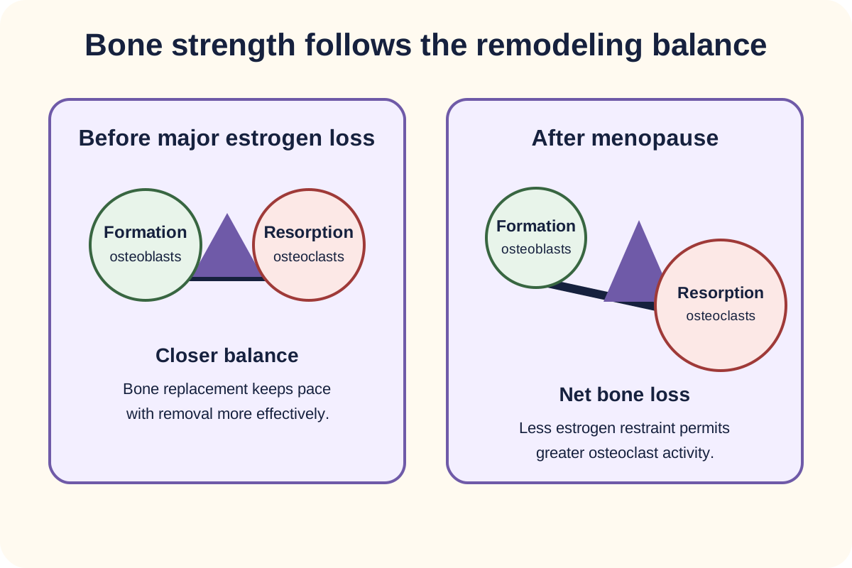
Bone is continuously renewed. Osteoclasts resorb old bone and osteoblasts replace it. Peak bone mass is built early in adulthood, then age, sex-hormone loss, disease, medications, nutrition, and activity alter the balance. After menopause, reduced estrogen signaling permits greater osteoclast activity, so resorption can exceed formation and trabecular-rich sites such as vertebrae lose strength quickly.
Compare the two sides: after estrogen loss, resorption can exceed formation, producing net bone loss. [1][9]
The first misconception to discard is that osteoporosis means poorly mineralized bone. In osteoporosis, the amount and architecture of bone are reduced, but the remaining matrix is usually mineralized normally. That is why routine serum calcium, phosphorus, and alkaline phosphatase often do not identify primary osteoporosis. Abnormal chemistry should instead raise the possibility of a secondary cause or a different metabolic bone disorder. [1][9]
Use chemistry to separate common bone disorders
Pattern
Calcium
Phosphorus
Alkaline phosphatase
PTH
Useful distinction
PatternPrimary osteoporosis
CalciumUsually normal
PhosphorusUsually normal
Alkaline phosphataseUsually normal
PTHUsually normal
Useful distinctionFragility and DXA drive the diagnosis
PatternOsteomalacia
CalciumCan be low
PhosphorusOften low
Alkaline phosphataseOften high
PTHOften high
Useful distinctionMineralization defect with bone pain, weakness, and sometimes Looser zones
PatternPrimary hyperparathyroidism
CalciumOften high
PhosphorusOften low
Alkaline phosphataseVariable
PTHHigh or inappropriately normal
Useful distinctionPTH is not suppressed by the hypercalcemia
PatternPaget disease
CalciumUsually normal
PhosphorusUsually normal
Alkaline phosphataseOften high
PTHUsually normal
Useful distinctionFocal high-turnover disease; bone pain or skull enlargement can occur
Learner action: read only the laboratory rows in the comparison above and name the disorder before reading the final column. If calcium and phosphorus are low while alkaline phosphatase is high, primary osteoporosis becomes a poor fit and a mineralization disorder rises. Apply the same sort whenever a low DXA value arrives with abnormal chemistry. [9]
A fracture changes the probability that the skeleton is fragile, but fracture site matters. A low-trauma hip or vertebral fracture in an adult age 50 or older supports treatment regardless of T-score. Pelvis, proximal humerus, and distal forearm fractures also deserve urgent osteoporosis evaluation, and in a patient with low bone mass they are treatment indications in the BHOF framework. The older shortcut that every low-trauma fracture automatically creates the same diagnostic rule is too broad. [1]
Checkpoint 1: A wrist fracture with a T-score of -1.9
A postmenopausal patient with a distal forearm fracture and low bone mass meets a BHOF secondary-fracture treatment indication. Do not describe this as the same BMD-independent rule used for hip or vertebral fracture. [1]
Apply that distinction to a new patient: if an older adult has sudden back pain, progressive height loss, or new kyphosis, consider an unrecognized vertebral compression fracture. Vertebral imaging can convert a vague history into a fracture-based risk assessment, and a silent vertebral fracture still matters clinically. [1][2]
Try it here · Checkpoint 1 of 3
Make your prediction before reading the choices. A first attempt is just a starting point.
Case 2
Show answer and explanations for case 2
A. No osteoporosis therapy is indicated because the T-score is above -2.5 (Why this does not fit)
Low bone mass plus a distal forearm fracture is a BHOF secondary-fracture treatment indication.
Reasoning steps for option A
Why might a femoral-neck T-score of -1.9 seem to argue against drug therapy?
It does not meet the -2.5 densitometric definition of osteoporosis.
What makes treatment indicated despite that T-score?
Under BHOF guidance, a distal forearm fragility fracture with low bone mass is itself a treatment indication.
B. The wrist fracture is identical to a hip fracture for every diagnostic rule (Why this does not fit)
Hip and vertebral fractures have the clearest BMD-independent treatment rule. Distal forearm fracture is handled with BMD context.
Reasoning steps for option B
What do a wrist fracture and a hip fracture share in this woman?
Both would be low-trauma fragility fractures that signal higher future fracture risk.
How does the rule for a distal radius fracture differ from the rule for a hip fracture?
A hip or vertebral fracture supports treatment regardless of BMD, whereas a distal forearm fracture is a treatment indication when low bone mass is present.
C. Start pharmacologic osteoporosis therapy (Best answer)
BHOF recommends treatment for distal forearm fracture when low bone mass is present in a postmenopausal woman or man age 50 or older.
Reasoning steps for option C
Which fracture did she sustain, and how?
A distal radius fracture from a standing-height fall, which is a low-trauma forearm fracture.
What does the femoral-neck T-score of -1.9 add?
It places her in the low bone mass range, which BHOF requires alongside a forearm fracture.
What decision follows from a forearm fracture plus low bone mass?
Start pharmacologic therapy, because BHOF treats this combination as a secondary-fracture indication.
D. Treatment depends only on whether serum calcium is low (Why this does not fit)
Serum calcium is not the main treatment gate for this fracture-risk pattern.
Reasoning steps for option D
Why might serum calcium seem relevant after a fracture?
Calcium is part of the routine evaluation for secondary causes of bone loss.
What actually decides treatment in this woman?
The combination of a fragility fracture and low bone mass, not the serum calcium value.
E. The fracture is irrelevant unless it recurs (Why this does not fit)
A first adult fracture is a major risk signal and should trigger osteoporosis assessment and, in this setting, treatment.
Reasoning steps for option E
Why might a single wrist fracture seem minor?
Distal radius fractures are common after falls and usually heal without lasting disability.
What does a first fragility fracture signal about future risk?
It marks increased risk of further fractures, and with low bone mass it warrants treatment now.
Takeaway: Distal forearm fracture plus low bone mass is a treatment indication, but it is not the same rule as hip or vertebral fracture.
DXA classifies density, while fracture history and FRAX classify risk
The standard abbreviation is DXA, dual-energy X-ray absorptiometry. In postmenopausal women and men age 50 or older, use the T-score. Osteoporosis by central DXA is a T-score of -2.5 or lower at the lumbar spine, total hip, or femoral neck, with the 33% radius usable in selected circumstances. Low bone mass is between -1.0 and -2.5. [2]
In females before menopause and males younger than 50, prefer the Z-score, which compares the patient with an age-matched reference population. A Z-score of -2.0 or lower is "below the expected range for age." In a younger adult, that result should prompt a search for secondary skeletal disease rather than automatic use of the older-adult densitometric label. [2]
FRAX answers a different question. It estimates 10-year fracture probability from age and clinical risk factors, with femoral-neck BMD added when available. In the United States, BHOF recommends considering pharmacologic therapy for low bone mass when the 10-year probability is at least 3% for hip fracture or at least 20% for major osteoporotic fracture. These are US intervention thresholds, not universal biological cutoffs. [1]
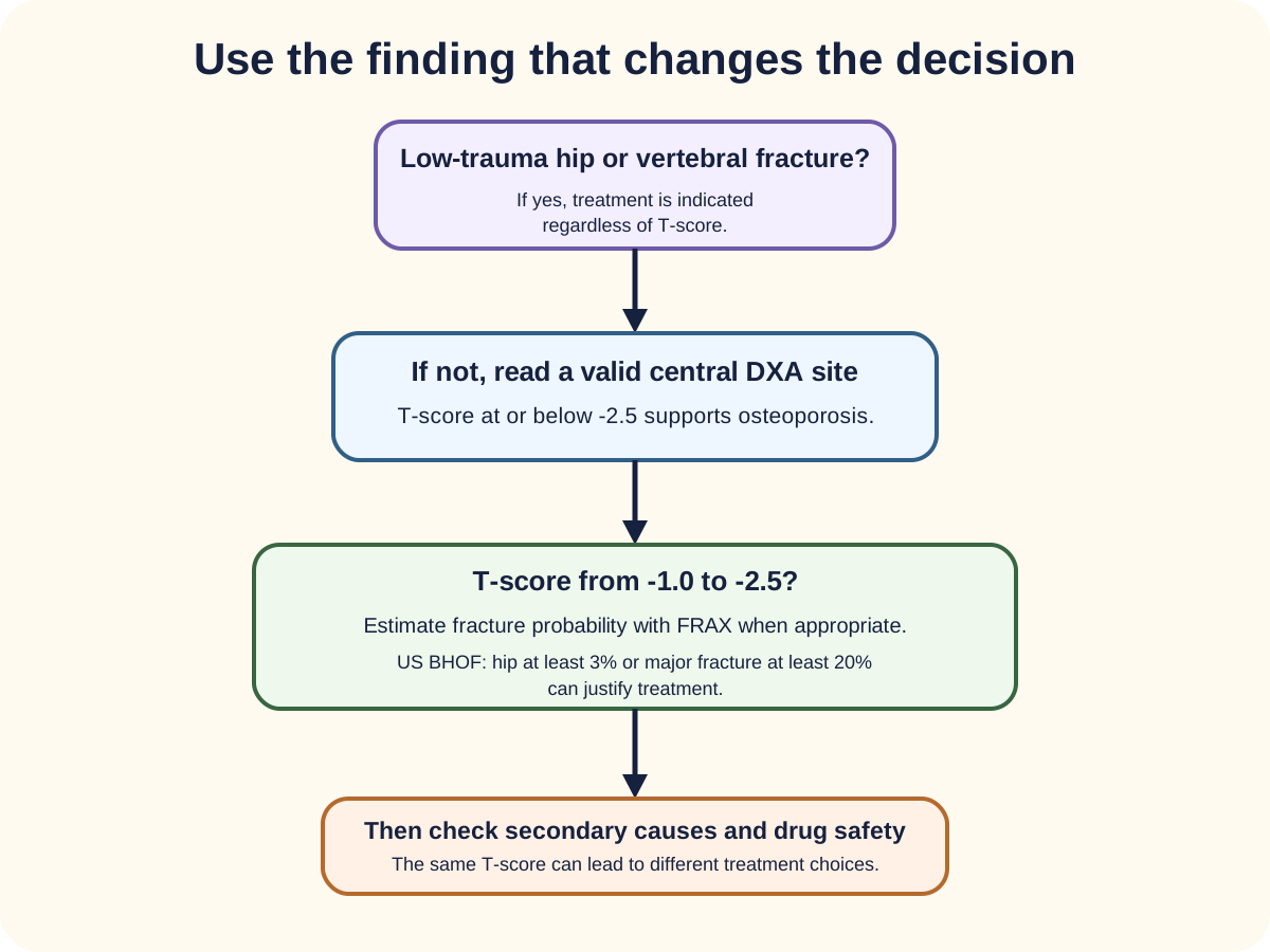
Read from the top: fracture history can determine treatment before the DXA threshold, while FRAX helps when BMD is in the low-bone-mass range. [1][2]
FRAX risk inputs include age, sex, body size, prior fracture, parental hip fracture, smoking, glucocorticoid exposure, rheumatoid arthritis, selected secondary osteoporosis, and alcohol exposure, with femoral-neck BMD when entered. A prior fracture is especially important because it predicts another fracture, while low body weight, tobacco, heavy alcohol exposure, and parental hip fracture add risk even before a new fracture occurs. [1]
First: fracture history Hip or vertebral fracture can establish a treatment indication regardless of T-score.
Second: DXA category T-score at or below -2.5 at a valid site supports osteoporosis in the appropriate age group.
Third: probability When T-score is between -1.0 and -2.5, FRAX can identify patients whose risk still justifies treatment.
Learner action: trace the decision figure from the top without skipping a branch. Start with hip or vertebral fracture, then read the valid DXA site, and use FRAX only when the patient remains in the low-bone-mass range. The consequence of skipping the first branch is predictable: a qualifying fracture can be missed because the T-score looks only moderately low. [1][2]
Checkpoint 2: T-score -1.8, FRAX hip risk 4%
The density category is low bone mass, but the US FRAX treatment threshold is crossed because hip risk is at least 3%. The treatment decision and the densitometric label answer different questions. [1]
Screening recommendations depend on the organization being used. The USPSTF recommends screening women age 65 or older and postmenopausal women younger than 65 who are at increased risk after clinical assessment. For men, the USPSTF concludes that evidence is insufficient to determine the balance of benefits and harms of population screening. ISCD and BHOF guidance, however, lists BMD testing for men age 70 or older and younger men with risk factors. State the framework rather than presenting these as if they were identical recommendations. [1][2][3]
Try it here · Checkpoint 2 of 3
Make your prediction before reading the choices. A first attempt is just a starting point.
Case 5
Show answer and explanations for case 5
A. No drug therapy because the major-fracture probability is below 20% (Why this does not fit)
Either US BHOF FRAX threshold can support treatment in low bone mass. The hip probability already exceeds its threshold.
Reasoning steps for option A
Which FRAX value appears to argue against treatment?
The major osteoporotic-fracture probability of 16% is below the 20% US threshold.
Which FRAX value meets a treatment threshold anyway?
The 10-year hip-fracture probability of 4% exceeds the 3% threshold, and either threshold is enough.
B. Treat because the hip-fracture probability exceeds 3% (Best answer)
In the BHOF framework, low bone mass plus a 10-year hip probability of at least 3% is a treatment indication.
Reasoning steps for option B
How is a femoral-neck T-score of -1.8 classified?
It is low bone mass, between -1.0 and -2.5.
How does each FRAX probability compare with its own threshold?
Hip risk of 4% is above the 3% cutoff, while major risk of 16% is below the 20% cutoff.
What decision follows when only one FRAX threshold is crossed?
Treat; low bone mass plus hip risk of at least 3% is a BHOF treatment indication on its own.
C. Diagnose osteomalacia because FRAX is high (Why this does not fit)
FRAX estimates fracture probability and does not diagnose a mineralization defect.
Reasoning steps for option C
Why might a high FRAX result seem to point to a bone disease?
It signals abnormal skeletal fragility, which could raise the question of a metabolic bone disorder.
What does FRAX actually estimate?
The 10-year probability of fracture; it does not diagnose a mineralization defect, and no abnormal chemistry is given.
D. Repeat FRAX after starting a bisphosphonate to confirm the indication (Why this does not fit)
The treatment indication is already present. FRAX is not a post-treatment confirmation test.
Reasoning steps for option D
Why might rechecking the risk estimate seem reasonable?
Monitoring response to therapy is part of osteoporosis care.
When is the treatment indication established?
Before treatment, from the untreated FRAX result; FRAX is not designed to confirm an indication once therapy starts.
E. Treat only if the T-score falls below -2.5 (Why this does not fit)
FRAX exists partly to identify high-risk patients whose T-scores remain between -1.0 and -2.5.
Reasoning steps for option E
Why might the T-score alone seem to decide treatment?
A T-score of -2.5 or lower is the densitometric definition of osteoporosis.
Why can this woman be treated with a T-score of -1.8?
FRAX identifies high absolute risk in low bone mass, and her hip probability crosses the treatment threshold.
Takeaway: In US practice, low bone mass can merit treatment when FRAX hip risk is at least 3% or major risk is at least 20%.
When bone loss is unexpected, ask what is stealing strength
A low density result should not end the evaluation. Secondary contributors are especially important with unexpectedly young age, fractures out of proportion to BMD, rapid loss, unusual laboratory findings, or an inadequate response to therapy. Common possibilities include glucocorticoids, hypogonadism, hyperthyroidism, hyperparathyroidism, celiac disease, chronic kidney disease, alcohol excess, and hematologic disease such as multiple myeloma. [9]
A practical initial evaluation commonly includes a CBC, serum calcium, phosphorus, creatinine or estimated kidney function, alkaline phosphatase, and 25-hydroxyvitamin D. Further testing follows the history and examination, such as PTH, thyroid testing, celiac serology, protein studies, or testosterone in men. A normal set of routine chemistries supports primary osteoporosis only after the clinical context is considered. [9]
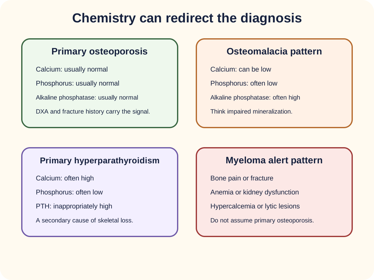
Pattern recognition is useful when the chemistry is abnormal. Hypercalcemia with an inappropriately high PTH suggests primary hyperparathyroidism. Low calcium or phosphorus with high alkaline phosphatase can support osteomalacia in the right context. Bone pain, anemia, renal dysfunction, hypercalcemia, and lytic lesions should redirect the evaluation toward myeloma rather than ordinary age-related osteoporosis. [9]
Use the pattern rather than one isolated value. Primary osteoporosis often has unrevealing routine mineral chemistry, while several mimics produce a different laboratory or systemic pattern. [9]
Learner action: use the laboratory-pattern figure as a four-way sort. Cover the panel titles, classify each pattern from the supplied values, then check the title. A mismatch should send you back to the abnormal value rather than toward a memorized disease label. In a new case, hypercalcemia with high PTH redirects toward parathyroid disease, while anemia, kidney dysfunction, hypercalcemia, or lytic lesions redirect toward myeloma. [9]
Glucocorticoids deserve their own branch because bone loss can begin early. The ACR guideline applies to adults receiving at least 2.5 mg prednisone equivalent daily for more than 3 months and recommends fracture-risk assessment as soon as possible. Drug treatment is then based on risk category rather than a blanket rule that every person above one dose threshold receives the same therapy. [4]
Use the same logic with height loss. Vertebral fractures can be painless, so historical height loss, kyphosis, or a suspicious DXA image can justify vertebral fracture assessment or lateral spine imaging. The point is not to wait for severe back pain before asking whether the spine has already fractured. [1][2]
Treatment choice follows fracture risk, contraindications, and what comes next
Every treatment plan sits on the same foundation: adequate calcium and vitamin D, weight-bearing and muscle-strengthening activity as tolerated, tobacco cessation, moderation of alcohol, and fall-risk reduction through vision, gait, home-hazard, and medication review. These steps support drug therapy; they do not replace pharmacologic treatment when fracture risk is high. [1]
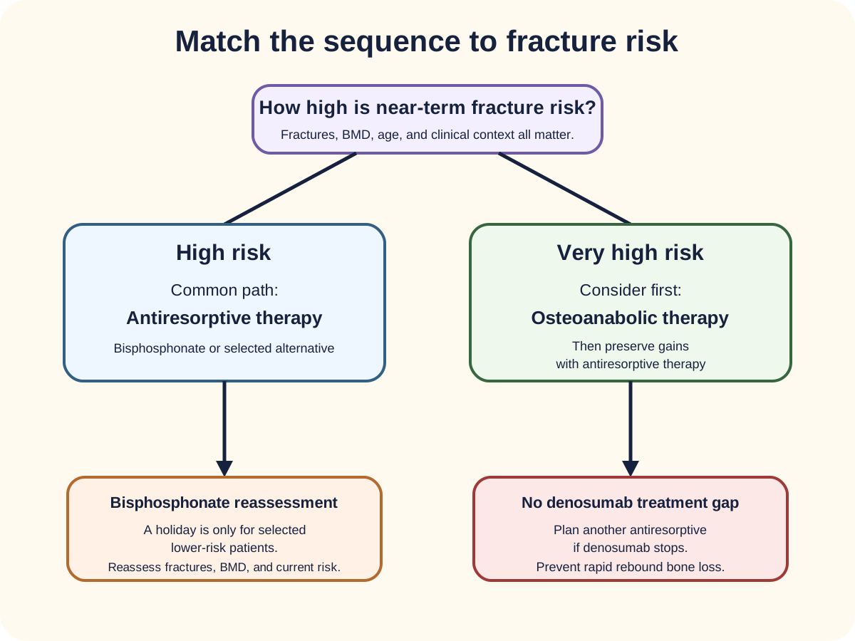
For many high-risk patients, an oral or intravenous bisphosphonate is an effective initial antiresorptive option. But "bisphosphonate first for everyone" is too simple. Recent major fractures, multiple vertebral fractures, or very low BMD can place a patient at very high or imminent risk, where an osteoanabolic-first strategy may produce faster and larger gains before antiresorptive consolidation. [8][10]
Compare the branches and then follow each to its next step. Treatment choice depends on fracture risk and must include the later transition plan. [1][8][10]
Denosumab inhibits RANKL and suppresses osteoclast formation and activity. Its effect reverses when dosing is delayed or stopped, so it should not be interrupted without planning subsequent antiresorptive therapy. At the same time, advanced chronic kidney disease is not a casual reason to prefer denosumab: the FDA added a boxed warning for severe hypocalcemia in advanced CKD, especially in dialysis and CKD-mineral bone disorder. [7][8]
Teriparatide and abaloparatide stimulate bone formation and are options for very high fracture risk. Teriparatide should be avoided in patients with important baseline osteosarcoma risks such as prior radiation involving the skeleton, Paget disease, bone metastases, skeletal malignancy, or open epiphyses. Current labeling no longer treats 2 years as an absolute lifetime prohibition: use beyond 2 years should only be considered when high fracture risk persists or returns. [6]
Raloxifene is useful mainly for vertebral-fracture reduction in selected postmenopausal women, particularly when breast-cancer risk reduction is also desirable and venous thrombosis risk is low; it can worsen hot flashes. Romosozumab can be considered for very high fracture risk, but the label advises against starting it within 1 year of myocardial infarction or stroke, and the Endocrine Society advises against it in women at high cardiovascular risk. After an osteoanabolic course, follow with antiresorptive therapy to preserve gains. [8][11]
Learner action: in the treatment-sequence figure, cover the lower boxes and choose the next therapy from the risk branch alone. Then reveal the lower box and check the transition. The important consequence is that very high fracture risk can justify an osteoanabolic-first plan, but the sequence is incomplete until antiresorptive therapy preserves the gain. [8][10]
Checkpoint 3: Multiple vertebral fractures and T-score -3.2
This is very high fracture risk, not merely a low DXA value. If no contraindication is present, an osteoanabolic-first strategy is reasonable, followed by antiresorptive therapy to preserve the new bone-density gain. [8][10]
Apply sequencing explicitly: if a patient improves on denosumab but must stop it, plan another antiresorptive rather than leaving a treatment gap. If a patient finishes an anabolic course, follow with antiresorptive therapy. Fracture prevention is a longitudinal plan, not a one-time prescription. [1][8][10]
Try it here · Checkpoint 3 of 3
Make your prediction before reading the choices. A first attempt is just a starting point.
Case 23
Show answer and explanations for case 23
A. Osteoanabolic first, then antiresorptive therapy (Best answer)
Multiple recent vertebral fractures and very low BMD place her at very high risk, where osteoanabolic-first treatment can be appropriate, followed by antiresorptive consolidation.
Reasoning steps for option A
What places her at very high fracture risk?
Three vertebral fractures within 18 months and a total-hip T-score of -3.2.
Which initial strategy suits very high risk?
Osteoanabolic therapy first, which builds bone faster and reduces fractures more than starting with an antiresorptive.
What should follow the anabolic course?
An antiresorptive to consolidate and preserve the gains.
B. Use calcium and vitamin D alone for 2 years before drug therapy (Why this does not fit)
Her fracture pattern represents very high risk that warrants effective pharmacologic treatment now.
Reasoning steps for option B
Why might supplements seem like a sensible first step?
Calcium and vitamin D belong in every osteoporosis plan.
Why is a 2-year delay harmful?
Multiple recent fractures signal imminent risk that needs effective drug therapy now.
C. Delay treatment until the T-score reaches -3.5 (Why this does not fit)
She already has both severe BMD loss and multiple fractures. Waiting for a lower number adds risk.
Reasoning steps for option C
Why might waiting for a lower T-score seem cautious?
Treatment decisions are often tied to T-score thresholds.
What does she already have that makes waiting pointless?
A T-score of -3.2 and three vertebral fractures, which already establish very high risk.
D. Choose raloxifene solely because it is an oral medication (Why this does not fit)
Raloxifene mainly reduces vertebral risk and is not the strongest fit for this very high-risk, multi-fracture presentation.
Reasoning steps for option D
Why might raloxifene seem appealing?
It is an oral tablet and reduces vertebral fractures.
Why is raloxifene a poor fit for her?
It has not been shown to reduce hip or nonvertebral fractures and is too weak for very high risk.
E. Use a drug holiday because repeated fractures imply too much antiresorption (Why this does not fit)
She has not been described as receiving long-term bisphosphonate therapy, and repeated fractures signal urgent risk reduction rather than a holiday.
Reasoning steps for option E
What could make a holiday seem relevant to repeated fractures?
Long-term bisphosphonate use is linked to atypical fractures, which is one reason holidays exist.
Why does that reasoning fail here?
She is not described as taking bisphosphonates, and repeated fractures call for urgent treatment.
Takeaway: Recent multiple vertebral fractures and very low BMD can justify an osteoanabolic-first sequence.
For oral alendronate, take the dose after getting up for the day, before food, drink, or other medication, with a full glass of plain water. Remain sitting or standing for at least 30 minutes and until after the first food of the day. This limits esophageal exposure and preserves absorption. Esophageal stricture or inability to remain upright makes oral therapy a poor choice. [5]
Long-term antiresorptive therapy has uncommon but important complications. New dull thigh or groin pain during prolonged bisphosphonate or denosumab therapy should prompt assessment for an atypical femur fracture. Osteonecrosis of the jaw is uncommon at osteoporosis doses but becomes relevant with exposed nonhealing jaw bone, especially after invasive dental procedures or with stronger oncology-dose exposures. These risks should be interpreted against the much larger fracture risk in patients who truly need treatment. [1][2]
Drug interruption rules differ by class. A bisphosphonate holiday can be considered in selected lower-risk patients after an appropriate treatment period because the drug remains in bone. Denosumab has no comparable residual effect, so delayed or stopped dosing can be followed by rapid bone loss and vertebral fractures unless another antiresorptive is arranged. [1][8]
Safety sort: symptom or situation to immediate concern
Prompt
First concern
PromptDysphagia or esophageal stricture
First concernAvoid routine oral alendronate exposure
PromptNew thigh or groin pain on prolonged antiresorptive therapy
First concernAssess for atypical femur fracture
PromptStopping or delaying denosumab
First concernPlan follow-on antiresorptive therapy
PromptDialysis with CKD-mineral bone disorder before denosumab
First concernAssess and reduce severe hypocalcemia risk
Learner action: cover the right column, name the first concern from the left column, then check the pairing. The visible consequence is a safety-specific action rather than a generic drug rule. Apply this sort when a stem gives a body-site symptom, a planned drug interruption, or advanced kidney disease. [1][5][7][8]
Monitoring should answer a clinical question. Follow-up DXA can assess response, detect significant loss, or guide a treatment change, but repeating scans too frequently does not make treatment safer. A new fracture, new risk factor, or major treatment decision can justify reassessment. [1][2]
When the stem seems crowded, sort it into four columns: fracture history, valid DXA result, secondary-cause evidence, and drug-specific safety. The best answer usually comes from the column that changes management, not from the most dramatic isolated fact.
Apply the lesson
Case 1
Show answer and explanations for case 1
A. The T-score is too high for osteoporosis treatment (Why this does not fit)
A T-score of -1.8 is low bone mass, but hip fracture history overrides the need for a densitometric osteoporosis threshold when deciding treatment.
Reasoning steps for option A
Why might the prior T-score of -1.8 seem to argue against treatment?
It sits in the low bone mass range, above the -2.5 densitometric cutoff for osteoporosis.
Which event in this history outweighs the T-score threshold?
A femoral-neck fracture from standing height is a low-trauma hip fracture, which supports treatment regardless of BMD.
B. Treat osteoporosis despite the T-score (Best answer)
A low-trauma hip fracture is a secondary-fracture treatment indication regardless of BMD. The prior T-score does not remove that risk.
Reasoning steps for option B
What kind of fracture did she have?
A femoral-neck fracture after a fall from standing height is a low-trauma hip fracture.
How should the older total-hip T-score of -1.8 be weighed?
It shows low bone mass, but it does not cancel the fracture-based treatment indication.
What general rule does her fracture illustrate?
Fracture history outranks densitometry: after a low-trauma hip or spine fracture, the T-score serves as a baseline for monitoring rather than a gate for starting therapy.
C. Normal serum calcium excludes skeletal fragility (Why this does not fit)
Primary osteoporosis often has normal routine mineral chemistry. Normal calcium does not negate a fragility hip fracture.
Reasoning steps for option C
Why could normal calcium, phosphorus, and alkaline phosphatase seem reassuring?
Normal chemistry makes a metabolic bone disorder such as osteomalacia or hyperparathyroidism less likely.
Does normal mineral chemistry rule out osteoporosis after this hip fracture?
No; primary osteoporosis usually has normal routine chemistry, and the fracture itself demonstrates skeletal fragility.
D. The fracture should be treated only if FRAX is at least 20% (Why this does not fit)
FRAX thresholds are used mainly when deciding treatment in low bone mass without a qualifying hip or vertebral fracture.
Reasoning steps for option D
Why does a FRAX threshold come to mind with a T-score of -1.8?
FRAX is the usual tool for deciding treatment when the T-score is in the low bone mass range.
When is FRAX unnecessary for the treatment decision?
After a low-trauma hip or vertebral fracture, treatment is indicated without reaching any FRAX threshold.
E. The pattern is more consistent with osteomalacia (Why this does not fit)
Osteomalacia is suggested by a mineralization disorder and often abnormal chemistry or bone pain, neither of which explains this fracture pattern.
Reasoning steps for option E
What could make osteomalacia enter the differential for a hip fracture with modest BMD loss?
Osteomalacia also weakens bone and lowers measured BMD, so DXA alone cannot separate it from osteoporosis.
Which supplied results argue against osteomalacia here?
Calcium, phosphorus, and alkaline phosphatase are normal, and no diffuse bone pain is described.
Takeaway: A hip or vertebral fragility fracture can establish a treatment indication regardless of T-score.
A T-score at or below -2.5 at a valid central DXA site, including the lumbar spine, supports osteoporosis in a postmenopausal woman.
Reasoning steps for option A
Which reference score applies to a 67-year-old postmenopausal woman?
The T-score, which compares her BMD with that of young adults.
Is the lumbar spine an acceptable site for diagnosis?
Yes; the lumbar spine, total hip, and femoral neck are all valid central DXA sites.
How is the classification made when the sites disagree?
The lowest valid T-score is used, so the spine value of -2.6 meets the osteoporosis threshold of -2.5 or lower.
B. Low bone mass only (Why this does not fit)
Although the hip values are in the low bone mass range, the lumbar-spine value crosses the diagnostic threshold.
Reasoning steps for option B
Why could low bone mass seem correct?
Both hip T-scores, -2.1 and -2.0, fall in the low bone mass range.
Which site determines her diagnosis?
The lumbar-spine T-score of -2.6 is the lowest valid value and crosses the -2.5 threshold.
C. Below expected range for age without osteoporosis (Why this does not fit)
That phrasing belongs to Z-score interpretation in younger adults, not T-score classification in this patient.
Reasoning steps for option C
Where does the phrase below the expected range for age come from?
It is the ISCD wording for a Z-score of -2.0 or lower in younger adults.
Why does that wording not apply to her?
She is postmenopausal, so her results are classified with T-scores, not Z-scores.
D. Normal bone density (Why this does not fit)
The lumbar-spine value is well below the normal threshold.
Reasoning steps for option D
What part of her history might make normal bone density seem likely?
She has never had a fracture, which can make her bone density seem unconcerning.
What do her DXA values show against a normal reading?
Normal requires T-scores of -1.0 or higher, and all three of her sites are at -2.0 or lower.
E. Osteoporosis only if FRAX hip risk is at least 3% (Why this does not fit)
FRAX thresholds help decide treatment in low bone mass. A valid T-score at or below -2.5 already meets the densitometric criterion.
Reasoning steps for option E
Why might FRAX seem necessary before labeling osteoporosis?
FRAX is commonly used to guide decisions in women without a prior fracture.
What role does FRAX play once a valid T-score is -2.5 or lower?
It plays no part in the label, because a spine T-score of -2.6 is diagnostic by itself; FRAX becomes useful for treatment decisions when scores sit in the low bone mass range.
Takeaway: Use the lowest valid diagnostic T-score at an accepted site in postmenopausal women and men age 50 or older.
A. Diagnose postmenopausal-pattern osteoporosis from the Z-score alone (Why this does not fit)
Younger adults are not classified by the older-adult T-score framework, and a low Z-score alone should not be treated as the same diagnosis.
Reasoning steps for option A
Why might a Z-score of -2.4 seem enough to call osteoporosis?
It is well below the expected range for age and sounds close to the -2.5 T-score cutoff.
Why is a postmenopausal-pattern diagnosis inappropriate here?
She is premenopausal, so the T-score framework does not apply, and a low Z-score alone does not diagnose osteoporosis.
B. Repeat DXA in one month before any other evaluation (Why this does not fit)
A one-month repeat is unlikely to provide meaningful biological change and delays evaluation of a plausible secondary cause.
Reasoning steps for option B
What could motivate repeating the scan before a workup?
Confirming an unexpectedly low result in a young woman can seem prudent.
Why is a repeat DXA in one month unhelpful?
The study was technically adequate, BMD does not change meaningfully in a month, and waiting delays the search for malabsorption.
C. Start raloxifene because the Z-score is below -2.0 (Why this does not fit)
Raloxifene is a postmenopausal therapy and does not address the likely secondary cause in this young patient.
Reasoning steps for option C
Why might a drug seem reasonable for a Z-score of -2.4?
The value is markedly low, which creates pressure to treat the number.
Why is raloxifene the wrong response in this woman?
Raloxifene is a therapy for postmenopausal women and does nothing for the likely underlying cause.
D. Evaluate for a secondary cause (Best answer)
A Z-score at or below -2.0 is below the expected range for age. Chronic diarrhea and iron deficiency make malabsorption or celiac disease particularly relevant.
Reasoning steps for option D
Which DXA score applies to a 34-year-old premenopausal woman?
The Z-score, which compares her with people of the same age and sex.
How is a Z-score of -2.4 described?
A Z-score of -2.0 or lower is below the expected range for age.
Which part of her history points to a specific secondary cause?
Chronic diarrhea with iron deficiency suggests malabsorption such as celiac disease, which should be evaluated.
E. Ignore the study because Z-scores cannot be interpreted (Why this does not fit)
Z-scores are the preferred DXA comparison in females before menopause, and this result is meaningfully low for age.
Reasoning steps for option E
Why might the result be dismissed?
She has no fracture, and the older-adult T-score thresholds cannot be applied to a premenopausal woman.
How should a Z-score be used before menopause?
It is the preferred DXA comparison in premenopausal women, and a value of -2.4 is meaningfully low for age.
Takeaway: A low Z-score in a younger adult should trigger a targeted search for secondary skeletal disease.
A. Wait until age 70 because screening is not recommended before then (Why this does not fit)
The USPSTF uses a two-step approach for postmenopausal women younger than 65 who have risk factors.
Reasoning steps for option A
Why might a 62-year-old seem too young for bone-density testing?
Age-based DXA thresholds of 65 for women and 70 for men can make her age seem ineligible.
What makes her eligible before age 65?
USPSTF recommends screening postmenopausal women younger than 65 with increased risk, and she has low weight, a parental hip fracture, and current smoking.
B. Order monthly serum calcium instead of bone-density testing (Why this does not fit)
Serum calcium does not substitute for osteoporosis screening with bone-density assessment.
Reasoning steps for option B
Why might a blood test seem like a simpler first step?
Serum calcium is inexpensive and is part of evaluating bone health.
What can serum calcium not tell you about her fracture risk?
It does not measure bone density; her risk factors call for risk assessment and DXA.
C. Screen only after the first fragility fracture (Why this does not fit)
Screening aims to identify high risk before a first fracture when possible.
Reasoning steps for option C
Why might waiting for a fracture seem acceptable?
She has never had a fracture, and a fracture is a strong trigger for treatment.
What is the purpose of screening a woman with her risk factors?
To find low bone density and treat it before a first fracture occurs.
D. Risk-assess, then obtain DXA if risk is increased (Best answer)
For postmenopausal women younger than 65 with risk factors, USPSTF recommends risk assessment followed by screening when risk is increased.
Reasoning steps for option D
Which USPSTF pathway applies to a postmenopausal woman aged 62?
The pathway for women younger than 65, which starts with clinical risk assessment.
Which of her features raise fracture risk?
Low body weight, a parental hip fracture, and current smoking.
What follows when a risk tool shows increased risk?
Bone-density testing with DXA.
E. Use a Z-score because she is younger than 65 (Why this does not fit)
She is postmenopausal, so the adult T-score framework applies; age 65 is a screening threshold, not the T-score versus Z-score boundary.
Reasoning steps for option E
Why might a Z-score seem appropriate for a woman younger than 65?
Age 65 is a familiar screening cutoff and can be mistaken for the boundary between score types.
Which score applies once a woman is postmenopausal?
The T-score, whether or not she has reached 65.
Takeaway: USPSTF screening before age 65 in postmenopausal women uses clinical risk assessment to select patients for DXA.
A. Age-related osteoporosis alone (Why this does not fit)
A fracture in an older adult can be osteoporotic, but anemia, renal dysfunction, hypercalcemia, and lytic lesions demand another diagnosis.
Reasoning steps for option A
Why might osteoporosis seem to explain a vertebral fracture at 71?
Vertebral compression fractures are common osteoporotic fractures in older adults.
Which findings cannot be explained by osteoporosis?
Anemia, kidney dysfunction, hypercalcemia, and punched-out skull lesions.
B. Osteomalacia from low vitamin D (Why this does not fit)
The supplied pattern does not show the typical low-mineral chemistry of osteomalacia, and punched-out lesions are not expected.
Reasoning steps for option B
What overlap with osteomalacia exists here?
Osteomalacia can also cause bone pain and fractures.
Which of his results argue against osteomalacia?
His calcium is high rather than low, and punched-out lytic lesions are not a mineralization defect.
C. Paget disease (Why this does not fit)
Paget disease can affect the skull, but punched-out lesions with anemia, renal dysfunction, and hypercalcemia suggest a hematologic process.
Reasoning steps for option C
Which finding could suggest Paget disease?
Paget disease often involves the skull in older adults.
How do the skull lesions and systemic findings point elsewhere?
Punched-out lytic lesions with anemia, kidney dysfunction, and hypercalcemia suggest a plasma cell neoplasm.
D. Multiple myeloma (Best answer)
The combination of anemia, kidney dysfunction, hypercalcemia, bone pain, and lytic lesions is classic for myeloma and requires protein studies and hematologic evaluation.
Reasoning steps for option D
Which systemic findings accompany his fracture?
Anemia, a creatinine of 2.0 mg/dL, and a calcium of 11.0 mg/dL.
What do punched-out skull lesions indicate?
Lytic bone destruction typical of multiple myeloma.
What evaluation should follow?
Serum and urine protein studies with free light chains, and hematologic assessment.
E. Uncomplicated primary hyperparathyroidism (Why this does not fit)
Hypercalcemia can fit hyperparathyroidism, but the anemia, renal dysfunction, and punched-out lesions strongly favor myeloma.
Reasoning steps for option E
Which finding could fit primary hyperparathyroidism?
His calcium of 11.0 mg/dL.
Which findings favor myeloma over hyperparathyroidism?
Anemia, kidney dysfunction, and punched-out lytic skull lesions.
Takeaway: Do not label every low-trauma vertebral fracture as primary osteoporosis when systemic findings suggest a secondary cause.
A. Wait until prednisone reaches 7.5 mg daily before assessing fracture risk (Why this does not fit)
The ACR guideline begins risk assessment at lower chronic doses when exposure is expected to exceed 3 months.
Reasoning steps for option A
Why might 7.5 mg daily seem like the threshold for concern?
Older guidance and many teaching sources used 7.5 mg daily for 3 months as the trigger for bone protection.
What threshold does the 2022 ACR guideline use?
At least 2.5 mg prednisone equivalent daily for more than 3 months, which her 5 mg for 8 months meets.
B. Assess fracture risk now and treat as indicated (Best answer)
At least 2.5 mg prednisone equivalent for more than 3 months triggers early assessment. Her prior fracture and low BMD make pharmacologic protection appropriate.
Reasoning steps for option B
Does her planned prednisone course meet the ACR trigger for assessment?
Yes; 5 mg daily for about 8 months exceeds 2.5 mg daily for more than 3 months.
Which features already raise her fracture risk?
A prior low-trauma humerus fracture and a femoral-neck T-score of -2.0.
What action follows now?
Assess fracture risk promptly and start pharmacologic protection as indicated rather than waiting.
C. Use calcium alone because her T-score is not below -2.5 (Why this does not fit)
Glucocorticoid exposure and prior fracture can justify treatment before the T-score reaches the densitometric osteoporosis threshold.
Reasoning steps for option C
Why might calcium alone seem adequate?
Her T-score of -2.0 is above the -2.5 osteoporosis threshold.
What justifies drug therapy despite that T-score?
Ongoing glucocorticoid exposure plus a prior fragility fracture place her at treatment-level risk.
D. Repeat DXA monthly and treat only after measurable decline (Why this does not fit)
Glucocorticoid bone loss can occur early, and monthly DXA is not an appropriate preventive strategy.
Reasoning steps for option D
Why might serial monitoring seem cautious?
Tracking BMD could document whether glucocorticoids are causing loss.
A. Take it with breakfast and calcium to reduce gastric irritation (Why this does not fit)
Food and calcium reduce alendronate absorption and should not accompany the dose.
Reasoning steps for option A
Why might taking it with breakfast seem kinder to the stomach?
Many oral drugs cause less upset when taken with food.
What do food and calcium do to alendronate?
They bind or block it and sharply reduce its absorption.
B. Take it at bedtime with a small amount of water (Why this does not fit)
Bedtime dosing increases esophageal exposure and does not follow the labeled administration instructions.
Reasoning steps for option B
Why might bedtime dosing seem convenient?
It separates the dose from breakfast, calcium, and daytime medicines.
Why is bedtime dosing unsafe?
Lying down soon after the dose raises the risk that the tablet injures the esophagus.
C. Take it with coffee, then wait 15 minutes before food (Why this does not fit)
Only plain water should be used, and the required interval is at least 30 minutes.
Reasoning steps for option C
What makes this option seem partly correct?
It does place a gap between the dose and food.
Which two details are wrong?
Only plain water is allowed, and the wait must be at least 30 minutes.
D. Take it on waking with plain water, remain upright 30 minutes, then delay food, calcium, and other oral medicines (Best answer)
Alendronate should be taken after rising with plain water, at least 30 minutes before the first food, beverage, or other oral medication, while the patient remains upright. This separates calcium and supports esophageal safety.
Reasoning steps for option D
What in her routine threatens alendronate absorption?
She takes calcium carbonate and her other oral medicines with breakfast.
How should the dose be taken?
After rising, with a full glass of plain water, at least 30 minutes before food, drinks, or other medicines.
How is esophageal injury reduced?
She swallows the tablet whole and stays upright for at least 30 minutes, until after her first food.
E. Chew the tablet with water to improve absorption (Why this does not fit)
The tablet should be swallowed rather than chewed or sucked, which can increase local irritation.
Reasoning steps for option E
Why might chewing seem to help?
Breaking a tablet can seem to speed its dissolution.
What does the label say about chewing?
The tablet must be swallowed whole, because chewing or sucking it can irritate the mouth and throat.
Takeaway: Take oral alendronate after rising with plain water, remain upright at least 30 minutes, and delay food, calcium, and other oral medicines.
A. Reassure her because normal calcium excludes fracture (Why this does not fit)
Atypical femur fractures can occur with normal routine chemistry.
Reasoning steps for option A
Why might normal calcium and vitamin D seem reassuring?
They make common metabolic causes of bone pain such as osteomalacia less likely.
Do normal labs exclude an atypical femur fracture?
No; these fractures reflect long-term suppression of bone remodeling in the femoral cortex, and serum calcium and vitamin D are usually normal when they occur.
B. Treat the symptom as lumbar radiculopathy without bone imaging (Why this does not fit)
The prolonged antiresorptive exposure and focal thigh pain make femoral assessment important before assuming a neurologic cause.
Reasoning steps for option B
Why might lumbar radiculopathy seem likely?
Lateral thigh pain in an older adult often comes from the lumbar spine.
What in her history makes bone imaging necessary first?
Seven years of bisphosphonate therapy with new focal thigh pain raises concern for an incomplete atypical femur fracture.
C. Image the femur for atypical fracture (Best answer)
Prodromal thigh or groin pain during long-term antiresorptive therapy should prompt assessment for an atypical femur fracture.
Reasoning steps for option C
Which exposure is relevant to her new thigh pain?
Seven years of oral bisphosphonate therapy.
What can dull lateral thigh pain without a fall represent in that setting?
A prodrome of an incomplete atypical femur fracture.
How should this be evaluated?
Image the femur, and consider the opposite femur too because these fractures are often bilateral.
D. Increase the bisphosphonate dose immediately (Why this does not fit)
Increasing antiresorptive exposure would be inappropriate before excluding an atypical femur fracture.
Reasoning steps for option D
Why might more bisphosphonate seem to help thigh pain?
Pain in a patient with osteoporosis can be misread as progressive bone loss.
Why is dose escalation inappropriate?
Prolonged antiresorptive exposure is the suspected cause, so more drug before excluding a femur fracture could worsen the problem.
E. Diagnose osteonecrosis of the jaw (Why this does not fit)
That complication presents in the jaw, not as isolated thigh pain.
Reasoning steps for option E
Why might osteonecrosis of the jaw come to mind?
It is another rare complication of long-term antiresorptive therapy.
Where does osteonecrosis of the jaw present?
In the jaw, as exposed bone, not as thigh pain.
Takeaway: New thigh or groin pain during prolonged antiresorptive therapy should trigger assessment for atypical femur fracture.
A. Atypical femur fracture (Why this does not fit)
That complication presents with femoral pain or fracture, not exposed jaw bone.
Reasoning steps for option A
Why might an atypical femur fracture be considered?
It is also a rare complication of long-term antiresorptive therapy.
Which features point away from an atypical femur fracture?
She has no thigh or groin pain, and the lesion is in the mandible.
B. Pill esophagitis (Why this does not fit)
Esophagitis causes swallowing or retrosternal symptoms rather than a nonhealing jaw lesion.
Reasoning steps for option B
Why might pill esophagitis come to mind?
Oral bisphosphonates are a recognized cause of esophageal injury.
What does the absence of dysphagia tell you?
Her problem is not esophageal; it is exposed jaw bone.
C. Primary hyperparathyroidism (Why this does not fit)
Hyperparathyroidism changes calcium and bone turnover but does not explain exposed nonhealing jaw bone after extraction.
Reasoning steps for option C
Why might a parathyroid disorder be considered in a woman on antiresorptives?
Hyperparathyroidism is a secondary cause of bone loss that is screened for in osteoporosis.
Why does hyperparathyroidism not explain this lesion?
It alters calcium and bone turnover but does not cause nonhealing exposed jaw bone after an extraction.
D. Osteomalacia (Why this does not fit)
Osteomalacia causes impaired mineralization and diffuse skeletal symptoms, not this focal dental presentation.
Reasoning steps for option D
Why might osteomalacia be raised when bone fails to heal?
Impaired mineralization can slow bone repair.
How does her presentation differ from osteomalacia?
Osteomalacia causes diffuse pain and weakness, not a focal exposed-bone lesion after dental work.
E. Medication-related osteonecrosis of the jaw (Best answer)
Antiresorptive exposure plus a nonhealing exposed jaw lesion after dental work is characteristic of medication-related osteonecrosis of the jaw. The absence of femoral and esophageal symptoms argues against the competing drug effects.
Reasoning steps for option E
Where is the lesion, and what preceded it?
Persistently exposed mandibular bone after a dental extraction.
Which drug exposure links to this lesion?
Long-term antiresorptive therapy.
How do the negative findings help?
No thigh pain and no dysphagia argue against atypical femur fracture and pill esophagitis.
Takeaway: Persistent exposed jaw bone after dental work and focal thigh or groin pain identify different uncommon antiresorptive complications.
A. Romosozumab is preferred because recent myocardial infarction is unrelated to its selection (Why this does not fit)
The romosozumab label advises against starting it within 1 year of myocardial infarction or stroke, and her infarction was 3 months ago.
Reasoning steps for option A
What makes romosozumab attractive for her?
Two recent vertebral fractures and a T-score of -3.1 put her at very high risk, where romosozumab is an option.
Why does her myocardial infarction exclude romosozumab?
Romosozumab carries a boxed cardiovascular warning and should not be started in anyone with a myocardial infarction or stroke in the past year; her infarction was only 3 months ago.
B. No pharmacologic therapy is appropriate after myocardial infarction (Why this does not fit)
Her cardiovascular history affects drug choice, not the need to reduce very high fracture risk.
Reasoning steps for option B
Why might her heart attack seem to push bone treatment aside?
A recent myocardial infarction makes clinicians cautious about new drugs.
Does her cardiac history remove the need for treatment?
No; it limits the choice of agent, while her fracture risk remains very high.
C. Consider a different osteoanabolic option that avoids the romosozumab cardiovascular concern in this very high-risk patient (Best answer)
Her multiple recent vertebral fractures and very low BMD support an osteoanabolic strategy, while recent myocardial infarction argues against romosozumab.
Reasoning steps for option C
How high is her fracture risk?
Very high: two recent vertebral fractures and a T-score of -3.1.
Which anabolic agent does her recent infarction rule out?
Romosozumab, which should not be started within a year of myocardial infarction.
What treatment concept fits?
Another osteoanabolic agent such as teriparatide or abaloparatide, since she has normal kidney function and no skeletal radiation, followed by an antiresorptive.
D. Calcium alone is sufficient because fractures have already occurred (Why this does not fit)
Calcium does not provide adequate fracture-risk reduction for very high-risk osteoporosis.
Reasoning steps for option D
Why might calcium seem like a safe choice after a heart attack?
It avoids adding a new prescription drug during cardiac recovery.
Why is calcium alone inadequate for her?
It does not reduce fracture risk enough in very high-risk osteoporosis with recent fractures.
E. A bisphosphonate holiday should start before any treatment (Why this does not fit)
A holiday is considered after selected periods of bisphosphonate therapy, not before treatment in a very high-risk patient.
Reasoning steps for option E
Why might a drug holiday come up in her plan?
Holidays are part of long-term bisphosphonate management.
Why is a holiday wrong for her?
A holiday follows years of bisphosphonate therapy; she has not started treatment and needs it now.
Takeaway: Very high fracture risk can support anabolic-first therapy, but cardiovascular history can narrow the anabolic choice.
A. Increase calcium intake alone (Why this does not fit)
Calcium does not correct the immediate fall hazards identified in the stem.
Reasoning steps for option A
Why might more calcium seem helpful?
She has osteoporosis, and calcium supports bone health.
What does calcium fail to address?
The sedative, poor vision, and loose rugs that are driving her falls.
B. Add a second antiresorptive without reviewing falls (Why this does not fit)
Stable BMD does not make the listed fall hazards irrelevant, and adding medication does not address them directly.
Reasoning steps for option B
Why might intensifying bone therapy seem logical?
Two recent falls raise concern that a fracture is coming.
Why is adding a second antiresorptive the wrong response?
Her BMD is stable, and the problem is fall hazards that another drug does not correct.
C. Stop all osteoporosis treatment because falls are the main problem (Why this does not fit)
Fall prevention and bone-directed therapy address different components of fracture risk and often belong together.
Reasoning steps for option C
Why might stopping bone therapy seem reasonable?
Her BMD is stable, and falls appear to be the main problem.
Why should her osteoporosis treatment continue?
Bone therapy and fall prevention reduce different parts of fracture risk and work together.
D. Repeat DXA every month until falls stop (Why this does not fit)
Frequent DXA does not address vision, sedating medication, or home hazards.
Reasoning steps for option D
Why might closer monitoring seem prudent after falls?
Falls raise concern that her bone health has worsened.
What does repeat DXA fail to change?
It measures BMD, which is stable, and does not address vision, sedation, or home hazards.
E. Address medications, vision, gait, balance, and home hazards (Best answer)
The stem identifies several modifiable fall risks that can be addressed through medication review, vision care, balance and gait interventions, and home-safety changes.
Reasoning steps for option E
What has changed in her fracture risk?
She has fallen twice in a month, while her BMD is stable on treatment.
Which fall drivers are modifiable?
A nighttime sedative, poor vision, loose rugs, and any gait or balance impairment.
What intervention follows?
Review and taper the sedative, correct vision, provide gait and balance training, and remove home hazards.
Takeaway: Fracture prevention includes both bone strength and fall-risk reduction.