Type 1 vs Type 2 Diabetes: Production, Resistance, and Crisis
Distinguish autoimmune insulin deficiency from type 2 resistance, interpret C-peptide and antibodies, and manage DKA, HHS, and therapy choices.
Type 1 and type 2 diabetes can produce the same high glucose, but they reach it by different paths. The central task is to decide whether the dominant problem is loss of insulin production or insulin resistance with progressive secretory failure, then use that distinction to interpret antibodies, C-peptide, acute crises, and treatment. [1]
Start with the biology, not the stereotype
A young thin patient is not automatically type 1, and an older patient with obesity is not automatically type 2. Type 1 diabetes is usually caused by autoimmune beta-cell destruction. Type 2 diabetes is a nonautoimmune progressive loss of adequate beta-cell secretion, commonly on a background of insulin resistance. Age, body size, and even the first acute crisis change probability, but no single clinical feature classifies every patient correctly. [1]
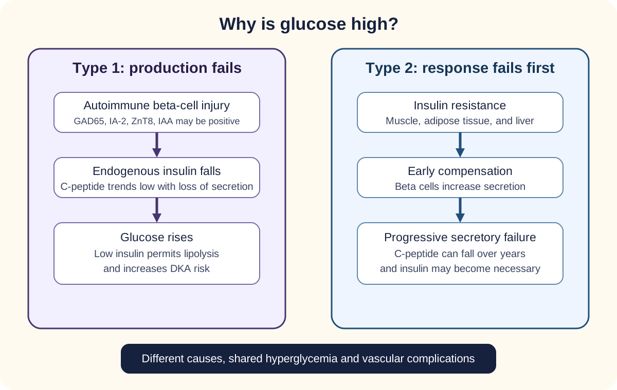
Different starting physiology can converge on hyperglycemia and shared complications. The IAA label means insulin autoantibodies, which are interpretable only before insulin treatment. [1]
The first diagram compares two trajectories. In type 1, immune injury reduces beta-cell mass until endogenous insulin becomes insufficient. In type 2, muscle, adipose tissue, and liver respond less effectively to insulin, so beta cells initially compensate with more secretion; over time, adequate secretion can no longer be maintained. Both paths can end in marked hyperglycemia and the same long-term vascular complications. [1][9]
Type 1 pattern
GAD65, IA-2, and ZnT8 autoantibodies, and insulin autoantibodies (IAA; valid only before insulin treatment), support autoimmune diabetes. HLA class II risk is strongly linked to DR3-DQ2 and DR4-DQ8 haplotypes, but HLA is not a routine diagnostic test. [1]
Type 2 pattern
Insulin resistance may coexist with normal or high insulin secretion early. Later, beta-cell secretory capacity can fall enough that insulin therapy is needed. Negative autoantibodies alone do not prove type 2 diabetes. [1]
Predict before opening: an adult with obesity presents in DKA. Does obesity settle the diabetes type?
No. DKA can occur in type 1 or type 2 diabetes, and obesity can occur in either. After the crisis resolves, classify with the whole clinical pattern and, when needed, autoantibodies and C-peptide. [1][3]
Transfer this rule: phenotype changes prior probability; immune markers and endogenous secretion answer a different question and can overturn a superficial age-or-body-size impression. [1]
Separate insulin production from insulin action
C-peptide is released in equimolar amounts with endogenous insulin when proinsulin is cleaved. Injected insulin does not contain C-peptide. That makes C-peptide useful when the question is, “How much insulin is this pancreas still making?” In insulin-treated patients with uncertain classification, a random C-peptide with a concurrent glucose can help, but timing matters and testing immediately after a hyperglycemic emergency can mislead. [1]
The insulin receptor itself is a receptor tyrosine kinase. In skeletal muscle and adipose tissue, insulin signaling increases surface GLUT4 and glucose uptake. Type 1 diabetes therefore has an intact receptor-signaling pathway but too little endogenous ligand; type 2 diabetes can have abundant insulin while downstream tissue response is impaired. [1][5]
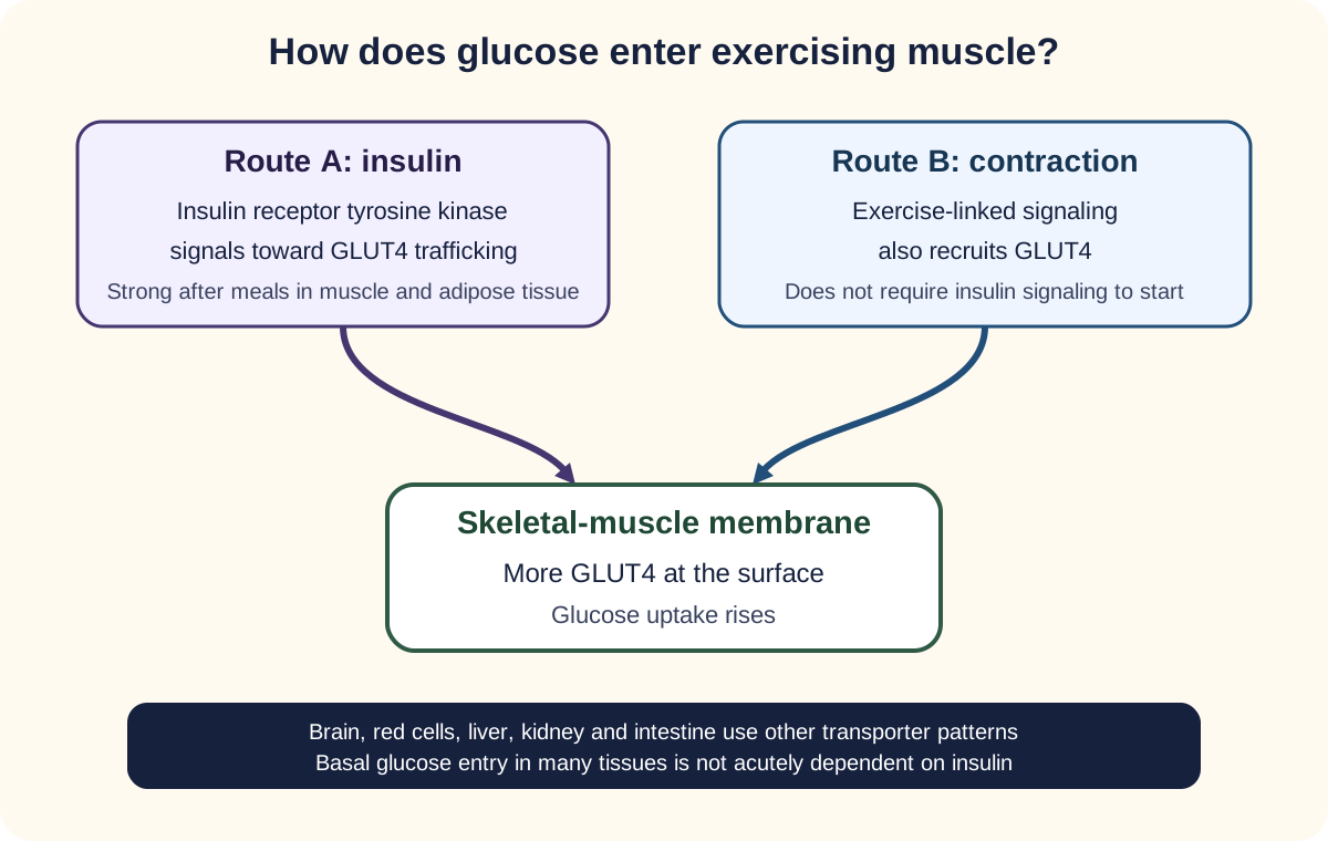
The old “seven tissues never need insulin” shortcut is too absolute. Many tissues use transporters that do not require acute insulin signaling for basal glucose entry. The clinically important contrast is that insulin strongly recruits GLUT4 in skeletal muscle and adipose tissue, while exercise can also recruit GLUT4 in skeletal muscle through contraction-linked signaling independent of insulin. [5]
Insulin and contraction use distinct signaling routes that converge on muscle GLUT4. [5]
Trace one glucose molecule
Resting muscle after a meal: insulin binds its receptor, signaling recruits GLUT4, and glucose entry rises.
Type 1 without enough insulin: that insulin-dependent recruitment is weak, while adipose lipolysis supplies fatty acids that the liver converts to ketones.
Exercising muscle: contraction-related pathways recruit GLUT4 even when insulin signaling is limited. [5]
Prediction: which result shows that circulating insulin came from the patient’s pancreas rather than an injection?
C-peptide. Injected insulin raises measured insulin without raising C-peptide, so measurable C-peptide, interpreted with the concurrent glucose and clinical setting, reflects endogenous secretion. [1]
Transfer this distinction to hypoglycemia: high insulin with high C-peptide indicates endogenous secretion or a secretagogue effect, whereas high insulin with suppressed C-peptide suggests exogenous insulin.
Prove diabetes, then classify it
In a nonpregnant person, diabetes can be diagnosed by A1C at least 6.5%, fasting plasma glucose at least 126 mg/dL, a 2-hour 75-g oral glucose tolerance value at least 200 mg/dL, or random plasma glucose at least 200 mg/dL with classic hyperglycemic symptoms or crisis. Without unequivocal hyperglycemia, confirmation is generally required. Prediabetes includes A1C 5.7% to 6.4%, fasting glucose 100 to 125 mg/dL, or 2-hour value 140 to 199 mg/dL. [1]
Once diabetes is established, classification is a separate step. For suspected adult type 1 diabetes, GAD antibodies are a useful first test; if negative, IA-2 and/or ZnT8 can add information. A minority of people with type 1 diabetes have no detectable islet autoantibodies, so a negative panel does not end the reasoning. [1]
Low C-peptide supports severe insulin deficiency, while clearly preserved C-peptide supports substantial endogenous secretion. Intermediate values overlap among type 1, monogenic diabetes, and long-duration type 2 diabetes. Adult-onset autoimmune diabetes, often called LADA in clinical use, belongs within the type 1 category rather than a separate “type 1.5” category. [1]
Pattern
Think next
PatternAutoantibody positive plus declining secretion
Think nextAutoimmune type 1 diabetes, regardless of adult age
Think nextConsider monogenic diabetes and genetic evaluation
PatternInsulin resistance phenotype with progressive secretion decline
Think nextType 2 diabetes remains likely, but classify from the full context
Prediction: a lean 19-year-old has mild stable fasting hyperglycemia, preserved C-peptide, negative antibodies, and affected relatives in three generations. Which category deserves attention?
Monogenic diabetes. The family pattern, preserved secretion, and absent autoimmunity should prevent an automatic type 1 label. [1]
The diagnostic threshold answers “is this diabetes?” The antibody and secretion pattern answers “what kind of diabetes is this?” Keep those questions separate. [1]
Try it here · Checkpoint 1 of 3
Make your prediction before reading the choices. A first attempt is just a starting point.
Case 4
Show answer and explanations for case 4
A. Monogenic diabetes (Best answer)
Young onset, preserved endogenous insulin, negative islet antibodies, and a vertical multigenerational pattern strongly suggest a monogenic form such as GCK- or HNF1A-related diabetes.
Reasoning steps for option A
What does a three-generation vertical family pattern suggest?
Diabetes in a father, grandmother, and great-grandmother suggests autosomal dominant transmission, typical of monogenic diabetes.
Which other findings strengthen the case for monogenic diabetes?
Lean body habitus, mild stable fasting hyperglycemia, negative islet antibodies, and preserved C-peptide make type 1 and type 2 diabetes less likely.
B. Autoimmune type 1 diabetes (Why this does not fit)
Type 1 can begin in adolescence, but the preserved secretion, negative antibodies, mild stable hyperglycemia, and vertical family pattern do not fit the usual autoimmune trajectory.
Reasoning steps for option B
Why is type 1 diabetes considered in a 19-year-old?
Adolescence and young adulthood are common ages for type 1 presentation.
What argues against autoimmune type 1 diabetes in this patient?
Negative islet antibodies, preserved C-peptide, and years of mild stable hyperglycemia do not fit autoimmune beta-cell destruction.
C. Typical youth-onset type 2 diabetes (Why this does not fit)
Type 2 can occur in adolescents, but this lean patient lacks an insulin-resistance phenotype and has a strong vertical family pattern that suggests a single-gene disorder.
Reasoning steps for option C
Why might a strong family history suggest youth-onset type 2 diabetes?
Type 2 diabetes is highly heritable and often clusters in families.
Which features make typical youth-onset type 2 diabetes less likely?
He has BMI 21 kg/m2 with no insulin-resistance phenotype, and the tidy dominant pattern of young-onset disease favors a single-gene cause.
D. Pancreatic exocrine diabetes (Why this does not fit)
Pancreatic disorders can cause diabetes, but no pancreatitis, malabsorption, surgery, or structural pancreatic disease is supplied.
Reasoning steps for option D
Why might preserved C-peptide be compatible with exocrine pancreatic diabetes?
Pancreatic disease can leave variable residual islet function.
What is missing for a pancreatic exocrine cause?
No pancreatitis, malabsorption, pancreatic surgery, or structural pancreatic disease is described, and exocrine disease does not explain the family pattern.
Takeaway: A multigenerational early-onset pattern with negative antibodies and preserved secretion should raise suspicion for monogenic diabetes.
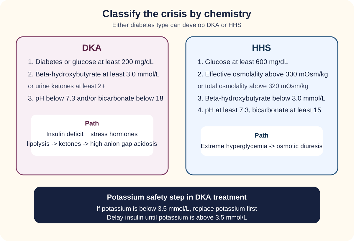
DKA and HHS are not reliable type labels. Both can occur in type 1 and type 2 diabetes, and mixed DKA/HHS presentations occur. Current adult consensus criteria define DKA by diabetes or hyperglycemia, significant ketonemia, and metabolic acidosis. HHS requires severe hyperglycemia and hyperosmolality without substantial ketonemia or acidosis. [3]
Use ketones, acid-base status, glucose, and osmolality to classify the crisis. [3]
For DKA, a plasma glucose at least 200 mg/dL or a prior diabetes diagnosis is paired with beta-hydroxybutyrate at least 3.0 mmol/L or urine ketones at least 2+, plus pH below 7.3 and/or bicarbonate below 18 mmol/L. This matters because SGLT2-associated DKA can occur with glucose below the 250 mg/dL threshold used in earlier criteria. [2][3]
For HHS, current consensus uses glucose at least 600 mg/dL, effective serum osmolality above 300 mOsm/kg or total osmolality above 320 mOsm/kg, beta-hydroxybutyrate below 3.0 mmol/L or urine ketones less than 2+, and pH at least 7.3 with bicarbonate at least 15 mmol/L. Profound osmotic diuresis makes dehydration and neurologic dysfunction prominent. [3]
DKA pathway
Insulin deficiency plus counterregulatory hormones increases lipolysis, hepatic ketogenesis, and high anion gap metabolic acidosis. Kussmaul respirations are respiratory compensation.
HHS pathway
Enough insulin effect usually limits major ketogenesis, but not the severe hyperglycemia that drives hyperosmolality and water loss. [3]
Potassium is the dangerous treatment pivot. Despite normal or high serum potassium on arrival, total-body potassium is depleted. Once serum potassium falls below 3.5 mmol/L, replace potassium and delay insulin until potassium rises above 3.5. When glucose falls below about 250 mg/dL during DKA treatment, add dextrose and continue insulin at an adjusted rate until ketoacidosis resolves. [3]
Prediction: glucose is 185 mg/dL, beta-hydroxybutyrate 5.2 mmol/L, pH 7.19, bicarbonate 12 mmol/L in a patient taking an SGLT2 inhibitor. Can this be DKA?
Yes. The ketonemia and acidosis satisfy the core DKA pattern, and current criteria do not require glucose above 250 mg/dL. [3]
Transfer the approach: ketones plus acidosis define the DKA pathway; marked hyperosmolality without substantial ketonemia or acidosis defines HHS. Do not use diabetes type as the crisis criterion. [3]
Try it here · Checkpoint 2 of 3
Make your prediction before reading the choices. A first attempt is just a starting point.
Case 12
Show answer and explanations for case 12
A. Start the insulin infusion immediately at the usual rate (Why this does not fit)
Insulin is essential for DKA, but at potassium 3.2 mmol/L it can drive potassium further into cells and increase arrhythmia risk.
Reasoning steps for option A
Why is insulin tempting in severe DKA?
Insulin is the treatment that stops ketogenesis, and pH 7.14 with beta-hydroxybutyrate 6.0 mmol/L shows severe ketoacidosis.
Why must insulin wait here?
Insulin drives potassium into cells, and starting it at potassium 3.2 mmol/L risks dangerous hypokalemia and arrhythmia.
B. Give sodium bicarbonate before any other therapy (Why this does not fit)
Routine bicarbonate is not the treatment priority at pH 7.14 and can worsen potassium shifts.
Reasoning steps for option B
Why might bicarbonate seem the logical first step?
With pH 7.14, giving base can look like direct correction of the acidosis.
Why is bicarbonate not the priority?
Routine bicarbonate is not recommended at pH 7.14, it can worsen hypokalemia, and potassium 3.2 mmol/L is the immediate danger.
C. Add dextrose because glucose is below 500 mg/dL (Why this does not fit)
Dextrose is added later as glucose approaches about 250 mg/dL while insulin continues to clear ketones; glucose 468 mg/dL does not trigger that step.
Reasoning steps for option C
Why might dextrose come up at this stage?
Dextrose is a standard part of later DKA therapy.
When is dextrose actually added?
When glucose falls below about 250 mg/dL during insulin treatment; glucose is 468 mg/dL and insulin has not started.
D. Replace potassium before insulin (Best answer)
Current adult consensus recommends potassium replacement and postponing insulin when potassium is below 3.5 mmol/L because insulin can worsen dangerous hypokalemia.
Reasoning steps for option D
Why does a potassium of 3.2 mmol/L matter so much in DKA?
Total-body potassium is depleted in DKA, and insulin will lower serum potassium further.
What sequence does current adult consensus recommend?
Replace potassium and delay insulin until potassium rises above 3.5 mmol/L, then start insulin.
Takeaway: In DKA with potassium below 3.5 mmol/L, replace potassium before starting insulin.
Type 1 diabetes requires insulin replacement. Delivery is by multiple daily injections of basal and prandial insulin or by insulin pump; the 2026 ADA Standards of Care recommend automated insulin delivery systems as the preferred method for people with type 1 diabetes. [10] Dose education includes carbohydrate intake, current glucose, expected activity, and correction needs. Oral secretagogues cannot replace absent beta-cell capacity. [2]
Type 2 therapy is no longer a single fixed ladder that always starts with metformin and only later considers organ protection. Treatment is individualized. In people with established or high-risk cardiovascular disease, heart failure, chronic kidney disease, and/or obesity, SGLT2 inhibitors and GLP-1-based therapy can be chosen for cardiorenal or weight benefit independent of whether metformin was used first or the current A1C. [2]
If a person with type 2 diabetes has severe hyperglycemia, catabolic symptoms, or glucose around 300 mg/dL or higher or A1C above 10%, insulin may be appropriate at presentation. Without severe hyperglycemia or crisis, a GLP-1-based therapy is preferred to insulin for many people who need additional injectable glucose-lowering treatment. [2]
Medication hazards still matter. Sulfonylureas can cause hypoglycemia because they stimulate insulin secretion. Pioglitazone can cause fluid retention and worsen heart failure; it is not recommended in symptomatic heart failure, and starting it in established NYHA class III or IV heart failure is contraindicated. [2][8]
Metformin remains effective and widely used, but renal function changes the plan. Current U.S. labeling contraindicates it below eGFR 30 mL/min/1.73 m2 and does not recommend starting it at eGFR 30 to 45. Renal impairment increases risk of metformin-associated lactic acidosis, a high anion gap acidosis with high lactate. [7]
Prediction: A1C is 7.0% in a person with type 2 diabetes and symptomatic heart failure. Is “A1C already at goal” a reason to ignore cardiorenal therapy?
No. An SGLT2 inhibitor with demonstrated heart-failure benefit may be indicated for organ protection even when additional glucose lowering is not the main reason for treatment. [2]
For many nonpregnant adults an A1C below 7% is appropriate, but targets are individualized when frailty, comorbidity, hypoglycemia, treatment burden, or personal goals change the balance. [4]
Try it here · Checkpoint 3 of 3
Make your prediction before reading the choices. A first attempt is just a starting point.
Case 15
Show answer and explanations for case 15
A. An SGLT2 inhibitor (Best answer)
In type 2 diabetes with heart failure or chronic kidney disease, an SGLT2 inhibitor with demonstrated benefit can be selected for cardiorenal protection independent of the current A1C or prior metformin use.
Reasoning steps for option A
Which comorbidities drive drug choice in this man?
Symptomatic heart failure with reduced ejection fraction and chronic kidney disease with eGFR 48 mL/min/1.73 m2.
Why is an SGLT2 inhibitor chosen when A1C is 7.1%?
SGLT2 inhibitors reduce heart-failure hospitalization and slow kidney disease progression, benefits independent of further glucose lowering.
B. Sulfonylurea (Why this does not fit)
Sulfonylureas lower glucose but add hypoglycemia risk and do not supply the same heart-failure and kidney-protection rationale.
Reasoning steps for option B
Why might a sulfonylurea be offered?
It is inexpensive and would lower A1C further.
Why does a sulfonylurea miss the goal of this question?
The priority is heart and kidney protection, which sulfonylureas do not provide, and they add hypoglycemia risk.
C. Pioglitazone (Why this does not fit)
Pioglitazone can improve insulin sensitivity but may cause fluid retention and worsen symptomatic heart failure.
Reasoning steps for option C
Why might pioglitazone appeal in type 2 diabetes?
It directly improves insulin sensitivity.
What makes pioglitazone harmful for this patient?
It causes fluid retention and can worsen symptomatic heart failure.
D. DPP-4 inhibitor (Why this does not fit)
DPP-4 inhibitors can lower glucose with little hypoglycemia, but the stem prioritizes demonstrated heart-failure and kidney benefit.
Reasoning steps for option D
Why might a DPP-4 inhibitor seem enough?
A1C is near target, and DPP-4 inhibitors lower glucose modestly with little hypoglycemia.
Why is a DPP-4 inhibitor not favored here?
It lacks the demonstrated heart-failure and kidney benefit that his comorbidities call for.
Takeaway: In type 2 diabetes with heart failure or CKD, therapy can be chosen for cardiorenal benefit independent of A1C.
Persistent hyperglycemia in either type can injure small vessels and large arteries. The classic microvascular domains are retinopathy, kidney disease, and neuropathy; macrovascular disease includes coronary, cerebrovascular, and peripheral arterial disease. Ongoing care therefore assesses complications and cardiovascular risk regardless of the original diabetes category. [9]
Two time-course patterns prevent classification errors. Early after type 1 diagnosis, surviving beta cells may temporarily recover secretion as glucotoxic stress falls, producing a partial remission or honeymoon phase with lower insulin needs and measurable C-peptide. In long-standing type 2 diabetes, the opposite trend can occur: progressive beta-cell failure lowers C-peptide and creates an eventual need for insulin without converting type 2 diabetes into autoimmune type 1. [1]
Pregnancy and childhood add useful transfer examples. Maternal hyperglycemia crosses the placenta and stimulates fetal insulin production. After delivery the maternal glucose supply stops abruptly while fetal insulin remains high, so an infant of a diabetic mother can develop neonatal hypoglycemia. This common mechanism is better described as fetal hyperinsulinism than by using “nesidioblastosis” as a routine label. [6]
In a previously dry child, new secondary enuresis with polyuria, polydipsia, weight loss, or candidal infection should prompt evaluation for hyperglycemia. The same osmotic diuresis that causes frequent daytime urination can reappear as bedwetting. [1]
Prediction: a person with established type 1 diabetes needs much less insulin six months after diagnosis and has measurable C-peptide. Has the autoimmune diagnosis disappeared?
No. Residual beta-cell secretion can create a partial remission. Insulin needs must be adjusted safely, but the underlying autoimmune classification remains. [1]
The final transfer rule is temporal: ask what the pancreas was doing before, during, and after the presentation. A single insulin or C-peptide value without its glucose level, treatment context, and disease duration can be misleading. [1]
Apply the distinctions
Case 1
Show answer and explanations for case 1
A. Ketosis-prone type 2 diabetes (Why this does not fit)
Ketosis-prone type 2 diabetes can present with DKA, but the positive GAD65 antibody and very low C-peptide after recovery support autoimmune beta-cell loss instead.
Reasoning steps for option A
Why is ketosis-prone type 2 diabetes tempting after a DKA presentation?
DKA can be the first presentation of ketosis-prone type 2 diabetes, especially in adults.
Which recovery results argue against ketosis-prone type 2 diabetes here?
A positive GAD65 antibody and very low C-peptide with glucose 172 mg/dL point to autoimmune beta-cell loss; ketosis-prone type 2 usually recovers secretion and is antibody-negative.
B. Glucocorticoid-induced diabetes (Why this does not fit)
Glucocorticoids can cause marked hyperglycemia through insulin resistance, but no exposure is supplied and the autoimmune marker plus low secretion point elsewhere.
Reasoning steps for option B
Why might a drug-induced cause be considered for glucose 386 mg/dL?
Glucocorticoids cause insulin resistance and can produce marked hyperglycemia.
What is missing for glucocorticoid-induced diabetes, and what points elsewhere?
No glucocorticoid exposure is described, and steroid-induced insulin resistance does not explain GAD65 positivity or very low C-peptide.
C. Autoimmune type 1 diabetes (Best answer)
The acute ketotic presentation establishes insulin-deficient physiology, and the later positive GAD65 antibody with very low C-peptide establishes autoimmune beta-cell failure.
Reasoning steps for option C
What does the crisis itself show about insulin supply?
pH 7.16 with beta-hydroxybutyrate 5.8 mmol/L shows insulin deficiency severe enough to allow unchecked ketogenesis.
Which post-recovery results confirm autoimmune type 1 diabetes?
Positive GAD65 antibodies plus very low C-peptide during glucose 172 mg/dL show autoimmune beta-cell destruction with little remaining secretion.
D. Hyperglycemic hyperosmolar state (Why this does not fit)
HHS can cause severe dehydration and neurologic symptoms, but the supplied acidosis and substantial beta-hydroxybutyrate define a ketotic acidotic crisis.
Reasoning steps for option D
Why does HHS come to mind with glucose 386 mg/dL and weeks of polyuria?
Prolonged osmotic diuresis causes dehydration, which is central to HHS.
Which laboratory values rule out isolated HHS?
pH 7.16 and beta-hydroxybutyrate 5.8 mmol/L define ketoacidosis, and glucose 386 mg/dL is below the HHS threshold of 600 mg/dL.
Takeaway: Autoantibodies and endogenous insulin secretion can classify diabetes after the acute crisis has been treated.
A. Progressive type 2 diabetes (Why this does not fit)
Type 2 diabetes can lose beta-cell capacity over time, but the short course and positive GAD65 antibody indicate autoimmunity rather than ordinary type 2 progression.
Reasoning steps for option A
Why does ordinary type 2 progression look plausible in this woman?
She has obesity, adult onset, and an initial response to metformin, all common in type 2 diabetes.
What makes ordinary type 2 progression the wrong explanation?
Type 2 secretory loss usually unfolds over years without autoimmunity; here control collapsed within 18 months and GAD65 is positive.
B. Adult autoimmune type 1 diabetes (Best answer)
Adult age and obesity do not exclude type 1 diabetes. Positive GAD65 antibodies plus declining endogenous secretion identify adult autoimmune type 1 diabetes.
Reasoning steps for option B
Why do obesity and metformin response fail to exclude type 1 diabetes?
Obesity is common in the general population and can coexist with autoimmune diabetes, and early residual secretion can make metformin briefly effective.
Which two findings confirm adult autoimmune type 1 diabetes?
A positive GAD65 antibody plus C-peptide of only 0.35 ng/mL with glucose 210 mg/dL show autoimmune loss of secretion.
C. GCK-related monogenic diabetes (Why this does not fit)
GCK-related diabetes often causes mild stable hyperglycemia, but it is antibody-negative and does not usually show rapid secretory decline.
Reasoning steps for option C
Why might a monogenic form be raised in an adult with mild early disease?
GCK-related diabetes causes mild hyperglycemia that often goes unrecognized until adulthood.
Which features of this course do not fit GCK-related diabetes?
GCK diabetes is antibody-negative and stable for decades, whereas she has GAD65 antibodies and rapidly worsening glycemia.
D. Pancreatogenic diabetes (Why this does not fit)
Pancreatic disease can reduce insulin secretion, but the stem provides no pancreatitis, pancreatic surgery, or exocrine dysfunction and does provide an autoimmune marker.
Reasoning steps for option D
Why could pancreatic disease explain a low C-peptide?
Destruction of pancreatic tissue removes islets and lowers insulin secretion.
What is absent for pancreatogenic diabetes, and what is present instead?
There is no pancreatitis, pancreatic surgery, or exocrine insufficiency, while GAD65 positivity directly indicates autoimmune injury.
Takeaway: Adult-onset autoimmune diabetes is type 1 diabetes even when obesity or initial metformin response resembles type 2.
A. The low C-peptide proves factitious insulin use (Why this does not fit)
Injected insulin does not contain C-peptide, but factitious use is not established by low C-peptide in a person with known diabetes and positive islet autoimmunity.
Reasoning steps for option A
Why might low C-peptide in an insulin user raise concern for factitious insulin?
Injected insulin contains no C-peptide, so exogenous insulin with suppressed C-peptide is the pattern of factitious hypoglycemia.
Why does that pattern not apply to this man?
He is prescribed insulin for known diabetes, is not hypoglycemic, and has positive GAD65 antibodies, so low C-peptide reflects deficient secretion.
B. The C-peptide should be ignored because insulin therapy makes it uninterpretable (Why this does not fit)
Insulin treatment does not make C-peptide useless. With a concurrent glucose and appropriate timing after crisis, it can help assess endogenous secretion.
Reasoning steps for option B
Why might ongoing basal-bolus insulin seem to invalidate the C-peptide?
Exogenous insulin lowers glucose and can reduce the stimulus for endogenous secretion.
Why is this particular C-peptide still interpretable?
C-peptide measures only pancreatic secretion, and here it is 0.10 ng/mL while glucose is 168 mg/dL, three months after the crisis.
C. The result demonstrates preserved beta-cell reserve (Why this does not fit)
A C-peptide of 0.10 ng/mL is not evidence of substantial reserve in this context.
Reasoning steps for option C
Why could a measurable C-peptide be misread as reassuring?
Any detectable value can look like preserved function if it is read without a reference scale.
What shows that 0.10 ng/mL is not preserved reserve?
The value is very low even though glucose of 168 mg/dL should be stimulating secretion.
D. Severe type 1 insulin deficiency (Best answer)
The test was obtained well after the acute crisis, glucose was concurrent, C-peptide is very low, and GAD65 is positive. Together these findings support autoimmune type 1 diabetes.
Reasoning steps for option D
Why is this C-peptide result trustworthy for classification?
It was drawn three months after the crisis with a concurrent glucose of 168 mg/dL, so it reflects stable secretory capacity.
What completes the case for severe type 1 insulin deficiency?
Positive GAD65 antibodies add independent evidence that autoimmune beta-cell destruction caused the very low secretion.
Takeaway: C-peptide is useful in insulin-treated patients when interpreted with concurrent glucose, timing, and autoimmune markers.
A. Repeat an A1C before diabetes can be diagnosed (Why this does not fit)
A confirmatory test is generally needed without unequivocal hyperglycemia, but classic symptoms plus random plasma glucose at least 200 mg/dL satisfy a diagnostic criterion directly.
Reasoning steps for option A
Why might a confirmatory A1C seem required?
Most abnormal results in asymptomatic people need a second test before diabetes is diagnosed.
Why is confirmation unnecessary for this woman?
Classic symptoms with random plasma glucose 218 mg/dL are unequivocal hyperglycemia and meet a diagnostic criterion on their own.
B. Order a 2-hour oral glucose tolerance test because random glucose is never diagnostic (Why this does not fit)
An OGTT is a valid diagnostic route, but random plasma glucose is diagnostic when it is at least 200 mg/dL and accompanied by classic hyperglycemic symptoms.
Reasoning steps for option B
Why might an oral glucose tolerance test be proposed?
A 2-hour value of at least 200 mg/dL is a valid diagnostic criterion.
What is wrong with claiming random glucose is never diagnostic?
Random plasma glucose of at least 200 mg/dL with classic symptoms is itself a recognized diagnostic criterion.
C. Diagnose diabetes from the random glucose plus classic symptoms (Best answer)
Polyuria, polydipsia, weight loss, and random plasma glucose above 200 mg/dL meet a diagnostic criterion for diabetes.
Reasoning steps for option C
Why do her symptoms matter when reading the random glucose?
Polyuria, polydipsia, and weight loss show that the glucose of 218 mg/dL reflects sustained symptomatic hyperglycemia.
Why can diabetes be diagnosed today without another test?
A random plasma glucose of at least 200 mg/dL with classic symptoms meets the criterion directly, so no second test is needed.
D. Label the result prediabetes until fasting glucose exceeds 126 mg/dL (Why this does not fit)
Prediabetes thresholds apply to lower glucose ranges and do not override a diagnostic random glucose accompanied by classic symptoms.
Reasoning steps for option D
Why might waiting for a fasting glucose seem reasonable?
Fasting plasma glucose is a common and familiar diagnostic route.
Why is a prediabetes label wrong here?
Prediabetes describes values below diabetes thresholds, while a symptomatic random glucose of 218 mg/dL already exceeds the diabetes threshold.
Takeaway: Random plasma glucose at least 200 mg/dL with classic hyperglycemic symptoms can diagnose diabetes.
A. G protein-coupled receptor (Why this does not fit)
Many metabolic hormones use G protein-coupled receptors, but the insulin receptor does not signal through a heterotrimeric G protein.
Reasoning steps for option A
Why might a G protein-coupled receptor seem a reasonable guess for insulin?
Many peptide hormones, such as glucagon, act through G protein-coupled surface receptors.
How does the insulin receptor differ from a G protein-coupled receptor?
It does not use a heterotrimeric G protein; ligand binding activates the receptor's own tyrosine kinase.
B. Intracellular nuclear receptor (Why this does not fit)
Nuclear receptors regulate transcription for lipid-soluble hormones, whereas insulin is a peptide that cannot diffuse through the plasma membrane to bind such a receptor.
Reasoning steps for option B
Why might a nuclear receptor be chosen for a hormone that changes cell metabolism?
Some hormones act by entering cells and binding intracellular receptors.
Why can insulin not use a nuclear receptor?
Insulin is a peptide that cannot cross the plasma membrane, so it must act on a cell-surface receptor.
C. Receptor tyrosine kinase (Best answer)
Insulin binds a receptor tyrosine kinase, leading to receptor autophosphorylation and downstream signaling that promotes GLUT4 trafficking in skeletal muscle and adipose tissue.
Reasoning steps for option C
How does insulin binding start the signal?
Insulin binds the receptor's extracellular alpha subunits, and the beta subunits autophosphorylate on tyrosine residues.
How does that receptor signal reach GLUT4 in muscle?
Receptor autophosphorylation starts downstream signaling that moves GLUT4 vesicles to the cell surface in skeletal muscle and fat.
D. Cytokine receptor with JAK-STAT signaling (Why this does not fit)
Some peptide hormones use JAK-STAT-associated receptors, but insulin uses a receptor with intrinsic kinase activity.
Reasoning steps for option D
Why might a JAK-STAT receptor seem plausible?
Growth hormone and prolactin are peptide hormones that signal through JAK-associated receptors.
What distinguishes the insulin receptor from a cytokine-type receptor?
The insulin receptor has intrinsic tyrosine kinase activity and does not need a separate JAK kinase.
Takeaway: Insulin signals through a receptor tyrosine kinase to increase GLUT4 surface availability in skeletal muscle and adipose tissue.
A. Increased hepatic GLUT2 insertion caused by exercise (Why this does not fit)
Hepatic GLUT2 is not the principal transporter responsible for contraction-stimulated skeletal-muscle glucose uptake.
Reasoning steps for option A
Why might the liver seem relevant after dinner?
The liver takes up and releases glucose and plays a large part in handling a meal.
Why does hepatic GLUT2 not explain this finding?
The increase is in exercising skeletal muscle, where contraction recruits GLUT4; hepatic GLUT2 is not regulated by exercise in this way.
B. Insulin-independent synthesis of new insulin receptors during the walk (Why this does not fit)
New receptor synthesis is too slow to explain an immediate exercise effect and does not account for contraction-specific glucose transport.
Reasoning steps for option B
Why might new insulin receptors seem a way around insulin resistance?
More receptors might seem able to offset weak insulin signaling.
Why can receptor synthesis not explain an effect during a 40-minute walk?
Building new receptors is too slow, and the uptake rises through a contraction pathway that bypasses insulin signaling.
C. Conversion of skeletal-muscle GLUT4 into GLUT2 (Why this does not fit)
Transporters do not change identity from GLUT4 to GLUT2 in response to exercise.
Reasoning steps for option C
Why might switching to a different transporter seem necessary?
If insulin cannot recruit GLUT4 normally, an insulin-independent transporter might seem required.
Why is a GLUT4-to-GLUT2 conversion impossible?
Transporters do not change isoform; exercise moves existing GLUT4 to the membrane through contraction signaling.
D. Contraction-linked GLUT4 trafficking (Best answer)
Muscle contraction activates signaling that increases GLUT4 at the membrane independently of insulin, allowing exercise to increase glucose uptake even when insulin action is impaired.
Reasoning steps for option D
What does glucose uptake rising during the walk indicate?
Muscle contraction itself creates a route to GLUT4 surface translocation.
Why does this route still work with impaired insulin sensitivity?
Contraction signaling, including AMPK and calcium pathways, bypasses the insulin receptor pathway that is resistant in type 2 diabetes.
Takeaway: Exercise can increase skeletal-muscle glucose uptake through contraction-stimulated GLUT4 trafficking that does not require insulin signaling.
A. Starvation ketosis without acidosis (Why this does not fit)
Reduced intake can raise ketones, but the marked ketonemia with pH 7.20 and bicarbonate 12 mmol/L represents substantial metabolic acidosis.
Reasoning steps for option A
Why might 24 hours of vomiting suggest starvation ketosis?
Poor intake lowers insulin and raises ketone production.
Why is starvation ketosis too mild an explanation?
Starvation ketosis rarely causes significant acidosis, whereas she has beta-hydroxybutyrate 5.1 mmol/L, pH 7.20, and bicarbonate 12 mmol/L.
B. Diabetic ketoacidosis with relatively low glucose (Best answer)
Current DKA criteria emphasize diabetes or hyperglycemia plus significant ketonemia and metabolic acidosis. SGLT2 therapy can be associated with DKA despite glucose below traditional thresholds.
Reasoning steps for option B
How does SGLT2 therapy change the usual DKA picture?
Urinary glucose loss keeps plasma glucose relatively low while insulin deficiency drives ketogenesis.
Which values confirm DKA despite glucose 184 mg/dL?
Beta-hydroxybutyrate 5.1 mmol/L with pH 7.20 and bicarbonate 12 mmol/L meet the ketonemia and acidosis criteria, and known diabetes satisfies the glucose domain.
C. Hyperglycemic hyperosmolar state (Why this does not fit)
HHS requires much more severe hyperglycemia and hyperosmolality with minimal ketonemia and no major acidosis.
Reasoning steps for option C
Why might HHS be considered in a woman with type 2 diabetes?
HHS occurs mostly in type 2 diabetes, often with intercurrent illness.
Which values are the opposite of HHS?
Glucose 184 mg/dL is far below 600 mg/dL, and marked ketonemia with pH 7.20 contradicts the minimal ketosis of HHS.
D. Metformin-associated lactic acidosis (Why this does not fit)
Metformin-associated acidosis is driven by lactate accumulation, not the supplied marked beta-hydroxybutyrate pattern.
Reasoning steps for option D
Why might metformin-associated lactic acidosis be considered?
It can present with vomiting and a high anion gap acidosis in a person with type 2 diabetes.
What identifies ketoacid rather than lactate as the acid here?
The measured acid is beta-hydroxybutyrate 5.1 mmol/L, and SGLT2 therapy is a known DKA setting; no metformin or lactate is described.
Takeaway: DKA can occur with glucose below 200 to 250 mg/dL, particularly with SGLT2 inhibitor exposure.
A. 26 mmol/L, supporting high anion gap ketoacidosis (Best answer)
The anion gap is 136 minus the sum of 100 and 10, which equals 26 mmol/L. In this ketotic hyperglycemic presentation, the unmeasured anions are consistent with ketoacids.
Reasoning steps for option A
How is the anion gap calculated from these values?
Sodium minus (chloride + bicarbonate): 136 minus 110 equals 26 mmol/L.
What does a gap of 26 mmol/L mean in this boy?
It is high, and with marked ketonemia and glucose 510 mg/dL the unmeasured anions are ketoacids.
B. 10 mmol/L, supporting a normal-gap acidosis (Why this does not fit)
10 mmol/L is the bicarbonate value, not the gap; the correct gap is 26 mmol/L, which is high, not normal.
Reasoning steps for option B
Why might 10 mmol/L be picked as the answer?
Ten is the bicarbonate value in the stem, and low bicarbonate does confirm metabolic acidosis.
What does the correct calculation show instead?
136 minus (100 + 10) equals 26 mmol/L, a high gap, not a normal one.
C. 36 mmol/L, supporting toxic alcohol ingestion (Why this does not fit)
36 results from subtracting only chloride and omitting bicarbonate; the correct gap is 26, and marked ketonemia already explains the acidosis.
Reasoning steps for option C
How could someone arrive at 36 mmol/L?
Subtracting only chloride from sodium gives 136 minus 100, which is 36.
Why is toxic alcohol ingestion not the conclusion?
The correct gap is 26 mmol/L, and marked ketonemia with severe hyperglycemia already explains the acidosis without another toxin.
D. 26 mmol/L, proving lactic acidosis (Why this does not fit)
The numeric calculation is correct, but an anion gap does not identify the acid by itself.
Reasoning steps for option D
Why might lactate be linked to this high gap?
Lactate is a common cause of a high anion gap acidosis.
Why can the gap not prove lactic acidosis?
A gap shows that unmeasured anions are present but not which ones, and marked ketonemia identifies ketoacids here.
Takeaway: Anion gap equals sodium minus (chloride + bicarbonate); the result must be interpreted with the measured acid and clinical setting.
A. Stop insulin because glucose is below 250 mg/dL (Why this does not fit)
Stopping insulin while ketonemia and acidosis persist can allow ketogenesis to continue or recur.
Reasoning steps for option A
Why might stopping insulin at glucose 232 mg/dL seem safe?
Glucose is falling toward the range where continued insulin could cause hypoglycemia.
Why would stopping insulin be harmful?
Beta-hydroxybutyrate 3.9 mmol/L and bicarbonate 14 mmol/L show that ketoacidosis has not resolved, so ketogenesis would continue.
B. Switch to subcutaneous therapy immediately because potassium is normal (Why this does not fit)
Normal potassium removes one barrier to insulin but does not establish DKA resolution.
Reasoning steps for option B
Why might normal potassium suggest switching to subcutaneous insulin?
Potassium 4.2 mmol/L removes one barrier to continuing therapy.
What must happen before transition to subcutaneous insulin?
DKA must resolve first, and persistent ketonemia with bicarbonate 14 mmol/L shows it has not.
C. Add dextrose and continue insulin (Best answer)
When glucose falls below about 250 mg/dL before ketoacidosis resolves, dextrose permits continued insulin to suppress ketogenesis without causing hypoglycemia.
Reasoning steps for option C
What does glucose 232 mg/dL with beta-hydroxybutyrate 3.9 mmol/L show?
Glucose has corrected faster than the ketoacidosis.
How does adding dextrose solve this mismatch?
Dextrose prevents hypoglycemia so insulin can continue until ketogenesis is suppressed and the acidosis clears.
D. Give bicarbonate to normalize bicarbonate before continuing insulin (Why this does not fit)
Bicarbonate is not routinely used for this degree of DKA and does not replace the need for insulin-mediated suppression of ketogenesis.
Reasoning steps for option D
Why might bicarbonate seem helpful at 14 mmol/L?
The low bicarbonate shows that metabolic acidosis persists.
Why is bicarbonate therapy not the next step?
Bicarbonate is not routinely given at this level; the acidosis resolves when continued insulin stops ketoacid production.
Takeaway: When glucose falls before DKA resolves, add dextrose and continue insulin so ketogenesis can be suppressed safely.
A. Metformin alone with home glucose checks (Why this does not fit)
Metformin can reduce hepatic glucose output and improve insulin sensitivity, but it cannot replace the insulin missing in autoimmune type 1 diabetes.
Reasoning steps for option A
Why might metformin come to mind for new diabetes?
Metformin is the most familiar first glucose-lowering drug.
Why can metformin not treat her diabetes alone?
Metformin lowers hepatic glucose output but cannot replace insulin that autoimmune beta-cell destruction has removed.
B. Basal-prandial insulin replacement (Best answer)
Type 1 diabetes requires insulin replacement. Basal plus prandial dosing or continuous subcutaneous infusion best approximates background and meal-related insulin needs.
Reasoning steps for option B
What does autoimmune beta-cell failure leave missing?
Both background (basal) and meal-related (prandial) insulin secretion are lost.
How does a basal-prandial plan match that physiology?
Basal insulin covers fasting needs and prandial insulin covers meals, delivered by injections, a pump, or preferably an automated insulin delivery system.
C. Sulfonylurea monotherapy to stimulate the remaining beta cells (Why this does not fit)
A secretagogue requires functioning beta cells and cannot reliably replace insulin in autoimmune type 1 diabetes.
Reasoning steps for option C
Why might a sulfonylurea seem useful soon after diagnosis?
Some newly diagnosed people with type 1 diabetes retain temporary beta-cell function.
Why is a secretagogue not adequate therapy?
Sulfonylureas need functioning beta cells, and residual secretion in type 1 diabetes is too little and too short-lived to replace insulin.
D. GLP-1 receptor agonist monotherapy because it lowers glucose with little hypoglycemia (Why this does not fit)
GLP-1-based therapy can be useful in type 2 diabetes, but it is not a substitute for insulin in established type 1 diabetes.
Reasoning steps for option D
Why might a GLP-1 receptor agonist seem attractive?
It lowers glucose with little hypoglycemia in type 2 diabetes.
Why is GLP-1 monotherapy wrong in type 1 diabetes?
It works mainly by enhancing insulin secretion, and this patient lacks the beta cells to respond, so insulin replacement is still required.
Takeaway: Type 1 diabetes requires basal and meal-related insulin replacement through injections or continuous infusion.
A sulfonylurea can lower glucose at low cost, but it can cause hypoglycemia and weight gain, which conflict with the stated weight priority.
Reasoning steps for option A
Why is glipizide a plausible add-on at A1C 8.3%?
It lowers glucose effectively and at low cost.
Why does glipizide conflict with her goals?
It tends to cause weight gain and hypoglycemia and offers no cardiovascular or weight benefit.
B. Sitagliptin (Why this does not fit)
A DPP-4 inhibitor is generally weight neutral and has low hypoglycemia risk, but it offers less weight reduction and does not match the cardiovascular and weight priorities as directly.
Reasoning steps for option B
Why might sitagliptin appeal?
It is weight neutral and has low hypoglycemia risk.
Why is sitagliptin a weaker fit than a GLP-1-based agent?
It produces little weight loss and has no proven cardiovascular benefit, while she has prior myocardial infarction and wants substantial weight reduction.
C. Pioglitazone (Why this does not fit)
Pioglitazone improves insulin sensitivity, but weight gain and edema make it poorly aligned with this patient's priorities.
Reasoning steps for option C
Why might pioglitazone be considered for insulin resistance?
It improves insulin sensitivity, which is central to type 2 diabetes with obesity.
Why is pioglitazone poorly aligned with this patient?
It causes weight gain and edema, which conflict with her top priority of weight reduction.
D. A GLP-1-based agent (Best answer)
A GLP-1-based agent with demonstrated cardiovascular benefit can address both established ASCVD and the patient's strong weight-reduction priority while lowering glucose.
Reasoning steps for option D
Which features define her treatment priorities?
Established ASCVD from prior myocardial infarction, BMI 38 kg/m2, and a stated goal of weight reduction.
How does a GLP-1-based agent meet all three needs?
Agents with proven cardiovascular benefit lower glucose, reduce cardiovascular events, and produce substantial weight loss.
Takeaway: Type 2 treatment selection should incorporate cardiovascular disease and weight goals, not only the A1C.
He has no ketoacidosis and is newly diagnosed with type 2 diabetes.
Why is waiting three months unsafe?
Glucose 356 mg/dL, A1C 11.4%, polyuria, and 8-kg weight loss show severe catabolic hyperglycemia that needs prompt drug therapy.
B. Initiate insulin to correct severe symptomatic hyperglycemia (Best answer)
Current type 2 guidance supports insulin when hyperglycemia is severe, especially with catabolic symptoms, glucose around 300 mg/dL or higher, or A1C above 10%.
Reasoning steps for option B
What do his weight loss and glucose level indicate?
Catabolic weight loss with glucose 356 mg/dL shows substantial insulin inadequacy.
Which guidance thresholds support starting insulin?
Insulin is favored with catabolic symptoms, glucose of about 300 mg/dL or higher, or A1C above 10%, and he meets all three.
C. Start a sulfonylurea only (Why this does not fit)
A sulfonylurea may lower glucose but does not provide the predictable control needed for this severe catabolic presentation and adds hypoglycemia risk.
Reasoning steps for option C
Why might a sulfonylurea seem able to act quickly?
Secretagogues can lower glucose within days in some people with type 2 diabetes.
Why is sulfonylurea monotherapy inadequate here?
It depends on remaining beta-cell function, gives less predictable control in severe catabolic hyperglycemia, and adds hypoglycemia risk.
D. Begin pioglitazone only (Why this does not fit)
Pioglitazone has a delayed glucose-lowering effect and is not appropriate as sole initial therapy for severe symptomatic hyperglycemia with catabolism.
Reasoning steps for option D
Why might pioglitazone be considered for type 2 diabetes?
Insulin resistance is a core feature of type 2 diabetes, and pioglitazone targets it.
Why is pioglitazone alone unsuitable?
Its glucose-lowering effect takes weeks to develop, which is too slow for severe symptomatic hyperglycemia with catabolism.
Takeaway: Severe symptomatic type 2 hyperglycemia with catabolic features is a setting in which insulin should be considered at presentation.
A. Start basal insulin because every injectable sequence begins with insulin (Why this does not fit)
Insulin is effective, but current type 2 guidance does not require insulin as the first injectable when severe hyperglycemia or crisis is absent.
Reasoning steps for option A
Why might basal insulin seem the natural first injectable?
Basal insulin is effective and has long been a standard injectable step.
Why is insulin not required first for her?
Without severe hyperglycemia, catabolism, or crisis, guidance prefers a GLP-1-based agent before insulin.
B. Add prandial insulin without basal insulin (Why this does not fit)
Prandial insulin alone does not fit usual initial insulin intensification and is unnecessary before trying a preferred noninsulin injectable in this stable setting.
Reasoning steps for option B
Why might prandial insulin be proposed?
Post-meal glucose can contribute to an A1C of 8.6%.
Why is prandial insulin alone a poor choice?
Insulin intensification usually starts with basal insulin, and a GLP-1-based agent is preferred before any insulin in this stable setting.
C. Use GLP-1-based therapy before insulin (Best answer)
When type 2 diabetes requires additional therapy and there is no severe hyperglycemia or crisis, GLP-1-based therapy is preferred to insulin for many patients, especially when obesity is also relevant.
Reasoning steps for option C
What features make her suitable for a noninsulin injectable?
She needs more glucose lowering but has no severe hyperglycemia, crisis, or catabolic weight loss.
Why is GLP-1-based therapy preferred over insulin for her?
It lowers glucose with little hypoglycemia and promotes weight loss, which matters because she has obesity.
D. Use a sulfonylurea because injectable therapy should be avoided (Why this does not fit)
The patient has specifically accepted injectable treatment, and a sulfonylurea would add hypoglycemia risk without addressing the preference for a weight-directed option.
Reasoning steps for option D
Why might an oral sulfonylurea be offered?
Oral therapy can seem simpler than any injection.
Why does a sulfonylurea not fit this patient?
She prefers an injectable, and a sulfonylurea adds hypoglycemia and weight gain without addressing obesity.
Takeaway: Without severe hyperglycemia or crisis, GLP-1-based therapy is preferred to insulin for many people with type 2 diabetes who need additional injectable treatment.
An SGLT2 inhibitor with heart-failure benefit is not the contraindicated option in this scenario.
Reasoning steps for option A
Why might empagliflozin draw attention in heart failure?
It is a diabetes drug discussed specifically in the context of heart failure.
Why is empagliflozin not the drug to reject?
SGLT2 inhibitors reduce heart-failure hospitalization and carry no heart-failure initiation contraindication.
B. Semaglutide (Why this does not fit)
A GLP-1-based agent may be considered for glucose, cardiovascular, or weight goals and does not carry the pioglitazone heart-failure initiation contraindication.
Reasoning steps for option B
Why might semaglutide be questioned in this man?
Any new drug in NYHA class III heart failure deserves a safety review.
Why is semaglutide not the contraindicated choice?
GLP-1 receptor agonists do not cause the fluid retention that makes a drug contraindicated in class III or IV heart failure.
C. Sitagliptin (Why this does not fit)
DPP-4 agents require individual safety assessment, but sitagliptin is not the drug with the pioglitazone boxed heart-failure warning supplied here.
Reasoning steps for option C
Why might sitagliptin be considered a safe addition?
It has low hypoglycemia risk when not combined with insulin or a secretagogue.
Why is sitagliptin not the labeled contraindication?
The labeled initiation contraindication in NYHA class III or IV heart failure applies to thiazolidinediones, not DPP-4 inhibitors.
D. Pioglitazone (Best answer)
Thiazolidinediones can cause fluid retention and exacerbate heart failure. Pioglitazone initiation is contraindicated in established NYHA class III or IV heart failure.
Reasoning steps for option D
Why is fluid retention dangerous for this patient?
He already has edema and exertional dyspnea from NYHA class III heart failure.
What does pioglitazone labeling say about this setting?
Pioglitazone causes fluid retention and is contraindicated for initiation in NYHA class III or IV heart failure.
Takeaway: Pioglitazone can worsen heart failure through fluid retention and should not be initiated in NYHA class III or IV heart failure.
Sulfonylureas close beta-cell ATP-sensitive potassium channels and stimulate insulin secretion even when glucose is low, so a missed meal can precipitate severe hypoglycemia.
Reasoning steps for option A
Why is a skipped lunch so dangerous with this regimen?
Glipizide keeps insulin secretion going regardless of glucose, so a missed meal leaves insulin unopposed.
How does glipizide cause hypoglycemia at the cellular level?
It closes beta-cell ATP-sensitive potassium channels, depolarizing the cell and releasing insulin even when glucose is low.
B. Metformin (Why this does not fit)
Metformin lowers hepatic glucose output and improves insulin sensitivity but does not usually cause hypoglycemia by itself because it does not force insulin secretion.
Reasoning steps for option B
Why might metformin be blamed?
It is one of the two glucose-lowering drugs he takes.
Why does metformin rarely cause hypoglycemia by itself?
It lowers hepatic glucose output and improves insulin sensitivity but does not stimulate insulin secretion.
C. Both drugs equally (Why this does not fit)
Combination therapy can influence overall glycemia, but the two drugs do not carry equal intrinsic hypoglycemia risk.
Reasoning steps for option C
Why might both drugs seem equally responsible?
Two glucose-lowering drugs together can lower glucose more than either alone.
Why are the two drugs not equal contributors?
Only glipizide forces glucose-independent insulin release, so it carries most of the hypoglycemia risk.
D. Neither drug because severe hypoglycemia implies type 1 diabetes (Why this does not fit)
People with type 2 diabetes can have severe medication-related hypoglycemia, especially with insulin or secretagogues.
Reasoning steps for option D
Why might severe hypoglycemia suggest type 1 diabetes?
Severe hypoglycemia is most familiar in insulin-treated type 1 diabetes.
Why does this event not change his diabetes type?
People with type 2 diabetes can have severe hypoglycemia from secretagogues or insulin, and glipizide is a direct cause.
Takeaway: Sulfonylureas can cause hypoglycemia because they stimulate insulin secretion even when glucose is low.
DKA is a high anion gap acidosis, but the absence of ketonemia and the marked lactate point to a different acid source.
Reasoning steps for option A
Why is DKA a reasonable first thought?
She has diabetes and a high anion gap metabolic acidosis.
What points away from DKA?
Ketonemia is absent, while lactate is 7.4 mmol/L in a patient taking metformin with eGFR 24 mL/min/1.73 m2.
B. Hyperglycemic hyperosmolar state (Why this does not fit)
HHS is defined by severe hyperglycemia and hyperosmolality and does not explain this high-lactate acidemia pattern.
Reasoning steps for option B
Why might HHS be considered in type 2 diabetes?
HHS is a common acute crisis in older people with type 2 diabetes.
Why does HHS not fit her presentation?
HHS is defined by severe hyperglycemia and hyperosmolality without major acidosis, but her dominant problem is lactic acidosis at pH 7.18.
C. Metformin-associated lactic acidosis (Best answer)
Metformin is substantially cleared by the kidney, and eGFR below 30 is a labeled contraindication. Severe renal impairment increases accumulation risk, and the high lactate with anion gap acidosis fits metformin-associated lactic acidosis.
Reasoning steps for option C
Why does eGFR 24 mL/min/1.73 m2 matter for metformin?
Metformin is cleared by the kidney and is contraindicated below eGFR 30, so it accumulates.
Which laboratory pattern confirms metformin-associated lactic acidosis?
Lactate 7.4 mmol/L with high anion gap acidosis and no ketonemia is the pattern described in the metformin boxed warning.
D. Respiratory acidosis from hypoventilation (Why this does not fit)
Respiratory acidosis would be driven by carbon dioxide retention and does not explain a high anion gap with lactate 7.4 mmol/L.
A. Conversion from type 1 to type 2 diabetes (Why this does not fit)
Diabetes types do not switch simply because insulin requirements change, and the established autoimmune diagnosis remains relevant.
Reasoning steps for option A
Why might a lower insulin requirement suggest type 2 diabetes?
Needing less insulin can look like a less insulin-deficient disease.
Why has her diabetes not converted to type 2?
She has antibody-positive type 1 diabetes diagnosed six months ago, and diabetes type does not change with insulin dose.
B. Partial remission (Best answer)
Soon after type 1 diagnosis, surviving beta cells can temporarily recover some secretion as metabolic stress improves, producing a honeymoon or partial-remission period with lower insulin needs and measurable C-peptide.
Reasoning steps for option B
What does measurable C-peptide six months after diagnosis show?
Surviving beta cells are still secreting some insulin.
Why is partial remission the best explanation?
Early after type 1 diagnosis, residual beta cells recover some function as glucose toxicity eases, lowering insulin needs temporarily.
C. Complete regeneration of the destroyed beta-cell population (Why this does not fit)
Measurable C-peptide does not prove full regeneration. Partial remission reflects residual functioning beta cells rather than complete restoration of beta-cell mass.
Detectable secretion can look like the islets have healed.
Why is complete regeneration not the explanation?
Partial remission comes from surviving beta cells working better, not restoration of the destroyed population, and it is usually temporary.
D. Insulin antibody interference with the A1C assay (Why this does not fit)
Insulin antibodies do not explain a stable A1C together with measurable C-peptide and lower physiologic insulin requirements.
Reasoning steps for option D
Why might insulin antibodies be raised in an insulin-treated patient?
Antibodies to therapeutic insulin can develop during treatment.
Why do insulin antibodies not explain her course?
They do not distort A1C, and her lower insulin need is matched by measurable endogenous C-peptide.
Takeaway: A honeymoon or partial-remission phase can temporarily lower insulin needs after type 1 diagnosis without changing the autoimmune classification.
A. New autoimmune destruction converting type 2 into type 1 diabetes (Why this does not fit)
The negative antibodies and long gradual history do not support a new autoimmune process, and type 2 diabetes does not need to convert categories to become insulin-requiring.
Reasoning steps for option A
Why might a C-peptide of 0.35 ng/mL suggest new autoimmunity?
Severe insulin deficiency is characteristic of type 1 diabetes.
What argues against new autoimmune destruction?
Islet antibodies are negative, and the decline occurred gradually over a 19-year type 2 course.
B. Metformin directly destroying beta cells (Why this does not fit)
Metformin is not a recognized cause of progressive beta-cell destruction and does not explain the natural decline in secretion over nearly two decades.
Reasoning steps for option B
Why might a drug be blamed for the fall in C-peptide?
When a laboratory value changes during long treatment, a drug effect is often considered.
Why is metformin not the cause?
Metformin does not destroy beta cells; progressive secretory loss is part of the natural history of type 2 diabetes.
C. An assay artifact caused by prior high C-peptide (Why this does not fit)
A major fall in C-peptide with concurrent hyperglycemia and worsening treatment response has physiologic meaning and is not explained by the earlier measurement.
Reasoning steps for option C
Why might a large change between two tests suggest lab error?
Assay variation can affect repeated measurements.
What shows the fall is real?
C-peptide dropped tenfold, from 3.4 to 0.35 ng/mL, during hyperglycemia, and glucose control worsened at the same time.
D. Progressive beta-cell secretory failure in type 2 diabetes (Best answer)
Type 2 diabetes includes progressive loss of adequate beta-cell insulin secretion. Over years, C-peptide can fall enough that insulin treatment becomes necessary even without autoimmunity.
Reasoning steps for option D
What does the C-peptide trend show about his beta cells?
Secretion has fallen from 3.4 to 0.35 ng/mL over five years, showing progressive loss of capacity.
Why is this progressive type 2 failure rather than type 1?
Negative islet antibodies and a long gradual course point to nonautoimmune beta-cell decline, which can make insulin necessary.
Takeaway: Long-standing type 2 diabetes can progress to marked endogenous insulin deficiency without becoming autoimmune type 1 diabetes.
Why does hypoglycemia appear two hours after birth?
Placental glucose supply stops at delivery while fetal insulin remains high, and high insulin and C-peptide confirm endogenous hyperinsulinism.
B. Transplacental maternal insulin continuing after birth (Why this does not fit)
Maternal insulin does not cross the placenta in a way that explains the infant's high C-peptide, and C-peptide confirms the infant is secreting insulin endogenously.
Reasoning steps for option B
Why might maternal insulin be suspected?
The mother has diabetes and may have been treated with insulin.
What proves the insulin is the infant's own?
The high C-peptide shows that the infant's pancreas is secreting insulin; maternal insulin does not cross the placenta in meaningful amounts.
C. Neonatal diabetic ketoacidosis (Why this does not fit)
DKA requires inadequate insulin and hyperglycemia, the opposite of the measured high insulin and glucose 28 mg/dL.
Reasoning steps for option C
Why might neonatal DKA be listed?
Neonatal diabetes can cause severe metabolic illness in newborns.
Why is DKA the opposite of this infant's findings?
DKA requires insulin deficiency and hyperglycemia, but this infant has high insulin and glucose 28 mg/dL.
D. Primary cortisol deficiency as the main mechanism (Why this does not fit)
Cortisol deficiency can contribute to hypoglycemia, but it does not explain inappropriately high insulin and C-peptide in this maternal-diabetes context.
Reasoning steps for option D
Why might cortisol deficiency be considered?
Counterregulatory hormone deficiency belongs in the differential of neonatal hypoglycemia.
Why is cortisol deficiency not the main mechanism?
It does not raise insulin, and inappropriately high insulin and C-peptide after maternal hyperglycemia directly explain the hypoglycemia.
Takeaway: An infant of a diabetic mother can develop neonatal hypoglycemia because fetal hyperinsulinism persists after placental glucose delivery ends.
A. Begin an enuresis alarm without laboratory testing (Why this does not fit)
An enuresis alarm is used for uncomplicated nocturnal enuresis, but new secondary enuresis with systemic hyperglycemic symptoms requires evaluation for an organic cause.
Reasoning steps for option A
Why might an enuresis alarm seem appropriate?
Alarms are effective treatment for uncomplicated nocturnal enuresis.
Why is an alarm without testing wrong for this boy?
Secondary enuresis with thirst, daytime polyuria, and candidal balanitis points to an organic cause such as diabetes.
B. Refer for circumcision as the primary treatment (Why this does not fit)
Treating local balanitis alone would miss the systemic pattern that links candidal infection with glycosuria and polyuria.
Reasoning steps for option B
Why might circumcision be considered?
He has candidal balanitis, a local genital infection.
What does focusing on the foreskin miss?
Candidal balanitis with thirst, polyuria, and new bedwetting suggests glycosuria from hyperglycemia, which needs testing first.
C. Check plasma glucose and urinalysis (Best answer)
Secondary enuresis with polyuria, polydipsia, and candidal infection is a classic setting in which new diabetes must be considered promptly. Plasma glucose and urine testing assess hyperglycemia and associated metabolic risk.
Reasoning steps for option C
How do his symptoms fit hyperglycemia?
Glucose-driven osmotic diuresis causes thirst, daytime frequency, and renewed bedwetting.
Why do plasma glucose and urinalysis come first?
They are quick, confirm or exclude hyperglycemia and glycosuria, and check for ketones, while candidal infection adds support for glycosuria.
D. Order a water-deprivation test before checking glucose (Why this does not fit)
Diabetes insipidus can cause thirst and polyuria, but the candidal infection and secondary enuresis should first prompt simple testing for hyperglycemia and glucosuria.
Reasoning steps for option D
Why might diabetes insipidus be considered?
It causes thirst and polyuria in children.
Why should glucose be checked before a water-deprivation test?
Candidal infection points to glycosuria, and a plasma glucose and urinalysis are simpler and directly test the most likely cause.
Takeaway: New secondary enuresis with polyuria and polydipsia should prompt evaluation for hyperglycemia rather than routine primary-enuresis treatment.