
GI
Biliary Atresia
Recognize infant cholestasis early, understand the limits of ultrasound and HIDA, and connect timely Kasai drainage with ongoing liver follow-up.
A thriving-looking infant can still have dangerous cholestasis. Pale stool and conjugated jaundice require action before the baby looks ill.
Recognize the pattern before naming the disease
Cholestasis means impaired formation or flow of bile. In an infant, an elevated direct or conjugated bilirubin concentration is not the expected physiologic jaundice of ordinary newborn adaptation. Physiologic and many feeding-associated patterns predominantly increase unconjugated bilirubin. Fractionation separates these possibilities by measuring total bilirubin and its direct or conjugated component. [1] [4]
Biliary atresia is a progressive destructive disease of the bile ducts. Bile cannot drain normally from the liver into the intestine. The infant may initially feed and gain weight, so appearance alone cannot exclude it. Persistent jaundice, very pale or acholic stools, dark urine, and a firm enlarged liver are important warning findings. Acholic means lacking the normal bile-related stool pigment. [1] [2]
Ask to see stool color rather than relying only on a label such as normal. Families and clinicians can miss a gradual loss of pigment. Review the infant age, feeding, growth, previous bilirubin measurements, and newborn-screen results. A prior newborn-jaundice diagnosis does not explain a later conjugated increase automatically. [1] [2]
At the 2- to 4-week visit, the AAP early-recognition approach emphasizes eye color, stool color, and prior direct or conjugated bilirubin results. The purpose is earlier detection, not permission to wait until that visit when pale stools or other warning findings are already present. An ill infant needs immediate stabilization as well as cholestasis evaluation. [1] [2]
Unconjugated pattern
Common newborn jaundice mechanisms concern production and conjugation. [4]
Conjugated pattern
Impaired bile handling requires evaluation for a pathologic cause. [1]
Appearance is not clearance
Feeding and weight gain do not rule out early biliary atresia. [2]
Connect failed drainage to stool, urine, and liver findings
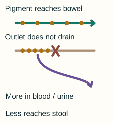
Hepatocytes normally conjugate bilirubin and secrete it into bile. Bile then flows through small intrahepatic ducts and the extrahepatic duct system toward the intestine. Biliary atresia damages this drainage route. Retained conjugated bilirubin accumulates in blood, and its filterable fraction can appear in urine. This explains how jaundice and dark urine can occur together. [1]
 Enlarge the whole image
One impaired bile route can reduce intestinal pigment while increasing conjugated pigment in blood and urine. This is not a stool-color screening card.
Bone Wizardry. Original educational schematic. Source and provenance
Less bilirubin reaches intestinal bacteria, so less downstream brown stool pigment is formed. The stool can become pale or white. Pale stool suggests reduced pigment delivery but does not by itself prove biliary atresia. Other severe cholestatic diseases can produce a similar appearance. The correct first conclusion is that the infant needs an expedited cholestasis assessment. [1]
Persistent bile retention injures the liver and promotes fibrosis, meaning scar formation. This can progress to cirrhosis and portal hypertension, in which pressure in the portal circulation rises. Later consequences include splenomegaly, ascites, poor growth, and impaired liver function. These are reasons to act early rather than wait for advanced physical findings. [1] [3]
Gamma-glutamyl transferase, or GGT, is often elevated in biliary atresia, but neither a high nor a normal value independently settles the diagnosis. Some inherited cholestatic disorders have low or normal GGT. Alkaline phosphatase is less specific in a growing infant because bone activity contributes to its concentration. Interpret the complete pattern instead of requiring every classic laboratory finding. [1]
- Duct injury Bile drainage becomes impaired. [1]
- Pigment redistribution Conjugated pigment rises in blood and can enter urine; intestinal pigment falls. [1]
- Progressive injury Chronic retention promotes fibrosis and later portal or synthetic complications. [1] [3]
Refer promptly while evaluating severity and treatable causes
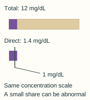
The 2017 NASPGHAN and ESPGHAN guideline recommends fractionating bilirubin in an infant still jaundiced after 2 weeks. A direct bilirubin greater than 1.0 mg/dL, approximately 17 micromol/L, is abnormal and warrants timely evaluation and referral. Do not require the direct fraction to exceed 20 percent of total before taking cholestasis seriously. Direct and conjugated assays differ, and the laboratory method and age-specific context still matter. [1] [4]
 Enlarge the whole image
The supplied infant case has direct bilirubin 1.4 mg/dL despite a total of 12. A small fraction does not erase an abnormal direct concentration.
Bone Wizardry. Original educational schematic. Source and provenance
Referral and initial investigation can proceed together. Measure liver enzymes, GGT, total and direct bilirubin, INR, glucose, and albumin to assess injury and function. INR describes clotting and can be prolonged by vitamin K deficiency or impaired hepatic synthesis. A concerning INR, hypoglycemia, bleeding, or an ill appearance changes the urgency of stabilization. [1]
Review the local newborn screen and assess infection, metabolic, endocrine, and genetic causes in parallel with duct evaluation. Examples include hypothyroidism, galactosemia, alpha-1-antitrypsin deficiency, cystic fibrosis, and Alagille syndrome. Blood or urine cultures and other investigations depend on the infant clinical presentation. Finding a possible alternative is not a reason to abandon assessment of bile-duct patency when concern remains. [1]
The goal is not to order every test sequentially before calling a specialist. Pediatric hepatology and surgery coordinate a focused workup that answers two questions promptly. Is there a treatable medical cause, and does the infant have an obstructed duct system requiring surgery? Prolonged serial testing can consume time during which liver injury continues. [1] [2]
- Identify cholestasis Use fractionated bilirubin rather than skin color alone. [1] [4]
- Assess immediate safety Check function, glucose, bleeding, and illness while arranging referral. [1]
- Investigate in parallel Medical causes and the need for anatomic assessment are evaluated together. [1]
Use ultrasound without treating a normal scan as exclusion
Ultrasound is the first anatomic imaging study in the cholestatic infant. It can assess the gallbladder, ducts, liver, vessels, spleen, and visible cystic lesions. A small or abnormal gallbladder, a triangular cord appearance near the porta hepatis, or associated anatomic abnormalities can strengthen suspicion for biliary atresia. None of these findings alone is a definitive diagnosis. [1]
 Enlarge the whole image
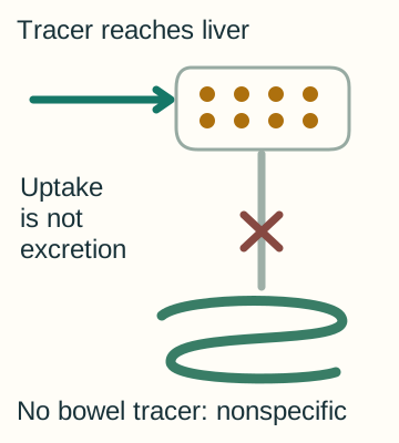
Tracer entry into the liver does not prove tracer exit into bowel. Nonexcretion has more than one possible cause.
Bone Wizardry. Original educational schematic. Source and provenance
The important negative result is also limited. A visible gallbladder, absence of duct dilation, or an otherwise reassuring ultrasound does not rule out biliary atresia. The ducts in this disorder may be obliterated rather than simply dilated behind a removable distal stone. Persistently pale stools and conjugated jaundice remain concerning even when the scan is not dramatic. [1]
Hepatobiliary scintigraphy, often called a HIDA scan, follows an injected tracer taken up by the liver and normally excreted into bile. Uptake into the liver does not establish that the ducts are patent. Nonexcretion into the bowel can occur in biliary atresia, but also in substantial intrahepatic cholestasis, neonatal hepatitis, and some infants receiving parenteral nutrition. [1]
Definite tracer passage into bowel argues against complete biliary obstruction, whereas absent passage is not proof of biliary atresia. The scan has limited specificity as a stand-alone diagnostic test. Preparing or waiting for scintigraphy should not delay the specialist-directed anatomic evaluation when suspicion is high. In particular, a prolonged attempt to improve tracer excretion is not an acceptable substitute for timely diagnosis. [1]
Ultrasound question
Are there visible structural findings or another obstructing lesion? [1]
HIDA question
Does tracer reach the intestine under the conditions of this study? [1]
Unanswered by either alone
Is the entire clinically relevant duct system patent, and what explains persistent cholestasis? [1]