Trace portal inflow and hepatic outflow obstruction, recognize bowel and liver emergencies, and choose treatment by vascular anatomy and clinical context.
Two patients have abdominal pain and abnormal veins near the liver. One has a tender, enlarged liver filling against a blocked exit. The other has an obstructed portal inlet and may have threatened intestinal drainage. Before choosing a treatment, trace where blood is trying to go, where resistance has appeared, and which organ is suffering.
Does blood fail to enter the liver or fail to leave it?
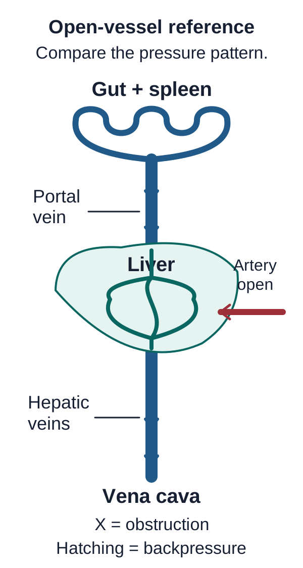
Start with the route rather than a memorized triad. Mesenteric and splenic venous blood reaches the portal vein, enters the liver and crosses the sinusoids. Blood then leaves through hepatic veins into the inferior vena cava and right atrium. The hepatic artery supplies a second inflow route. A portal vein and a hepatic vein therefore sit on opposite sides of the hepatic microcirculation. [1]
Budd-Chiari syndrome is an anatomic hepatic venous outflow obstruction. The affected territory can extend from small hepatic veins to the caval entrance into the right atrium. Primary disease arises within the venous outflow tract, commonly through thrombosis; secondary obstruction can result from external compression or tumor involvement. High right-sided cardiac pressure can also congest the liver, but cardiac congestive hepatopathy is not synonymous with primary Budd-Chiari syndrome. Sinusoidal obstruction syndrome is a separate small-vessel injury pattern. [1]
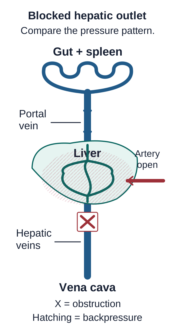
When the hepatic exit is obstructed, pressure accumulates in the sinusoidal bed. Congestion stretches the liver capsule, helping explain pain and tender hepatomegaly. Fluid formation contributes to ascites, and impaired tissue perfusion can injure the centrilobular region. Presentation ranges from subtle chronic disease to acute liver failure; the absence of the full pain-hepatomegaly-ascites triad does not exclude the diagnosis. [1]
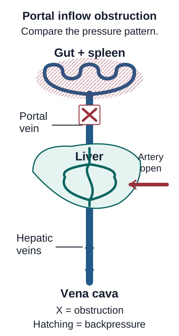
Portal vein thrombosis, or PVT, obstructs the portal trunk or its branches before blood has crossed the liver. Upstream splanchnic pressure can produce varices and splenomegaly. Arterial inflow and collateral portal delivery may preserve hepatic function, especially without pre-existing liver disease. Normal bilirubin or synthetic function therefore does not prove normal portal pressure. Extension into mesenteric veins creates a separate danger: impaired intestinal venous drainage. [1][3]
Predict before comparing. Trace the open vessels in the diagram. Open the portal-block comparison, then the outflow-block comparison. Identify whether the pressure pattern begins before the liver or within its sinusoidal bed. Each native comparison displays the selected obstruction and its pressure pattern without animation. Close it to return to the open-vessel reference; all three explanations remain readable below.
Reference route: portal blood crosses the liver and exits through hepatic veins toward the vena cava; hepatic arterial inflow is also open. Clinical basis. Open larger image.Block the portal inlet Predict where pressure rises. Open to compare the vessels.
The portal trunk is obstructed before the liver. An X marks the clot and hatching marks increased upstream splanchnic venous pressure. Arterial inflow remains open. Clinical basis. Open larger image.
Pressure rises on the upstream splanchnic side; the hepatic artery remains open.
Block the hepatic outlet Predict where pressure rises. Open to compare the vessels.
The hepatic venous outlet is obstructed after the sinusoidal bed. An X marks the block and hatching marks hepatic congestion. This simplified map highlights the sinusoidal pressure burden; portal pressure can also rise. Clinical basis. Open larger image.
Pressure rises in the sinusoidal bed because hepatic venous drainage is obstructed.
Open vessels: portal inflow crosses the sinusoidal bed and reaches the systemic venous outlet.
Portal block: the bowel and splenic venous side faces the obstruction. The hepatic artery remains open. Preserved arterial delivery does not reopen the portal trunk or normalize portal-hypertensive complications.
Hepatic outflow block: the sinusoidal bed cannot drain normally. Congestion can involve the liver and its upstream portal circulation. This is why preserving inflow alone does not solve an outflow problem.
Transfer the distinction to a patient with severe tricuspid regurgitation: if the hepatic veins are patent but right atrial pressure is high, blood faces an abnormally high downstream pressure rather than a primary hepatic venous clot. Similar ascites can arise from different vascular mechanisms. The pressure source and imaging must agree.
Why did this patient develop a splanchnic thrombus?
Does finding pancreatitis finish the investigation? Not when the history also contains an unprovoked leg thrombosis or a strong family history. Local and systemic contributors can coexist. Cirrhosis, abdominal malignancy, inflammation, pancreatitis, infection and abdominal surgery are important portal-thrombosis settings. Pregnancy, estrogen exposure and inherited or acquired thrombotic disorders can also contribute to splanchnic thrombosis. Treat a provoking condition without assuming it is the only cause. [1][4]
Myeloproliferative neoplasms, including polycythemia vera, are particularly important in primary splanchnic thrombosis. Consider a patient with a large spleen and new hepatic vein thrombosis but an apparently ordinary blood count. Portal-hypertensive sequestration, hemodilution or iron deficiency can obscure the usual count pattern. The thrombotic site can justify looking for a clonal driver before the count becomes striking. Baveno VII recommends JAK2 V617F testing in adults with primary splanchnic thrombosis; further molecular or marrow evaluation belongs with hematology when the initial result does not resolve the suspicion. [1][4]
Compare two histories. Patient A has noncirrhotic hepatic vein thrombosis, splenomegaly and a prior unexplained venous event. Patient B has cirrhosis-associated PVT with no additional thromboemboli, suspicious laboratory findings or relevant family history. Decide whose presentation gives an additional reason to investigate systemic thrombophilia.
Patient A needs a coordinated etiologic assessment rather than reassurance from a normal count. Patient B does not automatically need a broad hypercoagulable panel: the AGA cirrhosis-specific advice limits routine testing when additional suggestive evidence is absent. Tests should be selected for the clinical question and whether the result could change care. Antiphospholipid testing, evaluation for paroxysmal nocturnal hemoglobinuria in a compatible hemolytic or cytopenic presentation, and selected inherited thrombophilia testing are not interchangeable blanket panels. [2][4]
Timing matters as much as selection. Warfarin and liver disease can lower protein C and protein S; recent thrombosis and other illness can also complicate interpretation of natural-anticoagulant assays. A low result obtained under these conditions does not establish an inherited deficiency or justify diagnosing relatives. Arrange specialist interpretation and appropriately timed testing without interrupting necessary treatment just to obtain an unconfounded assay. [4]
For the next patient, record three separate questions: what local condition favors thrombosis, what systemic evidence remains unexplained, and which proposed test would change management? A visible provoking factor and an occult prothrombotic disorder can both be part of the answer.
Read the vascular image by following both ends of a vessel
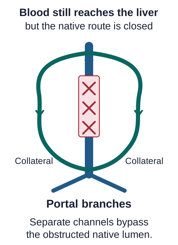
Are winding vessels proof that the native portal vein reopened? No. A portal cavernoma is a network of porto-portal collateral channels around a prior portal obstruction. Trace the channels to their destination: they can reconnect with intrahepatic portal branches even though the original extrahepatic lumen remains occluded. Hepatic venous collaterals in Budd-Chiari syndrome bypass a different part of the circulation. [1]
Trace both ends of each collateral to distinguish porto-portal bypass from a reopened native lumen or a portosystemic shunt. Clinical basis. Open larger image.
Trace the bypass. Follow one winding collateral from above the obstructed segment to the portal branches below it. Now point to the native lumen. The channel that carries some blood around the obstruction is not the channel that remains closed.
Collateral development is evidence of prior obstruction, not an exact calendar. Use symptom history, previous imaging and the current vascular appearance together. In the standard nomenclature, recent PVT is presumed present for less than six months and chronic PVT persists beyond six months. Document degree of obstruction, affected vessels and interval progression separately. If a high-quality scan showed a patent portal vein six weeks earlier, a newly seen collateral network cannot establish that the present thrombosis is older than six months. [1]
Contrast behavior answers another question. Bland thrombus generally appears as nonenhancing intraluminal material. Enhancement within the intravascular tissue, particularly when continuous with an adjacent tumor and accompanied by venous expansion, raises concern for malignant invasion. A patient with hepatocellular carcinoma can nevertheless have bland PVT. The cancer diagnosis alone must not substitute for an adequate multiphasic vascular assessment. [1][2]
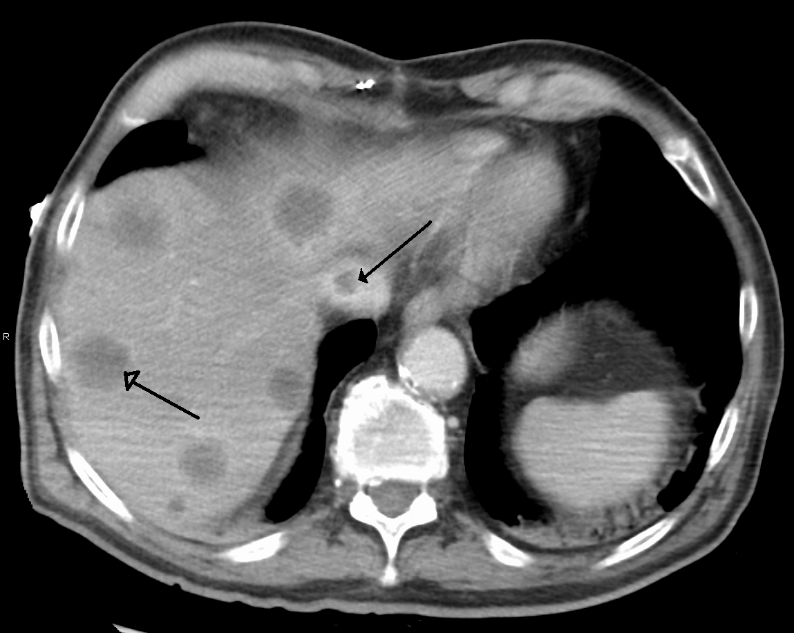
Clinical CT: image: inferior vena caval clot and hepatic metastases. A single section does not establish clot composition or map all hepatic outflow. Existing arrows are part of the original image. Image: James Heilman, MD. Source. CC BY-SA 3.0.
In the clinical CT, first identify the contrast-filled vena cava and the indicated filling defect, then note the separate liver lesions. Image: caval clot and hepatic metastases in a patient with Budd-Chiari syndrome. This single section does not map every hepatic vein, prove that the intravascular material is bland, or establish the cause of the obstruction. Carry the image finding forward as a localization question, not a complete diagnostic conclusion. [6]
Let the threatened organ determine urgency
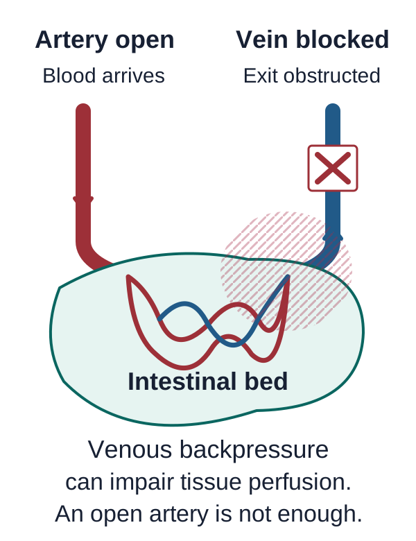
Why can an open mesenteric artery coexist with ischemic bowel? Arterial delivery is only one side of perfusion. Blood must also drain through the mesenteric venous bed. A new superior mesenteric vein obstruction raises venous backpressure, promotes bowel wall edema and can impair the pressure difference that supports tissue perfusion. An unchanged old portal cavernoma does not make a newly involved mesenteric territory harmless. [1][3]
Use the direction of flow to predict venous congestion, bowel edema and impaired perfusion when the superior mesenteric vein is obstructed. Clinical basis. Open larger image.
Predict the consequence. Trace blood from the patent artery through the intestinal bed toward the blocked vein. Predict whether venous pressure falls or rises, and whether arterial patency alone guarantees adequate tissue perfusion.
The venous pressure rises; arterial patency does not guarantee safety. Persistent or worsening severe pain, bloody diarrhea, increasing acidosis, deteriorating bowel enhancement or peritoneal signs demand urgent reassessment. No single laboratory result reliably excludes mesenteric ischemia. An initially normal lactate and a soft abdomen cannot justify postponing appropriate vascular imaging when clinical suspicion remains substantial. [3]
Peritonitis changes the plan. Guarding or rebound tenderness with suspected intestinal infarction requires urgent surgical assessment, resuscitation and coordinated treatment, not anticoagulation alone followed by delayed review. Without peritonitis or demonstrated nonviable bowel, mesenteric venous thrombosis can often begin with therapeutic anticoagulation and close inpatient reassessment. These are different clinical states, not two interchangeable treatments for every clot. [3]
The liver has its own emergency trajectory. Rapidly worsening synthetic function and encephalopathy in Budd-Chiari syndrome can indicate acute liver failure. Urgent transplant assessment and consideration of emergency decompression may proceed in parallel at an experienced center. A standard escalation sequence is not a requirement to wait through a fixed trial while the organ fails. [1]
Choose the study that answers the unanswered vascular question
Should every patient follow the same Doppler-first sequence? In a stable patient with suspected hepatic or portal venous disease, Doppler ultrasound is a useful initial vascular examination. It assesses patency, direction of flow and collateral circulation. But absent detected flow can reflect a difficult examination or very slow flow, not necessarily a fully occluded vessel. A technically limited test must not acquire more certainty when it is repeated in the chart. [1][2]
Contrast-enhanced CT or MR angiography confirms and maps the obstruction. Ask the report to identify the portal trunk and branches, hepatic veins, inferior vena cava and mesenteric extension; document residual lumen, prior imaging comparisons and relevant parenchymal or bowel changes. Assess for cirrhosis, malignancy, external compression and tumor within the vein. If a major hepatic venous obstruction is already demonstrated, a liver biopsy is not required merely to confirm Budd-Chiari syndrome. Selected imaging-negative small-vein disease is a different specialist diagnostic problem. [1][2]
Choose between two requests. One stable patient has an equivocal portal Doppler and no bowel symptoms. Another has persistent severe pain with concern for mesenteric ischemia. Decide whether either should wait for tomorrow's routine ultrasound appointment.
The stable patient needs suitable confirmatory contrast imaging and a complete vascular map. The patient with suspected mesenteric ischemia needs urgent CT angiography with arterial and venous assessment; do not let a routine elective sequence delay evaluation of bowel viability. A test is useful only if it answers the right question soon enough. [3]
After localization, assess consequences: hepatic function, ascites, variceal bleeding and bowel viability. Then connect the map to the cause investigation. Do not reverse that order by waiting for a broad thrombophilia panel before treating an urgent vascular or organ-threatening problem.
Treat thrombosis, pressure and organ failure as related but distinct problems
Why might anticoagulation be necessary but insufficient? It limits propagation and recurrence, yet a fixed outflow obstruction may continue to congest the liver. Primary Budd-Chiari syndrome generally requires long-term therapeutic anticoagulation, treatment of its cause and management of portal-hypertensive complications. Anticoagulant choice must account for bleeding, renal and hepatic function, drug interactions and planned procedures. Low-molecular-weight heparin is a common initial approach; routine systemic thrombolysis is not the default treatment. [1]
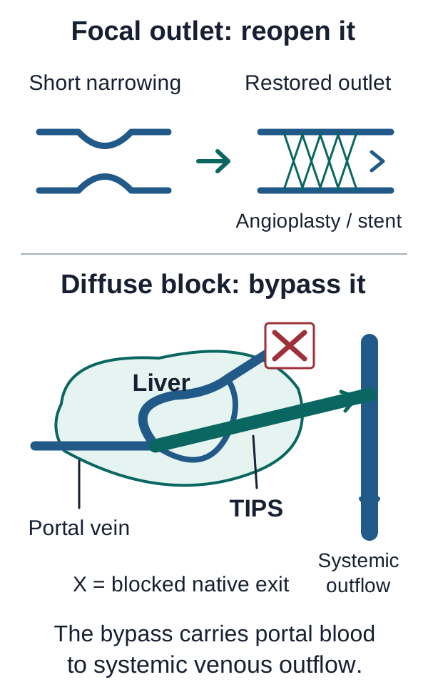
Match focal native outflow restoration to a suitable stenosis and portal-to-systemic decompression to persistent disease without a repairable outlet. Actual shunt anatomy requires expert planning. Clinical basis. Open larger image.
Match the route to the anatomy. In the upper drawing, identify the short native outlet that can be reopened. In the lower drawing, trace the alternative route when native drainage cannot be restored. Predict why stopping further thrombosis may still leave a pressure problem.
A suitable short stenosis can be treated with angioplasty and, when indicated, stenting. Persistent congestion or deterioration despite medical treatment, with no suitable native recanalization option, supports expert assessment for a transjugular intrahepatic portosystemic shunt (TIPS). The shunt supplies an alternative route to systemic venous outflow; the technical approach depends on the anatomy. Follow ascites formation and hepatic and renal function, not just whether a stent is visible on a scan. [1][5]
If function worsens after TIPS, reassess patency, adequacy of decompression and reversible causes. A patent shunt alone does not establish that decompression is sufficient. If hepatic failure progresses despite an adequately functioning intervention and appropriate treatment, transplant assessment becomes the priority. Interpret INR in the context of anticoagulant exposure rather than treating a warfarin-related INR change as direct evidence of hepatic deterioration. [1]
Recent PVT without cirrhosis: therapeutic anticoagulation is generally started promptly when not contraindicated and continued for at least six months. After that, a permanent prothrombotic state supports long-term treatment; continuing therapy can also be considered in other patients after individualized assessment. The goal may shift from reopening a recent clot to preventing recurrence. Treatment of a myeloproliferative disorder does not automatically eliminate the anticoagulation indication. [1]
Recent PVT with cirrhosis: consider degree, territory, progression, symptoms and transplant plans together. An incidental limited intrahepatic branch thrombus without ischemia or progression may be observed with imaging about every three months. More extensive main portal obstruction, mesenteric involvement, interval progression or transplant relevance favors anticoagulation assessment. Do not interpret less than 50% occlusion as a universal permission to observe: progression or a newly involved vascular bed can change the decision. Guidance contains overlapping categories for small main-portal lesions, so individualized specialist judgment remains important. [1][2]
Assess variceal bleeding prevention alongside treatment. The AGA advises variceal screening in cirrhotic PVT when the patient is not already receiving appropriate nonselective beta-blocker prophylaxis, but cautions against delaying indicated anticoagulation while pursuing screening. Active hemorrhage requires a coordinated urgent plan. Drug selection is not uniform across liver disease severity: selected compensated Child-Pugh A or B patients may receive a direct oral anticoagulant, but advanced hepatic dysfunction, renal impairment, interactions and agent-specific restrictions require particular care. [2]
Chronic complete cavernomatous PVT is not one universal treatment category. In cirrhosis, routine anticoagulation is not advised for longstanding complete occlusion with established collateralization solely to treat that PVT. Manage portal-hypertensive complications and consider separate anticoagulation indications or specialist transplant and intervention goals. By contrast, past PVT or cavernoma without cirrhosis and with a permanent prothrombotic state can still warrant long-term anticoagulation to prevent recurrence or extension. In an untreated noncirrhotic chronic cavernoma with high-risk varices, initiate appropriate bleeding prophylaxis before starting the planned anticoagulation. [1][2]
Apply this to a transplant candidate whose portal vein has reopened: maintaining patency until transplantation can remain the goal, so a good scan is not automatically a stopping instruction. Portal revascularization with TIPS is reserved for selected refractory complications or situations in which it improves transplant feasibility. At every reassessment, name the goal: prevent further thrombosis, restore a route, relieve pressure, or rescue failing tissue. [1][2]
Apply the vascular map to new patients
For each patient, identify the obstructed territory, the immediate organ threat and the purpose of the proposed treatment. Commit to an answer before comparing the explanations.
Case 1
Show answer and explanations for case 1
A. Portal pressure rises behind an obstructed hepatic inlet. (Why this does not fit)
A portal inlet obstruction raises pressure in the upstream splanchnic veins before blood crosses the liver. Her portal vein is patent; the two occluded vessels are hepatic veins on the exit side of the liver. It places the primary resistance before the sinusoids, whereas her demonstrated obstruction prevents the sinusoidal bed from draining.
Reasoning steps for option A
Where would an obstructed portal inlet raise pressure first?
A portal inlet obstruction raises pressure in the upstream splanchnic veins before blood crosses the liver.
Does this woman's portal vein contain the obstruction needed for that explanation?
Her portal vein is patent; the two occluded vessels are hepatic veins on the exit side of the liver.
Why does a portal-inlet explanation mislocalize her tender hepatomegaly?
It places the primary resistance before the sinusoids, whereas her demonstrated obstruction prevents the sinusoidal bed from draining.
B. Sinusoidal pressure rises behind an obstructed hepatic outlet. (Best answer)
The hepatic veins drain the sinusoids, so their occlusion places resistance downstream from the liver tissue. Continued portal and arterial inflow meets impaired venous drainage, raising pressure within the sinusoidal bed. Sinusoidal congestion enlarges the liver and stretches its capsule, producing tender hepatomegaly. Normal cardiac filling pressure favors the demonstrated local hepatic outflow obstruction rather than congestion transmitted from the heart.
Reasoning steps for option B
Where do the two occluded hepatic veins lie relative to the sinusoidal bed?
The hepatic veins drain the sinusoids, so their occlusion places resistance downstream from the liver tissue.
What happens to sinusoidal pressure when inflow stays open but these exits close?
Continued portal and arterial inflow meets impaired venous drainage, raising pressure within the sinusoidal bed.
How does that pressure increase explain the painful enlarged liver?
Sinusoidal congestion enlarges the liver and stretches its capsule, producing tender hepatomegaly.
What does normal right-sided filling pressure add to this localization?
Normal cardiac filling pressure favors the demonstrated local hepatic outflow obstruction rather than congestion transmitted from the heart.
C. Arterial perfusion pressure falls before the hepatic microcirculation. (Why this does not fit)
Loss of hepatic arterial perfusion impairs blood delivery into the hepatic microcirculation rather than venous drainage from it. The hepatic artery remains patent while two major hepatic veins are occluded. Obstructed venous drainage produces hepatic congestion and capsular stretch; an arterial inflow lesion is not demonstrated.
Reasoning steps for option C
Which side of hepatic circulation would an arterial pressure loss primarily impair?
Loss of hepatic arterial perfusion impairs blood delivery into the hepatic microcirculation rather than venous drainage from it.
Does the contrast study show an arterial interruption in this patient?
The hepatic artery remains patent while two major hepatic veins are occluded.
Which demonstrated vascular change better accounts for her capsular pain?
Obstructed venous drainage produces hepatic congestion and capsular stretch; an arterial inflow lesion is not demonstrated.
D. Right atrial pressure rises and is transmitted into patent hepatic veins. (Why this does not fit)
High right atrial pressure can travel backward through patent hepatic veins and congest the liver. Her right-sided filling pressure is normal, and imaging shows actual occlusion of two hepatic veins. The pressure source is a local blocked hepatic outlet, not elevated pressure transmitted through otherwise open outflow vessels.
Reasoning steps for option D
How could elevated right atrial pressure enlarge a liver with open hepatic veins?
High right atrial pressure can travel backward through patent hepatic veins and congest the liver.
Do her cardiac pressures and hepatic vein images match that cardiac mechanism?
Her right-sided filling pressure is normal, and imaging shows actual occlusion of two hepatic veins.
Why is transmitted cardiac pressure not the best explanation for these findings?
The pressure source is a local blocked hepatic outlet, not elevated pressure transmitted through otherwise open outflow vessels.
Takeaway: Localize the obstruction before predicting which vascular bed becomes congested.
A. Normal synthetic function indicates that splanchnic venous pressure has normalized. (Why this does not fit)
They indicate relatively preserved hepatic function despite the longstanding extrahepatic portal obstruction. Esophageal varices and splenomegaly indicate persistent upstream portal-hypertensive effects. Arterial and collateral blood delivery can sustain liver function while the blocked native portal trunk continues to elevate splanchnic pressure.
Reasoning steps for option A
What do near-normal bilirubin and synthetic function establish about this obstructed liver?
They indicate relatively preserved hepatic function despite the longstanding extrahepatic portal obstruction.
Which findings conflict with the claim that splanchnic pressure has normalized?
Esophageal varices and splenomegaly indicate persistent upstream portal-hypertensive effects.
Why cannot preserved synthetic function substitute for assessing portal pressure here?
Arterial and collateral blood delivery can sustain liver function while the blocked native portal trunk continues to elevate splanchnic pressure.
B. Hepatic venous obstruction has redirected blood into the esophageal veins. (Why this does not fit)
Hepatic venous obstruction can raise portal pressure and produce varices, so varices alone do not identify the obstructed vessel. CT identifies longstanding complete obstruction of the extrahepatic portal vein, an inflow vessel rather than a hepatic venous outlet. The supplied lesion lies before the liver, and the collateral channels compensate for portal obstruction rather than a demonstrated hepatic vein block.
Reasoning steps for option B
Could hepatic outflow obstruction also cause esophageal varices?
Hepatic venous obstruction can raise portal pressure and produce varices, so varices alone do not identify the obstructed vessel.
Which imaged vessel localizes this man's obstruction instead?
CT identifies longstanding complete obstruction of the extrahepatic portal vein, an inflow vessel rather than a hepatic venous outlet.
Why does redirecting blood from a blocked hepatic vein not fit this vascular map?
The supplied lesion lies before the liver, and the collateral channels compensate for portal obstruction rather than a demonstrated hepatic vein block.
C. Loss of arterial inflow has reduced sinusoidal pressure while portal flow is restored. (Why this does not fit)
This explanation would require interruption of the hepatic arterial supply, but CT shows a patent hepatic artery. The native extrahepatic portal vein remains completely obstructed despite the alternate portal channels. Arterial delivery is preserved rather than lost, and collateral delivery bypasses rather than reopens the obstructed portal trunk.
Reasoning steps for option C
What would have to be absent for loss of arterial inflow to explain his preserved function?
This explanation would require interruption of the hepatic arterial supply, but CT shows a patent hepatic artery.
Do his collateral channels establish restoration of the native portal trunk?
The native extrahepatic portal vein remains completely obstructed despite the alternate portal channels.
How do the artery and native portal lumen together contradict this option?
Arterial delivery is preserved rather than lost, and collateral delivery bypasses rather than reopens the obstructed portal trunk.
D. Alternate hepatic blood delivery coexists with increased splanchnic venous pressure. (Best answer)
The patent hepatic artery and collateral portal channels provide alternate hepatic blood delivery. They can support hepatic tissue function despite loss of flow through the native portal trunk. The splanchnic circulation still experiences increased pressure even though hepatic function is relatively preserved. Compensated hepatic blood delivery coexists with persistent prehepatic portal hypertension; preserved function does not imply normal pressure.
Reasoning steps for option D
Which two routes can still deliver blood despite complete extrahepatic portal obstruction?
The patent hepatic artery and collateral portal channels provide alternate hepatic blood delivery.
How can those routes account for the near-normal hepatic laboratory findings?
They can support hepatic tissue function despite loss of flow through the native portal trunk.
What do the varices and splenomegaly reveal about the upstream venous bed?
The splanchnic circulation still experiences increased pressure even though hepatic function is relatively preserved.
Which paired conclusion accommodates both the CT and laboratory findings?
Compensated hepatic blood delivery coexists with persistent prehepatic portal hypertension; preserved function does not imply normal pressure.
Takeaway: Preserved synthetic function does not exclude clinically important portal hypertension.
A. Increased venous backpressure reduces effective perfusion across the intestinal bed. (Best answer)
The new superior mesenteric vein thrombus impairs venous drainage from the intestinal bed. Rising downstream venous pressure reduces the arterial-to-venous pressure difference across the bowel microcirculation. An open artery permits delivery but cannot compensate fully for impaired drainage and elevated intestinal venous backpressure. The new clot involves bowel drainage territory and accompanies acute pain and edema, whereas the older portal collateral pattern is unchanged.
Reasoning steps for option A
Which newly obstructed vessel drains the edematous small bowel?
The new superior mesenteric vein thrombus impairs venous drainage from the intestinal bed.
How does this blocked venous exit change the pressure driving intestinal perfusion?
Rising downstream venous pressure reduces the arterial-to-venous pressure difference across the bowel microcirculation.
Why does the patent superior mesenteric artery not eliminate the ischemic threat?
An open artery permits delivery but cannot compensate fully for impaired drainage and elevated intestinal venous backpressure.
Why is the new mesenteric clot more relevant to this episode than the stable cavernoma?
The new clot involves bowel drainage territory and accompanies acute pain and edema, whereas the older portal collateral pattern is unchanged.
B. Increased hepatic venous pressure directly occludes the mesenteric artery. (Why this does not fit)
The clot is in the superior mesenteric vein, not a hepatic venous outlet or the mesenteric artery. The superior mesenteric artery is patent and no arterial embolus is present. The demonstrated mesenteric venous obstruction raises intestinal drainage pressure and can impair perfusion despite the open artery.
Reasoning steps for option B
Where is the new clot relative to the hepatic veins and mesenteric artery?
The clot is in the superior mesenteric vein, not a hepatic venous outlet or the mesenteric artery.
Which CT observation directly contradicts mesenteric arterial occlusion?
The superior mesenteric artery is patent and no arterial embolus is present.
What explains threatened bowel without invoking arterial closure from hepatic pressure?
The demonstrated mesenteric venous obstruction raises intestinal drainage pressure and can impair perfusion despite the open artery.
C. Decreased intestinal venous pressure increases fluid filtration into the bowel wall. (Why this does not fit)
Blocking the venous exit raises intestinal venous pressure rather than lowering it. Increased upstream hydrostatic pressure promotes fluid filtration into the bowel wall. It attributes edema to falling venous pressure, reversing the pressure increase produced by the new drainage obstruction.
Reasoning steps for option C
Would obstructing the superior mesenteric vein lower intestinal venous pressure?
Blocking the venous exit raises intestinal venous pressure rather than lowering it.
How does higher venous hydrostatic pressure account for the edematous bowel wall?
Increased upstream hydrostatic pressure promotes fluid filtration into the bowel wall.
Which directional error makes this explanation incompatible with the CT findings?
It attributes edema to falling venous pressure, reversing the pressure increase produced by the new drainage obstruction.
D. Restored portal trunk flow causes an acute loss of intestinal capillary blood. (Why this does not fit)
The portal cavernoma is stable; the newly demonstrated change is superior mesenteric venous thrombosis. Reopening an outlet would relieve obstruction, whereas a new mesenteric venous clot increases resistance to intestinal drainage. New mesenteric venous occlusion accounts for acute congestion and impaired perfusion without requiring undocumented portal recanalization.
Reasoning steps for option D
Does the scan demonstrate new restoration of native portal trunk flow?
The portal cavernoma is stable; the newly demonstrated change is superior mesenteric venous thrombosis.
Would reopening a portal outlet produce the same drainage problem as a new mesenteric clot?
Reopening an outlet would relieve obstruction, whereas a new mesenteric venous clot increases resistance to intestinal drainage.
Which interval finding links the acute pain to the bowel edema?
New mesenteric venous occlusion accounts for acute congestion and impaired perfusion without requiring undocumented portal recanalization.
Takeaway: An open mesenteric artery does not protect bowel from venous ischemia.
A. Primary thrombosis within the major hepatic venous outlets. (Why this does not fit)
Primary major hepatic vein thrombosis requires an obstructed hepatic venous outlet, not hepatic congestion alone. Doppler shows patent hepatic veins, and cross-sectional imaging shows no hepatic venous or caval block. Severe tricuspid regurgitation and high right atrial pressure explain congestion through open veins without primary hepatic vein thrombosis.
Reasoning steps for option A
What anatomic evidence would support primary major hepatic vein thrombosis?
Primary major hepatic vein thrombosis requires an obstructed hepatic venous outlet, not hepatic congestion alone.
How do the two vascular examinations address that proposed obstruction?
Doppler shows patent hepatic veins, and cross-sectional imaging shows no hepatic venous or caval block.
Why do ascites and hepatomegaly not overcome the absence of a venous clot here?
Severe tricuspid regurgitation and high right atrial pressure explain congestion through open veins without primary hepatic vein thrombosis.
B. Extrahepatic portal occlusion with porto-portal collateral formation. (Why this does not fit)
Extrahepatic portal occlusion blocks portal inflow before blood reaches the liver. Neither a portal occlusion nor a porto-portal collateral network is described; the measured abnormal pressure is in the right atrium. Elevated right atrial pressure is transmitted backward through the patent hepatic veins, fitting cardiac congestion rather than an unshown portal block.
Reasoning steps for option B
Where would extrahepatic portal occlusion place resistance relative to the liver?
Extrahepatic portal occlusion blocks portal inflow before blood reaches the liver.
Are a portal block and porto-portal collateral network demonstrated in this case?
Neither a portal occlusion nor a porto-portal collateral network is described; the measured abnormal pressure is in the right atrium.
Which mechanism connects the abnormal cardiac pressure to the pulsatile hepatic veins?
Elevated right atrial pressure is transmitted backward through the patent hepatic veins, fitting cardiac congestion rather than an unshown portal block.
C. Cardiac venous congestion transmitted backward through patent veins into the liver. (Best answer)
Severe tricuspid regurgitation raises right atrial pressure at the downstream end of hepatic venous drainage. Patent hepatic veins and cava allow elevated central venous pressure to be transmitted backward into the liver. They fit transmission of cardiac venous pressure and provide a hemodynamic explanation for the hepatic congestion. No anatomic hepatic outflow obstruction is demonstrated, while the cardiac pressure abnormality explains the ascites and enlarged liver.
Reasoning steps for option C
What does severe tricuspid regurgitation do to the pressure downstream of hepatic drainage?
Severe tricuspid regurgitation raises right atrial pressure at the downstream end of hepatic venous drainage.
Can that pressure reach the liver when the hepatic veins and cava remain open?
Patent hepatic veins and cava allow elevated central venous pressure to be transmitted backward into the liver.
How do marked venous pulsatility and a dilated cava support this mechanism?
They fit transmission of cardiac venous pressure and provide a hemodynamic explanation for the hepatic congestion.
Why should this congested liver not be labeled primary Budd-Chiari syndrome?
No anatomic hepatic outflow obstruction is demonstrated, while the cardiac pressure abnormality explains the ascites and enlarged liver.
D. Acute hepatic arterial occlusion with parenchymal ischemia. (Why this does not fit)
The proposed mechanism requires impaired hepatic arterial delivery from an acute arterial occlusion. These findings localize the pressure disturbance to the venous drainage side rather than demonstrating arterial occlusion. Backward transmission of elevated right atrial pressure through open hepatic veins explains the dilated cava, venous pulsatility and congested liver together.
Reasoning steps for option D
What vascular defect would acute hepatic arterial ischemia require in this explanation?
The proposed mechanism requires impaired hepatic arterial delivery from an acute arterial occlusion.
Do cardiac venous pulsatility and high right atrial pressure localize an arterial lesion?
These findings localize the pressure disturbance to the venous drainage side rather than demonstrating arterial occlusion.
Why does cardiac congestion better unite the liver, cava and echocardiographic findings?
Backward transmission of elevated right atrial pressure through open hepatic veins explains the dilated cava, venous pulsatility and congested liver together.
Takeaway: Hepatic congestion does not by itself establish Budd-Chiari syndrome.
A. CALR mutation testing alone. (Why this does not fit)
CALR testing can identify some myeloproliferative neoplasms when the initial JAK2 evaluation is negative. No negative JAK2 V617F result is supplied; this is her initial clonal-driver screen after hepatic vein thrombosis. It omits the JAK2 V617F screen indicated by the hepatic vein thrombosis, splenomegaly and compatible myeloproliferative symptoms.
Reasoning steps for option A
Which subgroup of myeloproliferative disorders can CALR testing help identify?
CALR testing can identify some myeloproliferative neoplasms when the initial JAK2 evaluation is negative.
Has this woman reached that later molecular-testing stage?
No negative JAK2 V617F result is supplied; this is her initial clonal-driver screen after hepatic vein thrombosis.
Why is CALR alone not the best initial assay for this splanchnic presentation?
It omits the JAK2 V617F screen indicated by the hepatic vein thrombosis, splenomegaly and compatible myeloproliferative symptoms.
B. JAK2 exon 12 mutation testing alone. (Why this does not fit)
Exon 12 testing can help evaluate selected patients with erythrocytosis after a negative JAK2 V617F assay. Neither a prior negative V617F assay nor established unexplained erythrocytosis is provided. Iron deficiency can obscure an erythrocytosis pattern, but her initial primary splanchnic thrombosis evaluation still calls for JAK2 V617F testing.
Reasoning steps for option B
What clinical setting particularly supports JAK2 exon 12 testing?
Exon 12 testing can help evaluate selected patients with erythrocytosis after a negative JAK2 V617F assay.
Does the current history establish V617F-negative unexplained erythrocytosis?
Neither a prior negative V617F assay nor established unexplained erythrocytosis is provided.
Why does the normal hematocrit not make exon 12 alone the preferred first screen?
Iron deficiency can obscure an erythrocytosis pattern, but her initial primary splanchnic thrombosis evaluation still calls for JAK2 V617F testing.
C. Initial JAK2 V617F mutation testing for occult myeloproliferative disease. (Best answer)
Hepatic vein thrombosis, marked splenomegaly and itching after warm showers raise suspicion for an occult myeloproliferative neoplasm. Iron deficiency can mask the usual erythrocytosis pattern, so a reference-range hematocrit does not exclude an underlying myeloproliferative disorder. JAK2 V617F testing is the appropriate initial molecular screen for an occult myeloproliferative driver in this setting. A negative V617F result would not exclude every clonal disorder; persistent suspicion can lead to tailored further testing with hematology.
Reasoning steps for option C
Which findings raise suspicion for a clonal driver despite reference-range blood counts?
Hepatic vein thrombosis, marked splenomegaly and itching after warm showers raise suspicion for an occult myeloproliferative neoplasm.
How can iron deficiency complicate interpretation of her normal hematocrit?
Iron deficiency can mask the usual erythrocytosis pattern, so a reference-range hematocrit does not exclude an underlying myeloproliferative disorder.
Which molecular assay is the initial screen for this primary splanchnic thrombosis pattern?
JAK2 V617F testing is the appropriate initial molecular screen for an occult myeloproliferative driver in this setting.
Does choosing V617F now mean a negative result would exclude every myeloproliferative neoplasm?
A negative V617F result would not exclude every clonal disorder; persistent suspicion can lead to tailored further testing with hematology.
D. BCR::ABL1 fusion testing alone. (Why this does not fit)
BCR::ABL1 identifies chronic myeloid leukemia rather than serving as the initial screen for the usual primary splanchnic thrombosis-associated driver. No leukocytosis or other characteristic chronic myeloid leukemia pattern is described. Those findings with splenomegaly support an occult myeloproliferative evaluation beginning with JAK2 V617F rather than BCR::ABL1 alone.
Reasoning steps for option D
Which clonal disorder is specifically identified by the BCR::ABL1 fusion?
BCR::ABL1 identifies chronic myeloid leukemia rather than serving as the initial screen for the usual primary splanchnic thrombosis-associated driver.
What supporting chronic myeloid leukemia pattern is missing from this presentation?
No leukocytosis or other characteristic chronic myeloid leukemia pattern is described.
Why should the unusual thrombosis and warm-shower itching direct the first assay elsewhere?
Those findings with splenomegaly support an occult myeloproliferative evaluation beginning with JAK2 V617F rather than BCR::ABL1 alone.
Takeaway: Normal blood counts do not exclude an occult myeloproliferative cause of splanchnic thrombosis.
A. Local inflammation may coexist with a systemic predisposition. (Best answer)
Pancreatic inflammation can promote thrombosis in adjacent portal venous territory. His prior unprovoked leg thrombosis predates the local inflammatory trigger and suggests an additional systemic predisposition. The family history independently supports investigating a thrombotic predisposition rather than attributing every event to pancreatitis. The local trigger and a systemic predisposition can coexist, so resolving pancreatitis does not remove the indication for a targeted etiologic assessment.
Reasoning steps for option A
How could the episode of pancreatitis contribute to portal thrombosis locally?
Pancreatic inflammation can promote thrombosis in adjacent portal venous territory.
Which earlier event remains unexplained by this episode of pancreatitis?
His prior unprovoked leg thrombosis predates the local inflammatory trigger and suggests an additional systemic predisposition.
What does his sister's recurrent venous thrombosis add to that concern?
The family history independently supports investigating a thrombotic predisposition rather than attributing every event to pancreatitis.
How should the resolved inflammation affect the remaining cause evaluation?
The local trigger and a systemic predisposition can coexist, so resolving pancreatitis does not remove the indication for a targeted etiologic assessment.
B. The timing establishes pancreatitis as the sole cause of thrombosis. (Why this does not fit)
It supports pancreatic inflammation as a plausible local contributor to the portal clot. The earlier unprovoked leg clot and his sister's recurrent thromboses are not explained by this episode of pancreatic inflammation. The personal and family histories supply independent evidence of systemic risk that remains relevant after the inflammation resolves.
Reasoning steps for option B
What does the temporal link between pancreatitis and portal thrombosis actually support?
It supports pancreatic inflammation as a plausible local contributor to the portal clot.
Can this local episode account for both the prior leg clot and his sister's events?
The earlier unprovoked leg clot and his sister's recurrent thromboses are not explained by this episode of pancreatic inflammation.
Why is naming pancreatitis as the sole cause premature?
The personal and family histories supply independent evidence of systemic risk that remains relevant after the inflammation resolves.
C. The family history establishes one specific inherited deficiency. (Why this does not fit)
A sibling's recurrent thrombosis can suggest a familial predisposition to venous clotting. It does not identify one specific inherited deficiency; the supplied history contains no diagnostic assay result. The history supports targeted evaluation and appropriately interpreted testing, not assignment of an untested inherited diagnosis.
Reasoning steps for option C
What can recurrent venous thrombosis in a sibling suggest about inherited risk?
A sibling's recurrent thrombosis can suggest a familial predisposition to venous clotting.
Does that family history identify which anticoagulant protein or pathway is abnormal?
It does not identify one specific inherited deficiency; the supplied history contains no diagnostic assay result.
What conclusion is justified before labeling a particular familial deficiency?
The history supports targeted evaluation and appropriately interpreted testing, not assignment of an untested inherited diagnosis.
D. The prior leg event establishes malignant portal vein invasion. (Why this does not fit)
Malignancy can promote systemic thrombosis, which is distinct from direct tumor invasion of a vessel. No abdominal tumor or enhancing intravascular tissue is supplied. A prior venous event signals possible systemic clotting risk but does not demonstrate tumor tissue within the portal lumen.
Reasoning steps for option D
How can cancer be related to venous thrombosis without growing inside the portal vein?
Malignancy can promote systemic thrombosis, which is distinct from direct tumor invasion of a vessel.
What evidence of portal tumor invasion is missing in this man's case?
No abdominal tumor or enhancing intravascular tissue is supplied.
Why cannot his prior unprovoked leg clot establish malignant portal invasion?
A prior venous event signals possible systemic clotting risk but does not demonstrate tumor tissue within the portal lumen.
Takeaway: Finding a provoking condition does not erase independent evidence of thrombophilia.
A. Order an inherited thrombophilia panel at this visit. (Why this does not fit)
The history supplies no earlier thrombosis, additional thromboemboli or family pattern suggesting an inherited disorder. Cirrhosis-associated PVT without additional suggestive evidence does not routinely require a broad inherited thrombophilia panel. The fully characterized portal clot and stable disease-consistent laboratory findings provide no additional indication for routine inherited testing now.
Reasoning steps for option A
Would an inherited panel answer a specific additional concern beyond this cirrhotic PVT?
The history supplies no earlier thrombosis, additional thromboemboli or family pattern suggesting an inherited disorder.
How does the cirrhosis context affect automatic inherited thrombophilia testing?
Cirrhosis-associated PVT without additional suggestive evidence does not routinely require a broad inherited thrombophilia panel.
Why is ordering the panel at this visit less appropriate than deferring it?
The fully characterized portal clot and stable disease-consistent laboratory findings provide no additional indication for routine inherited testing now.
B. Order a myeloproliferative mutation panel at this visit. (Why this does not fit)
Clonal-driver screening is important in primary noncirrhotic splanchnic thrombosis and other presentations with suggestive clinical or laboratory findings. She has cirrhosis-associated PVT, with stable blood counts appropriate for her liver disease and no additional thrombotic history. The absence of an additional clonal or thrombotic signal in this cirrhotic context supports deferring routine testing, not declaring normal counts universally exclusionary.
Reasoning steps for option B
In which splanchnic thrombosis setting is an occult myeloproliferative screen especially important?
Clonal-driver screening is important in primary noncirrhotic splanchnic thrombosis and other presentations with suggestive clinical or laboratory findings.
What distinguishes this woman's presentation from that primary noncirrhotic setting?
She has cirrhosis-associated PVT, with stable blood counts appropriate for her liver disease and no additional thrombotic history.
Why is an automatic mutation panel not indicated by this combination of findings?
The absence of an additional clonal or thrombotic signal in this cirrhotic context supports deferring routine testing, not declaring normal counts universally exclusionary.
C. Order antiphospholipid antibody testing at this visit. (Why this does not fit)
Antiphospholipid syndrome can cause venous thrombosis and change treatment when the clinical setting supports investigating it. No additional thromboemboli, suggestive laboratory abnormalities or relevant personal history are provided beyond the cirrhosis-associated portal clot. An antiphospholipid antibody assay still requires a clinical indication; cirrhotic PVT alone does not mandate routine hypercoagulable testing.
Reasoning steps for option C
Could antiphospholipid syndrome matter in a patient with an otherwise suggestive thrombotic history?
Antiphospholipid syndrome can cause venous thrombosis and change treatment when the clinical setting supports investigating it.
What extra thrombotic or laboratory signal supports that investigation here?
No additional thromboemboli, suggestive laboratory abnormalities or relevant personal history are provided beyond the cirrhosis-associated portal clot.
Why does choosing an acquired rather than inherited assay not solve the indication problem?
An antiphospholipid antibody assay still requires a clinical indication; cirrhotic PVT alone does not mandate routine hypercoagulable testing.
D. Defer additional thrombophilia testing at this visit unless new indications emerge. (Best answer)
CT confirms the new nonocclusive portal thrombus, defines its extent and shows no tumor. Additional thromboemboli, laboratory abnormalities or a family history suggesting thrombophilia could justify further evaluation, but none is supplied. Additional thrombophilia testing can be deferred because a routine hypercoagulable workup is not indicated for this presentation. Deferral reflects the current lack of a testing indication; new suggestive findings would warrant reassessment rather than a permanent exclusion of thrombophilia.
Reasoning steps for option D
What has contrast CT already established about the portal clot and possible tumor?
CT confirms the new nonocclusive portal thrombus, defines its extent and shows no tumor.
Which personal, family and laboratory clues would create an additional testing indication?
Additional thromboemboli, laboratory abnormalities or a family history suggesting thrombophilia could justify further evaluation, but none is supplied.
Which testing approach follows from cirrhosis plus the absence of those clues?
Additional thrombophilia testing can be deferred because a routine hypercoagulable workup is not indicated for this presentation.
Does deferring a panel establish that cirrhosis excludes every coexisting thrombophilia?
Deferral reflects the current lack of a testing indication; new suggestive findings would warrant reassessment rather than a permanent exclusion of thrombophilia.
Takeaway: The workup for noncirrhotic primary splanchnic thrombosis should not be copied indiscriminately into cirrhosis.
A. The combined low results establish two inherited anticoagulant deficiencies. (Why this does not fit)
Warfarin exposure and impaired hepatic synthesis can each lower protein C and protein S measurements. He was taking warfarin and had impaired hepatic synthetic function, with no pretreatment results for comparison. Both results can reflect the same acquired confounders, so they cannot establish inherited deficiency before appropriate interpretation and testing.
Reasoning steps for option A
What acquired influences could lower both measured protein C and protein S?
Warfarin exposure and impaired hepatic synthesis can each lower protein C and protein S measurements.
Which of those influences were present when this man's samples were obtained?
He was taking warfarin and had impaired hepatic synthetic function, with no pretreatment results for comparison.
Why do two low measurements not establish two inherited deficiencies in his family?
Both results can reflect the same acquired confounders, so they cannot establish inherited deficiency before appropriate interpretation and testing.
B. Confounded results do not establish an inherited deficiency. (Best answer)
Warfarin lowers vitamin K-dependent protein C and protein S, so reduced measurements during treatment need not reflect inherited deficiency. His impaired hepatic synthetic function can also reduce these anticoagulant proteins. Without unconfounded baseline results, the present assays cannot distinguish an inherited deficiency from acquired reductions. Specialist interpretation and appropriately timed evaluation are needed before familial counseling; necessary anticoagulation should not be interrupted simply to obtain assays.
Reasoning steps for option B
How does warfarin complicate interpretation of the low natural-anticoagulant levels?
Warfarin lowers vitamin K-dependent protein C and protein S, so reduced measurements during treatment need not reflect inherited deficiency.
What separate synthesis problem compounds that treatment-related confounding?
His impaired hepatic synthetic function can also reduce these anticoagulant proteins.
What does the absence of pretreatment values prevent the clinician from determining?
Without unconfounded baseline results, the present assays cannot distinguish an inherited deficiency from acquired reductions.
What should happen before these results become a diagnosis for his relatives?
Specialist interpretation and appropriately timed evaluation are needed before familial counseling; necessary anticoagulation should not be interrupted simply to obtain assays.
C. The results exclude a systemic prothrombotic predisposition. (Why this does not fit)
They are abnormal measurements obtained during warfarin exposure and impaired liver synthesis, not a reliable negative evaluation. They do not evaluate or exclude every inherited or acquired cause of thrombosis. The measurements neither establish inherited protein deficiency nor exclude a systemic predisposition, so interpretation must remain tied to the confounders.
Reasoning steps for option C
Are the low protein C and protein S results an unconfounded negative thrombophilia evaluation?
They are abnormal measurements obtained during warfarin exposure and impaired liver synthesis, not a reliable negative evaluation.
Can these two confounded assays rule out other systemic thrombotic predispositions?
They do not evaluate or exclude every inherited or acquired cause of thrombosis.
Which conclusion avoids both overdiagnosis and false reassurance from these results?
The measurements neither establish inherited protein deficiency nor exclude a systemic predisposition, so interpretation must remain tied to the confounders.
D. The results establish inadequate warfarin anticoagulation. (Why this does not fit)
Protein C and protein S assays investigate natural-anticoagulant levels rather than serving as the routine measure of warfarin treatment intensity. Their reduction is compatible with warfarin exposure and impaired hepatic synthesis, not proof of inadequate anticoagulation. Warfarin intensity requires its own clinically interpreted monitoring; these confounded thrombophilia assays cannot establish undertreatment or inherited disease.
Reasoning steps for option D
Do protein C and protein S assays directly measure warfarin treatment intensity?
Protein C and protein S assays investigate natural-anticoagulant levels rather than serving as the routine measure of warfarin treatment intensity.
What does their reduction during warfarin treatment actually tell us here?
Their reduction is compatible with warfarin exposure and impaired hepatic synthesis, not proof of inadequate anticoagulation.
Why should the anticoagulant-monitoring question remain separate from the family diagnosis question?
Warfarin intensity requires its own clinically interpreted monitoring; these confounded thrombophilia assays cannot establish undertreatment or inherited disease.
Takeaway: An abnormal thrombophilia assay must be interpreted before it becomes a familial diagnosis.
A. Noncontrast abdominal CT as the sole confirmatory study. (Why this does not fit)
The study has not established definite portal thrombus or mapped its extent and associated vascular findings. A noncontrast study lacks the vascular enhancement assessment needed to characterize patency, thrombus extent and possible malignant involvement. It leaves key confirmatory and mapping questions unresolved despite the availability of appropriate contrast-enhanced vascular imaging.
Reasoning steps for option A
What two vascular questions remain unanswered after absent flow on a limited Doppler?
The study has not established definite portal thrombus or mapped its extent and associated vascular findings.
Which needed information would a noncontrast CT fail to characterize adequately?
A noncontrast study lacks the vascular enhancement assessment needed to characterize patency, thrombus extent and possible malignant involvement.
Why is noncontrast CT alone insufficient when this patient can receive contrast?
It leaves key confirmatory and mapping questions unresolved despite the availability of appropriate contrast-enhanced vascular imaging.
B. Contrast-enhanced CT or MR angiography. (Best answer)
The technically limited examination may fail to detect slow flow and does not show definite intraluminal thrombus. Contrast vascular imaging can confirm or exclude thrombus and define the involved vessels and degree of lumen obstruction. It should assess associated malignancy and distinguish vascular tumor involvement from bland thrombus. He is stable without acute bowel symptoms and can receive contrast, allowing suitable confirmatory CT or MR angiography rather than treating a limited Doppler as definitive.
Reasoning steps for option B
Why is nondetection of portal flow not definitive proof of complete occlusion on this Doppler?
The technically limited examination may fail to detect slow flow and does not show definite intraluminal thrombus.
How can contrast-enhanced CT or MR angiography resolve the suspected portal obstruction?
Contrast vascular imaging can confirm or exclude thrombus and define the involved vessels and degree of lumen obstruction.
What additional cirrhosis-related diagnostic question should the cross-sectional study address?
It should assess associated malignancy and distinguish vascular tumor involvement from bland thrombus.
Why does this study fit the current clinical timing as well as the mapping need?
He is stable without acute bowel symptoms and can receive contrast, allowing suitable confirmatory CT or MR angiography rather than treating a limited Doppler as definitive.
C. Endoscopic ultrasonography of the periportal region. (Why this does not fit)
Endoscopic ultrasound can visualize portions of the portal circulation in selected circumstances. The unresolved portal clot requires assessment of its full abdominal extent, degree of obstruction and associated malignancy. Contrast-enhanced CT or MR angiography more directly supplies the complete cross-sectional vascular map needed after this limited Doppler.
Reasoning steps for option C
What can endoscopic ultrasound contribute to a periportal examination?
Endoscopic ultrasound can visualize portions of the portal circulation in selected circumstances.
What mapping requirement extends beyond this locally focused examination?
The unresolved portal clot requires assessment of its full abdominal extent, degree of obstruction and associated malignancy.
Why is a periportal endoscopic study not the preferred complete evaluation here?
Contrast-enhanced CT or MR angiography more directly supplies the complete cross-sectional vascular map needed after this limited Doppler.
D. Repeat optimized Doppler as the complete confirmatory evaluation. (Why this does not fit)
A better Doppler examination could clarify whether flow is present in the main portal vein. The cirrhotic PVT workup also needs cross-sectional characterization of clot extent, lumen obstruction and possible malignancy. Clarifying a local flow signal alone does not provide the complete anatomic and enhancement assessment needed to characterize suspected cirrhotic PVT.
Reasoning steps for option D
What part of the uncertain Doppler result could an optimized repeat clarify?
A better Doppler examination could clarify whether flow is present in the main portal vein.
Which additional questions would treating repeat Doppler as the whole evaluation omit?
The cirrhotic PVT workup also needs cross-sectional characterization of clot extent, lumen obstruction and possible malignancy.
Why does improved flow detection not eliminate the need for contrast mapping in this plan?
Clarifying a local flow signal alone does not provide the complete anatomic and enhancement assessment needed to characterize suspected cirrhotic PVT.
Takeaway: Confirm a suspected clot before treating a technically limited flow study as definitive.
A. Obtain a percutaneous biopsy before accepting the vascular diagnosis. (Why this does not fit)
Biopsy can help in selected suspected small-vessel disease when the relevant obstruction is not demonstrated by vascular imaging. Doppler and contrast MRI both directly demonstrate thrombotic obstruction of the major hepatic veins. Concordant imaging already establishes the symptomatic major hepatic outflow obstruction, so diagnostic biopsy would not resolve an unanswered vascular question.
Reasoning steps for option A
When might tissue help investigate hepatic outflow disease that vascular imaging has not established?
Biopsy can help in selected suspected small-vessel disease when the relevant obstruction is not demonstrated by vascular imaging.
What makes this woman's major-vessel findings different from that biopsy setting?
Doppler and contrast MRI both directly demonstrate thrombotic obstruction of the major hepatic veins.
Why should a percutaneous biopsy not be required before accepting her vascular diagnosis?
Concordant imaging already establishes the symptomatic major hepatic outflow obstruction, so diagnostic biopsy would not resolve an unanswered vascular question.
B. Require a transjugular biopsy before discussing anticoagulation. (Why this does not fit)
A transjugular approach changes how tissue is obtained, not whether tissue is necessary for the diagnosis. Two concordant vascular studies identify thrombotic obstruction of the major hepatic veins in a patient with compatible symptoms. No unresolved tissue diagnosis is supplied, and the hepatology team has already assessed anticoagulation for the imaging-confirmed obstruction.
Reasoning steps for option B
Does selecting a transjugular route change whether this patient needs diagnostic liver tissue?
A transjugular approach changes how tissue is obtained, not whether tissue is necessary for the diagnosis.
What diagnostic evidence already exists before considering that biopsy route?
Two concordant vascular studies identify thrombotic obstruction of the major hepatic veins in a patient with compatible symptoms.
Why is requiring transjugular biopsy before anticoagulation discussion an unnecessary delay?
No unresolved tissue diagnosis is supplied, and the hepatology team has already assessed anticoagulation for the imaging-confirmed obstruction.
C. Begin indicated treatment using the diagnostic vascular imaging. (Best answer)
They establish major hepatic venous outflow obstruction consistent with Budd-Chiari syndrome. Direct demonstration of major-vessel outflow obstruction does not require liver biopsy solely for diagnostic confirmation. She has no active bleeding and has undergone hepatology assessment for anticoagulation. The team should begin indicated disease-directed treatment rather than postponing it for an unnecessary confirmatory biopsy.
Reasoning steps for option C
What do the concordant Doppler and MRI findings establish in this symptomatic patient?
They establish major hepatic venous outflow obstruction consistent with Budd-Chiari syndrome.
Is biopsy needed merely to reconfirm the major hepatic venous thrombi?
Direct demonstration of major-vessel outflow obstruction does not require liver biopsy solely for diagnostic confirmation.
Which supplied treatment assessment supports proceeding rather than waiting for tissue?
She has no active bleeding and has undergone hepatology assessment for anticoagulation.
What should the team do with this already diagnostic vascular information?
The team should begin indicated disease-directed treatment rather than postponing it for an unnecessary confirmatory biopsy.
D. Repeat vascular imaging after several weeks before making a diagnosis. (Why this does not fit)
Interval imaging can help resolve an equivocal examination or document evolving vascular findings. Both Doppler and contrast MRI demonstrate the same thrombotic major hepatic vein obstruction. Waiting postpones indicated treatment of symptomatic, established hepatic outflow obstruction without a supplied diagnostic uncertainty to resolve.
Reasoning steps for option D
What sort of uncertainty could justify interval vascular imaging before assigning a diagnosis?
Interval imaging can help resolve an equivocal examination or document evolving vascular findings.
Do these two current studies leave the major hepatic obstruction equivocal?
Both Doppler and contrast MRI demonstrate the same thrombotic major hepatic vein obstruction.
What is lost by waiting several weeks to repeat an already answered diagnostic study?
Waiting postpones indicated treatment of symptomatic, established hepatic outflow obstruction without a supplied diagnostic uncertainty to resolve.
Takeaway: A biopsy is not required to reconfirm a major-vessel obstruction that imaging has established.
A. Porto-portal collaterals bypass a still-blocked native portal vein. (Best answer)
They reconnect with intrahepatic portal branches, keeping the bypass route within the portal circulation. The native segment has no residual lumen and remains obstructed. Porto-portal collaterals forming a portal cavernoma carry blood around the persistent native obstruction. Blood reaches the liver through alternate channels rather than through a reopened native portal vein.
Reasoning steps for option A
Where do the winding channels deliver blood after bypassing the blocked extrahepatic segment?
They reconnect with intrahepatic portal branches, keeping the bypass route within the portal circulation.
Does blood traverse the original extrahepatic portal lumen in this scan?
The native segment has no residual lumen and remains obstructed.
What name describes separate portal channels that bypass that closed native segment?
Porto-portal collaterals forming a portal cavernoma carry blood around the persistent native obstruction.
Why does restored hepatic blood delivery not mean restored normal portal anatomy?
Blood reaches the liver through alternate channels rather than through a reopened native portal vein.
B. Recanalization of the original portal lumen with restored normal anatomy. (Why this does not fit)
Recanalization would restore a flow channel within the previously obstructed native portal lumen. The scan shows no residual native lumen and instead identifies winding vessels outside the obstructed segment. The blood arrives through separate collateral channels, so delivery has improved without reopening the original portal segment.
Reasoning steps for option B
What would count as recanalization of the original extrahepatic portal vein?
Recanalization would restore a flow channel within the previously obstructed native portal lumen.
Which described imaging finding rules against that native-lumen route?
The scan shows no residual native lumen and instead identifies winding vessels outside the obstructed segment.
Why cannot the arrival of blood in intrahepatic branches establish normal anatomy?
The blood arrives through separate collateral channels, so delivery has improved without reopening the original portal segment.
C. A portosystemic shunt directly connecting the portal vein to the vena cava. (Why this does not fit)
A direct portosystemic shunt would empty portal blood into the systemic vena cava rather than into intrahepatic portal branches. The channels reconnect with intrahepatic portal branches; no connection to the vena cava is described. They identify a porto-portal bypass around extrahepatic portal obstruction, not a direct portosystemic shunt.
Reasoning steps for option C
Where would a direct portal-to-caval shunt terminate?
A direct portosystemic shunt would empty portal blood into the systemic vena cava rather than into intrahepatic portal branches.
Do the endpoints of these winding channels match a caval connection?
The channels reconnect with intrahepatic portal branches; no connection to the vena cava is described.
What circulation do those portal endpoints identify instead?
They identify a porto-portal bypass around extrahepatic portal obstruction, not a direct portosystemic shunt.
D. Hepatic venous collaterals bypassing occluded hepatic outflow vessels. (Why this does not fit)
Hepatic venous collaterals would bypass obstruction of the liver's venous outflow tract. Her hepatic veins are patent, while the obstructed native segment is the extrahepatic portal vein. The channels surround the portal obstruction and reconnect to portal branches, localizing compensation to portal inflow rather than hepatic venous drainage.
Reasoning steps for option D
Which obstructed territory would hepatic venous collaterals be bypassing?
Hepatic venous collaterals would bypass obstruction of the liver's venous outflow tract.
Does this woman's imaging show the hepatic outflow block that explanation needs?
Her hepatic veins are patent, while the obstructed native segment is the extrahepatic portal vein.
How do the collateral location and destination distinguish inflow from outflow bypass?
The channels surround the portal obstruction and reconnect to portal branches, localizing compensation to portal inflow rather than hepatic venous drainage.
Takeaway: Collateral blood delivery is not the same as recanalization of the native portal vein.
A. The collateral network establishes disease beyond six months, supporting observation alone. (Why this does not fit)
Chronicity beyond six months requires a compatible duration established from history or prior imaging, not collateral vessels alone. The portal vein was patent six weeks earlier, so the currently demonstrated thrombosis cannot have persisted for more than six months. The prior normal scan establishes recent thrombosis despite collateral formation, so a chronic-disease observation rationale does not fit.
Reasoning steps for option A
What evidence would be needed to classify this portal clot as older than six months?
Chronicity beyond six months requires a compatible duration established from history or prior imaging, not collateral vessels alone.
What upper bound does the adequate CT from six weeks earlier place on this obstruction?
The portal vein was patent six weeks earlier, so the currently demonstrated thrombosis cannot have persisted for more than six months.
Why cannot the collateral network justify observation based on the proposed chronicity?
The prior normal scan establishes recent thrombosis despite collateral formation, so a chronic-disease observation rationale does not fit.
B. The collateral network establishes native recanalization, supporting cessation of treatment. (Why this does not fit)
Flow would need to return through the original portal vein rather than only through vessels around it. It shows complete portal vein thrombosis, with separate periportal collateral channels. The collaterals provide a bypass but leave the recent native thrombus intact, so they do not establish the recanalization claimed by this option.
Reasoning steps for option B
Which lumen would need to reopen before native portal recanalization could be claimed?
Flow would need to return through the original portal vein rather than only through vessels around it.
What does the current CT show in that original portal vein?
It shows complete portal vein thrombosis, with separate periportal collateral channels.
Why do those channels not justify stopping antithrombotic treatment for restored native flow?
The collaterals provide a bypass but leave the recent native thrombus intact, so they do not establish the recanalization claimed by this option.
C. Complete occlusion establishes tumor invasion, supporting an oncologic rather than antithrombotic pathway. (Why this does not fit)
Complete obstruction can occur with bland thrombosis and is not specific for malignant invasion. No enhancing intravascular tumor or continuity with an adjacent malignancy is supplied. Those findings describe obstruction and a collateral response, not the composition of the intravascular material as tumor.
Reasoning steps for option C
Can complete portal occlusion occur with bland thrombus as well as tumor?
Complete obstruction can occur with bland thrombosis and is not specific for malignant invasion.
What tumor-specific evidence is absent from this man's imaging description?
No enhancing intravascular tumor or continuity with an adjacent malignancy is supplied.
Why does the combination of complete occlusion and collaterals not establish an oncologic pathway?
Those findings describe obstruction and a collateral response, not the composition of the intravascular material as tumor.
D. Recent portal thrombosis remains the relevant category, supporting therapeutic anticoagulation. (Best answer)
It places the current thrombosis within the recent category despite the newly visible collateral channels. Collateral development indicates a response to obstruction but does not prove persistence beyond six months. He has recent PVT without cirrhosis, intestinal ischemia or a bleeding contraindication. Therapeutic anticoagulation is appropriate for the recent noncirrhotic portal thrombosis rather than observation based on an incorrectly assigned chronic duration.
Reasoning steps for option D
How does documented portal patency six weeks earlier classify the current thrombus?
It places the current thrombosis within the recent category despite the newly visible collateral channels.
Why should collateral appearance not override that documented time interval?
Collateral development indicates a response to obstruction but does not prove persistence beyond six months.
Which supplied features support the recent noncirrhotic anticoagulation pathway?
He has recent PVT without cirrhosis, intestinal ischemia or a bleeding contraindication.
What initial antithrombotic direction follows from the duration and clinical context?
Therapeutic anticoagulation is appropriate for the recent noncirrhotic portal thrombosis rather than observation based on an incorrectly assigned chronic duration.
Takeaway: Use prior imaging to date thrombosis rather than assigning chronicity from a cavernoma alone.
A. A regressing vascular tumor coexists with progressive bland thrombosis. (Why this does not fit)
The arterial-enhancing tissue continuous with the hepatic tumor has enlarged. The separate nonenhancing filling defect has decreased in size during treatment. The tumor-like enhancing component is growing, while the bland-appearing nonenhancing component is shrinking, the opposite of this option's assignment.
Reasoning steps for option A
Which portal component has grown alongside the adjacent hepatic tumor?
The arterial-enhancing tissue continuous with the hepatic tumor has enlarged.
Which component has become smaller during anticoagulation instead?
The separate nonenhancing filling defect has decreased in size during treatment.
Why does describing tumor regression with bland-clot progression reverse this evidence?
The tumor-like enhancing component is growing, while the bland-appearing nonenhancing component is shrinking, the opposite of this option's assignment.
B. A resolving single bland thrombus explains both changing components. (Why this does not fit)
Regression of the nonenhancing filling defect during anticoagulation is compatible with resolving bland clot. The other component enhances, remains continuous with the hepatic tumor and enlarges as that tumor grows. The two components have different enhancement and interval behavior, so shrinkage of one does not establish that both represent resolving bland thrombus.
Reasoning steps for option B
Which interval change could a resolving bland thrombus explain?
Regression of the nonenhancing filling defect during anticoagulation is compatible with resolving bland clot.
What finding cannot be explained as uniform resolution of a single bland thrombus?
The other component enhances, remains continuous with the hepatic tumor and enlarges as that tumor grows.
Why must a response in the nonenhancing defect not be applied to the entire vein?
The two components have different enhancement and interval behavior, so shrinkage of one does not establish that both represent resolving bland thrombus.
C. Progressive cavernoma explains the shrinking defect and growing tissue. (Why this does not fit)
A cavernoma is a network of collateral channels around portal obstruction rather than an expanding mass of intraluminal tissue. No new collateral network is seen; the growing finding is enhancing tissue within the expanded portal vein. Intraluminal tumor-contiguous enhancing tissue differs from periportal collateral channels and does not explain the separate regressing nonenhancing defect as cavernoma.
Reasoning steps for option C
What would progressive portal cavernoma consist of anatomically?
A cavernoma is a network of collateral channels around portal obstruction rather than an expanding mass of intraluminal tissue.
Does the follow-up study show the new collateral network this explanation requires?
No new collateral network is seen; the growing finding is enhancing tissue within the expanded portal vein.
Why do the location and enhancement argue against cavernoma as the explanation for both changes?
Intraluminal tumor-contiguous enhancing tissue differs from periportal collateral channels and does not explain the separate regressing nonenhancing defect as cavernoma.
D. A regressing bland thrombotic component coexists with persistent vascular tumor. (Best answer)
They support vascular tumor within the portal vein, especially because that tissue enlarges with the adjacent hepatic tumor. Its nonenhancing appearance and regression are compatible with a bland thrombotic component. Bland thrombus and vascular tumor can coexist, allowing one component to regress while the malignant component persists and grows. The smaller bland component does not indicate disappearance of vascular tumor; enhancement, continuity and growth still support persistent malignant involvement.
Reasoning steps for option D
What do arterial enhancement and continuity with the hepatic tumor suggest about the growing tissue?
They support vascular tumor within the portal vein, especially because that tissue enlarges with the adjacent hepatic tumor.
How should the separate nonenhancing defect that shrinks on anticoagulation be interpreted?
Its nonenhancing appearance and regression are compatible with a bland thrombotic component.
Can the differing responses be reconciled without forcing one diagnosis onto all intravascular material?
Bland thrombus and vascular tumor can coexist, allowing one component to regress while the malignant component persists and grows.
What is the remaining significance of the enlarging enhancing tissue after partial clot regression?
The smaller bland component does not indicate disappearance of vascular tumor; enhancement, continuity and growth still support persistent malignant involvement.
Takeaway: Interpret each component by its enhancement, continuity and interval behavior rather than assigning one label to the whole vein.
A. Treat the venous material as tumor invasion because the patient has a cancer history. (Why this does not fit)
Hepatocellular carcinoma can invade the portal vein, but a cancer history alone does not establish malignant intravascular tissue. Expert review identifies nonenhancing thrombus separated from the treated tumor, without viable intravascular tumor. It would replace direct characterization of bland thrombus with an unsupported assumption based solely on the prior malignancy.
Reasoning steps for option A
Does a history of hepatocellular carcinoma by itself identify the portal filling defect as tumor?
Hepatocellular carcinoma can invade the portal vein, but a cancer history alone does not establish malignant intravascular tissue.
How does the adequate multiphasic CT characterize this particular defect?
Expert review identifies nonenhancing thrombus separated from the treated tumor, without viable intravascular tumor.
Why would assigning tumor invasion from the cancer history discard the decisive evidence?
It would replace direct characterization of bland thrombus with an unsupported assumption based solely on the prior malignancy.
B. Consider anticoagulation for progressive bland portal thrombosis. (Best answer)
The thrombus is nonenhancing, separate from the treated tumor and lacks viable intravascular tumor on expert multiphasic review. The occluded portion has increased from 20% to 70%, demonstrating substantial progression beyond half the lumen. The current recent main-portal thrombosis no longer fits the earlier small, nonprogressive appearance and favors anticoagulation assessment. No active bleeding or intestinal ischemia is described, supporting assessment for anticoagulation of progressive bland PVT rather than an unsupported tumor or immediate shunt pathway.
Reasoning steps for option B
Which imaging features support treating this as bland rather than malignant portal material?
The thrombus is nonenhancing, separate from the treated tumor and lacks viable intravascular tumor on expert multiphasic review.
How has the main portal obstruction changed during the two-month interval?
The occluded portion has increased from 20% to 70%, demonstrating substantial progression beyond half the lumen.
What does that progression imply for the initial limited-clot observation strategy?
The current recent main-portal thrombosis no longer fits the earlier small, nonprogressive appearance and favors anticoagulation assessment.
How do the supplied bleeding and bowel findings affect this management direction?
No active bleeding or intestinal ischemia is described, supporting assessment for anticoagulation of progressive bland PVT rather than an unsupported tumor or immediate shunt pathway.
C. Continue observation because the first scan showed less than 50% occlusion. (Why this does not fit)
Selected limited recent thrombosis in cirrhosis can be followed with serial imaging when the overall clinical context supports observation. The obstruction has progressed to 70% over two months. Substantial progression to more than half the main portal lumen changes the treatment balance even though the earliest scan showed a smaller clot.
Reasoning steps for option C
Why might observation have been considered when the portal obstruction was initially 20%?
Selected limited recent thrombosis in cirrhosis can be followed with serial imaging when the overall clinical context supports observation.
Which follow-up measurement invalidates treating that first percentage as the current state?
The obstruction has progressed to 70% over two months.
Why must the treatment decision use both the current extent and its trajectory?
Substantial progression to more than half the main portal lumen changes the treatment balance even though the earliest scan showed a smaller clot.
D. Proceed directly to TIPS because the main portal vein contains thrombus. (Why this does not fit)
Selected refractory portal-hypertensive complications or transplant-related vascular needs can justify portal revascularization with TIPS. No refractory complication, transplant vascular need or failure of anticoagulation is supplied. The immediate issue is anticoagulation assessment for recent progressive bland clot; the location alone does not establish a need for invasive shunting.
Reasoning steps for option D
What additional goals can justify portal revascularization with TIPS in cirrhotic PVT?
Selected refractory portal-hypertensive complications or transplant-related vascular needs can justify portal revascularization with TIPS.
Are those additional goals or failure of anticoagulation established in this case?
No refractory complication, transplant vascular need or failure of anticoagulation is supplied.
Why does finding progressive main portal thrombus not automatically make TIPS the first step?
The immediate issue is anticoagulation assessment for recent progressive bland clot; the location alone does not establish a need for invasive shunting.
Takeaway: Do not let a cancer diagnosis erase the distinction between bland thrombus and vascular invasion.
A. Repeat lactate in several hours and image only if it becomes abnormal. (Why this does not fit)
A normal initial lactate cannot reliably exclude early intestinal ischemia. Persistent severe pain and vomiting in a patient with a prothrombotic disorder remain concerning, and Doppler has not adequately assessed the mesenteric veins. Ischemic bowel can be present before lactate becomes abnormal, so conditioning imaging on a later elevation can delay diagnosis.
Reasoning steps for option A
Does a lactate of 1.5 mmol/L reliably exclude mesenteric ischemia in this symptomatic woman?
A normal initial lactate cannot reliably exclude early intestinal ischemia.
Which unresolved findings keep vascular imaging necessary despite that laboratory value?
Persistent severe pain and vomiting in a patient with a prothrombotic disorder remain concerning, and Doppler has not adequately assessed the mesenteric veins.
Why would waiting for lactate elevation create a dangerous diagnostic gate?
Ischemic bowel can be present before lactate becomes abnormal, so conditioning imaging on a later elevation can delay diagnosis.
B. Repeat routine Doppler the next day before considering cross-sectional imaging. (Why this does not fit)
It could not adequately assess the mesenteric veins, leaving a suspected vascular cause of severe pain unresolved. Possible mesenteric ischemia is a time-sensitive threat to bowel viability rather than a routine stable hepatic vascular question. Urgent definitive CT angiography with arterial and venous assessment should answer the unresolved vascular question without waiting until the next day.
Reasoning steps for option B
What limitation did the current Doppler leave in the mesenteric evaluation?
It could not adequately assess the mesenteric veins, leaving a suspected vascular cause of severe pain unresolved.
Why does the persistent pain make tomorrow's routine ultrasound an unsuitable next step?
Possible mesenteric ischemia is a time-sensitive threat to bowel viability rather than a routine stable hepatic vascular question.
What should replace the proposed delayed Doppler-first sequence?
Urgent definitive CT angiography with arterial and venous assessment should answer the unresolved vascular question without waiting until the next day.
C. Obtain urgent CT angiography with arterial and venous phase assessment. (Best answer)
Early mesenteric ischemia can occur before peritoneal signs develop or lactate rises. Persistent severe pain and vomiting with a prothrombotic disorder maintain concern for intestinal ischemia after an inadequate mesenteric Doppler. They must assess mesenteric arterial and venous obstruction as well as bowel enhancement to evaluate perfusion and viability. It should occur urgently rather than waiting for lactate elevation, because the clinical concern already warrants definitive imaging.
Reasoning steps for option C
Why do a soft abdomen and normal lactate not settle the cause of this severe pain?
Early mesenteric ischemia can occur before peritoneal signs develop or lactate rises.
What combination of symptoms and risk makes the remaining vascular uncertainty urgent?
Persistent severe pain and vomiting with a prothrombotic disorder maintain concern for intestinal ischemia after an inadequate mesenteric Doppler.
What must the arterial and venous phases of CT angiography assess?
They must assess mesenteric arterial and venous obstruction as well as bowel enhancement to evaluate perfusion and viability.
When should that CT angiographic assessment occur relative to repeat lactate testing?
It should occur urgently rather than waiting for lactate elevation, because the clinical concern already warrants definitive imaging.
D. Arrange elective MR angiography after outpatient symptom reassessment. (Why this does not fit)
MR angiography can characterize abdominal vessels in selected stable circumstances. Persistent severe pain and vomiting with ongoing concern for mesenteric ischemia create an urgent bowel-viability question. The plan postpones definitive evaluation of a time-sensitive organ threat; urgent CT angiography is needed rather than an elective outpatient pathway.
Reasoning steps for option D
Can MR angiography characterize abdominal vessels in a suitable stable setting?
MR angiography can characterize abdominal vessels in selected stable circumstances.
Which features make elective outpatient reassessment inappropriate in this case?
Persistent severe pain and vomiting with ongoing concern for mesenteric ischemia create an urgent bowel-viability question.
Why is the proposed elective timing wrong even though MR can image vessels?
The plan postpones definitive evaluation of a time-sensitive organ threat; urgent CT angiography is needed rather than an elective outpatient pathway.
Takeaway: A normal lactate is not a reason to defer appropriate vascular imaging.
A. Therapeutic anticoagulation with close inpatient bowel-viability reassessment. (Best answer)
They do not demonstrate nonviable bowel or peritonitis requiring immediate operative treatment. The confirmed acute superior mesenteric venous thrombus still requires therapeutic anticoagulation when not contraindicated. Bowel viability can deteriorate as mesenteric thrombosis evolves, so the current nonoperative approach depends on repeated clinical assessment. New guarding, rebound or evidence of nonviable bowel would require urgent surgical reassessment rather than continuation of anticoagulation alone.
Reasoning steps for option A
What do preserved bowel enhancement and the absence of peritoneal signs indicate about immediate surgery?
They do not demonstrate nonviable bowel or peritonitis requiring immediate operative treatment.
What active venous process still needs treatment despite those reassuring viability findings?
The confirmed acute superior mesenteric venous thrombus still requires therapeutic anticoagulation when not contraindicated.
Why must anticoagulation be paired with close inpatient bowel reassessment?
Bowel viability can deteriorate as mesenteric thrombosis evolves, so the current nonoperative approach depends on repeated clinical assessment.
What change would require escalation beyond this initial nonoperative plan?
New guarding, rebound or evidence of nonviable bowel would require urgent surgical reassessment rather than continuation of anticoagulation alone.
B. Immediate bowel resection based on the presence of mesenteric venous thrombus. (Why this does not fit)
Peritonitis or evidence of nonviable bowel would require urgent operative assessment, with resection directed at nonviable tissue. He has preserved bowel enhancement without guarding, rebound or shock, so nonviable bowel is not established. A mesenteric venous obstruction identifies a risk to bowel but does not prove that tissue is already nonviable and must be removed.
Reasoning steps for option B
What additional finding would turn mesenteric venous thrombosis into an immediate operative problem?
Peritonitis or evidence of nonviable bowel would require urgent operative assessment, with resection directed at nonviable tissue.
Do this man's current examination and contrast findings establish that condition?
He has preserved bowel enhancement without guarding, rebound or shock, so nonviable bowel is not established.
Why does the clot itself not justify immediate bowel resection?
A mesenteric venous obstruction identifies a risk to bowel but does not prove that tissue is already nonviable and must be removed.
C. Routine systemic thrombolysis before beginning anticoagulation. (Why this does not fit)
Therapeutic anticoagulation with close reassessment is the initial approach when there is no peritonitis or demonstrated nonviable bowel. No deterioration despite treatment or other expert-center rescue indication is described. Routine systemic lysis adds bleeding risk without a supplied indication to displace the appropriate initial anticoagulation strategy.
Reasoning steps for option C
What is the usual initial disease-directed treatment for mesenteric venous thrombosis without peritonitis?
Therapeutic anticoagulation with close reassessment is the initial approach when there is no peritonitis or demonstrated nonviable bowel.
What supplied evidence would favor a rescue thrombolytic strategy instead in this man?
No deterioration despite treatment or other expert-center rescue indication is described.
Why should systemic thrombolysis not routinely precede anticoagulation here?
Routine systemic lysis adds bleeding risk without a supplied indication to displace the appropriate initial anticoagulation strategy.
D. Observation without anticoagulation until the bowel loses contrast enhancement. (Why this does not fit)
Preserved enhancement does not remove the need to treat established acute mesenteric venous thrombosis. Waiting would allow possible progression to more advanced bowel injury before addressing the venous obstruction. Acute mesenteric venous thrombosis threatens intestinal drainage and warrants active anticoagulation, not an observation-only strategy borrowed from a different portal-thrombosis phenotype.
Reasoning steps for option D
Does preserved bowel enhancement remove the indication to treat an acute mesenteric venous clot?
Preserved enhancement does not remove the need to treat established acute mesenteric venous thrombosis.
What would waiting for loss of enhancement allow before treatment starts?
Waiting would allow possible progression to more advanced bowel injury before addressing the venous obstruction.
Why is observation alone for this clot different from surveillance of limited incidental cirrhotic PVT?
Acute mesenteric venous thrombosis threatens intestinal drainage and warrants active anticoagulation, not an observation-only strategy borrowed from a different portal-thrombosis phenotype.
Takeaway: Treat the thrombus while repeatedly assessing whether bowel injury requires intervention.
A. Continue the same anticoagulation regimen and reassess clinically the next day. (Why this does not fit)
Diffuse guarding and rebound tenderness indicate new peritonitis despite ongoing anticoagulation. Poor enhancement with pneumatosis and a lactate rise to 5.4 mmol/L increases concern for bowel infarction alongside the peritoneal signs. The new surgical abdominal emergency requires immediate bowel-viability assessment rather than delaying escalation until the next day.
Reasoning steps for option A
Which new examination findings mean this is no longer uncomplicated mesenteric venous thrombosis?
Diffuse guarding and rebound tenderness indicate new peritonitis despite ongoing anticoagulation.
How do poor bowel enhancement and rising lactate reinforce the need to change course?
Poor enhancement with pneumatosis and a lactate rise to 5.4 mmol/L increases concern for bowel infarction alongside the peritoneal signs.
Why is unchanged anticoagulation with next-day reassessment no longer adequate?
The new surgical abdominal emergency requires immediate bowel-viability assessment rather than delaying escalation until the next day.
B. Use catheter-directed thrombolysis before obtaining surgical assessment. (Why this does not fit)
Established peritoneal signs point to a surgical emergency in which assessment for nonviable bowel cannot wait for a lysis-first strategy. Her diffuse guarding and rebound require urgent operative assessment of possible infarcted bowel. Already nonviable bowel would still require surgical treatment; reopening the vein cannot substitute for evaluating and treating infarcted tissue.
Reasoning steps for option B
Which feature can distinguish selected endovascular rescue from an immediate surgical bowel emergency?
Established peritoneal signs point to a surgical emergency in which assessment for nonviable bowel cannot wait for a lysis-first strategy.
Does this woman's guarding and rebound fit a lysis-first approach without surgical assessment?
Her diffuse guarding and rebound require urgent operative assessment of possible infarcted bowel.
What organ problem could persist even if catheter-directed lysis restored venous flow?
Already nonviable bowel would still require surgical treatment; reopening the vein cannot substitute for evaluating and treating infarcted tissue.
C. Urgent operative assessment, resuscitation and coordinated antithrombotic care. (Best answer)
The new peritoneal signs indicate an abdominal emergency with concern for nonviable bowel rather than uncomplicated thrombosis. Poor bowel enhancement, pneumatosis and rising lactate support worsening ischemic injury in the setting of peritonitis. The team must assess bowel viability and address any infarcted tissue because anticoagulation alone cannot treat nonviable bowel. Resuscitation and coordinated antithrombotic management should proceed with urgent surgical care rather than a delayed imaging or anticoagulation-only plan.
Reasoning steps for option C
What severity change is signaled by diffuse guarding and rebound on anticoagulation?
The new peritoneal signs indicate an abdominal emergency with concern for nonviable bowel rather than uncomplicated thrombosis.
What combined imaging and laboratory changes support that concern?
Poor bowel enhancement, pneumatosis and rising lactate support worsening ischemic injury in the setting of peritonitis.
Why is urgent operative assessment necessary in addition to treating the thrombus?
The team must assess bowel viability and address any infarcted tissue because anticoagulation alone cannot treat nonviable bowel.
What care needs to accompany this urgent operative pathway?
Resuscitation and coordinated antithrombotic management should proceed with urgent surgical care rather than a delayed imaging or anticoagulation-only plan.
D. Obtain elective follow-up imaging after increasing the anticoagulant dose. (Why this does not fit)
Elective follow-up imaging can document the thrombus response when the clinical condition permits surveillance. Peritonitis, poorly enhancing bowel and increasing lactate indicate a possible bowel infarction emergency now. Dose adjustment does not replace immediate assessment of threatened bowel viability or surgical treatment of nonviable tissue.
Reasoning steps for option D
What is the purpose of elective clot-response imaging in an otherwise stable patient?
Elective follow-up imaging can document the thrombus response when the clinical condition permits surveillance.
Which current findings make that elective interval unsuitable for this patient?
Peritonitis, poorly enhancing bowel and increasing lactate indicate a possible bowel infarction emergency now.
Why cannot a higher anticoagulant dose make delayed imaging an adequate response?
Dose adjustment does not replace immediate assessment of threatened bowel viability or surgical treatment of nonviable tissue.
Takeaway: When bowel viability is threatened, address the organ emergency as well as the thrombus.
A. Prophylactic-dose low-molecular-weight heparin. (Why this does not fit)
Prophylactic dosing reduces venous thrombosis risk in selected patients who do not already require treatment for an established clot. She has confirmed symptomatic primary hepatic venous thrombosis, which is an established treatment indication. Prevention-intensity dosing does not replace therapeutic anticoagulation for the already demonstrated hepatic venous thrombosis.
Reasoning steps for option A
What clinical purpose does prophylactic-dose heparin serve before a treatment indication exists?
Prophylactic dosing reduces venous thrombosis risk in selected patients who do not already require treatment for an established clot.
Does this woman's vascular diagnosis represent risk alone or established thrombosis?
She has confirmed symptomatic primary hepatic venous thrombosis, which is an established treatment indication.
Why is prophylactic-dose LMWH the wrong intensity for her current venous process?
Prevention-intensity dosing does not replace therapeutic anticoagulation for the already demonstrated hepatic venous thrombosis.
B. Aspirin monotherapy. (Why this does not fit)
Aspirin inhibits platelet function and is an antiplatelet treatment, not a substitute for therapeutic anticoagulation. The patient has established symptomatic thrombosis of the hepatic venous outflow tract. Primary thrombotic Budd-Chiari syndrome calls for therapeutic anticoagulation; platelet inhibition alone does not fulfill that indication.
Reasoning steps for option B
Which component of clotting does aspirin target rather than providing therapeutic anticoagulation?
Aspirin inhibits platelet function and is an antiplatelet treatment, not a substitute for therapeutic anticoagulation.
What demonstrated vascular disease requires treatment beyond aspirin alone?
The patient has established symptomatic thrombosis of the hepatic venous outflow tract.
Why would aspirin monotherapy leave the initial Budd-Chiari antithrombotic plan incomplete?
Primary thrombotic Budd-Chiari syndrome calls for therapeutic anticoagulation; platelet inhibition alone does not fulfill that indication.
C. Systemic alteplase infusion as routine first-line treatment. (Why this does not fit)
Alteplase is a thrombolytic intended to promote clot breakdown rather than serving as routine anticoagulation. No fulminant failure or other selected rescue indication is described to justify routine systemic thrombolysis. A treatment considered in selected thrombotic situations is not the routine first-line strategy for this patient with primary Budd-Chiari syndrome.
Reasoning steps for option C
What additional treatment effect does systemic alteplase seek beyond limiting clot propagation?
Alteplase is a thrombolytic intended to promote clot breakdown rather than serving as routine anticoagulation.
Does this stable presentation supply a reason to make systemic lysis the default first treatment?
No fulminant failure or other selected rescue indication is described to justify routine systemic thrombolysis.
Why should possible expert-center use of lysis not displace the initial anticoagulation plan?
A treatment considered in selected thrombotic situations is not the routine first-line strategy for this patient with primary Budd-Chiari syndrome.
D. Therapeutic-dose low-molecular-weight heparin. (Best answer)
Vascular imaging confirms symptomatic primary hepatic venous thrombosis rather than a risk of future thrombosis alone. Her renal function is normal, and she has no active bleeding or previous heparin-induced thrombocytopenia. Therapeutic-dose low-molecular-weight heparin addresses the confirmed thrombosis; a prophylactic regimen does not provide the indicated treatment intensity. It begins the therapeutic anticoagulation strategy, with subsequent long-term treatment individualized alongside management of the outflow disease.
Reasoning steps for option D
What makes this hepatic venous process a therapeutic rather than prophylactic anticoagulation indication?
Vascular imaging confirms symptomatic primary hepatic venous thrombosis rather than a risk of future thrombosis alone.
Which stated features support using LMWH as the initial agent?
Her renal function is normal, and she has no active bleeding or previous heparin-induced thrombocytopenia.
What treatment intensity matches the established venous clot?
Therapeutic-dose low-molecular-weight heparin addresses the confirmed thrombosis; a prophylactic regimen does not provide the indicated treatment intensity.
How should this initial LMWH choice relate to continuing Budd-Chiari care?
It begins the therapeutic anticoagulation strategy, with subsequent long-term treatment individualized alongside management of the outflow disease.
Takeaway: Use treatment-intensity anticoagulation for established thrombosis rather than a prophylaxis regimen.
A. TIPS placement as the first intervention regardless of the focal lesion. (Why this does not fit)
TIPS can provide an alternative route for decompression when adequate native hepatic venous outflow cannot be reestablished. The venogram shows a short, traversable hepatic venous stenosis with a patent downstream cava. The supplied anatomy offers a directly treatable native outlet, so choosing a bypass regardless of that option does not best target the lesion.
Reasoning steps for option A
What outflow problem can TIPS address when native hepatic drainage cannot be restored?
TIPS can provide an alternative route for decompression when adequate native hepatic venous outflow cannot be reestablished.
What feature of this venogram makes native outflow restoration feasible first?
The venogram shows a short, traversable hepatic venous stenosis with a patent downstream cava.
Why should the focal lesion not be ignored in favor of automatic TIPS placement?
The supplied anatomy offers a directly treatable native outlet, so choosing a bypass regardless of that option does not best target the lesion.
B. Angioplasty with stenting when indicated at the focal stenosis. (Best answer)
Ascites persists despite anticoagulation and appropriate ascites treatment. The stenosis is short and traversable, and blood can drain into a patent downstream cava. It reopens the narrowed hepatic venous outlet to restore drainage through the native route. A repairable outlet remains available and hepatic function is not rapidly failing, supporting direct outflow restoration before escalation to TIPS or transplantation.
Reasoning steps for option B
What shows that medical treatment has not adequately relieved this man's outflow consequences?
Ascites persists despite anticoagulation and appropriate ascites treatment.
Which venographic features make the stenosis suitable for a native-route intervention?
The stenosis is short and traversable, and blood can drain into a patent downstream cava.
How would angioplasty with stenting when needed address the obstruction?
It reopens the narrowed hepatic venous outlet to restore drainage through the native route.
Why does this anatomy favor focal repair before bypass or organ replacement?
A repairable outlet remains available and hepatic function is not rapidly failing, supporting direct outflow restoration before escalation to TIPS or transplantation.
C. Liver transplantation before attempting outflow restoration. (Why this does not fit)
Fulminant or refractory hepatic failure can require liver transplantation. His liver is not rapidly failing, and the interventional team has identified a suitable native outflow lesion. A less invasive, anatomically appropriate repair remains available to relieve the persistent obstruction before considering organ replacement.
Reasoning steps for option C
What pattern of hepatic dysfunction can make transplantation necessary in Budd-Chiari syndrome?
Fulminant or refractory hepatic failure can require liver transplantation.
Does the current organ trajectory require bypassing the available focal repair?
His liver is not rapidly failing, and the interventional team has identified a suitable native outflow lesion.
Why is transplantation before attempting outflow restoration not the best next intervention?
A less invasive, anatomically appropriate repair remains available to relieve the persistent obstruction before considering organ replacement.
D. Continued anticoagulation alone without addressing the persistent stenosis. (Why this does not fit)
Anticoagulation limits propagation and recurrence of thrombosis. Persistent ascites accompanies a fixed, short hepatic venous stenosis despite medical management. Preventing further thrombosis does not necessarily relieve the established outlet resistance, and the suitable stenosis offers a direct means to improve drainage.
Reasoning steps for option D
Which part of the venous disease does continued anticoagulation address?
Anticoagulation limits propagation and recurrence of thrombosis.
What pressure-related problem has remained despite that treatment?
Persistent ascites accompanies a fixed, short hepatic venous stenosis despite medical management.
Why is anticoagulation alone insufficient as the whole plan for this repairable lesion?
Preventing further thrombosis does not necessarily relieve the established outlet resistance, and the suitable stenosis offers a direct means to improve drainage.
Takeaway: Look for a repairable native outlet before selecting a bypass for persistent congestion.
A. Expert placement of a transjugular intrahepatic portosystemic shunt. (Best answer)
They indicate continuing congestion and deterioration despite anticoagulation and medical therapy. Expert review finds diffuse hepatic venous occlusion without a focal lesion suitable for recanalization. It supplies an alternative route from the portal circulation to systemic venous outflow, reducing the burden of the obstructed native drainage. No cardiac contraindication to shunting or fulminant liver failure is identified, supporting expert TIPS placement for congestion unrelieved by medical therapy.
Reasoning steps for option A
What do recurrent tense ascites and worsening bilirubin reveal about medical treatment response?
They indicate continuing congestion and deterioration despite anticoagulation and medical therapy.
Why is reopening a short native hepatic vein lesion not available in this case?
Expert review finds diffuse hepatic venous occlusion without a focal lesion suitable for recanalization.
How does TIPS address the residual congestive physiology?
It supplies an alternative route from the portal circulation to systemic venous outflow, reducing the burden of the obstructed native drainage.
Which supplied severity and cardiac findings support assessing this decompressive intervention now?
No cardiac contraindication to shunting or fulminant liver failure is identified, supporting expert TIPS placement for congestion unrelieved by medical therapy.
B. Balloon dilation of a short hepatic venous stenosis. (Why this does not fit)
Balloon dilation requires a suitable focal narrowing that can be reopened to restore native drainage. It shows diffuse occlusion and specifically finds no short lesion that can be reopened. The treatment must match an available anatomic target, and the focal stenosis required by this option is absent.
Reasoning steps for option B
What anatomic target is required for balloon dilation of a short hepatic venous stenosis?
Balloon dilation requires a suitable focal narrowing that can be reopened to restore native drainage.
Does the expert vascular map identify such a focal target here?
It shows diffuse occlusion and specifically finds no short lesion that can be reopened.
Why does persistent congestion not by itself make balloon dilation feasible?
The treatment must match an available anatomic target, and the focal stenosis required by this option is absent.
C. Anticoagulant dose escalation as the sole treatment for persistent ascites. (Why this does not fit)
Anticoagulation limits further thrombosis and remains part of treatment for the primary venous process. No failure to achieve therapeutic anticoagulation is described; the stated problem is congestion despite medical treatment. Increasing anticoagulation does not create a drainage route through diffuse fixed venous occlusion, so persistent congestion needs decompressive assessment.
Reasoning steps for option C
What does anticoagulation control in primary hepatic venous thrombosis?
Anticoagulation limits further thrombosis and remains part of treatment for the primary venous process.
Is inadequate anticoagulant intensity supplied as the reason her ascites persists?
No failure to achieve therapeutic anticoagulation is described; the stated problem is congestion despite medical treatment.
Why would dose escalation alone fail to directly address the remaining obstruction?
Increasing anticoagulation does not create a drainage route through diffuse fixed venous occlusion, so persistent congestion needs decompressive assessment.
D. Immediate transplantation without assessing a decompressive option. (Why this does not fit)
Transplantation can be needed for fulminant or refractory hepatic failure. A decompressive shunt remains feasible after a cardiac assessment without a contraindication. Fulminant liver failure is absent and a feasible decompressive strategy remains available for the ongoing congestion.
Reasoning steps for option D
What disease trajectory can make transplantation the rescue option for hepatic outflow obstruction?
Transplantation can be needed for fulminant or refractory hepatic failure.
Which potentially useful intermediate intervention has not been exhausted in this patient?
A decompressive shunt remains feasible after a cardiac assessment without a contraindication.
Why is immediate transplantation without assessing decompression premature in this presentation?
Fulminant liver failure is absent and a feasible decompressive strategy remains available for the ongoing congestion.
Takeaway: Persistent congestion without a repairable outlet can require decompression rather than anticoagulation alone.
A. Urgent liver transplantation evaluation. (Best answer)
The shunt is patent and the measured pressure gradient is adequately reduced, confirming effective decompression rather than patency alone. It removes the identified reversible alternatives that might otherwise explain and redirect treatment of the deterioration. Rising bilirubin and creatinine, new encephalopathy and worsening synthetic function show progressive organ failure despite adequate decompression. Progressive hepatic failure despite adequate intervention and no identified reversible cause warrants urgent transplant assessment, even though the vascular procedure is functioning.
Reasoning steps for option A
What has reassessment established about the TIPS beyond simply seeing an open lumen?
The shunt is patent and the measured pressure gradient is adequately reduced, confirming effective decompression rather than patency alone.
What does exclusion of infection, recurrent clot and other treatable precipitants add?
It removes the identified reversible alternatives that might otherwise explain and redirect treatment of the deterioration.
Which ongoing findings indicate failure of organ recovery despite the functioning shunt?
Rising bilirubin and creatinine, new encephalopathy and worsening synthetic function show progressive organ failure despite adequate decompression.
Why is urgent transplant evaluation the next priority rather than declaring shunt success sufficient?
Progressive hepatic failure despite adequate intervention and no identified reversible cause warrants urgent transplant assessment, even though the vascular procedure is functioning.
B. Repeat shunt angioplasty solely because bilirubin continues to rise. (Why this does not fit)
A stenosis or inadequate decompression would provide a reason to consider shunt revision. Expert review confirms both shunt patency and an adequately reduced pressure gradient. Bilirubin can continue to rise from failing hepatic function despite adequate shunt performance, so repeating an intervention requires an actual correctable shunt problem.
Reasoning steps for option B
What shunt finding would provide a target for repeat angioplasty?
A stenosis or inadequate decompression would provide a reason to consider shunt revision.
Which reassessment results argue against that target in this patient?
Expert review confirms both shunt patency and an adequately reduced pressure gradient.
Why is a rising bilirubin alone not enough to justify repeat shunt angioplasty?
Bilirubin can continue to rise from failing hepatic function despite adequate shunt performance, so repeating an intervention requires an actual correctable shunt problem.
C. Increase anticoagulation intensity as the sole response to hepatic failure. (Why this does not fit)
New or recurrent thrombosis would require reassessment of the antithrombotic plan. Recurrent thrombosis has been excluded, and the shunt provides adequate decompression. It does not substitute for urgent assessment of continuing hepatic failure when there is no new thrombus or inadequate drainage to explain the deterioration.
Reasoning steps for option C
What recurrent vascular problem could make anticoagulation reassessment particularly relevant after TIPS?
New or recurrent thrombosis would require reassessment of the antithrombotic plan.
Is recurrent thrombosis the identified cause of this man's deterioration?
Recurrent thrombosis has been excluded, and the shunt provides adequate decompression.
It does not substitute for urgent assessment of continuing hepatic failure when there is no new thrombus or inadequate drainage to explain the deterioration.
D. Add a nonselective beta blocker as definitive rescue therapy. (Why this does not fit)
Nonselective beta blockers can reduce the risk of portal-hypertensive bleeding. The urgent problem is progressive hepatic failure with encephalopathy and worsening organ function despite an adequately functioning shunt. Reducing bleeding risk does not replace urgent transplant evaluation for continuing hepatic failure after adequate decompression.
Reasoning steps for option D
Which portal-hypertensive complication can a nonselective beta blocker help prevent in suitable patients?
Nonselective beta blockers can reduce the risk of portal-hypertensive bleeding.
Is bleeding prevention the principal rescue need created by this patient's current deterioration?
The urgent problem is progressive hepatic failure with encephalopathy and worsening organ function despite an adequately functioning shunt.
Why is adding a beta blocker not definitive rescue therapy for this problem?
Reducing bleeding risk does not replace urgent transplant evaluation for continuing hepatic failure after adequate decompression.
Takeaway: A patent vessel is not the same endpoint as recovery of the organ.
A. A fixed three-month anticoagulation trial before considering referral. (Why this does not fit)
A prolonged trial would assume time to assess medical response without an immediate organ-failure emergency. She rapidly develops encephalopathy, rising bilirubin and an INR of 2.8 before anticoagulation, and the team diagnoses acute liver failure. Acute liver failure requires urgent expert rescue assessment rather than waiting through a preset medical-treatment interval.
Reasoning steps for option A
What level of clinical stability would a prolonged trial before referral assume?
A prolonged trial would assume time to assess medical response without an immediate organ-failure emergency.
Which findings show this woman does not have that stable trajectory?
She rapidly develops encephalopathy, rising bilirubin and an INR of 2.8 before anticoagulation, and the team diagnoses acute liver failure.
Why is a fixed three-month delay inappropriate despite anticoagulation's role in Budd-Chiari syndrome?
Acute liver failure requires urgent expert rescue assessment rather than waiting through a preset medical-treatment interval.
B. Delay transplant discussion until emergency TIPS has demonstrably failed. (Why this does not fit)
Emergency TIPS can provide decompression in selected patients at an experienced center. Transplant evaluation can proceed in parallel with emergency decompression assessment during acute liver failure. Waiting for a sequential intervention to fail can delay transplant assessment while encephalopathy and hepatic synthetic failure are worsening.
Reasoning steps for option B
What could emergency TIPS potentially achieve during severe hepatic outflow obstruction?
Emergency TIPS can provide decompression in selected patients at an experienced center.
Does considering TIPS require postponing transplant evaluation until shunt failure is proven?
Transplant evaluation can proceed in parallel with emergency decompression assessment during acute liver failure.
Why is a TIPS-failure prerequisite hazardous in this rapidly deteriorating patient?
Waiting for a sequential intervention to fail can delay transplant assessment while encephalopathy and hepatic synthetic failure are worsening.
C. Urgent transplant assessment with parallel consideration of emergency TIPS at an expert center. (Best answer)
The coagulation abnormality precedes anticoagulant exposure and accompanies encephalopathy, supporting the team's diagnosis of acute liver failure. Rapid hepatic failure requires urgent expert escalation rather than a mandatory prolonged trial of medical therapy. Urgent transplant assessment and consideration of emergency TIPS can proceed in parallel. It keeps both organ replacement and decompressive options under timely review without forcing an unsafe wait for sequential treatment failure.
Reasoning steps for option C
Why does an INR of 2.8 before anticoagulation matter alongside new encephalopathy?
The coagulation abnormality precedes anticoagulant exposure and accompanies encephalopathy, supporting the team's diagnosis of acute liver failure.
How does that organ-failure trajectory change the usual staged treatment approach?
Rapid hepatic failure requires urgent expert escalation rather than a mandatory prolonged trial of medical therapy.
Which two rescue assessments can proceed together at the available specialist center?
Urgent transplant assessment and consideration of emergency TIPS can proceed in parallel.
Why is parallel assessment better matched to her worsening bilirubin and encephalopathy?
It keeps both organ replacement and decompressive options under timely review without forcing an unsafe wait for sequential treatment failure.
D. Treat ascites and defer vascular intervention until encephalopathy resolves. (Why this does not fit)
Ascites treatment addresses a complication of congestion as part of supportive care. It signals serious hepatic dysfunction and urgency rather than a reason to wait for spontaneous improvement before definitive assessment. Supportive ascites care alone does not address the rapidly failing liver or replace urgent evaluation of rescue options.
Reasoning steps for option D
What can ascites treatment address in hepatic venous outflow obstruction?
Ascites treatment addresses a complication of congestion as part of supportive care.
What does the new encephalopathy mean when the team has diagnosed acute liver failure?
It signals serious hepatic dysfunction and urgency rather than a reason to wait for spontaneous improvement before definitive assessment.
Why must vascular and transplant assessment not wait for encephalopathy to resolve?
Supportive ascites care alone does not address the rapidly failing liver or replace urgent evaluation of rescue options.
Takeaway: A stepwise strategy is not a requirement to postpone rescue assessment during acute liver failure.
A. Stop anticoagulation because the portal lumen has reopened. (Why this does not fit)
The portal lumen has reopened after anticoagulation, showing resolution of the local obstruction. The established myeloproliferative neoplasm persists as a prothrombotic condition. Recanalization does not remove his permanent thrombotic driver, so recurrence prevention remains a reason for long-term anticoagulation.
Reasoning steps for option A
What local treatment endpoint has the six-month scan achieved?
The portal lumen has reopened after anticoagulation, showing resolution of the local obstruction.
What ongoing systemic risk remains despite that recanalization?
The established myeloproliferative neoplasm persists as a prothrombotic condition.
Why is a patent portal lumen not a sufficient stopping rule for this man?
Recanalization does not remove his permanent thrombotic driver, so recurrence prevention remains a reason for long-term anticoagulation.
B. Replace anticoagulation with aspirin because the thrombus has resolved. (Why this does not fit)
Aspirin can have a role in selected myeloproliferative disorders as part of hematologic care. He has had noncirrhotic portal thrombosis and still has a persistent myeloproliferative neoplasm. Platelet-directed treatment does not automatically fulfill the continuing anticoagulation indication for recurrent splanchnic thrombosis prevention.
Reasoning steps for option B
Can aspirin have a separate role in treating selected myeloproliferative disorders?
Aspirin can have a role in selected myeloproliferative disorders as part of hematologic care.
What prior event and persistent condition create an anticoagulation indication beyond that aspirin role?
He has had noncirrhotic portal thrombosis and still has a persistent myeloproliferative neoplasm.
Why does clot resolution not make aspirin an automatic replacement for anticoagulation?
Platelet-directed treatment does not automatically fulfill the continuing anticoagulation indication for recurrent splanchnic thrombosis prevention.
C. Continue anticoagulation only until the blood count becomes normal. (Why this does not fit)
It measures a hematologic treatment response rather than proving disappearance of the underlying clonal disorder. Normal counts would not establish elimination of the persistent myeloproliferative prothrombotic state. The continuing indication is recurrence prevention from a persistent systemic risk, not correction of a single laboratory value.
Reasoning steps for option C
What does normalization of blood counts measure during treatment of a myeloproliferative neoplasm?
It measures a hematologic treatment response rather than proving disappearance of the underlying clonal disorder.
Would a normal count demonstrate that this man's persistent thrombotic driver has been eliminated?
Normal counts would not establish elimination of the persistent myeloproliferative prothrombotic state.
Why should anticoagulation duration not be tied solely to the next normal blood count?
The continuing indication is recurrence prevention from a persistent systemic risk, not correction of a single laboratory value.
D. Continue long-term anticoagulation to reduce recurrent thrombosis risk. (Best answer)
The treatment achieved portal recanalization, resolving the demonstrated local venous obstruction. The persistent myeloproliferative neoplasm remains a permanent prothrombotic condition despite its ongoing treatment. The goal is prevention of recurrent thrombosis rather than reopening a clot that is still present. Long-term anticoagulation remains appropriate after this noncirrhotic portal thrombosis because the systemic thrombotic driver persists.
Reasoning steps for option D
What did six months of anticoagulation accomplish in the portal vein?
The treatment achieved portal recanalization, resolving the demonstrated local venous obstruction.
Which established diagnosis continues to favor thrombosis after the vein reopens?
The persistent myeloproliferative neoplasm remains a permanent prothrombotic condition despite its ongoing treatment.
What becomes the principal reason for continuing anticoagulation after recanalization?
The goal is prevention of recurrent thrombosis rather than reopening a clot that is still present.
Which longer-term direction fits that persistent risk and the absence of a current bleeding contraindication?
Long-term anticoagulation remains appropriate after this noncirrhotic portal thrombosis because the systemic thrombotic driver persists.
Takeaway: The reason to continue treatment can be prevention of recurrence rather than opening a residual clot.
A. Start therapeutic anticoagulation for every incidental portal branch thrombus. (Why this does not fit)
Greater extent, symptoms, progression, mesenteric involvement or transplant relevance can favor anticoagulation assessment. None is supplied: the major vessels are patent, and there is no pain, ischemia, progression or transplant indication. The limited branch-only phenotype permits consideration of planned imaging surveillance rather than mandatory immediate anticoagulation.
Reasoning steps for option A
Which cirrhotic PVT features can favor therapeutic anticoagulation over observation?
Greater extent, symptoms, progression, mesenteric involvement or transplant relevance can favor anticoagulation assessment.
Which of those features accompanies this incidental 30% intrahepatic branch thrombus?
None is supplied: the major vessels are patent, and there is no pain, ischemia, progression or transplant indication.
Why is anticoagulation for every incidental branch clot too broad for this case?
The limited branch-only phenotype permits consideration of planned imaging surveillance rather than mandatory immediate anticoagulation.
B. Observe with repeat vascular imaging in about three months unless risk features emerge. (Best answer)
It occupies about 30% of one intrahepatic portal branch while the main portal, splenic and mesenteric veins remain patent. There is no abdominal pain, intestinal ischemia, documented progression or transplant indication. Repeat vascular imaging in about three months monitors whether the clot regresses, remains stable or progresses. Thrombus extent can change without new symptoms, so absence of pain does not replace scheduled vascular surveillance.
Reasoning steps for option B
How limited is the thrombus on the current vascular map?
It occupies about 30% of one intrahepatic portal branch while the main portal, splenic and mesenteric veins remain patent.
Which absent clinical and trajectory findings make observation reasonable here?
There is no abdominal pain, intestinal ischemia, documented progression or transplant indication.
What scheduled follow-up turns observation into an active management plan?
Repeat vascular imaging in about three months monitors whether the clot regresses, remains stable or progresses.
Why must the plan include imaging even while he remains asymptomatic?
Thrombus extent can change without new symptoms, so absence of pain does not replace scheduled vascular surveillance.
C. Proceed to portal revascularization with TIPS at diagnosis. (Why this does not fit)
Selected refractory portal-hypertensive complications or transplant-related vascular requirements can support TIPS assessment. No refractory complication or transplant indication is supplied, and major portal inflow remains patent. A limited incidental branch thrombus without additional high-risk features does not itself establish a need for an invasive decompressive bypass.
Reasoning steps for option C
Which complications or transplant needs could support portal revascularization with TIPS?
Selected refractory portal-hypertensive complications or transplant-related vascular requirements can support TIPS assessment.
Do this man's branch-limited clot and patent major vessels establish such a need?
No refractory complication or transplant indication is supplied, and major portal inflow remains patent.
Why is TIPS at diagnosis disproportionate to the demonstrated phenotype?
A limited incidental branch thrombus without additional high-risk features does not itself establish a need for an invasive decompressive bypass.
D. Reassess the portal circulation only if new symptoms develop. (Why this does not fit)
Portal thrombosis can progress without new symptoms. It could miss increased obstruction or extension into additional vascular territory while the patient still feels well. Scheduled repeat vascular imaging is required for the observation strategy, with earlier reassessment for concerning clinical changes.
Reasoning steps for option D
Would symptoms necessarily appear before this branch thrombus progressed?
Portal thrombosis can progress without new symptoms.
What important change could a symptoms-only follow-up plan therefore miss?
It could miss increased obstruction or extension into additional vascular territory while the patient still feels well.
What follow-up is required instead of waiting only for new symptoms?
Scheduled repeat vascular imaging is required for the observation strategy, with earlier reassessment for concerning clinical changes.
Takeaway: Observation is an active imaging plan, not abandonment of follow-up.
A. The current percentage favors continued observation regardless of extension. (Why this does not fit)
The percentage alone misses growth from 20% over six weeks and new superior mesenteric vein involvement. The main portal clot has enlarged and extended into a second vascular bed. Progression and mesenteric extension can favor anticoagulation assessment even when less than half of the main portal lumen is occluded.
Reasoning steps for option A
Would 40% main portal obstruction alone capture the change from the initial scan?
The percentage alone misses growth from 20% over six weeks and new superior mesenteric vein involvement.
Which two changes undermine treating this as stable, limited portal thrombosis?
The main portal clot has enlarged and extended into a second vascular bed.
Why cannot remaining below 50% automatically justify continued observation?
Progression and mesenteric extension can favor anticoagulation assessment even when less than half of the main portal lumen is occluded.
B. The lack of abdominal pain favors ending vascular surveillance. (Why this does not fit)
Absence of pain would not negate objectively documented progression of portal thrombosis. CT shows increased main portal obstruction and newly involved superior mesenteric venous territory. The evolving multivessel thrombosis requires continued reassessment and treatment review rather than symptom-based reassurance.
Reasoning steps for option B
Can absence of pain establish stable disease when interval CT shows clot extension?
Absence of pain would not negate objectively documented progression of portal thrombosis.
What worsening vascular findings would ending surveillance overlook in this case?
CT shows increased main portal obstruction and newly involved superior mesenteric venous territory.
Why is stopping surveillance the wrong response to that six-week comparison?
The evolving multivessel thrombosis requires continued reassessment and treatment review rather than symptom-based reassurance.
C. The newly involved mesenteric vein favors immediate bowel resection. (Why this does not fit)
Nonviable bowel requires surgical treatment; mesenteric clot location alone does not demonstrate infarction. The supplied assessment shows no bowel injury or peritonitis. It should prompt anticoagulation assessment and ongoing evaluation of bowel risk rather than immediate resection based solely on a newly involved vein.
Reasoning steps for option C
What complication of mesenteric venous thrombosis would justify bowel resection?
Nonviable bowel requires surgical treatment; mesenteric clot location alone does not demonstrate infarction.
Does this woman's current assessment establish nonviable bowel or peritonitis?
The supplied assessment shows no bowel injury or peritonitis.
How should the new mesenteric extension change management without assuming infarction?
It should prompt anticoagulation assessment and ongoing evaluation of bowel risk rather than immediate resection based solely on a newly involved vein.
D. Progression and mesenteric extension favor anticoagulation assessment. (Best answer)
It establishes interval progression rather than stable minimally occlusive thrombosis. Thrombosis now involves mesenteric venous drainage as well as the main portal vein, increasing concern for further extension and bowel consequences. Progression and mesenteric involvement favor anticoagulation assessment despite the main portal obstruction remaining at 40%. They provide no supplied bleeding contraindication or established surgical bowel emergency, supporting reassessment of antithrombotic treatment rather than observation by percentage alone.
Reasoning steps for option D
What does growth from 20% to 40% over six weeks establish about the main portal clot?
It establishes interval progression rather than stable minimally occlusive thrombosis.
What additional anatomic risk is created by extension into the superior mesenteric vein?
Thrombosis now involves mesenteric venous drainage as well as the main portal vein, increasing concern for further extension and bowel consequences.
How should those changes be weighed against the still-below-50% main portal measurement?
Progression and mesenteric involvement favor anticoagulation assessment despite the main portal obstruction remaining at 40%.
What do the absence of active bleeding, peritonitis and bowel injury contribute to the treatment review?
They provide no supplied bleeding contraindication or established surgical bowel emergency, supporting reassessment of antithrombotic treatment rather than observation by percentage alone.
Takeaway: A percentage threshold does not erase an adverse trajectory or additional vascular territory.
A. Manage portal-hypertensive complications without routine PVT anticoagulation. (Best answer)
They establish longstanding complete cavernomatous portal obstruction rather than a recent or progressing clot. This is chronic complete cavernomatous PVT in cirrhosis, not noncirrhotic cavernoma with a persistent systemic thrombophilia. There is no separate anticoagulation indication, transplant plan, intestinal ischemia or refractory portal-hypertensive complication. Care should focus on portal-hypertensive complications without routine anticoagulation solely for the longstanding complete cavernomatous PVT.
Reasoning steps for option A
What do complete occlusion and unchanged collateralization over eighteen months establish?
They establish longstanding complete cavernomatous portal obstruction rather than a recent or progressing clot.
Which underlying liver context determines the no-routine-anticoagulation recommendation being applied?
This is chronic complete cavernomatous PVT in cirrhosis, not noncirrhotic cavernoma with a persistent systemic thrombophilia.
Are separate anticoagulation, transplantation or refractory-complication indications supplied?
There is no separate anticoagulation indication, transplant plan, intestinal ischemia or refractory portal-hypertensive complication.
What treatment goal follows for this stable cirrhotic PVT presentation?
Care should focus on portal-hypertensive complications without routine anticoagulation solely for the longstanding complete cavernomatous PVT.
B. Use systemic thrombolysis to reopen the stable chronic portal segment. (Why this does not fit)
The stable longstanding portal segment represents an organized chronic obstruction rather than a newly formed or acutely deteriorating thrombus. No intestinal ischemia, interval progression or other rescue indication is supplied. The chronic organized lesion and stable cirrhotic context do not provide a rationale for routine systemic lysis of the portal obstruction.
Reasoning steps for option B
What distinguishes an eighteen-month stable obstruction from a selected recent-clot thrombolysis target?
The stable longstanding portal segment represents an organized chronic obstruction rather than a newly formed or acutely deteriorating thrombus.
What acute or rescue indication for systemic lysis is present in this description?
No intestinal ischemia, interval progression or other rescue indication is supplied.
Why is routine systemic thrombolysis not appropriate merely to reopen this old segment?
The chronic organized lesion and stable cirrhotic context do not provide a rationale for routine systemic lysis of the portal obstruction.
C. Begin therapeutic anticoagulation because chronicity makes reopening more likely. (Why this does not fit)
Chronic organization does not make a completely obstructed portal vein more responsive to a recanalization strategy. He has cirrhosis with complete cavernomatous obstruction that has remained unchanged for eighteen months. The option treats chronicity as a reason to expect better reopening, whereas routine anticoagulation is not advised solely for this stable chronic complete cirrhotic PVT.
Reasoning steps for option C
Does longer clot duration make reopening a completely obstructed portal segment more likely?
Chronic organization does not make a completely obstructed portal vein more responsive to a recanalization strategy.
What aspects of this man's anatomy and liver disease limit routine anticoagulation for this PVT?
He has cirrhosis with complete cavernomatous obstruction that has remained unchanged for eighteen months.
Why does the proposed chronicity-based rationale invert the treatment distinction?
The option treats chronicity as a reason to expect better reopening, whereas routine anticoagulation is not advised solely for this stable chronic complete cirrhotic PVT.
D. Place TIPS because collateral vessels establish a refractory complication. (Why this does not fit)
They show an alternate vascular response to longstanding portal obstruction. Collateral vessels alone establish none of those intervention indications, and the stem explicitly supplies no refractory complication or transplant plan. An invasive shunt requires an appropriate clinical goal; the stable collateral appearance by itself does not establish a refractory complication needing TIPS.
Reasoning steps for option D
What do the mature collateral vessels demonstrate about the prior portal obstruction?
They show an alternate vascular response to longstanding portal obstruction.
Do those vessels alone demonstrate refractory bleeding, ascites or a transplant vascular need?
Collateral vessels alone establish none of those intervention indications, and the stem explicitly supplies no refractory complication or transplant plan.
Why is collateralization not enough to justify TIPS in this patient?
An invasive shunt requires an appropriate clinical goal; the stable collateral appearance by itself does not establish a refractory complication needing TIPS.
Takeaway: State the population and treatment goal when applying the chronic cavernoma recommendation.
A. Avoid anticoagulation because cavernous transformation eliminates recurrent thrombosis risk. (Why this does not fit)
They can carry some blood around the blocked portal segment through alternate routes. They do not eliminate the systemic tendency to recurrent or extending thrombosis from the myeloproliferative disorder. Collateral compensation is not a cure for her permanent prothrombotic state, which remains relevant in noncirrhotic chronic cavernoma.
Reasoning steps for option A
What can cavernous collateral channels compensate for in an old portal obstruction?
They can carry some blood around the blocked portal segment through alternate routes.
Do those channels eliminate the persistent myeloproliferative neoplasm's thrombotic effect?
They do not eliminate the systemic tendency to recurrent or extending thrombosis from the myeloproliferative disorder.
Why is avoiding anticoagulation on the basis of collateral formation inappropriate here?
Collateral compensation is not a cure for her permanent prothrombotic state, which remains relevant in noncirrhotic chronic cavernoma.
B. Plan long-term anticoagulation for recurrent thrombosis prevention. (Best answer)
She has no cirrhosis and has a persistent myeloproliferative neoplasm, so a permanent systemic thrombotic driver remains. The goal is prevention of recurrent or extending thrombosis, not solely reopening the old obstructed portal segment. Appropriate bleeding prophylaxis has been initiated, and there is no active hemorrhage. Long-term anticoagulation should be planned with ongoing bleeding-risk management rather than applying the cirrhotic chronic-cavernoma rule to this noncirrhotic patient.
Reasoning steps for option B
Which feature distinguishes this cavernoma from chronic complete PVT in cirrhosis?
She has no cirrhosis and has a persistent myeloproliferative neoplasm, so a permanent systemic thrombotic driver remains.
What is the long-term anticoagulation goal when the old portal segment is already cavernomatous?
The goal is prevention of recurrent or extending thrombosis, not solely reopening the old obstructed portal segment.
How has the team addressed the high-risk varices before the planned anticoagulation?
Appropriate bleeding prophylaxis has been initiated, and there is no active hemorrhage.
Which combined strategy follows from the persistent clonal risk and treated bleeding risk?
Long-term anticoagulation should be planned with ongoing bleeding-risk management rather than applying the cirrhotic chronic-cavernoma rule to this noncirrhotic patient.
C. Use anticoagulation only if the native portal segment first recanalizes. (Why this does not fit)
It treats reopening of the old portal segment as necessary before addressing ongoing thrombotic risk. The myeloproliferative neoplasm continues to predispose to recurrent thrombosis and extension into other venous territory. Anticoagulation has a prevention goal independent of recanalizing the old segment, so requiring reopening first leaves the persistent systemic risk unaddressed.
Reasoning steps for option C
What does waiting for native recanalization treat as the prerequisite for anticoagulation?
It treats reopening of the old portal segment as necessary before addressing ongoing thrombotic risk.
Which risk persists even if the chronic native segment never reopens?
The myeloproliferative neoplasm continues to predispose to recurrent thrombosis and extension into other venous territory.
Why should recurrence prevention not wait for spontaneous native-lumen reopening?
Anticoagulation has a prevention goal independent of recanalizing the old segment, so requiring reopening first leaves the persistent systemic risk unaddressed.
D. Use variceal prophylaxis as a replacement for thrombotic-risk treatment. (Why this does not fit)
Variceal prophylaxis reduces the risk of portal-hypertensive bleeding. It does not treat the persistent myeloproliferative prothrombotic state. Bleeding risk and recurrent thrombosis risk coexist, requiring coordinated prophylaxis and an anticoagulation plan rather than substituting one for the other.
Reasoning steps for option D
Which danger is targeted by prophylaxis for this woman's high-risk varices?
Variceal prophylaxis reduces the risk of portal-hypertensive bleeding.
Does reducing variceal bleeding risk also treat the clonal tendency to form new thrombi?
It does not treat the persistent myeloproliferative prothrombotic state.
Why must variceal prophylaxis complement rather than replace thrombotic-risk treatment?
Bleeding risk and recurrent thrombosis risk coexist, requiring coordinated prophylaxis and an anticoagulation plan rather than substituting one for the other.
Takeaway: The cirrhotic chronic-cavernoma recommendation must not be generalized to noncirrhotic disease.
A. Stop anticoagulation because the six-month scan shows a patent portal vein. (Why this does not fit)
The main portal vein has fully recanalized after anticoagulation. She remains listed for transplantation, so maintaining portal patency until the operation remains important. Treatment duration must account for ongoing transplant-related protection from rethrombosis rather than ending automatically at the recanalization milestone.
Reasoning steps for option A
What does the six-month scan establish about the treated portal lumen?
The main portal vein has fully recanalized after anticoagulation.
Which ongoing clinical goal remains despite that favorable scan?
She remains listed for transplantation, so maintaining portal patency until the operation remains important.
Why is completing six months and reopening the vein not a sufficient stopping rule here?
Treatment duration must account for ongoing transplant-related protection from rethrombosis rather than ending automatically at the recanalization milestone.
B. Replace anticoagulation with prophylactic TIPS despite restored portal flow. (Why this does not fit)
It can help selected candidates when difficult portal vascular anatomy makes revascularization useful for transplant feasibility. The main portal vein is already patent, and no additional TIPS indication is described. Restored portal flow removes the proposed need for revascularization, while continued anticoagulation can maintain the achieved patency during the transplant wait.
Reasoning steps for option B
When can portal revascularization with TIPS help a transplant candidate?
It can help selected candidates when difficult portal vascular anatomy makes revascularization useful for transplant feasibility.
What anatomic problem would prophylactic TIPS be asked to correct in this woman's current scan?
The main portal vein is already patent, and no additional TIPS indication is described.
Why should successful anticoagulation not automatically be replaced by an invasive shunt?
Restored portal flow removes the proposed need for revascularization, while continued anticoagulation can maintain the achieved patency during the transplant wait.
C. Continue anticoagulation while awaiting transplantation. (Best answer)
The recent main portal thrombus has fully recanalized, leaving a patent portal vein. A patent portal vein helps preserve an adequate portal anastomosis for transplantation. Recanalization does not eliminate rethrombosis risk during the ongoing wait for transplantation. Anticoagulation should continue while she awaits transplantation, since there is no active bleeding and the portal-patency goal remains in place.
Reasoning steps for option C
What vascular result has been achieved after six months of tolerated anticoagulation?
The recent main portal thrombus has fully recanalized, leaving a patent portal vein.
Why is maintaining that patency particularly relevant while she remains listed?
A patent portal vein helps preserve an adequate portal anastomosis for transplantation.
Does the restored lumen remove the possibility of rethrombosis before the operation?
Recanalization does not eliminate rethrombosis risk during the ongoing wait for transplantation.
Which plan maintains the transplant-relevant result while treatment remains tolerated?
Anticoagulation should continue while she awaits transplantation, since there is no active bleeding and the portal-patency goal remains in place.
D. Use a lower-intensity prophylactic regimen until the next surveillance scan. (Why this does not fit)
It would lower the intensity of an anticoagulation regimen that achieved portal patency in a transplant candidate. No active bleeding or intolerance is described; she continues to tolerate the current treatment. The ongoing transplant indication supports maintaining the anticoagulation plan, and recanalization alone does not justify lowering its treatment intensity.
Reasoning steps for option D
What would reducing to a prophylactic regimen change about the established treatment plan?
It would lower the intensity of an anticoagulation regimen that achieved portal patency in a transplant candidate.
What new bleeding or treatment-tolerance problem is supplied to justify that reduction?
No active bleeding or intolerance is described; she continues to tolerate the current treatment.
Why is a patent six-month scan alone insufficient support for prophylactic-dose substitution?
The ongoing transplant indication supports maintaining the anticoagulation plan, and recanalization alone does not justify lowering its treatment intensity.
Takeaway: A successfully achieved vascular endpoint may need to be maintained until the planned operation.