Locate the bile-duct tumor, plan imaging and tissue sampling safely, and distinguish resection, selected transplantation, and systemic treatment pathways.
A small cancer at a shared bile-duct junction can cause more jaundice than a larger cancer in a peripheral branch. Follow bile flow first, then decide what information is needed without compromising a possible curative operation.
Where does bile stop?
Two patients have equally large tumors. One has a peripheral liver mass and normal bilirubin; the other has a lesion at the right-left duct junction and pale stools. Predict which parts of the tree lose their outlet before naming either tumor.
Cholangiocarcinoma is a malignant tumor with biliary epithelial differentiation. Most are adenocarcinomas, meaning cancers with gland-forming features. The biliary tree has branches inside the liver and larger ducts carrying bile toward the intestine. Naming the site of origin helps organize the workup, but a large tumor can cross boundaries and require careful specialist classification. [1][4]
Intrahepatic cholangiocarcinoma arises in ducts within the liver beyond the second-order duct branches. It often presents as a liver mass rather than an immediately obvious blocked main duct. Abdominal discomfort, loss of appetite, weight loss, or an incidental scan finding may lead to diagnosis. Absence of jaundice does not make a suspicious liver mass harmless. [1][4]
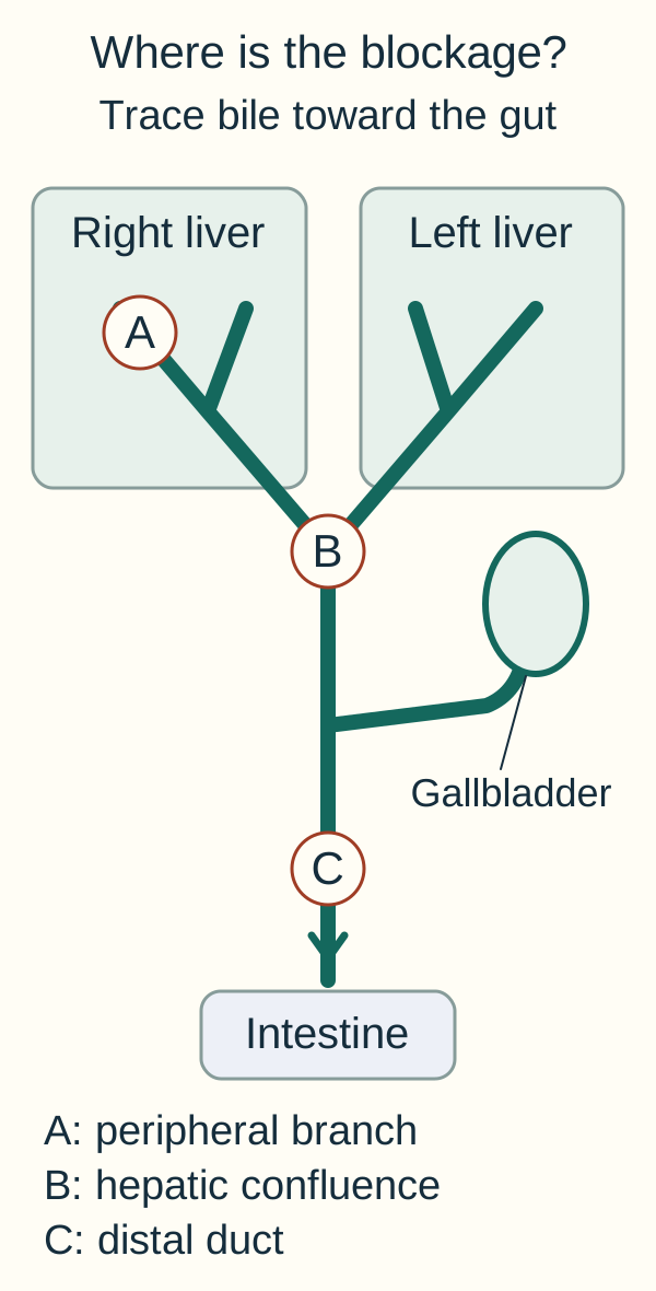
Perihilar cholangiocarcinoma affects the region around the right and left hepatic duct junction, extending between second-order ducts and the cystic duct insertion. The traditional name Klatskin tumor refers to this hilar location. Distal cholangiocarcinoma arises below the cystic duct insertion toward the ampulla. These extrahepatic tumors often restrict the shared route through which bile leaves the liver. [1][2]
An obstructing tumor can raise conjugated bilirubin in blood. Filterable conjugated pigment can darken urine, while reduced intestinal pigment delivery pales the stool. Pruritus and a cholestatic enzyme pattern support impaired bile handling. The pattern localizes a physiologic problem, not necessarily its cause, because a stone or a benign inflammatory stricture can produce similar findings. [1][4]
Trace, cover, predict. On the biliary map, trace a route from each liver side to the intestine. Cover point B at the confluence, then cover point C below the cystic duct insertion. Name which upstream regions can be affected in each position.
Trace each liver side to the intestine, then cover B or C and predict which ducts are upstream. A peripheral branch lesion can leave most central drainage patent. [1][2]
Blocking the confluence can separate both hepatic duct systems from their shared outlet. A distal obstruction can impede bile from both sides farther downstream. A peripheral branch lesion can leave most bile drainage intact. These are anatomic predictions; imaging must establish the actual extent and cause. [1][2]
Check the prediction: both hepatic systems dilated, distal duct not dilated
This pattern favors a proximal obstruction around their junction rather than an isolated distal blockage. Now test the opposite example: a peripheral mass with a patent central tree can be malignant without producing jaundice. [1][2]
What does this evidence actually establish?
A patient with a persistent irregular stricture has CA 19-9 of 620 U/mL during cholangitis and 42 U/mL after drainage and antibiotics; the laboratory upper limit is 37. Decide separately what the falling number says about the acute illness and what the remaining stricture still requires.
Primary sclerosing cholangitis creates a high-risk setting for cholangiocarcinoma. A new relevant stricture, worsening jaundice, unexplained weight loss, or a sustained change in the usual liver-test pattern needs evaluation. A pre-existing chronic diagnosis should not become a reason to dismiss new findings as routine fluctuation. Infection, benign obstruction, and cancer can also coexist. [1][2]
Other established risk settings include choledochal cysts and other congenital duct abnormalities, hepatolithiasis, and chronic infection with liver flukes such as Clonorchis sinensis or Opisthorchis viverrini. These conditions can create prolonged inflammation or abnormal drainage. Many affected patients have no recognized risk factor, so a reassuring exposure history cannot exclude the disease. [1][4]
CA 19-9 is a tumor-associated marker, not a cancer verdict. It can rise during benign obstruction or bacterial cholangitis. If drainage and infection improve, the marker may fall even when the original concern was serious. Conversely, some people do not produce appreciable CA 19-9 because of their Lewis-antigen biology, so a normal value cannot reliably exclude cancer. Interpret it beside imaging, tissue, and the clinical course. [1][2]
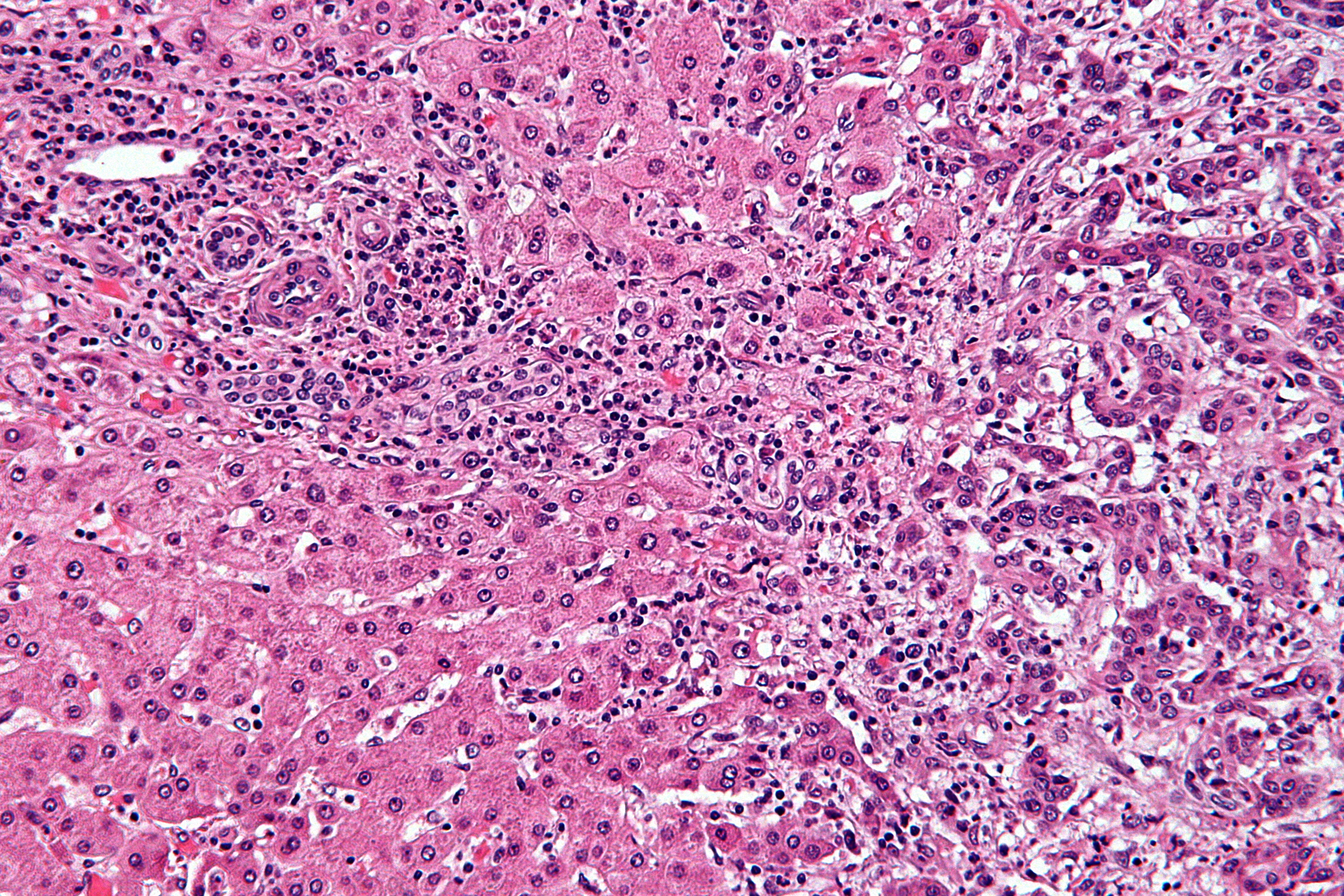
Histology often shows malignant glands embedded in a dense fibrous, or desmoplastic, stroma. This supports an adenocarcinoma pattern but must be distinguished from a metastasis from another organ and other primary liver tumors. Pathology, imaging, and the prior cancer history work together. Neither one stain nor a serum marker should be treated as a complete substitute for that assessment. [1][4]
Read tissue architecture, not the cancer label
In the accompanying H&E image, compare the crowded irregular glands in fibrous tissue on the right with the more orderly liver plates toward the lower left. Image: this as cholangiocarcinoma. Gland formation and desmoplasia support adenocarcinoma, but those features alone do not distinguish a biliary primary from every metastasis. [1]
Compare the irregular glands and surrounding fibrous tissue with the liver plates at lower left. Image: cholangiocarcinoma; gland-forming adenocarcinoma morphology still needs clinical and pathology correlation to establish origin. Image: Nephron, CC BY-SA 3.0, shown [1] Nephron; original source; CC BY-SA 3.0.
Separate the conclusions. Cover the source diagnosis and describe only what is visible. Then add a hypothetical history of colorectal cancer. What additional comparison becomes important?
The prior tumor and focused immunohistochemistry help establish origin; reserve material for molecular testing. For the marker example, the decline supports an obstruction or inflammation contribution, not proof that the persistent stricture is benign. [1][2]
Check the transfer: known cancer, repeatedly undetectable CA 19-9
Some patients produce little CA 19-9. Follow the disease with imaging and clinical assessment rather than treating the low marker as evidence of cure. The marker example is synthetic and is not a validated diagnostic cutoff exercise. [1][2]
Which information must come before a stent?
A stable patient has bilateral intrahepatic duct dilation, no fever and a normal blood pressure. Compare two orders: complete cross-sectional mapping before instrumentation, or stent first and reconstruct the original anatomy later.
In a stable patient, staging-quality imaging should precede biliary instrumentation when feasible. Stents, inflammation, and procedure-related changes can complicate later interpretation. Multiphasic contrast CT assesses the tumor, vessel relationships, nodes, and distant spread. The BSG framework includes imaging of the chest, abdomen, and pelvis rather than examining only the symptomatic duct segment. [1]
MRI with MRCP is especially useful for intrahepatic and perihilar tumors. MRCP shows the fluid-containing duct system without putting an endoscope into it. MRI can help define ductal extent, additional liver lesions, and the relationship between a mass and the biliary tree. For a distal tumor, a good CT may already provide the needed cross-sectional information; MRI is not automatically an extra requirement in every case. [1][2]
The team asks whether complete removal is feasible, whether important vessels or both sides of the duct system are involved, and how much functioning liver would remain. Future liver remnant means the liver tissue that must sustain the patient after a planned resection. Its volume, function, blood supply, and drainage matter. A small-looking tumor can still create a difficult operation if it sits in a critical location. [1]
This planned sequence does not justify delaying care for sepsis. A patient with fever, hypotension, confusion, and obstructed ducts needs urgent resuscitation, antibiotics, and a source-control plan. Imaging and communication should support timely drainage rather than become an elective checklist that prevents it. Once the immediate danger is addressed, the cancer workup still needs an organized completion plan. [1][2]
Change one fact. Keep the same duct obstruction, but replace the stable blood pressure with 86/52 mmHg and add confusion and rigors. State which part of the elective sequence can no longer be allowed to delay care.
Suspected infected obstruction with organ dysfunction requires resuscitation, prompt antibiotics and urgent drainage planning. Complete elective cancer staging is not a prerequisite for source control in an unstable patient. Essential procedural imaging and specialist communication should occur without avoidable delay. [1][2]
Check the transfer: a distal lesion already mapped by high-quality CT
MRI/MRCP is especially valuable for proximal and intrahepatic mapping; it is not automatically needed after adequate CT of a distal lesion. Choose the next study for the unanswered anatomic question, not to complete a fixed list. [1]
Can the right test use the wrong route?
Compare an inoperable peripheral liver mass with a small hilar lesion being evaluated for transplantation. Both need diagnostic evidence, but a needle path acceptable for the first patient can exclude the second from a transplant protocol.
Tissue sampling should answer a specific treatment question. An unresectable intrahepatic mass usually needs a core biopsy for histologic diagnosis and molecular profiling before systemic treatment. Sampling an accessible metastatic site may sometimes be preferable. The multidisciplinary team chooses the safest informative target and preserves enough tissue for the tests that could guide later therapy. [1][3][4]
For an extrahepatic stricture, ERCP can obtain brush cytology and intraductal biopsies while treating obstruction when indicated. Cytology examines sampled cells; FISH assesses selected chromosomal abnormalities. A negative brushing does not reliably exclude malignancy because the sample may miss the tumor. Persistently suspicious imaging may require additional sampling, follow-up imaging, or surgical discussion rather than a benign label based on one negative result. [1][2]
Endoscopic ultrasound can assess selected masses and lymph nodes. The important distinction is what is being sampled and where the needle travels. An unplanned transperitoneal biopsy of a perihilar primary can seed tumor and jeopardize eligibility for a specialized transplant pathway. Do not transfer the biopsy strategy for an unresectable liver mass automatically to a potentially transplantable hilar lesion. [1][2]
For potentially operable distal disease, BSG guidance discusses EUS with ERCP according to jaundice and the diagnostic need. Without jaundice, EUS first may avoid unnecessary ERCP complications. Occasionally a specialist team may proceed to surgery despite imperfect preoperative tissue confirmation when the evidence and potential benefit support it, after discussion of diagnostic uncertainty with the patient. That is not a general instruction to omit pathology. [1]
Drainage also needs a destination. In a planned major liver resection for perihilar disease, the future remnant liver is a key drainage target. In unresectable disease, the plan instead emphasizes relief of obstruction, infection control, and suitability for systemic therapy. Endoscopic, EUS-guided, or percutaneous approaches depend on anatomy and expertise. A stent is not a cure, and its monitoring or exchange plan belongs in the handoff. [1]
Drain the liver that will remain
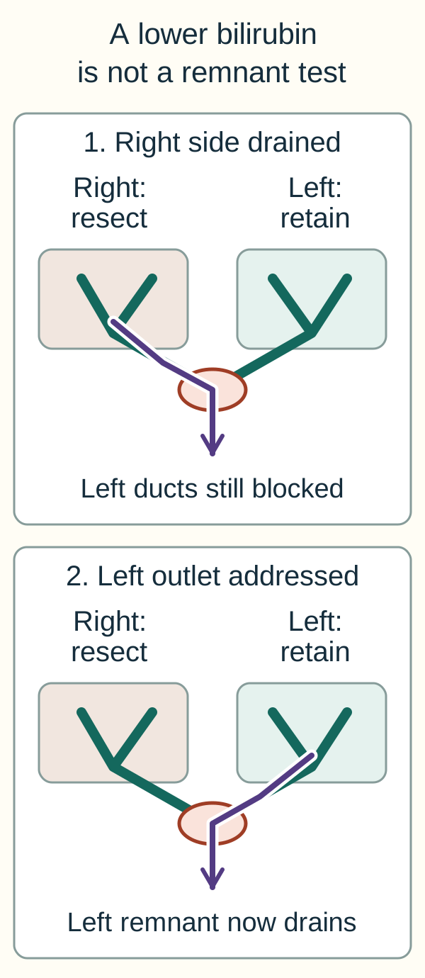
Use the remnant diagram: a right hepatectomy is planned, but only the right system has been drained. The bilirubin falls while the left ducts stay dilated. Trace the left-side outlet and decide whether the remaining liver has been adequately prepared.
Compare which lobe has an outlet, not just whether total bilirubin falls. Right-sided drainage does not establish preparation of a future left remnant. Addressing left drainage still does not prove sufficient volume, perfusion or function. The panels compare drainage targets, not device-placement instructions or a recommendation to remove an existing drain. [1][2]
A lower total bilirubin does not demonstrate drainage or sufficient function of the future left remnant. The team reassesses its duct drainage, volume, perfusion and function; selected patients need additional drainage or remnant augmentation before surgery. Drainage choices must also address infection in any persistently obstructed segments. [1][2]
Check the sampling prediction: negative brushing, suspicious persistent stricture
A negative sample can reflect missed tumor. Correlate with imaging and consider repeat or combined intraductal sampling, selected FISH and specialist surgical assessment. In a possible perihilar transplant pathway, contact the transplant team before sampling the primary through a transperitoneal route; appropriately selected node sampling answers a different staging question. [1][2]
Does unresectable mean the same plan for everyone?
Compare three patients: a fit patient with localized resectable intrahepatic disease, a patient with PSC and a small unresectable perihilar tumor without metastases, and a patient with lung metastases. Assign an initial treatment intent before selecting a drug.
Localized resectable disease should be assessed for an operation aiming for negative margins, called an R0 resection. Resectability depends on the full anatomy, spread, liver reserve, and patient fitness, not simply whether the tumor is called intrahepatic or extrahepatic. A technically removable lesion and a safe operation are related but different judgments. Experienced hepatobiliary teams bring both into the decision. [1][4]
For intrahepatic disease, treatment may involve liver resection with appropriate lymph-node assessment. Perihilar disease often requires extrahepatic duct resection together with major liver resection, often including the caudate lobe, and reconstruction of drainage. Distal cholangiocarcinoma commonly requires pancreaticoduodenectomy, the Whipple operation, because of the shared regional anatomy. A gallbladder-only operation does not remove a distal duct primary. [1][4]
Some highly selected patients with unresectable perihilar disease may enter a protocol combining neoadjuvant treatment and liver transplantation. Selection includes strict tumor, spread, staging, and medical criteria at an experienced center. The patient must be discussed before a biopsy that could close this option. This pathway is not a routine transplant indication for metastatic disease or for every intrahepatic tumor. [1][2]
After resection, recurrence remains a concern even when the visible tumor has been removed. BSG recommends consideration of a finite adjuvant capecitabine course, described as 24 weeks in that guideline. Adjuvant means treatment after the main local operation to reduce recurrence risk. The oncology team considers recovery, renal function, toxicity, pathology, and patient priorities rather than treating every postoperative patient as interchangeable. [1][4]
Assign the three patients. Give each patient in the opening comparison a different initial destination: resection assessment, specialized transplant-protocol assessment, or systemic disease control. Explain which finding separates the first two from the third.
Localized resectable disease merits an R0 surgical assessment. Selected small perihilar tumors, particularly in PSC, can merit neoadjuvant-transplant protocol assessment despite local unresectability. Distant spread instead supports systemic disease control; neither a stent nor routine transplantation eradicates metastatic disease. Protocol criteria vary by center and jurisdiction. [1][2]
Check the transfer: a regional node contains matching tumor during transplant staging
Confirmed nodal metastasis excludes the specialized perihilar transplant pathway described here. Local ductal unresectability and metastatic spread are different findings; one can motivate referral while the other defeats protocol eligibility. [2]
Does a gene name select a treatment?
Two reports both mention FGFR2. One identifies a fusion; the other reports a missense variant of uncertain significance. Decide whether the reports establish the same treatment eligibility, then consider stage, prior therapy and present fitness.
Unresectable or metastatic disease usually requires a disease-control plan rather than a routine curative resection. For an eligible patient, gemcitabine and cisplatin with durvalumab or pembrolizumab is an established first-line systemic approach in current guideline frameworks. These are alternative immunotherapy-containing regimens, not a recommendation to give both immune agents together. The exact regimen depends on approval, contraindications, and local practice. [1][3][4][5]
Fitness for treatment includes performance status, kidney function, marrow reserve, infection control, and the consequences of obstruction. A patient who is septic or has a blocked infected stent is not made ready for chemotherapy by a molecular report alone. A less intensive approach or supportive care may be appropriate when treatment burden outweighs expected benefit. This assessment should include the patient goals and change as the clinical state changes. [1][3][4]
Obtain molecular profiling early enough to influence later decisions, ideally before or during first-line treatment rather than only after every option has been exhausted. Potentially actionable findings include FGFR2 fusions or rearrangements, IDH1 mutation, BRAF V600E, HER2 overexpression or amplification, NTRK or RET fusions, and MSI-high or mismatch-repair-deficient disease. Their frequency and treatment implications differ; FGFR2 and IDH1 alterations are particularly associated with intrahepatic tumors. [1][3][4]
A biomarker name is not an automatic prescription. The exact alteration, assay, disease stage, prior treatment, and drug label must match. For example, the FDA zanidatamab approval concerns adults with previously treated unresectable or metastatic biliary tract cancer that is HER2-positive by IHC 3+ using an approved test. A vague report saying HER2 change does not by itself establish that indication. [6]
After progression, options can include a matched targeted treatment, a clinical trial, or further chemotherapy such as FOLFOX for a suitable patient without a useful target. The choice is not identical for every tumor with the same organ name. Reassessment after a major response can sometimes reopen local-treatment discussion, but response alone does not guarantee conversion to curative surgery. [1][3][4]
For an FGFR2 example, the FDA approved lirafugratinib on September 23, 2026 for adults with previously treated unresectable, locally advanced or metastatic cholangiocarcinoma with an FGFR2 fusion or other rearrangement. A missense variant of uncertain significance is not that finding. Other available options and sequencing require specialist review; this example is not a complete drug list or a comparative efficacy claim. [7]
Match four dimensions. For a proposed targeted drug, state the exact alteration, assay result, treatment setting and prior treatment. Then ask whether infection or organ dysfunction makes treatment unsafe today.
Actionable biology does not replace clinical fitness. Similarly, a report of HER2 amplification with IHC 2+ does not establish the IHC 3+ criterion in the cited US zanidatamab indication. No high PD-L1 threshold is required for the first-line chemotherapy-immunotherapy regimens described above. [3][5][6]
Check the transfer: no fusion was found, but the assay did not assess fusions
The report has not excluded a fusion. Review test coverage and tissue adequacy; complementary validated testing can be needed. Plan profiling before or during first-line therapy instead of discovering this gap only after progression. [1][3]
Can a responding cancer still cause an urgent problem?
A scan three weeks ago showed tumor shrinkage. Today the patient develops chills and recurrent jaundice with a biliary stent in place. Decide whether the old scan answers the new clinical question.
Relieving pain, itching, poor intake, and distress is part of cancer care at every stage. Palliative care can be provided alongside active oncology treatment; it is not restricted to the final days of life or to people who have stopped anticancer therapy. Nutrition support and an accessible specialist contact help turn a complex plan into something the patient can follow. [1]
After biliary stenting, give a clear plan for recognizing blockage or infection. Recurrent jaundice, fever, rigors, pain, or new illness warrants assessment rather than waiting for the next routine cancer appointment. The team should know which segments were drained, which device was placed, and whether a scheduled exchange or an on-demand intervention is planned. A procedure note alone is not a complete safety plan. [1]
After potentially curative treatment, imaging and clinical follow-up look for recurrence and treatment complications. During systemic treatment, response, tolerance, liver function, and patient priorities need repeated review. A lower CA 19-9 can support the overall assessment but cannot replace imaging or explain every change in symptoms. Surveillance and investigation of a new problem are separate reasons for a visit. [1][4]
Return to four linked questions. Where did the tumor arise? How far has it spread? Which diagnostic or drainage procedure preserves the best options? Is the present plan resection, a selected transplant protocol, or systemic disease control? The answer can evolve, but keeping these questions distinct prevents a useful test or symptom intervention from being mistaken for definitive cancer treatment. [1][2][3]
Choose the response to the new illness. Contrast urgent assessment of possible stent dysfunction with simply waiting for the next response scan. State what would make the situation an emergency.
Chills and recurrent jaundice warrant prompt assessment for recurrent obstruction and infection even during tumor response. Hypotension, confusion or other organ dysfunction require emergency care. Stent follow-up must identify the drained segments, device, responsible service and route to urgent help. [1][2]
Check the transfer: pain and poor intake without obstruction
Offer symptom treatment, nutrition and palliative support alongside desired anticancer treatment. Palliative care does not require stopping oncology therapy. Routine surveillance and investigation of a new symptom serve different purposes. [1]
Apply the lesson to clinical cases
You can pause here and return to any case. Choose an answer when ready, then compare the explanations.
Case 1
Show answer and explanations for case 1
A. A peripheral right intrahepatic duct (Why this does not fit)
A peripheral right duct drains only a limited portion of the right liver. An isolated peripheral lesion does not explain bilateral dilation with a normal duct below the junction.
Reasoning steps for option A
Which territory drains through a peripheral right duct?
A peripheral right duct drains only a limited portion of the right liver.
Does that location explain both dilated hepatic systems?
An isolated peripheral lesion does not explain bilateral dilation with a normal duct below the junction.
B. The right-left hepatic duct confluence (Best answer)
Both major intrahepatic duct systems are dilated. The common downstream duct is not dilated, placing the obstruction proximally. The right-left hepatic confluence connects both systems and fits a perihilar primary.
Reasoning steps for option B
Where is dilation present?
Both major intrahepatic duct systems are dilated.
Where does dilation stop?
The common downstream duct is not dilated, placing the obstruction proximally.
Which shared junction fits that distribution?
The right-left hepatic confluence connects both systems and fits a perihilar primary.
C. The gallbladder neck and cystic duct (Why this does not fit)
A neck lesion or impacted stone can compress the adjacent common hepatic duct. The lesion arises in the bile duct wall and the gallbladder wall is normal, favoring a perihilar duct primary.
Reasoning steps for option C
How can a gallbladder-neck process cause jaundice?
A neck lesion or impacted stone can compress the adjacent common hepatic duct.
Which finding identifies a different primary site here?
The lesion arises in the bile duct wall and the gallbladder wall is normal, favoring a perihilar duct primary.
D. The intrapancreatic common bile duct (Why this does not fit)
The proximal extrahepatic duct and both intrahepatic systems lie upstream. The extrahepatic duct below the hepatic junction remains normal rather than sharing the upstream dilation.
Reasoning steps for option D
What lies upstream of the intrapancreatic common duct?
The proximal extrahepatic duct and both intrahepatic systems lie upstream.
Which duct-caliber finding argues against that distal site?
The extrahepatic duct below the hepatic junction remains normal rather than sharing the upstream dilation.
Takeaway: Localize an obstruction from the distribution of dilated and nondilated ducts, then identify its site of origin.
A. Microscopic nerve involvement beside the original focus (Why this does not fit)
Tumor invasion around nerves can contribute to pain and local spread. No central obstruction is supplied, so it does not directly explain loss of intestinal pigment and conjugated pigment in urine.
Reasoning steps for option A
What can perineural invasion help explain?
Tumor invasion around nerves can contribute to pain and local spread.
Does microscopic invasion beside a peripheral focus block the shared outlet?
No central obstruction is supplied, so it does not directly explain loss of intestinal pigment and conjugated pigment in urine.
B. A second small focus confined to the right periphery (Why this does not fit)
A new peripheral focus can increase disease burden while much of the duct system remains patent. The described small peripheral focus does not establish substantial obstruction of bile delivery to the intestine.
Reasoning steps for option B
Can another peripheral focus increase cancer burden?
A new peripheral focus can increase disease burden while much of the duct system remains patent.
Which missing effect is needed for these new symptoms?
The described small peripheral focus does not establish substantial obstruction of bile delivery to the intestine.
C. A small noncompressive regional lymph-node deposit (Why this does not fit)
A regional tumor deposit affects staging and may obstruct structures if it becomes compressive. The node is explicitly small and noncompressive, so it does not account for the new obstructive pigment pattern.
Reasoning steps for option C
Can regional nodal spread matter clinically?
A regional tumor deposit affects staging and may obstruct structures if it becomes compressive.
What excludes compression as the supplied explanation?
The node is explicitly small and noncompressive, so it does not account for the new obstructive pigment pattern.
D. Extension across the right-left hepatic duct junction (Best answer)
Reduced pigment reaches the intestine while conjugated bilirubin reaches the urine, supporting obstructed bile flow. The peripheral primary had left the central drainage routes patent. Crossing the hepatic duct junction can impede the common outlet from major liver regions.
Reasoning steps for option D
What do pale stools and dark urine together suggest?
Reduced pigment reaches the intestine while conjugated bilirubin reaches the urine, supporting obstructed bile flow.
Why was bilirubin previously normal?
The peripheral primary had left the central drainage routes patent.
Which new extension changes that relationship?
Crossing the hepatic duct junction can impede the common outlet from major liver regions.
Takeaway: Predict jaundice from impaired central bile drainage rather than from tumor size or cancer burden alone.
A. ERCP with empiric extraction of an occult duct stone (Why this does not fit)
Stones commonly cause cholestatic obstruction. A persistent irregular stricture, weight loss and no stone on MRCP require a malignancy-focused assessment rather than empiric extraction.
Reasoning steps for option A
Why might a stone initially be considered?
Stones commonly cause cholestatic obstruction.
Which observations favor a different assessment here?
A persistent irregular stricture, weight loss and no stone on MRCP require a malignancy-focused assessment rather than empiric extraction.
B. Interval MRCP after a prolonged observation period (Why this does not fit)
Specialist follow-up imaging can help resolve selected indeterminate strictures. Progressive symptoms and a fixed irregular narrowing warrant prompt staging and diagnostic review, not delay based on absent risk factors.
Reasoning steps for option B
When can interval imaging help?
Specialist follow-up imaging can help resolve selected indeterminate strictures.
Why is prolonged observation insufficient in this presentation?
Progressive symptoms and a fixed irregular narrowing warrant prompt staging and diagnostic review, not delay based on absent risk factors.
C. Glucocorticoid treatment before diagnostic sampling (Why this does not fit)
IgG4-related sclerosing cholangitis can cause a biliary stricture and respond to glucocorticoids. No supporting IgG4-related findings or completed malignancy evaluation are supplied; empiric steroids can obscure an important competing diagnosis.
Reasoning steps for option C
Which benign disease can resemble a malignant stricture?
IgG4-related sclerosing cholangitis can cause a biliary stricture and respond to glucocorticoids.
What is missing before choosing that treatment?
No supporting IgG4-related findings or completed malignancy evaluation are supplied; empiric steroids can obscure an important competing diagnosis.
D. Hepatobiliary staging review with directed sampling (Best answer)
The fixed irregular common hepatic duct narrowing is not explained by a visible stone. No. Cholangiocarcinoma can arise without a recognized risk factor. Arrange timely hepatobiliary staging review and location-appropriate sampling while assessing benign mimics.
Reasoning steps for option D
What does the imaging demonstrate?
The fixed irregular common hepatic duct narrowing is not explained by a visible stone.
Do absent PSC, cysts and fluke exposure exclude cancer?
No. Cholangiocarcinoma can arise without a recognized risk factor.
What follows from the progressive course?
Arrange timely hepatobiliary staging review and location-appropriate sampling while assessing benign mimics.
Takeaway: Risk factors modify suspicion; their absence does not dismiss a progressive irregular biliary stricture.
A. Acute obstruction contributed; the stricture still needs assessment (Best answer)
Fever and bilirubin improved while CA 19-9 fell markedly. Obstruction or infection contributed to the initial marker concentration. An irregular stricture remains and still needs evaluation; marker improvement does not establish its cause.
Reasoning steps for option A
What changed after antibiotics and drainage?
Fever and bilirubin improved while CA 19-9 fell markedly.
What does that coupled response support?
Obstruction or infection contributed to the initial marker concentration.
What important abnormality persists independently?
An irregular stricture remains and still needs evaluation; marker improvement does not establish its cause.
B. The marker decline resolves concern about the persistent stricture (Why this does not fit)
A large decline accompanies resolution of acute inflammation and obstruction. Cancer can coexist with those reversible processes, and the irregular stricture remains on imaging.
Reasoning steps for option B
Why might the decline seem reassuring?
A large decline accompanies resolution of acute inflammation and obstruction.
Why does it not settle the lesion diagnosis?
Cancer can coexist with those reversible processes, and the irregular stricture remains on imaging.
C. Residual marker abnormality establishes a malignant stricture (Why this does not fit)
A value of 42 U/mL remains just above the supplied upper limit of 37. CA 19-9 is not specific for cancer, so the remaining abnormality cannot replace imaging and tissue assessment.
Reasoning steps for option C
Is the repeat concentration completely normal?
A value of 42 U/mL remains just above the supplied upper limit of 37.
Does that small residual difference establish malignancy?
CA 19-9 is not specific for cancer, so the remaining abnormality cannot replace imaging and tissue assessment.
D. The initial high value reflects a marker-nonproducing phenotype (Why this does not fit)
Some patients have little circulating CA 19-9 despite measurable cancer. The initial concentration of 620 demonstrates substantial marker production; the issue is nonspecific production during illness.
Reasoning steps for option D
Why do nonproducing phenotypes matter?
Some patients have little circulating CA 19-9 despite measurable cancer.
Which measurement contradicts that explanation here?
The initial concentration of 620 demonstrates substantial marker production; the issue is nonspecific production during illness.
Takeaway: Interpret a marker trend alongside both the treated obstruction and any persistent structural lesion.
A. Compare serum bilirubin before and after biliary drainage (Why this does not fit)
Bilirubin can improve when bile drainage improves. Drainage relief does not establish disappearance or shrinkage of the CT-visible tumor.
Reasoning steps for option A
What does bilirubin improvement measure most directly?
Bilirubin can improve when bile drainage improves.
Why is that not an adequate tumor-response measure here?
Drainage relief does not establish disappearance or shrinkage of the CT-visible tumor.
B. Compare interval CT with baseline lesion measurements (Best answer)
It was below 2 U/mL despite a measurable pretreatment tumor. An unchanged low value cannot distinguish treatment response from persistent or growing disease. Interval imaging against baseline lesion measurements addresses response, alongside clinical assessment.
Reasoning steps for option B
Was CA 19-9 measurable when tumor was definitely present?
It was below 2 U/mL despite a measurable pretreatment tumor.
How does that affect serial marker interpretation?
An unchanged low value cannot distinguish treatment response from persistent or growing disease.
Which assessment directly compares the measurable disease?
C. Compare serial CA 19-9 with the laboratory upper limit (Why this does not fit)
A marker that reliably reflects a individual disease can supplement response assessment. No. It was undetectable before treatment, so remaining below the upper limit does not classify stable burden or response.
Reasoning steps for option C
Why are serial tumor markers sometimes useful?
A marker that reliably reflects a individual disease can supplement response assessment.
Does this marker have that baseline relationship?
No. It was undetectable before treatment, so remaining below the upper limit does not classify stable burden or response.
D. Compare appetite and weight with pretreatment symptoms (Why this does not fit)
Symptoms and nutritional change help assess function and treatment tolerance. Supportive care and toxicity can change these measures independently of the measurable tumor; imaging remains necessary.
Reasoning steps for option D
What do appetite and weight contribute?
Symptoms and nutritional change help assess function and treatment tolerance.
Why is symptom improvement not enough to classify tumor response?
Supportive care and toxicity can change these measures independently of the measurable tumor; imaging remains necessary.
Takeaway: An uninformative baseline tumor marker stays uninformative as a response measure; compare measurable disease with imaging.
A. Compare the archived tumor and select focused ancillary tests (Best answer)
Malignant glands establish an adenocarcinoma pattern, not a unique site of origin. The archived colorectal tumor permits direct comparison with the new lesion. Focused testing addresses the differential while conserving limited tissue for predictive molecular studies.
Reasoning steps for option A
What has the core established?
Malignant glands establish an adenocarcinoma pattern, not a unique site of origin.
Which additional evidence is particularly relevant?
The archived colorectal tumor permits direct comparison with the new lesion.
Why should ancillary tests be selected rather than indiscriminate?
Focused testing addresses the differential while conserving limited tissue for predictive molecular studies.
B. Apply an extensive immunostain panel before reviewing prior tissue (Why this does not fit)
Selected stains can help separate competing primary sites. The prior tumor provides a focused differential, and exhausting a limited core may prevent later molecular profiling.
Reasoning steps for option B
Why can immunohistochemistry be useful?
Selected stains can help separate competing primary sites.
Why not begin with an extensive untargeted panel?
The prior tumor provides a focused differential, and exhausting a limited core may prevent later molecular profiling.
C. Assign a biliary primary from the gland-and-stroma pattern (Why this does not fit)
Desmoplastic stroma with malignant glands is compatible with cholangiocarcinoma. Metastatic adenocarcinoma can share this pattern, especially with a prior colorectal primary.
Reasoning steps for option C
What does the fibrous gland-forming pattern suggest?
Desmoplastic stroma with malignant glands is compatible with cholangiocarcinoma.
What competing origin remains unresolved?
Metastatic adenocarcinoma can share this pattern, especially with a prior colorectal primary.
D. Assign a colorectal metastasis from the prior cancer history (Why this does not fit)
A previous colorectal adenocarcinoma makes metastatic recurrence a plausible explanation for a liver mass. A second primary remains possible; morphology, focused testing and imaging must be correlated before assigning origin.
Reasoning steps for option D
Why is colorectal metastasis a serious possibility?
A previous colorectal adenocarcinoma makes metastatic recurrence a plausible explanation for a liver mass.
Why does history alone not establish it?
A second primary remains possible; morphology, focused testing and imaging must be correlated before assigning origin.
Takeaway: Use morphology to define the differential, then compare relevant prior tissue while conserving material for profiling.
A. ERCP-directed sampling followed by immediate metal stenting (Why this does not fit)
ERCP can sample a stricture and decompress an obstructed duct. The patient is stable and has not had imaging to define tumor extent, vessels and the drainage plan.
Reasoning steps for option A
What can ERCP accomplish?
ERCP can sample a stricture and decompress an obstructed duct.
Why should elective instrumentation not lead this sequence?
The patient is stable and has not had imaging to define tumor extent, vessels and the drainage plan.
B. PET-CT staging followed by ERCP-directed duct assessment (Why this does not fit)
PET-CT can contribute to detection of nodal or distant disease in selected staging pathways. It does not replace multiphasic CT and proximal duct mapping before elective hilar intervention.
Reasoning steps for option B
What question can PET-CT sometimes help answer?
PET-CT can contribute to detection of nodal or distant disease in selected staging pathways.
Which necessary anatomic information is still missing?
It does not replace multiphasic CT and proximal duct mapping before elective hilar intervention.
C. Staging laparoscopy followed by cross-sectional duct mapping (Why this does not fit)
It can reveal occult spread before a major planned operation. Cross-sectional staging must first define the suspected tumor and guide selection for invasive staging.
Reasoning steps for option C
Why is staging laparoscopy considered in some patients?
It can reveal occult spread before a major planned operation.
Why is it not the first investigation here?
Cross-sectional staging must first define the suspected tumor and guide selection for invasive staging.
D. Staging CT with MRI/MRCP and hepatobiliary review (Best answer)
A proximal obstruction needs assessment of both duct systems and their surrounding anatomy. No fever, hypotension or organ dysfunction is supplied. Obtain staging CT with MRI/MRCP for proximal extent and coordinate intervention with the hepatobiliary team.
Reasoning steps for option D
What does bilateral intrahepatic dilation suggest?
A proximal obstruction needs assessment of both duct systems and their surrounding anatomy.
Is there an emergency requiring immediate drainage before full elective mapping?
No fever, hypotension or organ dysfunction is supplied.
Which sequence preserves the needed information?
Obtain staging CT with MRI/MRCP for proximal extent and coordinate intervention with the hepatobiliary team.
Takeaway: In a stable proximal obstruction, obtain staging and ductal anatomy before elective instrumentation.
A. Prompt antibiotics with urgent coordinated biliary drainage (Best answer)
Recurrent dilation above a biliary stent with rigors and jaundice points to infected duct obstruction. Hypotension, confusion and rising creatinine indicate severe systemic illness. Urgent coordinated decompression provides source control while resuscitation and antibiotics continue.
Reasoning steps for option A
Where is the likely infectious source?
Recurrent dilation above a biliary stent with rigors and jaundice points to infected duct obstruction.
What indicates organ dysfunction?
Hypotension, confusion and rising creatinine indicate severe systemic illness.
What must accompany antimicrobial therapy?
Urgent coordinated decompression provides source control while resuscitation and antibiotics continue.
B. Prompt antibiotics with urgent laparoscopic cholecystectomy (Why this does not fit)
A gallbladder-centered infection can require gallbladder-directed treatment. Recurrent duct dilation above a stent and no gallbladder-wall thickening favor obstructed bile ducts; cholecystectomy does not address that obstruction.
Reasoning steps for option B
When would the gallbladder be the source-control target?
A gallbladder-centered infection can require gallbladder-directed treatment.
Which findings instead localize this emergency?
Recurrent duct dilation above a stent and no gallbladder-wall thickening favor obstructed bile ducts; cholecystectomy does not address that obstruction.
C. Prompt antibiotics with delayed drainage after renal recovery (Why this does not fit)
Renal dysfunction affects resuscitation and procedural planning. The infected obstruction may be causing the kidney injury; waiting for recovery can leave its cause untreated.
Reasoning steps for option C
Why does renal dysfunction matter for procedures?
Renal dysfunction affects resuscitation and procedural planning.
Why should recovery not be a prerequisite for source control?
The infected obstruction may be causing the kidney injury; waiting for recovery can leave its cause untreated.
D. Prompt antibiotics with drainage after a 72-hour response trial (Why this does not fit)
Antibiotics treat bacteria and are required promptly. Shock and organ dysfunction with ongoing obstruction require urgent source-control planning rather than an elective response trial.
Reasoning steps for option D
Can antibiotics sometimes improve cholangitis?
Antibiotics treat bacteria and are required promptly.
Why is a fixed 72-hour wait inappropriate here?
Shock and organ dysfunction with ongoing obstruction require urgent source-control planning rather than an elective response trial.
Takeaway: Combine the anatomic source with organ dysfunction: infected biliary obstruction requires antibiotics, resuscitation and urgent drainage.
A. Assess left-lobe reserve and plan remnant-directed preparation (Best answer)
The left lobe is the future liver remnant. No. Bilirubin can fall after right-sided drainage while the left ducts remain obstructed. Left-remnant drainage and functional reserve require review, with drainage or augmentation chosen as needed.
Reasoning steps for option A
Which liver must sustain function after right hepatectomy?
The left lobe is the future liver remnant.
Does the serum bilirubin establish drainage of that lobe?
No. Bilirubin can fall after right-sided drainage while the left ducts remain obstructed.
What therefore needs assessment before proceeding?
Left-remnant drainage and functional reserve require review, with drainage or augmentation chosen as needed.
B. Proceed after documenting a further fall in total bilirubin (Why this does not fit)
At least some bile drainage or handling has improved. Persistently dilated left ducts and unmeasured left functional reserve prevent clearance for surgery from total bilirubin alone.
Reasoning steps for option B
What does falling bilirubin demonstrate?
At least some bile drainage or handling has improved.
What crucial regional question remains unanswered?
Persistently dilated left ducts and unmeasured left functional reserve prevent clearance for surgery from total bilirubin alone.
C. Drain more right-lobe segments before repeating serum tests (Why this does not fit)
It could increase drainage from tissue currently contributing to the serum result. The right lobe is planned for resection; the undrained future left remnant remains the relevant concern.
Reasoning steps for option C
Why might more right-sided drainage lower bilirubin?
It could increase drainage from tissue currently contributing to the serum result.
Why does that not solve the stated postoperative risk?
The right lobe is planned for resection; the undrained future left remnant remains the relevant concern.
D. Increase resection extent to encompass the undrained ducts (Why this does not fit)
A larger resection may be needed in selected patients to achieve adequate margins. Further tissue loss may worsen residual liver reserve; drainage and function of the intended remnant need assessment first.
Reasoning steps for option D
What is the oncologic reason to extend a resection?
A larger resection may be needed in selected patients to achieve adequate margins.
Why is it not the answer to an unprepared remnant?
Further tissue loss may worsen residual liver reserve; drainage and function of the intended remnant need assessment first.
Takeaway: A global bilirubin response cannot substitute for regional assessment of the future liver remnant.
A. Image-guided core biopsy of an accessible tumor deposit (Best answer)
The disease is represented by peripheral hepatic tumor deposits, not a dominant accessible extrahepatic stricture. The team needs tumor architecture for diagnosis and sufficient tumor material for molecular testing. Image-guided core sampling of a safely accessible tumor deposit targets the relevant tissue.
Reasoning steps for option A
Where is the tissue target?
The disease is represented by peripheral hepatic tumor deposits, not a dominant accessible extrahepatic stricture.
What kind of information is required?
The team needs tumor architecture for diagnosis and sufficient tumor material for molecular testing.
Which route matches both requirements?
Image-guided core sampling of a safely accessible tumor deposit targets the relevant tissue.
B. ERCP brush sampling of the extrahepatic duct lumen (Why this does not fit)
It samples cells from the duct lumen and a reachable stricture. There is no dominant accessible extrahepatic stricture; brushing a normal-caliber duct may miss the peripheral tumor.
Reasoning steps for option B
What tissue does ERCP brushing usually sample?
It samples cells from the duct lumen and a reachable stricture.
Why is that a poor target here?
There is no dominant accessible extrahepatic stricture; brushing a normal-caliber duct may miss the peripheral tumor.
C. Percutaneous core biopsy of uninvolved background liver (Why this does not fit)
It can characterize accompanying parenchymal disease such as fibrosis or inflammation. Uninvolved liver does not supply the tumor needed to establish cancer type and predictive alterations.
Reasoning steps for option C
What can background liver biopsy assess?
It can characterize accompanying parenchymal disease such as fibrosis or inflammation.
Why does it fail the requested purpose?
Uninvolved liver does not supply the tumor needed to establish cancer type and predictive alterations.
D. Transjugular sampling of non-targeted liver parenchyma (Why this does not fit)
It can obtain liver parenchyma when patient-specific bleeding or vascular considerations favor that route. The requested specimen must contain a tumor deposit, and no reason to substitute non-targeted tissue is supplied.
Reasoning steps for option D
When can a transjugular liver approach be useful?
It can obtain liver parenchyma when patient-specific bleeding or vascular considerations favor that route.
Why is non-targeted parenchymal sampling inadequate here?
The requested specimen must contain a tumor deposit, and no reason to substitute non-targeted tissue is supplied.
Takeaway: Match the biopsy target and specimen type to the diagnostic and molecular question.
Twenty-four of the 40 patients with cancer had negative brushings: 24/40 = 60%. No. It asks about all negative tests, so the denominator must also include the 59 true negatives.
Reasoning steps for option A
Which fraction equals 60%?
Twenty-four of the 40 patients with cancer had negative brushings: 24/40 = 60%.
Is the question restricted to patients already known to have cancer?
No. It asks about all negative tests, so the denominator must also include the 59 true negatives.
B. 40% (Why this does not fit)
Sixteen of the 40 patients with cancer tested positive: 16/40 = 40% sensitivity in this synthetic cohort. The question starts with a negative test and asks how many of those patients still had cancer, reversing the conditioning group.
Reasoning steps for option B
Which fraction equals 40%?
Sixteen of the 40 patients with cancer tested positive: 16/40 = 40% sensitivity in this synthetic cohort.
Why is sensitivity not the requested result?
The question starts with a negative test and asks how many of those patients still had cancer, reversing the conditioning group.
C. 29% (Best answer)
There are 24 false negatives plus 59 true negatives, totaling 83. Twenty-four of the 83 negative tests occurred in patients with cancer. 24/83 is approximately 29%; a negative result does not rule out cancer.
Reasoning steps for option C
How many negative brushings are there?
There are 24 false negatives plus 59 true negatives, totaling 83.
How many of those negatives occurred in patients with cancer?
Twenty-four of the 83 negative tests occurred in patients with cancer.
What proportion follows?
24/83 is approximately 29%; a negative result does not rule out cancer.
D. 94% (Why this does not fit)
Sixteen of 17 positive brushings were in patients with cancer: 16/(16+1), approximately 94%. The requested group is negative brushings, not positive brushings; its relevant fraction is 24/(24+59).
Reasoning steps for option D
Which fraction is approximately 94%?
Sixteen of 17 positive brushings were in patients with cancer: 16/(16+1), approximately 94%.
Which group is being asked about?
The requested group is negative brushings, not positive brushings; its relevant fraction is 24/(24+59).
Takeaway: For disease after a negative result, count false negatives among all negative results; do not substitute sensitivity or positive predictive value.
A. Intraductal forceps sampling during a planned ERCP (Why this does not fit)
It obtains tissue from within the duct stricture. It is not an unplanned transperitoneal needle path through the hilar primary; diagnostic strategy still belongs to the specialist protocol.
Reasoning steps for option A
What does intraductal forceps sampling target?
It obtains tissue from within the duct stricture.
Why is it different from the procedure being flagged?
It is not an unplanned transperitoneal needle path through the hilar primary; diagnostic strategy still belongs to the specialist protocol.
B. ERCP brush cytology from within the duct stricture (Why this does not fit)
The brush samples the stricture from the duct lumen. No. The warning specifically concerns transperitoneal primary-tumor sampling, not a blanket ban on diagnostic evidence.
Reasoning steps for option B
What is the route for ERCP brush cytology?
The brush samples the stricture from the duct lumen.
Does the named route introduce the same peritoneal seeding concern?
No. The warning specifically concerns transperitoneal primary-tumor sampling, not a blanket ban on diagnostic evidence.
C. Transperitoneal needle biopsy of the hilar primary (Best answer)
A specialized neoadjuvant-transplant pathway remains possible for this selected small perihilar lesion. Tumor dissemination along that route can compromise transplant eligibility. Pause and confirm the acceptable diagnostic strategy with the transplant team.
Reasoning steps for option C
Which curative option remains under evaluation?
A specialized neoadjuvant-transplant pathway remains possible for this selected small perihilar lesion.
What risk does a transperitoneal primary-tumor needle path create?
Tumor dissemination along that route can compromise transplant eligibility.
What should happen before that proposed biopsy?
Pause and confirm the acceptable diagnostic strategy with the transplant team.
D. EUS-guided sampling of the indeterminate regional node (Why this does not fit)
It could detect nodal metastasis that excludes the transplant pathway. The target is a staging node, not the primary; the transplant team can use appropriately selected nodal sampling to assess eligibility.
Reasoning steps for option D
What question would regional-node sampling answer?
It could detect nodal metastasis that excludes the transplant pathway.
Why is this not equivalent to puncturing the hilar primary?
The target is a staging node, not the primary; the transplant team can use appropriately selected nodal sampling to assess eligibility.
Takeaway: Before sampling perihilar disease, distinguish the primary-tumor route from a staging-node target and protect a possible transplant pathway.
A. ERCP with diagnostic sampling and preoperative stenting (Why this does not fit)
It can sample the duct and treat clinically important obstruction. There is no jaundice, cholangitis or drainage requirement; BSG favors EUS first in this setting to limit avoidable ERCP complications.
Reasoning steps for option A
Why might ERCP be useful for distal obstruction?
It can sample the duct and treat clinically important obstruction.
Which absent problem makes immediate stenting unnecessary here?
There is no jaundice, cholangitis or drainage requirement; BSG favors EUS first in this setting to limit avoidable ERCP complications.
B. Staging laparoscopy before further endoscopic evaluation (Why this does not fit)
It can identify occult peritoneal or liver-surface spread before a major operation. The team needs local diagnostic information about the distal lesion; EUS addresses that before selecting further invasive staging.
Reasoning steps for option B
What can selected staging laparoscopy detect?
It can identify occult peritoneal or liver-surface spread before a major operation.
What is the immediate unanswered question?
The team needs local diagnostic information about the distal lesion; EUS addresses that before selecting further invasive staging.
C. Percutaneous needle sampling of the distal duct primary (Why this does not fit)
It can provide diagnostic tumor tissue when the target and route are appropriate. Potentially operable distal disease has an endoscopic diagnostic pathway, and percutaneous primary-tumor biopsy can create avoidable dissemination risk.
Reasoning steps for option C
What can needle biopsy provide?
It can provide diagnostic tumor tissue when the target and route are appropriate.
Why is an unplanned percutaneous route not preferred here?
Potentially operable distal disease has an endoscopic diagnostic pathway, and percutaneous primary-tumor biopsy can create avoidable dissemination risk.
D. Endoscopic ultrasound with lesion-directed assessment (Best answer)
No. Bilirubin is normal and there is no cholangitis or stated drainage requirement. Local assessment of the distal lesion and surrounding structures will inform diagnosis and surgery. EUS first obtains local information without imposing unnecessary ERCP and stent risks.
Reasoning steps for option D
Is therapeutic biliary drainage needed now?
No. Bilirubin is normal and there is no cholangitis or stated drainage requirement.
What additional information is needed?
Local assessment of the distal lesion and surrounding structures will inform diagnosis and surgery.
Which BSG sequence fits both facts?
EUS first obtains local information without imposing unnecessary ERCP and stent risks.
Takeaway: For potentially operable distal disease without jaundice, EUS-first assessment can avoid unnecessary ERCP risk.
A. Major hepatectomy with caudate and hilar duct resection (Why this does not fit)
Perihilar disease may require major hepatectomy, caudate resection and biliary reconstruction. It is in the intrapancreatic common bile duct, separate from the hepatic hilum.
Reasoning steps for option A
Which location often needs liver and hilar duct resection?
Perihilar disease may require major hepatectomy, caudate resection and biliary reconstruction.
Where is this primary instead centered?
It is in the intrapancreatic common bile duct, separate from the hepatic hilum.
B. Pancreaticoduodenectomy with regional nodal assessment (Best answer)
The distal bile duct traverses the pancreatic head near the duodenum. No distant spread is found and the patient is fit for surgery. Pancreaticoduodenectomy is the usual curative-intent framework for an intrapancreatic distal duct primary.
Reasoning steps for option B
What anatomy contains the primary?
The distal bile duct traverses the pancreatic head near the duodenum.
Has staging or fitness ruled out an operative approach?
No distant spread is found and the patient is fit for surgery.
Which operation addresses that shared region?
Pancreaticoduodenectomy is the usual curative-intent framework for an intrapancreatic distal duct primary.
C. Cholecystectomy with adjacent hepatic-bed resection (Why this does not fit)
That framework addresses selected gallbladder cancers. The tumor arises in the distal bile duct and is separate from the gallbladder, so the primary would remain outside that field.
Reasoning steps for option C
What primary site is addressed by gallbladder and hepatic-bed surgery?
That framework addresses selected gallbladder cancers.
Which supplied site makes it inadequate?
The tumor arises in the distal bile duct and is separate from the gallbladder, so the primary would remain outside that field.
D. Segmental duct excision with hepaticojejunostomy (Why this does not fit)
It limits the operation to a duct segment with reconstruction of bile drainage. The surrounding pancreatic and duodenal anatomy and margin requirements commonly require pancreaticoduodenectomy.
Reasoning steps for option D
Why can segmental duct excision appear attractive?
It limits the operation to a duct segment with reconstruction of bile drainage.
Why is that not the usual approach for this intrapancreatic primary?
The surrounding pancreatic and duodenal anatomy and margin requirements commonly require pancreaticoduodenectomy.
Takeaway: Select the operative framework from the primary site and regional anatomy, not simply from the presence of jaundice.
A. Neoadjuvant chemoimmunotherapy before reassessing resection (Why this does not fit)
Selected borderline or initially inoperable disease may undergo systemic treatment and reassessment. The supplied lesion is already anatomically resectable with adequate reserve; routine neoadjuvant chemotherapy is not the cited BSG approach for such disease.
Reasoning steps for option A
When can systemic therapy precede local treatment?
Selected borderline or initially inoperable disease may undergo systemic treatment and reassessment.
What argues against making it routine before surgery here?
The supplied lesion is already anatomically resectable with adequate reserve; routine neoadjuvant chemotherapy is not the cited BSG approach for such disease.
B. Referral for a neoadjuvant-transplant protocol (Why this does not fit)
Small perihilar disease, particularly with PSC, may qualify under specialized protocols. No. This is a resectable intrahepatic tumor in noncirrhotic liver, not the selected unresectable perihilar setting.
Reasoning steps for option B
Which cholangiocarcinoma setting has an established selected transplant pathway?
Small perihilar disease, particularly with PSC, may qualify under specialized protocols.
Does this patient share that surgical problem?
No. This is a resectable intrahepatic tumor in noncirrhotic liver, not the selected unresectable perihilar setting.
C. Resection planning with regional lymph-node assessment (Best answer)
There is a solitary segment VI tumor without nodal or distant deposits or major vascular involvement. The noncirrhotic liver and adequate anticipated remnant support that possibility. Hepatobiliary resection planning with appropriate nodal assessment offers the relevant curative-intent pathway.
Reasoning steps for option C
What does staging show about disease extent?
There is a solitary segment VI tumor without nodal or distant deposits or major vascular involvement.
Can sufficient functional liver remain?
The noncirrhotic liver and adequate anticipated remnant support that possibility.
Which intent should lead?
Hepatobiliary resection planning with appropriate nodal assessment offers the relevant curative-intent pathway.
D. Definitive stereotactic radiotherapy with imaging follow-up (Why this does not fit)
It can contribute to treatment of selected locally advanced or medically inoperable disease. The patient has suitable anatomy and reserve for a potentially curative resection, which should be evaluated rather than displaced by a nonsurgical default.
Reasoning steps for option D
When can stereotactic radiotherapy be considered?
It can contribute to treatment of selected locally advanced or medically inoperable disease.
What available option makes it less appropriate as the lead here?
The patient has suitable anatomy and reserve for a potentially curative resection, which should be evaluated rather than displaced by a nonsurgical default.
Takeaway: Combine anatomic resectability, spread and remnant reserve before assigning localized disease to systemic-only care.
A. The tumor closely involves adjacent portal structures (Why this does not fit)
It can make conventional surgery technically difficult or unsuitable. No. It can be part of the reason for referral; full protocol staging determines eligibility.
Reasoning steps for option A
How can local portal involvement affect ordinary resection?
It can make conventional surgery technically difficult or unsuitable.
Does local unresectability alone exclude the specialized pathway?
No. It can be part of the reason for referral; full protocol staging determines eligibility.
B. A regional-node biopsy shows matching adenocarcinoma (Best answer)
A small perihilar tumor with local unresectability in PSC can fit an initial referral scenario. It establishes metastatic spread beyond the primary tumor. Confirmed nodal metastasis excludes the selected neoadjuvant-transplant pathway described in the guidance.
Reasoning steps for option B
Why was a transplant protocol being considered?
A small perihilar tumor with local unresectability in PSC can fit an initial referral scenario.
What does matching adenocarcinoma in a regional node establish?
It establishes metastatic spread beyond the primary tumor.
How does that change protocol eligibility?
Confirmed nodal metastasis excludes the selected neoadjuvant-transplant pathway described in the guidance.
C. The tumor involves ducts on both sides of the hilum (Why this does not fit)
It may prevent a conventional resection from leaving adequate drainage and margins. It describes local duct extent, not proven metastatic spread; local unresectability is already the basis for this referral.
Reasoning steps for option C
What problem can bilateral ductal involvement create?
It may prevent a conventional resection from leaving adequate drainage and margins.
Why is this not equivalent to a positive node?
It describes local duct extent, not proven metastatic spread; local unresectability is already the basis for this referral.
D. Background liver tissue shows advanced PSC fibrosis (Why this does not fit)
It can reduce liver reserve and complicate conventional resection. No. PSC-associated perihilar cancer is an important selected transplant setting; fibrosis does not establish spread.
Reasoning steps for option D
Why does background PSC fibrosis matter?
It can reduce liver reserve and complicate conventional resection.
Does PSC fibrosis itself redirect the case to metastatic treatment?
No. PSC-associated perihilar cancer is an important selected transplant setting; fibrosis does not establish spread.
Takeaway: Local unresectability can motivate transplant evaluation; confirmed nodal spread excludes the specialized perihilar protocol.
A. Gemcitabine-cisplatin with durvalumab for advanced disease (Why this does not fit)
It is a first-line option for suitable patients with unresectable advanced or metastatic biliary cancer. The patient has had an R0 resection with no detected residual disease, so the task is postoperative recurrence-risk reduction.
Reasoning steps for option A
Where is gemcitabine-cisplatin with immunotherapy established?
It is a first-line option for suitable patients with unresectable advanced or metastatic biliary cancer.
What clinical state is supplied instead?
The patient has had an R0 resection with no detected residual disease, so the task is postoperative recurrence-risk reduction.
B. An FGFR inhibitor as routine postoperative therapy (Why this does not fit)
It may inform a matched therapy in an appropriate advanced-disease setting. The alteration does not convert an advanced-disease indication into an established adjuvant standard after R0 resection.
Reasoning steps for option B
Why does the FGFR2 fusion matter?
It may inform a matched therapy in an appropriate advanced-disease setting.
Why does it not select routine postoperative FGFR inhibition?
The alteration does not convert an advanced-disease indication into an established adjuvant standard after R0 resection.
C. FOLFOX under the later-line advanced-disease framework (Why this does not fit)
It is a later-line option for suitable advanced disease after prior therapy, particularly without a useful matched target. There is no described progression after advanced-disease treatment; the patient is in the postoperative adjuvant setting.
Reasoning steps for option C
When is FOLFOX commonly discussed in this lesson?
It is a later-line option for suitable advanced disease after prior therapy, particularly without a useful matched target.
Is that treatment course present here?
There is no described progression after advanced-disease treatment; the patient is in the postoperative adjuvant setting.
D. A finite postoperative course of adjuvant capecitabine (Best answer)
No. Absence of tumor at the resection margins does not eliminate microscopic recurrence risk. The aim is to reduce recurrence risk after a potentially curative operation, not treat measurable advanced disease. Consider a finite 24-week course of adjuvant capecitabine, individualized to recovery, fitness and preferences.
Reasoning steps for option D
Does an R0 margin guarantee that cancer cannot recur?
No. Absence of tumor at the resection margins does not eliminate microscopic recurrence risk.
What is the current treatment intent?
The aim is to reduce recurrence risk after a potentially curative operation, not treat measurable advanced disease.
What does the cited BSG framework recommend discussing?
Consider a finite 24-week course of adjuvant capecitabine, individualized to recovery, fitness and preferences.
Takeaway: Classify the treatment setting before applying a molecular result: adjuvant and advanced-disease indications are not interchangeable.
A. Gemcitabine-cisplatin with durvalumab (Best answer)
Good performance status, adequate renal function, functioning drainage and no immune contraindication support it. No. These are not required positive biomarkers for the cited chemotherapy-immunotherapy approach. Gemcitabine-cisplatin with durvalumab is an established first-line option; pembrolizumab is an alternative partner with chemotherapy, not a second immune drug to add.
Reasoning steps for option A
Is the patient suitable for an intensive first-line approach?
Good performance status, adequate renal function, functioning drainage and no immune contraindication support it.
Does low PD-L1 or intact mismatch repair exclude this combination?
No. These are not required positive biomarkers for the cited chemotherapy-immunotherapy approach.
Which listed regimen fits the treatment line?
Gemcitabine-cisplatin with durvalumab is an established first-line option; pembrolizumab is an alternative partner with chemotherapy, not a second immune drug to add.
B. Gemcitabine as single-agent treatment (Why this does not fit)
A less intensive regimen can be considered for substantial debility or limited tolerance of platinum-based treatment. The patient has good performance status, adequate organ function and no stated contraindication to the established combination.
Reasoning steps for option B
When can gemcitabine alone be appropriate?
A less intensive regimen can be considered for substantial debility or limited tolerance of platinum-based treatment.
Which supplied findings favor the combination instead?
The patient has good performance status, adequate organ function and no stated contraindication to the established combination.
C. Fluorouracil-leucovorin-oxaliplatin (Why this does not fit)
It is a recognized later-line discussion for suitable advanced disease after prior treatment. This patient is treatment-naive and eligible for the first-line gemcitabine-cisplatin-immunotherapy approach.
Reasoning steps for option C
When is FOLFOX useful in this framework?
It is a recognized later-line discussion for suitable advanced disease after prior treatment.
Why is it not the preferred listed initial regimen?
This patient is treatment-naive and eligible for the first-line gemcitabine-cisplatin-immunotherapy approach.
D. Pembrolizumab as single-agent treatment (Why this does not fit)
Selected MSI-high or mismatch-repair-deficient disease may support an appropriate immunotherapy indication. The tumor is mismatch-repair proficient, and the cited unselected first-line pembrolizumab approach combines it with gemcitabine-cisplatin rather than using it alone.
Reasoning steps for option D
When can an immune agent alone have a biomarker-directed role?
Selected MSI-high or mismatch-repair-deficient disease may support an appropriate immunotherapy indication.
What is different in this patient?
The tumor is mismatch-repair proficient, and the cited unselected first-line pembrolizumab approach combines it with gemcitabine-cisplatin rather than using it alone.
Takeaway: For fit advanced disease, first-line chemoimmunotherapy does not require high PD-L1 expression; do not substitute biomarker-directed monotherapy.
A. Start dose-reduced gemcitabine while monitoring renal recovery (Why this does not fit)
Renal and hepatic dysfunction can change tolerance of systemic treatment. Rigors with an obstructed stent suggest active infected obstruction requiring source control before elective cancer therapy.
Reasoning steps for option A
Why might dose adjustment be considered?
Renal and hepatic dysfunction can change tolerance of systemic treatment.
What problem is not solved by lowering the dose?
Rigors with an obstructed stent suggest active infected obstruction requiring source control before elective cancer therapy.
B. Treat infected obstruction and reassess systemic-treatment fitness (Best answer)
An obstructed stent with rigors and recurrent jaundice suggests infection with impaired drainage. Yes. Infection, cholestasis and acute kidney injury can make a previously appropriate regimen unsafe now. Treat infection and restore appropriate drainage, then reassess organ function, performance status and goals.
Reasoning steps for option B
What ties the new symptoms and laboratory changes together?
An obstructed stent with rigors and recurrent jaundice suggests infection with impaired drainage.
Could this explain an acute decline in treatment fitness?
Yes. Infection, cholestasis and acute kidney injury can make a previously appropriate regimen unsafe now.
What should precede a new regimen decision?
Treat infection and restore appropriate drainage, then reassess organ function, performance status and goals.
C. Start a matched targeted drug while arranging elective stent review (Why this does not fit)
It can identify future matched treatment options. Targeted therapy does not replace antibiotics and source control for infected obstruction and may still be unsafe during acute organ dysfunction.
Reasoning steps for option C
What does a completed molecular profile contribute?
It can identify future matched treatment options.
Why does it not make a targeted drug the immediate solution?
Targeted therapy does not replace antibiotics and source control for infected obstruction and may still be unsafe during acute organ dysfunction.
D. Substitute a less nephrotoxic regimen and begin treatment today (Why this does not fit)
Persistent renal impairment may require a different cancer-treatment plan. The acute infected obstruction is potentially reversible and must be treated before selecting an elective replacement regimen.
Reasoning steps for option D
Why might another regimen be considered after kidney injury?
Persistent renal impairment may require a different cancer-treatment plan.
What must be addressed before deciding that the impairment is a new baseline?
The acute infected obstruction is potentially reversible and must be treated before selecting an elective replacement regimen.
Takeaway: Treat reversible infected obstruction before interpreting acute organ dysfunction as the new baseline for cancer-therapy selection.
A. Review assay coverage and obtain complementary validated testing (Best answer)
It tested only alterations within the validated scope of the limited DNA panel. Several gene fusions were explicitly outside that scope, so the report does not exclude them. Review coverage and remaining tissue with molecular pathology, then obtain appropriate complementary testing before or during first-line therapy.
Reasoning steps for option A
What did the current negative report actually test?
It tested only alterations within the validated scope of the limited DNA panel.
What important category remains unassessed?
Several gene fusions were explicitly outside that scope, so the report does not exclude them.
What approach preserves timely treatment options?
Review coverage and remaining tissue with molecular pathology, then obtain appropriate complementary testing before or during first-line therapy.
B. Repeat the same DNA assay on a second cut of the tumor core (Why this does not fit)
It can help when specimen adequacy or technical failure invalidated the first result. The assay lacks validated fusion coverage; repeating the same assay does not add that missing capability and consumes tissue.
Reasoning steps for option B
What can repeating an assay sometimes address?
It can help when specimen adequacy or technical failure invalidated the first result.
What is the actual limitation here?
The assay lacks validated fusion coverage; repeating the same assay does not add that missing capability and consumes tissue.
C. Reserve further molecular assessment until radiographic progression (Why this does not fit)
Many matched therapies are used after prior systemic treatment. Testing delays and insufficient remaining tissue may prevent a useful result when the next treatment decision is urgent.
Reasoning steps for option C
Why might later profiling seem sufficient?
Many matched therapies are used after prior systemic treatment.
What disadvantage follows from waiting for progression?
Testing delays and insufficient remaining tissue may prevent a useful result when the next treatment decision is urgent.
D. Use a negative plasma-only panel to close tissue profiling (Why this does not fit)
It can detect some tumor alterations when enough circulating tumor DNA is present. A negative result can reflect limited shedding or assay coverage, so it does not reliably exclude an untested tissue fusion.
Reasoning steps for option D
What can a plasma assay add?
It can detect some tumor alterations when enough circulating tumor DNA is present.
Why can a negative plasma-only result not close this gap?
A negative result can reflect limited shedding or assay coverage, so it does not reliably exclude an untested tissue fusion.
Takeaway: A negative molecular report excludes only what an adequate validated assay assessed; plan complementary profiling early.
A. A variant in FGFR2 establishes eligibility for lirafugratinib (Why this does not fit)
FGFR2 fusions or rearrangements can define eligibility for a matched treatment in advanced cholangiocarcinoma. It contains a missense variant of uncertain significance, not an established qualifying fusion or rearrangement.
Reasoning steps for option A
Why does the FGFR2 gene name attract attention?
FGFR2 fusions or rearrangements can define eligibility for a matched treatment in advanced cholangiocarcinoma.
What does this report contain instead?
It contains a missense variant of uncertain significance, not an established qualifying fusion or rearrangement.
B. Progression establishes eligibility for a targeted FGFR2 drug (Why this does not fit)
The patient has previously treated advanced disease rather than an untreated or adjuvant setting. No. Treatment line does not change an uncertain missense variant into an FGFR2 fusion or rearrangement.
Reasoning steps for option B
What part of the treatment setting does progression support?
The patient has previously treated advanced disease rather than an untreated or adjuvant setting.
Does that establish the separate biomarker requirement?
No. Treatment line does not change an uncertain missense variant into an FGFR2 fusion or rearrangement.
C. An adequate fusion-negative assay should be treated as positive (Why this does not fit)
An inadequate specimen or assay that lacks fusion coverage may fail to exclude a fusion. The assay is validated for fusions and the tumor specimen is adequate; no qualifying rearrangement is reported.
Reasoning steps for option C
Why can a negative result sometimes need clarification?
An inadequate specimen or assay that lacks fusion coverage may fail to exclude a fusion.
Which supplied facts address that concern here?
The assay is validated for fusions and the tumor specimen is adequate; no qualifying rearrangement is reported.
D. The report does not establish the required FGFR2 alteration (Best answer)
The patient has previously treated unresectable disease, which fits that part of the indication. A missense variant of uncertain significance does not establish an FGFR2 fusion or other rearrangement. The report does not establish on-label lirafugratinib eligibility; specialist review should consider other supported options rather than treating the gene name alone.
Reasoning steps for option D
Does the clinical setting resemble the cited drug indication?
The patient has previously treated unresectable disease, which fits that part of the indication.
Does the exact reported alteration fit?
A missense variant of uncertain significance does not establish an FGFR2 fusion or other rearrangement.
What conclusion is justified?
The report does not establish on-label lirafugratinib eligibility; specialist review should consider other supported options rather than treating the gene name alone.
Takeaway: Match alteration class as well as treatment setting; an FGFR2 variant of uncertain significance is not a qualifying fusion.
A. The treatment setting and biomarker criterion are both met (Why this does not fit)
The patient has metastatic biliary tract cancer previously treated with a gemcitabine-containing regimen. The cited US zanidatamab indication specifies HER2 IHC 3+; IHC 2+ with amplification does not establish that result.
Reasoning steps for option A
Which treatment-setting requirements are supplied?
The patient has metastatic biliary tract cancer previously treated with a gemcitabine-containing regimen.
Which separate requirement prevents concluding that both are met?
The cited US zanidatamab indication specifies HER2 IHC 3+; IHC 2+ with amplification does not establish that result.
B. The setting is unmet, but the biomarker criterion is met (Why this does not fit)
HER2 amplification can be relevant to some treatment or trial frameworks. The previously treated metastatic setting fits, while the specified IHC 3+ result is not established; the statement reverses both assessments.
Reasoning steps for option B
Why might the amplification appear to meet a HER2 criterion?
HER2 amplification can be relevant to some treatment or trial frameworks.
Why is the combined statement wrong here?
The previously treated metastatic setting fits, while the specified IHC 3+ result is not established; the statement reverses both assessments.
C. The setting is met, but the biomarker criterion is unmet (Best answer)
Yes. This is previously treated metastatic biliary tract cancer. It requires HER2 IHC 3+ disease detected by an approved test. No. The clinical setting fits, but the stated specimen does not establish the specified IHC result; review testing and other options without assuming label equivalence.
Reasoning steps for option C
Does the patient meet the treatment-setting component?
Yes. This is previously treated metastatic biliary tract cancer.
What exact biomarker result does the cited US indication require?
It requires HER2 IHC 3+ disease detected by an approved test.
Does IHC 2+ with amplification establish that criterion?
No. The clinical setting fits, but the stated specimen does not establish the specified IHC result; review testing and other options without assuming label equivalence.
D. The treatment setting and biomarker criterion are both unmet (Why this does not fit)
No. IHC 2+ is not the IHC 3+ result required by the cited indication. The cancer is metastatic and has already received systemic therapy, so the setting component is present.
Reasoning steps for option D
Is the supplied biomarker result sufficient?
No. IHC 2+ is not the IHC 3+ result required by the cited indication.
Why is it wrong to reject the treatment setting as well?
The cancer is metastatic and has already received systemic therapy, so the setting component is present.
Takeaway: Assess disease setting and exact assay criterion separately; HER2 amplification is not automatically interchangeable with an IHC 3+ label requirement.
A. Capecitabine under a postoperative adjuvant plan (Why this does not fit)
It is discussed after potentially curative resection to reduce recurrence risk. New lung deposits represent progression of metastatic disease after systemic therapy, not a postoperative disease-free state.
Reasoning steps for option A
What is the purpose of adjuvant capecitabine?
It is discussed after potentially curative resection to reduce recurrence risk.
Why is that framework inapplicable here?
New lung deposits represent progression of metastatic disease after systemic therapy, not a postoperative disease-free state.
B. Durvalumab maintenance without the chemotherapy (Why this does not fit)
Maintenance may be part of a first-line regimen in patients without progression. New metastatic deposits establish progression despite the initial response, requiring a new treatment assessment.
Reasoning steps for option B
When can maintenance immune treatment fit a planned course?
Maintenance may be part of a first-line regimen in patients without progression.
What prevents treating this as uncomplicated maintenance?
New metastatic deposits establish progression despite the initial response, requiring a new treatment assessment.
C. FOLFOX after documented first-line progression (Best answer)
No. New lung deposits indicate progression on the first-line course. Adequate molecular assessment finds no available matched option and performance status remains good. FOLFOX is a supported later-line discussion, with clinical trials, tolerance and goals considered alongside it.
Reasoning steps for option C
Has the earlier response persisted?
No. New lung deposits indicate progression on the first-line course.
Is there a useful matched target or a major stated fitness barrier?
Adequate molecular assessment finds no available matched option and performance status remains good.
Which listed next-line plan fits?
FOLFOX is a supported later-line discussion, with clinical trials, tolerance and goals considered alongside it.
D. Cisplatin-gemcitabine with a switch to pembrolizumab (Why this does not fit)
Yes, each can be paired with gemcitabine-cisplatin in an appropriate first-line regimen. No. That first-line evidence does not validate simply exchanging one immune agent for the other while reusing a progressing regimen.
Reasoning steps for option D
Are pembrolizumab and durvalumab both first-line chemotherapy partners?
Yes, each can be paired with gemcitabine-cisplatin in an appropriate first-line regimen.
Does their availability establish benefit from switching partners after progression?
No. That first-line evidence does not validate simply exchanging one immune agent for the other while reusing a progressing regimen.
Takeaway: After confirmed progression, reassess fitness and matched targets before selecting a next-line regimen; an initial response is not a maintenance indication forever.
A. Repeat CA 19-9 before deciding whether acute evaluation is needed (Why this does not fit)
Yes. It can rise nonspecifically with these acute biliary problems. No. The symptoms and stent history already warrant prompt assessment; waiting for a marker can delay needed care.
Reasoning steps for option A
Can CA 19-9 change during obstruction or infection?
Yes. It can rise nonspecifically with these acute biliary problems.
Should that result determine whether to evaluate chills and jaundice?
No. The symptoms and stent history already warrant prompt assessment; waiting for a marker can delay needed care.
B. Assess promptly for recurrent obstruction and stent-related infection (Best answer)
It documents tumor response three weeks earlier, not present stent patency. Stent dysfunction with infected obstruction is a major concern. Arrange prompt acute assessment; hypotension, confusion or other organ dysfunction would require emergency care.
Reasoning steps for option B
What does the prior scan establish?
It documents tumor response three weeks earlier, not present stent patency.
What new process is suggested by chills and recurrent jaundice?
Stent dysfunction with infected obstruction is a major concern.
What response follows even without current hypotension?
Arrange prompt acute assessment; hypotension, confusion or other organ dysfunction would require emergency care.
C. Wait for the next routine CT because the last scan showed response (Why this does not fit)
It suggests the cancer had become smaller at the previous assessment. Mechanical blockage and infection can develop between scans even while the cancer is responding.
Reasoning steps for option C
Why might the recent response seem reassuring?
It suggests the cancer had become smaller at the previous assessment.
Why does that not justify routine follow-up only?
Mechanical blockage and infection can develop between scans even while the cancer is responding.
D. Change the cancer regimen because recurrent jaundice proves progression (Why this does not fit)
Progression can cause recurrent obstruction. Stent blockage and infection can produce the same symptoms and require acute evaluation before attributing them to cancer progression.
Reasoning steps for option D
Can tumor progression cause recurrent jaundice?
Progression can cause recurrent obstruction.
Why is a regimen change not established by these symptoms alone?
Stent blockage and infection can produce the same symptoms and require acute evaluation before attributing them to cancer progression.
Takeaway: A recent tumor response does not establish current stent patency or exclude cholangitis.
A. Continue oncology care with analgesics as the sole added support (Why this does not fit)
They can address pain when selected and monitored appropriately. Itching, reduced intake, distress and impaired home function require a broader symptom and support plan.
Reasoning steps for option A
Can analgesics help?
They can address pain when selected and monitored appropriately.
Which other active needs would remain untreated?
Itching, reduced intake, distress and impaired home function require a broader symptom and support plan.
B. Escalate cancer therapy to address presumed occult progression (Why this does not fit)
Progression can worsen pain, appetite and function. Imaging shows disease control and drainage assessment shows no acute complication; assess and treat symptoms rather than escalating cancer treatment without evidence.
Reasoning steps for option B
Can progressive cancer worsen symptoms?
Progression can worsen pain, appetite and function.
What evidence argues against presuming progression here?
Imaging shows disease control and drainage assessment shows no acute complication; assess and treat symptoms rather than escalating cancer treatment without evidence.
C. Add palliative symptom care and nutrition alongside oncology (Best answer)
Pain, itching, poor intake and distress remain clinically significant. No. The patient explicitly wishes to continue it while improving function at home. Integrate specialist palliative symptom care and nutrition with ongoing oncology treatment.
Reasoning steps for option C
Are important needs present despite disease control?
Pain, itching, poor intake and distress remain clinically significant.
Does the patient want to stop anticancer treatment?
No. The patient explicitly wishes to continue it while improving function at home.
Which plan addresses both?
Integrate specialist palliative symptom care and nutrition with ongoing oncology treatment.
D. Replace the active cancer plan with hospice-only care (Why this does not fit)
It can prioritize comfort when that care model matches the clinical situation and patient preferences. The patient wants continued anticancer therapy and has disease control; symptom needs alone do not require a hospice-only replacement plan.
Reasoning steps for option D
What can hospice-focused care provide when consistent with goals?
It can prioritize comfort when that care model matches the clinical situation and patient preferences.
Why does it not fit the stated decision?
The patient wants continued anticancer therapy and has disease control; symptom needs alone do not require a hospice-only replacement plan.
Takeaway: Palliative symptom care and nutrition can accompany active oncology treatment; use the clinical situation and patient goals rather than an end-of-life stereotype.