Connect cirrhotic architecture and portal pressure to ascites, bleeding, brain and kidney changes, then choose prevention, surveillance and timely referral.
How can one patient have a swollen abdomen, low arterial filling and only mildly abnormal liver enzymes? Cirrhosis changes both hepatic architecture and blood flow. Follow those changes to distinguish injury from reserve, interpret ascitic fluid, treat urgent complications and recognize when prevention or transplant assessment is due.
Architecture is not an enzyme value
Can a person with nearly normal aminotransferases have advanced cirrhosis? Yes. Injury, function and pressure describe different properties. AST and ALT reflect hepatocyte injury, not the amount of working liver remaining. Albumin and INR help assess synthesis, bilirubin reflects excretory function, and platelets may indirectly signal portal hypertension. None should be interpreted alone. Albumin also falls with inflammation or protein loss; INR is affected by anticoagulants and vitamin K status. [8]
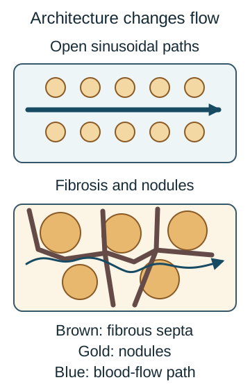
Cirrhosis combines diffuse bridging fibrosis with regenerative nodules. Repeated inflammation or toxic-metabolic injury activates hepatic stellate cells into matrix-producing, contractile cells. Fibrous septa connect vascular regions and surround nodules; narrowed and distorted sinusoids impede portal blood flow. Contractile vascular tone adds to the fixed resistance. Loss of hepatocytes also reduces synthesis, detoxification and bilirubin handling.
Trace the portal inflow through the upper model, then through the narrowed lower paths. Fibrosis changes both resistance and access to functioning hepatocytes. [8]
Predict: a patient has a nodular liver, splenomegaly and platelets of 85,000/mm3, but ALT of 29 U/L. Does the ALT rule out advanced disease?
Check the interpretation
No. The structural and portal findings remain important despite little current enzyme leakage. The alternative inference, that normal ALT means preserved architecture, confuses injury with accumulated fibrosis. Assess architecture and reserve separately from current injury.
Common causes include alcohol-associated liver disease, metabolic dysfunction-associated steatotic liver disease (MASLD), chronic hepatitis B or C, autoimmune hepatitis, and chronic cholestatic disease such as primary biliary cholangitis or primary sclerosing cholangitis. More than one cause can coexist. Consider hemochromatosis, Wilson disease and alpha-1 antitrypsin deficiency when age, phenotype or family history fits; do not attribute unexplained disease to alcohol or body weight alone. [7]
Noninvasive fibrosis assessment and imaging often establish the clinical diagnosis; biopsy is reserved for uncertainty or an unresolved cause rather than required in every patient. Inflammation and venous congestion can increase liver stiffness without equivalent fibrosis. Treat the driver with supported alcohol abstinence, antiviral therapy, metabolic risk management or disease-specific therapy. Improvement does not immediately eliminate established portal or cancer risk. [8][1]
Apply it: after hepatitis C cure, ALT may normalize before portal collaterals disappear. Continue assessment of complications instead of declaring cirrhosis absent.
Why a swollen patient can have arterial underfilling
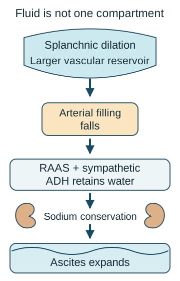
The apparent contradiction is real: total body fluid can increase while effective arterial blood volume falls. Resistance within the liver raises upstream portal pressure. Splanchnic arterial vasodilation, partly mediated by nitric oxide, reduces effective arterial filling and can increase portal inflow. The kidney responds to the arterial signal, not to the visible abdominal fluid. [2][3]
Compare the enlarged fluid reservoir with the smaller effective arterial compartment. [2,3]
Renin-angiotensin-aldosterone and sympathetic activation favor sodium retention; nonosmotic antidiuretic hormone release retains water. Portal hydrostatic pressure and excess lymph formation favor ascites. Retaining proportionally more water can produce dilutional hyponatremia. Severe circulatory dysfunction and renal vasoconstriction contribute to hepatorenal physiology, but not every acute kidney injury in cirrhosis is hepatorenal syndrome. [5]
Portal blood also finds collateral paths into systemic veins. Gastroesophageal varices can bleed, splenic congestion contributes to thrombocytopenia and sometimes leukopenia, and bypassing hepatic detoxification increases exposure to gut-derived substances. Reduced thrombopoietin and other factors also contribute to low platelets, so the count is neither a direct pressure measurement nor a direct synthesis test. [8]
Trace three arrows from splanchnic dilation to the kidney. Predict whether urinary sodium conservation can increase despite worsening edema.
Check the renal response
Yes. Arterial underfilling activates sodium-conserving signals. The tempting alternative, that edema must suppress retention, treats all fluid compartments as equivalent. Visible fluid excess does not establish adequate arterial filling.
For sinusoidal disease, hepatic venous pressure gradient (HVPG) above 5 mmHg indicates portal hypertension; at least 10 mmHg defines clinically significant portal hypertension (CSPH). HVPG estimates the sinusoidal gradient and can underestimate presinusoidal disease. Varices or portosystemic collaterals can establish clinically important portal hypertension without catheter measurement. [1]
Compensated cirrhosis has no history of overt ascites, variceal hemorrhage or overt hepatic encephalopathy. CSPH and unbled varices can already be present. A first overt complication marks decompensation and changes prognosis. Jaundice and kidney dysfunction can accompany progression. Tiny imaging-only fluid collections and covert cognitive deficits are not interchangeable with these overt events. [1]
Apply it: new moderate ascites changes the clinical stage even if bilirubin and creatinine remain near baseline. Do not require kidney failure before recognizing decompensation.
Ask the fluid three separate questions
Does painless ascites mean uncomplicated cirrhosis? No. Perform diagnostic paracentesis for new ascites and promptly in hospitalized patients with cirrhosis and ascites, even without fever or tenderness. Also sample when infection, encephalopathy, bleeding or renal deterioration occurs. Obtain cell count with differential, albumin and total protein; inoculate culture bottles at the bedside when infection is suspected. Routine plasma transfusion to normalize cirrhotic INR before paracentesis is not recommended. [2]
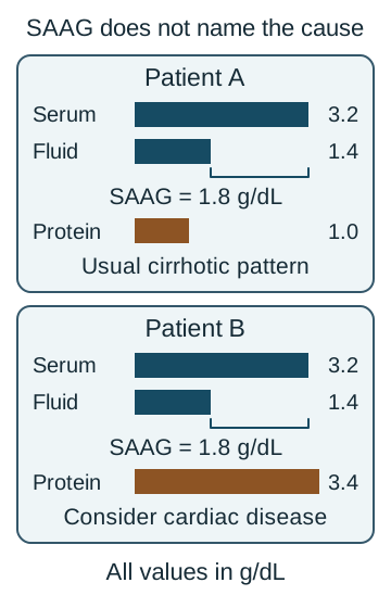
Pressure: SAAG is same-day serum albumin minus ascitic albumin. A gradient at least 1.1 g/dL supports portal hypertension; it does not identify cirrhosis by itself. Protein: high-SAAG fluid with protein below 2.5 g/dL is typical of cirrhosis. High-SAAG fluid with protein at least 2.5 g/dL raises cardiac congestion or hepatic venous outflow disease. Interpret the pattern alongside examination, imaging and time course. [2]
The two examples have the same SAAG. Total protein adds information about the cause; it is not subtracted to calculate SAAG. [2]
A SAAG below 1.1 g/dL suggests a nonportal process, such as peritoneal malignancy, tuberculosis, pancreatic disease or nephrotic physiology. Order cytology, mycobacterial studies or amylase according to the clinical question rather than indiscriminately. Mixed causes and discordant measurements require reassessment.
Calculate: serum albumin 3.2 and ascitic albumin 1.4 g/dL. Now compare ascitic protein 1.0 versus 3.4 g/dL. Which result makes you inspect the jugular venous pressure and heart more closely?
Check the comparison
Both gradients are 1.8 g/dL. Protein of 3.4 g/dL makes cardiac congestion more likely than the usual cirrhotic pattern. Low protein supports cirrhosis but does not prove it. SAAG identifies a pressure pattern; protein helps distinguish its causes.
Infection: an ascitic polymorphonuclear neutrophil (PMN) count at least 250/mm3 requires prompt empiric treatment for presumed spontaneous bacterial peritonitis (SBP), while excluding a surgically treatable source when indicated. Multiply total cells by the neutrophil fraction if only a differential is reported. Negative cultures do not negate neutrocytic ascites. Select antibiotics by illness severity, healthcare exposure and local resistance; a third-generation cephalosporin is appropriate for many community-acquired episodes. Albumin is especially important with renal dysfunction or high-risk disease; a standard SBP schedule is 1.5 g/kg on day 1 and 1 g/kg on day 3, with volume monitoring. Polymicrobial growth, focal peritoneal signs or poor response should prompt evaluation for secondary peritonitis. [2][5]
For uncomplicated ascites, use moderate sodium restriction, commonly about 2 g sodium daily, while protecting nutrition. Spironolactone counters aldosterone; a loop diuretic such as furosemide is often added. Monitor weight, blood pressure, creatinine, sodium and potassium. Routine water restriction is unnecessary unless clinically significant hyponatremia warrants it. Tense ascites benefits from therapeutic paracentesis. When more than 5 L is drained, give albumin based on the entire volume; the BSG/BASL regimen is 8 g/L. Seven liters therefore corresponds to 56 g, not 16 g. Smaller drainage volumes may still warrant albumin in high-risk patients. [2][3]
Apply it: recurrent fluid despite tolerated treatment, or diuretic toxicity preventing adequate treatment, warrants serial paracentesis, selected TIPS assessment and transplant evaluation. A low SAAG instead requires investigation of another cause, not simply stronger diuresis.
Lower portal pressure without ignoring the circulation
Is every portal-pressure treatment appropriate during hemorrhagic shock? No. Prevention and emergency hemostasis use different approaches. In compensated cirrhosis with CSPH, consider a nonselective beta blocker (NSBB), with carvedilol generally preferred if tolerated. Beta-1 blockade reduces cardiac output; beta-2 blockade reduces splanchnic inflow through unopposed vasoconstriction. Carvedilol also reduces intrahepatic resistance through alpha-1 blockade. Hypotension limits its use. A patient already receiving an appropriate NSBB to prevent decompensation does not need screening endoscopy solely to decide whether to start that therapy. If NSBBs are unsuitable, use an endoscopic assessment strategy; ligation prevents first bleeding from high-risk esophageal varices. [1]
During suspected acute variceal hemorrhage, secure airway and circulation as needed, begin vasoactive therapy such as octreotide and antibiotic prophylaxis such as ceftriaxone, and arrange urgent endoscopy after resuscitation, generally within 12 hours. Esophageal varices are treated with band ligation. Gastric varices may require tissue adhesive or endovascular therapy rather than routine esophageal banding. Do not wait for endoscopic confirmation to start portal-directed medication and antibiotics. [1]
Use a restrictive red-cell strategy, commonly targeting hemoglobin about 7 to 8 g/dL, individualized for active shock, ongoing loss and cardiovascular disease. An early hemoglobin can underestimate acute blood loss. Routine fresh frozen plasma to normalize INR can add harmful volume without correcting the complex hemostatic balance. An NSBB is not the immediate hemostatic treatment for an unstable bleed. [1]
A bleeding patient has been resuscitated. Before the scope, choose the missing treatment if octreotide is already running: antibiotic prophylaxis or an oral beta blocker?
Check the urgent choice
Antibiotic prophylaxis belongs to acute care because infection risk increases with gastrointestinal bleeding. The beta blocker is important later but does not provide acute hemostasis in an unstable patient. Distinguish treatment of the active bleed from prevention of the next one.
Trace a portal bypass
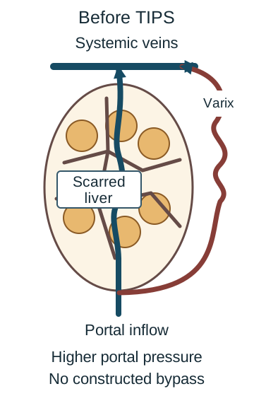
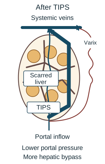
A transjugular intrahepatic portosystemic shunt (TIPS) creates a low-resistance connection from portal to hepatic venous blood. It can decompress varices and improve selected recurrent or refractory ascites. It also increases systemic delivery of blood that bypasses hepatic processing and increases venous return. Heart failure, pulmonary vascular disease, infection and severe recurrent encephalopathy matter when selecting an elective candidate. [1][2]
Starting state for the optional tracing activity. Trace the thick portal path up to the liver, then identify the collateral. [1,2]
Predict two changes after a bypass: what happens to upstream pressure, and what happens to blood bypassing hepatocytes? Compare the two diagrams, then open each explanation to check your prediction. Close the explanations to try again; both diagrams remain visible.
Changed state: the labeled bypass provides another route around hepatic resistance. [1,2] Compare the pressure path after TIPS
Upstream pressure generally falls. The bypass does not dissolve fibrous septa. Pressure relief is not restoration of liver architecture.
Compare systemic exposure after TIPS
More portal blood can reach the systemic circulation without hepatic processing, so encephalopathy risk can increase even as ascites improves. A prediction of better detoxification confuses decompression with hepatocyte function. A pressure benefit can have a detoxification cost.
The complete comparison is the same without an activity: TIPS lowers the portal gradient, may improve bleeding or ascites, increases bypass and venous return, and can worsen encephalopathy or cardiac failure. After a controlled bleed without TIPS, secondary prevention usually combines an NSBB with repeat ligation. Selected high-risk bleeds, including Child-Pugh C scores 10 to 13 or Child-Pugh B above 7 with active bleeding at initial endoscopy, warrant early specialist consideration of preemptive TIPS within 72 hours, ideally sooner. [1]
Apply it: less abdominal fluid but new confusion after TIPS is physiologically coherent. Assess for encephalopathy and other precipitants rather than assuming that successful decompression excludes a complication.
Brain and kidney changes require a fresh differential
Does confusion in cirrhosis automatically mean ammonia toxicity? No. Overt hepatic encephalopathy is a clinical syndrome of impaired attention, disorientation, altered alertness and often asterixis in the appropriate setting. Reduced detoxification, shunting and inflammation contribute, but hypoglycemia, stroke, medication toxicity, alcohol withdrawal, infection and other causes remain possible. Focal findings or an atypical course demand an alternative evaluation. An ammonia concentration alone neither establishes severity nor directs lactulose dosing. [3][8]
For example, constipation and a new opioid can precipitate an episode, while gastrointestinal bleeding supplies a nitrogen load and infection adds systemic inflammation. Treat the precipitant and give lactulose for acute overt encephalopathy, with hospital care and airway protection when severity requires. After recovery, titrate to about 2 to 3 soft stools daily, not persistent diarrhea. Excess catharsis causes dehydration and electrolyte losses that can themselves worsen cognition. Add rifaximin for recurrent episodes despite appropriate lactulose; the adult recurrence-prevention dose is 550 mg twice daily. Consider the label's precautions, including increased systemic exposure in severe hepatic impairment. [3][10]
A patient is disoriented after six watery stools daily for three days, with orthostasis and a rising creatinine. Predict whether simply increasing lactulose is likely to help.
Check the competing explanation
More catharsis can worsen hypovolemia. Correct losses, reassess dosing and search for additional causes. Constipation would support insufficient catharsis, but that is not this patient's finding. Titrate treatment to cognition, hydration and stool quality, not to an ammonia target.
Do not restrict protein to treat encephalopathy. Preserve muscle and nutrition, commonly targeting 1.2 to 1.5 g/kg ideal body weight daily, with regular meals and a late snack. Review sedatives and avoid unnecessary nephrotoxic drugs, particularly NSAIDs. Educate the patient and support person about recurrent confusion and when urgent care is needed. [3]
For the kidney, first recognize the change: creatinine rising at least 0.3 mg/dL in 48 hours or at least 50% within seven days meets AKI criteria; low urine output also matters. A rise from 0.6 to 1.0 mg/dL can be important despite an apparently normal final value. Assess volume, bleeding, infection, medications, urinary sediment and obstruction. Cirrhosis predisposes to hypovolemia, acute tubular injury and HRS-AKI, sometimes together. [5]
Current ADQI-ICA HRS-AKI criteria require cirrhosis with ascites, qualifying AKI, no improvement within 24 hours after adequate resuscitation when indicated, and no strong alternative primary explanation. They do not require an automatic 48-hour albumin trial in every patient. Avoid indiscriminate fluid administration to someone already overloaded. Specialist treatment of established HRS-AKI uses vasoconstrictor therapy, often terlipressin, with carefully adjusted albumin and transplant assessment; respiratory and ischemic risk requires monitoring. [5]
Apply it: if creatinine falls promptly after correcting diuretic-related depletion, do not label that episode HRS-AKI solely because urine sodium was low.
Plan before the next complication
Can a modest risk score justify waiting after the first decompensation? No. MELD 3.0 estimates short-term mortality using bilirubin, INR, creatinine, sodium, albumin and a sex term, with defined interactions and limits. It supports allocation and prognosis but does not directly include ascites, encephalopathy or frailty. Child-Pugh includes bilirubin, albumin, INR, ascites and encephalopathy and supplies a different clinical summary. Neither replaces judgment. [6][8]
First decompensation should prompt hepatology and transplant referral discussion while the acute event is treated. Recurrent ascites, bleeding, encephalopathy, frailty, HCC and kidney dysfunction strengthen the need for assessment. Evaluation addresses candidacy, comorbidities, nutrition, substance-use treatment, support and patient goals; referral is not a promise of listing. The burden of encephalopathy may be incompletely represented by listing criteria. [1][3][9]
For treatment-eligible patients with cirrhosis, HCC surveillance generally uses ultrasound plus alpha-fetoprotein (AFP) every six months. A normal AFP does not exclude HCC. After hepatitis C cure, established cirrhosis still warrants surveillance. Child-Pugh C patients who are not transplant candidates generally do not benefit from routine surveillance; eligibility and competing illness belong beside the recommendation, not in a distant exception. [4]
A new solid lesion at least 1 cm, AFP at least 20 ng/mL or a rising AFP pattern prompts diagnostic multiphasic CT or contrast-enhanced MRI. A subcentimeter lesion usually receives repeat ultrasound and AFP in 3 to 6 months. Persistently inadequate ultrasound visualization may require an alternative surveillance modality. Suspicion does not make every tiny lesion an immediate biopsy target. [4]
Compare two otherwise eligible patients: one has a new 7 mm lesion and stable normal AFP; the other has a new 16 mm lesion and normal AFP. Which can usually have short-interval ultrasound follow-up?
Check the surveillance branch
The 7 mm lesion generally receives ultrasound and AFP in 3 to 6 months. The 16 mm lesion requires diagnostic multiphasic imaging despite normal AFP. Combine lesion size, AFP trend and image quality when selecting follow-up.
Apply it: at discharge after a first episode of ascites, organize fluid management and laboratory monitoring, portal-bleeding prevention, cancer surveillance, nutrition, appropriate vaccination and transplant assessment in parallel. Reversible symptoms do not erase the prognostic event.
A. Compensated disease without clinically important portal hypertension (Why this does not fit)
She has never had ascites, overt encephalopathy or gastrointestinal bleeding. The absence of an overt decompensating event supports compensation. No. These values help assess synthetic reserve, whereas paraesophageal collaterals and splenomegaly indicate a portal-pressure problem. The collateral vessels demonstrate portal hypertension despite preserved laboratory values. Compensation does not remove the need to prevent portal complications.
Reasoning steps for option A
Which parts of her history support calling the cirrhosis compensated?
She has never had ascites, overt encephalopathy or gastrointestinal bleeding. The absence of an overt decompensating event supports compensation.
Do albumin 3.8 g/dL and INR 1.1 exclude elevated portal pressure?
No. These values help assess synthetic reserve, whereas paraesophageal collaterals and splenomegaly indicate a portal-pressure problem.
Why is the claim of no clinically important portal hypertension inconsistent with her imaging?
The collateral vessels demonstrate portal hypertension despite preserved laboratory values. Compensation does not remove the need to prevent portal complications.
B. Compensated disease with clinically important portal hypertension (Best answer)
They show that portal blood has developed collateral pathways, supporting clinically important portal hypertension. She has no history of overt ascites, encephalopathy or hemorrhage. Collaterals can be present before any of these events. She has compensated cirrhosis with clinically important portal hypertension. Preventive care is relevant before her first overt complication.
Reasoning steps for option B
What do paraesophageal collaterals reveal about her portal circulation?
They show that portal blood has developed collateral pathways, supporting clinically important portal hypertension.
Why does portal hypertension not make this woman decompensated?
She has no history of overt ascites, encephalopathy or hemorrhage. Collaterals can be present before any of these events.
How should her preserved clinical stage and abnormal portal circulation be combined?
She has compensated cirrhosis with clinically important portal hypertension. Preventive care is relevant before her first overt complication.
C. Decompensated disease defined by collateral vessels (Why this does not fit)
Yes. Collateral pathways can develop while the patient has never experienced ascites, variceal hemorrhage or overt encephalopathy. An overt decompensating event, rather than collateral vessels alone, would support that classification. The stem explicitly excludes the relevant clinical history. They establish a pressure-related vascular change, not a bleeding event. Her current classification remains compensated despite the need for portal-risk prevention.
Reasoning steps for option C
Can collateral vessels appear before a patient has an overt cirrhotic complication?
Yes. Collateral pathways can develop while the patient has never experienced ascites, variceal hemorrhage or overt encephalopathy.
Which event would be needed to support the proposed decompensated classification here?
An overt decompensating event, rather than collateral vessels alone, would support that classification. The stem explicitly excludes the relevant clinical history.
Why should her unbled paraesophageal collaterals not be treated as a prior hemorrhage?
They establish a pressure-related vascular change, not a bleeding event. Her current classification remains compensated despite the need for portal-risk prevention.
D. Resolved cirrhosis with residual vascular findings (Why this does not fit)
ALT reflects current hepatocyte injury. Little enzyme elevation can coexist with advanced architectural damage. No. A normal injury marker does not demonstrate regression of biopsy-proven cirrhosis, and her collateral vessels remain clinically important. That label would dismiss ongoing portal risk without evidence that cirrhosis has resolved. Her structural history and current collaterals still require assessment.
Reasoning steps for option D
What does ALT 31 U/L measure that differs from accumulated cirrhotic fibrosis?
ALT reflects current hepatocyte injury. Little enzyme elevation can coexist with advanced architectural damage.
Does a normal ALT overturn her earlier biopsy diagnosis?
No. A normal injury marker does not demonstrate regression of biopsy-proven cirrhosis, and her collateral vessels remain clinically important.
What would be missed by labeling the vascular findings merely residual after resolution?
That label would dismiss ongoing portal risk without evidence that cirrhosis has resolved. Her structural history and current collaterals still require assessment.
Takeaway: Compensated cirrhosis can already have clinically significant portal hypertension.
A. Malignant infiltration of the peritoneum (Why this does not fit)
Peritoneal malignancy can produce protein-rich ascites. The protein concentration alone therefore does not identify the cause. The gradient is 3.4 minus 1.7, or 1.7 g/dL. This high SAAG supports a pressure-driven process rather than the usual low-gradient peritoneal pattern. High jugular venous pressure, severe tricuspid regurgitation and right ventricular dilation supply a direct congestive explanation for the high-gradient fluid.
Reasoning steps for option A
Why could ascitic protein of 3.5 g/dL initially suggest a peritoneal malignancy?
Peritoneal malignancy can produce protein-rich ascites. The protein concentration alone therefore does not identify the cause.
How does the albumin gradient weigh against a primarily peritoneal explanation?
The gradient is 3.4 minus 1.7, or 1.7 g/dL. This high SAAG supports a pressure-driven process rather than the usual low-gradient peritoneal pattern.
Which findings make cardiac congestion more coherent than peritoneal carcinomatosis?
High jugular venous pressure, severe tricuspid regurgitation and right ventricular dilation supply a direct congestive explanation for the high-gradient fluid.
B. Leakage of fluid from the pancreatic duct (Why this does not fit)
The high total protein of 3.5 g/dL can occur in pancreatic ascites, so protein alone could make duct leakage seem plausible. No. Pancreatic leakage usually produces a low gradient, whereas this gradient supports a portal-pressure mechanism. The elevated jugular pressure and right-sided cardiac abnormalities explain hepatic congestion. High protein does not outweigh this combined pressure and cardiac evidence.
Reasoning steps for option B
Which fluid feature overlaps with ascites from a pancreatic duct leak?
The high total protein of 3.5 g/dL can occur in pancreatic ascites, so protein alone could make duct leakage seem plausible.
Does the 1.7 g/dL SAAG match the usual pancreatic-ascites pattern?
No. Pancreatic leakage usually produces a low gradient, whereas this gradient supports a portal-pressure mechanism.
Why should the pancreatic hypothesis give way to the documented venous findings?
The elevated jugular pressure and right-sided cardiac abnormalities explain hepatic congestion. High protein does not outweigh this combined pressure and cardiac evidence.
C. Urinary protein loss from nephrotic disease (Why this does not fit)
Urinary protein loss can reduce oncotic pressure and cause edema. Edema by itself does not distinguish that mechanism from venous congestion. A SAAG of 1.7 g/dL supports a hydrostatic hepatic pressure process rather than isolated loss of oncotic pressure. The high jugular venous pressure with severe tricuspid regurgitation and right ventricular dilation points to right-sided congestion as the stronger explanation.
Reasoning steps for option C
How could nephrotic urinary protein loss account for ankle edema?
Urinary protein loss can reduce oncotic pressure and cause edema. Edema by itself does not distinguish that mechanism from venous congestion.
What does the high SAAG add beyond the presence of edema?
A SAAG of 1.7 g/dL supports a hydrostatic hepatic pressure process rather than isolated loss of oncotic pressure.
Which circulatory abnormality is left unexplained by isolated nephrotic protein loss?
The high jugular venous pressure with severe tricuspid regurgitation and right ventricular dilation points to right-sided congestion as the stronger explanation.
D. Hepatic congestion from right-sided cardiac disease (Best answer)
The SAAG is 3.4 - 1.7 = 1.7 g/dL, at or above the 1.1 g/dL threshold for portal-hypertensive (high-gradient) ascites. Ascitic total protein is 3.5 g/dL, at or above the 2.5 g/dL threshold that, with a high SAAG, favors cardiac post-sinusoidal congestion over cirrhosis. Elevated jugular venous pressure, severe tricuspid regurgitation and right ventricular dilation support this cardiac cause.
Reasoning steps for option D
What is the SAAG when serum albumin is 3.4 and ascitic albumin is 1.7 g/dL?
The SAAG is 1.7 g/dL, above the 1.1 g/dL threshold supporting portal hypertension. The gradient alone does not distinguish cardiac from cirrhotic ascites.
How does ascitic protein of 3.5 g/dL refine this high-gradient pattern?
It is above 2.5 g/dL. High-SAAG, high-protein fluid favors cardiac congestion over the usual low-protein pattern of cirrhotic ascites.
What links the fluid profile to right-sided cardiac disease in this man?
Elevated jugular pressure, severe tricuspid regurgitation and a dilated right ventricle support hepatic venous congestion as the cause of the pressure-driven ascites.
Takeaway: High SAAG with high protein and venous congestion supports cardiac ascites.
The SAAG is 0.6 g/dL, below 1.1 g/dL. This is not the expected portal-hypertensive fluid pattern simply because she has cirrhosis. Irregular peritoneal thickening, weight loss and early satiety accompany the low-gradient ascites, making a peritoneal malignancy an important competing cause. Ascitic cytology directly investigates a malignant cause. A negative result does not necessarily end evaluation when the peritoneal imaging and clinical features remain concerning.
Reasoning steps for option A
Why does subtracting ascitic albumin 2.4 from serum albumin 3.0 change the ascites differential?
The SAAG is 0.6 g/dL, below 1.1 g/dL. This is not the expected portal-hypertensive fluid pattern simply because she has cirrhosis.
Which CT and clinical findings make malignant peritoneal disease a targeted concern?
Irregular peritoneal thickening, weight loss and early satiety accompany the low-gradient ascites, making a peritoneal malignancy an important competing cause.
What can cytology contribute, and what would a negative result not settle?
Ascitic cytology directly investigates a malignant cause. A negative result does not necessarily end evaluation when the peritoneal imaging and clinical features remain concerning.
B. Repeat screening endoscopy (Why this does not fit)
It would assess gastroesophageal variceal risk, which concerns portal-hypertension complications rather than the cause of this atypical fluid. No. Those findings direct attention to a peritoneal process, which screening endoscopy does not investigate. Known cirrhosis does not explain away low-gradient ascites with an abnormal peritoneum. The additional investigation should target that competing cause.
Reasoning steps for option B
What cirrhosis-related problem would repeat screening endoscopy assess?
It would assess gastroesophageal variceal risk, which concerns portal-hypertension complications rather than the cause of this atypical fluid.
Would examining varices explain a SAAG of 0.6 g/dL with irregular peritoneal thickening?
No. Those findings direct attention to a peritoneal process, which screening endoscopy does not investigate.
Why should variceal screening not replace the immediate investigation of her weight loss and ascites?
Known cirrhosis does not explain away low-gradient ascites with an abnormal peritoneum. The additional investigation should target that competing cause.
C. Hepatic venous pressure measurement (Why this does not fit)
It could characterize portal hypertension, a plausible background complication of her established cirrhosis. The SAAG of 0.6 g/dL and irregular peritoneal thickening favor investigation of a nonportal peritoneal process; CT also shows no hepatic venous obstruction. A second cause of ascites can coexist with cirrhosis. Measuring portal pressure would not directly investigate the suspected peritoneal malignancy.
Reasoning steps for option C
What question would hepatic venous pressure measurement answer in this patient?
It could characterize portal hypertension, a plausible background complication of her established cirrhosis.
Which fluid and CT results make pressure measurement less directly useful than peritoneal testing?
The SAAG of 0.6 g/dL and irregular peritoneal thickening favor investigation of a nonportal peritoneal process; CT also shows no hepatic venous obstruction.
Why does established cirrhosis not justify ignoring the discordant gradient?
A second cause of ascites can coexist with cirrhosis. Measuring portal pressure would not directly investigate the suspected peritoneal malignancy.
D. A 24-hour urine sodium collection (Why this does not fit)
It can help assess sodium intake or the response to diuretics, which is useful when managing sodium retention. No. It measures renal sodium handling rather than identifying the peritoneal abnormality responsible for this atypical ascites pattern. That approach would assume routine cirrhotic ascites despite weight loss, peritoneal thickening and a SAAG of 0.6 g/dL. The cause needs targeted investigation.
Reasoning steps for option D
What would a 24-hour urine sodium collection usually help assess during ascites care?
It can help assess sodium intake or the response to diuretics, which is useful when managing sodium retention.
Can urinary sodium explain her protein-rich, low-SAAG fluid and peritoneal thickening?
No. It measures renal sodium handling rather than identifying the peritoneal abnormality responsible for this atypical ascites pattern.
What diagnostic error would occur if this new effusion were managed only as sodium retention?
That approach would assume routine cirrhotic ascites despite weight loss, peritoneal thickening and a SAAG of 0.6 g/dL. The cause needs targeted investigation.
Takeaway: Cirrhosis does not eliminate a second cause of ascites.
A. Observe pending a positive ascitic culture (Why this does not fit)
It means no organism has yet been recovered. It does not negate an elevated ascitic neutrophil count. Yes. The absolute PMN count is 800 times 0.40, or 320/mm3, above the 250/mm3 threshold for prompt empiric treatment. Neutrocytic ascites can occur without these symptoms or positive cultures. Waiting would delay treatment of presumed SBP in a patient with worsening kidney function.
Reasoning steps for option A
What does no growth at 24 hours establish about an ascitic culture?
It means no organism has yet been recovered. It does not negate an elevated ascitic neutrophil count.
Does 800 nucleated cells/mm3 with 40% neutrophils already cross the treatment threshold?
Yes. The absolute PMN count is 800 times 0.40, or 320/mm3, above the 250/mm3 threshold for prompt empiric treatment.
Why is waiting for culture positivity unsafe despite no fever or tenderness?
Neutrocytic ascites can occur without these symptoms or positive cultures. Waiting would delay treatment of presumed SBP in a patient with worsening kidney function.
B. Treat with lactulose alone and repeat the count after recovery (Why this does not fit)
New disorientation in cirrhosis supports evaluating and treating encephalopathy. Lactulose can address that syndrome but does not treat an ascitic infection. The calculated count is 320/mm3, supporting presumed SBP. Infection can precipitate encephalopathy and requires treatment alongside the cognitive complication. It postpones treatment of the probable infectious trigger. Lactulose alone leaves neutrocytic ascites and the associated renal risk unaddressed.
Reasoning steps for option B
What part of his presentation makes lactulose relevant?
New disorientation in cirrhosis supports evaluating and treating encephalopathy. Lactulose can address that syndrome but does not treat an ascitic infection.
How could the fluid PMN result connect the confusion to an untreated precipitant?
The calculated count is 320/mm3, supporting presumed SBP. Infection can precipitate encephalopathy and requires treatment alongside the cognitive complication.
Why is repeating the cell count only after cognitive recovery insufficient?
It postpones treatment of the probable infectious trigger. Lactulose alone leaves neutrocytic ascites and the associated renal risk unaddressed.
C. Start empiric antibiotics and albumin with volume monitoring (Best answer)
Multiply 800 nucleated cells/mm3 by the neutrophil fraction of 0.40 to obtain 320 PMNs/mm3. It warrants prompt empiric treatment for presumed SBP. Neither absence of tenderness nor an early negative culture cancels the neutrophil-based treatment threshold. The renal deterioration strengthens the indication for albumin alongside antibiotics. Volume monitoring remains important while treating this high-risk presentation.
Reasoning steps for option C
How is the absolute ascitic PMN count obtained from the reported differential?
Multiply 800 nucleated cells/mm3 by the neutrophil fraction of 0.40 to obtain 320 PMNs/mm3.
What does a count of 320/mm3 require when the examination is quiet and culture remains negative?
It warrants prompt empiric treatment for presumed SBP. Neither absence of tenderness nor an early negative culture cancels the neutrophil-based treatment threshold.
Why does the creatinine rise from 0.9 to 1.5 mg/dL matter to the albumin component?
The renal deterioration strengthens the indication for albumin alongside antibiotics. Volume monitoring remains important while treating this high-risk presentation.
D. Increase diuretics to reduce the infected fluid volume (Why this does not fit)
Diuretics can reduce uncomplicated ascitic fluid accumulation, but removing retained fluid does not eradicate infection. A count of 320/mm3 crosses the threshold for presumed SBP treatment. Increasing diuretics is not a substitute for antimicrobial therapy. Creatinine rising from 0.9 to 1.5 mg/dL signals AKI. Further diuresis may worsen perfusion while leaving the infectious cause untreated.
Reasoning steps for option D
What aspect of his disease might make stronger diuresis seem useful?
Diuretics can reduce uncomplicated ascitic fluid accumulation, but removing retained fluid does not eradicate infection.
What immediate problem is demonstrated by the calculated PMN count?
A count of 320/mm3 crosses the threshold for presumed SBP treatment. Increasing diuretics is not a substitute for antimicrobial therapy.
How does his rising creatinine change the risk of intensifying diuretics?
Creatinine rising from 0.9 to 1.5 mg/dL signals AKI. Further diuresis may worsen perfusion while leaving the infectious cause untreated.
Takeaway: Absolute PMN count, not culture positivity or pain, determines prompt SBP treatment.
A. Delay sampling until plasma lowers the INR below 1.5 (Why this does not fit)
A prolonged INR can create concern about procedural bleeding, but cirrhotic INR does not reliably represent the full hemostatic balance. Platelets are 72,000/mm3, with no active bleeding, anticoagulant use or disseminated coagulation. The supplied findings do not justify routine correction before diagnostic paracentesis. It delays fluid sampling for fever and worsening kidney function. Suspected infected ascites needs prompt investigation rather than routine plasma normalization of cirrhotic INR.
Reasoning steps for option A
Why might INR 1.9 appear to justify plasma before paracentesis?
A prolonged INR can create concern about procedural bleeding, but cirrhotic INR does not reliably represent the full hemostatic balance.
Do her platelet count and history establish a need for routine prophylactic blood products?
Platelets are 72,000/mm3, with no active bleeding, anticoagulant use or disseminated coagulation. The supplied findings do not justify routine correction before diagnostic paracentesis.
What is the cost of waiting for an INR below 1.5 in this admission?
It delays fluid sampling for fever and worsening kidney function. Suspected infected ascites needs prompt investigation rather than routine plasma normalization of cirrhotic INR.
B. Perform prompt ultrasound-guided diagnostic paracentesis (Best answer)
Hospitalization with ascites, fever and deteriorating kidney function raises concern for an ascitic infection that needs prompt fluid assessment. Without another supplied bleeding disorder, active bleeding or anticoagulant exposure, these cirrhotic laboratory results do not require routine prophylactic blood products before paracentesis. An accessible fluid pocket allows selection of a suitable ultrasound-guided site. Sampling can then provide the cell count and culture needed to assess infection.
Reasoning steps for option B
Which new features make diagnostic paracentesis urgent rather than elective?
Hospitalization with ascites, fever and deteriorating kidney function raises concern for an ascitic infection that needs prompt fluid assessment.
Why can the procedure proceed despite INR 1.9 and platelets of 72,000/mm3?
Without another supplied bleeding disorder, active bleeding or anticoagulant exposure, these cirrhotic laboratory results do not require routine prophylactic blood products before paracentesis.
How does the ultrasound finding support the proposed sampling approach?
An accessible fluid pocket allows selection of a suitable ultrasound-guided site. Sampling can then provide the cell count and culture needed to assess infection.
C. Begin higher-dose diuretics and sample if fever persists (Why this does not fit)
They increase fluid removal for sodium and water retention. That is different from diagnosing an infection in the ascitic fluid. They suggest a new complication requiring prompt sampling. More diuresis could worsen renal perfusion without identifying or treating infected ascites. A possible ascitic infection remains uninvestigated. The immediate priority is diagnostic paracentesis, not escalation of fluid-removal medication.
Reasoning steps for option C
What problem do higher diuretic doses treat when ascites is uncomplicated?
They increase fluid removal for sodium and water retention. That is different from diagnosing an infection in the ascitic fluid.
Why are fever and worsening creatinine reasons not to postpone the tap for a diuretic trial?
They suggest a new complication requiring prompt sampling. More diuresis could worsen renal perfusion without identifying or treating infected ascites.
What remains untreated while waiting to see whether her fever persists?
A possible ascitic infection remains uninvestigated. The immediate priority is diagnostic paracentesis, not escalation of fluid-removal medication.
D. Use abdominal CT instead of fluid sampling (Why this does not fit)
It can depict abdominal anatomy and suggest alternative diagnoses, so it may be useful for selected additional questions. CT does not provide an ascitic PMN count or culture. Those fluid measurements are needed to evaluate the suspected infection here. A suitable ultrasound-guided sampling site is available. Her fever and renal deterioration require direct fluid assessment, which CT cannot replace.
Reasoning steps for option D
What could abdominal CT contribute in a patient with ascites and fever?
It can depict abdominal anatomy and suggest alternative diagnoses, so it may be useful for selected additional questions.
Which infection measurements cannot be obtained by replacing a tap with CT?
CT does not provide an ascitic PMN count or culture. Those fluid measurements are needed to evaluate the suspected infection here.
Why does finding an accessible pocket favor sampling rather than imaging alone?
A suitable ultrasound-guided sampling site is available. Her fever and renal deterioration require direct fluid assessment, which CT cannot replace.
Takeaway: Do not delay diagnostic paracentesis for routine correction of cirrhotic INR.
Subtracting the 5 L threshold from 7 L leaves 2 L; multiplying only those 2 L by 8 g/L produces 16 g. No. It determines when routine replacement is indicated under the stated protocol, not which drained liters count toward the dose. Apply 8 g/L to all 7 L, giving 56 g. Counting only the excess 2 L underestimates the replacement.
Reasoning steps for option A
Which volume calculation produces the proposed 16 g albumin order?
Subtracting the 5 L threshold from 7 L leaves 2 L; multiplying only those 2 L by 8 g/L produces 16 g.
Does the greater-than-5-L threshold define a volume to subtract?
No. It determines when routine replacement is indicated under the stated protocol, not which drained liters count toward the dose.
How should the 16 g calculation be corrected for the actual drainage?
Apply 8 g/L to all 7 L, giving 56 g. Counting only the excess 2 L underestimates the replacement.
B. 40 g of albumin (Why this does not fit)
Forty grams covers 5 L. It therefore accounts for only part of the documented 7 L drainage. They also count toward replacement once drainage exceeds 5 L. The threshold does not cap the volume used for dosing. Seven liters multiplied by 8 g/L gives 56 g of albumin. Stopping at 40 g omits replacement for 2 L.
Reasoning steps for option B
How many liters does 40 g cover at the prescribed 8 g/L rate?
Forty grams covers 5 L. It therefore accounts for only part of the documented 7 L drainage.
What happens to the remaining 2 L under the total-volume regimen?
They also count toward replacement once drainage exceeds 5 L. The threshold does not cap the volume used for dosing.
What order includes both the first 5 L and the remaining 2 L?
Seven liters multiplied by 8 g/L gives 56 g of albumin. Stopping at 40 g omits replacement for 2 L.
C. 48 g of albumin (Why this does not fit)
Dividing 48 g by 8 g/L gives 6 L, which is less than the volume actually removed. The procedure drained 7 L. The stated regimen uses that entire recorded volume after the replacement threshold is crossed. One liter remains uncounted, requiring another 8 g at the specified rate. The correct total is 56 g.
Reasoning steps for option C
What drainage volume would correspond to a 48 g order at 8 g/L?
Dividing 48 g by 8 g/L gives 6 L, which is less than the volume actually removed.
Which recorded procedure detail rules out using a 6 L calculation?
The procedure drained 7 L. The stated regimen uses that entire recorded volume after the replacement threshold is crossed.
How much albumin is missing from the proposed 48 g order?
One liter remains uncounted, requiring another 8 g at the specified rate. The correct total is 56 g.
D. 56 g of albumin (Best answer)
Yes. Seven liters exceeds 5 L, and no pulmonary edema or clinical heart failure is supplied to complicate applying this stated regimen. Seven liters times 8 g/L equals 56 g of albumin. The calculation includes every drained liter, not just the amount beyond 5 L. Albumin replacement reduces post-paracentesis circulatory dysfunction. Using all 7 L yields the protocol-specific order of 56 g.
Reasoning steps for option D
Does removing 7 L meet the service threshold for routine albumin replacement?
Yes. Seven liters exceeds 5 L, and no pulmonary edema or clinical heart failure is supplied to complicate applying this stated regimen.
What dose results from applying 8 g/L to the entire drained volume?
Seven liters times 8 g/L equals 56 g of albumin. The calculation includes every drained liter, not just the amount beyond 5 L.
Which post-drainage problem is this total-volume replacement intended to reduce?
Albumin replacement reduces post-paracentesis circulatory dysfunction. Using all 7 L yields the protocol-specific order of 56 g.
Takeaway: At the stated 8 g/L regimen, 7 L of drainage requires 56 g of albumin.
A. Hold diuretics, restore intravascular volume and reassess (Best answer)
The rapid weight loss, orthostasis and nearly resolved edema support excessive diuresis with intravascular depletion rather than uncontrolled fluid retention. Reduced effective circulating volume can cause hypovolemic hyponatremia and impaired renal perfusion. The creatinine rise from 0.9 to 1.7 mg/dL indicates associated kidney injury. Holding them stops further medication-driven losses. Restore volume carefully and reassess kidney function, sodium and other contributors before restarting or adjusting treatment.
Reasoning steps for option A
What do a 5 kg loss in five days and new orthostasis suggest about the diuretic response?
The rapid weight loss, orthostasis and nearly resolved edema support excessive diuresis with intravascular depletion rather than uncontrolled fluid retention.
How can depletion explain both sodium 124 mmol/L and rising creatinine?
Reduced effective circulating volume can cause hypovolemic hyponatremia and impaired renal perfusion. The creatinine rise from 0.9 to 1.7 mg/dL indicates associated kidney injury.
Why should both diuretics be held before reassessing treatment intensity?
Holding them stops further medication-driven losses. Restore volume carefully and reassess kidney function, sodium and other contributors before restarting or adjusting treatment.
B. Continue both drugs and restrict water to 500 mL daily (Why this does not fit)
It may help selected patients with hypervolemic, dilutional hyponatremia. Sodium 124 mmol/L alone does not establish that pattern. She has lost 5 kg quickly, become orthostatic and nearly lost her edema after starting diuretics. These findings support depletion. It could worsen the existing circulatory deficit. The immediate plan should correct depletion rather than impose more losses and restriction.
Reasoning steps for option B
In which volume pattern could water restriction help a low sodium concentration?
It may help selected patients with hypervolemic, dilutional hyponatremia. Sodium 124 mmol/L alone does not establish that pattern.
Which findings conflict with assuming water excess is her main immediate problem?
She has lost 5 kg quickly, become orthostatic and nearly lost her edema after starting diuretics. These findings support depletion.
What could continuing diuretics plus a 500 mL water limit do to her kidney perfusion?
It could worsen the existing circulatory deficit. The immediate plan should correct depletion rather than impose more losses and restriction.
C. Increase spironolactone while stopping furosemide (Why this does not fit)
Stronger aldosterone blockade can continue to increase sodium loss. Stopping the loop diuretic does not make further spironolactone escalation harmless. Orthostasis, rapid weight loss and nearly resolved edema suggest overdiuresis, not persistent uncontrolled retention requiring stronger treatment. Circulation, sodium and kidney function need reassessment and correction of depletion. Increasing spironolactone now could prolong the cause of the renal deterioration.
Reasoning steps for option C
What renal effect would increasing spironolactone have despite stopping furosemide?
Stronger aldosterone blockade can continue to increase sodium loss. Stopping the loop diuretic does not make further spironolactone escalation harmless.
Why is more sodium loss poorly matched to her current examination?
Orthostasis, rapid weight loss and nearly resolved edema suggest overdiuresis, not persistent uncontrolled retention requiring stronger treatment.
What should happen before either diuretic is retitrated in this woman?
Circulation, sodium and kidney function need reassessment and correction of depletion. Increasing spironolactone now could prolong the cause of the renal deterioration.
D. Diagnose HRS-AKI and give vasoconstrictors before assessing volume (Why this does not fit)
This clinical setting can develop hepatorenal circulatory dysfunction, but cirrhosis and an acute creatinine rise are not sufficient to establish HRS-AKI. The onset after diuretics, 5 kg weight loss, orthostasis and resolving edema strongly support diuretic-related volume depletion. The reversible depletion must be assessed and corrected before attributing the injury to HRS-AKI. The response to appropriate volume treatment informs that distinction.
Reasoning steps for option D
Why does cirrhosis with ascites place HRS-AKI in the differential?
This clinical setting can develop hepatorenal circulatory dysfunction, but cirrhosis and an acute creatinine rise are not sufficient to establish HRS-AKI.
Which supplied evidence identifies an alternative perfusion problem that needs attention first?
The onset after diuretics, 5 kg weight loss, orthostasis and resolving edema strongly support diuretic-related volume depletion.
Why is immediate vasoconstrictor treatment without assessing volume premature?
The reversible depletion must be assessed and corrected before attributing the injury to HRS-AKI. The response to appropriate volume treatment informs that distinction.
Takeaway: Hyponatremia with orthostasis after rapid diuresis calls for depletion assessment, not automatic water restriction.
A. Arrange elective TIPS to prevent a first decompensating event (Why this does not fit)
TIPS can decompress portal pressure in selected bleeding or ascites settings. This does not make it routine prevention for every patient with varices. He has never bled or developed ascites and remains compensated. Small unbled varices do not establish a routine prophylactic shunt indication. His blood pressure is adequate and no beta-blocker contraindication is supplied. Tolerated medical portal-pressure treatment offers a less invasive preventive approach.
Reasoning steps for option A
What can a portal shunt accomplish in selected advanced cirrhotic complications?
TIPS can decompress portal pressure in selected bleeding or ascites settings. This does not make it routine prevention for every patient with varices.
Which missing clinical events argue against prophylactic TIPS in this man?
He has never bled or developed ascites and remains compensated. Small unbled varices do not establish a routine prophylactic shunt indication.
What preventive opportunity makes an elective shunt unnecessary as the initial strategy?
His blood pressure is adequate and no beta-blocker contraindication is supplied. Tolerated medical portal-pressure treatment offers a less invasive preventive approach.
B. Observe until varices enlarge before addressing portal pressure (Why this does not fit)
Small varices can appear less urgent than large bleeding-prone varices, especially in a patient who has never had an overt complication. Yes. Their presence provides evidence of clinically significant portal hypertension even though the patient remains compensated. An appropriate NSBB can help prevent a first decompensating event. Prevention is not limited to treating varices only after they become large.
Reasoning steps for option B
Why might the small size of the varices tempt someone to delay portal treatment?
Small varices can appear less urgent than large bleeding-prone varices, especially in a patient who has never had an overt complication.
Do small unbled varices still establish clinically significant portal hypertension?
Yes. Their presence provides evidence of clinically significant portal hypertension even though the patient remains compensated.
Which benefit could be missed by waiting for the varices to enlarge?
An appropriate NSBB can help prevent a first decompensating event. Prevention is not limited to treating varices only after they become large.
C. Offer carvedilol with blood pressure and tolerability monitoring (Best answer)
The esophageal varices establish clinically significant portal hypertension. Abstinence treats the cause but does not erase established portal risk. Blood pressure is 124/76 mmHg with pulse 78/min, and no asthma, heart block or kidney injury is supplied to argue against this preventive option. He can receive portal-pressure prevention before a first decompensating event. Monitoring helps keep that treatment tolerable rather than waiting for bleeding or ascites.
Reasoning steps for option C
Which finding creates a portal-pressure prevention indication despite sustained abstinence?
The esophageal varices establish clinically significant portal hypertension. Abstinence treats the cause but does not erase established portal risk.
What supplied vital signs and history support offering carvedilol?
Blood pressure is 124/76 mmHg with pulse 78/min, and no asthma, heart block or kidney injury is supplied to argue against this preventive option.
Why pair carvedilol with blood pressure and tolerability monitoring now?
He can receive portal-pressure prevention before a first decompensating event. Monitoring helps keep that treatment tolerable rather than waiting for bleeding or ascites.
D. Arrange elective endoscopic ligation of the esophageal varices (Why this does not fit)
It can prevent bleeding from selected high-risk esophageal varices through local treatment of the varices. This compensated patient has small varices and no supplied contraindication to a tolerated NSBB, which addresses prevention of decompensation more broadly. The question concerns a first decompensating event, not only variceal bleeding. Portal-pressure treatment addresses that broader goal in this eligible patient.
Reasoning steps for option D
What complication does elective endoscopic ligation primarily aim to prevent?
It can prevent bleeding from selected high-risk esophageal varices through local treatment of the varices.
How do small varices and available NSBB treatment limit the appeal of ligation here?
This compensated patient has small varices and no supplied contraindication to a tolerated NSBB, which addresses prevention of decompensation more broadly.
Why is local variceal treatment not equivalent to the preventive goal in the question?
The question concerns a first decompensating event, not only variceal bleeding. Portal-pressure treatment addresses that broader goal in this eligible patient.
Takeaway: Consider an NSBB in compensated cirrhosis with CSPH, not only after large varices develop.
A. Pantoprazole with oral carvedilol to lower portal pressure (Why this does not fit)
Acid suppression may be used while evaluating an undifferentiated upper gastrointestinal bleed. It does not supply the full empiric variceal-bleeding treatment. Carvedilol is preventive portal-pressure therapy, not immediate hemorrhage control in shock. Further pressure-lowering therapy is poorly matched to this unstable circulation. This pair omits an acute portal vasoactive drug such as octreotide and antibiotic prophylaxis. Both belong in suspected variceal-bleeding care before endoscopic confirmation.
Reasoning steps for option A
What role could pantoprazole have while the source of hematemesis is still uncertain?
Acid suppression may be used while evaluating an undifferentiated upper gastrointestinal bleed. It does not supply the full empiric variceal-bleeding treatment.
Why does blood pressure 88/54 mmHg make oral carvedilol a poor acute hemostatic choice?
Carvedilol is preventive portal-pressure therapy, not immediate hemorrhage control in shock. Further pressure-lowering therapy is poorly matched to this unstable circulation.
Which two early treatment components are absent from pantoprazole plus carvedilol?
This pair omits an acute portal vasoactive drug such as octreotide and antibiotic prophylaxis. Both belong in suspected variceal-bleeding care before endoscopic confirmation.
B. Fresh frozen plasma plus intravenous vitamin K (Why this does not fit)
It aims at correction of coagulation abnormalities rather than direct treatment of the suspected portal-hypertensive bleeding source. Cirrhotic hemostatic balance is not captured by INR alone. Plasma can add harmful volume without controlling portal hemorrhage. Resuscitation is already underway; vasoactive treatment and antibiotic prophylaxis should accompany urgent endoscopic care. Plasma plus vitamin K does not replace that bundle.
Reasoning steps for option B
What presumed bleeding mechanism is the plasma and vitamin K combination trying to address?
It aims at correction of coagulation abnormalities rather than direct treatment of the suspected portal-hypertensive bleeding source.
Why is routine plasma normalization of cirrhotic INR not the appropriate empiric strategy?
Cirrhotic hemostatic balance is not captured by INR alone. Plasma can add harmful volume without controlling portal hemorrhage.
What should take priority over routine laboratory correction during this hematemesis episode?
Resuscitation is already underway; vasoactive treatment and antibiotic prophylaxis should accompany urgent endoscopic care. Plasma plus vitamin K does not replace that bundle.
C. Oral rifaximin with lactulose to treat encephalopathy (Why this does not fit)
These are gut-directed therapies for encephalopathy. That complication may become relevant after a gastrointestinal bleed, but it is not the immediate hemorrhage treatment. No. They do not provide the acute portal vasoactive effect or endoscopic hemostasis needed for the bleeding source. Gut-directed encephalopathy treatment does not replace systemic antibiotic prophylaxis in acute variceal hemorrhage. The proposed pair leaves the early bleeding bundle incomplete.
Reasoning steps for option C
Which complication of cirrhosis is treated with rifaximin and lactulose?
These are gut-directed therapies for encephalopathy. That complication may become relevant after a gastrointestinal bleed, but it is not the immediate hemorrhage treatment.
Can these oral drugs control the suspected variceal source in a patient with shock?
No. They do not provide the acute portal vasoactive effect or endoscopic hemostasis needed for the bleeding source.
Why does rifaximin in this pair not fulfill the antibiotic component of acute bleeding care?
Gut-directed encephalopathy treatment does not replace systemic antibiotic prophylaxis in acute variceal hemorrhage. The proposed pair leaves the early bleeding bundle incomplete.
D. Intravenous octreotide plus antibiotic prophylaxis (Best answer)
Cirrhosis with major hematemesis and hypotension raises strong suspicion of variceal hemorrhage, even though the source has not yet been confirmed. Octreotide provides acute vasoactive therapy directed at portal bleeding, while antibiotics address the infection risk associated with the hemorrhage. No. Start them from presentation while resuscitation and urgent endoscopy proceed. Endoscopic confirmation is not a prerequisite for these empiric measures.
Reasoning steps for option D
Why is a variceal source a strong concern before endoscopy in this presentation?
Cirrhosis with major hematemesis and hypotension raises strong suspicion of variceal hemorrhage, even though the source has not yet been confirmed.
Which distinct problems do octreotide and antibiotic prophylaxis address?
Octreotide provides acute vasoactive therapy directed at portal bleeding, while antibiotics address the infection risk associated with the hemorrhage.
Should these drugs wait until resuscitation and endoscopy have established the source?
No. Start them from presentation while resuscitation and urgent endoscopy proceed. Endoscopic confirmation is not a prerequisite for these empiric measures.
Takeaway: Start vasoactive therapy and antibiotic prophylaxis early in suspected variceal bleeding.
A. Transfuse red cells to a hemoglobin of 10 g/dL (Why this does not fit)
Selected cardiovascular disease, ongoing shock or continued blood loss may justify an individualized higher target. Those circumstances are not supplied here. They accompany controlled bleeding and restored perfusion. A stable hemoglobin of 7.8 g/dL is within the usual restrictive target in this nonischemic patient. Unnecessary transfusion adds volume and can worsen portal pressure. Monitoring is preferable to raising hemoglobin beyond the indicated target here.
Reasoning steps for option A
Which circumstances could justify a higher red-cell target than usual restrictive care?
Selected cardiovascular disease, ongoing shock or continued blood loss may justify an individualized higher target. Those circumstances are not supplied here.
How do the stable 7.8 g/dL readings and normal lactate affect a proposed 10 g/dL target?
They accompany controlled bleeding and restored perfusion. A stable hemoglobin of 7.8 g/dL is within the usual restrictive target in this nonischemic patient.
What harm can additional transfusion to 10 g/dL introduce after successful ligation?
Unnecessary transfusion adds volume and can worsen portal pressure. Monitoring is preferable to raising hemoglobin beyond the indicated target here.
B. Withhold red cells and monitor for recurrent bleeding (Best answer)
There is no further hematemesis, blood pressure is 116/70 mmHg, lactate has normalized and hemoglobin is stable on two measurements. No. Controlled bleeding and adequate perfusion support a restrictive strategy; the hemoglobin value alone does not indicate another transfusion here. Continue monitoring for recurrent hemorrhage and changes in perfusion or hemoglobin. Withholding an unnecessary unit does not mean ending observation.
Reasoning steps for option B
Which findings show that the red-cell decision is being made after hemostasis rather than during shock?
There is no further hematemesis, blood pressure is 116/70 mmHg, lactate has normalized and hemoglobin is stable on two measurements.
Does a stable hemoglobin of 7.8 g/dL require another unit without coronary disease or ischemic symptoms?
No. Controlled bleeding and adequate perfusion support a restrictive strategy; the hemoglobin value alone does not indicate another transfusion here.
What should accompany withholding further red cells after the controlled variceal bleed?
Continue monitoring for recurrent hemorrhage and changes in perfusion or hemoglobin. Withholding an unnecessary unit does not mean ending observation.
C. Transfuse red cells to a hemoglobin of 12 g/dL (Why this does not fit)
It aims toward a normal hemoglobin value, but normalization is not the usual acute transfusion goal after variceal hemostasis. Bleeding is controlled, lactate and blood pressure indicate restored perfusion, and there are no ischemic symptoms or coronary disease. It exposes him to unnecessary red cells and volume without a supplied indication. Restrictive use avoids treating a normal laboratory value as the required endpoint.
Reasoning steps for option C
Why might a target of 12 g/dL appear reassuring after a major bleed?
It aims toward a normal hemoglobin value, but normalization is not the usual acute transfusion goal after variceal hemostasis.
What evidence makes normalization unnecessary in this particular patient?
Bleeding is controlled, lactate and blood pressure indicate restored perfusion, and there are no ischemic symptoms or coronary disease.
What is the consequence of transfusing from a stable 7.8 g/dL toward 12 g/dL here?
It exposes him to unnecessary red cells and volume without a supplied indication. Restrictive use avoids treating a normal laboratory value as the required endpoint.
D. Give one red-cell unit now and recheck hemoglobin (Why this does not fit)
A single-unit approach can limit exposure when a transfusion is actually indicated. It does not create an indication by itself. Serial hemoglobin is stable at 7.8 g/dL after hemostasis, perfusion is restored and no ischemic condition is supplied. Monitor bleeding, circulation and hemoglobin without first administering an unnecessary unit. Reassessment does not require transfusion in this stable setting.
Reasoning steps for option D
When is giving one unit and reassessing a useful red-cell strategy?
A single-unit approach can limit exposure when a transfusion is actually indicated. It does not create an indication by itself.
Which findings argue that even the first additional unit is unnecessary now?
Serial hemoglobin is stable at 7.8 g/dL after hemostasis, perfusion is restored and no ischemic condition is supplied.
How can the team retain reassessment without the proposed immediate transfusion?
Monitor bleeding, circulation and hemoglobin without first administering an unnecessary unit. Reassessment does not require transfusion in this stable setting.
Takeaway: After hemostasis, stable perfusion and absence of ischemia support restrictive red-cell use.
A. Repeat endoscopic ligation without beta blockade (Why this does not fit)
Ligation alone can be used when a nonselective beta blocker cannot be tolerated or is contraindicated. No. Blood pressure is 118/72 mmHg, creatinine is at baseline and no beta-blocker contraindication is supplied. It omits complementary medical portal-pressure reduction. After a bleed without TIPS, tolerated secondary prevention usually combines an NSBB with serial ligation.
Reasoning steps for option A
When might repeat ligation alone be a reasonable secondary-prevention approach?
Ligation alone can be used when a nonselective beta blocker cannot be tolerated or is contraindicated.
Does her follow-up assessment supply a reason to omit beta blockade?
No. Blood pressure is 118/72 mmHg, creatinine is at baseline and no beta-blocker contraindication is supplied.
What preventive mechanism is missing from repeat ligation alone after her confirmed hemorrhage?
It omits complementary medical portal-pressure reduction. After a bleed without TIPS, tolerated secondary prevention usually combines an NSBB with serial ligation.
B. Use carvedilol alone without further endoscopy (Why this does not fit)
As an NSBB, carvedilol lowers portal pressure and contributes to prevention of recurrent hemorrhage. She has survived a confirmed bleed and has not received TIPS. Serial ligation is ordinarily added when feasible for secondary prevention. Primary-prevention simplifications do not replace the combined strategy after a documented hemorrhage. Her plan should retain both pressure reduction and local variceal treatment.
Reasoning steps for option B
What useful effect would carvedilol provide after her esophageal variceal bleed?
As an NSBB, carvedilol lowers portal pressure and contributes to prevention of recurrent hemorrhage.
Why does that pressure effect not justify ending endoscopic treatment in this case?
She has survived a confirmed bleed and has not received TIPS. Serial ligation is ordinarily added when feasible for secondary prevention.
Which prevention distinction makes carvedilol alone an incomplete plan here?
Primary-prevention simplifications do not replace the combined strategy after a documented hemorrhage. Her plan should retain both pressure reduction and local variceal treatment.
C. Combine an NSBB with serial endoscopic ligation (Best answer)
She has survived an esophageal variceal hemorrhage treated with ligation. The goal is now to prevent recurrence, not a first bleed. She has adequate blood pressure, baseline kidney function and no supplied beta-blocker contraindication. TIPS has not replaced this preventive approach. The NSBB reduces portal pressure while serial ligation treats the varices locally. Their combination is standard secondary prophylaxis in this setting.
Reasoning steps for option C
Which historical event makes this secondary rather than primary prophylaxis?
She has survived an esophageal variceal hemorrhage treated with ligation. The goal is now to prevent recurrence, not a first bleed.
What makes an NSBB plus further ligation feasible at her follow-up?
She has adequate blood pressure, baseline kidney function and no supplied beta-blocker contraindication. TIPS has not replaced this preventive approach.
How do the two components complement one another after her bleed?
The NSBB reduces portal pressure while serial ligation treats the varices locally. Their combination is standard secondary prophylaxis in this setting.
D. Schedule surveillance endoscopy without prophylaxis (Why this does not fit)
It establishes a substantial risk of recurrent hemorrhage requiring active prevention, rather than observation alone. No. Her blood pressure and kidney function are suitable, and no beta-blocker contraindication is supplied. Surveillance can identify varices but does not itself provide pressure reduction or therapeutic ligation. A documented bleed calls for active secondary prevention.
Reasoning steps for option D
What does a survived variceal hemorrhage establish that surveillance alone does not address?
It establishes a substantial risk of recurrent hemorrhage requiring active prevention, rather than observation alone.
Is an inability to provide prophylaxis documented at this follow-up?
No. Her blood pressure and kidney function are suitable, and no beta-blocker contraindication is supplied.
Why is scheduling endoscopy without prophylactic treatment insufficient after her first bleed?
Surveillance can identify varices but does not itself provide pressure reduction or therapeutic ligation. A documented bleed calls for active secondary prevention.
Takeaway: After a variceal bleed without TIPS, secondary prevention generally combines an NSBB and ligation.
A. Preemptive covered TIPS within 72 hours (Best answer)
It is Child-Pugh C and lies within the 10 to 13 range considered for preemptive TIPS after acute variceal hemorrhage. No. Initial hemostasis does not eliminate the early risk associated with this high-risk liver-disease category. Evaluate preemptive covered TIPS within 72 hours rather than waiting for recurrent bleeding. The supplied score and eligibility support assessment during that early window.
Reasoning steps for option A
Where does a Child-Pugh score of 11 fall in the high-risk bleeding criteria?
It is Child-Pugh C and lies within the 10 to 13 range considered for preemptive TIPS after acute variceal hemorrhage.
Does successful initial band ligation remove the risk addressed by preemptive TIPS?
No. Initial hemostasis does not eliminate the early risk associated with this high-risk liver-disease category.
What TIPS timing should be evaluated when major contraindications are absent?
Evaluate preemptive covered TIPS within 72 hours rather than waiting for recurrent bleeding. The supplied score and eligibility support assessment during that early window.
B. Rescue TIPS only if bleeding recurs during this admission (Why this does not fit)
It addresses failure of bleeding control or recurrent bleeding in an appropriate patient, rather than being planned before that failure occurs. A score of 11 identifies the high-risk group for preemptive assessment even though vasoactive treatment and ligation have achieved initial hemostasis. That restriction postpones an indicated early assessment until another bleeding event. Preemptive TIPS is considered to address risk before rescue becomes necessary.
Reasoning steps for option B
What clinical situation does rescue TIPS ordinarily address?
It addresses failure of bleeding control or recurrent bleeding in an appropriate patient, rather than being planned before that failure occurs.
Why does the documented Child-Pugh score argue against waiting for rescue criteria?
A score of 11 identifies the high-risk group for preemptive assessment even though vasoactive treatment and ligation have achieved initial hemostasis.
What opportunity is lost by limiting TIPS to recurrence during this admission?
That restriction postpones an indicated early assessment until another bleeding event. Preemptive TIPS is considered to address risk before rescue becomes necessary.
C. Elective TIPS after completion of a six-week ligation program (Why this does not fit)
It moves the decision well beyond the early preemptive window used for selected high-risk acute variceal bleeding. Child-Pugh 11 places him in the high-risk Child-Pugh C group considered for early TIPS, with no major contraindication supplied. Control of the current bleed does not remove his early risk. A six-week delay would miss the within-72-hour preemptive assessment opportunity.
Reasoning steps for option C
What does waiting for a six-week ligation program do to the proposed TIPS timing?
It moves the decision well beyond the early preemptive window used for selected high-risk acute variceal bleeding.
Which supplied risk marker makes that delay inappropriate for this presentation?
Child-Pugh 11 places him in the high-risk Child-Pugh C group considered for early TIPS, with no major contraindication supplied.
Why should initial hemostasis not redirect this patient to an automatically delayed elective decision?
Control of the current bleed does not remove his early risk. A six-week delay would miss the within-72-hour preemptive assessment opportunity.
D. No TIPS consideration unless refractory ascites develops (Why this does not fit)
Yes. Selected recurrent or refractory ascites can prompt TIPS assessment, but it is not the only reason to consider a shunt. His acute variceal bleed with Child-Pugh C score 11 identifies a high-risk bleeding presentation that merits preemptive TIPS assessment. High-risk acute variceal bleeding independently supports early evaluation. Waiting for refractory ascites would ignore the current bleeding-related indication.
Reasoning steps for option D
Is refractory ascites a possible TIPS indication independent of this bleeding admission?
Yes. Selected recurrent or refractory ascites can prompt TIPS assessment, but it is not the only reason to consider a shunt.
Which indication is already present before any refractory ascites develops?
His acute variceal bleed with Child-Pugh C score 11 identifies a high-risk bleeding presentation that merits preemptive TIPS assessment.
Why is making ascites a prerequisite inconsistent with the supplied risk category?
High-risk acute variceal bleeding independently supports early evaluation. Waiting for refractory ascites would ignore the current bleeding-related indication.
Takeaway: Eligible patients with Child-Pugh C scores 10 to 13 merit early preemptive TIPS assessment after variceal bleeding.
A. Higher portal pressure with greater hepatic toxin clearance (Why this does not fit)
No. The improvement in ascites fits portal decompression through a patent shunt, not an increase in upstream pressure. Greater clearance would favor improved cognition rather than the new encephalopathy-like syndrome. The proposed clearance change points in the wrong direction. TIPS generally lowers portal pressure while allowing more portal solutes to bypass hepatic processing. It does not pair higher pressure with better detoxification.
Reasoning steps for option A
Would higher portal pressure account for needing less ascites drainage after TIPS?
No. The improvement in ascites fits portal decompression through a patent shunt, not an increase in upstream pressure.
Would greater hepatic toxin clearance predict her new disorientation and asterixis?
Greater clearance would favor improved cognition rather than the new encephalopathy-like syndrome. The proposed clearance change points in the wrong direction.
Which two effects of a patent portal bypass contradict this proposed pair?
TIPS generally lowers portal pressure while allowing more portal solutes to bypass hepatic processing. It does not pair higher pressure with better detoxification.
B. Lower portal pressure with greater systemic portal-solute delivery (Best answer)
It supports lower portal pressure and improvement of a pressure-related complication after TIPS. The low-resistance portal-to-systemic route allows more blood to reach the systemic circulation without hepatic processing. Decompression improves fluid control, while increased bypass can worsen encephalopathy. The pressure benefit does not imply improved detoxification.
Reasoning steps for option B
What does reduced drainage need suggest about pressure upstream of her patent shunt?
It supports lower portal pressure and improvement of a pressure-related complication after TIPS.
How can the same shunt increase systemic exposure to gut-derived portal solutes?
The low-resistance portal-to-systemic route allows more blood to reach the systemic circulation without hepatic processing.
Why can less ascites and new asterixis coexist three weeks after TIPS?
Decompression improves fluid control, while increased bypass can worsen encephalopathy. The pressure benefit does not imply improved detoxification.
C. Lower portal pressure with an increase in hepatocyte mass (Why this does not fit)
Lower portal pressure fits the reduced need for ascites drainage after a patent TIPS. No. TIPS creates a vascular bypass; it does not generate hepatocytes or restore hepatic processing capacity. Improved hepatic processing is not the mechanism of TIPS. Increased bypass of existing hepatocytes, rather than regeneration, can explain the encephalopathy despite less ascites.
Reasoning steps for option C
Which half of the lower-pressure and increased-hepatocyte-mass option fits the drainage response?
Lower portal pressure fits the reduced need for ascites drainage after a patent TIPS.
Does constructing a TIPS increase hepatocyte mass?
No. TIPS creates a vascular bypass; it does not generate hepatocytes or restore hepatic processing capacity.
Why does the tissue-regeneration claim fail to explain her new confusion?
Improved hepatic processing is not the mechanism of TIPS. Increased bypass of existing hepatocytes, rather than regeneration, can explain the encephalopathy despite less ascites.
D. Higher portal pressure with reduced splanchnic blood inflow (Why this does not fit)
Less ascites after a patent shunt is consistent with portal decompression. Higher upstream pressure would not explain that benefit. No. The key change is the bypass route around hepatic processing, not merely a reduction in blood arriving from the splanchnic circulation. It must explain disorientation and asterixis through increased hepatic bypass. The proposed higher-pressure and lower-inflow pair fails to connect both outcomes.
Reasoning steps for option D
How does less ascites challenge the claim that portal pressure has risen?
Less ascites after a patent shunt is consistent with portal decompression. Higher upstream pressure would not explain that benefit.
Would reduced splanchnic inflow alone explain the new systemic solute exposure?
No. The key change is the bypass route around hepatic processing, not merely a reduction in blood arriving from the splanchnic circulation.
What must a paired explanation account for besides the fall in drainage need?
It must explain disorientation and asterixis through increased hepatic bypass. The proposed higher-pressure and lower-inflow pair fails to connect both outcomes.
Takeaway: TIPS can improve pressure-related complications while increasing encephalopathy risk.
A. Start rifaximin alone and continue the oxycodone regimen (Why this does not fit)
Rifaximin can reduce recurrence as an adjunct to lactulose. That role does not make rifaximin alone a complete initial plan for this episode. Four days of constipation and recent opioid exposure remain important contributors. The proposed plan neither corrects the constipation with initial bowel therapy nor reassesses the opioid. Initial overt encephalopathy care includes lactulose and correction of precipitants. Rifaximin alone while maintaining oxycodone leaves the case-specific contributors in place.
Reasoning steps for option A
What is the established recurrence-prevention role of rifaximin in encephalopathy?
Rifaximin can reduce recurrence as an adjunct to lactulose. That role does not make rifaximin alone a complete initial plan for this episode.
Which two supplied precipitants remain unaddressed by keeping oxycodone and using rifaximin alone?
Four days of constipation and recent opioid exposure remain important contributors. The proposed plan neither corrects the constipation with initial bowel therapy nor reassesses the opioid.
Why is this incomplete despite rifaximin being a gut-directed treatment?
Initial overt encephalopathy care includes lactulose and correction of precipitants. Rifaximin alone while maintaining oxycodone leaves the case-specific contributors in place.
B. Stop the opioid and defer bowel therapy until ammonia returns (Why this does not fit)
Recent oxycodone exposure is a plausible contributor to the disorientation and constipation. Medication review addresses a treatable precipitant. No. The clinical syndrome supports treatment while competing causes are evaluated; ammonia is not required to begin disease-directed therapy. Four days of constipation remains a potential precipitant. Stopping the opioid alone does not replace prompt lactulose and bowel management.
Reasoning steps for option B
Why is stopping or reassessing the new opioid a relevant part of care?
Recent oxycodone exposure is a plausible contributor to the disorientation and constipation. Medication review addresses a treatable precipitant.
Must an ammonia result return before treating disorientation with asterixis in this setting?
No. The clinical syndrome supports treatment while competing causes are evaluated; ammonia is not required to begin disease-directed therapy.
What is left untreated by delaying bowel therapy until that laboratory result arrives?
Four days of constipation remains a potential precipitant. Stopping the opioid alone does not replace prompt lactulose and bowel management.
C. Begin lactulose but continue the opioid without reassessment (Why this does not fit)
Lactulose is initial disease-directed therapy for overt encephalopathy and also addresses the constipation reported in the stem. The recently prescribed centrally depressant opioid remains a potential contributor to cognitive impairment and constipation. Treating the syndrome does not remove the need to correct triggers. The temporal opioid exposure still requires reassessment alongside bowel therapy.
Reasoning steps for option C
Which supplied problems can lactulose address during this episode?
Lactulose is initial disease-directed therapy for overt encephalopathy and also addresses the constipation reported in the stem.
What plausible precipitant persists if the oxycodone regimen is continued without reassessment?
The recently prescribed centrally depressant opioid remains a potential contributor to cognitive impairment and constipation.
Why is starting lactulose not enough to justify leaving the medication plan untouched?
Treating the syndrome does not remove the need to correct triggers. The temporal opioid exposure still requires reassessment alongside bowel therapy.
D. Start lactulose for constipation and reassess oxycodone (Best answer)
New disorientation and asterixis in cirrhosis support the syndrome. Glucose, oxygenation and examination do not identify a competing focal emergency, while infection and bleeding are still being assessed. They identify treatable contributors. Lactulose provides initial disease-directed bowel therapy, and the opioid should be reassessed rather than continued automatically. He is awake and can swallow safely. Start lactulose while correcting constipation, reviewing the opioid and continuing evaluation for other precipitants.
Reasoning steps for option D
Which findings support treating overt encephalopathy while the differential evaluation continues?
New disorientation and asterixis in cirrhosis support the syndrome. Glucose, oxygenation and examination do not identify a competing focal emergency, while infection and bleeding are still being assessed.
How do constipation and recent oxycodone exposure shape the initial treatment plan?
They identify treatable contributors. Lactulose provides initial disease-directed bowel therapy, and the opioid should be reassessed rather than continued automatically.
What makes initiating oral bowel therapy feasible in this patient?
He is awake and can swallow safely. Start lactulose while correcting constipation, reviewing the opioid and continuing evaluation for other precipitants.
Takeaway: Treat overt encephalopathy with precipitant correction and lactulose, not an isolated ammonia target.
A. Further increase lactulose until the ammonia result normalizes (Why this does not fit)
They reacted to an ammonia concentration above the laboratory range rather than a stated clinical need for more catharsis. Eight watery stools daily, orthostasis and sodium 149 mmol/L point to dehydration and water loss after the dose increase. It could intensify fluid and electrolyte losses and further impair circulation. Dosing should address clinical response and tolerable stool output, not ammonia normalization.
Reasoning steps for option A
What prompted the family to intensify lactulose before the new deterioration?
They reacted to an ammonia concentration above the laboratory range rather than a stated clinical need for more catharsis.
Which findings argue against further escalation as the response to recurrent inattention?
Eight watery stools daily, orthostasis and sodium 149 mmol/L point to dehydration and water loss after the dose increase.
How could another lactulose increase worsen the supplied problem?
It could intensify fluid and electrolyte losses and further impair circulation. Dosing should address clinical response and tolerable stool output, not ammonia normalization.
B. Continue the current dose and restrict free water (Why this does not fit)
Selected dilutional hyponatremia may call for water restriction. That is different from this patient with sodium 149 mmol/L. They support water and circulating-volume loss from excessive catharsis, rather than water excess. The plan continues the gastrointestinal losses while limiting replacement. It would worsen the dehydration associated with her renewed inattention.
Reasoning steps for option B
What sodium disorder sometimes supports restricting free water in cirrhosis?
Selected dilutional hyponatremia may call for water restriction. That is different from this patient with sodium 149 mmol/L.
What do hypernatremia and orthostasis suggest after eight watery stools each day?
They support water and circulating-volume loss from excessive catharsis, rather than water excess.
Why would maintaining the high lactulose dose and restricting water compound the problem?
The plan continues the gastrointestinal losses while limiting replacement. It would worsen the dehydration associated with her renewed inattention.
C. Replace fluid losses, adjust lactulose and reassess other causes (Best answer)
The family doubled lactulose before she developed eight watery stools daily, orthostasis and hypernatremia. This sequence supports excessive catharsis as a precipitant. Replace the fluid losses and adjust lactulose toward soft, tolerable stools rather than ongoing watery diarrhea. This treats the circulatory problem instead of chasing ammonia. Dehydration is the supplied precipitating problem, but cognitive deterioration can have additional contributors. Correct the losses while continuing the clinical evaluation.
Reasoning steps for option C
What timing links her recurrent inattention to an adverse treatment effect?
The family doubled lactulose before she developed eight watery stools daily, orthostasis and hypernatremia. This sequence supports excessive catharsis as a precipitant.
Which two parts of the treatment plan directly address those losses?
Replace the fluid losses and adjust lactulose toward soft, tolerable stools rather than ongoing watery diarrhea. This treats the circulatory problem instead of chasing ammonia.
Why should other causes still be reassessed after recognizing over-catharsis?
Dehydration is the supplied precipitating problem, but cognitive deterioration can have additional contributors. Correct the losses while continuing the clinical evaluation.
D. Add rifaximin while keeping the current lactulose dose unchanged (Why this does not fit)
It may be appropriate for recurrence prevention, but that role does not correct an immediate adverse effect of excessive lactulose. No. The eight watery stools daily would remain a source of dehydration and electrolyte disturbance. Orthostasis and sodium 149 mmol/L show an immediate fluid-loss problem. Adding another drug while leaving the excessive dose unchanged misses that precipitant.
Reasoning steps for option D
What benefit could rifaximin provide in a patient with recurrent encephalopathy?
It may be appropriate for recurrence prevention, but that role does not correct an immediate adverse effect of excessive lactulose.
Would adding rifaximin stop the water loss produced by the unchanged lactulose dose?
No. The eight watery stools daily would remain a source of dehydration and electrolyte disturbance.
Why is correcting the lactulose-related problem necessary even if adjunctive therapy is later indicated?
Orthostasis and sodium 149 mmol/L show an immediate fluid-loss problem. Adding another drug while leaving the excessive dose unchanged misses that precipitant.
Takeaway: Excess lactulose can precipitate cognitive deterioration through dehydration and electrolyte disturbance.
A. Add rifaximin 550 mg twice daily to the lactulose regimen (Best answer)
He has had three hospitalizations in six months despite reliable lactulose use, an appropriate stool target and attention to common precipitants. The labeled regimen is 550 mg twice daily. In this patient it is added to the tolerated lactulose regimen rather than replacing it. Severe hepatic impairment carries increased systemic exposure and requires attention to label precautions. Continue lactulose adherence and precipitant review as part of combined prevention.
Reasoning steps for option A
What distinguishes this recurrence history from an isolated adequately treated episode?
He has had three hospitalizations in six months despite reliable lactulose use, an appropriate stool target and attention to common precipitants.
Which rifaximin regimen is used for adult overt encephalopathy recurrence prevention?
The labeled regimen is 550 mg twice daily. In this patient it is added to the tolerated lactulose regimen rather than replacing it.
What precautions and ongoing care remain relevant after adding rifaximin?
Severe hepatic impairment carries increased systemic exposure and requires attention to label precautions. Continue lactulose adherence and precipitant review as part of combined prevention.
B. Continue lactulose alone with more frequent ammonia measurements (Why this does not fit)
He reports reliable dosing and two to three soft stools daily. The history does not suggest inadequate bowel treatment or unresolved constipation. No. Laboratory surveillance does not prevent clinical recurrence. Repeated overt episodes despite adequate lactulose support an adjunctive treatment. The recurrent admissions show a need beyond appropriate lactulose alone. Adding rifaximin addresses recurrence, whereas additional ammonia measurements do not.
Reasoning steps for option B
What evidence shows that lactulose is already being used appropriately?
He reports reliable dosing and two to three soft stools daily. The history does not suggest inadequate bowel treatment or unresolved constipation.
Would more frequent ammonia measurements address three recurrent hospitalizations?
No. Laboratory surveillance does not prevent clinical recurrence. Repeated overt episodes despite adequate lactulose support an adjunctive treatment.
Why is continuing unchanged monotherapy an insufficient response to this clinical course?
The recurrent admissions show a need beyond appropriate lactulose alone. Adding rifaximin addresses recurrence, whereas additional ammonia measurements do not.
C. Additional lactulose targeting six watery stools daily (Why this does not fit)
No. It is the appropriate target, so the stem does not demonstrate insufficient catharsis. Watery diarrhea increases dehydration and electrolyte-loss risk without established added protection against encephalopathy. Add recurrence-prevention therapy such as rifaximin to the tolerated regimen. More catharsis is not the solution to recurrent episodes despite appropriate lactulose.
Reasoning steps for option C
Is his current output of two to three soft stools below the intended lactulose target?
No. It is the appropriate target, so the stem does not demonstrate insufficient catharsis.
What problem could targeting six watery stools introduce?
Watery diarrhea increases dehydration and electrolyte-loss risk without established added protection against encephalopathy.
How should prevention be intensified without driving his stool output beyond the target?
Add recurrence-prevention therapy such as rifaximin to the tolerated regimen. More catharsis is not the solution to recurrent episodes despite appropriate lactulose.
D. Replace lactulose with rifaximin monotherapy (Why this does not fit)
Poor lactulose tolerance can lead to individualized alternatives. The question does not describe that circumstance. He reliably takes lactulose and has two to three soft stools daily, demonstrating a tolerated and appropriately targeted regimen. Repeated episodes despite adequate lactulose justify an adjunct. Adding rifaximin preserves useful tolerated therapy instead of removing it.
Reasoning steps for option D
What circumstance could make rifaximin without lactulose reasonable in individualized care?
Poor lactulose tolerance can lead to individualized alternatives. The question does not describe that circumstance.
Which findings argue against replacing his lactulose rather than adding to it?
He reliably takes lactulose and has two to three soft stools daily, demonstrating a tolerated and appropriately targeted regimen.
Why does his recurrence history support combination treatment rather than substitution?
Repeated episodes despite adequate lactulose justify an adjunct. Adding rifaximin preserves useful tolerated therapy instead of removing it.
A. Double lactulose until the value is normal (Why this does not fit)
No. Its physiologic role does not mean that an isolated elevated measurement should determine lactulose escalation. She is alert, oriented and functioning at baseline, has no asterixis and reports two soft stools daily. Both clinical recovery and stool output are appropriate. Unneeded escalation can cause diarrhea and fluid losses. Treatment should follow cognition and tolerable stool output rather than an isolated laboratory target.
Reasoning steps for option A
Does ammonia participating in encephalopathy make its blood concentration a sufficient dosing target?
No. Its physiologic role does not mean that an isolated elevated measurement should determine lactulose escalation.
Which clinical findings oppose doubling lactulose at this visit?
She is alert, oriented and functioning at baseline, has no asterixis and reports two soft stools daily. Both clinical recovery and stool output are appropriate.
What adverse effect could result from pursuing a normal ammonia value despite this recovery?
Unneeded escalation can cause diarrhea and fluid losses. Treatment should follow cognition and tolerable stool output rather than an isolated laboratory target.
B. Continue the regimen without ammonia-driven escalation (Best answer)
She is alert, oriented and functioning at baseline without asterixis. There is no supplied clinical deterioration. No. The stool target is appropriate and the isolated laboratory result does not justify increasing treatment. Continue clinical monitoring and recurrence prevention. Maintaining a working regimen avoids unnecessary ammonia-driven escalation without abandoning follow-up.
Reasoning steps for option B
What demonstrates that the treated cognitive syndrome has resolved at this visit?
She is alert, oriented and functioning at baseline without asterixis. There is no supplied clinical deterioration.
Does an elevated ammonia concentration outweigh two soft stools daily and recovered cognition?
No. The stool target is appropriate and the isolated laboratory result does not justify increasing treatment.
What care continues when the regimen is left unchanged?
Continue clinical monitoring and recurrence prevention. Maintaining a working regimen avoids unnecessary ammonia-driven escalation without abandoning follow-up.
C. Stop lactulose because cognition is normal (Why this does not fit)
It supports clinical recovery and treatment success. It does not establish that the underlying recurrence risk has disappeared. She has previously had cirrhosis-related encephalopathy, so the need for recurrence prevention persists despite current normal function. Declining escalation preserves an effective preventive regimen. Stopping it solely because cognition is normal mistakes recovery for permanent removal of recurrence risk.
Reasoning steps for option C
What does normal cognition during lactulose treatment establish about the present episode?
It supports clinical recovery and treatment success. It does not establish that the underlying recurrence risk has disappeared.
Which part of her history remains relevant despite a symptom-free visit?
She has previously had cirrhosis-related encephalopathy, so the need for recurrence prevention persists despite current normal function.
Why is stopping lactulose different from declining to escalate it for ammonia alone?
Declining escalation preserves an effective preventive regimen. Stopping it solely because cognition is normal mistakes recovery for permanent removal of recurrence risk.
D. Add rifaximin solely to normalize the laboratory value (Why this does not fit)
Rifaximin can reduce recurrence of overt encephalopathy. Its use should address a clinical prevention need rather than merely an abnormal laboratory value. No. She is functioning at baseline with an appropriate stool pattern, and the proposed addition is based solely on elevated ammonia. The clinical syndrome has resolved. An ammonia-only goal does not establish a benefit from adding rifaximin to the current regimen.
Reasoning steps for option D
What clinical goal can justify adding rifaximin in cirrhosis?
Rifaximin can reduce recurrence of overt encephalopathy. Its use should address a clinical prevention need rather than merely an abnormal laboratory value.
Does this visit provide clinical deterioration or a stated recurrent-episode indication for adding it?
No. She is functioning at baseline with an appropriate stool pattern, and the proposed addition is based solely on elevated ammonia.
Why is normalization of this isolated result not enough to justify an adjunct?
The clinical syndrome has resolved. An ammonia-only goal does not establish a benefit from adding rifaximin to the current regimen.
Takeaway: Ammonia alone should not direct escalation in a clinically recovered patient.
A. No AKI; maintain diuretic treatment at the current doses (Why this does not fit)
It lies within the laboratory reference interval of 0.6 to 1.2 mg/dL. That interval does not account for his lower individual baseline. The increase is 0.4 mg/dL within 48 hours, exceeding the 0.3 mg/dL threshold for AKI despite the normal-range final value. Vomiting, poor intake and orthostasis suggest a perfusion-related contributor. The qualifying AKI requires volume and medication assessment rather than reassurance from the reference range.
Reasoning steps for option A
Why might creatinine 1.0 mg/dL be incorrectly interpreted as excluding AKI?
It lies within the laboratory reference interval of 0.6 to 1.2 mg/dL. That interval does not account for his lower individual baseline.
What does the change from 0.6 to 1.0 mg/dL over 36 hours show?
The increase is 0.4 mg/dL within 48 hours, exceeding the 0.3 mg/dL threshold for AKI despite the normal-range final value.
Why should current diuretic doses not be continued without reassessing this change?
Vomiting, poor intake and orthostasis suggest a perfusion-related contributor. The qualifying AKI requires volume and medication assessment rather than reassurance from the reference range.
B. Established HRS-AKI; proceed without assessing volume status (Why this does not fit)
He has cirrhosis with ascites and a qualifying acute creatinine increase. That context warrants considering HRS among the possible causes. Vomiting and poor intake with orthostasis identify potentially reversible volume depletion that first requires assessment and correction. A response to appropriate correction of losses helps distinguish depletion from HRS physiology. Cirrhosis with AKI does not establish the diagnosis without that clinical evaluation.
Reasoning steps for option B
Which background findings make HRS-AKI a possible concern?
He has cirrhosis with ascites and a qualifying acute creatinine increase. That context warrants considering HRS among the possible causes.
Which recent events prevent establishing HRS-AKI from the creatinine rise alone?
Vomiting and poor intake with orthostasis identify potentially reversible volume depletion that first requires assessment and correction.
Why is omitting volume assessment incompatible with attributing this episode to HRS-AKI?
A response to appropriate correction of losses helps distinguish depletion from HRS physiology. Cirrhosis with AKI does not establish the diagnosis without that clinical evaluation.
C. Intrinsic glomerular disease; proceed directly to renal biopsy (Why this does not fit)
It would investigate structural glomerular disease. Structural causes belong in the differential but are not established by an acute creatinine rise alone. His sediment is bland, while vomiting, poor intake and orthostasis provide prominent evidence of recent volume losses. The immediate reversible contributor is suspected depletion. Assess and correct it while evaluating other causes rather than proceeding directly to biopsy without supporting glomerular findings.
Reasoning steps for option C
What broad category of kidney injury would a glomerular biopsy investigate?
It would investigate structural glomerular disease. Structural causes belong in the differential but are not established by an acute creatinine rise alone.
What evidence favors initial perfusion assessment over immediate glomerular biopsy?
His sediment is bland, while vomiting, poor intake and orthostasis provide prominent evidence of recent volume losses.
Why should the supplied clinical pattern guide the first step rather than the mere possibility of structural disease?
The immediate reversible contributor is suspected depletion. Assess and correct it while evaluating other causes rather than proceeding directly to biopsy without supporting glomerular findings.
D. AKI; assess and correct depletion, and check other causes (Best answer)
The increase exceeds 0.3 mg/dL within 48 hours. AKI is present even though the final creatinine is within the laboratory reference interval. Vomiting, poor intake and orthostasis indicate recent losses and reduced circulating volume. Bland sediment supports beginning with careful perfusion assessment. Assess and correct volume depletion carefully, reassess kidney function and continue checking for other causes. Do not label HRS-AKI solely from cirrhosis and a creatinine rise.
Reasoning steps for option D
Which AKI threshold is met by the 0.4 mg/dL rise over 36 hours?
The increase exceeds 0.3 mg/dL within 48 hours. AKI is present even though the final creatinine is within the laboratory reference interval.
What points toward depletion as a contributor to the qualifying AKI?
Vomiting, poor intake and orthostasis indicate recent losses and reduced circulating volume. Bland sediment supports beginning with careful perfusion assessment.
What should follow recognizing the injury and the recent fluid losses?
Assess and correct volume depletion carefully, reassess kidney function and continue checking for other causes. Do not label HRS-AKI solely from cirrhosis and a creatinine rise.
Takeaway: An apparently normal creatinine can represent AKI when the baseline is lower.
A. Use baseline creatinine below 1.0 mg/dL to exclude HRS-AKI (Why this does not fit)
No. The diagnostic change is assessed against her own baseline, not a requirement that the baseline exceed 1.0 mg/dL. Creatinine has doubled within seven days, exceeding the 50% increase criterion. The low initial value does not negate the qualifying injury. She has cirrhosis with ascites and persistent qualifying AKI after correction of depletion. HRS-AKI must be assessed from that context rather than ruled out by the baseline value.
Reasoning steps for option A
Does a baseline creatinine of 0.8 mg/dL prevent this patient from meeting AKI criteria?
No. The diagnostic change is assessed against her own baseline, not a requirement that the baseline exceed 1.0 mg/dL.
How does an increase from 0.8 to 1.6 mg/dL over three days compare with the relative AKI threshold?
Creatinine has doubled within seven days, exceeding the 50% increase criterion. The low initial value does not negate the qualifying injury.
Why is baseline-based exclusion of HRS-AKI inappropriate after her resuscitation course?
She has cirrhosis with ascites and persistent qualifying AKI after correction of depletion. HRS-AKI must be assessed from that context rather than ruled out by the baseline value.
B. Require another 48 hours of albumin before evaluating HRS-AKI (Why this does not fit)
Older frameworks used a prolonged albumin challenge before assigning HRS-AKI. The question specifically asks for the 2024 ADQI-ICA approach. Initial depletion has been corrected, yet creatinine and urine output have not improved over the next 24 hours. The stated 2024 approach does not require systematic 48-hour albumin administration after adequate resuscitation. Automatic additional volume can accumulate when the patient is already euvolemic.
Reasoning steps for option B
What earlier diagnostic practice does an automatic additional 48 hours of albumin resemble?
Older frameworks used a prolonged albumin challenge before assigning HRS-AKI. The question specifically asks for the 2024 ADQI-ICA approach.
What response assessment has already been completed in this euvolemic patient?
Initial depletion has been corrected, yet creatinine and urine output have not improved over the next 24 hours.
Why should another fixed albumin course not be required before HRS-AKI evaluation?
The stated 2024 approach does not require systematic 48-hour albumin administration after adequate resuscitation. Automatic additional volume can accumulate when the patient is already euvolemic.
C. Evaluate HRS-AKI now without requiring prolonged albumin (Best answer)
She has cirrhosis with ascites and creatinine doubling from 0.8 to 1.6 mg/dL over three days, which qualifies as AKI. Nonresponse after appropriate resuscitation, with no strong alternative primary cause supplied, supports considering HRS-AKI without requiring an automatic prolonged albumin trial. Mixed causes of AKI can coexist. The current findings support HRS-AKI consideration, but no single sediment result or time threshold alone proves it.
Reasoning steps for option C
Which findings establish the clinical and renal context needed to evaluate HRS-AKI?
She has cirrhosis with ascites and creatinine doubling from 0.8 to 1.6 mg/dL over three days, which qualifies as AKI.
What does persistent dysfunction for 24 hours after correcting depletion add to the assessment?
Nonresponse after appropriate resuscitation, with no strong alternative primary cause supplied, supports considering HRS-AKI without requiring an automatic prolonged albumin trial.
Why does evaluating HRS-AKI now still require continued reassessment?
Mixed causes of AKI can coexist. The current findings support HRS-AKI consideration, but no single sediment result or time threshold alone proves it.
D. Use bland sediment to establish HRS-AKI irrespective of the course (Why this does not fit)
It is compatible with functional circulatory dysfunction, including HRS physiology. It is not specific to that diagnosis. Volume depletion can produce a bland sediment. The patient initially had depletion, showing why the sediment must be interpreted with the treatment response. Use the qualifying creatinine change, cirrhosis with ascites, nonresponse after adequate resuscitation and absence of a strong alternative primary cause. Bland sediment cannot replace that course-based assessment.
Reasoning steps for option D
What can a bland urinary sediment suggest in a patient with renal dysfunction?
It is compatible with functional circulatory dysfunction, including HRS physiology. It is not specific to that diagnosis.
Which other supplied renal mechanism can also occur with bland sediment?
Volume depletion can produce a bland sediment. The patient initially had depletion, showing why the sediment must be interpreted with the treatment response.
What evidence beyond sediment is needed to apply the HRS-AKI approach here?
Use the qualifying creatinine change, cirrhosis with ascites, nonresponse after adequate resuscitation and absence of a strong alternative primary cause. Bland sediment cannot replace that course-based assessment.
Takeaway: HRS-AKI assessment is volume-directed rather than an obligatory 48-hour albumin delay.
A. Repeat ultrasound in six months (Why this does not fit)
An unremarkable assessment can return to routine surveillance. This ultrasound instead identifies a new solid lesion. Its size exceeds the 1 cm diagnostic threshold in a patient with cirrhosis. AFP 6 ng/mL does not make the positive ultrasound finding unremarkable. The lesion needs diagnostic characterization now. Waiting on a routine surveillance schedule would delay investigation of a potentially malignant mass.
Reasoning steps for option A
What type of surveillance result would usually support another routine six-month ultrasound?
An unremarkable assessment can return to routine surveillance. This ultrasound instead identifies a new solid lesion.
Which property of the 1.6 cm observation changes the pathway despite normal AFP?
Its size exceeds the 1 cm diagnostic threshold in a patient with cirrhosis. AFP 6 ng/mL does not make the positive ultrasound finding unremarkable.
Why would a six-month wait be an inappropriate response to this positive screen?
The lesion needs diagnostic characterization now. Waiting on a routine surveillance schedule would delay investigation of a potentially malignant mass.
B. Diagnostic multiphasic contrast-enhanced CT or MRI (Best answer)
It is a new solid lesion measuring 1.6 cm in a patient with cirrhosis who remains eligible for cancer treatment, exceeding the 1 cm evaluation threshold. No. Normal AFP lacks the sensitivity to exclude HCC and does not cancel the imaging-based reason for investigation. It can characterize enhancement and washout of the mass. This follows the lesion-specific diagnostic pathway rather than relying on AFP alone.
Reasoning steps for option B
Why does this liver observation warrant diagnostic evaluation rather than routine surveillance?
It is a new solid lesion measuring 1.6 cm in a patient with cirrhosis who remains eligible for cancer treatment, exceeding the 1 cm evaluation threshold.
Does AFP 6 ng/mL exclude HCC in the observed lesion?
No. Normal AFP lacks the sensitivity to exclude HCC and does not cancel the imaging-based reason for investigation.
What can multiphasic contrast-enhanced CT or MRI add to the ultrasound finding?
It can characterize enhancement and washout of the mass. This follows the lesion-specific diagnostic pathway rather than relying on AFP alone.
C. Repeat AFP alone in three months (Why this does not fit)
AFP is not sufficiently sensitive to exclude HCC. A normal value does not characterize the structural lesion already seen on ultrasound. It would leave the nature of the 1.6 cm solid lesion uncharacterized. The lesion itself already meets the threshold for diagnostic imaging. Multiphasic imaging can assess the lesion directly. Another AFP measurement cannot replace the indicated characterization of a positive surveillance finding.
Reasoning steps for option C
What limitation prevents a normal AFP result from resolving the new mass finding?
AFP is not sufficiently sensitive to exclude HCC. A normal value does not characterize the structural lesion already seen on ultrasound.
What would repeating AFP alone in three months leave unanswered?
It would leave the nature of the 1.6 cm solid lesion uncharacterized. The lesion itself already meets the threshold for diagnostic imaging.
Why must the next investigation remain directed at the mass rather than another isolated marker?
Multiphasic imaging can assess the lesion directly. Another AFP measurement cannot replace the indicated characterization of a positive surveillance finding.
D. Routine random liver biopsy without lesion characterization (Why this does not fit)
Selected indeterminate lesions may require biopsy after appropriate characterization. That does not make a routine random biopsy the first step here. A random sample does not directly characterize the observed lesion. Diagnostic multiphasic imaging is the appropriate initial targeted evaluation. Characteristic multiphasic findings may establish a diagnosis noninvasively. The normal AFP result does not justify bypassing lesion characterization.
Reasoning steps for option D
When can biopsy be useful during investigation of a liver lesion?
Selected indeterminate lesions may require biopsy after appropriate characterization. That does not make a routine random biopsy the first step here.
Why is a random liver sample poorly matched to the documented 1.6 cm mass?
A random sample does not directly characterize the observed lesion. Diagnostic multiphasic imaging is the appropriate initial targeted evaluation.
What potential diagnostic benefit favors imaging before an untargeted biopsy?
Characteristic multiphasic findings may establish a diagnosis noninvasively. The normal AFP result does not justify bypassing lesion characterization.
Takeaway: A new liver lesion at least 1 cm requires diagnostic imaging despite normal AFP.
A. Repeat ultrasound and AFP in 3 to 6 months (Best answer)
Seven millimeters is below 1 cm. With adequate ultrasound visualization and stable normal AFP, this falls into the subcentimeter surveillance recall pathway. Very small observations are difficult to characterize definitively. Repeating imaging and AFP in 3 to 6 months permits reassessment without treating the current finding as a confirmed diagnosis. The ultrasound is adequate, AFP is stable at 5 ng/mL and there is no prior HCC. These features support repeat ultrasound and AFP rather than automatic immediate biopsy or multiphasic MRI.
Reasoning steps for option A
How does a 7 mm observation compare with the size threshold for immediate diagnostic imaging?
Seven millimeters is below 1 cm. With adequate ultrasound visualization and stable normal AFP, this falls into the subcentimeter surveillance recall pathway.
Why is short-interval ultrasound plus AFP preferable to assuming this tiny observation is either malignant or benign?
Very small observations are difficult to characterize definitively. Repeating imaging and AFP in 3 to 6 months permits reassessment without treating the current finding as a confirmed diagnosis.
Which supplied findings support using that short-interval pathway in this woman?
The ultrasound is adequate, AFP is stable at 5 ng/mL and there is no prior HCC. These features support repeat ultrasound and AFP rather than automatic immediate biopsy or multiphasic MRI.
B. Repeat AFP alone in 12 months (Why this does not fit)
It would not show whether the 7 mm structural observation has changed. A stable blood marker cannot replace imaging of that finding. It is longer than the 3 to 6 months used for a subcentimeter observation with adequate visualization and stable normal AFP. The lesion still needs imaging follow-up, and a year delays the recommended reassessment. Retain ultrasound plus AFP at a short interval rather than relying on annual AFP alone.
Reasoning steps for option B
What part of the surveillance finding would an AFP measurement alone fail to reassess?
It would not show whether the 7 mm structural observation has changed. A stable blood marker cannot replace imaging of that finding.
How does a 12-month interval compare with the recommended recall interval for this observation?
It is longer than the 3 to 6 months used for a subcentimeter observation with adequate visualization and stable normal AFP.
Why are both the proposed test choice and timing inadequate despite AFP remaining normal?
The lesion still needs imaging follow-up, and a year delays the recommended reassessment. Retain ultrasound plus AFP at a short interval rather than relying on annual AFP alone.
C. Proceed immediately to percutaneous biopsy (Why this does not fit)
A subcentimeter observation may be difficult to sample and characterize. Immediate percutaneous biopsy is not the routine first recall step for this supplied pattern. The observation is below 1 cm, the ultrasound is otherwise adequate and AFP has remained normal and stable. There is no supplied prior HCC to alter that context. Repeat ultrasound and AFP in 3 to 6 months and reassess if the finding evolves. This preserves follow-up without treating a tiny observation as an immediate biopsy target.
Reasoning steps for option C
What practical limitation affects immediate biopsy of a 7 mm liver observation?
A subcentimeter observation may be difficult to sample and characterize. Immediate percutaneous biopsy is not the routine first recall step for this supplied pattern.
Which findings support reassessment before attempting tissue sampling?
The observation is below 1 cm, the ultrasound is otherwise adequate and AFP has remained normal and stable. There is no supplied prior HCC to alter that context.
How can the observation be followed without dismissing the possibility of malignancy?
Repeat ultrasound and AFP in 3 to 6 months and reassess if the finding evolves. This preserves follow-up without treating a tiny observation as an immediate biopsy target.
D. Obtain immediate diagnostic multiphasic MRI (Why this does not fit)
Larger observations or other concerning surveillance findings can warrant multiphasic CT or MRI. The size and marker context determine the recall branch. It remains below 1 cm, with adequate ultrasound visualization and stable AFP of 5 ng/mL. The supplied pattern supports short-interval ultrasound follow-up. It reassesses a very small observation that is difficult to characterize definitively. The recommended 3 to 6 month recall preserves surveillance without automatic escalation to the larger-lesion pathway.
Reasoning steps for option D
What surveillance findings ordinarily favor diagnostic multiphasic imaging rather than short-interval ultrasound?
Larger observations or other concerning surveillance findings can warrant multiphasic CT or MRI. The size and marker context determine the recall branch.
Why does this 7 mm observation not automatically follow the larger-lesion pathway?
It remains below 1 cm, with adequate ultrasound visualization and stable AFP of 5 ng/mL. The supplied pattern supports short-interval ultrasound follow-up.
What does repeating ultrasound plus AFP accomplish before ordering immediate MRI here?
It reassesses a very small observation that is difficult to characterize definitively. The recommended 3 to 6 month recall preserves surveillance without automatic escalation to the larger-lesion pathway.
Takeaway: Subcentimeter observations generally receive ultrasound and AFP reassessment in 3 to 6 months.
A. Stop ultrasound and AFP surveillance after hepatitis C cure (Why this does not fit)
Viral cure reduces ongoing injury and future cancer risk. It addresses the cause of disease but does not erase the consequences of established cirrhosis. He had biopsy-proven cirrhosis before antiviral therapy. Residual HCC risk remains relevant even with normal ALT and no ascites after cure. Established cirrhosis continues to justify HCC surveillance after viral cure. His treatment eligibility makes ongoing detection useful rather than removing the surveillance indication.
Reasoning steps for option A
What important benefit does sustained virologic response provide after hepatitis C treatment?
Viral cure reduces ongoing injury and future cancer risk. It addresses the cause of disease but does not erase the consequences of established cirrhosis.
Which pretreatment finding prevents treating this man as someone whose cirrhosis risk never developed?
He had biopsy-proven cirrhosis before antiviral therapy. Residual HCC risk remains relevant even with normal ALT and no ascites after cure.
Why is ending ultrasound and AFP surveillance inappropriate while cancer treatment remains feasible?
Established cirrhosis continues to justify HCC surveillance after viral cure. His treatment eligibility makes ongoing detection useful rather than removing the surveillance indication.
B. Replace imaging with annual AFP measurements alone (Why this does not fit)
AFP alone is insufficiently sensitive for HCC detection and does not replace liver imaging. A normal marker cannot reliably exclude cancer. No. The documented pretreatment cirrhosis remains important despite improved injury markers and absence of ascites. It omits ultrasound and extends testing beyond the approximately six-month interval. Continue combined ultrasound and AFP surveillance while treatment remains feasible.
Reasoning steps for option B
Why would annual AFP alone be less informative than combined surveillance?
AFP alone is insufficiently sensitive for HCC detection and does not replace liver imaging. A normal marker cannot reliably exclude cancer.
Does normal ALT after sustained virologic response remove the imaging indication?
No. The documented pretreatment cirrhosis remains important despite improved injury markers and absence of ascites.
Which two features of the proposed annual marker-only plan depart from the needed surveillance?
It omits ultrasound and extends testing beyond the approximately six-month interval. Continue combined ultrasound and AFP surveillance while treatment remains feasible.
C. Repeat ultrasound and AFP at two-year surveillance intervals (Why this does not fit)
Both are reassuring about the treated cause and the prior assessment. Neither establishes that future HCC risk from established cirrhosis has disappeared. Ultrasound plus AFP generally remains due at approximately six-month intervals when the patient is eligible for cancer treatment. It was a normal assessment at that earlier time, not a replacement for future surveillance. The next combined study is due now rather than on a two-year schedule.
Reasoning steps for option C
Why could viral cure and a previously normal ultrasound make a longer interval seem reasonable?
Both are reassuring about the treated cause and the prior assessment. Neither establishes that future HCC risk from established cirrhosis has disappeared.
What interval remains appropriate with biopsy-proven cirrhosis after sustained virologic response?
Ultrasound plus AFP generally remains due at approximately six-month intervals when the patient is eligible for cancer treatment.
Why does the study performed six months ago not justify waiting another two years?
It was a normal assessment at that earlier time, not a replacement for future surveillance. The next combined study is due now rather than on a two-year schedule.
D. Continue ultrasound and AFP now, then every six months (Best answer)
Biopsy-proven cirrhosis preceded treatment, and he remains eligible for cancer-directed care. Viral cure does not eliminate the associated residual cancer risk. Those studies were performed six months ago. A prior normal result does not replace the next scheduled combined surveillance assessment. Continue ultrasound plus AFP now and at approximately six-month intervals. Normal ALT and absence of ascites do not end surveillance for established cirrhosis.
Reasoning steps for option D
What preserves the indication for HCC surveillance after hepatitis C cure in this man?
Biopsy-proven cirrhosis preceded treatment, and he remains eligible for cancer-directed care. Viral cure does not eliminate the associated residual cancer risk.
How does the date of the last normal ultrasound and AFP determine what is due now?
Those studies were performed six months ago. A prior normal result does not replace the next scheduled combined surveillance assessment.
What ongoing schedule fits his persistent risk and treatment eligibility?
Continue ultrasound plus AFP now and at approximately six-month intervals. Normal ALT and absence of ascites do not end surveillance for established cirrhosis.
Takeaway: Hepatitis C cure does not end HCC surveillance in established cirrhosis.
A. Wait until the MELD score exceeds 30 before referral (Why this does not fit)
It estimates important short-term mortality risk and informs allocation. It is useful but does not directly capture every burden of decompensated cirrhosis. Two admissions for overt encephalopathy and repeated therapeutic paracenteses within four months demonstrate recurrent decompensation despite preserved kidney function. It would ignore clinical burdens not directly represented by the score and postpone candidacy assessment. Referral need not wait for an extreme laboratory-based risk score.
Reasoning steps for option A
What does MELD 3.0 contribute to transplant planning that makes the score relevant?
It estimates important short-term mortality risk and informs allocation. It is useful but does not directly capture every burden of decompensated cirrhosis.
Which complications show substantial burden despite her score of 12?
Two admissions for overt encephalopathy and repeated therapeutic paracenteses within four months demonstrate recurrent decompensation despite preserved kidney function.
Why would waiting for a score above 30 risk delaying useful evaluation?
It would ignore clinical burdens not directly represented by the score and postpone candidacy assessment. Referral need not wait for an extreme laboratory-based risk score.
B. Refer only if surveillance identifies HCC (Why this does not fit)
HCC is one reason to consider transplantation. It is not the only condition that can justify evaluation in a patient with cirrhosis. Recurrent ascites requiring drainage and repeated overt encephalopathy admissions demonstrate a substantial decompensation burden on their own. Waiting for malignancy would disregard the existing recurrent complications. Transplant assessment can proceed without HCC while treatment of the cause and complications continues.
Reasoning steps for option B
Why is HCC sometimes relevant to transplant referral?
HCC is one reason to consider transplantation. It is not the only condition that can justify evaluation in a patient with cirrhosis.
What supplied events support referral even without a cancer diagnosis?
Recurrent ascites requiring drainage and repeated overt encephalopathy admissions demonstrate a substantial decompensation burden on their own.
Why should surveillance for HCC not become the gatekeeper for this evaluation?
Waiting for malignancy would disregard the existing recurrent complications. Transplant assessment can proceed without HCC while treatment of the cause and complications continues.
C. Begin transplant evaluation discussion now (Best answer)
They show recurrent decompensation, not simply stable compensated cirrhosis. That clinical burden remains important despite preserved kidney function. The score summarizes laboratory-based short-term risk but does not directly include ascites or encephalopathy. Her recurrent clinical events add information beyond that modest score. Evaluation can begin alongside treatment of the cause and complications. It permits candidacy assessment but does not guarantee listing or replace the role of MELD in allocation.
Reasoning steps for option C
What do repeated paracenteses and two overt encephalopathy admissions imply about her clinical stage?
They show recurrent decompensation, not simply stable compensated cirrhosis. That clinical burden remains important despite preserved kidney function.
Why does MELD 3.0 of 12 not settle whether evaluation should begin?
The score summarizes laboratory-based short-term risk but does not directly include ascites or encephalopathy. Her recurrent clinical events add information beyond that modest score.
What does beginning transplant evaluation discussion now mean for current treatment and eventual listing?
Evaluation can begin alongside treatment of the cause and complications. It permits candidacy assessment but does not guarantee listing or replace the role of MELD in allocation.
D. Defer referral while each complication responds temporarily to treatment (Why this does not fit)
It demonstrates control of a particular episode or symptom. It does not establish that the underlying risk of further decompensation has resolved. Their recurrence over four months indicates an ongoing burden that a short-lived response does not erase. Clinical events remain relevant alongside the MELD score. Candidacy assessment takes time and can proceed while complications are treated. Temporary improvement should not postpone discussion of the longer-term transplant option.
Reasoning steps for option D
What does temporary improvement after lactulose or paracentesis demonstrate?
It demonstrates control of a particular episode or symptom. It does not establish that the underlying risk of further decompensation has resolved.
Why do repeated admissions and drainage procedures matter even when each intervention works temporarily?
Their recurrence over four months indicates an ongoing burden that a short-lived response does not erase. Clinical events remain relevant alongside the MELD score.
What planning opportunity is lost by repeatedly postponing referral after each response?
Candidacy assessment takes time and can proceed while complications are treated. Temporary improvement should not postpone discussion of the longer-term transplant option.
Takeaway: Recurrent decompensation can warrant transplant assessment despite a modest MELD 3.0 score.
A. Dilutional thrombocytopenia from reduced albumin synthesis (Why this does not fit)
Albumin remains 3.7 g/dL rather than showing the proposed reduction. Platelet count and albumin synthesis describe different processes. They provide anatomical evidence of portal hypertension and splenic congestion, which can account for platelet sequestration. Thrombocytopenia can reflect portal and splenic changes despite preserved synthetic markers. The count does not establish dilution from reduced albumin synthesis in this setting.
Reasoning steps for option A
Which laboratory result challenges the proposed loss of albumin synthesis as the explanation?
Albumin remains 3.7 g/dL rather than showing the proposed reduction. Platelet count and albumin synthesis describe different processes.
What do the enlarged spleen and new collaterals contribute that a dilutional explanation misses?
They provide anatomical evidence of portal hypertension and splenic congestion, which can account for platelet sequestration.
Why should the falling platelet count not be treated as a direct measure of albumin production?
Thrombocytopenia can reflect portal and splenic changes despite preserved synthetic markers. The count does not establish dilution from reduced albumin synthesis in this setting.
B. Splenic sequestration from portal hypertension (Best answer)
Portal congestion can enlarge the spleen and promote platelet sequestration. This connects the anatomical finding with the declining count. New portosystemic collaterals support portal hypertension. Together with splenomegaly, they provide a coherent explanation for the trend from 155,000 to 79,000/mm3. Reduced thrombopoietin and other factors can also contribute to thrombocytopenia. Interpret the count with anatomy and clinical context rather than as a direct measurement of pressure or hepatic synthesis.
Reasoning steps for option B
How can splenic enlargement lower the circulating platelet count in portal hypertension?
Portal congestion can enlarge the spleen and promote platelet sequestration. This connects the anatomical finding with the declining count.
Which CT finding independently supports the portal-pressure explanation?
New portosystemic collaterals support portal hypertension. Together with splenomegaly, they provide a coherent explanation for the trend from 155,000 to 79,000/mm3.
Why should this explanation not make the platelet count a direct portal-pressure or synthesis test?
Reduced thrombopoietin and other factors can also contribute to thrombocytopenia. Interpret the count with anatomy and clinical context rather than as a direct measurement of pressure or hepatic synthesis.
C. Immune-mediated platelet destruction (Why this does not fit)
Immune-mediated destruction can lower platelets without requiring broad abnormalities in other coagulation measurements. It remains a possible mechanism in the general differential. No. A normal INR does not diagnose immune thrombocytopenia and does not explain the new collateral vessels. An enlarged spleen with portosystemic collaterals points to portal hypertension and splenic congestion. That anatomical pattern better explains the gradual platelet decline.
Reasoning steps for option C
What makes immune platelet destruction a possible general explanation for thrombocytopenia?
Immune-mediated destruction can lower platelets without requiring broad abnormalities in other coagulation measurements. It remains a possible mechanism in the general differential.
Does INR 1.1 provide positive evidence that immune destruction is occurring here?
No. A normal INR does not diagnose immune thrombocytopenia and does not explain the new collateral vessels.
Which positive findings favor sequestration over isolated immune destruction in this case?
An enlarged spleen with portosystemic collaterals points to portal hypertension and splenic congestion. That anatomical pattern better explains the gradual platelet decline.
D. Platelet consumption from disseminated coagulation (Why this does not fit)
Platelet consumption can occur with an acute coagulopathy. A low platelet count alone does not establish that process. Collaterals reflect chronic altered venous flow and support portal hypertension. They are not evidence of disseminated coagulation. The gradual decline with splenomegaly and new collaterals favors a chronic portal mechanism rather than the proposed acute consumptive explanation. Interpret the trend together with the anatomy.
Reasoning steps for option D
How could disseminated coagulation lower platelets in an appropriate clinical setting?
Platelet consumption can occur with an acute coagulopathy. A low platelet count alone does not establish that process.
Why do portosystemic collaterals not demonstrate platelet consumption?
Collaterals reflect chronic altered venous flow and support portal hypertension. They are not evidence of disseminated coagulation.
How does the year-long platelet trend fit the supplied splenic and vascular findings?
The gradual decline with splenomegaly and new collaterals favors a chronic portal mechanism rather than the proposed acute consumptive explanation. Interpret the trend together with the anatomy.
Takeaway: Falling platelets with splenomegaly and collaterals supports portal hypertension rather than directly measuring synthesis.
A. Select elective TIPS based on the recurrence of ascites alone (Why this does not fit)
Recurrence despite tolerated medical therapy can prompt consideration of portal decompression. That establishes a possible indication, not automatic procedural candidacy. TIPS increases venous return. Severe symptomatic right-heart failure and marked pulmonary hypertension create major cardiopulmonary risk from that added load. It removes one possible concern but does not resolve the major supplied cardiac and pulmonary vascular risks. Candidacy requires more than identifying recurrent ascites.
Reasoning steps for option A
What makes recurrent ascites a reason to assess TIPS as a possible intervention?
Recurrence despite tolerated medical therapy can prompt consideration of portal decompression. That establishes a possible indication, not automatic procedural candidacy.
How would a portal shunt affect the circulation of someone with severe right ventricular failure?
TIPS increases venous return. Severe symptomatic right-heart failure and marked pulmonary hypertension create major cardiopulmonary risk from that added load.
Why is the absence of current infection insufficient to make elective TIPS appropriate?
It removes one possible concern but does not resolve the major supplied cardiac and pulmonary vascular risks. Candidacy requires more than identifying recurrent ascites.
B. Increase diuretics above tolerated doses before further paracentesis (Why this does not fit)
Diuretics can control fluid when tolerated. Escalation above previously tolerated doses is not equivalent to safely optimizing the existing treatment. Further escalation risks kidney injury and electrolyte disturbance. It does not remove the cardiopulmonary constraint that complicates consideration of a shunt. Monitored symptom control and specialist assessment remain appropriate when tolerated therapy is insufficient. Known intolerance should not be overridden as a prerequisite to paracentesis.
Reasoning steps for option B
Why does the phrase tolerated medical therapy matter before increasing diuretics further?
Diuretics can control fluid when tolerated. Escalation above previously tolerated doses is not equivalent to safely optimizing the existing treatment.
Which complications can follow pushing diuretics beyond this patient's known tolerance?
Further escalation risks kidney injury and electrolyte disturbance. It does not remove the cardiopulmonary constraint that complicates consideration of a shunt.
Why should further drainage not be automatically deferred until those excessive doses have been tried?
Monitored symptom control and specialist assessment remain appropriate when tolerated therapy is insufficient. Known intolerance should not be overridden as a prerequisite to paracentesis.
C. Select a permanent peritoneal drain for routine first-line treatment (Why this does not fit)
Selected palliative-care situations may warrant a long-term drain after assessment of goals and risks. It is not a routine first-line choice for recurrent cirrhotic ascites. No. Absence of infection now does not eliminate the infection risk of a long-term drain. That risk remains part of the selection decision. One procedure being risky does not make another risk-free or first-line. Serial paracentesis and specialist planning allow alternatives to be considered in the context of his goals and overall risk.
Reasoning steps for option C
In what context might a long-term peritoneal drain be considered?
Selected palliative-care situations may warrant a long-term drain after assessment of goals and risks. It is not a routine first-line choice for recurrent cirrhotic ascites.
Does the lack of current infection remove the infection concern associated with a permanent drain?
No. Absence of infection now does not eliminate the infection risk of a long-term drain. That risk remains part of the selection decision.
Why does severe TIPS-related cardiac risk not automatically justify routine permanent drainage?
One procedure being risky does not make another risk-free or first-line. Serial paracentesis and specialist planning allow alternatives to be considered in the context of his goals and overall risk.
D. Continue paracentesis, indicated albumin and specialist review (Best answer)
It provides symptom control while severe right ventricular failure and pulmonary hypertension limit elective shunt candidacy. A portal bypass would increase venous return. Give albumin according to the volume removed and the patient's clinical risk. The indication is linked to the drainage and circulatory context rather than assumed identical for every procedure. Assess alternatives, including transplant or palliative approaches as appropriate to candidacy and goals. Recurrent ascites warrants planning, but the supplied cardiopulmonary risk prevents treating it as automatic TIPS eligibility.
Reasoning steps for option D
Why can serial paracentesis remain useful when recurrent ascites might otherwise prompt TIPS?
It provides symptom control while severe right ventricular failure and pulmonary hypertension limit elective shunt candidacy. A portal bypass would increase venous return.
How should albumin be incorporated into the planned drainage strategy?
Give albumin according to the volume removed and the patient's clinical risk. The indication is linked to the drainage and circulatory context rather than assumed identical for every procedure.
What should the specialist review address beyond continuing drainage?
Assess alternatives, including transplant or palliative approaches as appropriate to candidacy and goals. Recurrent ascites warrants planning, but the supplied cardiopulmonary risk prevents treating it as automatic TIPS eligibility.
Takeaway: Evaluate cardiopulmonary tolerance before using TIPS for recurrent ascites.
A. Urgent abdominal imaging and assessment for a surgically treatable source (Best answer)
After 48 hours she remains febrile, has worsening focal tenderness, grows several enteric organisms and shows no meaningful PMN decline. The combined pattern raises concern for secondary peritonitis. A surgically treatable source may require source control rather than antimicrobial treatment alone. Urgent abdominal imaging and source assessment address that possibility. Reassess antimicrobial therapy while urgently investigating the source. Persistent focal deterioration should reopen the diagnosis rather than preserve an unchanged SBP-only plan.
Reasoning steps for option A
Which combination of findings makes the course atypical for uncomplicated presumed SBP?
After 48 hours she remains febrile, has worsening focal tenderness, grows several enteric organisms and shows no meaningful PMN decline. The combined pattern raises concern for secondary peritonitis.
What additional treatment need could a secondary abdominal source create?
A surgically treatable source may require source control rather than antimicrobial treatment alone. Urgent abdominal imaging and source assessment address that possibility.
What should happen to antimicrobial management while the source is investigated?
Reassess antimicrobial therapy while urgently investigating the source. Persistent focal deterioration should reopen the diagnosis rather than preserve an unchanged SBP-only plan.
B. Continue the same regimen unchanged until the culture becomes sterile (Why this does not fit)
Some infections improve slowly, so a single symptom does not always establish treatment failure. This case supplies several additional concerning findings. Worsening focal tenderness, multiple enteric organisms and a PMN count that has not meaningfully declined suggest an abdominal source requiring further investigation. Imaging and assessment for source control could be postponed despite suspected secondary peritonitis. Antibiotics alone may not address a surgically treatable source.
Reasoning steps for option B
Why might persistent fever alone sometimes lead to continued observation on antibiotics?
Some infections improve slowly, so a single symptom does not always establish treatment failure. This case supplies several additional concerning findings.
Which findings make simply waiting for sterile cultures a poor response here?
Worsening focal tenderness, multiple enteric organisms and a PMN count that has not meaningfully declined suggest an abdominal source requiring further investigation.
What important intervention could be delayed by continuing the regimen unchanged?
Imaging and assessment for source control could be postponed despite suspected secondary peritonitis. Antibiotics alone may not address a surgically treatable source.
C. Stop antibiotics because mixed growth proves contamination (Why this does not fit)
Yes, contamination is possible. The culture result must still be interpreted alongside the patient's symptoms and fluid response rather than dismissed automatically. Several enteric organisms occur with persistent fever, worsening focal tenderness and ongoing neutrocytosis after treatment. Together they raise concern for secondary peritonitis. The clinical and fluid findings support an ongoing infectious process needing reassessment and source investigation. Mixed growth does not justify withdrawing treatment on the assumption of contamination.
Reasoning steps for option C
Can mixed growth in an ascitic culture ever reflect contamination?
Yes, contamination is possible. The culture result must still be interpreted alongside the patient's symptoms and fluid response rather than dismissed automatically.
Which simultaneous findings argue against treating this mixed growth as proof of contamination?
Several enteric organisms occur with persistent fever, worsening focal tenderness and ongoing neutrocytosis after treatment. Together they raise concern for secondary peritonitis.
Why would stopping antibiotics be unsafe in this deteriorating patient?
The clinical and fluid findings support an ongoing infectious process needing reassessment and source investigation. Mixed growth does not justify withdrawing treatment on the assumption of contamination.
D. Repeat paracentesis in one week before considering further investigation (Why this does not fit)
Serial fluid counts can help assess the response to treatment. This patient already has evidence that the PMN count has not meaningfully declined. She is deteriorating with focal tenderness and polymicrobial fluid after 48 hours. The present course already warrants investigation for a secondary source. Arrange urgent imaging and source-control assessment while reassessing therapy. Delayed sampling should not postpone evaluation of potentially treatable secondary pathology.
Reasoning steps for option D
What useful information can repeat paracentesis provide during treatment of presumed SBP?
Serial fluid counts can help assess the response to treatment. This patient already has evidence that the PMN count has not meaningfully declined.
What makes a one-week delay inappropriate despite the usefulness of follow-up sampling?
She is deteriorating with focal tenderness and polymicrobial fluid after 48 hours. The present course already warrants investigation for a secondary source.
What should replace waiting for another scheduled tap before further investigation?
Arrange urgent imaging and source-control assessment while reassessing therapy. Delayed sampling should not postpone evaluation of potentially treatable secondary pathology.
Takeaway: Persistent neutrocytic ascites with focal signs and polymicrobial growth warrants secondary-peritonitis evaluation.
A. Maintain protein restriction until ammonia is normal (Why this does not fit)
Dietary nitrogen contributes to the rationale behind that concern, but an ammonia-focused restriction strategy does not establish appropriate prevention of encephalopathy. He has substantial muscle loss and poor intake despite recovery of cognition. Continuing restriction would perpetuate malnutrition rather than address his current risk. An isolated laboratory target should not prolong a harmful nutritional restriction. Adequate intake is needed while the clinical risks and precipitants of encephalopathy are managed.
Reasoning steps for option A
Why could concern about ammonia make the family think protein restriction is protective?
Dietary nitrogen contributes to the rationale behind that concern, but an ammonia-focused restriction strategy does not establish appropriate prevention of encephalopathy.
What consequence of two months of restriction is already evident in this man?
He has substantial muscle loss and poor intake despite recovery of cognition. Continuing restriction would perpetuate malnutrition rather than address his current risk.
Why should a normal ammonia result not be required before restoring adequate protein?
An isolated laboratory target should not prolong a harmful nutritional restriction. Adequate intake is needed while the clinical risks and precipitants of encephalopathy are managed.
B. Avoid evening food to reduce overnight nitrogen delivery (Why this does not fit)
It extends the overnight fasting period. Prolonged fasting intensifies the catabolic state rather than helping a patient who is already losing muscle. Substantial muscle loss and poor intake follow two months of protein restriction. His current problem calls for restoring nutrition and shortening fasting intervals. A late snack can reduce the prolonged fasting interval as part of regular intake. It supports the nutritional strategy rather than withholding food to pursue a nitrogen-delivery target.
Reasoning steps for option B
How does avoiding evening food change the fasting interval in cirrhosis?
It extends the overnight fasting period. Prolonged fasting intensifies the catabolic state rather than helping a patient who is already losing muscle.
Which supplied findings make extending that fast particularly poorly suited to him?
Substantial muscle loss and poor intake follow two months of protein restriction. His current problem calls for restoring nutrition and shortening fasting intervals.
What is the role of a late snack compared with avoiding evening food?
A late snack can reduce the prolonged fasting interval as part of regular intake. It supports the nutritional strategy rather than withholding food to pursue a nitrogen-delivery target.
C. Restore adequate protein with regular meals and a late snack (Best answer)
No. He has no current disorientation but has developed muscle loss and poor intake during restriction. Those findings call for nutritional restoration. Adequate protein is often targeted at 1.2 to 1.5 g/kg ideal body weight daily. That addresses nutritional needs instead of maintaining a restriction that worsens muscle loss. Regular intake limits prolonged fasting and makes nutritional restoration part of the daily plan. Preserve nutrition while continuing to manage encephalopathy precipitants.
Reasoning steps for option C
Does recovery from encephalopathy make prolonged protein restriction an appropriate preventive strategy?
No. He has no current disorientation but has developed muscle loss and poor intake during restriction. Those findings call for nutritional restoration.
What protein intake range is commonly used to support nutrition in this cirrhosis setting?
Adequate protein is often targeted at 1.2 to 1.5 g/kg ideal body weight daily. That addresses nutritional needs instead of maintaining a restriction that worsens muscle loss.
Why combine adequate protein with regular meals and a late snack?
Regular intake limits prolonged fasting and makes nutritional restoration part of the daily plan. Preserve nutrition while continuing to manage encephalopathy precipitants.
D. Provide most daily protein in one large evening meal (Why this does not fit)
Increasing total protein addresses the deficiency created by prolonged restriction. Restoring quantity is important, but meal distribution also matters. Concentrating most protein into one meal may reduce tolerability and leaves long intervals without intake. It does not correct the fasting pattern effectively. Spread intake through regular meals and include a late snack rather than concentrating most protein in one large meal. This supports nutrition while shortening prolonged fasting intervals.
Reasoning steps for option D
Which part of concentrating protein in one evening meal recognizes a genuine need?
Increasing total protein addresses the deficiency created by prolonged restriction. Restoring quantity is important, but meal distribution also matters.
Why might a single large meal be poorly tolerated by a patient with poor intake?
Concentrating most protein into one meal may reduce tolerability and leaves long intervals without intake. It does not correct the fasting pattern effectively.
How should protein restoration be distributed to better address his muscle loss?
Spread intake through regular meals and include a late snack rather than concentrating most protein in one large meal. This supports nutrition while shortening prolonged fasting intervals.
Takeaway: Do not use prolonged protein restriction to prevent hepatic encephalopathy.
A. Discontinue it permanently because an episode of AKI has occurred (Why this does not fit)
Repeated hypotension and kidney deterioration during diarrhea-related depletion indicate poor circulatory tolerance. Continuing pressure-lowering treatment unchanged may be unsafe during the episode. Her prior variceal hemorrhage remains relevant to secondary prevention. An episode of AKI does not erase that ongoing bleeding-risk indication. After perfusion and kidney function recover, treatment can often be reintroduced with reassessment. Acute intolerance should not automatically be treated as permanent intolerance.
Reasoning steps for option A
Why can this acute episode justify interrupting carvedilol?
Repeated hypotension and kidney deterioration during diarrhea-related depletion indicate poor circulatory tolerance. Continuing pressure-lowering treatment unchanged may be unsafe during the episode.
Which preventive indication remains after the acute depletion resolves?
Her prior variceal hemorrhage remains relevant to secondary prevention. An episode of AKI does not erase that ongoing bleeding-risk indication.
Why does an acute interruption not establish a reason for permanent discontinuation?
After perfusion and kidney function recover, treatment can often be reintroduced with reassessment. Acute intolerance should not automatically be treated as permanent intolerance.
B. Temporarily reduce or hold it while correcting the circulatory problem (Best answer)
Systolic pressure is below 90 mmHg, and the estimated mean arterial pressure is about 60 mmHg. These readings show significant hypotension during the depletion episode. Renal deterioration accompanies the low pressure and diarrhea-related volume loss. Temporarily reducing or holding carvedilol helps avoid maintaining poorly tolerated pressure-lowering therapy while correcting the circulatory problem. Reassess perfusion, kidney recovery and tolerability after correcting the precipitant. The preventive indication persists even though the safe dose may change during and after the acute event.
Reasoning steps for option B
What do repeated pressures of 84/48 mmHg indicate about circulatory tolerance?
Systolic pressure is below 90 mmHg, and the estimated mean arterial pressure is about 60 mmHg. These readings show significant hypotension during the depletion episode.
How does creatinine rising from 0.9 to 1.5 mg/dL strengthen the reason to reassess carvedilol now?
Renal deterioration accompanies the low pressure and diarrhea-related volume loss. Temporarily reducing or holding carvedilol helps avoid maintaining poorly tolerated pressure-lowering therapy while correcting the circulatory problem.
What should determine whether carvedilol is reintroduced after this interruption?
Reassess perfusion, kidney recovery and tolerability after correcting the precipitant. The preventive indication persists even though the safe dose may change during and after the acute event.
C. Continue unchanged because prior bleeding outweighs blood pressure concerns (Why this does not fit)
A prior hemorrhage establishes a secondary-prevention need. Carvedilol can contribute to that prevention when it is tolerated. Repeated blood pressure of 84/48 mmHg and creatinine rising to 1.5 mg/dL during volume depletion indicate poor tolerance now, even without recurrent bleeding. Therapy can be temporarily reduced or held while the precipitant is corrected, then reassessed for reintroduction. Prevention must remain compatible with adequate perfusion.
Reasoning steps for option C
Why does her history of variceal bleeding make continued prevention important?
A prior hemorrhage establishes a secondary-prevention need. Carvedilol can contribute to that prevention when it is tolerated.
Which current findings prevent that indication from overriding circulatory safety?
Repeated blood pressure of 84/48 mmHg and creatinine rising to 1.5 mg/dL during volume depletion indicate poor tolerance now, even without recurrent bleeding.
Why is maintaining the dose unchanged not the only way to preserve long-term prevention?
Therapy can be temporarily reduced or held while the precipitant is corrected, then reassessed for reintroduction. Prevention must remain compatible with adequate perfusion.
D. Replace it immediately with a higher-dose nonselective beta blocker (Why this does not fit)
No. Traditional nonselective beta blockers also affect cardiac output and circulatory reserve. A different drug in the class does not remove the immediate depletion problem. Stronger blockade could further compromise tolerance while blood pressure is 84/48 mmHg and kidney function is worsening. It does not correct the losses causing the circulatory deficit. Correct the volume-related problem and reassess perfusion and kidney function first. Subsequent preventive treatment should be retitrated to tolerability rather than escalated during depletion.
Reasoning steps for option D
Would switching from carvedilol to another nonselective beta blocker eliminate circulatory effects?
No. Traditional nonselective beta blockers also affect cardiac output and circulatory reserve. A different drug in the class does not remove the immediate depletion problem.
What could a higher dose of beta blockade do during her persistent hypotension?
Stronger blockade could further compromise tolerance while blood pressure is 84/48 mmHg and kidney function is worsening. It does not correct the losses causing the circulatory deficit.
When should the preventive beta-blocker regimen be reconsidered rather than intensified immediately?
Correct the volume-related problem and reassess perfusion and kidney function first. Subsequent preventive treatment should be retitrated to tolerability rather than escalated during depletion.
Takeaway: An NSBB may be temporarily unsafe during hypotension despite a continuing preventive indication.