Distinguish reflux, pills, eosinophilic and infectious disease, then use injury depth to assess caustic burns, vomiting tears, and esophageal webs.
First decide whether this is a problem of injured lining, impaired food transit or a wall that no longer contains its contents. Then use exposure, immune status, tissue findings and physiology to choose the next step.
Locate the injury before naming it
Does food hurt as it passes, or does it fail to pass? Odynophagia is painful swallowing. Dysphagia is impaired transit. They may coexist, but neither symptom alone identifies the cause or depth of injury.
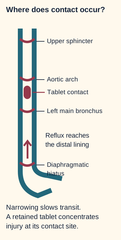
Picture two patients. One has burning and sour regurgitation after meals for years. The other develops abrupt pain after taking doxycycline with a sip of water and lying down. Before reading further, point to the expected injury on the contact map.
Predict the location before naming the cause: retained tablets often injure a focal middle segment; repeated reflux commonly injures the distal lining. These are common patterns, not exclusive locations. [3][7]
Repeated reflux versus retained tablets
Reflux repeatedly exposes the distal esophagus to gastric contents. Inflammation may erode the lining; repeated injury and repair can produce a fibrotic peptic stricture. Barrett metaplasia is another reflux-associated consequence, not another name for a stricture. Heartburn relief does not establish that a new swallowing problem is harmless. Dysphagia, bleeding or weight loss warrants investigation, usually with upper endoscopy rather than a longer empiric treatment trial. [3]
A retained tablet dissolves against a small area of mucosa. Doxycycline and other tetracyclines, bisphosphonates, potassium chloride, iron and some NSAIDs can cause this contact injury. Little water, recumbency and impaired transit prolong exposure. Mid-esophageal ulcers are common near external compression, although location is not exclusive. The upper sphincter, aortic arch, left main bronchus and diaphragmatic hiatus are useful anatomic landmarks where transit or instrumentation can become difficult. [7]
Test your prediction: the long reflux history points toward distal exposure; the abrupt tablet-associated history points toward focal contact. A proton pump inhibitor reduces acid but does not prevent a tablet from lodging. Review the offending medicine with the treating clinician, pause or replace it when indicated, and follow its water and upright-position instructions. Do not crush an unfamiliar formulation or simply resume the same harmful dosing routine.
For alendronate, new painful or difficult swallowing warrants prescriber contact before further doses. Healing alone does not make the same regimen suitable: a patient who still cannot remain upright for at least 30 minutes needs medication and route reassessment rather than a meal, antacid or antifungal workaround. [12]
For uncomplicated typical reflux without alarm features, an appropriately timed PPI trial is reasonable. Taking a conventional PPI before a meal, rather than after symptoms begin, supports acid suppression. In contrast, complete obstruction with inability to swallow saliva, severe acute chest pain, major hematemesis or shock requires urgent assessment. [1][3]
Apply it elsewhere: a patient says the burning is better but meat increasingly sticks. The treatment response describes acid symptoms; it does not explain or dismiss the progressive obstruction. Consider stricture, eosinophilic disease and malignancy rather than escalating acid therapy alone.
A tablet rests near the middle-esophageal narrowing. What changes locally?
Dissolved drug contacts one mucosal area for longer, concentrating injury rather than requiring a whole-esophagus acid disorder.
Which part of the dosing routine reduces that contact?
Adequate water and an upright position, following the specific product instructions, support transit; the suspected culprit also needs clinical review.
Would improved heartburn explain newly progressive solid-food dysphagia?
No. Symptom relief does not exclude structural disease; new alarm features require investigation.
Use the host, then choose the right tissue
White material is not automatically fungus, and a ring is not a complete diagnosis. Ask what the patient experiences, what host factors are present and which cells a biopsy must capture.
Eosinophilic inflammation can narrow a flexible tube
A young adult with asthma and eczema repeatedly needs food removed from the esophagus. Concentric rings, longitudinal furrows and white exudates support eosinophilic esophagitis, but endoscopy can also look normal. Atopy increases suspicion; its absence does not exclude disease. Chronic inflammation and remodeling can reduce distensibility and cause solid-food impaction. A current impaction with inability to handle secretions needs emergency endoscopic care, not a delayed outpatient allergy workup. [1][2]
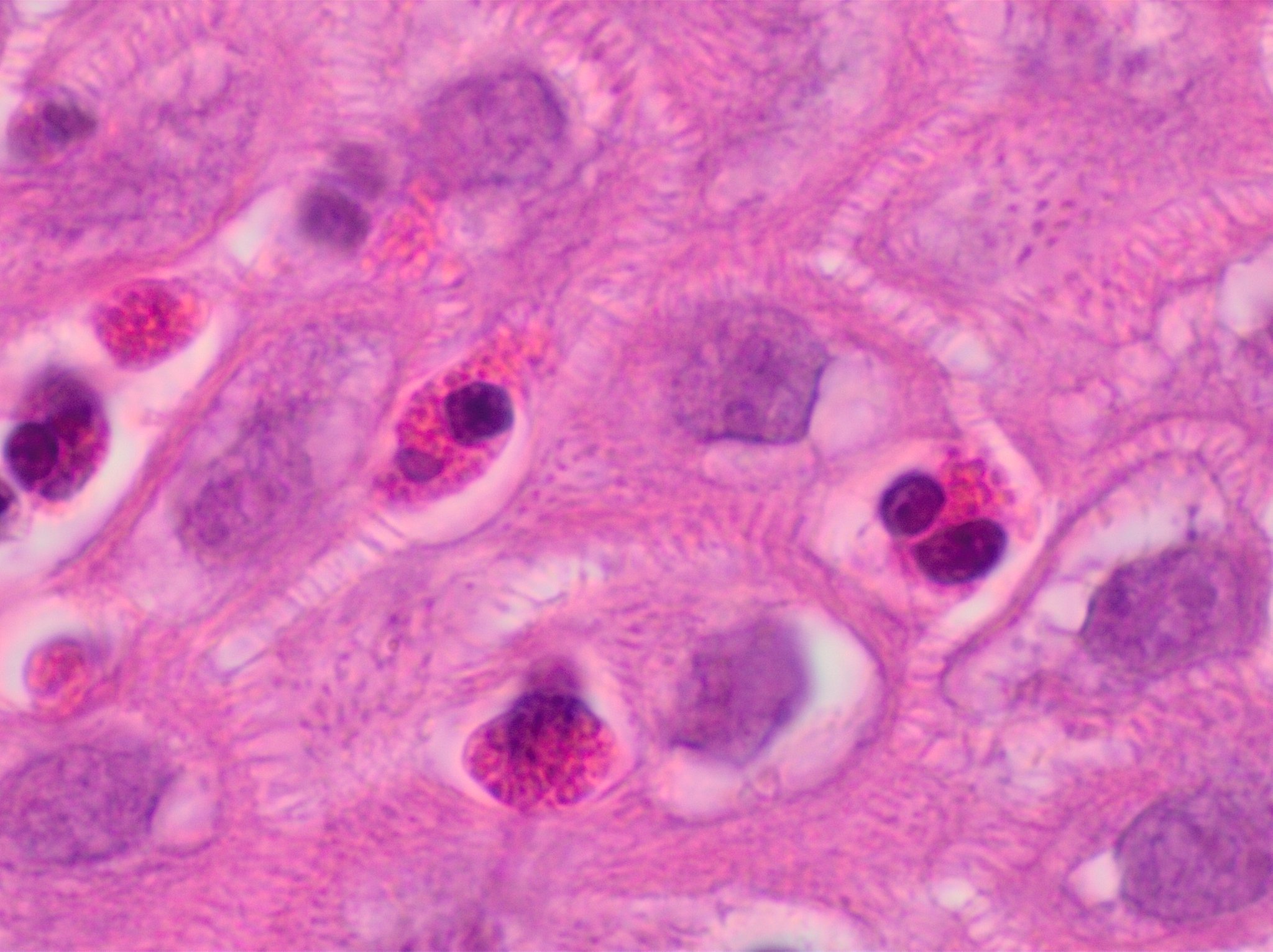
Find the granular cells, then ask what else is needed: this uncalibrated image illustrates eosinophils but cannot supply a diagnostic high-power-field count. Symptoms, adequate biopsies and alternative causes still matter. Image: Mikael Häggström, M.D.; original source; CC0 1.0. [2].
Predict what the photograph cannot tell you: it demonstrates eosinophils, but it does not supply symptoms, calibrated field area or sampling from the whole esophagus. Diagnosis combines esophageal dysfunction with at least 15 eosinophils per high-power field on biopsy and evaluation for other causes of esophageal eosinophilia. Obtain at least six targeted biopsies from two esophageal levels. A single normal distal fragment cannot reliably exclude a patchy process. Treatment at the time of sampling can also suppress findings; diagnostic preparation should be planned by the clinician. [2]
PPI response does not exclude eosinophilic esophagitis. PPIs, swallowed topical corticosteroids and an appropriately supported elimination diet are treatment options selected with the patient. Dilation can treat a narrowed lumen, but it does not treat the inflammation. Assess symptoms together with endoscopic and histologic response, because swallowing can improve while inflammation persists. [2]
Impaired cellular immunity increases concern for infectious esophagitis, especially when swallowing is painful. Candida commonly produces adherent white plaques or pseudomembranes. Yeast and pseudohyphae invading tissue support the diagnosis. Oral thrush may be absent. Candida albicans is common, but the endoscopic appearance does not identify the species. Esophageal candidiasis requires systemic therapy, usually fluconazole when appropriate; topical oral nystatin is not sufficient. In a suitable clinical setting, a treatment trial can precede endoscopy. Persistent symptoms despite an adequate course require reassessment rather than assuming every white lesion is Candida. [4][6]
A usual fluconazole course for esophageal candidiasis is 14 to 21 days. Improvement during the first week supports response but does not replace completing the course. Persistent symptoms after an adequate initial trial warrant reassessment before blindly extending or changing therapy. [4]
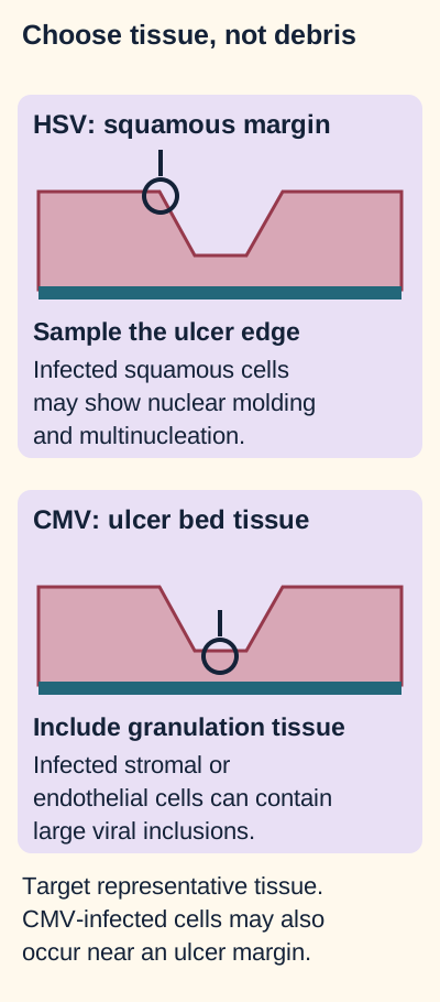
Ulcer shape narrows the differential, but an adequate sample must contain the cells infected by the suspected organism. Avoid treating the center of necrotic debris as an adequate viral biopsy. [5][6]
Match the suspected virus to representative tissue
Pattern that raises suspicion
Tissue that can answer the question
Pattern that raises suspicionHSV: often several small, sharply defined ulcers
Tissue that can answer the questionViable squamous epithelium at ulcer margins. Look for multinucleation, nuclear molding and chromatin margination.
Pattern that raises suspicionCMV: often larger, elongated distal ulcers
Tissue that can answer the questionGranulation tissue with stromal and endothelial cells, commonly in the ulcer bed. Cytomegalic cells with inclusions and confirmatory staining support invasive disease.
Choose the next sample: an ulcer-center specimen contains only dead debris. Repeating that exact sample may repeat the failure. For suspected HSV, include the squamous edge; for suspected CMV, include representative granulation tissue. CMV-infected endothelial cells can also occur near an ulcer margin, so the principle is infected cell type, not a rigid geographic rule. Endoscopic patterns overlap and are not independently definitive. [5][6]
CMV detected in blood, or detected by tissue PCR without compatible histopathology, does not by itself prove CMV esophagitis. Severe confirmed CMV esophagitis that prevents oral intake generally requires IV ganciclovir; renal function, blood counts and treatment response require monitoring. Oral valganciclovir can be appropriate when disease severity and absorption permit. [5]
Transfer: two patients have white esophageal lesions. Recurrent impaction with multi-level eosinophilia supports eosinophilic disease; painful swallowing with tissue-invasive fungal forms supports candidiasis. The color alone cannot select treatment.
Do white lesions establish Candida by color alone?
No. Fungal plaques and eosinophilic exudates can resemble each other. Host factors, symptoms and tissue findings determine the interpretation.
Why might a viral biopsy fail even when an ulcer is visible?
Necrotic debris or the wrong tissue compartment may omit infected cells. Include squamous ulcer margins for HSV and representative stromal or endothelial tissue for CMV.
What would persistent eosinophilia after successful dilation mean?
The lumen is wider, but inflammation remains active. Mechanical relief does not replace anti-inflammatory management.
Ask whether the wall still contains the lumen
Would severe pain or a large amount of blood tell you exactly how far an injury extends? No. Use the anatomy and evidence of a leak, not a symptom-to-depth shortcut.
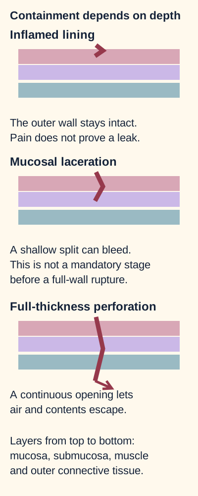
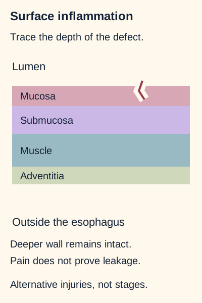
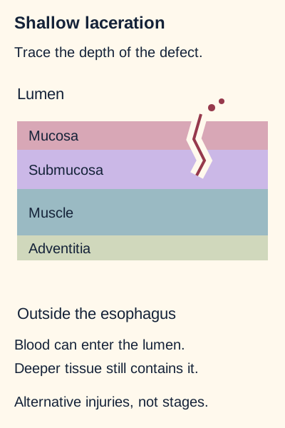
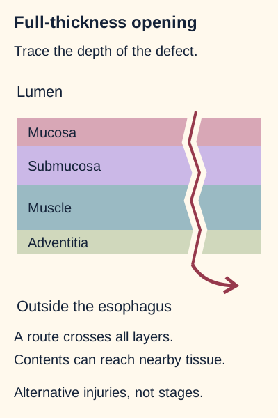
Follow the red defect rather than the symptom label. Only the full-wall defect connects the lumen to adjacent tissue in this simplified diagram; shallow injury can still cause major bleeding. [1][9]
The mucosa lines the lumen; the submucosa supports it and contains vessels. Injury here can cause inflammation, ulceration, bleeding and later fibrosis. The muscular wall contributes propulsion and containment. Most of the esophagus has an outer adventitial connective-tissue surface rather than a complete serosal covering. A full-thickness defect therefore exposes nearby spaces to saliva, gastric contents and microbes. [1]
Compare three wall-depth examples
Predict whether there is a continuous route from the lumen to surrounding tissue in each example. These are alternative injuries, not stages that every patient passes through.
Worked comparison: surface inflammation leaves deeper containment intact. A shallow mucosal or submucosal laceration can bleed while the outer wall remains closed. A full-thickness opening permits air and contents to reach adjacent tissues. Neither pain severity nor blood volume alone determines depth. The diagrams are qualitative, not rupture-pressure calculators.
Surface inflammation: can contents reach the outside?
Deeper wall remains intact. Pain does not prove leakage. This is an alternative injury, not a step in a required progression. [1][9]
The injury is confined to the lining. Intact deeper tissue separates the lumen from the outside. Pain can be substantial without a full-wall leak.
Transfer: painful swallowing after a retained tablet does not, by itself, establish perforation; evaluate new severe chest pain or systemic illness separately.
Shallow laceration: does bleeding prove a full-wall opening?
Blood can enter the lumen. Deeper tissue still contains it. This is an alternative injury, not a step in a required progression. [1][9]
The split can injure superficial vessels while the deeper wall remains intact. Blood can enter the lumen without a path into the mediastinum.
Transfer: significant hematemesis can require resuscitation and hemostasis even when the lesion is not a perforation.
Full-thickness defect: which new space becomes exposed?
A route crosses all layers. Contents can reach nearby tissue. This is an alternative injury, not a step in a required progression. [1][9]
The defect crosses the complete wall and connects the lumen with surrounding tissue. Saliva and gastric contents can contaminate mediastinal or pleural spaces.
Transfer: minimal hematemesis does not make a full-wall defect harmless. Urgent leak assessment and specialist management are needed.
Thoracic perforation can contaminate the mediastinum and pleural space, producing mediastinitis, an effusion, hydropneumothorax or empyema. Air may track into the neck and produce crepitus. But mediastinal air can also arise from the respiratory tract; its presence does not prove an esophageal defect. [11] Conversely, absent air on a plain chest film does not exclude one. [1]
Apply the distinction: a shallow junctional tear can cause substantial hemorrhage and require hemostasis. A small full-wall defect can demand urgent leak assessment even before obvious external swelling develops. Depth informs the threat; physiology determines immediate stabilization.
Which defect first connects the lumen to adjacent spaces in the drawing?
The full-wall defect. A shallow laceration can bleed while the deeper wall remains continuous.
Does mediastinal air independently identify the leaking organ?
No. Respiratory air dissection is another source; the clinical setting and complete imaging assessment matter.
Would neck crepitus prove that the esophageal defect is cervical?
No. Air can track upward from the mediastinum through connected tissue planes.
Caustic injury: a normal mouth cannot clear the esophagus
A patient swallowed a corrosive cleaner and has drooling and chest pain, but no obvious oral burn. Is the esophagus safe? The mouth is not a reliable depth gauge for the swallowed exposure.
Alkalis classically produce liquefactive injury and acids classically produce coagulative injury. Both can cause deep necrosis and perforation. Concentration, amount, contact time and the agent's properties matter more than memorizing a harmless-acid versus harmful-alkali rule. The injury may also involve the airway and stomach. [1][10]
Use the full-wall drawing above: predict what failure of deep tissue would do even if the surface examination seemed modest. Once a continuous defect develops, contamination can extend beyond the lumen. Early stabilization includes airway assessment, avoidance of oral intake, treatment of shock and prompt specialist and poison-center involvement. Do not induce vomiting, attempt chemical neutralization or insert a gastric tube blindly; these can add injury or aspiration risk. [10]
In adults with significant caustic exposure, contrast-enhanced CT helps assess injury extent and depth. Absent wall enhancement raises concern for transmural necrosis. Endoscopy is selected when CT is unavailable, unsuitable or uncertain; pediatric assessment often favors endoscopy rather than automatically applying an adult CT-first pathway. Suspected necrosis or perforation requires urgent surgical assessment, not a delay for routine endoscopic staging. Airway compromise or shock must be addressed before nonessential diagnostic testing. [1][10]
Change the time point: after surviving the acute injury, a patient develops progressively difficult solid-food transit over several weeks. Healing with fibrosis can create a stricture. That delayed narrowing is a different problem from an acute free leak and requires planned specialist assessment; successful early resuscitation does not eliminate later swallowing complications.
Can a normal mouth exclude injury after a caustic ingestion?
No. The esophagus can be injured despite few visible oral findings, especially when drooling or odynophagia is present.
Why does absent wall enhancement change the question?
It raises concern for nonviable deep tissue rather than uncomplicated surface inflammation, requiring urgent specialist assessment.
What new problem can scar formation produce weeks later?
A fibrotic stricture can progressively obstruct solids even after the initial leak or necrosis concern has resolved.
After vomiting, separate bleeding from loss of containment
Two patients retch forcefully. One then vomits blood; the other develops abrupt severe chest pain and respiratory distress. The shared trigger does not imply the same lesion or treatment.
Pressure can rise abruptly during vomiting, especially when the upper esophageal sphincter does not relax appropriately. A Mallory-Weiss tear is a nonperforating longitudinal laceration near the gastroesophageal junction, involving mucosa and sometimes submucosa. Boerhaave syndrome is a spontaneous full-thickness rupture related to the pressure event, commonly in the distal thoracic esophagus. A Mallory-Weiss tear does not have to occur first. [1][9]
Different problems after the same trigger
Finding
What it changes
FindingJunctional laceration with active luminal bleeding
What it changesAssess hemorrhage, resuscitate and use endoscopic hemostasis when indicated.
FindingChest pain, systemic illness or imaging evidence of a leak
What it changesUrgently assess wall disruption and contamination while resuscitation proceeds.
FindingA reassuring plain film or absent neck crepitus
What it changesNeither excludes perforation when the clinical presentation remains concerning.
Many Mallory-Weiss bleeds stop without a hemostatic procedure, but some are severe. Persistent bleeding, recurrent bleeding or unstable physiology changes management. Upper endoscopy can identify the laceration and treat active bleeding with an appropriate endoscopic technique. Do not select major thoracic surgery just because blood followed vomiting. Equally, do not dismiss serious chest pain as a shallow tear merely because hematemesis also occurred. [9] A nonbleeding visible vessel in a patient who has rebled is not equivalent to a low-risk tear that has remained quiet. [9]
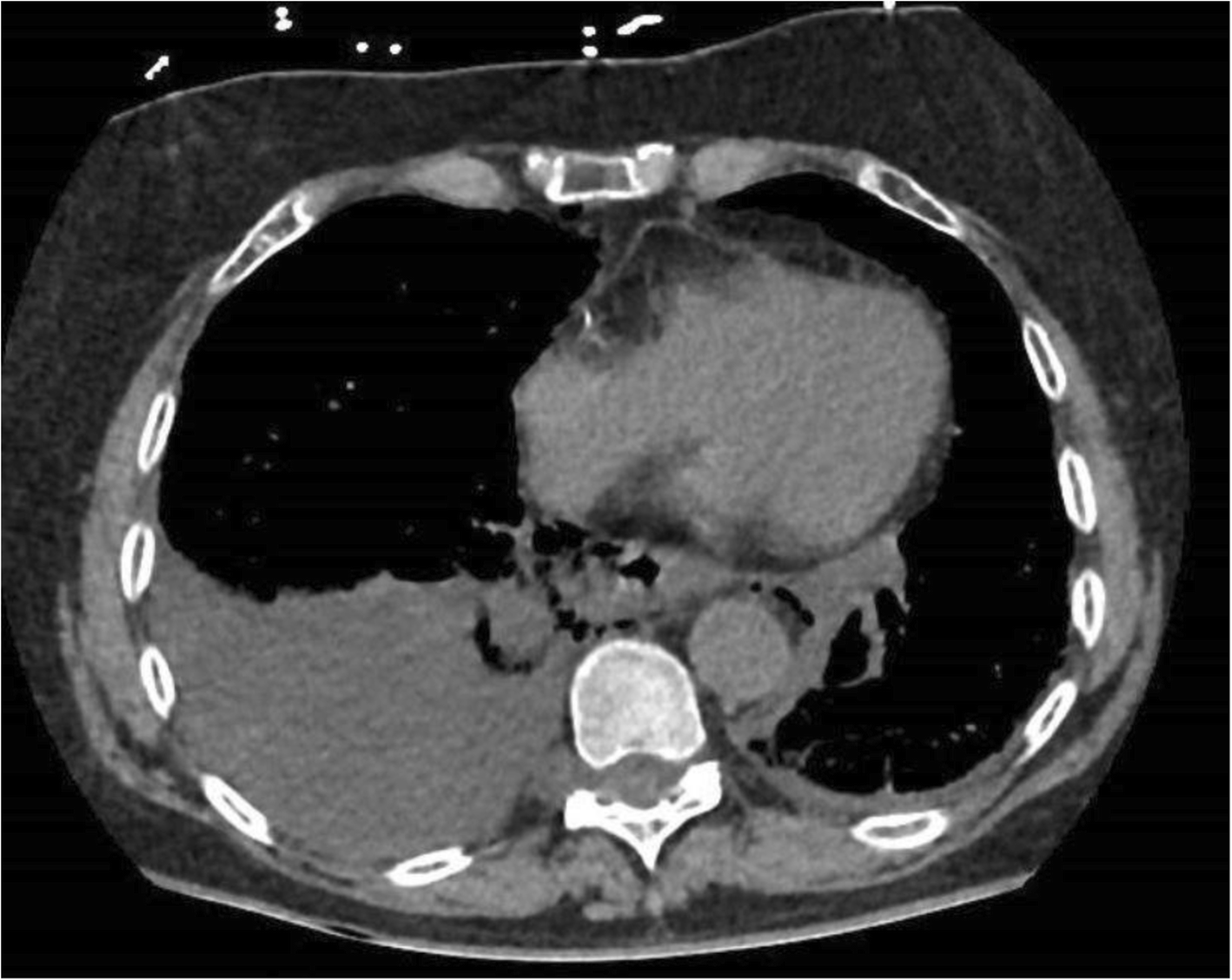
In the published case, wall disruption and mediastinal air accompany a right pleural effusion. Right-sided fluid is possible: the usual distal left-sided rupture pattern is not an exclusion rule. Interpret the full CT and clinical setting, not this single slice alone. Image: Chirica M, Kelly MD, Siboni S, et al.; original source; CC BY 4.0. [1].
Inspect and predict: the published CT has fluid in the patient's right pleural space as well as mediastinal abnormalities. Does right-sided fluid contradict perforation? No. Laterality and the complete vomiting-pain-crepitus triad are not required diagnostic criteria. Contrast-enhanced CT with an appropriate esophageal-leak protocol is a central investigation when perforation is suspected. A water-soluble contrast study or carefully selected endoscopy may contribute when uncertainty remains; investigation should not postpone treatment of an unstable patient. [1]
Suspected perforation requires urgent specialist involvement, no oral intake, IV broad-spectrum antimicrobial coverage and resuscitation. The team assesses leak location, size, contamination, physiology and nutrition. Free thoracic leakage with sepsis or shock requires urgent surgical source-control assessment, including defect treatment and drainage of contaminated spaces. Sealing a defect does not remove an established pleural collection. Selected stable patients with an early contained leak, limited contamination and close specialist surveillance can receive nonoperative management. This is active inpatient treatment with nutritional planning and readiness to intervene, not reassurance or routine discharge. Delayed recognition permits mediastinitis and sepsis to worsen. [1]
Transfer to instrumentation: a perforation recognized during endoscopic dilation is iatrogenic, not Boerhaave syndrome. Early recognition may permit endoscopic closure, but containment, antibiotics, drainage needs and clinical monitoring still require assessment.
What separates an actively bleeding laceration from a contaminating leak?
Bleeding arises from injured vessels; a full-wall defect also provides an exit for luminal contents into adjacent spaces.
Why are a contained leak and a freely contaminating leak managed differently?
Containment limits initial spread. Stability, lack of sepsis and expert surveillance are essential to selected nonoperative care; free contamination with shock needs urgent source control.
Does a negative plain chest film settle a concerning post-emesis presentation?
No. A plain film may miss perforation; urgent appropriate CT assessment is needed when clinical concern persists.
A proximal web is a structural finding, not the end of the investigation
A patient has fatigue, intermittent difficulty with solid food, hemoglobin 9.2 g/dL, mean corpuscular volume 68 fL and ferritin 4 ng/mL. A swallow study shows a thin proximal mucosal shelf. What connects these findings, and what still needs an explanation?
The combination of iron-deficiency anemia, dysphagia and a proximal esophageal web is Plummer-Vinson syndrome. The web is a thin mucosal projection that can obstruct solids. Glossitis, angular cheilitis, pallor or spoon-shaped nails may accompany iron deficiency. The syndrome's pathogenesis is not fully established: iron-dependent epithelial or muscular dysfunction is a proposed contributor, not a proven single explanation. A web can also occur without this complete syndrome. [8]
Trace the route of a food bolus: a thin shelf high in the esophagus explains intermittent solid-food obstruction. A distal ring has a different location. A motility disorder such as achalasia more often affects both solids and liquids and involves impaired lower-sphincter relaxation rather than a proximal shelf. Symptoms guide investigation, but do not substitute for defining the lesion.
Iron replacement addresses the deficiency and may improve swallowing. The cause of the deficiency still requires investigation, including appropriate evaluation for blood loss, impaired absorption or malignancy. A persistent obstructive web may need endoscopic dilation. Merely normalizing ferritin does not guarantee that an established physical narrowing has disappeared. [8]
Plummer-Vinson syndrome is associated with squamous-cell carcinoma of the hypopharynx and upper esophagus. This is an association, not a prediction that every patient will develop cancer. Follow-up should be individualized; recurrent or progressive dysphagia, weight loss or a new voice change requires reassessment rather than automatic attribution to a familiar web. No fixed surveillance interval is prescribed in this lesson; arrange follow-up with the treating specialists. [8]
Change one finding: the original patient now has a corrected blood count but progressively worsening swallowing and weight loss. Iron deficiency no longer explains the entire presentation. Reassess the upper aerodigestive tract for malignancy and other structural disease rather than repeating iron treatment alone.
What links low ferritin, a proximal web and solid-food dysphagia?
Together they support the Plummer-Vinson pattern, but the cause of iron deficiency still needs investigation.
What if iron indices normalize but a confirmed obstructive web persists?
The physical narrowing may need endoscopic dilation; more iron alone does not directly widen an established web.
What if the swallowing pattern becomes progressive with weight loss?
Reassess for malignancy and other structural disease instead of assuming recurrence of the prior benign explanation.
Practice the decisions
Use the history and new findings to choose one best answer. These original educational cases are not validated replicas of a licensing examination.
Case 1
Show answer and explanations for case 1
A. Ambulatory esophageal pH monitoring (Why this does not fit)
Ambulatory pH monitoring measures reflux burden when reflux diagnosis or persistent reflux symptoms need clarification. Reflux monitoring cannot directly evaluate or biopsy the structural cause of progressive dysphagia and weight loss.
Reasoning steps for option A
What does pH monitoring assess?
Ambulatory pH monitoring measures reflux burden when reflux diagnosis or persistent reflux symptoms need clarification.
Why is it not the priority here?
Reflux monitoring cannot directly evaluate or biopsy the structural cause of progressive dysphagia and weight loss.
B. Upper endoscopy with mucosal biopsies (Best answer)
Progressive solid-food dysphagia and weight loss have developed despite relief of burning. A stricture or malignancy can impair transit while acid symptoms improve. Upper endoscopy can define the obstruction and obtain tissue where indicated.
Reasoning steps for option B
Which symptom has changed?
Progressive solid-food dysphagia and weight loss have developed despite relief of burning.
Does acid symptom improvement explain that change?
No. A stricture or malignancy can impair transit while acid symptoms improve.
Which test addresses that concern?
Upper endoscopy can define the obstruction and obtain tissue where indicated.
C. High-resolution esophageal manometry (Why this does not fit)
Manometry characterizes motor disorders when motility is the unresolved question. The new progressive solid-food obstruction and weight loss require structural and mucosal evaluation first.
Reasoning steps for option C
When does manometry help?
Manometry characterizes motor disorders when motility is the unresolved question.
What must be addressed first?
The new progressive solid-food obstruction and weight loss require structural and mucosal evaluation first.
D. Barium contrast examination of swallowing (Why this does not fit)
A contrast study can delineate a structural narrowing and may help selected swallowing evaluations. Progressive dysphagia with weight loss warrants direct mucosal assessment and the ability to obtain tissue, which endoscopy provides.
Reasoning steps for option D
What can esophagography demonstrate?
A contrast study can delineate a structural narrowing and may help selected swallowing evaluations.
Why should it not replace the prioritized investigation here?
Progressive dysphagia with weight loss warrants direct mucosal assessment and the ability to obtain tissue, which endoscopy provides.
Takeaway: A response in heartburn does not dismiss newly progressive dysphagia or weight loss.
A. Loss of inhibitory neurons at the lower sphincter (Why this does not fit)
Loss of inhibitory myenteric signaling can prevent lower-sphincter relaxation in achalasia. A short scarred structural narrowing with reflux-associated inflammation is not a neuronal relaxation defect.
Reasoning steps for option A
How can neuronal loss impair swallowing?
Loss of inhibitory myenteric signaling can prevent lower-sphincter relaxation in achalasia.
Why does it not explain this lesion?
A short scarred structural narrowing with reflux-associated inflammation is not a neuronal relaxation defect.
B. Iron-associated formation of a cervical mucosal web (Why this does not fit)
A proximal esophageal web can obstruct solids in Plummer-Vinson syndrome. The demonstrated abnormality is a fibrotic distal stricture, not a thin cervical shelf.
Reasoning steps for option B
What lesion can accompany iron deficiency?
A proximal esophageal web can obstruct solids in Plummer-Vinson syndrome.
Which supplied feature argues against that lesion?
The demonstrated abnormality is a fibrotic distal stricture, not a thin cervical shelf.
C. Viral invasion of ulcer-bed endothelial cells (Why this does not fit)
CMV may infect endothelial and stromal cells in esophageal ulcers. The case describes chronic distal scarring without the ulcerative tissue evidence needed to attribute it to CMV.
Reasoning steps for option C
Which infection targets these cells?
CMV may infect endothelial and stromal cells in esophageal ulcers.
Why is this not the best explanation?
The case describes chronic distal scarring without the ulcerative tissue evidence needed to attribute it to CMV.
D. Repeated reflux injury with fibrotic repair (Best answer)
The fixed narrowing lies in the distal segment repeatedly exposed to refluxate. Fibrosis without malignancy or substantial eosinophilia supports scarring rather than a tumor or eosinophilic process. Fibrotic repair reduces luminal diameter and creates a peptic stricture.
Reasoning steps for option D
Where is the abnormality located?
The fixed narrowing lies in the distal segment repeatedly exposed to refluxate.
What does the biopsy add?
Fibrosis without malignancy or substantial eosinophilia supports scarring rather than a tumor or eosinophilic process.
How does that produce dysphagia?
Fibrotic repair reduces luminal diameter and creates a peptic stricture.
Takeaway: Distal location, repeated reflux and fibrotic histology support a peptic stricture.
A. Take after dinner and then lie down (Why this does not fit)
Patients may associate food with less medication-related stomach discomfort. Lying down immediately after the tablet can prolong esophageal contact despite the change in meal timing.
Reasoning steps for option A
What concern might meal-based dosing address?
Patients may associate food with less medication-related stomach discomfort.
What exposure does this plan leave unchanged?
Lying down immediately after the tablet can prolong esophageal contact despite the change in meal timing.
B. Add acid suppression before the bedtime dose (Why this does not fit)
A PPI reduces gastric acid exposure and may support healing. Acid suppression does not correct the delayed transit of a bedtime tablet taken with very little water.
Reasoning steps for option B
What does acid suppression reduce?
A PPI reduces gastric acid exposure and may support healing.
Why does it not prevent these focal ulcers?
Acid suppression does not correct the delayed transit of a bedtime tablet taken with very little water.
C. Take with adequate water and remain upright (Best answer)
A poorly washed-down tablet was followed immediately by recumbency. Delayed tablet transit can concentrate dissolved drug against mucosa near an anatomic narrowing. Water and an upright position, following product-specific directions, reduce prolonged esophageal contact.
Reasoning steps for option C
Which exposure preceded the focal ulcers?
A poorly washed-down tablet was followed immediately by recumbency.
Why is the middle segment involved?
Delayed tablet transit can concentrate dissolved drug against mucosa near an anatomic narrowing.
Which change addresses that process?
Water and an upright position, following product-specific directions, reduce prolonged esophageal contact.
D. Take with less water to limit regurgitation (Why this does not fit)
The patient may worry that a larger bedtime drink will provoke regurgitation. Inadequate water makes tablet retention more likely rather than preventing the focal contact injury.
Reasoning steps for option D
Why might the patient reduce fluid intake?
The patient may worry that a larger bedtime drink will provoke regurgitation.
How does that affect the demonstrated mechanism?
Inadequate water makes tablet retention more likely rather than preventing the focal contact injury.
Takeaway: Prevent recurrent contact injury by correcting administration and reviewing the medicine, not by masking pain alone.
A. Select a nonoral osteoporosis treatment with the prescriber (Best answer)
Recumbent tablet dosing preceded a focal ulcer that healed when alendronate was withheld. The patient remains unable to follow the required upright administration instructions. The prescriber should select a suitable nonoral osteoporosis strategy after reviewing its own risks and eligibility.
Reasoning steps for option A
What links the medication to the ulcer?
Recumbent tablet dosing preceded a focal ulcer that healed when alendronate was withheld.
Which risk persists despite healing?
The patient remains unable to follow the required upright administration instructions.
What changes the exposure while preserving treatment?
The prescriber should select a suitable nonoral osteoporosis strategy after reviewing its own risks and eligibility.
B. Restart the tablet with additional acid suppression (Why this does not fit)
Acid suppression may support mucosal healing or treat concurrent reflux. The patient still cannot remain upright, so acid suppression does not prevent renewed alendronate contact injury.
Reasoning steps for option B
What can acid suppression help?
Acid suppression may support mucosal healing or treat concurrent reflux.
What risk remains after the ulcer heals?
The patient still cannot remain upright, so acid suppression does not prevent renewed alendronate contact injury.
C. Restart the tablet after the morning meal (Why this does not fit)
Food may seem to protect the digestive tract from a tablet that previously caused pain. Alendronate requires fasting administration, and taking it after food does not correct the inability to remain upright.
Reasoning steps for option C
Why might a meal seem protective?
Food may seem to protect the digestive tract from a tablet that previously caused pain.
Why is this not the appropriate workaround?
Alendronate requires fasting administration, and taking it after food does not correct the inability to remain upright.
D. Restart the tablet with an oral antifungal (Why this does not fit)
An antifungal would treat a susceptible fungal infection rather than tablet retention. There is no fungal invasion, while the timing, localization and response to withdrawal support medication contact injury.
Reasoning steps for option D
What would an antifungal address?
An antifungal would treat a susceptible fungal infection rather than tablet retention.
What argues against that treatment here?
There is no fungal invasion, while the timing, localization and response to withdrawal support medication contact injury.
Takeaway: Healing does not remove a persistent administration barrier. Reassess osteoporosis therapy rather than restarting oral alendronate when upright dosing is not possible.
A. Reflux esophagitis responding to acid suppression (Why this does not fit)
PPIs are familiar treatments for acid-related symptoms and can improve swallowing discomfort. PPI responsiveness no longer serves as an exclusion criterion for eosinophilic esophagitis.
Reasoning steps for option A
Why could reflux seem plausible?
PPIs are familiar treatments for acid-related symptoms and can improve swallowing discomfort.
What assumption is incorrect?
PPI responsiveness no longer serves as an exclusion criterion for eosinophilic esophagitis.
B. Eosinophilic esophagitis responding histologically to PPI (Best answer)
Esophageal dysfunction and multi-level counts above 15 eosinophils per high-power field supported eosinophilic esophagitis after alternative causes were evaluated. Symptoms and tissue eosinophilia both improve during PPI treatment. PPIs are a treatment for eosinophilic esophagitis, and response does not exclude it.
Reasoning steps for option B
Did the initial assessment meet a disease pattern?
Esophageal dysfunction and multi-level counts above 15 eosinophils per high-power field supported eosinophilic esophagitis after alternative causes were evaluated.
What does the follow-up demonstrate?
Symptoms and tissue eosinophilia both improve during PPI treatment.
Does that response negate the original diagnosis?
No. PPIs are a treatment for eosinophilic esophagitis, and response does not exclude it.
C. Eosinophilic esophagitis remaining histologically active (Why this does not fit)
Eosinophilic esophagitis can recur and requires continued assessment. The reported eosinophil count has fallen below 15 per high-power field with symptom improvement, rather than demonstrating persistent histologic activity.
Reasoning steps for option C
Why is long-term follow-up still important?
Eosinophilic esophagitis can recur and requires continued assessment.
What do the current data show?
The reported eosinophil count has fallen below 15 per high-power field with symptom improvement, rather than demonstrating persistent histologic activity.
D. Peptic stenosis persisting after resolution of inflammation (Why this does not fit)
A fixed narrowing can require mechanical treatment to improve transit. The multi-level eosinophilia and treatment-associated histologic response establish an inflammatory component that dilation alone would not address.
Reasoning steps for option D
Why might dilation be considered in a patient with impactions?
A fixed narrowing can require mechanical treatment to improve transit.
Why is that not the best interpretation of these data?
The multi-level eosinophilia and treatment-associated histologic response establish an inflammatory component that dilation alone would not address.
Takeaway: PPI-responsive tissue eosinophilia can be treated eosinophilic esophagitis, not evidence against the diagnosis.
A. Obtain six biopsies distributed across two esophageal levels (Best answer)
Recurrent impaction in a patient with atopy indicates a plausible esophageal inflammatory disorder even with a normal surface appearance. One distal fragment can miss patchy eosinophilia. At least six targeted biopsies from two esophageal levels provide more representative sampling.
Reasoning steps for option A
What keeps eosinophilic disease in the differential?
Recurrent impaction in a patient with atopy indicates a plausible esophageal inflammatory disorder even with a normal surface appearance.
How adequate is the existing sample?
One distal fragment can miss patchy eosinophilia.
What improves the assessment?
At least six targeted biopsies from two esophageal levels provide more representative sampling.
B. Accept the distal biopsy as excluding eosinophilic disease (Why this does not fit)
The sampled fragment has a low eosinophil count and endoscopy looks normal. Neither a normal-appearing esophagus nor one negative distal fragment reliably excludes patchy eosinophilic esophagitis.
Reasoning steps for option B
What might seem reassuring?
The sampled fragment has a low eosinophil count and endoscopy looks normal.
Why is exclusion premature?
Neither a normal-appearing esophagus nor one negative distal fragment reliably excludes patchy eosinophilic esophagitis.
C. Diagnose eosinophilic disease from the atopic presentation (Why this does not fit)
Asthma and recurrent impaction increase clinical suspicion for eosinophilic esophagitis. The diagnosis still requires representative histology and assessment for other causes of esophageal eosinophilia.
Reasoning steps for option C
Why does atopy matter?
Asthma and recurrent impaction increase clinical suspicion for eosinophilic esophagitis.
Why is that not enough to diagnose it?
The diagnosis still requires representative histology and assessment for other causes of esophageal eosinophilia.
D. Use food-allergy testing to identify the esophageal cause (Why this does not fit)
Eosinophilic esophagitis is associated with an atopic background. It does not establish the required esophageal tissue diagnosis or explain the obstructive episodes by itself.
Reasoning steps for option D
Why might allergy testing seem relevant?
Eosinophilic esophagitis is associated with an atopic background.
Does serum allergy testing replace the esophageal evaluation?
No. It does not establish the required esophageal tissue diagnosis or explain the obstructive episodes by itself.
Takeaway: A negative limited sample is not a negative adequately sampled esophagus.
A. Repeat dilation to address the persistent biopsy abnormality (Why this does not fit)
Dilation can improve a residual or recurrent fixed narrowing. Mechanical widening does not directly suppress eosinophilic inflammation.
Reasoning steps for option A
What can another dilation accomplish?
Dilation can improve a residual or recurrent fixed narrowing.
Why would it not be expected to normalize this biopsy?
Mechanical widening does not directly suppress eosinophilic inflammation.
B. Add swallowed budesonide to the treatment plan (Best answer)
Dilation increased the caliber of the narrowed lumen and improved food transit. The persistently high biopsy count shows ongoing eosinophilic inflammation despite the PPI trial. A shared decision about additional anti-inflammatory therapy, such as swallowed budesonide, addresses tissue disease rather than diameter alone.
Reasoning steps for option B
What did dilation change?
Dilation increased the caliber of the narrowed lumen and improved food transit.
What did it fail to resolve?
The persistently high biopsy count shows ongoing eosinophilic inflammation despite the PPI trial.
What treatment addresses that remaining process?
A shared decision about additional anti-inflammatory therapy, such as swallowed budesonide, addresses tissue disease rather than diameter alone.
C. Continue observation after the improvement in swallowing (Why this does not fit)
The patient now passes food more easily after the lumen was widened. The repeat count of 52 eosinophils per high-power field documents persistent inflammatory activity.
Reasoning steps for option C
Why might symptoms suggest success?
The patient now passes food more easily after the lumen was widened.
What contradicts complete disease control?
The repeat count of 52 eosinophils per high-power field documents persistent inflammatory activity.
D. Substitute systemic fluconazole for the current PPI (Why this does not fit)
Candida esophagitis is treated with systemic antifungal therapy. The established disease and current biopsy show eosinophilic inflammation, not tissue-invasive fungi.
Reasoning steps for option D
Which disease responds to fluconazole?
Candida esophagitis is treated with systemic antifungal therapy.
Why is that not supported here?
The established disease and current biopsy show eosinophilic inflammation, not tissue-invasive fungi.
Takeaway: Dilation treats narrowing; persistent eosinophilia still needs anti-inflammatory management.
A. Within 24 hours: endoscopy and multilevel esophageal biopsy sampling (Why this does not fit)
Multilevel esophageal biopsies can investigate a patchy inflammatory disorder such as eosinophilic esophagitis. Inability to swallow saliva indicates complete obstruction, which requires emergency relief rather than the less urgent incomplete-obstruction window.
Reasoning steps for option A
Which part of this plan addresses recurrent impaction?
Multilevel esophageal biopsies can investigate a patchy inflammatory disorder such as eosinophilic esophagitis.
Why is the timing insufficient?
Inability to swallow saliva indicates complete obstruction, which requires emergency relief rather than the less urgent incomplete-obstruction window.
B. Within 6 hours: endoscopy and serum food-specific IgE testing (Why this does not fit)
Complete obstruction with retained saliva creates aspiration risk even when oxygenation is currently normal. Serum food-allergy testing does not establish the tissue diagnosis of eosinophilic esophagitis.
Reasoning steps for option B
Why is emergency endoscopy appropriate?
Complete obstruction with retained saliva creates aspiration risk even when oxygenation is currently normal.
Why is the subsequent test inadequate?
Serum food-allergy testing does not establish the tissue diagnosis of eosinophilic esophagitis.
C. Within 6 hours: endoscopy and multilevel esophageal biopsy sampling (Best answer)
Saliva cannot pass, indicating complete obstruction that needs emergency endoscopic relief. Repeated solid-food impaction with atopy raises concern for an underlying inflammatory narrowing, including eosinophilic esophagitis. After safe bolus clearance, obtain representative biopsies from at least two esophageal levels; atopy alone does not establish the diagnosis.
Repeated solid-food impaction with atopy raises concern for an underlying inflammatory narrowing, including eosinophilic esophagitis.
How is that possibility evaluated?
After safe bolus clearance, obtain representative biopsies from at least two esophageal levels; atopy alone does not establish the diagnosis.
D. Within 24 hours: endoscopy and serum food-specific IgE testing (Why this does not fit)
Previous impactions with asthma suggest an allergic-associated disorder, and the patient is not hypoxemic. Normal oxygenation does not make complete obstruction nonurgent, and allergy testing cannot replace representative esophageal histology.
Reasoning steps for option D
Why might this combination seem attractive?
Previous impactions with asthma suggest an allergic-associated disorder, and the patient is not hypoxemic.
Which two requirements does it miss?
Normal oxygenation does not make complete obstruction nonurgent, and allergy testing cannot replace representative esophageal histology.
Takeaway: Inability to handle secretions determines emergency timing; recurrent impaction requires an underlying-disease assessment with representative esophageal tissue.
A. Continue fluconazole to complete a 14- to 21-day course (Best answer)
Yeast and pseudohyphae invading symptomatic esophageal tissue establish candidiasis despite the absence of oral thrush. Symptoms have responded to an active systemic agent, but seven days is shorter than the recommended treatment course. Continue the tolerated fluconazole regimen to complete 14 to 21 days rather than stopping or switching to topical treatment.
Reasoning steps for option A
What do the biopsy findings establish?
Yeast and pseudohyphae invading symptomatic esophageal tissue establish candidiasis despite the absence of oral thrush.
What does the improvement after seven days show?
Symptoms have responded to an active systemic agent, but seven days is shorter than the recommended treatment course.
What plan fits both findings?
Continue the tolerated fluconazole regimen to complete 14 to 21 days rather than stopping or switching to topical treatment.
B. Replace fluconazole with topical nystatin for seven days (Why this does not fit)
Topical nystatin can treat superficial oral candidiasis. The documented infection involves esophageal tissue and still requires a full systemic treatment course.
Reasoning steps for option B
Which site can topical nystatin treat?
Topical nystatin can treat superficial oral candidiasis.
Why is this not an appropriate step-down?
The documented infection involves esophageal tissue and still requires a full systemic treatment course.
C. Stop antifungal therapy and arrange symptom-based follow-up (Why this does not fit)
Normal eating and resolution of odynophagia demonstrate an early clinical response. Symptom resolution after seven days does not establish completion of the recommended 14- to 21-day systemic course.
Reasoning steps for option C
Why might stopping seem reasonable?
Normal eating and resolution of odynophagia demonstrate an early clinical response.
What remains incomplete?
Symptom resolution after seven days does not establish completion of the recommended 14- to 21-day systemic course.
D. Continue fluconazole as indefinite suppressive therapy (Why this does not fit)
Recurrent esophageal candidiasis can justify a suppressive regimen together with management of the underlying immune deficiency. This is a responding episode without prior recurrence; the immediate need is completion of the treatment course, not indefinite suppression.
Reasoning steps for option D
When might suppressive fluconazole be considered?
Recurrent esophageal candidiasis can justify a suppressive regimen together with management of the underlying immune deficiency.
Why is indefinite suppression not the best plan here?
This is a responding episode without prior recurrence; the immediate need is completion of the treatment course, not indefinite suppression.
Takeaway: Early symptomatic improvement supports response but does not shorten esophageal candidiasis treatment to one week. Complete the systemic course.
A. Extend the initial fluconazole trial before additional testing (Why this does not fit)
Esophageal candidiasis usually receives a treatment course longer than one week. Persistent poor intake and no response after an adequate initial trial warrant reassessment, even though one week is not the full treatment duration.
Reasoning steps for option A
Why might completing a course seem reasonable?
Esophageal candidiasis usually receives a treatment course longer than one week.
Why is waiting alone inadequate?
Persistent poor intake and no response after an adequate initial trial warrant reassessment, even though one week is not the full treatment duration.
B. Replace systemic fluconazole with topical oral nystatin (Why this does not fit)
Topical nystatin can treat superficial oral Candida disease. It does not provide the systemic treatment required for esophageal candidiasis or investigate the lack of response.
Reasoning steps for option B
What does topical nystatin treat?
Topical nystatin can treat superficial oral Candida disease.
Why is this a step in the wrong direction?
It does not provide the systemic treatment required for esophageal candidiasis or investigate the lack of response.
C. Perform endoscopy with representative biopsies and fungal studies (Best answer)
There has been no symptomatic improvement despite an appropriate systemic antifungal exposure. An alternative infection, resistant Candida or a noninfectious lesion may account for the symptoms. Endoscopy with representative sampling evaluates lesion morphology and obtains tissue rather than assuming the original diagnosis.
Reasoning steps for option C
What has the treatment trial shown?
There has been no symptomatic improvement despite an appropriate systemic antifungal exposure.
What possibilities remain?
An alternative infection, resistant Candida or a noninfectious lesion may account for the symptoms.
Which test can distinguish these possibilities?
Endoscopy with representative sampling evaluates lesion morphology and obtains tissue rather than assuming the original diagnosis.
D. Begin empiric ganciclovir for an opportunistic viral infection (Why this does not fit)
Immunosuppression with persistent odynophagia raises concern for an ulcerative viral process. Immune status alone does not establish invasive CMV disease; targeted assessment can identify viral, fungal or noninfectious injury.
Reasoning steps for option D
Why might CMV enter the differential?
Immunosuppression with persistent odynophagia raises concern for an ulcerative viral process.
Why is immune status alone insufficient?
Immune status alone does not establish invasive CMV disease; targeted assessment can identify viral, fungal or noninfectious injury.
Takeaway: Failure to improve after an adequate empiric trial should prompt reassessment, not an automatic drug substitution.
A. Viable squamous epithelium from an ulcer margin (Best answer)
Several small sharply demarcated ulcers raise suspicion for HSV, although tissue is needed to distinguish overlapping causes. Necrotic debris lacks the viable squamous cells in which HSV cytopathic changes are typically identified. A representative ulcer margin supplies viable squamous epithelium for histology and appropriate viral testing.
Reasoning steps for option A
Which viral process does this pattern suggest?
Several small sharply demarcated ulcers raise suspicion for HSV, although tissue is needed to distinguish overlapping causes.
Why was the initial result uninformative?
Necrotic debris lacks the viable squamous cells in which HSV cytopathic changes are typically identified.
What sample addresses both the suspicion and the limitation?
A representative ulcer margin supplies viable squamous epithelium for histology and appropriate viral testing.
B. Necrotic material from the center of an ulcer (Why this does not fit)
The necrotic center is the most visibly disrupted part of the ulcer. Necrotic material alone can again omit the viable squamous cells that demonstrate HSV changes.
Reasoning steps for option B
Why might the center attract sampling?
The necrotic center is the most visibly disrupted part of the ulcer.
Why would repeating it risk the same failure?
Necrotic material alone can again omit the viable squamous cells that demonstrate HSV changes.
C. Granulation tissue from the deepest ulcer bed (Why this does not fit)
Ulcer-bed stromal and endothelial cells are useful targets when CMV is suspected. HSV assessment specifically benefits from including viable squamous epithelium rather than only the granulation bed.
Reasoning steps for option C
For which infection can this tissue be valuable?
Ulcer-bed stromal and endothelial cells are useful targets when CMV is suspected.
Why is it not the best HSV-directed specimen?
HSV assessment specifically benefits from including viable squamous epithelium rather than only the granulation bed.
D. Normal squamous mucosa distant from the ulcers (Why this does not fit)
HSV infects squamous cells, and unaffected mucosa provides viable epithelium. Sampling away from the active lesion may omit the infected cells causing that ulcer.
Reasoning steps for option D
Why might preserved squamous tissue seem useful?
HSV infects squamous cells, and unaffected mucosa provides viable epithelium.
Why is it less informative than a representative ulcer margin?
Sampling away from the active lesion may omit the infected cells causing that ulcer.
Takeaway: For a debris-only biopsy with suspected HSV, change the sampled tissue to viable squamous ulcer margin.
A. More superficial squamous epithelium from the same site (Why this does not fit)
HSV cytopathic changes are commonly seen in squamous epithelial cells. Repeating superficial sampling may again omit the stromal and endothelial tissue needed to evaluate suspected CMV.
Reasoning steps for option A
Which infection often favors squamous-margin sampling?
HSV cytopathic changes are commonly seen in squamous epithelial cells.
Why is repetition insufficient for this question?
Repeating superficial sampling may again omit the stromal and endothelial tissue needed to evaluate suspected CMV.
B. Surface brushings from the adjacent esophageal mucosa (Why this does not fit)
Surface brushings may identify fungal organisms when a plaque-forming process is present. They do not provide the representative ulcer tissue needed to establish CMV-associated histopathology.
Reasoning steps for option B
When can plaque brushings be informative?
Surface brushings may identify fungal organisms when a plaque-forming process is present.
Why do they not resolve this ulcer question?
They do not provide the representative ulcer tissue needed to establish CMV-associated histopathology.
C. Plasma for quantitative viral DNA measurement (Why this does not fit)
Plasma PCR can detect circulating CMV DNA in an immunosuppressed patient. Viremia may occur without esophageal disease, while tissue disease may occur without detectable plasma DNA.
Reasoning steps for option C
What does plasma PCR detect?
Plasma PCR can detect circulating CMV DNA in an immunosuppressed patient.
Why is it not a tissue-disease substitute?
Viremia may occur without esophageal disease, while tissue disease may occur without detectable plasma DNA.
D. Ulcer-associated granulation tissue containing endothelial cells (Best answer)
The samples contain superficial epithelium but omit the granulation tissue where relevant infected cells may be found. CMV commonly infects stromal and endothelial cells within ulcer-associated tissue. Representative ulcer-bed granulation tissue can capture these cells; adequate ulcer margins may also contain infected endothelium.
Reasoning steps for option D
What limitation does the pathology report identify?
The samples contain superficial epithelium but omit the granulation tissue where relevant infected cells may be found.
Which cells are important for CMV assessment?
CMV commonly infects stromal and endothelial cells within ulcer-associated tissue.
What repeat sample addresses the limitation?
Representative ulcer-bed granulation tissue can capture these cells; adequate ulcer margins may also contain infected endothelium.
Takeaway: For suspected CMV, adequacy depends on including representative stromal and endothelial tissue, not merely repeating a superficial sample.
A. Begin ganciclovir directed at the positive plasma result (Why this does not fit)
Advanced HIV and detectable CMV DNA increase awareness of possible CMV disease. The positive blood test does not demonstrate CMV-associated esophageal tissue injury.
Reasoning steps for option A
Why is CMV a reasonable consideration?
Advanced HIV and detectable CMV DNA increase awareness of possible CMV disease.
Which evidentiary step is missing?
The positive blood test does not demonstrate CMV-associated esophageal tissue injury.
B. Begin fluconazole directed at the esophageal tissue findings (Best answer)
The esophageal biopsy demonstrates fungal invasion at the symptomatic site. It establishes CMV DNA in blood, not necessarily invasive CMV disease in the esophagus. Treat the documented Candida esophagitis and reassess persistent or changing symptoms rather than diagnosing CMV from viremia alone.
Reasoning steps for option B
Which result directly establishes local disease?
The esophageal biopsy demonstrates fungal invasion at the symptomatic site.
What does the positive plasma PCR establish?
It establishes CMV DNA in blood, not necessarily invasive CMV disease in the esophagus.
How should treatment be directed now?
Treat the documented Candida esophagitis and reassess persistent or changing symptoms rather than diagnosing CMV from viremia alone.
C. Begin both fluconazole and ganciclovir for the detected organisms (Why this does not fit)
More than one infection can occur in an immunocompromised host. One pathogen invades the sampled esophagus; the other is detected only in blood without supporting local disease findings.
Reasoning steps for option C
Can coinfection occur?
More than one infection can occur in an immunocompromised host.
Why is it not established by these results?
One pathogen invades the sampled esophagus; the other is detected only in blood without supporting local disease findings.
D. Observe without antimicrobials and repeat microbiologic testing (Why this does not fit)
Microorganism detection without tissue injury may not establish the cause of symptoms. The Candida organisms are demonstrated invading the symptomatic esophageal mucosa.
Reasoning steps for option D
Why is colonization relevant to interpreting microbiology?
Microorganism detection without tissue injury may not establish the cause of symptoms.
What rules out simple dismissal of both findings?
The Candida organisms are demonstrated invading the symptomatic esophageal mucosa.
Takeaway: A positive blood PCR should not outweigh the actual tissue evidence of the symptomatic lesion.
Oral valganciclovir can be used when disease severity and gastrointestinal tolerance permit. The patient cannot tolerate oral medication, so the route does not meet the immediate need.
Reasoning steps for option A
When is this an appropriate CMV treatment?
Oral valganciclovir can be used when disease severity and gastrointestinal tolerance permit.
What makes it unsuitable as the initial choice here?
The patient cannot tolerate oral medication, so the route does not meet the immediate need.
B. Intravenous ganciclovir (Best answer)
The ulcer tissue contains compatible cytopathic changes and CMV immunostaining. An oral drug cannot be relied upon when the patient cannot take or absorb it. IV ganciclovir treats severe CMV esophagitis, with renal and hematologic monitoring and later oral transition when appropriate.
Reasoning steps for option B
What establishes the target infection?
The ulcer tissue contains compatible cytopathic changes and CMV immunostaining.
How does oral intolerance affect treatment?
An oral drug cannot be relied upon when the patient cannot take or absorb it.
Which initial therapy fits the disease and route?
IV ganciclovir treats severe CMV esophagitis, with renal and hematologic monitoring and later oral transition when appropriate.
C. Intravenous acyclovir (Why this does not fit)
Acyclovir is used for HSV infection. The infected endothelial cells and CMV-specific staining establish CMV rather than HSV disease.
Reasoning steps for option C
Which esophageal pathogen can acyclovir target?
Acyclovir is used for HSV infection.
Why is it not the correct pathogen-directed choice?
The infected endothelial cells and CMV-specific staining establish CMV rather than HSV disease.
D. Intravenous fluconazole (Why this does not fit)
IV fluconazole can provide systemic antifungal treatment when oral intake is limited. The biopsy establishes CMV-associated disease, which requires antiviral rather than antifungal therapy.
Reasoning steps for option D
Why might this route seem helpful?
IV fluconazole can provide systemic antifungal treatment when oral intake is limited.
What does the tissue result show instead?
The biopsy establishes CMV-associated disease, which requires antiviral rather than antifungal therapy.
Takeaway: The tissue identifies CMV; severe disease with oral intolerance determines the initial IV route.
A. Medical observation with acid suppression and antiemetics (Why this does not fit)
A shallow tear with stopped bleeding, stable physiology and no high-risk findings can be managed supportively. The patient has already rebled with hypotension and has a visible vessel, so observation is no longer the preferred strategy.
Reasoning steps for option A
When can observation be reasonable?
A shallow tear with stopped bleeding, stable physiology and no high-risk findings can be managed supportively.
Which supplied event changes that assessment?
The patient has already rebled with hypotension and has a visible vessel, so observation is no longer the preferred strategy.
B. Angiographic embolization as the initial hemostatic procedure (Why this does not fit)
Embolization may help control bleeding when endoscopic treatment fails or cannot control an accessible source. The source is visible during endoscopy and no endoscopic hemostasis has yet been attempted.
Reasoning steps for option B
What role can angiographic treatment have?
Embolization may help control bleeding when endoscopic treatment fails or cannot control an accessible source.
Why is it not the first intervention here?
The source is visible during endoscopy and no endoscopic hemostasis has yet been attempted.
C. Endoscopic hemostasis at the identified laceration (Best answer)
Both examinations identify the same post-retching junctional laceration without another bleeding lesion. Fresh hematemesis and hypotension document rebleeding, while the visible vessel provides a target at the source. Endoscopic hemostasis treats the laceration before escalation to angiographic or operative rescue.
Reasoning steps for option C
What identifies the source of the recurrent blood loss?
Both examinations identify the same post-retching junctional laceration without another bleeding lesion.
Why is a nonbleeding appearance not reassuring enough?
Fresh hematemesis and hypotension document rebleeding, while the visible vessel provides a target at the source.
Which intervention fits this untreated accessible source?
Endoscopic hemostasis treats the laceration before escalation to angiographic or operative rescue.
D. Operative oversewing as the initial hemostatic procedure (Why this does not fit)
Operative treatment may be needed for bleeding that cannot be controlled by less invasive methods. There is no perforation, and the demonstrated laceration has not yet received endoscopic hemostatic treatment.
Reasoning steps for option D
When can surgery become necessary?
Operative treatment may be needed for bleeding that cannot be controlled by less invasive methods.
Why is it premature in this case?
There is no perforation, and the demonstrated laceration has not yet received endoscopic hemostatic treatment.
Takeaway: A quiet endoscopic moment does not erase a documented rebleeding episode. Recurrent hemorrhage with a visible vessel supports endoscopic hemostasis.
A. Provide supportive care and monitor for rebleeding (Best answer)
Endoscopy identifies a nonperforating junctional tear without current high-risk bleeding findings. Bleeding has stopped and physiology is stable after initial support. Supportive treatment, symptom control and appropriate observation are reasonable rather than automatic hemostatic or surgical intervention.
Reasoning steps for option A
What does the endoscopy establish?
Endoscopy identifies a nonperforating junctional tear without current high-risk bleeding findings.
How does the clinical course modify urgency?
Bleeding has stopped and physiology is stable after initial support.
Which approach fits those combined findings?
Supportive treatment, symptom control and appropriate observation are reasonable rather than automatic hemostatic or surgical intervention.
B. Apply endoscopic clips to the identified laceration (Why this does not fit)
Active or recurrent bleeding and selected high-risk findings can justify endoscopic treatment. There is no current bleeding, visible vessel or coagulopathy to make prophylactic treatment of this shallow tear the preferred approach.
Reasoning steps for option B
When would hemostasis be indicated?
Active or recurrent bleeding and selected high-risk findings can justify endoscopic treatment.
What is missing in this presentation?
There is no current bleeding, visible vessel or coagulopathy to make prophylactic treatment of this shallow tear the preferred approach.
C. Arrange operative repair of the distal esophagus (Why this does not fit)
A full-thickness defect with leakage or uncontrolled complications can need surgical intervention. The observed lesion is shallow, bleeding has stopped and there is no clinical evidence of perforation.
Reasoning steps for option C
When is repair considered?
A full-thickness defect with leakage or uncontrolled complications can need surgical intervention.
Why is that disproportionate here?
The observed lesion is shallow, bleeding has stopped and there is no clinical evidence of perforation.
D. Request angiographic embolization of the bleeding source (Why this does not fit)
Angiographic embolization can be a rescue strategy for ongoing hemorrhage that is not controlled endoscopically. Bleeding has stopped, physiology is stable and the endoscopy shows no high-risk bleeding finding requiring source intervention.
Reasoning steps for option D
When can embolization help?
Angiographic embolization can be a rescue strategy for ongoing hemorrhage that is not controlled endoscopically.
What makes that escalation unnecessary now?
Bleeding has stopped, physiology is stable and the endoscopy shows no high-risk bleeding finding requiring source intervention.
Takeaway: Management follows current bleeding and physiology, not the word tear alone.
A. Obtain urgent contrast-enhanced CT with a leak-assessment protocol (Best answer)
Abrupt severe pain after forceful emesis with tachycardia and tachypnea raises concern for perforation. Absence of visible mediastinal air on a plain radiograph does not exclude an esophageal defect. Urgent CT using an appropriate leak-assessment protocol should accompany specialist evaluation and stabilization.
Reasoning steps for option A
Which clinical pattern remains concerning?
Abrupt severe pain after forceful emesis with tachycardia and tachypnea raises concern for perforation.
How much does the plain film reduce that concern?
Absence of visible mediastinal air on a plain radiograph does not exclude an esophageal defect.
Which investigation addresses the unresolved risk?
Urgent CT using an appropriate leak-assessment protocol should accompany specialist evaluation and stabilization.
B. Repeat plain chest radiography after observation (Why this does not fit)
A later film might reveal indirect signs such as mediastinal air or pleural fluid. Abrupt post-emesis pain with abnormal vital signs already warrants urgent assessment for a leak, despite the first normal film.
Reasoning steps for option B
What might repeat radiographs reveal?
A later film might reveal indirect signs such as mediastinal air or pleural fluid.
Why is observation insufficient now?
Abrupt post-emesis pain with abnormal vital signs already warrants urgent assessment for a leak, despite the first normal film.
C. Perform diagnostic upper endoscopy as the initial study (Why this does not fit)
Carefully selected endoscopy may help when CT findings remain uncertain or another specific indication exists. CT assesses both the esophageal region and extraesophageal contamination; diagnostic insufflation can worsen an unrecognized defect.
Reasoning steps for option C
When can diagnostic endoscopy contribute?
Carefully selected endoscopy may help when CT findings remain uncertain or another specific indication exists.
Why is CT preferred first in this stable patient?
CT assesses both the esophageal region and extraesophageal contamination; diagnostic insufflation can worsen an unrecognized defect.
D. Obtain ambulatory esophageal pH monitoring after symptom control (Why this does not fit)
Ambulatory pH monitoring quantifies reflux exposure when a chronic reflux diagnosis needs clarification. It does not assess the acute wall disruption or contamination suggested by severe post-emesis pain and abnormal vital signs.
Reasoning steps for option D
What question does ambulatory pH monitoring answer?
Ambulatory pH monitoring quantifies reflux exposure when a chronic reflux diagnosis needs clarification.
What current risk does it fail to evaluate?
It does not assess the acute wall disruption or contamination suggested by severe post-emesis pain and abnormal vital signs.
Takeaway: A concerning post-emesis presentation needs leak assessment even when the plain film and neck examination are unrevealing.
A. Air leaking through a full-thickness esophageal defect (Why this does not fit)
Esophageal perforation can release air into the mediastinum. The demonstrated bronchovascular air route and negative complete esophageal assessment favor a respiratory source.
Reasoning steps for option A
Why might rupture initially be considered?
Esophageal perforation can release air into the mediastinum.
What evidence favors the alternative here?
The demonstrated bronchovascular air route and negative complete esophageal assessment favor a respiratory source.
B. Alveolar air dissecting along bronchovascular connective tissue (Best answer)
Air follows bronchovascular tissue toward the mediastinum rather than being localized to a demonstrated esophageal defect. The esophageal assessment and clinical findings do not support perforation in this case. Respiratory air dissection explains the mediastinal air and demonstrates why that finding alone is not proof of esophageal rupture.
Reasoning steps for option B
What route does CT demonstrate?
Air follows bronchovascular tissue toward the mediastinum rather than being localized to a demonstrated esophageal defect.
What does the accompanying evaluation show?
The esophageal assessment and clinical findings do not support perforation in this case.
Which interpretation combines these results?
Respiratory air dissection explains the mediastinal air and demonstrates why that finding alone is not proof of esophageal rupture.
C. Air escaping from a disrupted central airway (Why this does not fit)
Disruption of the trachea or a main bronchus can release respiratory gas into mediastinal tissues. The described air tracks along peripheral bronchovascular tissue after coughing rather than arising from a demonstrated central airway defect.
Reasoning steps for option C
Can a central airway injury produce mediastinal air?
Disruption of the trachea or a main bronchus can release respiratory gas into mediastinal tissues.
What route does the supplied CT instead demonstrate?
The described air tracks along peripheral bronchovascular tissue after coughing rather than arising from a demonstrated central airway defect.
D. Gas produced within an infected mediastinal collection (Why this does not fit)
A gas-containing mediastinal infection can be a cause of extraesophageal gas. The patient lacks systemic illness and fluid collections, and CT shows bronchovascular air tracking after prolonged coughing.
Reasoning steps for option D
Can infection produce mediastinal gas?
A gas-containing mediastinal infection can be a cause of extraesophageal gas.
Which findings favor a different mechanism here?
The patient lacks systemic illness and fluid collections, and CT shows bronchovascular air tracking after prolonged coughing.
Takeaway: Mediastinal air has more than one source; identify the route and evaluate the esophagus when indicated.
A. Continue inpatient antibiotics with serial clinical assessment (Why this does not fit)
Selected stable patients with an early contained leak and minimal contamination may receive monitored specialist nonoperative care. Free pleural contamination and shock require urgent source control rather than antibiotics and surveillance without intervention.
Reasoning steps for option A
When can intensive nonoperative care be appropriate?
Selected stable patients with an early contained leak and minimal contamination may receive monitored specialist nonoperative care.
Which findings exclude that simple strategy here?
Free pleural contamination and shock require urgent source control rather than antibiotics and surveillance without intervention.
B. Seal the esophageal defect and defer pleural drainage (Why this does not fit)
A successful seal can limit further entry of esophageal contents into adjacent tissues. The pleural space is already freely contaminated, so controlling the opening does not remove the existing septic collection.
Reasoning steps for option B
What can sealing the defect accomplish?
A successful seal can limit further entry of esophageal contents into adjacent tissues.
Why is a seal without timely drainage incomplete here?
The pleural space is already freely contaminated, so controlling the opening does not remove the existing septic collection.
C. Arrange urgent operative source control and pleural drainage (Best answer)
The esophagus has a full-thickness defect with free contamination of the mediastinum and pleural space. Shock, fever and elevated lactate indicate uncontrolled contamination rather than a stable contained injury. Urgent surgical source control must address the defect and contaminated spaces while resuscitation and IV antibiotics continue.
Reasoning steps for option C
What does extraluminal contrast establish?
The esophagus has a full-thickness defect with free contamination of the mediastinum and pleural space.
How does physiology affect eligibility for observation?
Shock, fever and elevated lactate indicate uncontrolled contamination rather than a stable contained injury.
What must definitive treatment address?
Urgent surgical source control must address the defect and contaminated spaces while resuscitation and IV antibiotics continue.
D. Drain the pleural collection and defer defect control (Why this does not fit)
Drainage reduces contaminated fluid in the pleural space and contributes to source control. An uncontrolled full-thickness esophageal leak can continue to replenish contamination despite drainage.
Reasoning steps for option D
What does pleural drainage treat?
Drainage reduces contaminated fluid in the pleural space and contributes to source control.
Why is deferring defect control inadequate here?
An uncontrolled full-thickness esophageal leak can continue to replenish contamination despite drainage.
Takeaway: Free contamination plus shock is not the setting for a contained-leak observation strategy.
A. Discharge with oral antibiotics and scheduled reassessment (Why this does not fit)
Containment limits the initial spread of leaked contents. Containment may fail and infection can progress, so this approach depends on close specialist monitoring.
Reasoning steps for option A
Why does containment improve the outlook?
Containment limits the initial spread of leaked contents.
Why is discharge not justified?
Containment may fail and infection can progress, so this approach depends on close specialist monitoring.
B. Perform esophagectomy with diversion of the proximal segment (Why this does not fit)
Uncontrolled contamination, necrosis or clinical deterioration can require operative treatment. The supplied early contained injury and stable physiology meet conditions in which a selected nonoperative strategy can be considered.
Reasoning steps for option B
Why might aggressive surgery be considered?
Uncontrolled contamination, necrosis or clinical deterioration can require operative treatment.
Why is it not obligatory for this patient?
The supplied early contained injury and stable physiology meet conditions in which a selected nonoperative strategy can be considered.
C. Resume oral feeding during symptom-based inpatient observation (Why this does not fit)
A patient may be able to swallow despite a wall defect. Oral intake can add material to the leak before the team has established a safe nutrition and healing plan.
Reasoning steps for option C
Why is preserved swallowing not enough?
A patient may be able to swallow despite a wall defect.
What risk does unrestricted intake create?
Oral intake can add material to the leak before the team has established a safe nutrition and healing plan.
D. Provide monitored inpatient nonoperative care with escalation readiness (Best answer)
The injury is early and localized, with no free contamination, sepsis or obstructing disease. An experienced team must provide close surveillance and the ability to intervene promptly if the patient worsens. Nonoperative management includes inpatient surveillance, no oral intake, IV antimicrobials and nutritional planning, with prompt intervention if deterioration occurs.
Reasoning steps for option D
What makes a monitored approach plausible?
The injury is early and localized, with no free contamination, sepsis or obstructing disease.
What system of care is also required?
An experienced team must provide close surveillance and the ability to intervene promptly if the patient worsens.
What does nonoperative management mean here?
Nonoperative management includes inpatient surveillance, no oral intake, IV antimicrobials and nutritional planning, with prompt intervention if deterioration occurs.
Takeaway: Selected nonoperative care depends on containment, stability and specialist surveillance together.
A. Endoscopic closure followed by discharge after recovery (Why this does not fit)
Closing a small recognized defect may prevent continuing leakage. Prior contamination, closure durability and clinical deterioration still require assessment and surveillance.
Reasoning steps for option A
Why could technical closure be reassuring?
Closing a small recognized defect may prevent continuing leakage.
What uncertainty remains?
Prior contamination, closure durability and clinical deterioration still require assessment and surveillance.
B. Observation of the defect followed by inpatient monitoring (Why this does not fit)
An instrument-related perforation is iatrogenic rather than a spontaneous vomiting-associated rupture. Both can create a full-wall defect, and the small accessible defect should not be left untreated merely because its cause is known.
Reasoning steps for option B
Why distinguish the cause?
An instrument-related perforation is iatrogenic rather than a spontaneous vomiting-associated rupture.
Why does cause not remove the urgency?
Both can create a full-wall defect, and the small accessible defect should not be left untreated merely because its cause is known.
C. Delayed closure after clinical evidence of mediastinal infection (Why this does not fit)
Fever or fluid collections can demonstrate the consequences of ongoing contamination. Early recognition permits intervention before the leak produces preventable infectious complications.
Reasoning steps for option C
Why might later findings seem to clarify severity?
Fever or fluid collections can demonstrate the consequences of ongoing contamination.
Why should treatment not wait for them?
Early recognition permits intervention before the leak produces preventable infectious complications.
D. Endoscopic closure followed by inpatient specialist monitoring (Best answer)
The small defect is identified while experienced endoscopic treatment is available. Antibiotics, leak assessment, possible drainage and clinical surveillance still depend on contamination and the subsequent clinical course. Prompt endoscopic closure can be paired with inpatient specialist evaluation rather than delaying all treatment or assuming closure alone is sufficient.
Reasoning steps for option D
What advantage does immediate recognition provide?
The small defect is identified while experienced endoscopic treatment is available.
Does closing it end all management needs?
No. Antibiotics, leak assessment, possible drainage and clinical surveillance still depend on contamination and the subsequent clinical course.
What is the appropriate combined strategy?
Prompt endoscopic closure can be paired with inpatient specialist evaluation rather than delaying all treatment or assuming closure alone is sufficient.
Takeaway: Early iatrogenic perforation can permit endoscopic closure, but containment and surveillance still matter.
A. A separate leak from the cervical esophageal lumen (Why this does not fit)
Neck crepitus can occur when a nearby structure releases gas into cervical tissues. A distal thoracic leak is already established, and continuous tissue planes provide a route to the neck without another injury.
Reasoning steps for option A
Why might a cervical defect enter the differential?
Neck crepitus can occur when a nearby structure releases gas into cervical tissues.
Why is a second defect not the best inference here?
A distal thoracic leak is already established, and continuous tissue planes provide a route to the neck without another injury.
B. Gas confined inside a dilated cervical esophageal lumen (Why this does not fit)
Gas within the cervical esophageal lumen remains inside the swallowing passage. Crepitus reflects gas outside the lumen in soft tissues, not simply a dilated gas-containing esophagus.
Reasoning steps for option B
What does intraluminal gas occupy?
Gas within the cervical esophageal lumen remains inside the swallowing passage.
Why would that not explain palpable neck crepitus?
Crepitus reflects gas outside the lumen in soft tissues, not simply a dilated gas-containing esophagus.
C. Gas confined inside the cervical venous circulation (Why this does not fit)
Venous gas would occupy a vascular compartment rather than cervical soft tissue. Palpable soft-tissue crepitus after a thoracic leak is explained by extravascular tissue spread rather than isolated venous gas.
Reasoning steps for option C
Which compartment does this option propose?
Venous gas would occupy a vascular compartment rather than cervical soft tissue.
Why does that not fit the supplied finding?
Palpable soft-tissue crepitus after a thoracic leak is explained by extravascular tissue spread rather than isolated venous gas.
D. Extraluminal gas in cervical connective-tissue planes (Best answer)
The demonstrated distal thoracic defect allows luminal gas into surrounding mediastinal tissues. Mediastinal and cervical connective-tissue planes are continuous, allowing gas to track upward. Extraluminal gas within cervical tissue planes can account for crepitus without requiring a separate cervical perforation.
Reasoning steps for option D
Where has the gas escaped initially?
The demonstrated distal thoracic defect allows luminal gas into surrounding mediastinal tissues.
How can that affect the neck?
Mediastinal and cervical connective-tissue planes are continuous, allowing gas to track upward.
What should cervical imaging therefore show?
Extraluminal gas within cervical tissue planes can account for crepitus without requiring a separate cervical perforation.
Takeaway: Cervical crepitus can reflect air tracking from a thoracic leak through continuous tissue planes; its location does not prove a second esophageal defect.
A. Obtain contrast-enhanced CT to assess gastrointestinal injury depth (Why this does not fit)
Contrast-enhanced CT can assess the extent and depth of significant gastrointestinal caustic injury in an adult. The newly threatened airway requires stabilization before diagnostic staging away from immediate airway support.
Reasoning steps for option A
What question can CT address?
Contrast-enhanced CT can assess the extent and depth of significant gastrointestinal caustic injury in an adult.
What makes transport for CT the wrong first step?
The newly threatened airway requires stabilization before diagnostic staging away from immediate airway support.
B. Secure the airway with an experienced airway team (Best answer)
New hoarseness, inspiratory noise and retractions indicate evolving upper-airway compromise after the caustic exposure. Preserved oxygen saturation and a normal oral examination do not negate clinically progressive airway obstruction. An experienced airway team should secure the threatened airway while emergency specialist and poison-center care continue.
Reasoning steps for option B
What does the interval change in breathing suggest?
New hoarseness, inspiratory noise and retractions indicate evolving upper-airway compromise after the caustic exposure.
Do the reassuring measurements remove that concern?
Preserved oxygen saturation and a normal oral examination do not negate clinically progressive airway obstruction.
What must precede gastrointestinal staging?
An experienced airway team should secure the threatened airway while emergency specialist and poison-center care continue.
C. Perform upper endoscopy to grade the mucosal injury (Why this does not fit)
Endoscopy can evaluate mucosal injury when selected by the specialist team after initial stabilization. Progressive upper-airway obstruction is the current threat, so gastrointestinal endoscopy must not delay airway protection.
Reasoning steps for option C
When can endoscopy be useful?
Endoscopy can evaluate mucosal injury when selected by the specialist team after initial stabilization.
Why should it not precede the immediate intervention here?
Progressive upper-airway obstruction is the current threat, so gastrointestinal endoscopy must not delay airway protection.
D. Place an enteral tube to begin early nutrition support (Why this does not fit)
Patients unable to eat may need a specialist nutritional plan during recovery. The airway is deteriorating, and blind passage of a tube through an acutely injured esophagus can cause additional harm.
Reasoning steps for option D
Why is nutrition relevant after severe injury?
Patients unable to eat may need a specialist nutritional plan during recovery.
Why is tube placement not the first action here?
The airway is deteriorating, and blind passage of a tube through an acutely injured esophagus can cause additional harm.
Takeaway: Evolving airway signs after caustic exposure take priority over gastrointestinal staging. A normal mouth and preserved oxygen saturation do not exclude a threatened airway.
A. Proceed first to endoscopic grading of mucosal injury (Why this does not fit)
Endoscopy can assess mucosal injury in selected patients after stabilization. CT and physiology already raise concern for transmural necrosis, which needs urgent operative assessment.
Reasoning steps for option A
Why can endoscopy be useful after caustic injury?
Endoscopy can assess mucosal injury in selected patients after stabilization.
Why should it not be the delaying step here?
CT and physiology already raise concern for transmural necrosis, which needs urgent operative assessment.
B. Observe in intensive care with repeat CT after an interval (Why this does not fit)
Stable patients without evidence of full-thickness necrosis may undergo monitored nonoperative care. Nonenhancing wall with rising lactate and acidosis raises concern for nonviable tissue that needs urgent surgical assessment.
Reasoning steps for option B
When is monitored reassessment appropriate?
Stable patients without evidence of full-thickness necrosis may undergo monitored nonoperative care.
Why should that plan not delay consultation here?
Nonenhancing wall with rising lactate and acidosis raises concern for nonviable tissue that needs urgent surgical assessment.
C. Obtain urgent surgical assessment for transmural necrosis (Best answer)
Absent postcontrast wall enhancement raises concern for nonviable esophageal tissue rather than superficial inflammation. Rising lactate and acidosis reinforce concern for significant tissue injury and clinical deterioration. Urgent surgical assessment should not be delayed for routine endoscopic staging when transmural necrosis is suspected.
Reasoning steps for option C
What does absent wall enhancement suggest in this setting?
Absent postcontrast wall enhancement raises concern for nonviable esophageal tissue rather than superficial inflammation.
What does the worsening metabolic picture add?
Rising lactate and acidosis reinforce concern for significant tissue injury and clinical deterioration.
What takes priority?
Urgent surgical assessment should not be delayed for routine endoscopic staging when transmural necrosis is suspected.
D. Begin enteral tube feeding while reassessing the laboratory results (Why this does not fit)
A safe enteral route can support recovery when a patient cannot eat. The possible transmural necrosis and deteriorating physiology require urgent surgical evaluation before routine nutritional tube placement.
Reasoning steps for option D
What benefit does enteral nutrition provide?
A safe enteral route can support recovery when a patient cannot eat.
What must be resolved first in this presentation?
The possible transmural necrosis and deteriorating physiology require urgent surgical evaluation before routine nutritional tube placement.
Takeaway: Caustic injury with nonenhancing wall and deteriorating physiology requires urgent assessment for necrosis.
A. Full-wall discontinuity with leakage into adjacent tissues (Why this does not fit)
A full-wall defect permits luminal contents to escape into adjacent tissues. Imaging shows a fixed narrowing without extraluminal contrast, and symptoms develop during recovery rather than with systemic contamination.
Reasoning steps for option A
What would a full-wall defect allow?
A full-wall defect permits luminal contents to escape into adjacent tissues.
Which findings argue against that being the current problem?
Imaging shows a fixed narrowing without extraluminal contrast, and symptoms develop during recovery rather than with systemic contamination.
B. Myenteric neuronal loss with impaired sphincter relaxation (Why this does not fit)
Achalasia impairs lower-sphincter relaxation and esophageal propulsion. The study demonstrates a focal fixed narrowing at the prior injury site rather than an unexplained lower-sphincter motility defect.
Reasoning steps for option B
What disorder can result from inhibitory neuronal loss?
Achalasia impairs lower-sphincter relaxation and esophageal propulsion.
Why is that not the best explanation here?
The study demonstrates a focal fixed narrowing at the prior injury site rather than an unexplained lower-sphincter motility defect.
C. Fungal mucosal invasion with acute inflammatory ulceration (Why this does not fit)
Fungal invasion can produce acute odynophagia and plaque-forming or ulcerative mucosal disease. Delayed solid-food obstruction with a smooth fixed narrowing during caustic recovery fits a structural healing complication more directly.
Reasoning steps for option C
What symptoms can fungal esophagitis cause?
Fungal invasion can produce acute odynophagia and plaque-forming or ulcerative mucosal disease.
Why do these findings favor a different process?
Delayed solid-food obstruction with a smooth fixed narrowing during caustic recovery fits a structural healing complication more directly.
D. Collagen deposition with reduced luminal distensibility (Best answer)
Solids increasingly stick while liquids pass, supporting a structural limitation of luminal caliber. The fixed narrowing develops weeks after caustic injury at the same segment, favoring contraction during repair. Collagen-rich fibrosis reduces distensibility and narrows the lumen, producing a post-caustic stricture.
Reasoning steps for option D
What does the swallowing pattern suggest?
Solids increasingly stick while liquids pass, supporting a structural limitation of luminal caliber.
How do timing and localization narrow the mechanism?
The fixed narrowing develops weeks after caustic injury at the same segment, favoring contraction during repair.
What tissue change produces that consequence?
Collagen-rich fibrosis reduces distensibility and narrows the lumen, producing a post-caustic stricture.
Takeaway: Delayed fixed narrowing after caustic injury reflects fibrotic healing, not proof of continuing perforation or a new sphincter disorder.
A. Treat reflux and reassess the blood count after therapy (Why this does not fit)
Acid suppression treats reflux-associated mucosal injury. A reflux-focused plan would not explain the marked iron deficiency or proximal web and could delay assessment of blood loss or malabsorption.
Reasoning steps for option A
When can acid suppression improve esophageal symptoms?
A reflux-focused plan would not explain the marked iron deficiency or proximal web and could delay assessment of blood loss or malabsorption.
B. Dilate the web and reassess iron indices after treatment (Why this does not fit)
Dilation can widen a persistently obstructive mucosal web. The anemia and its underlying cause still require treatment and investigation even when transit improves.
Reasoning steps for option B
What part of the problem can dilation treat?
Dilation can widen a persistently obstructive mucosal web.
Why is it incomplete as the whole plan?
The anemia and its underlying cause still require treatment and investigation even when transit improves.
C. Replace iron and investigate the cause of the deficiency (Best answer)
Microcytic anemia with a markedly low ferritin supports iron deficiency. Iron-deficiency anemia, dysphagia and a proximal web form the Plummer-Vinson pattern. Replace iron while investigating why the deficiency occurred and assessing the obstructive symptoms.
Reasoning steps for option C
What do the laboratory results establish?
Microcytic anemia with a markedly low ferritin supports iron deficiency.
How does the proximal web fit?
Iron-deficiency anemia, dysphagia and a proximal web form the Plummer-Vinson pattern.
What still needs attention beyond the syndrome label?
Replace iron while investigating why the deficiency occurred and assessing the obstructive symptoms.
D. Treat impaired lower-sphincter relaxation with botulinum toxin (Why this does not fit)
Achalasia causes impaired esophageal transit through disordered propulsion and lower-sphincter relaxation. A proximal structural web together with iron-deficiency anemia supports a different lesion and management priority.
Reasoning steps for option D
Why is achalasia considered in dysphagia?
Achalasia causes impaired esophageal transit through disordered propulsion and lower-sphincter relaxation.
Which data favor a different explanation?
A proximal structural web together with iron-deficiency anemia supports a different lesion and management priority.
Takeaway: Recognizing Plummer-Vinson syndrome should begin, not end, the iron-deficiency investigation.
A. Repeat iron replacement and reassess swallowing after treatment (Why this does not fit)
Iron deficiency was part of the prior swallowing syndrome. The current indices are normal, and progressive dysphagia, weight loss and voice change require another assessment.
Reasoning steps for option A
Why could recurrent deficiency initially be considered?
Iron deficiency was part of the prior swallowing syndrome.
What prevents it from explaining the whole presentation now?
The current indices are normal, and progressive dysphagia, weight loss and voice change require another assessment.
B. Schedule dilation directed at the previously identified web (Why this does not fit)
A proximal web may recur or remain mechanically obstructive. New progressive symptoms and weight loss cannot safely be assumed to be the previously documented benign web.
Reasoning steps for option B
Why might dilation seem familiar?
A proximal web may recur or remain mechanically obstructive.
Why must the evaluation come first?
New progressive symptoms and weight loss cannot safely be assumed to be the previously documented benign web.
C. Continue scheduled surveillance after correction of the anemia (Why this does not fit)
Iron replacement treats deficiency and may improve related symptoms. It does not prove that new alarm symptoms are benign or that the associated malignancy concern has disappeared.
Reasoning steps for option C
Why is correcting anemia beneficial?
Iron replacement treats deficiency and may improve related symptoms.
What conclusion does correction not establish?
It does not prove that new alarm symptoms are benign or that the associated malignancy concern has disappeared.
D. Evaluate the upper aerodigestive tract for a new lesion (Best answer)
Iron indices are corrected, while swallowing has progressively worsened with weight loss and a new voice change. The Plummer-Vinson association includes upper-esophageal and hypopharyngeal squamous-cell carcinoma, and new alarm symptoms require reassessment. Prompt upper aerodigestive evaluation should assess malignancy and other structural disease rather than attributing everything to the old web.
Reasoning steps for option D
What has changed from the original syndrome?
Iron indices are corrected, while swallowing has progressively worsened with weight loss and a new voice change.
Why does that matter in this clinical history?
The Plummer-Vinson association includes upper-esophageal and hypopharyngeal squamous-cell carcinoma, and new alarm symptoms require reassessment.
What should not be deferred?
Prompt upper aerodigestive evaluation should assess malignancy and other structural disease rather than attributing everything to the old web.
Takeaway: New alarm symptoms after corrected iron deficiency require a fresh structural and malignancy assessment.
A. Perform endoscopic dilation of the persistent obstructive web (Best answer)
The iron deficiency has been corrected. A persistent proximal web still narrows the passage for solid food. Endoscopic dilation can address the web when symptoms persist despite correction of iron deficiency.
Reasoning steps for option A
What component of the original problem has improved?
The iron deficiency has been corrected.
What structural problem remains documented?
A persistent proximal web still narrows the passage for solid food.
Which treatment targets that residual obstruction?
Endoscopic dilation can address the web when symptoms persist despite correction of iron deficiency.
B. Escalate iron replacement for the persistent swallowing symptoms (Why this does not fit)
Iron replacement treats the deficiency and can improve swallowing in some patients with this syndrome. The iron indices are corrected while a physical obstruction remains demonstrable.
Reasoning steps for option B
Why was iron appropriate initially?
Iron replacement treats the deficiency and can improve swallowing in some patients with this syndrome.
Why is escalation not the direct solution now?
The iron indices are corrected while a physical obstruction remains demonstrable.
C. Inject botulinum toxin into the lower esophageal sphincter (Why this does not fit)
Selected motility disorders involve impaired relaxation at the distal sphincter. The confirmed obstruction is a thin proximal web rather than a distal sphincter relaxation disorder.
Reasoning steps for option C
Which problem can lower-sphincter therapy address?
Selected motility disorders involve impaired relaxation at the distal sphincter.
Why does that not target this symptom source?
The confirmed obstruction is a thin proximal web rather than a distal sphincter relaxation disorder.
D. Start swallowed corticosteroids for an eosinophilic inflammatory process (Why this does not fit)
Swallowed corticosteroids can treat established eosinophilic esophageal inflammation. The reassessment identifies a persistent obstructive web, without evidence establishing eosinophilic disease as the remaining cause.
Reasoning steps for option D
When are swallowed corticosteroids useful?
Swallowed corticosteroids can treat established eosinophilic esophageal inflammation.
What evidence directs treatment elsewhere?
The reassessment identifies a persistent obstructive web, without evidence establishing eosinophilic disease as the remaining cause.
Takeaway: Corrected blood indices do not guarantee that an established obstructing web has disappeared.