Trace groin hernia entrances, derive spermatic-cord coverings, and distinguish the nerves and persistent channels that shape inguinal disease.
Two groin swellings can look similar while taking different routes through the abdominal wall. The useful question is not simply "Where is the lump?" but "Where did it enter, and what did it pass beside?" Reconstructing that route explains hernia type, spermatic-cord coverings and the nerves at risk during repair.
By the end, you should be able to trace a sac relative to the inferior epigastric vessels and inguinal ligament, derive the cord's coverings from the abdominal wall, and distinguish a nerve inside the canal from a nerve inside the cord. Begin with the rings, then add walls, coverings, contents and neighboring spaces.
Name the route from its entrance, not its endpoint
Does a sac near the superficial ring have to be indirect? No. Both direct and indirect inguinal hernias can reach that region. The neck, where the sac passes through the abdominal wall, identifies the route more reliably than the final swelling. The inguinal canal is a short oblique passage running inferomedially above and roughly parallel to the inguinal ligament. Medial means toward the midline; lateral means away from it. [1][9]
The deep ring is an opening in transversalis fascia, lateral to the inferior epigastric vessels. An indirect hernia enters here and follows the canal, potentially continuing through the superficial ring into the scrotum or labia majora. Reaching those external structures is possible, not required. The superficial ring is a triangular opening in external oblique aponeurosis just above the pubic tubercle. The rings belong to different layers and are not stacked directly over one another. [1][2]
In contrast, a direct hernia pushes through the posterior wall within Hesselbach triangle, medial to the inferior epigastric vessels. Its boundaries are the lateral edge of rectus abdominis medially, inferior epigastric vessels laterally and inguinal ligament inferiorly. It bypasses the deep ring. Direct hernias usually reflect acquired wall weakness; age alone cannot classify a sac. [1][9]
Trace one entrance at a time
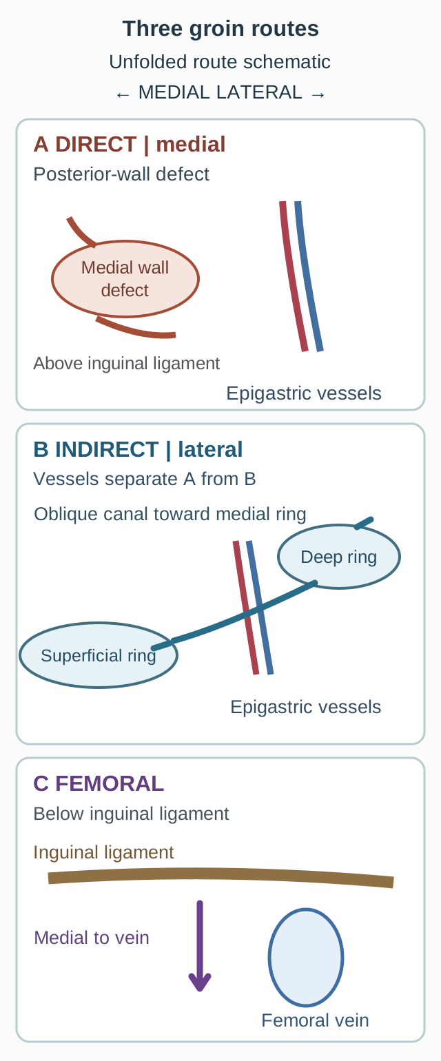
Use the route diagram to follow each path from the abdominal side toward the groin. First identify the epigastric vessels, then the ligament. Keep a finger beside the entrance as you trace, rather than following only the largest part of the swelling.
Trace the entrance of each route before naming its external swelling. Left is medial in every panel; [1][4][7][9]
Trace a neck lateral to the epigastric vessels that enters the deep ring.
Return to the abdominal side. Trace a defect medial to the same vessels, above the ligament.
Return again. Trace an entrance below the ligament, medial to the femoral vein.
Check the lateral entrance
The first path is indirect because it uses the deep ring.
Check the medial entrance
The second path is direct because it crosses the posterior wall within Hesselbach triangle.
Check the subinguinal entrance
The third path is femoral because it enters below the ligament beside the femoral vein.
The complete comparison remains visible in the diagram: indirect uses the lateral ring, direct crosses the medial wall, and femoral lies below the ligament. Now imagine the lateral sac lengthening toward the scrotum. Its name does not change because its entrance has not changed. Likewise, seeing a sac at the superficial ring does not identify which entrance it used. [1][7][9]
The midpoint of the inguinal ligament is halfway between the anterior superior iliac spine and pubic tubercle; the deep ring lies just above it. The mid-inguinal point instead uses the pubic symphysis and approximates the femoral arterial crossing. The two landmarks are not interchangeable. [1][7]
Build a tunnel, not a stack of four identical layers
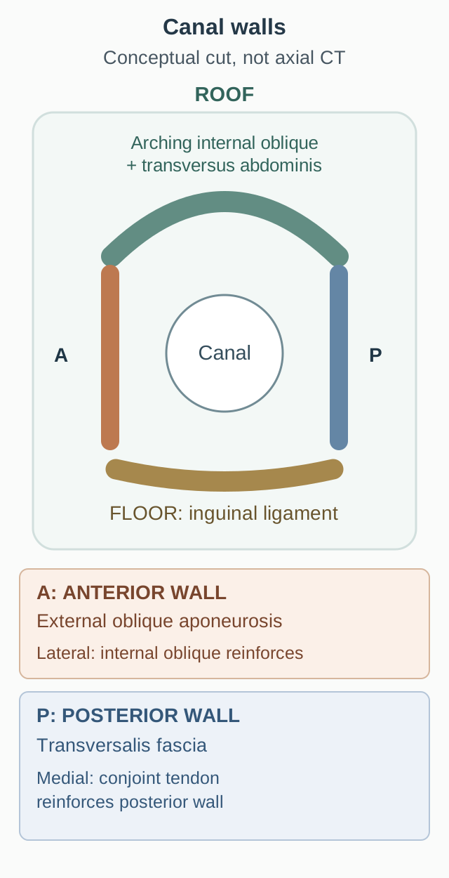
If external oblique forms the front wall, why does strengthening only that layer not address a direct hernia? The canal sits between layers. Its front, back, roof and floor are different boundaries, not four consecutive wrappers around the cord. The cross-sectional diagram separates those directions;
Anterior wall
External oblique aponeurosis forms the main front wall. Internal oblique reinforces it laterally, near the deep ring.
Posterior wall
Transversalis fascia forms the main back wall. Medially, aponeurotic fibers of internal oblique and transversus abdominis provide reinforcement traditionally described as the conjoint tendon or inguinal falx. Its configuration varies.
Roof
Arching fibers of internal oblique and transversus abdominis pass above the canal.
Floor
The shelving edge of the inguinal ligament supports the canal below. The lacunar ligament reinforces the medial part. [1][9]
Picture an open anterior repair. After the external oblique aponeurosis is opened, the canal contents are exposed. A broad bulge behind those contents, medial to the epigastric vessels, reflects failure of the posterior wall, not a new opening through the roof. Surgeons may call that posterior wall the inguinal "floor" in operative descriptions; in a four-wall anatomy question, the inferior floor is the inguinal ligament. Read the orientation before choosing a structure. [9]
Try the comparison: point first to the lateral reinforcement in front, then to the medial reinforcement behind. Which structure would support a medial posterior-wall repair?
Compare the main wall with its regional reinforcement. Letters A and P provide identification independent of color. This is not an axial scan. [1][9]Check the medial support
The conjoint region supports the medial posterior wall; lateral internal-oblique fibers support the anterior wall.
The distinction explains why the same muscle can contribute to more than one boundary. Internal oblique helps the anterior wall laterally, arches across the roof and contributes aponeurotic fibers medially. Transfer that relationship to a new operative view: opening external oblique gains access, but the weakness of a direct hernia remains behind the cord. [1][9]
Descent explains both coverings and persistent channels
Should every muscle crossed on a drawing supply a cord covering? No. The descent pathway passes beneath an arch rather than piercing every layer in a straight line. During development, the processus vaginalis is a peritoneal outpouching associated with the gubernaculum and the route of testicular descent. It extends through the deep ring and canal before the testis completes its descent. The testis is not a free organ floating within the lumen of that outpouching. [3]
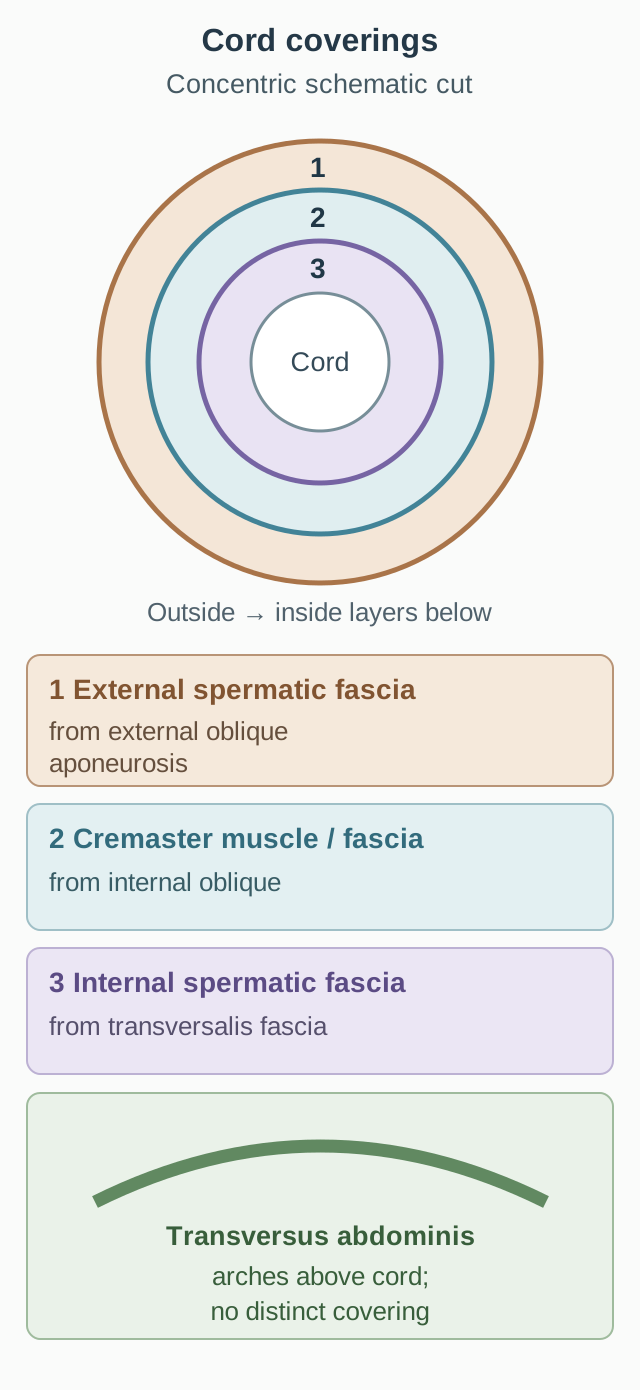
Follow the covering diagram from deep to superficial. Transversalis fascia continues as internal spermatic fascia; internal oblique contributes cremaster muscle and fascia; external oblique aponeurosis supplies external spermatic fascia at the superficial ring. The letters I, C and E name the resulting coverings from inside outward, not the names of the abdominal-wall muscles. [2]
Read the numbered coverings from outside inward, then reverse the sequence to reconstruct descent. The final arch depicts a relationship, not a fourth coat. [1][2][3]
Transversus abdominis arches above the route and contributes no distinct spermatic-cord covering in the standard anatomic description. It can therefore help form the roof and medial posterior reinforcement without supplying a fourth wrapper. The tunica vaginalis is different again: it is the distal peritoneal remnant around the testis, not another abdominal-wall-derived spermatic fascia. [1][2][3]
Build the dissection backward: imagine opening external spermatic fascia and then encountering contractile fibers before reaching internal spermatic fascia. Name the middle layer and its parent muscle.
Check the middle covering
The middle layer is cremaster muscle and fascia, derived from internal oblique.
Reverse the direction for transfer. At the deep ring, the first named spermatic covering comes from transversalis fascia, not transversus abdominis. Similar names do not mean identical tissue. [2]
Ask what remains open
The proximal processus vaginalis normally loses its communication with the peritoneal cavity; its distal portion persists as the tunica vaginalis. Closure timing varies, and a patent channel need not be symptomatic. When fluid passes from the peritoneal cavity toward the testis, the result is a communicating hydrocele. When bowel, omentum or another abdominal structure enters the same route, it is an indirect hernia. They may coexist. [3]
Open proximally and distally: fluid can extend from the abdomen into the scrotum.
Open proximally but closed above the testis: a funicular hydrocele can fill along the cord without surrounding the testis.
Closed at both ends with a persistent middle segment: an encysted cord hydrocele holds an isolated fluid collection.
Compare two ultrasound reports: fluid around the testis enlarges with standing in one child; an isolated fluid pocket above the testis has no demonstrated connection and does not change with straining in another. Predict which collection can exchange fluid with the abdomen.
Check the communication prediction
The first pattern supports an open communication; the isolated, unchanged pocket supports an encysted segment.
This is a pathway comparison, not a guarantee from a single bedside sign. Dynamic imaging and the full examination distinguish fluid from tissue and establish continuity. A new painful or irreducible swelling needs assessment rather than reassurance from the word hydrocele. [3][6]
A cord covering is not a cord content
Does everything in the inguinal canal belong to the spermatic cord? The canal contains a wrapped bundle plus structures traveling beside it. The covering diagram shows the wrappers; the list below identifies what they enclose. [1][2]
The ductus deferens transports sperm.
The testicular artery, artery to the ductus deferens and cremasteric artery supply different components.
The pampiniform venous plexus drains the testis and participates in heat exchange with the testicular artery.
The genital branch of the genitofemoral nerve supplies the cremaster; autonomic fibers accompany vessels.
Testicular lymphatics accompany the cord toward abdominal para-aortic nodes.
A remnant of the processus vaginalis may remain along the cord. [2][3]
The ilioinguinal nerve typically accompanies the cord along its anterior surface and is not counted as a true spermatic-cord content. Its relationship to the cremasteric layers varies, so this schematic relationship is not an invariant operative identification rule. [5] Do not count the femoral nerve, which passes into the thigh beneath the inguinal ligament, as either one. [1][7]
Predict the result: in typical anatomy, after the cord is isolated, a small nerve remains on its surface, outside its coverings. A second nerve runs with the cord and activates cremaster. Which one can be identified from each relationship?
Check the surface nerve
The usual surface nerve is ilioinguinal.
Check the motor pathway
The genital branch of the genitofemoral nerve supplies cremaster.
These routes explain a useful reflex comparison. In the usual cremasteric reflex, upper medial thigh stimulation provides an afferent signal through the ilioinguinal nerve, and the genital branch carries the efferent signal to cremaster. A selective injury to either limb can impair the reflex; an absent reflex alone does not identify which limb failed. Transfer the same reasoning to operative identification: combine course and function rather than choosing a nerve from one symptom. [2][5]
Below the ligament is a different entrance
Can a groin lump above the skin crease exclude a femoral hernia? Surface position can mislead as a sac enlarges. Locate its neck relative to the inguinal ligament and femoral vein. A femoral hernia enters the femoral canal below the ligament and medial to the vein, generally inferolateral to the pubic tubercle. An inguinal entrance is above the ligament. [7][9]
Use the lower route in the first diagram as a comparison. Beneath the ligament, the order from lateral to medial is femoral nerve, artery, vein and femoral canal. The nerve is outside the femoral sheath; the artery, vein and canal occupy its compartments. The canal normally accommodates loose tissue and lymphatics and permits venous expansion. [7]
Localize before naming: an ultrasound report places a sac neck medial to the femoral vein and inferior to the ligament. A second report places a neck lateral to the inferior epigastric vessels above the ligament. Name the two routes before considering the patient's age or sex.
Check the comparison
The first route is femoral; the second is indirect inguinal.
The two vessel landmarks answer different questions. Inferior epigastric vessels separate direct from indirect inguinal entrances; the femoral vein locates the femoral canal. Neither vessel should be substituted for the other. [1][7]
One region, several potential defects
The myopectineal orifice is the broader weak region containing the direct, indirect and femoral sites. The inguinal ligament divides its inguinal region above from its femoral region below. A sufficiently broad preperitoneal repair can cover all three potential entrances; covering only the deep ring does not address a separate medial posterior-wall or femoral defect. This is an anatomical rationale, not a universal prescription for a particular mesh or operation. Repair selection depends on the patient, hernia, expertise and shared decision-making. [4][6]
Safety transfer: a previously reducible bulge that becomes acutely irreducible needs urgent surgical assessment. Increasing pain, vomiting, obstruction or concerning skin findings raise suspicion of compromised bowel. A normal lactate does not safely exclude a small ischemic segment. Suspected strangulation calls for immediate emergency surgical management, not delayed observation to perfect the anatomical label. The 2023 guideline distinguishes acute irreducibility from confirmed strangulation and notes that emergency evidence is often low quality. [6]
Two nerves share an exit without sharing an entrance
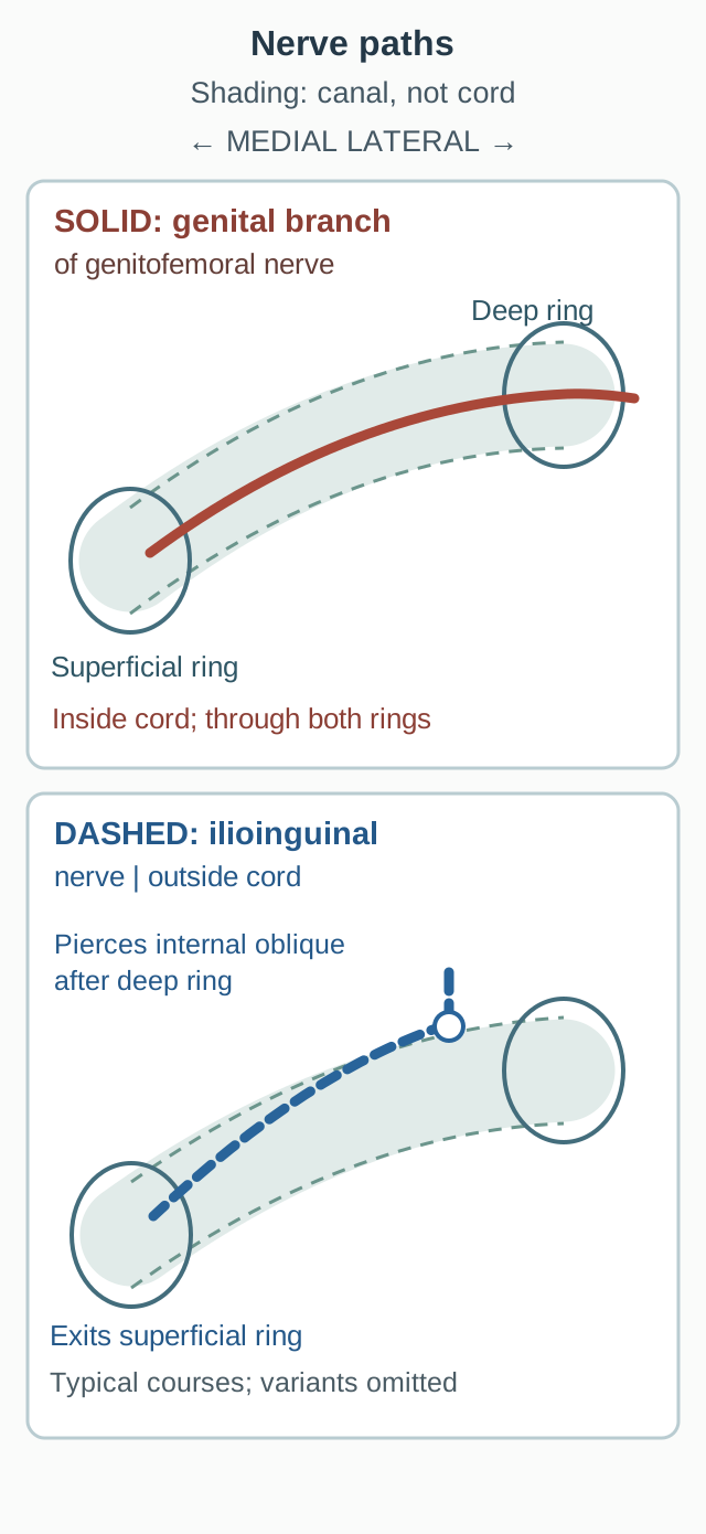
Does exiting the superficial ring prove that a nerve entered through the deep ring? Trace the two paths in the nerve diagram. The genital branch of the genitofemoral nerve enters the deep ring with the cord. The ilioinguinal nerve typically pierces internal oblique to join the canal farther along, bypassing the deep ring. Both can emerge near the superficial ring. [1][5]
Trace typical courses. Ilioinguinal is not counted as a true cord content, but actual relationships to cremasteric layers vary. Solid and dashed lines distinguish paths independently of color. [1][2][5]
Predict selective exposure: a dissection stays at the deep ring without opening the lateral muscular wall. Which of these two nerves is expected in that field?
Check the deep-ring nerve
The genital branch is expected at the deep ring; the usual ilioinguinal route joins distally.
The consequence matters in repair: preserving a nerve seen beside the cord does not establish that every cord-associated nerve is intact. Conversely, numbness alone cannot prove an exact nerve injury because sensory territories overlap and branching varies. Use the observed route, motor function and examination together. These diagrams show typical anatomy, not a substitute for direct identification. [5]
Do not confuse the genital branch with the femoral branch of the same nerve. The femoral branch passes beneath the inguinal ligament to supply skin over the femoral triangle; it does not provide the genital branch's cremaster motor output. [10]
The canal exists without a spermatic cord
In females, the uterine round ligament traverses the deep ring, canal and superficial ring toward the labia majora. The ilioinguinal nerve still joins through the wall rather than the deep ring; the genital branch accompanies the round ligament. The developmental peritoneal extension accompanying this pathway is called the canal of Nuck when persistent. It can permit an indirect hernia or form a hydrocele. A female inguinal sac may contain bowel or an ovary, so a groin mass should not be assumed to contain only fat. [1][3]
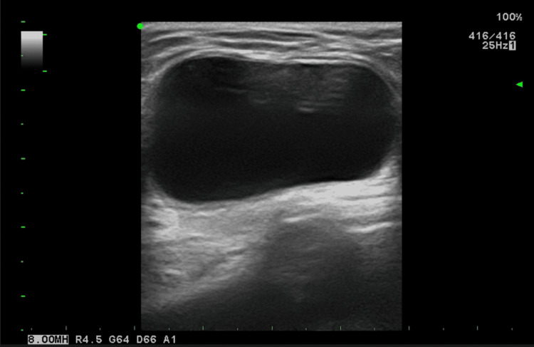
The accompanying published ultrasound shows a canal-of-Nuck hydrocele from the authors' patient. Identify the dark fluid-filled region and the brighter tissue deep to it, consistent with posterior acoustic enhancement. This still image supports a fluid collection but does not establish communication with the peritoneal cavity; that requires additional dynamic imaging or anatomical evidence. The image is a real clinical example, not a diagram and not an assessment of your diagnostic accuracy. [8]
Figure 3, Hydrocele of the Canal of Nuck: A Review, by. Image: Nattawut Keeratibharat and Jirapa Chansangrat, Cureus 2022, doi:10.7759/cureus.23757. Original published image, CC BY 4.0. Use the cyst and posterior enhancement to support a fluid interpretation, not to infer communication. Nattawut Keeratibharat and Jirapa Chansangrat; original source; CC BY 4.0. [8].
Interpret the limit: does this still image alone establish an open connection with the abdomen?
Check what the image cannot establish
No. Communication requires additional dynamic imaging or anatomical evidence.
A cystic appearance supports a fluid collection, while continuity, changes with straining, Doppler findings and any bowel or ovarian tissue refine the diagnosis. Transfer the developmental explanation rather than a sex-based shortcut: persistent peritoneal pathways can cause disease in either inguinal canal. [3][8]
Apply the anatomy to a new route
Case 1
Show answer and explanations for case 1
A. Internal oblique muscle (Why this does not fit)
Internal oblique contributes cremaster muscle and fascia, the middle covering. No. The lateral entrance is the deep ring, where transversalis fascia supplies the innermost sleeve.
Reasoning steps for option A
Which cord covering comes from internal oblique?
Internal oblique contributes cremaster muscle and fascia, the middle covering.
Is the sampled sleeve the middle covering?
No. The lateral entrance is the deep ring, where transversalis fascia supplies the innermost sleeve.
B. External oblique aponeurosis (Why this does not fit)
Its aponeurosis supplies external spermatic fascia at the superficial ring. No. This lateral neck enters through the deep ring; the superficial exit and outer sleeve occur farther along the route.
Reasoning steps for option B
Where does external oblique contribute a spermatic covering?
Its aponeurosis supplies external spermatic fascia at the superficial ring.
Does that explain a sleeve sampled at this abdominal entrance?
No. This lateral neck enters through the deep ring; the superficial exit and outer sleeve occur farther along the route.
C. Transversalis fascia (Best answer)
That combination identifies the indirect route through the deep inguinal ring. Transversalis fascia forms the deep-ring margin and continues as internal spermatic fascia, outside the peritoneal sac.
Reasoning steps for option C
Which entrance is identified by a lateral neck following the cord?
That combination identifies the indirect route through the deep inguinal ring.
Which tissue continues from that aperture into the inner sleeve?
Transversalis fascia forms the deep-ring margin and continues as internal spermatic fascia, outside the peritoneal sac.
D. Transversus abdominis muscle (Why this does not fit)
Its arch passes above the canal and does not supply a distinct spermatic covering. Transversalis fascia, not transversus muscle, contributes the sleeve at the deep-ring entrance.
Reasoning steps for option D
How does transversus abdominis relate to the descending cord?
Its arch passes above the canal and does not supply a distinct spermatic covering.
Which similarly named tissue supplies the observed inner sleeve instead?
Transversalis fascia, not transversus muscle, contributes the sleeve at the deep-ring entrance.
Takeaway: Identify the entrance before assigning a covering; the spermatic sleeve is not the peritoneal sac wall.
A. External oblique and internal oblique (Why this does not fit)
External oblique forms the main anterior wall and the superficial ring. No. The described neck is in the direct region; its conventional aponeurotic reinforcement combines internal oblique with transversus abdominis.
Reasoning steps for option A
What canal support does external oblique chiefly provide?
External oblique forms the main anterior wall and the superficial ring.
Does adding internal oblique make this the medial posterior reinforcement?
No. The described neck is in the direct region; its conventional aponeurotic reinforcement combines internal oblique with transversus abdominis.
B. Internal oblique and transversus abdominis (Best answer)
Above the ligament, this interval is the direct region of the posterior wall. Aponeurotic fibers of internal oblique and transversus abdominis form the conventional conjoint region, with variable fusion.
Reasoning steps for option B
Which canal wall contains a neck between rectus and the epigastric vessels?
Above the ligament, this interval is the direct region of the posterior wall.
Which fibers reinforce that wall medially?
Aponeurotic fibers of internal oblique and transversus abdominis form the conventional conjoint region, with variable fusion.
C. External oblique and transversus abdominis (Why this does not fit)
Transversus abdominis contributes medial aponeurotic reinforcement. External oblique chiefly forms the anterior wall; internal oblique is the partner in the conventional medial posterior reinforcement.
Reasoning steps for option C
Which member of this pair contributes to the conjoint region?
Which proposed partner conflicts with the localized wall?
External oblique chiefly forms the anterior wall; internal oblique is the partner in the conventional medial posterior reinforcement.
D. Rectus abdominis and internal oblique (Why this does not fit)
Its lateral border marks the medial boundary of the direct inguinal region. No. The reinforcing fibers come from internal oblique and transversus abdominis, not rectus itself.
Reasoning steps for option D
What role does rectus have in locating this sac?
Its lateral border marks the medial boundary of the direct inguinal region.
Does being a boundary make rectus a source of conjoint fibers?
No. The reinforcing fibers come from internal oblique and transversus abdominis, not rectus itself.
Takeaway: The borders locate a direct defect; the conjoint region reinforces it but need not form one discrete fused tendon.
A. Reduced leg arterial inflow with initially preserved venous return (Why this does not fit)
The femoral vein lies immediately lateral; the artery lies farther lateral. No. The first adjacent major vessel is venous, so impaired venous return is the expected initial effect.
Reasoning steps for option A
Which major vessel lies directly lateral to the femoral canal?
The femoral vein lies immediately lateral; the artery lies farther lateral.
Would early lateral expansion preferentially obstruct arterial inflow?
No. The first adjacent major vessel is venous, so impaired venous return is the expected initial effect.
B. Increased leg venous return with initially preserved arterial inflow (Why this does not fit)
It can compress the femoral vein and increase resistance to outflow from the leg. No. Initial arterial preservation fits, but venous return would be impeded rather than increased.
Reasoning steps for option B
What does lateral expansion from this compartment do to the adjacent vein?
It can compress the femoral vein and increase resistance to outflow from the leg.
Would that increase venous return while arterial flow persists?
No. Initial arterial preservation fits, but venous return would be impeded rather than increased.
C. Reduced leg arterial inflow with increased venous drainage (Why this does not fit)
The sac first meets the femoral vein on the lateral border of the canal. Neither is the expected early pattern. Compression impedes venous outflow before substantial arterial compression is expected.
Reasoning steps for option C
Which vascular compartment is encountered before the artery?
The sac first meets the femoral vein on the lateral border of the canal.
Do reduced arterial inflow and improved venous drainage follow?
Neither is the expected early pattern. Compression impedes venous outflow before substantial arterial compression is expected.
D. Reduced leg venous return with initially preserved arterial inflow (Best answer)
The entrance is femoral; the canal is medial to the femoral vein within the sheath. Venous outflow from the leg becomes restricted while the farther-lateral artery can initially remain patent.
Reasoning steps for option D
Where does the imaged entrance place the sac relative to the vessels?
The entrance is femoral; the canal is medial to the femoral vein within the sheath.
What follows when expansion compresses that lateral neighbor?
Venous outflow from the leg becomes restricted while the farther-lateral artery can initially remain patent.
Takeaway: Predict compression from the deep neck and its neighbors, not from the height of the skin bulge.
The deep ring lies in transversalis fascia and contributes the internal spermatic sleeve. No. The medial sac bypasses the deep ring, whereas both described sacs reach the superficial exit.
Reasoning steps for option A
Which part of the lateral sac's route involves transversalis fascia?
The deep ring lies in transversalis fascia and contributes the internal spermatic sleeve.
Is that the common terminal covering of both sacs?
No. The medial sac bypasses the deep ring, whereas both described sacs reach the superficial exit.
B. External oblique aponeurosis (Best answer)
An indirect sac traverses the canal from the deep ring, while a direct sac enters medially and can also reach the superficial ring. The superficial ring lies in external oblique aponeurosis, which supplies external spermatic fascia.
Reasoning steps for option B
How can these differently located entrances reach one canal exit?
An indirect sac traverses the canal from the deep ring, while a direct sac enters medially and can also reach the superficial ring.
Which tissue contributes the outer fascia at that shared exit?
The superficial ring lies in external oblique aponeurosis, which supplies external spermatic fascia.
C. Internal oblique aponeurosis (Why this does not fit)
Internal oblique supplies the cremasteric middle covering, not the superficial ring's outer fascia. A covering acquired at the exit is derived from external oblique aponeurosis.
Reasoning steps for option C
Which spermatic covering is associated with internal oblique?
Internal oblique supplies the cremasteric middle covering, not the superficial ring's outer fascia.
What does the common terminal location require instead?
A covering acquired at the exit is derived from external oblique aponeurosis.
D. Transversus abdominis aponeurosis (Why this does not fit)
No. It arches above the canal and contributes regional support rather than a separate cord covering. Acquisition at the shared superficial exit identifies external oblique aponeurosis.
Reasoning steps for option D
Does transversus abdominis provide a distinct spermatic coat?
No. It arches above the canal and contributes regional support rather than a separate cord covering.
Which observed feature identifies the source of the sampled coat?
Acquisition at the shared superficial exit identifies external oblique aponeurosis.
Takeaway: Different inguinal entrances can share an exit and its external spermatic covering.
A. External oblique aponeurosis (Why this does not fit)
External oblique aponeurosis supplies the external spermatic fascia. No. The failed effector is the cremasteric muscle layer, which derives from internal oblique.
Reasoning steps for option A
Which layer derives from external oblique?
External oblique aponeurosis supplies the external spermatic fascia.
Can loss of that noncontractile fascia explain failure of direct muscle stimulation?
No. The failed effector is the cremasteric muscle layer, which derives from internal oblique.
B. Internal oblique muscle (Best answer)
They localize the response failure to the muscle effector rather than the tested sensory input or motor axons. The cremasteric muscle and fascia derive conventionally from internal oblique.
Reasoning steps for option B
What do intact motor conduction and failed direct muscle stimulation localize?
They localize the response failure to the muscle effector rather than the tested sensory input or motor axons.
Which abdominal muscle supplies that effector in the middle covering?
The cremasteric muscle and fascia derive conventionally from internal oblique.
C. Transversalis fascia (Why this does not fit)
It contributes the internal spermatic fascia, the innermost sleeve. No. The sleeve is intact and noncontractile; failure is in the middle cremasteric muscle layer.
Reasoning steps for option C
Which covering is derived from transversalis fascia?
It contributes the internal spermatic fascia, the innermost sleeve.
Does the case identify that sleeve as the failed contractile structure?
No. The sleeve is intact and noncontractile; failure is in the middle cremasteric muscle layer.
D. Transversus abdominis muscle (Why this does not fit)
It arches above the canal without supplying a distinct cord coat. No. That coat is cremaster, derived from internal oblique despite its proximity to transversus.
Reasoning steps for option D
How does transversus abdominis relate to the cord coverings?
It arches above the canal without supplying a distinct cord coat.
Could it be the source of the nonresponsive middle muscle coat?
No. That coat is cremaster, derived from internal oblique despite its proximity to transversus.
Takeaway: A functioning motor axon cannot contract a damaged effector; cremaster is the internal-oblique contribution to the cord.
A. The posterior wall medial to the inferior epigastric vessels (Why this does not fit)
A direct inguinal hernia enters medial to the inferior epigastric vessels. No. A persistent processus vaginalis connects the peritoneum to the scrotum through the deep ring, not a new medial wall defect.
Reasoning steps for option A
What hernia route enters through the medial posterior wall?
A direct inguinal hernia enters medial to the inferior epigastric vessels.
Does that route explain bowel following this demonstrated fluid connection?
No. A persistent processus vaginalis connects the peritoneum to the scrotum through the deep ring, not a new medial wall defect.
B. The deep ring lateral to the inferior epigastric vessels (Best answer)
A patent processus vaginalis provides a communicating peritoneal extension. It takes an indirect inguinal route through the deep ring, lateral to the inferior epigastric vessels.
Reasoning steps for option B
What does fluid continuity from the abdomen to the testis imply?
A patent processus vaginalis provides a communicating peritoneal extension.
Where must bowel enter when it follows that same extension?
It takes an indirect inguinal route through the deep ring, lateral to the inferior epigastric vessels.
C. The femoral canal medial to the femoral vein (Why this does not fit)
It lies below the ligament, medial to the femoral vein. No. The persistent developmental channel follows the inguinal route above the ligament rather than the femoral canal.
Reasoning steps for option C
Where is the femoral canal relative to the inguinal ligament?
It lies below the ligament, medial to the femoral vein.
Does that compartment provide the demonstrated peritoneum-to-testis channel?
No. The persistent developmental channel follows the inguinal route above the ligament rather than the femoral canal.
D. The superficial ring within the external oblique aponeurosis (Why this does not fit)
It is the external-oblique exit after passage through the inguinal canal. No. Bowel first enters at the deep ring; reaching the superficial ring does not make it the peritoneal entrance.
Reasoning steps for option D
What role does the superficial ring have in an indirect route?
It is the external-oblique exit after passage through the inguinal canal.
Is that the abdominal entrance requested here?
No. Bowel first enters at the deep ring; reaching the superficial ring does not make it the peritoneal entrance.
Takeaway: Fluid and bowel can use the same patent processus vaginalis; the contents change, not the deep-ring entrance.
A. Funicular cord hydrocele; no abdominal refilling (Why this does not fit)
A funicular hydrocele remains open proximally to the peritoneum while ending above the testis. No. The absence of abdominal refilling fits the new state, but the retained fluid is now isolated between two sealed ends.
Reasoning steps for option A
Which connection defines the initial funicular collection?
A funicular hydrocele remains open proximally to the peritoneum while ending above the testis.
Does that classification still fit after confirmed proximal closure?
No. The absence of abdominal refilling fits the new state, but the retained fluid is now isolated between two sealed ends.
B. Funicular cord hydrocele; persistent abdominal refilling (Why this does not fit)
A patent proximal channel would still connect the cord collection with the peritoneal cavity. No. Complete proximal closure removes both the funicular configuration and its route for abdominal refilling.
Reasoning steps for option B
What would permit persistent pressure-driven abdominal refilling?
A patent proximal channel would still connect the cord collection with the peritoneal cavity.
Is that necessary channel present at the later assessment?
No. Complete proximal closure removes both the funicular configuration and its route for abdominal refilling.
C. Encysted cord hydrocele; no abdominal refilling (Best answer)
The residual fluid occupies an isolated cord segment, the configuration of an encysted hydrocele. There is no open abdominal channel through which additional peritoneal fluid can enter; retained fluid need not disappear immediately.
Reasoning steps for option C
What is the later configuration after adding a proximal seal to the existing distal seal?
The residual fluid occupies an isolated cord segment, the configuration of an encysted hydrocele.
How does that configuration affect refilling during abdominal pressure rises?
There is no open abdominal channel through which additional peritoneal fluid can enter; retained fluid need not disappear immediately.
D. Encysted cord hydrocele; persistent abdominal refilling (Why this does not fit)
Both the proximal and distal ends of the residual cord cavity are closed. No. Demonstrated new inflow would contradict the stated complete proximal seal and require reassessment of the connection.
Reasoning steps for option D
What does the encysted classification say about the two ends?
Both the proximal and distal ends of the residual cord cavity are closed.
Can that closed configuration support continuing abdominal refilling?
No. Demonstrated new inflow would contradict the stated complete proximal seal and require reassessment of the connection.
Takeaway: A new proximal seal changes a funicular connection into an isolated cord cavity; it does not instantly remove retained fluid.
A. The superficial ring in external oblique aponeurosis (Why this does not fit)
The superficial ring is the distal exit through external oblique aponeurosis. No. Fill-dependent scrotal fluid suggests an abdominal communication that first reaches the canal through the deep ring.
Reasoning steps for option A
Which part of an inguinoscrotal route crosses external oblique?
The superficial ring is the distal exit through external oblique aponeurosis.
Does that explain the first abdominal passage of dialysate?
No. Fill-dependent scrotal fluid suggests an abdominal communication that first reaches the canal through the deep ring.
B. A medial posterior-wall defect in transversalis fascia (Why this does not fit)
It would create a direct inguinal entrance through the medial posterior wall. No such defect is present. Dialysate-dependent peritesticular fluid instead supports a persistent processus vaginalis using the lateral deep-ring route.
Reasoning steps for option B
What route would a medial transversalis defect create?
It would create a direct inguinal entrance through the medial posterior wall.
Does the supplied pattern support a new medial defect?
No such defect is present. Dialysate-dependent peritesticular fluid instead supports a persistent processus vaginalis using the lateral deep-ring route.
C. The deep ring in lateral transversalis fascia (Best answer)
The peritesticular space is communicating with the peritoneal cavity through a persistent processus vaginalis. The deep inguinal ring is an opening in transversalis fascia lateral to the inferior epigastric vessels.
Reasoning steps for option C
What does the repeated fill-and-drain relationship suggest about the scrotal fluid?
The peritesticular space is communicating with the peritoneal cavity through a persistent processus vaginalis.
Which abdominal-wall aperture starts that communicating route?
The deep inguinal ring is an opening in transversalis fascia lateral to the inferior epigastric vessels.
D. The femoral ring below the inguinal ligament (Why this does not fit)
It would enter the subinguinal femoral canal, medial to the femoral vein. No. A patent processus vaginalis follows the inguinal canal above the ligament rather than the femoral compartment.
Reasoning steps for option D
Where would a femoral-ring entrance direct the fluid initially?
It would enter the subinguinal femoral canal, medial to the femoral vein.
Does that explain this developmental route to the testicular serosal space?
No. A patent processus vaginalis follows the inguinal canal above the ligament rather than the femoral compartment.
Takeaway: A fill-dependent scrotal fluid collection can reveal a persistent peritoneal connection; identify its entrance rather than its distal exit.
It enters through internal oblique distal to the deep ring rather than using the abdominal ring. No. The communicating canal-of-Nuck tract enters at the deep ring, which the usual ilioinguinal course bypasses.
Reasoning steps for option A
How does the ilioinguinal nerve usually join the canal?
It enters through internal oblique distal to the deep ring rather than using the abdominal ring.
Does sharing the superficial exit mean it shares this fluid tract's abdominal entrance?
No. The communicating canal-of-Nuck tract enters at the deep ring, which the usual ilioinguinal course bypasses.
B. Iliohypogastric nerve (Why this does not fit)
It supplies the lower abdominal wall and suprapubic region rather than traversing the canal through the deep ring. No. The demonstrated extension follows the deep-ring inguinal route, shared by the genital genitofemoral branch.
Reasoning steps for option B
What is the usual distal route of the iliohypogastric nerve?
It supplies the lower abdominal wall and suprapubic region rather than traversing the canal through the deep ring.
Does that course follow the communicating inguinolabial extension?
No. The demonstrated extension follows the deep-ring inguinal route, shared by the genital genitofemoral branch.
C. Genital branch of the genitofemoral nerve (Best answer)
A patent canal of Nuck, the female peritoneal extension along the inguinal route, communicates through the deep ring. The genital branch of the genitofemoral nerve enters the deep ring; the usual ilioinguinal nerve joins farther distally.
Reasoning steps for option C
Which developmental route explains demonstrated peritoneal inflow into an inguinolabial collection?
A patent canal of Nuck, the female peritoneal extension along the inguinal route, communicates through the deep ring.
Which nerve normally passes through that same abdominal aperture?
The genital branch of the genitofemoral nerve enters the deep ring; the usual ilioinguinal nerve joins farther distally.
D. Femoral branch of the genitofemoral nerve (Why this does not fit)
It passes beneath the inguinal ligament toward skin over the femoral triangle. No. The tract uses the deep inguinal ring above the ligament, not the subinguinal sensory route.
Reasoning steps for option D
Where does the femoral genitofemoral branch pass relative to the ligament?
It passes beneath the inguinal ligament toward skin over the femoral triangle.
Is that the entrance of the communicating inguinolabial tract?
No. The tract uses the deep inguinal ring above the ligament, not the subinguinal sensory route.
Takeaway: Establish communication before mapping a fluid tract; the genital branch, not the usual ilioinguinal nerve, shares its deep-ring entrance.
A. Venous congestion with diminished cooling of incoming arterial blood (Best answer)
The pampiniform venous plexus drains the testis and lies in a heat-exchanging arrangement around the artery. Venous congestion develops and effective countercurrent cooling of incoming arterial blood is impaired; initial arterial patency does not prevent these effects.
Reasoning steps for option A
Which network is identified by thin-walled vessels surrounding the testicular artery?
The pampiniform venous plexus drains the testis and lies in a heat-exchanging arrangement around the artery.
What follows from obstructing that network despite preserved arterial inflow?
Venous congestion develops and effective countercurrent cooling of incoming arterial blood is impaired; initial arterial patency does not prevent these effects.
B. Venous congestion with increased cooling of incoming arterial blood (Why this does not fit)
Yes. It fits restricted outflow through the pampiniform plexus. No. Reduced venous circulation impairs the normal countercurrent exchange rather than improving it.
Reasoning steps for option B
Does distal venous distension support congestion?
Yes. It fits restricted outflow through the pampiniform plexus.
Would obstructing that plexus increase effective arterial precooling?
No. Reduced venous circulation impairs the normal countercurrent exchange rather than improving it.
C. Venous decompression with diminished cooling of incoming arterial blood (Why this does not fit)
No. Continued inflow with restricted outflow favors venous distension and congestion. Diminished cooling fits injury to the pampiniform heat-exchange arrangement, but venous decompression does not.
Reasoning steps for option C
Would an obstruction decompress veins on the testicular side?
No. Continued inflow with restricted outflow favors venous distension and congestion.
Which part of this proposed effect nevertheless fits the network's role?
Diminished cooling fits injury to the pampiniform heat-exchange arrangement, but venous decompression does not.
D. Venous decompression with increased cooling of incoming arterial blood (Why this does not fit)
Outflow and circulating venous contact would need to improve, rather than become obstructed. No. The findings instead predict congestion and impaired arterial precooling while inflow is initially preserved.
Reasoning steps for option D
What would be required for venous decompression with better heat exchange?
Outflow and circulating venous contact would need to improve, rather than become obstructed.
Does the observed distended, poorly flowing plexus support either proposed improvement?
No. The findings instead predict congestion and impaired arterial precooling while inflow is initially preserved.
Takeaway: Preserved arterial inflow does not preserve venous drainage or pampiniform heat exchange after venous obstruction.
A. Stimulus not perceived; reflex contraction preserved (Why this does not fit)
B joins through internal oblique and is the typical ilioinguinal nerve, so the usual sensory pathway remains intact. Both halves conflict: perception should remain, while dividing the deep-ring genital branch interrupts cremasteric motor output.
Reasoning steps for option A
Which preserved nerve supplies the usual upper medial thigh sensory route?
B joins through internal oblique and is the typical ilioinguinal nerve, so the usual sensory pathway remains intact.
Which half of this proposed response conflicts with dividing A?
Both halves conflict: perception should remain, while dividing the deep-ring genital branch interrupts cremasteric motor output.
B. Stimulus not perceived; reflex contraction impaired (Why this does not fit)
Yes. A follows the genital genitofemoral course and supplies cremaster in the male counterpart. No. B is the separate, preserved ilioinguinal pathway supplying the usual sensory input.
Reasoning steps for option B
Does division of the deep-ring nerve explain impaired contraction?
Yes. A follows the genital genitofemoral course and supplies cremaster in the male counterpart.
Does the same lesion require loss of the stated thigh stimulus?
No. B is the separate, preserved ilioinguinal pathway supplying the usual sensory input.
C. Stimulus perceived; reflex contraction preserved (Why this does not fit)
The wall-entering ilioinguinal route B is intact under the stipulated typical anatomy. No. A is the genital motor branch, so preserved sensory input does not restore its interrupted output.
Reasoning steps for option C
Why is preserved perception compatible with this operation?
The wall-entering ilioinguinal route B is intact under the stipulated typical anatomy.
Can a normal reflex contraction follow through the divided nerve A?
No. A is the genital motor branch, so preserved sensory input does not restore its interrupted output.
D. Stimulus perceived; reflex contraction impaired (Best answer)
A is the genital genitofemoral branch through the deep ring; B is ilioinguinal through internal oblique. The usual thigh stimulus remains perceived, but the cremasteric motor response is impaired despite an intact muscle.
Reasoning steps for option D
Which nerves are identified by the two different canal entrances?
A is the genital genitofemoral branch through the deep ring; B is ilioinguinal through internal oblique.
What functions remain after A is divided and B is spared?
The usual thigh stimulus remains perceived, but the cremasteric motor response is impaired despite an intact muscle.
Takeaway: Shared passage through part of the canal does not make the two nerves functionally interchangeable.
A. The nerve ends in suprapubic skin without traversing the canal (Why this does not fit)
That course favors iliohypogastric rather than ilioinguinal anatomy. No. Typical ilioinguinal injury better fits that functional pattern; a suprapubic course would support a different localization.
Reasoning steps for option A
Which nerve is suggested by a suprapubic course without canal transit?
That course favors iliohypogastric rather than ilioinguinal anatomy.
Does it best corroborate this loss of the usual thigh afferent with preserved genital motor function?
No. Typical ilioinguinal injury better fits that functional pattern; a suprapubic course would support a different localization.
B. The nerve enters through internal oblique distal to the deep ring (Best answer)
The usual ilioinguinal sensory pathway is implicated, although cutaneous territories overlap. The ilioinguinal nerve typically enters through internal oblique distal to the deep ring rather than accompanying the cord through that ring.
Reasoning steps for option B
What pathway is implicated when usual stimulus perception is lost but the tested motor branch and muscle respond?
The usual ilioinguinal sensory pathway is implicated, although cutaneous territories overlap.
What anatomical course would strengthen that functional localization?
The ilioinguinal nerve typically enters through internal oblique distal to the deep ring rather than accompanying the cord through that ring.
C. The nerve passes beneath the ligament beside the femoral artery (Why this does not fit)
The femoral branch of the genitofemoral nerve passes beneath the ligament toward the femoral triangle. No. Under the stated typical anatomy, that afferent is ilioinguinal and enters through the canal wall.
Reasoning steps for option C
Which small sensory branch follows this subinguinal route?
The femoral branch of the genitofemoral nerve passes beneath the ligament toward the femoral triangle.
Would that be the best match for the usual upper medial thigh reflex afferent?
No. Under the stated typical anatomy, that afferent is ilioinguinal and enters through the canal wall.
D. The nerve follows the spermatic cord through the deep ring (Why this does not fit)
The genital branch of the genitofemoral nerve accompanies the cord through the deep ring. Its continuity and evoked cremaster contraction are established, so this route does not corroborate the separate sensory lesion.
Reasoning steps for option D
Which relevant nerve uses the deep-ring cord route?
The genital branch of the genitofemoral nerve accompanies the cord through the deep ring.
What supplied finding argues against that being the injured nerve?
Its continuity and evoked cremaster contraction are established, so this route does not corroborate the separate sensory lesion.
Takeaway: Functional testing suggests a pathway; its actual course supplies corroboration when sensory territories overlap.
A. Genital branch interruption between the two stimulation points (Best answer)
It localizes an interruption between the two stimulation sites, because the distal axons and muscle still generate contraction. The genital branch of the genitofemoral nerve supplies cremaster; its focal interruption explains the stimulation pattern.
Reasoning steps for option A
What does distal success with proximal failure localize on the same motor nerve?
It localizes an interruption between the two stimulation sites, because the distal axons and muscle still generate contraction.
Which nerve normally supplies this cord-associated motor route?
The genital branch of the genitofemoral nerve supplies cremaster; its focal interruption explains the stimulation pattern.
B. Sensory receptor failure within the upper medial thigh skin (Why this does not fit)
The patient reports perceiving upper medial thigh stimulation. No. Those tests bypass the skin receptors and localize failure within the tested motor route.
Reasoning steps for option B
What evidence shows that the applied skin stimulus reaches conscious perception?
The patient reports perceiving upper medial thigh stimulation.
Could receptor failure explain unequal responses to proximal and distal motor-nerve stimulation?
No. Those tests bypass the skin receptors and localize failure within the tested motor route.
C. Ilioinguinal interruption at its entry through internal oblique (Why this does not fit)
It carries the usual upper medial thigh sensory input rather than cremasteric motor output. No. Failure confined between two sites on the cord-associated motor nerve implicates the genital branch instead.
Reasoning steps for option C
Which reflex role usually belongs to ilioinguinal?
It carries the usual upper medial thigh sensory input rather than cremasteric motor output.
Would an isolated ilioinguinal interruption create the tested motor-nerve dissociation?
No. Failure confined between two sites on the cord-associated motor nerve implicates the genital branch instead.
D. Cremaster muscle failure beyond an intact genital motor branch (Why this does not fit)
Both show that the cremasteric effector can contract. Between the proximal and distal nerve sites, not in the muscle beyond a supposedly intact nerve.
Reasoning steps for option D
What do direct muscle and distal nerve stimulation establish?
Both show that the cremasteric effector can contract.
Where must the remaining failure lie when proximal stimulation fails?
Between the proximal and distal nerve sites, not in the muscle beyond a supposedly intact nerve.
Takeaway: Compare stimulation sites on the same nerve; an intact distal response localizes failure upstream of the responding segment.
A. Stimulate the central end of the wall-entering nerve (Best answer)
The ilioinguinal afferent pathway is implicated; it typically joins the canal through internal oblique. Its central end remains connected toward the spinal cord, so stimulation there bypasses the peripheral interruption and can recruit the intact motor output.
Reasoning steps for option A
Which usual sensory pathway fits loss of the thigh input with intact tested motor output?
The ilioinguinal afferent pathway is implicated; it typically joins the canal through internal oblique.
Which side of its interruption can still send the stimulus toward the spinal circuit?
Its central end remains connected toward the spinal cord, so stimulation there bypasses the peripheral interruption and can recruit the intact motor output.
B. Stimulate the peripheral end of the wall-entering nerve (Why this does not fit)
Yes. It identifies the typical ilioinguinal pathway supplying the missing input. No. Stimulation there remains separated from the central circuit, so it does not bypass the interruption.
Reasoning steps for option B
Does the wall-entering route identify the appropriate usual afferent?
Yes. It identifies the typical ilioinguinal pathway supplying the missing input.
Can its disconnected peripheral end deliver impulses across a complete interruption to the spinal cord?
No. Stimulation there remains separated from the central circuit, so it does not bypass the interruption.
C. Stimulate the central end of the deep-ring nerve (Why this does not fit)
It is the genital genitofemoral pathway, whose motor output has already been shown to function. No. That would test a different nerve, not continuity from the central end of the implicated ilioinguinal input.
Reasoning steps for option C
Which pathway is represented by the deep-ring nerve in this reflex comparison?
It is the genital genitofemoral pathway, whose motor output has already been shown to function.
Does testing its central end specifically test the interrupted original thigh afferent?
No. That would test a different nerve, not continuity from the central end of the implicated ilioinguinal input.
D. Stimulate the peripheral end of the deep-ring nerve (Why this does not fit)
It can directly drive the intact cremasteric muscle distal to the stimulus. No. Direct motor activation bypasses rather than tests the afferent and central portions of the reflex.
Reasoning steps for option D
What response can stimulation of the peripheral genital motor branch produce?
It can directly drive the intact cremasteric muscle distal to the stimulus.
Would that demonstrate recruitment through the original injured sensory pathway and spinal circuit?
No. Direct motor activation bypasses rather than tests the afferent and central portions of the reflex.
Takeaway: A central afferent stimulus can bypass a peripheral sensory interruption; direct motor stimulation answers a different question.
A. Reduced pinprick detection in the skin over the femoral triangle (Why this does not fit)
B is the femoral genitofemoral branch, which supplies skin over the femoral triangle. No. It is compatible with the proposed focal sensory-branch injury.
Reasoning steps for option A
Which sensory territory belongs to the subinguinal branch B?
B is the femoral genitofemoral branch, which supplies skin over the femoral triangle.
Does reduced pinprick in that territory require an additional lesion?
No. It is compatible with the proposed focal sensory-branch injury.
B. Burning neuropathic pain in the skin over the femoral triangle (Why this does not fit)
Yes. A focal sensory-nerve injury can produce neuropathic pain in its supplied territory. No. It can accompany injury to the same femoral genitofemoral sensory branch.
Reasoning steps for option B
Can an injured cutaneous nerve produce pain as well as numbness?
Yes. A focal sensory-nerve injury can produce neuropathic pain in its supplied territory.
Would pain confined to this mapped territory contradict isolated B injury?
No. It can accompany injury to the same femoral genitofemoral sensory branch.
C. Reduced sensory conduction across the injured subinguinal nerve segment (Why this does not fit)
Sensory conduction through that injured segment can be reduced. No. It is a direct consequence within the already implicated subinguinal sensory branch.
Reasoning steps for option C
What electrophysiological effect can a focal injury within B produce?
Sensory conduction through that injured segment can be reduced.
Does that finding extend the lesion beyond B's demonstrated course?
No. It is a direct consequence within the already implicated subinguinal sensory branch.
D. Absent reflex cremaster contraction after the usual thigh stimulus (Best answer)
No. That output normally travels through the separate genital branch A and the deep ring. The isolated B lesion is insufficient; an additional central or motor-pathway problem must be considered rather than assuming A injury is proved.
Reasoning steps for option D
Does branch B carry cremasteric motor output?
No. That output normally travels through the separate genital branch A and the deep ring.
What does absent reflex contraction imply when the usual afferent and muscle test intact?
The isolated B lesion is insufficient; an additional central or motor-pathway problem must be considered rather than assuming A injury is proved.
Takeaway: A subinguinal sensory-branch injury does not by itself explain loss of cremasteric motor output.
A. The anterior wall near the superficial inguinal ring (Why this does not fit)
External oblique aponeurosis forms those anterior structures. No. The injured muscle is transversus abdominis, whose medial aponeurotic contribution supports the posterior wall.
Reasoning steps for option A
Which tissue principally forms the anterior wall and superficial ring?
External oblique aponeurosis forms those anterior structures.
Is external oblique the muscle identified deep to internal oblique?
No. The injured muscle is transversus abdominis, whose medial aponeurotic contribution supports the posterior wall.
B. The posterior wall medial to the inferior epigastric vessels (Best answer)
Transversus abdominis has that relationship; preservation of the cremasteric covering is consistent with sparing its internal-oblique source. It contributes with internal oblique to the conventional conjoint region reinforcing the medial posterior wall, medial to the inferior epigastric vessels.
Reasoning steps for option B
Which muscle lies deep to internal oblique and arches without adding a cord covering?
Transversus abdominis has that relationship; preservation of the cremasteric covering is consistent with sparing its internal-oblique source.
Where does the damaged medial aponeurosis normally contribute support?
It contributes with internal oblique to the conventional conjoint region reinforcing the medial posterior wall, medial to the inferior epigastric vessels.
C. The anterior wall lateral to the inferior epigastric vessels (Why this does not fit)
Internal oblique reinforces the anterior wall laterally. No. The stem identifies the deeper transversus layer and its medial aponeurotic support, not lateral anterior reinforcement by internal oblique.
Reasoning steps for option C
Which muscle provides lateral anterior-wall reinforcement?
Internal oblique reinforces the anterior wall laterally.
Does this fit the identified injured muscle and its torn medial continuation?
No. The stem identifies the deeper transversus layer and its medial aponeurotic support, not lateral anterior reinforcement by internal oblique.
D. The floor beside the medial edge of the femoral vein (Why this does not fit)
The inguinal ligament forms the floor, with medial lacunar reinforcement. No. It removes a component of medial posterior-wall reinforcement rather than the floor beside the femoral vessels.
Reasoning steps for option D
Which structure primarily forms the inguinal canal floor?
The inguinal ligament forms the floor, with medial lacunar reinforcement.
Does tearing the medial transversus aponeurosis primarily remove that ligamentous floor?
No. It removes a component of medial posterior-wall reinforcement rather than the floor beside the femoral vessels.
Takeaway: Transversus supplies no distinct cord coat, but it contributes both to the roof and to medial posterior-wall reinforcement.
A. Direct posterior-wall region and lateral deep-ring region (Why this does not fit)
It would address the neck between rectus and the epigastric vessels above the ligament. No. The deep-ring site is already covered, while the subinguinal neck requires inclusion of the femoral region.
Reasoning steps for option A
Which newly demonstrated entrance would direct-region coverage address?
It would address the neck between rectus and the epigastric vessels above the ligament.
Does repeating deep-ring coverage include the other new neck?
No. The deep-ring site is already covered, while the subinguinal neck requires inclusion of the femoral region.
B. Direct posterior-wall region and superficial-ring exit (Why this does not fit)
No. It is an inguinal exit through external oblique above the ligament. The below-ligament neck medial to the femoral vein is femoral and would remain outside these two proposed additions.
Reasoning steps for option B
Is the superficial ring a separate abdominal entrance corresponding to the lower neck?
No. It is an inguinal exit through external oblique above the ligament.
Which new site would remain absent from this proposed map?
The below-ligament neck medial to the femoral vein is femoral and would remain outside these two proposed additions.
C. Femoral-canal region and lateral deep-ring region (Why this does not fit)
It includes the below-ligament entrance medial to the femoral vein. The new medial inguinal neck in the direct posterior-wall region remains unaccounted for.
Reasoning steps for option C
Which new neck does femoral-canal coverage include?
It includes the below-ligament entrance medial to the femoral vein.
What remains when the other addition repeats an already covered lateral site?
The new medial inguinal neck in the direct posterior-wall region remains unaccounted for.
D. Direct posterior-wall region and femoral-canal region (Best answer)
The rectus-to-epigastric interval identifies a direct entrance, and the below-ligament medial-to-vein neck identifies a femoral entrance. Adding the direct posterior-wall and femoral-canal regions includes the two remaining entrances; actual operative planning still requires individualized assessment.
Reasoning steps for option D
Which entrances are newly present after accounting for the existing lateral patch?
The rectus-to-epigastric interval identifies a direct entrance, and the below-ligament medial-to-vein neck identifies a femoral entrance.
Which additions complete the mapped inventory without counting the lateral site twice?
Adding the direct posterior-wall and femoral-canal regions includes the two remaining entrances; actual operative planning still requires individualized assessment.
Takeaway: Inventory all demonstrated entrances before comparing coverage; an already covered deep ring does not account for direct and femoral sites.
A. Through the medial posterior wall, bypassing the deep inguinal ring (Best answer)
Y remains above the inferior extension and medial to the existing lateral coverage. Y is a direct entrance through the medial posterior inguinal wall and does not require passage through the deep ring.
Reasoning steps for option A
Which neck lies beyond the two stated areas of coverage?
Y remains above the inferior extension and medial to the existing lateral coverage.
What route follows from Y's relationship to the epigastric vessels?
Y is a direct entrance through the medial posterior inguinal wall and does not require passage through the deep ring.
B. Through the femoral canal, below the ligament and medial to the vein (Why this does not fit)
Z lies below the ligament and medial to the femoral vein, identifying the femoral route. No. The inferior extension surrounds Z; the remaining gap is the medial inguinal site above the ligament.
Reasoning steps for option B
Which mapped neck would use the femoral canal?
Z lies below the ligament and medial to the femoral vein, identifying the femoral route.
Does the map leave that specific entrance uncovered?
No. The inferior extension surrounds Z; the remaining gap is the medial inguinal site above the ligament.
C. No residual route; direct, indirect and femoral entrances are covered (Why this does not fit)
No. Coverage must be compared with each entrance rather than counted by the number of patches. Its upper edge ends at the ligament, whereas Y lies above it and medial to the original lateral coverage.
Reasoning steps for option C
Do two covered necks necessarily account for all three mapped sites?
No. Coverage must be compared with each entrance rather than counted by the number of patches.
Which boundary prevents this extension from including Y?
Its upper edge ends at the ligament, whereas Y lies above it and medial to the original lateral coverage.
D. Through the deep inguinal ring, following the cord toward its exit (Why this does not fit)
X is lateral to the epigastric vessels above the ligament, identifying the indirect entrance. No. X is already surrounded by the original coverage; the medial posterior-wall route at Y remains possible.
Reasoning steps for option D
Which mapped neck begins the cord-following deep-ring route?
X is lateral to the epigastric vessels above the ligament, identifying the indirect entrance.
Is X the residual gap after the proposed addition?
No. X is already surrounded by the original coverage; the medial posterior-wall route at Y remains possible.
Takeaway: Coverage boundaries, not the number of covered sites, determine which route remains possible.
A. Primary mesenteric arterial occlusion with widespread bowel injury and delayed systemic marker accumulation (Why this does not fit)
It would threaten bowel in the affected arterial territory rather than being confined to a short loop at one groin neck. The main arteries are patent and impaired enhancement is confined to the trapped loop, favoring local neck compression rather than a widespread arterial event.
Reasoning steps for option A
What distribution would a primary major mesenteric arterial occlusion usually produce?
It would threaten bowel in the affected arterial territory rather than being confined to a short loop at one groin neck.
Which findings instead localize the threat to the hernia?
The main arteries are patent and impaired enhancement is confined to the trapped loop, favoring local neck compression rather than a widespread arterial event.
B. Focal neck compression involving a short bowel loop and limited systemic lactate release (Best answer)
They indicate entrapment of a short loop with suspected local perfusion compromise in a femoral hernia, not merely an external swelling. A small threatened bowel segment may not release enough lactate to produce an abnormal systemic value. Suspected strangulation requires emergency surgical assessment without waiting for the marker to rise.
Reasoning steps for option B
What do paired transitions at one tight neck and reduced loop enhancement suggest?
They indicate entrapment of a short loop with suspected local perfusion compromise in a femoral hernia, not merely an external swelling.
Why does normal lactate not remove that concern?
A small threatened bowel segment may not release enough lactate to produce an abnormal systemic value. Suspected strangulation requires emergency surgical assessment without waiting for the marker to rise.
C. Diffuse functional bowel paralysis with preserved mural perfusion and normal systemic marker production (Why this does not fit)
No. The focal entrance and exit transitions indicate mechanical entrapment rather than diffuse failure of motility. No. Reduced enhancement raises concern about local blood supply, which a normal systemic marker cannot safely dismiss.
Reasoning steps for option C
Would diffuse functional ileus explain a short loop constrained at a single neck?
No. The focal entrance and exit transitions indicate mechanical entrapment rather than diffuse failure of motility.
Does preserved perfusion fit the trapped loop's enhancement?
No. Reduced enhancement raises concern about local blood supply, which a normal systemic marker cannot safely dismiss.
D. Isolated luminal narrowing at the neck with preserved mural perfusion and reassuring biochemical results (Why this does not fit)
Yes, but obstruction alone does not account for every observed feature in this case. Reduced mural enhancement raises concern about perfusion compromise. A normal lactate is not sufficient reassurance about a short trapped segment's viability.
Reasoning steps for option D
Can mechanical obstruction occur without vascular compromise?
Yes, but obstruction alone does not account for every observed feature in this case.
Which finding prevents assuming this loop remains well perfused?
Reduced mural enhancement raises concern about perfusion compromise. A normal lactate is not sufficient reassurance about a short trapped segment's viability.
Takeaway: A normal systemic lactate does not exclude a short strangulating bowel segment; suspected strangulation needs emergency assessment.
A. The obstructing direct route remains; the fat-containing indirect route is addressed (Why this does not fit)
The sac with a transition from dilated to decompressed bowel contains the obstructing lesion. No. A lateral above-ligament neck is indirect; the medial fat-containing sac is the direct route.
Reasoning steps for option A
Which sac contains the evidence of intestinal obstruction?
The sac with a transition from dilated to decompressed bowel contains the obstructing lesion.
Does its lateral neck make it a direct route?
No. A lateral above-ligament neck is indirect; the medial fat-containing sac is the direct route.
B. The fat-containing direct route remains; the obstructing indirect route is addressed (Why this does not fit)
Yes. The fat-containing medial sac is direct and the bowel-containing lateral sac is indirect. No. The plan addresses the medial direct neck, not the lateral indirect neck containing the obstruction.
Reasoning steps for option B
Does this option correctly name the two sacs?
Yes. The fat-containing medial sac is direct and the bowel-containing lateral sac is indirect.
Does it correctly compare those sacs with the stated coverage?
No. The plan addresses the medial direct neck, not the lateral indirect neck containing the obstruction.
C. The fat-containing indirect route remains; the obstructing direct route is addressed (Why this does not fit)
Its neck is lateral to the inferior epigastric vessels, identifying an indirect entrance. No. That reverses the content-to-route assignment; the medial intervention addresses the separate fat-containing direct sac.
Reasoning steps for option C
What entrance defines the bowel-containing sac in this case?
Its neck is lateral to the inferior epigastric vessels, identifying an indirect entrance.
Could treating the medial neck address it as a direct obstruction?
No. That reverses the content-to-route assignment; the medial intervention addresses the separate fat-containing direct sac.
D. The obstructing indirect route remains; the fat-containing direct route is addressed (Best answer)
Following the obstructing bowel identifies the lateral deep-ring entrance and therefore the indirect route. The obstructing indirect route remains unaddressed. The medial direct fat sac is not the source of the demonstrated bowel transition; urgent evaluation of the obstruction remains necessary.
Reasoning steps for option D
Which entrance must be followed from the bowel transition?
Following the obstructing bowel identifies the lateral deep-ring entrance and therefore the indirect route.
What remains after the separately located medial neck is addressed?
The obstructing indirect route remains unaddressed. The medial direct fat sac is not the source of the demonstrated bowel transition; urgent evaluation of the obstruction remains necessary.
Takeaway: Follow the obstructing bowel to its own neck; treating a separate inguinal defect does not address that route.
A depends on the deep ring, whereas B uses a separate above-ligament entrance that bypasses it. The indirect deep-ring entrance A is lateral; the direct posterior-wall entrance B is medial.
Reasoning steps for option A
What does selective obstruction of A by the deep-ring seal establish?
A depends on the deep ring, whereas B uses a separate above-ligament entrance that bypasses it.
Where are those two entrances relative to the epigastric vessels?
The indirect deep-ring entrance A is lateral; the direct posterior-wall entrance B is medial.
B. A lateral; B lateral (Why this does not fit)
Under the model's usual anatomy, both would use the deep-ring route. No. B's unaffected passage indicates a separate direct entrance medial to the vessels.
Reasoning steps for option B
Would two lateral indirect necks both depend on the deep-ring opening?
Under the model's usual anatomy, both would use the deep-ring route.
Does continued passage of B with that opening sealed support two lateral entrances?
No. B's unaffected passage indicates a separate direct entrance medial to the vessels.
C. A medial; B medial (Why this does not fit)
No. It traverses the medial posterior wall and bypasses the ring. No. A's dependence on that ring identifies the lateral indirect entrance, although B is medial.
Reasoning steps for option C
Would a medial direct entrance require an open deep ring?
No. It traverses the medial posterior wall and bypasses the ring.
Can A therefore be medial when a ring-only seal blocks it?
No. A's dependence on that ring identifies the lateral indirect entrance, although B is medial.
D. A medial; B lateral (Why this does not fit)
A is blocked by a seal limited to that ring. No. It reverses the routes: A is lateral and B is medial despite their shared external exit.
Reasoning steps for option D
Which sac behaves like the one using the lateral deep ring?
A is blocked by a seal limited to that ring.
Does assigning the lateral neck to B preserve that experimental relationship?
No. It reverses the routes: A is lateral and B is medial despite their shared external exit.
Takeaway: A shared external exit does not identify the entrance; a deep-ring-only seal affects the indirect route, not the direct route.
A. A femoral entrance outside the repair's documented inferior boundary (Best answer)
A below-ligament neck immediately medial to the femoral vein identifies the femoral canal. It lies inferior to the documented coverage. This does not itself prove failure of the prior inguinal repair or distinguish a new from a previously unrecognized femoral defect.
Reasoning steps for option A
Which route is identified by the actual neck rather than the skin projection?
A below-ligament neck immediately medial to the femoral vein identifies the femoral canal.
How does that entrance compare with a repair ending at the ligament?
It lies inferior to the documented coverage. This does not itself prove failure of the prior inguinal repair or distinguish a new from a previously unrecognized femoral defect.
B. A femoral entrance inside the repair's documented inferior boundary (Why this does not fit)
Yes. Its subinguinal position medial to the vein is characteristic of a femoral entrance. No. A boundary ending at the inguinal ligament does not include the below-ligament femoral entrance.
Reasoning steps for option B
Does the femoral label fit the imaged neck?
Yes. Its subinguinal position medial to the vein is characteristic of a femoral entrance.
Was that entrance included by the recorded inferior boundary?
No. A boundary ending at the inguinal ligament does not include the below-ligament femoral entrance.
C. An indirect entrance outside the repair's documented lateral boundary (Why this does not fit)
At the deep ring above the ligament, lateral to the inferior epigastric vessels. No. Imaging identifies a femoral neck, and the prior record already includes the lateral inguinal region.
Reasoning steps for option C
Where would an indirect abdominal entrance lie?
At the deep ring above the ligament, lateral to the inferior epigastric vessels.
Do either the imaged entrance or the recorded lateral coverage support this comparison?
No. Imaging identifies a femoral neck, and the prior record already includes the lateral inguinal region.
D. A direct entrance inside the repair's documented medial boundary (Why this does not fit)
It enters above the ligament through the medial posterior inguinal wall. No. The actual neck is femoral and outside the recorded inferior boundary, not a demonstrated direct recurrence.
Reasoning steps for option D
Where does a direct neck enter relative to the ligament and epigastric vessels?
It enters above the ligament through the medial posterior inguinal wall.
Can the height of the external mass override the imaged subinguinal neck?
No. The actual neck is femoral and outside the recorded inferior boundary, not a demonstrated direct recurrence.
Takeaway: Classify the new neck before interpreting prior repair coverage; a high skin bulge does not establish inguinal recurrence.
A. Anterior continuity restored; posterior defect also supported (Why this does not fit)
The anterior wall's aponeurotic continuity is restored. No. The neck is in the direct posterior-wall region, which receives no support from the stated limited closure.
Reasoning steps for option A
What layer is restored by reapproximating the external-oblique incision?
The anterior wall's aponeurotic continuity is restored.
Does that repair the wall identified by this medial sac neck?
No. The neck is in the direct posterior-wall region, which receives no support from the stated limited closure.
B. Anterior continuity absent; posterior defect still unsupported (Why this does not fit)
No. That is the specific continuity restored by the closure. The posterior defect remains unsupported because the medial sac's deeper entrance was not addressed.
Reasoning steps for option B
Does closing the external-oblique incision leave anterior continuity absent?
No. That is the specific continuity restored by the closure.
Which part of this option correctly describes the residual problem?
The posterior defect remains unsupported because the medial sac's deeper entrance was not addressed.
C. Anterior continuity restored; posterior defect still unsupported (Best answer)
The direct entrance involves the posterior inguinal wall rather than the external-oblique access layer. Anterior continuity is restored, but the posterior defect remains unsupported. Exposure and treatment of the defect are different anatomical acts.
Reasoning steps for option C
Which wall is implicated by a medial above-ligament sac neck?
The direct entrance involves the posterior inguinal wall rather than the external-oblique access layer.
What remains after closing only the access layer?
Anterior continuity is restored, but the posterior defect remains unsupported. Exposure and treatment of the defect are different anatomical acts.
D. Anterior continuity absent; posterior defect now supported (Why this does not fit)
The tissue at the direct neck or its deeper supporting wall would need to be addressed, which the exercise excludes. It restores anterior continuity, so this option reverses both the restored and the still-unsupported compartments.
Reasoning steps for option D
Which proposed restoration would support the posterior defect?
The tissue at the direct neck or its deeper supporting wall would need to be addressed, which the exercise excludes.
What does the actual external-oblique closure restore instead?
It restores anterior continuity, so this option reverses both the restored and the still-unsupported compartments.
Takeaway: Closing an anterior access incision does not by itself support a separate posterior-wall defect.
A. Greater narrowing of the superficial external-oblique exit (Why this does not fit)
The superficial exit is an aperture in external oblique aponeurosis. No. Their regional contribution is medial posterior-wall reinforcement, not the external-oblique aperture.
Reasoning steps for option A
Which tissue chiefly defines the superficial ring?
The superficial exit is an aperture in external oblique aponeurosis.
Does selectively tensioning the described deeper slips chiefly narrow that unchanged exit?
No. Their regional contribution is medial posterior-wall reinforcement, not the external-oblique aperture.
B. Greater tightening of the lateral deep-ring fascial margin (Why this does not fit)
The deep ring is lateral to the epigastric vessels, whereas the slips supply medial support. No. The margin is held unchanged; the predicted effect is resistance at the medial posterior wall.
Reasoning steps for option B
Where is the deep-ring margin compared with the described pubic slips?
The deep ring is lateral to the epigastric vessels, whereas the slips supply medial support.
Does the demonstration change that lateral fascial margin?
No. The margin is held unchanged; the predicted effect is resistance at the medial posterior wall.
C. Greater resistance to bulging through the femoral canal (Why this does not fit)
Below the inguinal ligament, medial to the femoral vein. No. Their medial posterior inguinal support is above the ligament; the femoral compartment is not the stated target of the tension.
Reasoning steps for option C
Where would a femoral sac enter?
Below the inguinal ligament, medial to the femoral vein.
Is that the wall reinforced by the identified conjoint-region slips?
No. Their medial posterior inguinal support is above the ligament; the femoral compartment is not the stated target of the tension.
D. Greater resistance to bulging of the medial posterior wall (Best answer)
They identify the conventional conjoint reinforcement of the medial posterior wall, even without one discrete fused tendon. It should increase resistance to medial posterior-wall bulging without requiring a change at the separate deep ring or superficial exit.
Reasoning steps for option D
What region do these muscle sources, pubic attachments and position deep to the cord identify?
They identify the conventional conjoint reinforcement of the medial posterior wall, even without one discrete fused tendon.
What should tension in that regional reinforcement resist?
It should increase resistance to medial posterior-wall bulging without requiring a change at the separate deep ring or superficial exit.
Takeaway: A single fused conjoint tendon is not required for regional aponeurotic support of the medial posterior wall.
A. Ilioinguinal nerve before it enters the inguinal canal (Why this does not fit)
It would affect the usual upper medial thigh sensory pathway, which is preserved in this case. No. Those findings point to two genitofemoral branch functions rather than an isolated ilioinguinal lesion.
Reasoning steps for option A
Which deficit would a usual ilioinguinal lesion most directly affect?
It would affect the usual upper medial thigh sensory pathway, which is preserved in this case.
Would it also explain genital motor-conduction loss and femoral-triangle sensory loss?
No. Those findings point to two genitofemoral branch functions rather than an isolated ilioinguinal lesion.
B. Genital genitofemoral branch after it enters the deep ring (Why this does not fit)
Loss of cremasteric motor conduction fits injury to the genital branch. No. It does not account for the femoral-triangle deficit attributed to the femoral genitofemoral branch.
Reasoning steps for option B
Which finding fits a genital-branch lesion at the deep ring?
Loss of cremasteric motor conduction fits injury to the genital branch.
Does a lesion after entry also involve the separate subinguinal sensory branch?
No. It does not account for the femoral-triangle deficit attributed to the femoral genitofemoral branch.
C. Genitofemoral trunk before its genital and femoral branches separate (Best answer)
Cremasteric motor output implicates the genital branch, while the mapped femoral-triangle sensory deficit implicates the femoral branch. They share the genitofemoral trunk before bifurcation. That location explains both deficits while preserving the separate usual ilioinguinal sensory input.
Reasoning steps for option C
Which two functions must a single lesion affect?
Cremasteric motor output implicates the genital branch, while the mapped femoral-triangle sensory deficit implicates the femoral branch.
Where do those two affected pathways remain together while the ilioinguinal route remains separate?
They share the genitofemoral trunk before bifurcation. That location explains both deficits while preserving the separate usual ilioinguinal sensory input.
D. Femoral genitofemoral branch after it passes beneath the ligament (Why this does not fit)
It fits sensory loss over the femoral triangle. No. The genital branch has already separated and takes the deep-ring route, so a more proximal common lesion is needed to explain both findings.
Reasoning steps for option D
Which deficit fits a subinguinal femoral-branch lesion?
It fits sensory loss over the femoral triangle.
Would that distal branch injury also impair the genital motor pathway?
No. The genital branch has already separated and takes the deep-ring route, so a more proximal common lesion is needed to explain both findings.
Takeaway: Deficits in both genitofemoral branches suggest their shared proximal trunk, not a distal lesion of either branch alone.