Trace gastrointestinal ligaments by their attachments, contents and spaces, then apply the anatomy to hepatic inflow control and surgical injury.
A surgeon can reach the same space by two routes yet encounter different vessels. A clamp can stop bleeding without identifying a single injured vessel. Learn these six peritoneal attachments as connections between organs, then use their contents to predict what a clinical finding means.
Six ligaments, six connections
Why can a thin peritoneal fold bleed heavily? These abdominal ligaments are usually two layers of peritoneum with tissue, vessels, lymphatics and nerves between them. They are not simply the tough straps found around joints. Name the endpoints first, then predict what travels between them. The peritoneal cavity is the potential space beside these folds, not the tissue inside a ligament. [1]
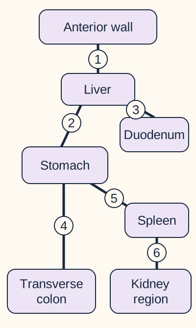
Connection map, not a surgical view or a scale drawing. Numbers refer to the six explanations immediately below.
1. Falciform: liver to the anterior abdominal wall and diaphragm. Its free inferior edge contains the ligamentum teres hepatis and paraumbilical veins. It is a ventral-mesentery derivative but is not part of the lesser omentum. [4]
2. Hepatogastric, also called gastrohepatic: liver to the lesser curvature of the stomach. This is the broad, relatively thin portion of the lesser omentum. Gastric vessels follow its gastric attachment. A replaced or accessory left hepatic artery may also cross this fold; a thin appearance does not establish an empty surgical plane. [1][6]
3. Hepatoduodenal: liver to the proximal duodenum. The thick free right edge of the lesser omentum contains the portal vein, proper hepatic artery and common bile duct. [3]
4. Gastrocolic: greater curvature of the stomach to transverse colon. It is a major component of the greater omentum. Gastroepiploic, also called gastroomental, vessels run near the gastric attachment. [1]
5. Gastrosplenic: proximal greater curvature to splenic hilum. It carries short gastric and left gastroepiploic vessels. 6. Splenorenal, also called lienorenal: splenic hilum to the posterior abdominal wall over the left kidney. Distal splenic vessels and the pancreatic tail are the important neighbors at the hilum; the word renal does not put the renal artery inside this fold. [1][8]
Trace the two separate paths leaving the spleen in the map. Which reaches the stomach, and which returns toward the posterior wall? Point to each endpoint before naming a vessel.
Check the two splenic paths
The stomach-facing path is gastrosplenic and carries gastric branches. The posterior path is splenorenal and carries the hilar splenic vessels beside the pancreatic tail. A shared splenic endpoint does not mean shared contents.
Apply this to a new situation: an operative note names a stomach-to-spleen attachment, while a drain later suggests pancreatic injury. Those findings should prompt review of whether dissection also reached the deeper splenic pedicle, rather than assuming every splenic attachment contains pancreas.
Borders of the lesser sac
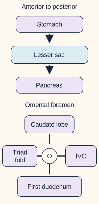
Does reaching tissue behind the stomach necessarily mean entering the retroperitoneum? No. The lesser sac, or omental bursa, is a peritoneal recess posterior to the stomach and lesser omentum and anterior to the peritoneal covering of the pancreas. A collection in that recess is different from inflammation within pancreatic or mesenteric tissue. Use the surfaces bounding the fluid, not merely the word posterior. [1][2]
The upper panel is a front-to-back stack, not a gravity map. In the lower boundary diagram, left means anterior and right means posterior; O is the opening and IVC is inferior vena cava.
The omental foramen, also called the epiploic foramen or foramen of Winslow, is the natural communication between greater and lesser sacs. Its anterior boundary is the hepatoduodenal ligament; its posterior boundary is the peritoneum over the inferior vena cava. The caudate lobe is above it, and the first part of the duodenum is below it. A finger behind the free lesser-omental edge is therefore behind the portal triad, not inside the portal vein. [2]
The gastrosplenic and splenorenal folds enclose the splenic recess at the left end. They do not create a second normal opening equivalent to the omental foramen. A bowel loop passing through the foramen can occupy the lesser sac; identifying its neck between the triad anteriorly and vena cava posteriorly localizes that route. [2]
Surgical entry through the gastrocolic ligament provides another route to the space behind the stomach. This is an intentionally created opening, not the natural foramen. The stomach is above that attachment and the transverse colon below. The gastroepiploic arcade near the stomach and the colonic blood supply require separate attention; this lesson does not define a universally vessel-free incision. [1]
Cover the lower diagram with your hand. Reconstruct the four boundaries using liver, duodenum, triad and vena cava. Then compare the natural entrance with the stomach-to-colon route.
Check which route was created
Opening the gastrocolic attachment creates access to the lesser sac. Passing behind the hepatoduodenal free edge uses an existing foramen. The destination can be identical while the structures at risk differ.
For transfer, imagine a collection that displaces the stomach anteriorly and lies in front of the pancreas. That relationship supports lesser-sac localization. It does not, by itself, identify the cause of the collection or choose a drainage procedure.
The transverse mesocolon connects the transverse colon to the posterior region and contains middle colic vessels. It is not the same sheet as the gastrocolic attachment. Within either fold, tissue and vessels lie between peritoneal layers; free lesser-sac fluid lies outside those tissue layers. Contiguous disease following a vessel within a ligament therefore differs from free spread through peritoneal fluid. [1]
The portal triad and Pringle maneuver
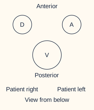
If a clamp stops liver bleeding, has it identified the injured vessel? Not necessarily. The hepatoduodenal ligament contains two blood inflow routes and a bile outflow route. In the usual arrangement near its free edge, the portal vein is posterior; the proper hepatic artery is anterior and toward the patient's left, while the common bile duct is anterior and toward the patient's right. Orient the patient before translating these relationships to an image. [1][3]
Typical arrangement near the free edge of the lesser omentum. A = proper hepatic artery; D = common bile duct; V = portal vein. Individual anatomy can vary.
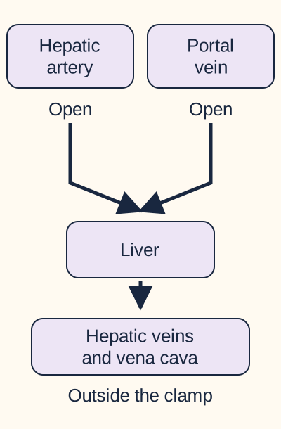
The Pringle maneuver compresses the hepatoduodenal ligament, interrupting the usual portal venous and hepatic arterial inflow. It does not directly compress the hepatic veins or retrohepatic inferior vena cava. The common bile duct accompanies the inflow vessels but is not another source of blood. Temporary inflow interruption can cause ischemia; the anatomy exercise below is not a clamp-duration or operative-management guide. [3]
Trace the paths before and during compression
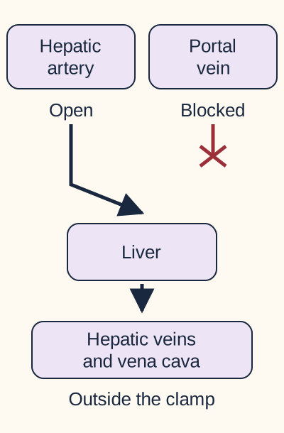
Start with both incoming paths open. Predict what remains connected if only the portal path closes, then compare that with compression of the whole triad fold. The intermediate state isolates a variable for learning; it is not a Pringle maneuver.
Conceptual patency map, not measured flow, a clamp-time guide, or a procedural instruction. Predict, then inspect selective portal occlusion
Conceptual patency map, not measured flow, a clamp-time guide, or a procedural instruction.
One usual inflow route remains: the hepatic artery. Closing the portal path alone cannot represent compression of the whole hepatoduodenal ligament.
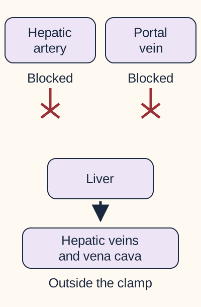
Predict, then inspect compression of the triad fold
Conceptual patency map, not measured flow, a clamp-time guide, or a procedural instruction.
Both usual incoming blood paths are interrupted. The hepatic veins and vena cava remain outside the compression. Their drawn connection denotes an uncompressed route, not a promise of unchanged flow.
Close either answer to compare with the starting diagram again. The complete relationship is also readable here: selective portal occlusion leaves arterial inflow; effective triad compression interrupts both usual inflows; neither directly closes the hepatic venous outflow structures. These immediate visual comparisons need no motion, timer, stored state or JavaScript.
Marked reduction in bleeding supports an inflow-dependent source but does not distinguish the hepatic artery from the portal vein. Persistent bleeding needs a check of whether inflow is actually controlled and whether an aberrant artery bypasses the compressed route. When control is confirmed and blood comes from behind the liver, hepatic venous or retrohepatic caval injury becomes especially concerning. Persistent bleeding alone is not proof of one named vein. [5]
For a new case, keep the clamp unchanged but add Doppler evidence of continued left-lobe arterial perfusion. That finding weakens a simple claim that all inflow has stopped. Look for an arterial variant rather than treating the word Pringle as a substitute for the observed anatomy.
What each ligament carries
During splenic surgery, why can gastric bleeding and pancreatic leakage arise from different attachments? The gastrosplenic fold carries branches toward the stomach. The splenorenal fold carries the distal splenic pedicle beside the pancreatic tail. Return to the two separate splenic links in the attachment map and follow each away from the hilum. [1][8]
Short gastric arteries cross toward the fundus. The left gastroepiploic artery continues along the greater curvature and communicates with the right-sided gastroepiploic supply. The right and left gastric arteries instead follow the lesser curvature. Thus, a fundal branch crossing from the spleen, a vessel following the greater curvature, and a vessel following the lesser curvature are three different anatomical descriptions. Specify both attachment and territory before selecting a vessel. [1]
Near the splenic hilum, the splenic artery and vein have a close relationship with the pancreatic tail in the splenorenal attachment. This does not mean the entire splenic artery lies inside that ligament: its more proximal course follows the pancreas. Likewise, renal vessels are posterior neighbors serving the kidney, not the structures carried to the splenic hilum. [1][8]
A pancreatic injury may become apparent through enzyme-rich drainage after splenectomy, even when bleeding was controlled during the operation. Such a finding supports pancreatic leakage in context. It does not establish the severity or formal grade of a postoperative pancreatic fistula from one laboratory value. Operative location, timing, imaging and clinical course still matter. [7]
Compare two operative descriptions: a short branch runs from splenic hilum directly into the fundus; a separate pedicle passes beside the pancreatic tail toward the posterior wall. Predict which attachment was encountered in each description.
Check the contrasting attachments
The fundal branch crosses the gastrosplenic ligament. The pedicle beside the pancreatic tail is in the splenorenal region. Use the second endpoint and adjacent organ, not the shared word splenic, to localize injury.
Transfer this distinction to a scan showing both spleen and kidney enhancing normally after an operation but fluid collecting at a distal pancreatic defect. Patent neighboring organs do not exclude injury to the pancreatic tissue between them.
Ventral versus dorsal mesentery
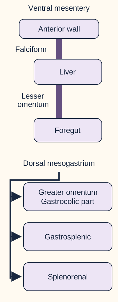
Why does the falciform ligament share an origin with the lesser omentum without belonging to it? Developmental family and adult attachment are different questions. The ventral mesentery persists in the foregut region rather than extending along the entire gut. As the liver grows within it, tissue between liver and anterior wall becomes the falciform ligament, while tissue between liver and foregut becomes the lesser omentum. Its stomach-facing part is hepatogastric and its duodenum-facing free edge is hepatoduodenal. [4]
The ventral panel shows the liver between two residual attachments. The dorsal branches indicate shared developmental origin, not a sequence or identical adult names.
The dorsal mesogastrium is the stomach's posterior mesenteric attachment. Stomach rotation and growth alter its position and enlarge the recess behind the stomach. Its extension contributes to the greater omentum, and its relationship with the developing spleen produces gastrosplenic and splenorenal attachments. The spleen develops from mesenchyme within the dorsal mesogastrium. Most pancreatic tissue becomes fixed posteriorly, while the tail retains its close hilar relationship in the splenorenal fold. [1][2][8]
Cover the labels beside the liver in the ventral panel. Follow the tissue toward the body wall, then toward the foregut. Name the two surviving attachments. Now trace the separate dorsal branches without treating them as a chronological sequence.
Check the developmental reconstruction
Wall to liver becomes falciform; liver to stomach and proximal duodenum becomes lesser omentum. The dorsal family contains the greater omentum and splenic ligaments. Shared origin explains related folds but does not erase their distinct endpoints.
For transfer, a developmental defect involving the stomach's posterior mesenteric attachment can affect splenic support while leaving the falciform ligament present. The preserved anterior attachment does not demonstrate that posterior development was normal.
Lesser versus greater omentum
Can one short label replace a full anatomical description? Use the name omentum to organize the map, then return to individual attachments when an operation or imaging finding requires precision. The lesser omentum consists of hepatogastric and hepatoduodenal portions. The greater omentum hangs from the greater curvature, descends anterior to bowel and returns toward the transverse colon; the gastrocolic attachment is central to the stomach-to-colon relationship. Gastrosplenic and splenorenal folds share dorsal mesogastric ancestry, but that fact should not be used to label every splenic ligament as the gastrocolic apron. [1][2]
The falciform ligament adds another practical distinction. Its free-edge cord, the ligamentum teres hepatis, is the remnant of the left umbilical vein. The ligamentum venosum is the remnant of the ductus venosus, a different fetal connection. Paraumbilical veins travel beside the round-ligament region and can provide collateral flow between the portal system and veins around the umbilicus in portal hypertension. A fibrous remnant and a nearby patent vein are not interchangeable structures. [4]
An ultrasound report describes a large vein running from the left portal region toward the umbilicus, while an operative report describes a cord at the lower edge of a liver-to-wall fold. Predict the fold, then decide whether both reports describe the same kind of structure.
Check the vein and cord distinction
Both localize to the falciform region, but the flowing collateral is a vein and the round ligament is a fetal venous remnant. A recognizable ligament is not assurance that its neighboring tissue lacks patent vessels.
Use three questions on a new case: What two endpoints are described? Which vessel or organ shares that attachment? What finding should follow if that structure is compressed, divided or injured? Check a competing location against the actual observations before naming the answer. The cases below are original educational scenarios, not patient records.
Apply the lesson
Case 1
Show answer and explanations for case 1
A. Left gastric branches (Why this does not fit)
Left gastric branches supply the lesser-curvature region. The injured branches cross from the splenic side directly into the fundus, not along the lesser curvature. Use both the attachment and supplied gastric region.
Reasoning steps for option A
Case context: During splenectomy, bleeding begins from several small vessels in a fold attached to the gastric fundus. The larger vessel following the greater curvature remains intact. Why could Left gastric branches initially fit the anatomy?
Left gastric branches supply the lesser-curvature region.
Case context: During splenectomy, bleeding begins from several small vessels in a fold attached to the gastric fundus. The larger vessel following the greater curvature remains intact. Which finding argues against Left gastric branches?
The injured branches cross from the splenic side directly into the fundus, not along the lesser curvature.
Case context: During splenectomy, bleeding begins from several small vessels in a fold attached to the gastric fundus. The larger vessel following the greater curvature remains intact. What discriminator separates Left gastric branches from the correct choice?
Use both the attachment and supplied gastric region.
B. Left gastroepiploic branches (Why this does not fit)
The left gastroepiploic artery travels from the splenic region along the greater curvature. The stem explicitly preserves the larger greater-curvature vessel while describing several short fundal branches. Distinguish fundal branches from the longitudinal greater-curvature arcade.
Reasoning steps for option B
Case context: During splenectomy, bleeding begins from several small vessels in a fold attached to the gastric fundus. The larger vessel following the greater curvature remains intact. Why could Left gastroepiploic branches initially fit the anatomy?
The left gastroepiploic artery travels from the splenic region along the greater curvature.
Case context: During splenectomy, bleeding begins from several small vessels in a fold attached to the gastric fundus. The larger vessel following the greater curvature remains intact. Which finding argues against Left gastroepiploic branches?
The stem explicitly preserves the larger greater-curvature vessel while describing several short fundal branches.
Case context: During splenectomy, bleeding begins from several small vessels in a fold attached to the gastric fundus. The larger vessel following the greater curvature remains intact. What discriminator separates Left gastroepiploic branches from the correct choice?
Distinguish fundal branches from the longitudinal greater-curvature arcade.
C. Short gastric branches (Best answer)
Short gastric vessels cross the gastrosplenic fold to the fundus. Several small fundal branches with an intact greater-curvature arcade identify these vessels. Stomach-to-spleen attachments can contain more than one gastric vessel type.
Reasoning steps for option C
Which attachment links the fundus to the splenic hilum?
The gastrosplenic ligament.
What does the intact longitudinal arcade exclude as the described injury?
The injury is in the separately described direct fundal branches rather than the greater-curvature arcade.
Which branches fit that route?
Short gastric branches.
Case context: During splenectomy, bleeding begins from several small vessels in a fold attached to the gastric fundus. The larger vessel following the greater curvature remains intact. What discriminator separates Short gastric branches from the correct choice?
Stomach-to-spleen attachments can contain more than one gastric vessel type.
D. Right gastric branches (Why this does not fit)
Right gastric branches participate in the lesser-curvature supply. The fundal splenic-side attachment and intact greater-curvature arcade do not locate a right gastric branch. A gastric destination alone does not identify the artery.
Reasoning steps for option D
Case context: During splenectomy, bleeding begins from several small vessels in a fold attached to the gastric fundus. The larger vessel following the greater curvature remains intact. Why could Right gastric branches initially fit the anatomy?
Right gastric branches participate in the lesser-curvature supply.
Case context: During splenectomy, bleeding begins from several small vessels in a fold attached to the gastric fundus. The larger vessel following the greater curvature remains intact. Which finding argues against Right gastric branches?
The fundal splenic-side attachment and intact greater-curvature arcade do not locate a right gastric branch.
Case context: During splenectomy, bleeding begins from several small vessels in a fold attached to the gastric fundus. The larger vessel following the greater curvature remains intact. What discriminator separates Right gastric branches from the correct choice?
A gastric destination alone does not identify the artery.
Takeaway: Identify the ligament and then distinguish a direct fundal branch from the greater-curvature arcade.
The pancreatic tail lies beside the splenic hilar vessels in the splenorenal region. The distal pancreatic defect and enzyme-rich fluid identify tissue at that posterior hilar attachment. An anatomical localization does not assign a formal postoperative fistula grade.
Reasoning steps for option A
Which organ matches the imaged defect and enzyme-rich drainage?
The pancreas.
Which pancreatic region is adjacent to the former splenic hilum?
The pancreatic tail.
Which attachment contains that hilar relationship?
The splenorenal attachment.
Case context: On postoperative day 4 after splenectomy, a patient has persistent upper abdominal drainage. Drain amylase is 6200 U/L; the laboratory serum upper limit is 100 U/L. CT shows a small... What discriminator separates Splenorenal ligament from the correct choice?
An anatomical localization does not assign a formal postoperative fistula grade.
B. Gastrosplenic ligament (Why this does not fit)
The gastrosplenic attachment is adjacent to the operative field and carries gastric vessels. A demonstrated distal pancreatic defect is better localized to the deeper splenorenal region than to the stomach-facing attachment. Separate gastric vascular injury from pancreatic tissue injury.
Reasoning steps for option B
Case context: On postoperative day 4 after splenectomy, a patient has persistent upper abdominal drainage. Drain amylase is 6200 U/L; the laboratory serum upper limit is 100 U/L. CT shows a small... Why could Gastrosplenic ligament initially fit the anatomy?
The gastrosplenic attachment is adjacent to the operative field and carries gastric vessels.
Case context: On postoperative day 4 after splenectomy, a patient has persistent upper abdominal drainage. Drain amylase is 6200 U/L; the laboratory serum upper limit is 100 U/L. CT shows a small... Which finding argues against Gastrosplenic ligament?
A demonstrated distal pancreatic defect is better localized to the deeper splenorenal region than to the stomach-facing attachment.
Case context: On postoperative day 4 after splenectomy, a patient has persistent upper abdominal drainage. Drain amylase is 6200 U/L; the laboratory serum upper limit is 100 U/L. CT shows a small... What discriminator separates Gastrosplenic ligament from the correct choice?
Separate gastric vascular injury from pancreatic tissue injury.
C. Gastrocolic ligament (Why this does not fit)
The gastrocolic attachment is a route into the lesser sac. Entering that space is different from the posterior hilar attachment containing the pancreatic tail. The access route and the injured structure can occupy different planes.
Reasoning steps for option C
Case context: On postoperative day 4 after splenectomy, a patient has persistent upper abdominal drainage. Drain amylase is 6200 U/L; the laboratory serum upper limit is 100 U/L. CT shows a small... Why could Gastrocolic ligament initially fit the anatomy?
The gastrocolic attachment is a route into the lesser sac.
Case context: On postoperative day 4 after splenectomy, a patient has persistent upper abdominal drainage. Drain amylase is 6200 U/L; the laboratory serum upper limit is 100 U/L. CT shows a small... Which finding argues against Gastrocolic ligament?
Entering that space is different from the posterior hilar attachment containing the pancreatic tail.
Case context: On postoperative day 4 after splenectomy, a patient has persistent upper abdominal drainage. Drain amylase is 6200 U/L; the laboratory serum upper limit is 100 U/L. CT shows a small... What discriminator separates Gastrocolic ligament from the correct choice?
The access route and the injured structure can occupy different planes.
D. Hepatogastric ligament (Why this does not fit)
The hepatogastric fold joins liver to lesser curvature. The described defect is at the distal pancreas near the splenic hilum, not in the liver-facing gastric fold. Use the localized defect rather than the broad phrase upper abdominal surgery.
Reasoning steps for option D
Case context: On postoperative day 4 after splenectomy, a patient has persistent upper abdominal drainage. Drain amylase is 6200 U/L; the laboratory serum upper limit is 100 U/L. CT shows a small... Why could Hepatogastric ligament initially fit the anatomy?
The hepatogastric fold joins liver to lesser curvature.
Case context: On postoperative day 4 after splenectomy, a patient has persistent upper abdominal drainage. Drain amylase is 6200 U/L; the laboratory serum upper limit is 100 U/L. CT shows a small... Which finding argues against Hepatogastric ligament?
The described defect is at the distal pancreas near the splenic hilum, not in the liver-facing gastric fold.
Case context: On postoperative day 4 after splenectomy, a patient has persistent upper abdominal drainage. Drain amylase is 6200 U/L; the laboratory serum upper limit is 100 U/L. CT shows a small... What discriminator separates Hepatogastric ligament from the correct choice?
Use the localized defect rather than the broad phrase upper abdominal surgery.
Takeaway: A pancreatic defect beside the splenic hilum points to the splenorenal attachment.
The left gastric artery supplies the lesser curvature. The observed vessel crosses from the splenic hilum and follows the greater curvature. A side designation is less useful than origin and course together.
Reasoning steps for option A
Case context: During mobilization of the proximal greater curvature, an arterial branch is seen crossing from the splenic hilum and continuing along the gastric body. Short branches ending directly in... Why could Left gastric artery initially fit the anatomy?
The left gastric artery supplies the lesser curvature.
Case context: During mobilization of the proximal greater curvature, an arterial branch is seen crossing from the splenic hilum and continuing along the gastric body. Short branches ending directly in... Which finding argues against Left gastric artery?
The observed vessel crosses from the splenic hilum and follows the greater curvature.
Case context: During mobilization of the proximal greater curvature, an arterial branch is seen crossing from the splenic hilum and continuing along the gastric body. Short branches ending directly in... What discriminator separates Left gastric artery from the correct choice?
A side designation is less useful than origin and course together.
B. Right gastric artery (Why this does not fit)
The right gastric artery also follows the lesser curvature. Neither a splenic origin nor a greater-curvature course fits this artery. Do not exchange the two gastric arcades.
Reasoning steps for option B
Case context: During mobilization of the proximal greater curvature, an arterial branch is seen crossing from the splenic hilum and continuing along the gastric body. Short branches ending directly in... Why could Right gastric artery initially fit the anatomy?
The right gastric artery also follows the lesser curvature.
Case context: During mobilization of the proximal greater curvature, an arterial branch is seen crossing from the splenic hilum and continuing along the gastric body. Short branches ending directly in... Which finding argues against Right gastric artery?
Neither a splenic origin nor a greater-curvature course fits this artery.
Case context: During mobilization of the proximal greater curvature, an arterial branch is seen crossing from the splenic hilum and continuing along the gastric body. Short branches ending directly in... What discriminator separates Right gastric artery from the correct choice?
Do not exchange the two gastric arcades.
C. Short gastric artery (Why this does not fit)
Short gastric arteries arise on the splenic side and pass to the fundus. Those separately preserved vessels end in the fundus rather than continuing along the gastric body. Direct fundal supply differs from a longitudinal gastroomental course.
Reasoning steps for option C
Case context: During mobilization of the proximal greater curvature, an arterial branch is seen crossing from the splenic hilum and continuing along the gastric body. Short branches ending directly in... Why could Short gastric artery initially fit the anatomy?
Short gastric arteries arise on the splenic side and pass to the fundus.
Case context: During mobilization of the proximal greater curvature, an arterial branch is seen crossing from the splenic hilum and continuing along the gastric body. Short branches ending directly in... Which finding argues against Short gastric artery?
Those separately preserved vessels end in the fundus rather than continuing along the gastric body.
Case context: During mobilization of the proximal greater curvature, an arterial branch is seen crossing from the splenic hilum and continuing along the gastric body. Short branches ending directly in... What discriminator separates Short gastric artery from the correct choice?
Direct fundal supply differs from a longitudinal gastroomental course.
D. Left gastroepiploic artery (Best answer)
The left gastroepiploic artery passes from the splenic region toward the greater curvature. Its continuation along the body distinguishes it from the preserved short gastric branches. Follow an artery beyond its origin before assigning its name.
Reasoning steps for option D
Which course distinguishes this vessel from the short gastric branches?
It continues along the greater curvature of the gastric body.
What artery follows that course from the splenic region?
The left gastroepiploic artery.
Case context: During mobilization of the proximal greater curvature, an arterial branch is seen crossing from the splenic hilum and continuing along the gastric body. Short branches ending directly in... What discriminator separates Left gastroepiploic artery from the correct choice?
Follow an artery beyond its origin before assigning its name.
Takeaway: A splenic-side artery continuing along the greater curvature is the left gastroepiploic artery.
A. Hepatoduodenal ligament (Why this does not fit)
The usual hepatic arterial route runs in the hepatoduodenal free edge. The affected artery instead originates from the left gastric artery, and right-lobe arterial and main portal signals remain present. A regional perfusion loss can identify an arterial route outside the usual pedicle.
Reasoning steps for option A
Case context: During gastric mobilization for esophagectomy, left-lobe hepatic arterial Doppler flow disappears after division of a peritoneal sheet. Right-lobe arterial flow and main portal flow... Why could Hepatoduodenal ligament initially fit the anatomy?
The usual hepatic arterial route runs in the hepatoduodenal free edge.
Case context: During gastric mobilization for esophagectomy, left-lobe hepatic arterial Doppler flow disappears after division of a peritoneal sheet. Right-lobe arterial flow and main portal flow... Which finding argues against Hepatoduodenal ligament?
The affected artery instead originates from the left gastric artery, and right-lobe arterial and main portal signals remain present.
Case context: During gastric mobilization for esophagectomy, left-lobe hepatic arterial Doppler flow disappears after division of a peritoneal sheet. Right-lobe arterial flow and main portal flow... What discriminator separates Hepatoduodenal ligament from the correct choice?
A regional perfusion loss can identify an arterial route outside the usual pedicle.
B. Hepatogastric ligament (Best answer)
A replaced left hepatic artery from the left gastric artery may cross the hepatogastric sheet. Loss of left-lobe arterial flow during gastric mobilization fits interruption of the documented variant in this attachment. Combine regional perfusion with the documented arterial origin.
Reasoning steps for option B
Which flow is lost while the other measured inflows remain?
Left-lobe hepatic arterial flow.
Which preoperative origin identifies the affected variant?
The left gastric artery supplies the replaced left hepatic artery.
Which attachment carries that gastric-to-hepatic route?
The hepatogastric ligament.
Case context: During gastric mobilization for esophagectomy, left-lobe hepatic arterial Doppler flow disappears after division of a peritoneal sheet. Right-lobe arterial flow and main portal flow... What discriminator separates Hepatogastric ligament from the correct choice?
Combine regional perfusion with the documented arterial origin.
C. Gastrosplenic ligament (Why this does not fit)
The gastrosplenic fold contains gastric branches arising on the splenic side. The injured hepatic route was traced to the left gastric artery rather than the splenic hilum. Use the demonstrated origin rather than gastric proximity alone.
Reasoning steps for option C
Case context: During gastric mobilization for esophagectomy, left-lobe hepatic arterial Doppler flow disappears after division of a peritoneal sheet. Right-lobe arterial flow and main portal flow... Why could Gastrosplenic ligament initially fit the anatomy?
The gastrosplenic fold contains gastric branches arising on the splenic side.
Case context: During gastric mobilization for esophagectomy, left-lobe hepatic arterial Doppler flow disappears after division of a peritoneal sheet. Right-lobe arterial flow and main portal flow... Which finding argues against Gastrosplenic ligament?
The injured hepatic route was traced to the left gastric artery rather than the splenic hilum.
Case context: During gastric mobilization for esophagectomy, left-lobe hepatic arterial Doppler flow disappears after division of a peritoneal sheet. Right-lobe arterial flow and main portal flow... What discriminator separates Gastrosplenic ligament from the correct choice?
Use the demonstrated origin rather than gastric proximity alone.
D. Falciform ligament (Why this does not fit)
The falciform attachment reaches the liver from the anterior wall. The documented artery approaches from the left gastric route, not the abdominal wall. Different liver-facing attachments transmit different vascular routes.
Reasoning steps for option D
Case context: During gastric mobilization for esophagectomy, left-lobe hepatic arterial Doppler flow disappears after division of a peritoneal sheet. Right-lobe arterial flow and main portal flow... Why could Falciform ligament initially fit the anatomy?
The falciform attachment reaches the liver from the anterior wall.
Case context: During gastric mobilization for esophagectomy, left-lobe hepatic arterial Doppler flow disappears after division of a peritoneal sheet. Right-lobe arterial flow and main portal flow... Which finding argues against Falciform ligament?
The documented artery approaches from the left gastric route, not the abdominal wall.
Case context: During gastric mobilization for esophagectomy, left-lobe hepatic arterial Doppler flow disappears after division of a peritoneal sheet. Right-lobe arterial flow and main portal flow... What discriminator separates Falciform ligament from the correct choice?
Different liver-facing attachments transmit different vascular routes.
Takeaway: A replaced left hepatic artery from the left gastric artery may traverse the hepatogastric ligament.
A. Left renal artery and vein (Why this does not fit)
Renal hilar vessels supply and drain the kidney. The renal hilum is separately visualized posterior to the attachment carrying the distal pancreatic lesion. An attachment over the kidney does not contain the renal pedicle.
Reasoning steps for option A
Case context: A distal pancreatic lesion extends through a posterior attachment toward the splenic hilum. The left renal hilum is separately visualized behind the involved tissue. Why could Left renal artery and vein initially fit the anatomy?
Renal hilar vessels supply and drain the kidney.
Case context: A distal pancreatic lesion extends through a posterior attachment toward the splenic hilum. The left renal hilum is separately visualized behind the involved tissue. Which finding argues against Left renal artery and vein?
The renal hilum is separately visualized posterior to the attachment carrying the distal pancreatic lesion.
Case context: A distal pancreatic lesion extends through a posterior attachment toward the splenic hilum. The left renal hilum is separately visualized behind the involved tissue. What discriminator separates Left renal artery and vein from the correct choice?
An attachment over the kidney does not contain the renal pedicle.
B. Left gastric artery and vein (Why this does not fit)
Left gastric vessels follow the lesser-curvature route. The lesion follows distal pancreas toward the splenic hilum rather than the lesser curvature. Use continuity with the diseased organ to localize the vascular corridor.
Reasoning steps for option B
Case context: A distal pancreatic lesion extends through a posterior attachment toward the splenic hilum. The left renal hilum is separately visualized behind the involved tissue. Why could Left gastric artery and vein initially fit the anatomy?
Left gastric vessels follow the lesser-curvature route.
Case context: A distal pancreatic lesion extends through a posterior attachment toward the splenic hilum. The left renal hilum is separately visualized behind the involved tissue. Which finding argues against Left gastric artery and vein?
The lesion follows distal pancreas toward the splenic hilum rather than the lesser curvature.
Case context: A distal pancreatic lesion extends through a posterior attachment toward the splenic hilum. The left renal hilum is separately visualized behind the involved tissue. What discriminator separates Left gastric artery and vein from the correct choice?
Use continuity with the diseased organ to localize the vascular corridor.
C. Splenic artery and vein (Best answer)
Distal splenic vessels share the splenorenal hilar attachment with the pancreatic tail. A lesion following distal pancreas toward the splenic hilum can encase that pair. The hilar segment of the splenic pedicle is distinct from the renal hilum.
Reasoning steps for option C
Which vascular pair accompanies the distal pancreas toward the splenic hilum?
The distal splenic artery and vein.
Why is the renal pair a poorer fit?
The renal hilum is separately visualized behind the involved attachment.
Case context: A distal pancreatic lesion extends through a posterior attachment toward the splenic hilum. The left renal hilum is separately visualized behind the involved tissue. What discriminator separates Splenic artery and vein from the correct choice?
The hilar segment of the splenic pedicle is distinct from the renal hilum.
D. Middle colic artery and vein (Why this does not fit)
Middle colic vessels run in the transverse mesocolon. The involved connection reaches the splenic hilum rather than anchoring the transverse colon. A posterior mesenteric attachment is identified by its organ endpoint.
Reasoning steps for option D
Case context: A distal pancreatic lesion extends through a posterior attachment toward the splenic hilum. The left renal hilum is separately visualized behind the involved tissue. Why could Middle colic artery and vein initially fit the anatomy?
Middle colic vessels run in the transverse mesocolon.
Case context: A distal pancreatic lesion extends through a posterior attachment toward the splenic hilum. The left renal hilum is separately visualized behind the involved tissue. Which finding argues against Middle colic artery and vein?
The involved connection reaches the splenic hilum rather than anchoring the transverse colon.
Case context: A distal pancreatic lesion extends through a posterior attachment toward the splenic hilum. The left renal hilum is separately visualized behind the involved tissue. What discriminator separates Middle colic artery and vein from the correct choice?
A posterior mesenteric attachment is identified by its organ endpoint.
Takeaway: The distal pancreas and splenic vascular pedicle share the splenorenal connection.
A. Right and left gastroepiploic arteries (Best answer)
The gastroepiploic arteries supply the greater-curvature arcade in the gastrocolic region. The affected gastric territory and preserved lesser-curvature and colonic vessels localize injury to this arcade. Use perfusion territory to verify the vessel at risk in an operative attachment.
Reasoning steps for option A
Which gastric region has lost perfusion?
The greater curvature of the gastric body.
Which competing vascular territories remain patent?
The lesser-curvature gastric and assessed middle colic vessels.
Which arcade fits the affected territory and divided attachment?
The right and left gastroepiploic arcade in the gastrocolic region.
Case context: After separation of a sheet between the gastric body and transverse colon, focal ischemia appears along the gastric greater curvature. Angiography shows that lesser-curvature gastric... What discriminator separates Right and left gastroepiploic arteries from the correct choice?
Use perfusion territory to verify the vessel at risk in an operative attachment.
B. Right and left gastric arteries (Why this does not fit)
These arteries supply the lesser-curvature arcade. Their angiographic patency and the greater-curvature ischemia make them a poorer match. Distinguish the two gastric arterial territories rather than treating every gastric artery as equivalent.
Reasoning steps for option B
Case context: After separation of a sheet between the gastric body and transverse colon, focal ischemia appears along the gastric greater curvature. Angiography shows that lesser-curvature gastric... Why could Right and left gastric arteries initially fit the anatomy?
These arteries supply the lesser-curvature arcade.
Case context: After separation of a sheet between the gastric body and transverse colon, focal ischemia appears along the gastric greater curvature. Angiography shows that lesser-curvature gastric... Which finding argues against Right and left gastric arteries?
Their angiographic patency and the greater-curvature ischemia make them a poorer match.
Case context: After separation of a sheet between the gastric body and transverse colon, focal ischemia appears along the gastric greater curvature. Angiography shows that lesser-curvature gastric... What discriminator separates Right and left gastric arteries from the correct choice?
Distinguish the two gastric arterial territories rather than treating every gastric artery as equivalent.
C. Middle and right colic arteries (Why this does not fit)
Colic vessels can be injured when dissection enters colonic mesentery. The injury produces gastric greater-curvature ischemia while the assessed middle colic branches remain patent. The affected organ helps separate adjacent vascular planes.
Reasoning steps for option C
Case context: After separation of a sheet between the gastric body and transverse colon, focal ischemia appears along the gastric greater curvature. Angiography shows that lesser-curvature gastric... Why could Middle and right colic arteries initially fit the anatomy?
Colic vessels can be injured when dissection enters colonic mesentery.
Case context: After separation of a sheet between the gastric body and transverse colon, focal ischemia appears along the gastric greater curvature. Angiography shows that lesser-curvature gastric... Which finding argues against Middle and right colic arteries?
The injury produces gastric greater-curvature ischemia while the assessed middle colic branches remain patent.
Case context: After separation of a sheet between the gastric body and transverse colon, focal ischemia appears along the gastric greater curvature. Angiography shows that lesser-curvature gastric... What discriminator separates Middle and right colic arteries from the correct choice?
The affected organ helps separate adjacent vascular planes.
D. Short gastric and left gastric arteries (Why this does not fit)
These arteries supply the fundal and lesser-curvature regions. The lesion follows the greater curvature of the gastric body, with documented patency of the lesser-curvature vessels. Combine vessel patency with the distribution of ischemia.
Reasoning steps for option D
Case context: After separation of a sheet between the gastric body and transverse colon, focal ischemia appears along the gastric greater curvature. Angiography shows that lesser-curvature gastric... Why could Short gastric and left gastric arteries initially fit the anatomy?
These arteries supply the fundal and lesser-curvature regions.
Case context: After separation of a sheet between the gastric body and transverse colon, focal ischemia appears along the gastric greater curvature. Angiography shows that lesser-curvature gastric... Which finding argues against Short gastric and left gastric arteries?
The lesion follows the greater curvature of the gastric body, with documented patency of the lesser-curvature vessels.
Case context: After separation of a sheet between the gastric body and transverse colon, focal ischemia appears along the gastric greater curvature. Angiography shows that lesser-curvature gastric... What discriminator separates Short gastric and left gastric arteries from the correct choice?
Combine vessel patency with the distribution of ischemia.
Takeaway: The greater-curvature arcade lies on the gastric side of the gastrocolic attachment.
A hepatogastric route can approach the region behind the lesser omentum. The specified approach is below the greater curvature next to the transverse colon, not through the liver-facing lesser-curvature sheet. Match an access route to the operative approach as well as the target space.
Reasoning steps for option A
Case context: A stable patient undergoes exploration of a localized upper abdominal collection. CT shows the stomach displaced anteriorly and the pancreas posterior to the fluid. The team approaches... Why could Hepatogastric ligament initially fit the anatomy?
A hepatogastric route can approach the region behind the lesser omentum.
Case context: A stable patient undergoes exploration of a localized upper abdominal collection. CT shows the stomach displaced anteriorly and the pancreas posterior to the fluid. The team approaches... Which finding argues against Hepatogastric ligament?
The specified approach is below the greater curvature next to the transverse colon, not through the liver-facing lesser-curvature sheet.
Case context: A stable patient undergoes exploration of a localized upper abdominal collection. CT shows the stomach displaced anteriorly and the pancreas posterior to the fluid. The team approaches... What discriminator separates Hepatogastric ligament from the correct choice?
Match an access route to the operative approach as well as the target space.
B. Gastrocolic ligament (Best answer)
The gastrocolic attachment connects greater curvature to transverse colon. Fluid between stomach and pancreas localizes the lesser sac, and the specified inferior gastric approach traverses this attachment. Different entrances into the same space encounter different structures.
Reasoning steps for option B
Where is fluid between stomach and pancreas localized?
The lesser sac.
Which two organs define the specified approach?
The greater curvature of the stomach and transverse colon.
Which attachment joins those endpoints?
The gastrocolic ligament.
Case context: A stable patient undergoes exploration of a localized upper abdominal collection. CT shows the stomach displaced anteriorly and the pancreas posterior to the fluid. The team approaches... What discriminator separates Gastrocolic ligament from the correct choice?
Different entrances into the same space encounter different structures.
C. Splenorenal ligament (Why this does not fit)
The splenorenal region lies near the left margin of the lesser sac. The approach is from below the gastric body above the transverse colon rather than through the posterior splenic pedicle. A lateral boundary is not the specified anterior access plane.
Reasoning steps for option C
Case context: A stable patient undergoes exploration of a localized upper abdominal collection. CT shows the stomach displaced anteriorly and the pancreas posterior to the fluid. The team approaches... Why could Splenorenal ligament initially fit the anatomy?
The splenorenal region lies near the left margin of the lesser sac.
Case context: A stable patient undergoes exploration of a localized upper abdominal collection. CT shows the stomach displaced anteriorly and the pancreas posterior to the fluid. The team approaches... Which finding argues against Splenorenal ligament?
The approach is from below the gastric body above the transverse colon rather than through the posterior splenic pedicle.
Case context: A stable patient undergoes exploration of a localized upper abdominal collection. CT shows the stomach displaced anteriorly and the pancreas posterior to the fluid. The team approaches... What discriminator separates Splenorenal ligament from the correct choice?
A lateral boundary is not the specified anterior access plane.
D. Transverse mesocolon (Why this does not fit)
The transverse mesocolon attaches colon to the posterior abdominal wall. The stated route passes between greater curvature and colon rather than through the colon-to-posterior-wall attachment. Distinguish gastrocolic tissue from the mesocolon beneath it.
Reasoning steps for option D
Case context: A stable patient undergoes exploration of a localized upper abdominal collection. CT shows the stomach displaced anteriorly and the pancreas posterior to the fluid. The team approaches... Why could Transverse mesocolon initially fit the anatomy?
The transverse mesocolon attaches colon to the posterior abdominal wall.
Case context: A stable patient undergoes exploration of a localized upper abdominal collection. CT shows the stomach displaced anteriorly and the pancreas posterior to the fluid. The team approaches... Which finding argues against Transverse mesocolon?
The stated route passes between greater curvature and colon rather than through the colon-to-posterior-wall attachment.
Case context: A stable patient undergoes exploration of a localized upper abdominal collection. CT shows the stomach displaced anteriorly and the pancreas posterior to the fluid. The team approaches... What discriminator separates Transverse mesocolon from the correct choice?
Distinguish gastrocolic tissue from the mesocolon beneath it.
Takeaway: Localize the lesser sac first, then use the specified approach to choose the attachment.
The transverse colon is an inferior neighbor of the gastric region. The neck between triad and vena cava identifies the omental foramen, whose inferior border is not the transverse colon. First identify the opening, then assign its boundaries.
Reasoning steps for option A
Case context: CT in a patient with acute bowel obstruction shows a distended bowel loop behind the stomach. Its neck passes posterior to the portal triad and anterior to the inferior vena cava. Why could Transverse colon initially fit the anatomy?
The transverse colon is an inferior neighbor of the gastric region.
Case context: CT in a patient with acute bowel obstruction shows a distended bowel loop behind the stomach. Its neck passes posterior to the portal triad and anterior to the inferior vena cava. Which finding argues against Transverse colon?
The neck between triad and vena cava identifies the omental foramen, whose inferior border is not the transverse colon.
Case context: CT in a patient with acute bowel obstruction shows a distended bowel loop behind the stomach. Its neck passes posterior to the portal triad and anterior to the inferior vena cava. What discriminator separates Transverse colon from the correct choice?
First identify the opening, then assign its boundaries.
B. Pancreatic body (Why this does not fit)
The pancreatic body lies posterior to much of the lesser sac. The described neck is at the natural entrance between the triad and vena cava, not over the pancreatic body. A sac wall and the border of its entrance are different landmarks.
Reasoning steps for option B
Case context: CT in a patient with acute bowel obstruction shows a distended bowel loop behind the stomach. Its neck passes posterior to the portal triad and anterior to the inferior vena cava. Why could Pancreatic body initially fit the anatomy?
The pancreatic body lies posterior to much of the lesser sac.
Case context: CT in a patient with acute bowel obstruction shows a distended bowel loop behind the stomach. Its neck passes posterior to the portal triad and anterior to the inferior vena cava. Which finding argues against Pancreatic body?
The described neck is at the natural entrance between the triad and vena cava, not over the pancreatic body.
Case context: CT in a patient with acute bowel obstruction shows a distended bowel loop behind the stomach. Its neck passes posterior to the portal triad and anterior to the inferior vena cava. What discriminator separates Pancreatic body from the correct choice?
A sac wall and the border of its entrance are different landmarks.
C. Caudate liver lobe (Why this does not fit)
The caudate lobe borders the omental foramen. It is superior to the neck rather than inferior. Preserve the directional relationship after identifying the correct opening.
Reasoning steps for option C
Case context: CT in a patient with acute bowel obstruction shows a distended bowel loop behind the stomach. Its neck passes posterior to the portal triad and anterior to the inferior vena cava. Why could Caudate liver lobe initially fit the anatomy?
The caudate lobe borders the omental foramen.
Case context: CT in a patient with acute bowel obstruction shows a distended bowel loop behind the stomach. Its neck passes posterior to the portal triad and anterior to the inferior vena cava. Which finding argues against Caudate liver lobe?
It is superior to the neck rather than inferior.
Case context: CT in a patient with acute bowel obstruction shows a distended bowel loop behind the stomach. Its neck passes posterior to the portal triad and anterior to the inferior vena cava. What discriminator separates Caudate liver lobe from the correct choice?
Preserve the directional relationship after identifying the correct opening.
D. First duodenal part (Best answer)
The first duodenal part forms the lower border of the omental foramen. The neck lies between the portal triad and vena cava, establishing which opening is involved. Foramen localization turns a bowel-position finding into a boundary prediction.
Reasoning steps for option D
Which opening lies between portal triad and vena cava?
The omental foramen.
What forms its inferior border?
The first part of the duodenum.
Case context: CT in a patient with acute bowel obstruction shows a distended bowel loop behind the stomach. Its neck passes posterior to the portal triad and anterior to the inferior vena cava. What discriminator separates First duodenal part from the correct choice?
Foramen localization turns a bowel-position finding into a boundary prediction.
Takeaway: The first part of the duodenum lies below the omental foramen.
The vena cava is a boundary of the omental foramen. It lies posterior to that communication rather than anterior. Distinguish the two vascular boundaries of the same opening.
Reasoning steps for option A
Case context: A posterior gastric perforation has released contrast into a confined pocket anterior to the pancreas. On subsequent images, a narrow extension reaches the greater peritoneal compartment... Why could Inferior vena cava initially fit the anatomy?
The vena cava is a boundary of the omental foramen.
Case context: A posterior gastric perforation has released contrast into a confined pocket anterior to the pancreas. On subsequent images, a narrow extension reaches the greater peritoneal compartment... Which finding argues against Inferior vena cava?
It lies posterior to that communication rather than anterior.
Case context: A posterior gastric perforation has released contrast into a confined pocket anterior to the pancreas. On subsequent images, a narrow extension reaches the greater peritoneal compartment... What discriminator separates Inferior vena cava from the correct choice?
Distinguish the two vascular boundaries of the same opening.
B. First part of duodenum (Why this does not fit)
The proximal duodenum borders the foramen connecting the sacs. It forms the inferior boundary, not the anterior boundary. Correct regional anatomy still requires the requested direction.
Reasoning steps for option B
Case context: A posterior gastric perforation has released contrast into a confined pocket anterior to the pancreas. On subsequent images, a narrow extension reaches the greater peritoneal compartment... Why could First part of duodenum initially fit the anatomy?
The proximal duodenum borders the foramen connecting the sacs.
Case context: A posterior gastric perforation has released contrast into a confined pocket anterior to the pancreas. On subsequent images, a narrow extension reaches the greater peritoneal compartment... Which finding argues against First part of duodenum?
It forms the inferior boundary, not the anterior boundary.
Case context: A posterior gastric perforation has released contrast into a confined pocket anterior to the pancreas. On subsequent images, a narrow extension reaches the greater peritoneal compartment... What discriminator separates First part of duodenum from the correct choice?
Correct regional anatomy still requires the requested direction.
C. Hepatoduodenal ligament (Best answer)
The triad-containing free lesser-omental edge forms the anterior border of the omental foramen. The initial collection is in the lesser sac and reaches the greater sac through its natural communication. The anterior border of the opening is a vessel-containing fold.
Reasoning steps for option C
Which space initially receives the posterior gastric leak?
The lesser sac.
Which natural opening connects that space to the greater sac?
The omental foramen.
What is immediately anterior to that opening?
The hepatoduodenal ligament.
Case context: A posterior gastric perforation has released contrast into a confined pocket anterior to the pancreas. On subsequent images, a narrow extension reaches the greater peritoneal compartment... What discriminator separates Hepatoduodenal ligament from the correct choice?
The anterior border of the opening is a vessel-containing fold.
D. Hepatogastric ligament (Why this does not fit)
The hepatogastric sheet is anterior to a portion of the lesser sac. It does not form the free-edge anterior border of the natural foramen. Separate the broad lesser-sac wall from the specific entrance.
Reasoning steps for option D
Case context: A posterior gastric perforation has released contrast into a confined pocket anterior to the pancreas. On subsequent images, a narrow extension reaches the greater peritoneal compartment... Why could Hepatogastric ligament initially fit the anatomy?
The hepatogastric sheet is anterior to a portion of the lesser sac.
Case context: A posterior gastric perforation has released contrast into a confined pocket anterior to the pancreas. On subsequent images, a narrow extension reaches the greater peritoneal compartment... Which finding argues against Hepatogastric ligament?
It does not form the free-edge anterior border of the natural foramen.
Case context: A posterior gastric perforation has released contrast into a confined pocket anterior to the pancreas. On subsequent images, a narrow extension reaches the greater peritoneal compartment... What discriminator separates Hepatogastric ligament from the correct choice?
Separate the broad lesser-sac wall from the specific entrance.
Takeaway: Fluid behind the stomach reaches the greater sac naturally through the omental foramen, anteriorly bounded by the hepatoduodenal ligament.
The hepatogastric sheet follows the lesser-curvature vessels toward the liver and continues with the triad-containing hepatoduodenal edge. The gastric-vessel origin and subsequent bile-duct and portal-vein involvement identify these adjoining parts. Trace contiguous tissue through both ends of a disease route.
Reasoning steps for option A
Which attachment follows the lesser-curvature gastric vessels toward the liver?
The hepatogastric attachment.
Which adjacent attachment contains the involved bile duct and portal vein?
The hepatoduodenal free edge.
Case context: A gastric cancer near the lesser curvature shows continuous soft-tissue extension along the gastric vessels toward the liver. At the lower hepatic pedicle, the same process surrounds the... What discriminator separates Hepatogastric and hepatoduodenal from the correct choice?
Trace contiguous tissue through both ends of a disease route.
B. Gastrosplenic and splenorenal (Why this does not fit)
These folds provide a route between proximal stomach and the posterior splenic region. The supplied route follows lesser-curvature vessels to the hepatic pedicle without splenic hilar extension. Do not substitute a different stomach-to-solid-organ route.
Reasoning steps for option B
Case context: A gastric cancer near the lesser curvature shows continuous soft-tissue extension along the gastric vessels toward the liver. At the lower hepatic pedicle, the same process surrounds the... Why could Gastrosplenic and splenorenal initially fit the anatomy?
These folds provide a route between proximal stomach and the posterior splenic region.
Case context: A gastric cancer near the lesser curvature shows continuous soft-tissue extension along the gastric vessels toward the liver. At the lower hepatic pedicle, the same process surrounds the... Which finding argues against Gastrosplenic and splenorenal?
The supplied route follows lesser-curvature vessels to the hepatic pedicle without splenic hilar extension.
Case context: A gastric cancer near the lesser curvature shows continuous soft-tissue extension along the gastric vessels toward the liver. At the lower hepatic pedicle, the same process surrounds the... What discriminator separates Gastrosplenic and splenorenal from the correct choice?
Do not substitute a different stomach-to-solid-organ route.
C. Gastrocolic and transverse mesocolon (Why this does not fit)
These attachments link the greater-curvature and transverse-colon regions. The involved gastric vessels and hepatic triad are not on the described colonic route. The gastric curvature and destination discriminate competing tissue corridors.
Reasoning steps for option C
Case context: A gastric cancer near the lesser curvature shows continuous soft-tissue extension along the gastric vessels toward the liver. At the lower hepatic pedicle, the same process surrounds the... Why could Gastrocolic and transverse mesocolon initially fit the anatomy?
These attachments link the greater-curvature and transverse-colon regions.
Case context: A gastric cancer near the lesser curvature shows continuous soft-tissue extension along the gastric vessels toward the liver. At the lower hepatic pedicle, the same process surrounds the... Which finding argues against Gastrocolic and transverse mesocolon?
The involved gastric vessels and hepatic triad are not on the described colonic route.
Case context: A gastric cancer near the lesser curvature shows continuous soft-tissue extension along the gastric vessels toward the liver. At the lower hepatic pedicle, the same process surrounds the... What discriminator separates Gastrocolic and transverse mesocolon from the correct choice?
The gastric curvature and destination discriminate competing tissue corridors.
D. Falciform and splenorenal (Why this does not fit)
The falciform approaches the liver from the wall and the splenorenal approaches the splenic hilum posteriorly. Neither pairing provides the observed continuous lesser-curvature-to-hepatic-pedicle path. Contiguous extension must follow anatomically connected tissue.
Reasoning steps for option D
Case context: A gastric cancer near the lesser curvature shows continuous soft-tissue extension along the gastric vessels toward the liver. At the lower hepatic pedicle, the same process surrounds the... Why could Falciform and splenorenal initially fit the anatomy?
The falciform approaches the liver from the wall and the splenorenal approaches the splenic hilum posteriorly.
Case context: A gastric cancer near the lesser curvature shows continuous soft-tissue extension along the gastric vessels toward the liver. At the lower hepatic pedicle, the same process surrounds the... Which finding argues against Falciform and splenorenal?
Neither pairing provides the observed continuous lesser-curvature-to-hepatic-pedicle path.
Case context: A gastric cancer near the lesser curvature shows continuous soft-tissue extension along the gastric vessels toward the liver. At the lower hepatic pedicle, the same process surrounds the... What discriminator separates Falciform and splenorenal from the correct choice?
Contiguous extension must follow anatomically connected tissue.
Takeaway: Lesser-curvature tissue can connect to the hepatic pedicle through the two portions of the lesser omentum.
A. Reduced hepatic arterial inflow from celiac branches (Why this does not fit)
The hepatic artery supplies arterial inflow and usually occupies an anterior left-sided position in the triad. The narrowed lumen is posterior to the identified pulsatile anterior structure. Identify the vessel before predicting which component of hepatic inflow is impaired.
Reasoning steps for option A
Case context: Ultrasonography of a mass at the free edge of the lesser omentum identifies three adjacent structures. The anterior structure on the patient left is pulsatile; the anterior structure on... Why could Reduced hepatic arterial inflow from celiac branches initially fit the anatomy?
The hepatic artery supplies arterial inflow and usually occupies an anterior left-sided position in the triad.
Case context: Ultrasonography of a mass at the free edge of the lesser omentum identifies three adjacent structures. The anterior structure on the patient left is pulsatile; the anterior structure on... Which finding argues against Reduced hepatic arterial inflow from celiac branches?
The narrowed lumen is posterior to the identified pulsatile anterior structure.
Case context: Ultrasonography of a mass at the free edge of the lesser omentum identifies three adjacent structures. The anterior structure on the patient left is pulsatile; the anterior structure on... What discriminator separates Reduced hepatic arterial inflow from celiac branches from the correct choice?
Identify the vessel before predicting which component of hepatic inflow is impaired.
B. Impaired bile drainage through the extrahepatic duct (Why this does not fit)
The bile duct has no blood-flow signal and usually lies anteriorly on the patient right. The compressed structure is the posterior blood-containing lumen, not that nonvascular anterior structure. Use flow characteristics and position together.
Reasoning steps for option B
Case context: Ultrasonography of a mass at the free edge of the lesser omentum identifies three adjacent structures. The anterior structure on the patient left is pulsatile; the anterior structure on... Why could Impaired bile drainage through the extrahepatic duct initially fit the anatomy?
The bile duct has no blood-flow signal and usually lies anteriorly on the patient right.
Case context: Ultrasonography of a mass at the free edge of the lesser omentum identifies three adjacent structures. The anterior structure on the patient left is pulsatile; the anterior structure on... Which finding argues against Impaired bile drainage through the extrahepatic duct?
The compressed structure is the posterior blood-containing lumen, not that nonvascular anterior structure.
Case context: Ultrasonography of a mass at the free edge of the lesser omentum identifies three adjacent structures. The anterior structure on the patient left is pulsatile; the anterior structure on... What discriminator separates Impaired bile drainage through the extrahepatic duct from the correct choice?
Use flow characteristics and position together.
C. Impaired hepatic venous drainage to the vena cava (Why this does not fit)
Hepatic veins drain the liver into the vena cava. They are not the largest posterior lumen within the triad-containing free lesser-omental edge. Posterior within a ligament differs from posterior to the liver.
Reasoning steps for option C
Case context: Ultrasonography of a mass at the free edge of the lesser omentum identifies three adjacent structures. The anterior structure on the patient left is pulsatile; the anterior structure on... Why could Impaired hepatic venous drainage to the vena cava initially fit the anatomy?
Hepatic veins drain the liver into the vena cava.
Case context: Ultrasonography of a mass at the free edge of the lesser omentum identifies three adjacent structures. The anterior structure on the patient left is pulsatile; the anterior structure on... Which finding argues against Impaired hepatic venous drainage to the vena cava?
They are not the largest posterior lumen within the triad-containing free lesser-omental edge.
Case context: Ultrasonography of a mass at the free edge of the lesser omentum identifies three adjacent structures. The anterior structure on the patient left is pulsatile; the anterior structure on... What discriminator separates Impaired hepatic venous drainage to the vena cava from the correct choice?
Posterior within a ligament differs from posterior to the liver.
D. Reduced portal inflow from the intestine and spleen (Best answer)
The portal vein lies posterior to the anterior artery and duct in the usual triad arrangement. The narrowed posterior lumen therefore compromises portal venous inflow rather than arterial, biliary or hepatic venous flow. A spatial identification becomes a physiological prediction only after vessel identity is established.
Reasoning steps for option D
Which triad structures are identified anteriorly?
A pulsatile artery on the patient left and a nonvascular duct on the patient right.
Which vessel lies posterior to them?
The portal vein.
Which inflow does narrowing that vein impair?
Portal venous inflow from the intestine and spleen.
Case context: Ultrasonography of a mass at the free edge of the lesser omentum identifies three adjacent structures. The anterior structure on the patient left is pulsatile; the anterior structure on... What discriminator separates Reduced portal inflow from the intestine and spleen from the correct choice?
A spatial identification becomes a physiological prediction only after vessel identity is established.
Takeaway: The posterior triad vessel is the portal vein, not a hepatic vein.
The proper hepatic artery is contained in the same free edge. Persistent arterial flow and a biliary leak with upstream duct dilation favor duct injury rather than isolated arterial transection. Structures sharing a fold produce different consequences when damaged.
Reasoning steps for option A
Case context: After repair of a small injury in the free edge of the lesser omentum, a drain becomes bile stained and imaging shows proximal extrahepatic duct dilation. Portal venous and hepatic... Why could Proper hepatic artery initially fit the anatomy?
The proper hepatic artery is contained in the same free edge.
Case context: After repair of a small injury in the free edge of the lesser omentum, a drain becomes bile stained and imaging shows proximal extrahepatic duct dilation. Portal venous and hepatic... Which finding argues against Proper hepatic artery?
Persistent arterial flow and a biliary leak with upstream duct dilation favor duct injury rather than isolated arterial transection.
Case context: After repair of a small injury in the free edge of the lesser omentum, a drain becomes bile stained and imaging shows proximal extrahepatic duct dilation. Portal venous and hepatic... What discriminator separates Proper hepatic artery from the correct choice?
Structures sharing a fold produce different consequences when damaged.
B. Common bile duct (Best answer)
The common bile duct accompanies the portal vein and proper hepatic artery in the hepatoduodenal ligament. Bile drainage and upstream extrahepatic dilation localize the complication to this nonvascular member of the triad. A biliary complication can occur even when both hepatic inflow routes are patent.
Reasoning steps for option B
Which conduit fits bile leakage with upstream duct dilation?
The extrahepatic bile duct.
Which such duct occupies the injured free lesser-omental edge?
The common bile duct.
Case context: After repair of a small injury in the free edge of the lesser omentum, a drain becomes bile stained and imaging shows proximal extrahepatic duct dilation. Portal venous and hepatic... What discriminator separates Common bile duct from the correct choice?
A biliary complication can occur even when both hepatic inflow routes are patent.
C. Main portal vein (Why this does not fit)
The portal vein is the posterior vascular member of the triad. Its continued Doppler flow does not explain bile-stained drainage with duct dilation. Use the type of fluid and the affected conduit, not location alone.
Reasoning steps for option C
Case context: After repair of a small injury in the free edge of the lesser omentum, a drain becomes bile stained and imaging shows proximal extrahepatic duct dilation. Portal venous and hepatic... Why could Main portal vein initially fit the anatomy?
The portal vein is the posterior vascular member of the triad.
Case context: After repair of a small injury in the free edge of the lesser omentum, a drain becomes bile stained and imaging shows proximal extrahepatic duct dilation. Portal venous and hepatic... Which finding argues against Main portal vein?
Its continued Doppler flow does not explain bile-stained drainage with duct dilation.
Case context: After repair of a small injury in the free edge of the lesser omentum, a drain becomes bile stained and imaging shows proximal extrahepatic duct dilation. Portal venous and hepatic... What discriminator separates Main portal vein from the correct choice?
Use the type of fluid and the affected conduit, not location alone.
D. Right hepatic vein (Why this does not fit)
A hepatic vein injury can cause hemorrhage from the liver. Hepatic veins are outside the free lesser-omental edge and do not account for the demonstrated biliary findings. Hepatic venous outflow is not part of the portal triad.
Reasoning steps for option D
Case context: After repair of a small injury in the free edge of the lesser omentum, a drain becomes bile stained and imaging shows proximal extrahepatic duct dilation. Portal venous and hepatic... Why could Right hepatic vein initially fit the anatomy?
A hepatic vein injury can cause hemorrhage from the liver.
Case context: After repair of a small injury in the free edge of the lesser omentum, a drain becomes bile stained and imaging shows proximal extrahepatic duct dilation. Portal venous and hepatic... Which finding argues against Right hepatic vein?
Hepatic veins are outside the free lesser-omental edge and do not account for the demonstrated biliary findings.
Case context: After repair of a small injury in the free edge of the lesser omentum, a drain becomes bile stained and imaging shows proximal extrahepatic duct dilation. Portal venous and hepatic... What discriminator separates Right hepatic vein from the correct choice?
Hepatic venous outflow is not part of the portal triad.
Takeaway: Duct injury can coexist with preserved hepatic arterial and portal venous flow.
A. Arterial flow present; portal flow absent; bleeding diminishes (Why this does not fit)
Selective portal interruption leaves hepatic arterial inflow present. A reduction with this pattern would support portal rather than specifically arterial dependence. Identify which route is interrupted while the competing route remains patent.
Reasoning steps for option A
Case context: Compression of the hepatoduodenal ligament reduces liver-laceration bleeding while both usual blood inflow signals disappear. The initial response does not separate the two... Why could Arterial flow present; portal flow absent; bleeding diminishes initially fit the anatomy?
Case context: Compression of the hepatoduodenal ligament reduces liver-laceration bleeding while both usual blood inflow signals disappear. The initial response does not separate the two... Which finding argues against Arterial flow present; portal flow absent; bleeding diminishes?
A reduction with this pattern would support portal rather than specifically arterial dependence.
Case context: Compression of the hepatoduodenal ligament reduces liver-laceration bleeding while both usual blood inflow signals disappear. The initial response does not separate the two... What discriminator separates Arterial flow present; portal flow absent; bleeding diminishes from the correct choice?
Identify which route is interrupted while the competing route remains patent.
B. Arterial flow absent; portal flow absent; bleeding diminishes (Why this does not fit)
Combined interruption can reduce inflow-dependent bleeding. This repeats the initial nondiscriminating condition because neither route remains separately testable. Changing two inflow routes together does not separate their contributions.
Reasoning steps for option B
Case context: Compression of the hepatoduodenal ligament reduces liver-laceration bleeding while both usual blood inflow signals disappear. The initial response does not separate the two... Why could Arterial flow absent; portal flow absent; bleeding diminishes initially fit the anatomy?
Combined interruption can reduce inflow-dependent bleeding.
Case context: Compression of the hepatoduodenal ligament reduces liver-laceration bleeding while both usual blood inflow signals disappear. The initial response does not separate the two... Which finding argues against Arterial flow absent; portal flow absent; bleeding diminishes?
This repeats the initial nondiscriminating condition because neither route remains separately testable.
Case context: Compression of the hepatoduodenal ligament reduces liver-laceration bleeding while both usual blood inflow signals disappear. The initial response does not separate the two... What discriminator separates Arterial flow absent; portal flow absent; bleeding diminishes from the correct choice?
Changing two inflow routes together does not separate their contributions.
Selective arterial interruption leaves portal venous inflow present. Bleeding decreases while only the arterial route is interrupted, specifically supporting arterial dependence. A selective perturbation is more discriminating than simultaneous interruption of both inflows.
Reasoning steps for option C
Why does the initial response not separate the two usual inflows?
Both inflows disappear together.
Which later pattern isolates the arterial contribution?
Arterial flow is absent while portal flow remains present.
What does decreased bleeding under that selective condition support?
Dependence of the bleeding on hepatic arterial inflow.
Case context: Compression of the hepatoduodenal ligament reduces liver-laceration bleeding while both usual blood inflow signals disappear. The initial response does not separate the two... What discriminator separates Arterial flow absent; portal flow present; bleeding diminishes from the correct choice?
A selective perturbation is more discriminating than simultaneous interruption of both inflows.
D. Arterial flow present; portal flow present; bleeding diminishes (Why this does not fit)
Bleeding can diminish for reasons other than inflow interruption. Neither measured inflow has been interrupted, so this pattern does not isolate arterial dependence. A response is informative only when matched to the variable actually changed.
Reasoning steps for option D
Case context: Compression of the hepatoduodenal ligament reduces liver-laceration bleeding while both usual blood inflow signals disappear. The initial response does not separate the two... Why could Arterial flow present; portal flow present; bleeding diminishes initially fit the anatomy?
Bleeding can diminish for reasons other than inflow interruption.
Case context: Compression of the hepatoduodenal ligament reduces liver-laceration bleeding while both usual blood inflow signals disappear. The initial response does not separate the two... Which finding argues against Arterial flow present; portal flow present; bleeding diminishes?
Neither measured inflow has been interrupted, so this pattern does not isolate arterial dependence.
Case context: Compression of the hepatoduodenal ligament reduces liver-laceration bleeding while both usual blood inflow signals disappear. The initial response does not separate the two... What discriminator separates Arterial flow present; portal flow present; bleeding diminishes from the correct choice?
A response is informative only when matched to the variable actually changed.
Takeaway: Selective interruption of arterial flow with portal flow preserved supports arterial dependence of the bleeding.
A. Hepatic veins or retrohepatic vena cava (Best answer)
These venous outflow structures lie posteriorly and outside the inflow clamp. Confirmed inflow control with persistent blood from behind the liver strongly directs attention to this group. Persistent bleeding is most informative when clamp effectiveness and the bleeding location are known.
Reasoning steps for option A
What has been verified about hepatic inflow?
The assessment confirms control of the usual inflows and finds no bypassing arterial flow.
Where does the remaining blood emerge?
Behind the liver.
Which uncompressed structures fit that location?
The hepatic veins and retrohepatic inferior vena cava.
Case context: After a deep posterior liver laceration, bleeding continues despite compression of the hepatoduodenal ligament. Intraoperative assessment confirms control of portal and hepatic arterial... What discriminator separates Hepatic veins or retrohepatic vena cava from the correct choice?
Persistent bleeding is most informative when clamp effectiveness and the bleeding location are known.
B. Proper hepatic artery or intrahepatic branches (Why this does not fit)
Arterial branches can produce brisk hemorrhage from liver lacerations. Effective hepatic arterial inflow control and the posterior origin make this a poorer explanation than an outflow injury. Do not ignore verified control of the proposed supplying route.
Reasoning steps for option B
Case context: After a deep posterior liver laceration, bleeding continues despite compression of the hepatoduodenal ligament. Intraoperative assessment confirms control of portal and hepatic arterial... Why could Proper hepatic artery or intrahepatic branches initially fit the anatomy?
Arterial branches can produce brisk hemorrhage from liver lacerations.
Case context: After a deep posterior liver laceration, bleeding continues despite compression of the hepatoduodenal ligament. Intraoperative assessment confirms control of portal and hepatic arterial... Which finding argues against Proper hepatic artery or intrahepatic branches?
Effective hepatic arterial inflow control and the posterior origin make this a poorer explanation than an outflow injury.
Case context: After a deep posterior liver laceration, bleeding continues despite compression of the hepatoduodenal ligament. Intraoperative assessment confirms control of portal and hepatic arterial... What discriminator separates Proper hepatic artery or intrahepatic branches from the correct choice?
Do not ignore verified control of the proposed supplying route.
C. Main portal vein or intrahepatic branches (Why this does not fit)
Portal inflow can contribute substantially to liver bleeding. Portal inflow has been controlled while posterior bleeding persists. Separate portal venous inflow from hepatic venous outflow.
Reasoning steps for option C
Case context: After a deep posterior liver laceration, bleeding continues despite compression of the hepatoduodenal ligament. Intraoperative assessment confirms control of portal and hepatic arterial... Why could Main portal vein or intrahepatic branches initially fit the anatomy?
Portal inflow can contribute substantially to liver bleeding.
Case context: After a deep posterior liver laceration, bleeding continues despite compression of the hepatoduodenal ligament. Intraoperative assessment confirms control of portal and hepatic arterial... Which finding argues against Main portal vein or intrahepatic branches?
Portal inflow has been controlled while posterior bleeding persists.
Case context: After a deep posterior liver laceration, bleeding continues despite compression of the hepatoduodenal ligament. Intraoperative assessment confirms control of portal and hepatic arterial... What discriminator separates Main portal vein or intrahepatic branches from the correct choice?
Separate portal venous inflow from hepatic venous outflow.
D. Short gastric or left gastroepiploic arteries (Why this does not fit)
These vessels can bleed in upper abdominal surgery. The observed source is behind the liver after a posterior hepatic laceration, not at the stomach-to-spleen attachment. Localize the bleeding before choosing a nearby abdominal artery.
Reasoning steps for option D
Case context: After a deep posterior liver laceration, bleeding continues despite compression of the hepatoduodenal ligament. Intraoperative assessment confirms control of portal and hepatic arterial... Why could Short gastric or left gastroepiploic arteries initially fit the anatomy?
These vessels can bleed in upper abdominal surgery.
Case context: After a deep posterior liver laceration, bleeding continues despite compression of the hepatoduodenal ligament. Intraoperative assessment confirms control of portal and hepatic arterial... Which finding argues against Short gastric or left gastroepiploic arteries?
The observed source is behind the liver after a posterior hepatic laceration, not at the stomach-to-spleen attachment.
Case context: After a deep posterior liver laceration, bleeding continues despite compression of the hepatoduodenal ligament. Intraoperative assessment confirms control of portal and hepatic arterial... What discriminator separates Short gastric or left gastroepiploic arteries from the correct choice?
Localize the bleeding before choosing a nearby abdominal artery.
Takeaway: Verified inflow control plus posterior liver bleeding raises concern for hepatic venous or retrohepatic caval injury.
The splenorenal attachment carries splenic hilar vessels. A left gastric-to-left hepatic route does not follow the posterior splenic pedicle. Identify the arterial origin and destination rather than using left-sided location alone.
Reasoning steps for option A
Case context: During liver-trauma exploration, compression of the hepatoduodenal ligament abolishes portal flow and the usual proper hepatic arterial signal, but left-lobe arterial perfusion and focal... Why could Splenorenal ligament initially fit the anatomy?
The splenorenal attachment carries splenic hilar vessels.
Case context: During liver-trauma exploration, compression of the hepatoduodenal ligament abolishes portal flow and the usual proper hepatic arterial signal, but left-lobe arterial perfusion and focal... Which finding argues against Splenorenal ligament?
A left gastric-to-left hepatic route does not follow the posterior splenic pedicle.
Case context: During liver-trauma exploration, compression of the hepatoduodenal ligament abolishes portal flow and the usual proper hepatic arterial signal, but left-lobe arterial perfusion and focal... What discriminator separates Splenorenal ligament from the correct choice?
Identify the arterial origin and destination rather than using left-sided location alone.
B. Gastrocolic ligament (Why this does not fit)
The gastrocolic attachment contains the greater-curvature arterial arcade near the stomach. The persistent route arises from the left gastric artery and enters the liver, not the greater-curvature arcade. Gastric and gastroepiploic arterial courses are distinct.
Reasoning steps for option B
Case context: During liver-trauma exploration, compression of the hepatoduodenal ligament abolishes portal flow and the usual proper hepatic arterial signal, but left-lobe arterial perfusion and focal... Why could Gastrocolic ligament initially fit the anatomy?
The gastrocolic attachment contains the greater-curvature arterial arcade near the stomach.
Case context: During liver-trauma exploration, compression of the hepatoduodenal ligament abolishes portal flow and the usual proper hepatic arterial signal, but left-lobe arterial perfusion and focal... Which finding argues against Gastrocolic ligament?
The persistent route arises from the left gastric artery and enters the liver, not the greater-curvature arcade.
Case context: During liver-trauma exploration, compression of the hepatoduodenal ligament abolishes portal flow and the usual proper hepatic arterial signal, but left-lobe arterial perfusion and focal... What discriminator separates Gastrocolic ligament from the correct choice?
Gastric and gastroepiploic arterial courses are distinct.
C. Falciform ligament (Why this does not fit)
The falciform ligament approaches the liver from the anterior wall. The documented variant comes from the left gastric artery rather than the wall-facing attachment. A hepatic destination does not imply a falciform route.
Reasoning steps for option C
Case context: During liver-trauma exploration, compression of the hepatoduodenal ligament abolishes portal flow and the usual proper hepatic arterial signal, but left-lobe arterial perfusion and focal... Why could Falciform ligament initially fit the anatomy?
The falciform ligament approaches the liver from the anterior wall.
Case context: During liver-trauma exploration, compression of the hepatoduodenal ligament abolishes portal flow and the usual proper hepatic arterial signal, but left-lobe arterial perfusion and focal... Which finding argues against Falciform ligament?
The documented variant comes from the left gastric artery rather than the wall-facing attachment.
Case context: During liver-trauma exploration, compression of the hepatoduodenal ligament abolishes portal flow and the usual proper hepatic arterial signal, but left-lobe arterial perfusion and focal... What discriminator separates Falciform ligament from the correct choice?
A hepatic destination does not imply a falciform route.
D. Hepatogastric ligament (Best answer)
An aberrant left hepatic artery from the left gastric artery may traverse the hepatogastric sheet. This route explains persistent left-lobe arterial flow despite control of the usual triad pedicle. Before attributing residual bleeding to outflow injury, assess whether an arterial route bypasses the clamp.
Reasoning steps for option D
What finding contradicts complete arterial inflow interruption?
Left-lobe arterial perfusion persists.
Which documented artery can provide that perfusion?
The left hepatic artery arising from the left gastric artery.
Which attachment can carry that route outside the usual pedicle?
The hepatogastric ligament.
Case context: During liver-trauma exploration, compression of the hepatoduodenal ligament abolishes portal flow and the usual proper hepatic arterial signal, but left-lobe arterial perfusion and focal... What discriminator separates Hepatogastric ligament from the correct choice?
Before attributing residual bleeding to outflow injury, assess whether an arterial route bypasses the clamp.
Takeaway: Observed residual hepatic arterial flow can reveal a route outside the usual compressed pedicle.
The proper hepatic artery usually occupies the anterior patient-left position. Its pulsatile signal persists after the first compression, so it was not the isolated posterior structure. Map the persisting and absent signals before identifying the target.
Reasoning steps for option A
Case context: During sequential vascular assessment in liver surgery, compression first abolishes flow in the large posterior lumen of the portal triad while the anterior patient-left lumen remains... Why could Proper hepatic artery initially fit the anatomy?
The proper hepatic artery usually occupies the anterior patient-left position.
Case context: During sequential vascular assessment in liver surgery, compression first abolishes flow in the large posterior lumen of the portal triad while the anterior patient-left lumen remains... Which finding argues against Proper hepatic artery?
Its pulsatile signal persists after the first compression, so it was not the isolated posterior structure.
Case context: During sequential vascular assessment in liver surgery, compression first abolishes flow in the large posterior lumen of the portal triad while the anterior patient-left lumen remains... What discriminator separates Proper hepatic artery from the correct choice?
Map the persisting and absent signals before identifying the target.
B. Main portal vein (Best answer)
The portal vein is the large posterior triad vessel. Loss of its flow with preserved anterior arterial pulsatility indicates selective portal rather than combined inflow interruption. One lost inflow signal does not establish that the whole triad fold was compressed.
Reasoning steps for option B
Which triad vessel is large and posterior?
The portal vein.
What does persisting anterior patient-left pulsatility indicate?
The hepatic artery is still perfused.
What does the initial compression therefore represent?
Selective portal occlusion rather than combined usual inflow interruption.
Case context: During sequential vascular assessment in liver surgery, compression first abolishes flow in the large posterior lumen of the portal triad while the anterior patient-left lumen remains... What discriminator separates Main portal vein from the correct choice?
One lost inflow signal does not establish that the whole triad fold was compressed.
C. Common bile duct (Why this does not fit)
The bile duct occupies an anterior patient-right position and carries no blood-flow signal. The first compression abolishes flow in a large posterior vascular lumen. A duct cannot account for disappearance of a venous Doppler signal.
Reasoning steps for option C
Case context: During sequential vascular assessment in liver surgery, compression first abolishes flow in the large posterior lumen of the portal triad while the anterior patient-left lumen remains... Why could Common bile duct initially fit the anatomy?
The bile duct occupies an anterior patient-right position and carries no blood-flow signal.
Case context: During sequential vascular assessment in liver surgery, compression first abolishes flow in the large posterior lumen of the portal triad while the anterior patient-left lumen remains... Which finding argues against Common bile duct?
The first compression abolishes flow in a large posterior vascular lumen.
Case context: During sequential vascular assessment in liver surgery, compression first abolishes flow in the large posterior lumen of the portal triad while the anterior patient-left lumen remains... What discriminator separates Common bile duct from the correct choice?
A duct cannot account for disappearance of a venous Doppler signal.
D. Right hepatic vein (Why this does not fit)
A hepatic vein drains the liver into the vena cava outside the triad fold. The observed lumen is within the triad and is paired with a persisting anterior artery. Distinguish position within the inflow pedicle from hepatic venous drainage above it.
Reasoning steps for option D
Case context: During sequential vascular assessment in liver surgery, compression first abolishes flow in the large posterior lumen of the portal triad while the anterior patient-left lumen remains... Why could Right hepatic vein initially fit the anatomy?
A hepatic vein drains the liver into the vena cava outside the triad fold.
Case context: During sequential vascular assessment in liver surgery, compression first abolishes flow in the large posterior lumen of the portal triad while the anterior patient-left lumen remains... Which finding argues against Right hepatic vein?
The observed lumen is within the triad and is paired with a persisting anterior artery.
Case context: During sequential vascular assessment in liver surgery, compression first abolishes flow in the large posterior lumen of the portal triad while the anterior patient-left lumen remains... What discriminator separates Right hepatic vein from the correct choice?
Distinguish position within the inflow pedicle from hepatic venous drainage above it.
Takeaway: The posterior triad vein can be isolated without interrupting the anterior hepatic artery.
The omental foramen communicates between the two peritoneal sacs and has these four boundaries. The combined caudate, duodenal, triad and caval relationships identify this natural passage. A complete boundary set is more reliable than one nearby organ.
Reasoning steps for option A
Which opening fits the supplied four borders?
The omental foramen.
Why does a gastrocolic defect not fit?
Its edges relate to the stomach and transverse colon rather than the four supplied borders.
Case context: At exploration for an internal hernia, the bowel neck lies below the caudate liver lobe and above the first duodenal part. The portal triad is anterior to the neck, and the vena cava is... What discriminator separates Omental foramen from the correct choice?
A complete boundary set is more reliable than one nearby organ.
B. Esophageal hiatus (Why this does not fit)
The esophageal hiatus lies near the upper stomach and liver. It does not have the first duodenum below it and the triad and vena cava as its anterior and posterior walls. Do not confuse a diaphragmatic passage with an intraperitoneal communication.
Reasoning steps for option B
Case context: At exploration for an internal hernia, the bowel neck lies below the caudate liver lobe and above the first duodenal part. The portal triad is anterior to the neck, and the vena cava is... Why could Esophageal hiatus initially fit the anatomy?
The esophageal hiatus lies near the upper stomach and liver.
Case context: At exploration for an internal hernia, the bowel neck lies below the caudate liver lobe and above the first duodenal part. The portal triad is anterior to the neck, and the vena cava is... Which finding argues against Esophageal hiatus?
It does not have the first duodenum below it and the triad and vena cava as its anterior and posterior walls.
Case context: At exploration for an internal hernia, the bowel neck lies below the caudate liver lobe and above the first duodenal part. The portal triad is anterior to the neck, and the vena cava is... What discriminator separates Esophageal hiatus from the correct choice?
Do not confuse a diaphragmatic passage with an intraperitoneal communication.
C. Transverse mesocolic defect (Why this does not fit)
A mesocolic defect can permit an internal hernia. The described neck is between the hepatic pedicle and vena cava rather than through the colon-to-posterior-wall mesentery. Internal hernias are localized by their neck, not simply by displaced bowel.
Reasoning steps for option C
Case context: At exploration for an internal hernia, the bowel neck lies below the caudate liver lobe and above the first duodenal part. The portal triad is anterior to the neck, and the vena cava is... Why could Transverse mesocolic defect initially fit the anatomy?
A mesocolic defect can permit an internal hernia.
Case context: At exploration for an internal hernia, the bowel neck lies below the caudate liver lobe and above the first duodenal part. The portal triad is anterior to the neck, and the vena cava is... Which finding argues against Transverse mesocolic defect?
The described neck is between the hepatic pedicle and vena cava rather than through the colon-to-posterior-wall mesentery.
Case context: At exploration for an internal hernia, the bowel neck lies below the caudate liver lobe and above the first duodenal part. The portal triad is anterior to the neck, and the vena cava is... What discriminator separates Transverse mesocolic defect from the correct choice?
Internal hernias are localized by their neck, not simply by displaced bowel.
D. Gastrocolic defect (Why this does not fit)
A gastrocolic defect can create access behind the stomach. Its edges are related to the greater curvature and transverse colon rather than the four supplied boundaries. Distinguish a defect in an attachment from the normal omental foramen.
Reasoning steps for option D
Case context: At exploration for an internal hernia, the bowel neck lies below the caudate liver lobe and above the first duodenal part. The portal triad is anterior to the neck, and the vena cava is... Why could Gastrocolic defect initially fit the anatomy?
A gastrocolic defect can create access behind the stomach.
Case context: At exploration for an internal hernia, the bowel neck lies below the caudate liver lobe and above the first duodenal part. The portal triad is anterior to the neck, and the vena cava is... Which finding argues against Gastrocolic defect?
Its edges are related to the greater curvature and transverse colon rather than the four supplied boundaries.
Case context: At exploration for an internal hernia, the bowel neck lies below the caudate liver lobe and above the first duodenal part. The portal triad is anterior to the neck, and the vena cava is... What discriminator separates Gastrocolic defect from the correct choice?
Distinguish a defect in an attachment from the normal omental foramen.
Takeaway: The four boundaries together identify the omental foramen.
The hepatogastric attachment approaches the liver from the lesser curvature. The demonstrated channel runs toward the umbilicus rather than along the lesser-curvature gastric vessels. Portal collateral pathways are distinguished by their extrahepatic destination.
Reasoning steps for option A
Case context: A patient with cirrhosis has prominent veins radiating from the umbilicus. Doppler shows a venous channel carrying blood from the left portal region toward those abdominal wall veins.... Why could Hepatogastric ligament initially fit the anatomy?
The hepatogastric attachment approaches the liver from the lesser curvature.
Case context: A patient with cirrhosis has prominent veins radiating from the umbilicus. Doppler shows a venous channel carrying blood from the left portal region toward those abdominal wall veins.... Which finding argues against Hepatogastric ligament?
The demonstrated channel runs toward the umbilicus rather than along the lesser-curvature gastric vessels.
Case context: A patient with cirrhosis has prominent veins radiating from the umbilicus. Doppler shows a venous channel carrying blood from the left portal region toward those abdominal wall veins.... What discriminator separates Hepatogastric ligament from the correct choice?
Portal collateral pathways are distinguished by their extrahepatic destination.
B. Splenorenal ligament (Why this does not fit)
The splenorenal region can be relevant to splenic venous anatomy near the left kidney. The imaged destination is the anterior umbilical wall, not the renal or splenic hilar region. Do not exchange abdominal wall collaterals with posterior venous relationships.
Reasoning steps for option B
Case context: A patient with cirrhosis has prominent veins radiating from the umbilicus. Doppler shows a venous channel carrying blood from the left portal region toward those abdominal wall veins.... Why could Splenorenal ligament initially fit the anatomy?
The splenorenal region can be relevant to splenic venous anatomy near the left kidney.
Case context: A patient with cirrhosis has prominent veins radiating from the umbilicus. Doppler shows a venous channel carrying blood from the left portal region toward those abdominal wall veins.... Which finding argues against Splenorenal ligament?
The imaged destination is the anterior umbilical wall, not the renal or splenic hilar region.
Case context: A patient with cirrhosis has prominent veins radiating from the umbilicus. Doppler shows a venous channel carrying blood from the left portal region toward those abdominal wall veins.... What discriminator separates Splenorenal ligament from the correct choice?
Do not exchange abdominal wall collaterals with posterior venous relationships.
C. Falciform ligament (Best answer)
Paraumbilical veins course in the falciform region beside the round ligament of the liver. Portal-to-umbilical flow and radiating wall veins identify this anterior collateral route. The falciform region can contain enlarged patent veins in portal hypertension.
Reasoning steps for option C
What does portal-to-umbilical venous flow represent?
A paraumbilical collateral route.
Which attachment spans the liver-to-anterior-wall path?
The falciform ligament.
Case context: A patient with cirrhosis has prominent veins radiating from the umbilicus. Doppler shows a venous channel carrying blood from the left portal region toward those abdominal wall veins.... What discriminator separates Falciform ligament from the correct choice?
The falciform region can contain enlarged patent veins in portal hypertension.
D. Hepatoduodenal ligament (Why this does not fit)
The main portal vein passes through the hepatoduodenal attachment. The question concerns the collateral leaving the left portal region for the umbilical wall, not the main inflow vessel arriving at the liver. Trace a collateral beyond the main portal pedicle.
Reasoning steps for option D
Case context: A patient with cirrhosis has prominent veins radiating from the umbilicus. Doppler shows a venous channel carrying blood from the left portal region toward those abdominal wall veins.... Why could Hepatoduodenal ligament initially fit the anatomy?
The main portal vein passes through the hepatoduodenal attachment.
Case context: A patient with cirrhosis has prominent veins radiating from the umbilicus. Doppler shows a venous channel carrying blood from the left portal region toward those abdominal wall veins.... Which finding argues against Hepatoduodenal ligament?
The question concerns the collateral leaving the left portal region for the umbilical wall, not the main inflow vessel arriving at the liver.
Case context: A patient with cirrhosis has prominent veins radiating from the umbilicus. Doppler shows a venous channel carrying blood from the left portal region toward those abdominal wall veins.... What discriminator separates Hepatoduodenal ligament from the correct choice?
Trace a collateral beyond the main portal pedicle.
Takeaway: Paraumbilical collateral flow follows the falciform region toward the abdominal wall.
The ductus venosus forms the ligamentum venosum after birth. The described cord runs from the umbilicus in the falciform free edge rather than along the separate ductus-venosus remnant. Distinguish the umbilical inflow route from the fetal intrahepatic bypass.
Reasoning steps for option A
Case context: During laparoscopy, a fibrous cord is identified at the free lower edge of a liver-to-anterior-wall fold. It extends from the umbilicus toward the left portal region and has no... Why could Ductus venosus initially fit the anatomy?
The ductus venosus forms the ligamentum venosum after birth.
Case context: During laparoscopy, a fibrous cord is identified at the free lower edge of a liver-to-anterior-wall fold. It extends from the umbilicus toward the left portal region and has no... Which finding argues against Ductus venosus?
The described cord runs from the umbilicus in the falciform free edge rather than along the separate ductus-venosus remnant.
Case context: During laparoscopy, a fibrous cord is identified at the free lower edge of a liver-to-anterior-wall fold. It extends from the umbilicus toward the left portal region and has no... What discriminator separates Ductus venosus from the correct choice?
Distinguish the umbilical inflow route from the fetal intrahepatic bypass.
B. Left umbilical vein (Best answer)
The left umbilical vein becomes the ligamentum teres hepatis. The umbilical-to-liver cord in the falciform free edge identifies that remnant, separate from adjacent patent veins. A nearby collateral channel is not the same structure as the fibrous round ligament.
Reasoning steps for option B
Which adult cord lies in the falciform free edge?
The ligamentum teres hepatis.
Which fetal vessel forms that cord?
The left umbilical vein.
Which different remnant comes from the ductus venosus?
The ligamentum venosum.
Case context: During laparoscopy, a fibrous cord is identified at the free lower edge of a liver-to-anterior-wall fold. It extends from the umbilicus toward the left portal region and has no... What discriminator separates Left umbilical vein from the correct choice?
A nearby collateral channel is not the same structure as the fibrous round ligament.
C. Right umbilical vein (Why this does not fit)
The right umbilical vein is an embryonic venous structure that normally regresses earlier. It is not the persisting fetal umbilical vein that forms the postnatal round ligament of the liver. Left-right fetal venous asymmetry determines the named adult remnant.
Reasoning steps for option C
Case context: During laparoscopy, a fibrous cord is identified at the free lower edge of a liver-to-anterior-wall fold. It extends from the umbilicus toward the left portal region and has no... Why could Right umbilical vein initially fit the anatomy?
The right umbilical vein is an embryonic venous structure that normally regresses earlier.
Case context: During laparoscopy, a fibrous cord is identified at the free lower edge of a liver-to-anterior-wall fold. It extends from the umbilicus toward the left portal region and has no... Which finding argues against Right umbilical vein?
It is not the persisting fetal umbilical vein that forms the postnatal round ligament of the liver.
Case context: During laparoscopy, a fibrous cord is identified at the free lower edge of a liver-to-anterior-wall fold. It extends from the umbilicus toward the left portal region and has no... What discriminator separates Right umbilical vein from the correct choice?
Left-right fetal venous asymmetry determines the named adult remnant.
D. Vitelline venous network (Why this does not fit)
The vitelline venous system contributes to portal-system development. The specific umbilical free-edge cord is a remnant of the left umbilical vein rather than the portal-forming network. An adult structure near the portal region need not derive from the same fetal vein.
Reasoning steps for option D
Case context: During laparoscopy, a fibrous cord is identified at the free lower edge of a liver-to-anterior-wall fold. It extends from the umbilicus toward the left portal region and has no... Why could Vitelline venous network initially fit the anatomy?
The vitelline venous system contributes to portal-system development.
Case context: During laparoscopy, a fibrous cord is identified at the free lower edge of a liver-to-anterior-wall fold. It extends from the umbilicus toward the left portal region and has no... Which finding argues against Vitelline venous network?
The specific umbilical free-edge cord is a remnant of the left umbilical vein rather than the portal-forming network.
Case context: During laparoscopy, a fibrous cord is identified at the free lower edge of a liver-to-anterior-wall fold. It extends from the umbilicus toward the left portal region and has no... What discriminator separates Vitelline venous network from the correct choice?
An adult structure near the portal region need not derive from the same fetal vein.
Takeaway: The falciform free-edge cord is ligamentum teres hepatis, derived from the left umbilical vein.
A. Ventral foregut mesentery (Why this does not fit)
Ventral foregut mesentery produces the falciform ligament and lesser omentum. The intact liver-to-wall fold belongs to that family, while the two abnormal splenic attachments do not. Preserved ventral anatomy does not establish normal dorsal attachment development.
Reasoning steps for option A
Case context: A child with recurrent abdominal discomfort has a spleen that changes position on serial imaging. Laparoscopy shows attenuated stomach-to-spleen and spleen-to-posterior-wall attachments,... Why could Ventral foregut mesentery initially fit the anatomy?
Ventral foregut mesentery produces the falciform ligament and lesser omentum.
Case context: A child with recurrent abdominal discomfort has a spleen that changes position on serial imaging. Laparoscopy shows attenuated stomach-to-spleen and spleen-to-posterior-wall attachments,... Which finding argues against Ventral foregut mesentery?
The intact liver-to-wall fold belongs to that family, while the two abnormal splenic attachments do not.
Case context: A child with recurrent abdominal discomfort has a spleen that changes position on serial imaging. Laparoscopy shows attenuated stomach-to-spleen and spleen-to-posterior-wall attachments,... What discriminator separates Ventral foregut mesentery from the correct choice?
Preserved ventral anatomy does not establish normal dorsal attachment development.
B. Dorsal midgut mesentery (Why this does not fit)
Dorsal midgut mesentery supports more distal bowel and its associated mesenteries. The abnormal attachments link stomach, spleen and the left posterior region rather than the midgut loops. Match the gut region as well as the anterior or posterior position.
Reasoning steps for option B
Case context: A child with recurrent abdominal discomfort has a spleen that changes position on serial imaging. Laparoscopy shows attenuated stomach-to-spleen and spleen-to-posterior-wall attachments,... Why could Dorsal midgut mesentery initially fit the anatomy?
Dorsal midgut mesentery supports more distal bowel and its associated mesenteries.
Case context: A child with recurrent abdominal discomfort has a spleen that changes position on serial imaging. Laparoscopy shows attenuated stomach-to-spleen and spleen-to-posterior-wall attachments,... Which finding argues against Dorsal midgut mesentery?
The abnormal attachments link stomach, spleen and the left posterior region rather than the midgut loops.
Case context: A child with recurrent abdominal discomfort has a spleen that changes position on serial imaging. Laparoscopy shows attenuated stomach-to-spleen and spleen-to-posterior-wall attachments,... What discriminator separates Dorsal midgut mesentery from the correct choice?
Match the gut region as well as the anterior or posterior position.
C. Dorsal hindgut mesentery (Why this does not fit)
Dorsal hindgut mesentery contributes to distal colonic mesenteric relationships. The observed deficiency is in the upper gastric-splenic attachments, not a distal colonic attachment. Posterior location alone does not specify the correct mesenteric segment.
Reasoning steps for option C
Case context: A child with recurrent abdominal discomfort has a spleen that changes position on serial imaging. Laparoscopy shows attenuated stomach-to-spleen and spleen-to-posterior-wall attachments,... Why could Dorsal hindgut mesentery initially fit the anatomy?
Dorsal hindgut mesentery contributes to distal colonic mesenteric relationships.
Case context: A child with recurrent abdominal discomfort has a spleen that changes position on serial imaging. Laparoscopy shows attenuated stomach-to-spleen and spleen-to-posterior-wall attachments,... Which finding argues against Dorsal hindgut mesentery?
The observed deficiency is in the upper gastric-splenic attachments, not a distal colonic attachment.
Case context: A child with recurrent abdominal discomfort has a spleen that changes position on serial imaging. Laparoscopy shows attenuated stomach-to-spleen and spleen-to-posterior-wall attachments,... What discriminator separates Dorsal hindgut mesentery from the correct choice?
Posterior location alone does not specify the correct mesenteric segment.
D. Dorsal gastric mesentery (Best answer)
The dorsal mesogastrium gives rise to the gastrosplenic and splenorenal attachments. Their shared abnormality with preserved falciform attachment fits a dorsal rather than ventral family. Developmental origin organizes related structures without requiring all abdominal folds to be abnormal.
Reasoning steps for option D
Which attachments are abnormal?
The gastrosplenic and splenorenal attachments.
Which shared fetal tissue gives rise to them?
The dorsal mesogastrium.
What does an intact falciform ligament show about the contrasting family?
That particular ventral-derived attachment is preserved.
Case context: A child with recurrent abdominal discomfort has a spleen that changes position on serial imaging. Laparoscopy shows attenuated stomach-to-spleen and spleen-to-posterior-wall attachments,... What discriminator separates Dorsal gastric mesentery from the correct choice?
Developmental origin organizes related structures without requiring all abdominal folds to be abnormal.
Takeaway: Gastrosplenic and splenorenal attachments share dorsal mesogastric ancestry.
The falciform ligament and both parts of the lesser omentum derive from ventral mesentery. The two disrupted liver-facing folds form the lesser omentum, whereas the falciform remains a separate liver-to-wall attachment. Shared embryologic origin does not imply identical adult omental membership.
Reasoning steps for option A
Which omentum is formed by the two identified folds?
The lesser omentum.
Which separate fold shares its ventral origin?
The falciform ligament.
Case context: During reconstruction after upper abdominal trauma, two disrupted folds are identified: one connects the liver to the lesser gastric curvature and the other connects the liver to the... What discriminator separates Falciform ligament from the correct choice?
Shared embryologic origin does not imply identical adult omental membership.
B. Gastrosplenic ligament (Why this does not fit)
The gastrosplenic attachment is another foregut-associated peritoneal fold. It derives from dorsal rather than ventral mesogastrium. Foregut association does not erase the ventral-dorsal distinction.
Reasoning steps for option B
Case context: During reconstruction after upper abdominal trauma, two disrupted folds are identified: one connects the liver to the lesser gastric curvature and the other connects the liver to the... Why could Gastrosplenic ligament initially fit the anatomy?
The gastrosplenic attachment is another foregut-associated peritoneal fold.
Case context: During reconstruction after upper abdominal trauma, two disrupted folds are identified: one connects the liver to the lesser gastric curvature and the other connects the liver to the... Which finding argues against Gastrosplenic ligament?
It derives from dorsal rather than ventral mesogastrium.
Case context: During reconstruction after upper abdominal trauma, two disrupted folds are identified: one connects the liver to the lesser gastric curvature and the other connects the liver to the... What discriminator separates Gastrosplenic ligament from the correct choice?
Foregut association does not erase the ventral-dorsal distinction.
C. Splenorenal ligament (Why this does not fit)
The splenorenal attachment has a developmental relationship with the stomach and spleen. Its dorsal origin differs from the paired liver-facing folds. The splenic family is not part of the lesser omentum.
Reasoning steps for option C
Case context: During reconstruction after upper abdominal trauma, two disrupted folds are identified: one connects the liver to the lesser gastric curvature and the other connects the liver to the... Why could Splenorenal ligament initially fit the anatomy?
The splenorenal attachment has a developmental relationship with the stomach and spleen.
Case context: During reconstruction after upper abdominal trauma, two disrupted folds are identified: one connects the liver to the lesser gastric curvature and the other connects the liver to the... Which finding argues against Splenorenal ligament?
Its dorsal origin differs from the paired liver-facing folds.
Case context: During reconstruction after upper abdominal trauma, two disrupted folds are identified: one connects the liver to the lesser gastric curvature and the other connects the liver to the... What discriminator separates Splenorenal ligament from the correct choice?
The splenic family is not part of the lesser omentum.
D. Gastrocolic ligament (Why this does not fit)
The gastrocolic attachment belongs to the greater-omental region. It has dorsal ancestry rather than the ventral origin of the identified lesser-omental pair. Distinguish named omenta before assigning shared derivatives.
Reasoning steps for option D
Case context: During reconstruction after upper abdominal trauma, two disrupted folds are identified: one connects the liver to the lesser gastric curvature and the other connects the liver to the... Why could Gastrocolic ligament initially fit the anatomy?
The gastrocolic attachment belongs to the greater-omental region.
Case context: During reconstruction after upper abdominal trauma, two disrupted folds are identified: one connects the liver to the lesser gastric curvature and the other connects the liver to the... Which finding argues against Gastrocolic ligament?
It has dorsal ancestry rather than the ventral origin of the identified lesser-omental pair.
Case context: During reconstruction after upper abdominal trauma, two disrupted folds are identified: one connects the liver to the lesser gastric curvature and the other connects the liver to the... What discriminator separates Gastrocolic ligament from the correct choice?
Distinguish named omenta before assigning shared derivatives.
Takeaway: Falciform and lesser omentum share ventral origin, but falciform is not lesser omentum.
A. Free dissemination within peritoneal fluid (Why this does not fit)
Free intraperitoneal spread can distribute disease over peritoneal surfaces. The tumor instead follows a continuous vessel-associated tissue sleeve beneath an intact covering surface. Distinguish tissue inside a fold from the peritoneal cavity beside it.
Reasoning steps for option A
Case context: CT shows a gastric tumor extending as a continuous sleeve of tissue along lesser-curvature vessels into the adjacent liver-facing fold. The covering peritoneal surface remains intact,... Why could Free dissemination within peritoneal fluid initially fit the anatomy?
Free intraperitoneal spread can distribute disease over peritoneal surfaces.
Case context: CT shows a gastric tumor extending as a continuous sleeve of tissue along lesser-curvature vessels into the adjacent liver-facing fold. The covering peritoneal surface remains intact,... Which finding argues against Free dissemination within peritoneal fluid?
The tumor instead follows a continuous vessel-associated tissue sleeve beneath an intact covering surface.
Case context: CT shows a gastric tumor extending as a continuous sleeve of tissue along lesser-curvature vessels into the adjacent liver-facing fold. The covering peritoneal surface remains intact,... What discriminator separates Free dissemination within peritoneal fluid from the correct choice?
Distinguish tissue inside a fold from the peritoneal cavity beside it.
B. Discontinuous hematogenous seeding of liver (Why this does not fit)
Hematogenous spread can produce separate liver lesions. The scan demonstrates direct continuity along an attachment without separate liver deposits. Continuous extension and distant blood-borne deposits are different patterns.
Reasoning steps for option B
Case context: CT shows a gastric tumor extending as a continuous sleeve of tissue along lesser-curvature vessels into the adjacent liver-facing fold. The covering peritoneal surface remains intact,... Why could Discontinuous hematogenous seeding of liver initially fit the anatomy?
Hematogenous spread can produce separate liver lesions.
Case context: CT shows a gastric tumor extending as a continuous sleeve of tissue along lesser-curvature vessels into the adjacent liver-facing fold. The covering peritoneal surface remains intact,... Which finding argues against Discontinuous hematogenous seeding of liver?
The scan demonstrates direct continuity along an attachment without separate liver deposits.
Case context: CT shows a gastric tumor extending as a continuous sleeve of tissue along lesser-curvature vessels into the adjacent liver-facing fold. The covering peritoneal surface remains intact,... What discriminator separates Discontinuous hematogenous seeding of liver from the correct choice?
Continuous extension and distant blood-borne deposits are different patterns.
C. Contiguous spread within subperitoneal tissue (Best answer)
Subperitoneal tissue within ligaments provides continuity between abdominal organs. A vessel-associated extension under intact peritoneum matches this route rather than free-cavity dissemination. Peritoneal ligaments are tissue corridors, not empty channels of peritoneal fluid.
Reasoning steps for option C
Is the tumor on a free peritoneal surface or within a covered tissue sleeve?
It is within a covered vessel-associated tissue sleeve.
Which compartment contains tissue inside a ligament?
The subperitoneal tissue compartment.
Which growth pattern matches direct continuity?
Contiguous extension rather than separate deposits.
Case context: CT shows a gastric tumor extending as a continuous sleeve of tissue along lesser-curvature vessels into the adjacent liver-facing fold. The covering peritoneal surface remains intact,... What discriminator separates Contiguous spread within subperitoneal tissue from the correct choice?
Peritoneal ligaments are tissue corridors, not empty channels of peritoneal fluid.
D. Retrograde spread within the bile duct (Why this does not fit)
A biliary route follows the duct lumen or wall toward the hepatic pedicle. The observed initial route follows gastric vessels within a liver-facing fold rather than the bile duct. Match the visible conduit to the proposed route of spread.
Reasoning steps for option D
Case context: CT shows a gastric tumor extending as a continuous sleeve of tissue along lesser-curvature vessels into the adjacent liver-facing fold. The covering peritoneal surface remains intact,... Why could Retrograde spread within the bile duct initially fit the anatomy?
A biliary route follows the duct lumen or wall toward the hepatic pedicle.
Case context: CT shows a gastric tumor extending as a continuous sleeve of tissue along lesser-curvature vessels into the adjacent liver-facing fold. The covering peritoneal surface remains intact,... Which finding argues against Retrograde spread within the bile duct?
The observed initial route follows gastric vessels within a liver-facing fold rather than the bile duct.
Case context: CT shows a gastric tumor extending as a continuous sleeve of tissue along lesser-curvature vessels into the adjacent liver-facing fold. The covering peritoneal surface remains intact,... What discriminator separates Retrograde spread within the bile duct from the correct choice?
Match the visible conduit to the proposed route of spread.
Takeaway: A tumor following vessels inside a fold occupies subperitoneal tissue, not the free peritoneal cavity.
Portal venous obstruction can produce abdominal venous congestion. The portal vein is patent and lies in the displaced anterior triad, not in the compressed posterior border. Use the demonstrated vessel patency and the affected drainage territory.
Reasoning steps for option A
Case context: A mass expands the neck of the recess behind the stomach, below the caudate lobe and above the first duodenum. It displaces the portal triad anteriorly and compresses the large vessel... Why could Main portal vein initially fit the anatomy?
Portal venous obstruction can produce abdominal venous congestion.
Case context: A mass expands the neck of the recess behind the stomach, below the caudate lobe and above the first duodenum. It displaces the portal triad anteriorly and compresses the large vessel... Which finding argues against Main portal vein?
The portal vein is patent and lies in the displaced anterior triad, not in the compressed posterior border.
Case context: A mass expands the neck of the recess behind the stomach, below the caudate lobe and above the first duodenum. It displaces the portal triad anteriorly and compresses the large vessel... What discriminator separates Main portal vein from the correct choice?
Use the demonstrated vessel patency and the affected drainage territory.
B. Left renal vein (Why this does not fit)
Left renal venous obstruction affects renal outflow on that side. The described posterior boundary of the omental foramen is the vena cava, and the new edema is bilateral. A regional renal outflow problem is different from caval outflow obstruction.
Reasoning steps for option B
Case context: A mass expands the neck of the recess behind the stomach, below the caudate lobe and above the first duodenum. It displaces the portal triad anteriorly and compresses the large vessel... Why could Left renal vein initially fit the anatomy?
Left renal venous obstruction affects renal outflow on that side.
Case context: A mass expands the neck of the recess behind the stomach, below the caudate lobe and above the first duodenum. It displaces the portal triad anteriorly and compresses the large vessel... Which finding argues against Left renal vein?
The described posterior boundary of the omental foramen is the vena cava, and the new edema is bilateral.
Case context: A mass expands the neck of the recess behind the stomach, below the caudate lobe and above the first duodenum. It displaces the portal triad anteriorly and compresses the large vessel... What discriminator separates Left renal vein from the correct choice?
A regional renal outflow problem is different from caval outflow obstruction.
C. Main splenic vein (Why this does not fit)
Splenic venous obstruction can congest the splenic and gastric venous territory. The compressed vessel lies behind the natural lesser-sac entrance, not along the pancreatic-splenic course. Connect the boundary to the drainage territory rather than choosing any large abdominal vein.
Reasoning steps for option C
Case context: A mass expands the neck of the recess behind the stomach, below the caudate lobe and above the first duodenum. It displaces the portal triad anteriorly and compresses the large vessel... Why could Main splenic vein initially fit the anatomy?
Splenic venous obstruction can congest the splenic and gastric venous territory.
Case context: A mass expands the neck of the recess behind the stomach, below the caudate lobe and above the first duodenum. It displaces the portal triad anteriorly and compresses the large vessel... Which finding argues against Main splenic vein?
The compressed vessel lies behind the natural lesser-sac entrance, not along the pancreatic-splenic course.
Case context: A mass expands the neck of the recess behind the stomach, below the caudate lobe and above the first duodenum. It displaces the portal triad anteriorly and compresses the large vessel... What discriminator separates Main splenic vein from the correct choice?
Connect the boundary to the drainage territory rather than choosing any large abdominal vein.
D. Inferior vena cava (Best answer)
The vena cava forms the posterior boundary of the omental foramen. The identified neck and bilateral lower-body venous congestion support obstruction of that boundary vessel. A lesser-sac entrance lesion can affect systemic venous return without obstructing the portal vein.
Reasoning steps for option D
Which natural opening fits the caudate and duodenal boundaries?
The omental foramen.
Which vessel lies behind that opening?
The inferior vena cava.
Which finding supports a systemic rather than portal outflow problem?
Bilateral leg edema develops while the portal vein remains patent.
Case context: A mass expands the neck of the recess behind the stomach, below the caudate lobe and above the first duodenum. It displaces the portal triad anteriorly and compresses the large vessel... What discriminator separates Inferior vena cava from the correct choice?
A lesser-sac entrance lesion can affect systemic venous return without obstructing the portal vein.
Takeaway: The vena cava lies behind the omental foramen and carries systemic venous return from below the liver.
The gastrocolic sheet connects greater curvature to transverse colon. The injured middle colic branch lies in the colon-to-posterior-wall attachment, while the gastric arcade is preserved. An intended access plane is not proof of the plane actually entered.
Reasoning steps for option A
Case context: During an attempted approach behind the gastric body, a vessel in a colon-to-posterior-wall sheet is injured. Angiography localizes the injury to a middle colic branch; the gastric... Why could Gastrocolic ligament initially fit the anatomy?
The gastrocolic sheet connects greater curvature to transverse colon.
Case context: During an attempted approach behind the gastric body, a vessel in a colon-to-posterior-wall sheet is injured. Angiography localizes the injury to a middle colic branch; the gastric... Which finding argues against Gastrocolic ligament?
The injured middle colic branch lies in the colon-to-posterior-wall attachment, while the gastric arcade is preserved.
Case context: During an attempted approach behind the gastric body, a vessel in a colon-to-posterior-wall sheet is injured. Angiography localizes the injury to a middle colic branch; the gastric... What discriminator separates Gastrocolic ligament from the correct choice?
An intended access plane is not proof of the plane actually entered.
B. Transverse mesocolon (Best answer)
The transverse mesocolon contains middle colic vessels and anchors the transverse colon posteriorly. The branch identity and posterior colonic attachment identify this deeper sheet. Separate the gastrocolic route to the lesser sac from the colonic vascular mesentery.
Reasoning steps for option B
Which artery identifies the injured vascular territory?
A middle colic branch.
Which attachment contains that branch?
The transverse mesocolon.
Which preserved vessels help distinguish the intended gastric route?
The greater-curvature gastric arcade remains intact.
Case context: During an attempted approach behind the gastric body, a vessel in a colon-to-posterior-wall sheet is injured. Angiography localizes the injury to a middle colic branch; the gastric... What discriminator separates Transverse mesocolon from the correct choice?
Separate the gastrocolic route to the lesser sac from the colonic vascular mesentery.
C. Hepatogastric ligament (Why this does not fit)
The hepatogastric sheet contains lesser-curvature gastric vessels. A middle colic branch and a posterior colonic attachment do not fit this liver-facing sheet. Use the injured artery to verify the tissue plane.
Reasoning steps for option C
Case context: During an attempted approach behind the gastric body, a vessel in a colon-to-posterior-wall sheet is injured. Angiography localizes the injury to a middle colic branch; the gastric... Why could Hepatogastric ligament initially fit the anatomy?
The hepatogastric sheet contains lesser-curvature gastric vessels.
Case context: During an attempted approach behind the gastric body, a vessel in a colon-to-posterior-wall sheet is injured. Angiography localizes the injury to a middle colic branch; the gastric... Which finding argues against Hepatogastric ligament?
A middle colic branch and a posterior colonic attachment do not fit this liver-facing sheet.
Case context: During an attempted approach behind the gastric body, a vessel in a colon-to-posterior-wall sheet is injured. Angiography localizes the injury to a middle colic branch; the gastric... What discriminator separates Hepatogastric ligament from the correct choice?
Use the injured artery to verify the tissue plane.
D. Gastrosplenic ligament (Why this does not fit)
The gastrosplenic sheet carries short gastric and left gastroepiploic vessels. The angiogram instead identifies a middle colic branch with an intact gastric arcade. Colonic and gastric arterial findings discriminate nearby upper abdominal attachments.
Reasoning steps for option D
Case context: During an attempted approach behind the gastric body, a vessel in a colon-to-posterior-wall sheet is injured. Angiography localizes the injury to a middle colic branch; the gastric... Why could Gastrosplenic ligament initially fit the anatomy?
The gastrosplenic sheet carries short gastric and left gastroepiploic vessels.
Case context: During an attempted approach behind the gastric body, a vessel in a colon-to-posterior-wall sheet is injured. Angiography localizes the injury to a middle colic branch; the gastric... Which finding argues against Gastrosplenic ligament?
The angiogram instead identifies a middle colic branch with an intact gastric arcade.
Case context: During an attempted approach behind the gastric body, a vessel in a colon-to-posterior-wall sheet is injured. Angiography localizes the injury to a middle colic branch; the gastric... What discriminator separates Gastrosplenic ligament from the correct choice?
Colonic and gastric arterial findings discriminate nearby upper abdominal attachments.
Takeaway: A middle colic vessel identifies transverse mesocolon, not the gastrocolic attachment.
The splenic vein drains the spleen through the hilar splenorenal region. Preserved arterial enhancement with hilar venous interruption and patent downstream portal and renal veins localizes obstruction here. Interpret inflow and outflow separately, even when they travel together.
Reasoning steps for option A
Does the spleen still receive arterial inflow?
Yes, arterial enhancement is preserved.
Where is venous passage interrupted?
At the splenic hilum.
Which candidate veins are separately shown to be patent?
The main portal vein and left renal vein.
Which vessel fits the remaining localized obstruction?
The splenic vein.
Case context: After dissection near the posterior splenic attachment, CT shows a newly congested spleen with preserved arterial enhancement. Venous contrast stops at the splenic hilum. The main portal... What discriminator separates Splenic vein from the correct choice?
Interpret inflow and outflow separately, even when they travel together.
B. Main portal vein (Why this does not fit)
Main portal obstruction can impair venous drainage from the spleen and bowel. The main portal vein is directly shown to remain patent, with interruption localized to the splenic hilum. Prefer the demonstrated segmental obstruction over an unobserved downstream lesion.
Reasoning steps for option B
Case context: After dissection near the posterior splenic attachment, CT shows a newly congested spleen with preserved arterial enhancement. Venous contrast stops at the splenic hilum. The main portal... Why could Main portal vein initially fit the anatomy?
Main portal obstruction can impair venous drainage from the spleen and bowel.
Case context: After dissection near the posterior splenic attachment, CT shows a newly congested spleen with preserved arterial enhancement. Venous contrast stops at the splenic hilum. The main portal... Which finding argues against Main portal vein?
The main portal vein is directly shown to remain patent, with interruption localized to the splenic hilum.
Case context: After dissection near the posterior splenic attachment, CT shows a newly congested spleen with preserved arterial enhancement. Venous contrast stops at the splenic hilum. The main portal... What discriminator separates Main portal vein from the correct choice?
Prefer the demonstrated segmental obstruction over an unobserved downstream lesion.
C. Left renal vein (Why this does not fit)
The left renal vein drains the adjacent kidney and can be confused with a vessel in a renal-named fold. It is patent and the kidney enhances normally, while the abnormality is at the splenic hilum. The splenorenal name does not make renal vessels the splenic outflow route.
Reasoning steps for option C
Case context: After dissection near the posterior splenic attachment, CT shows a newly congested spleen with preserved arterial enhancement. Venous contrast stops at the splenic hilum. The main portal... Why could Left renal vein initially fit the anatomy?
The left renal vein drains the adjacent kidney and can be confused with a vessel in a renal-named fold.
Case context: After dissection near the posterior splenic attachment, CT shows a newly congested spleen with preserved arterial enhancement. Venous contrast stops at the splenic hilum. The main portal... Which finding argues against Left renal vein?
It is patent and the kidney enhances normally, while the abnormality is at the splenic hilum.
Case context: After dissection near the posterior splenic attachment, CT shows a newly congested spleen with preserved arterial enhancement. Venous contrast stops at the splenic hilum. The main portal... What discriminator separates Left renal vein from the correct choice?
The splenorenal name does not make renal vessels the splenic outflow route.
D. Left gastric vein (Why this does not fit)
The left gastric vein drains gastric territory into the portal system. The observed venous interruption is at the splenic hilum rather than the lesser-curvature gastric venous route. Use the site of contrast interruption to distinguish related portal tributaries.
Reasoning steps for option D
Case context: After dissection near the posterior splenic attachment, CT shows a newly congested spleen with preserved arterial enhancement. Venous contrast stops at the splenic hilum. The main portal... Why could Left gastric vein initially fit the anatomy?
The left gastric vein drains gastric territory into the portal system.
Case context: After dissection near the posterior splenic attachment, CT shows a newly congested spleen with preserved arterial enhancement. Venous contrast stops at the splenic hilum. The main portal... Which finding argues against Left gastric vein?
The observed venous interruption is at the splenic hilum rather than the lesser-curvature gastric venous route.
Case context: After dissection near the posterior splenic attachment, CT shows a newly congested spleen with preserved arterial enhancement. Venous contrast stops at the splenic hilum. The main portal... What discriminator separates Left gastric vein from the correct choice?
Use the site of contrast interruption to distinguish related portal tributaries.
Takeaway: The splenorenal attachment carries splenic venous outflow, not renal venous outflow.