
GI
Intussusception
Recognize telescoping bowel before late bleeding, interpret pediatric ultrasound, select safe reduction, and distinguish recurrent, lead-point and adult patterns.
A comfortable interval does not rule out an intestinal emergency. A child with intussusception may look well between episodes of pain. Start with the recurring pattern, not a demand for bloody stool or a palpable mass. Then decide whether the bowel can be reduced safely without surgery.
Follow what gets pulled into the next segment
In intussusception, one segment of bowel slides inside the next. The entering segment is the intussusceptum; the receiving segment is the intussuscipiens. Mesentery travels with the entering bowel, so telescoping can obstruct both the lumen and vascular drainage. In young children, the common pattern is ileum entering the colon through the ileocecal region. [1] [4] [5]
 Enlarge the whole image
Compare the outer receiving bowel with the entering inner layers and carried tissue. The drawing explains the arrangement; it is not a patient ultrasound.
Bone Wizardry. Original Bone Wizardry schematic. Source and provenance
The blood supply is part of the problem
Venous congestion produces swelling in the trapped bowel. Swelling makes the passage tighter and can progress to bowel-wall ischemia. Injured mucosa may bleed; advanced injury can lead to necrosis, perforation, peritonitis, or shock. This explains why a child with repeated pain and vomiting needs assessment before late bleeding appears. [1]
Most young children with ileocolic intussusception have no identified pathological lead point. A recent viral illness may be part of the history, but it is not proof of a specific cause. In older children or recurrent cases, an anatomical lead point becomes more important to investigate. The same telescoping shape can therefore have different underlying causes. [1] [2]
- Bowel slides inward. The entering segment carries its mesentery into the receiving bowel. [1] [4]
- Drainage becomes congested. Edema increases trapping and can compromise perfusion. [1]
- Mucosa and wall can become ischemic. Bleeding and perforation are potential consequences, not required starting features. [1]
Think beyond a blocked tube. The trapped bowel carries vessels that can be compressed too. [1]
Recognize the pattern before the classic stool
A typical presentation includes episodes of distress or cramping pain, pallor, and vomiting. The child may draw the knees upward and then settle between episodes. That return to apparent comfort can be misleading. Repeated episodes with a similar pattern deserve attention even when the examination between them is less dramatic. [1] [4]
Two reasons the diagnosis is missed
Unexplained lethargy can be the main symptom. The child may appear unusually sleepy, pale, or less responsive without the expected obvious pain story. Early diarrhea can also misdirect the assessment toward gastroenteritis. Neither finding excludes intussusception. Consider the full course, including intermittent distress and vomiting, rather than treating one symptom as the entire diagnosis. [1]
Red currant-jelly stool is a late, less common finding. It reflects blood and mucus associated with bowel injury. The familiar triad of pain, a palpable mass, and bloody stool is often incomplete. Waiting for all three risks delaying treatment. A sausage-shaped abdominal mass can support suspicion when present, but absence of a palpable mass is not a rule-out test. [1]
Bilious vomiting, increasing distention, guarding, or shock raises concern for obstruction or advanced injury. Stabilization and surgical involvement should not wait while the team tries to obtain every textbook sign. These changes also affect whether a radiological reduction attempt is appropriate. [1]
Earlier pattern
Episodic pain, pallor, vomiting, or otherwise unexplained lethargy can be enough to trigger evaluation. [1]
Later danger signs
Bleeding, peritoneal signs, or shock suggest progression and change treatment urgency. [1]
No bloody stool does not mean no intussusception. Use the recurring clinical pattern rather than waiting for a complete triad. [1]
Use ultrasound to answer the bowel question
Ultrasound is the initial study of choice for suspected pediatric intussusception. The concentric appearance of bowel inside bowel produces a target pattern in cross-section; a lengthwise view can have a pseudokidney appearance. The image is a way to identify the telescoping anatomy, not a substitute for assessing hydration, perfusion, pain, and peritoneal signs. [1] [4] [5]
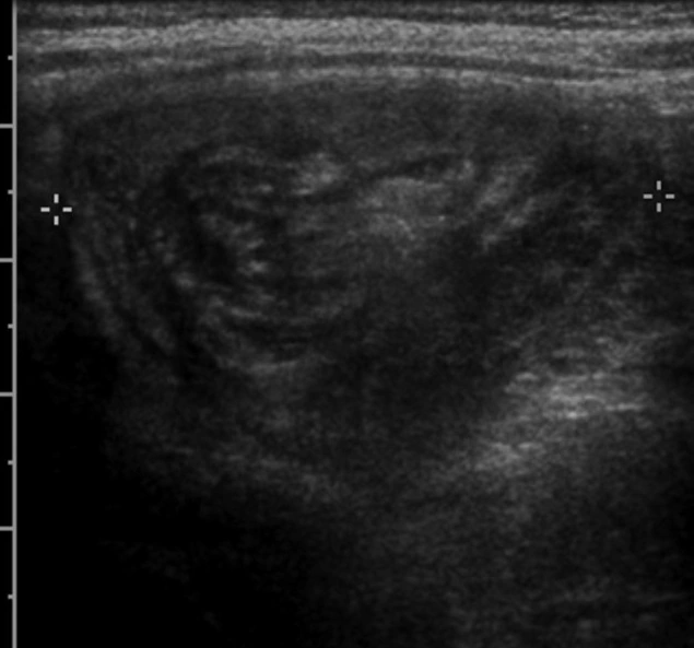 Enlarge the whole image
This real ultrasound shows the layered bowel arrangement. Compare its long-axis appearance with the separate cross-sectional schematic. A still image alone does not establish tissue viability.
Frank Gaillard. CC BY-SA 3.0. Source and provenance

A negative screening look is not the same as exclusion
Appropriately trained clinicians may confirm intussusception using point-of-care ultrasound. The RCH guideline cautions against using POCUS to exclude it. A negative limited scan should not end the evaluation of a child with convincing symptoms. Obtain appropriate experienced imaging and reconsider other causes if no bowel abnormality is found. [1]
A plain abdominal radiograph may be useful when obstruction or perforation is suspected or when access to ultrasound is limited. A normal radiograph does not exclude intussusception. Do not let a reassuring gas pattern delay transfer or definitive imaging when clinical concern remains high. Free air, in contrast, changes the treatment pathway because it suggests perforation. [1]
Not every telescoping finding is persistent ileocolic disease. A short ileoileal intussusception may resolve spontaneously, particularly when symptoms are mild and there is no obstruction or concerning lead point. The location, clinical course, and follow-up imaging matter. Do not send every transient small-bowel finding automatically to an ileocolic enema pathway. [1]
Suspected ileocolic disease
Experienced ultrasound and assessment for reduction eligibility are central. [1]
Negative limited test
Persistent clinical suspicion still needs adequate evaluation. [1]
Transient small-bowel finding
Interpret location, symptoms, obstruction, and persistence before choosing treatment. [1]
The test must match the remaining question. A normal radiograph or limited POCUS cannot cancel a convincing clinical pattern. [1]