Connect abnormal intestinal fixation to obstruction and ischemia, interpret imaging without false reassurance, and explain urgent treatment and Ladd repair.
A baby can have threatened bowel while the abdomen still feels soft. The central question is not simply whether the intestine is in the wrong place: has abnormal fixation allowed obstruction, and is blood flow now at risk? Follow the anatomy to the imaging decision, then to the operation that preserves bowel.
By the end, you should be able to distinguish a congenital rotational abnormality from an active twist, interpret an incomplete imaging report, and decide when another study would delay necessary treatment. Start with fixation, trace perfusion, then connect landmarks to test selection, urgency, and repair.
Why does a misplaced intestine become able to twist?
Malrotation is abnormal intestinal rotation and fixation; volvulus is twisting of bowel and its mesentery. One creates susceptibility to the other, but they are not synonyms. During normal development, the midgut lengthens outside the abdominal cavity, returns, and completes approximately 270 degrees of counterclockwise rotation around the superior mesenteric artery, viewed from the front. Fixation then establishes a broad mesenteric attachment. This developmental rotation is different from the later pathological twist. [1]
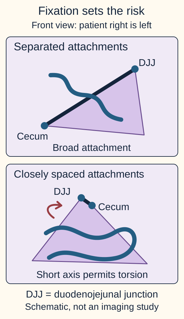
The duodenojejunal junction, where duodenum becomes jejunum near the ligament of Treitz, normally lies high on the patient's left. The cecum descends to the right lower abdomen. Their separation creates a broad, oblique mesenteric root. In classic malrotation, the junction remains rightward and the cecum may be high or unusually mobile. Closely spaced attachment points leave a narrow stalk containing the superior mesenteric vessels. The width of this attachment, not bowel position alone, explains the danger. [1]
Compare the broad and narrow attachments in the fixation diagram. Trace the line between the duodenojejunal junction and cecum with a finger or your eyes. Predict which arrangement lets a large length of bowel rotate around a short axis.
Separated attachment points create a broad root; closely spaced points create an axis around which the midgut can twist. Compare the attachment lengths, not the absolute size of the bowel loops. [1][5]Check the attachment prediction
The narrow attachment permits the mesentery to turn around its vascular stalk. Position suggests the abnormality; fixation explains the risk.
A second problem does not require twisting. Peritoneal Ladd bands can extend from the abnormally positioned cecum across the duodenum toward the right lateral abdominal wall. They compress the duodenum from outside, producing partial or complete obstruction even when the mesenteric vessels are not twisted. A band compresses a tube; volvulus twists a vascular attachment as well as bowel. [1] [7]
Apply the distinction to an older child with episodic vomiting: relief between episodes does not prove normal fixation. Partial band obstruction or intermittent torsion can explain fluctuating symptoms. Malrotation can first present in childhood or adulthood, sometimes with chronic pain, feeding intolerance or poor growth rather than a neonatal catastrophe. [4] [7]
Which fails first: drainage or delivery?
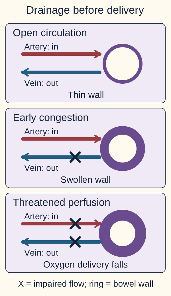
The superior mesenteric artery supplies the midgut; the superior mesenteric vein drains much of the same territory. In a typical midgut volvulus, the bowel turns clockwise around the artery's axis. Compression of thin-walled veins impairs outflow early, while some arterial blood may still enter. Venous congestion therefore produces a swollen bowel wall before complete loss of arterial perfusion. Continued torsion compromises inflow and can lead to infarction, perforation and sepsis. [1] [4] [5]
Use the three-state vascular diagram as a tracing exercise. In the open state, trace blood into and out of the bowel. In the congested state, stop the outward route but leave the inward route partly open. Predict the effect on wall thickness before comparing that state with the ischemic state.
Follow inflow and outflow in each state. Impaired drainage can swell the bowel while arterial inflow persists; later inflow compromise threatens oxygen delivery. Cross marks represent impaired flow, not a guaranteed complete occlusion. [1][4]Check the wall-thickness prediction
The wall becomes edematous as blood and fluid accumulate. Detectable arterial flow does not guarantee adequate tissue perfusion.
The visible result is a congested, swollen bowel that can progress to inadequate oxygen delivery. A patent proximal artery is therefore not reassurance that the bowel downstream is safe. Conversely, dusky bowel immediately after detorsion is not automatically dead: congestion can improve after the stalk is untwisted. Surgeons reassess viability rather than deciding from a single color observation. [4] [5]
Now apply the model to a baby whose vomiting is followed by abdominal tenderness, bloody stool and poor perfusion. Those findings suggest more than a blocked lumen. Acidosis and rising lactate may accompany ischemia or shock, but there is no laboratory value that must become abnormal before urgent action is justified. Early examination and laboratory findings can be deceptively mild. Delay risks extensive bowel loss, intestinal failure requiring parenteral nutrition, and death. [1] [4] [5]
What does each abnormal landmark actually establish?
A report of reversed vessels and a report of spiraling vessels answer different questions. The first concerns orientation; the second suggests active torsion. First identify the normal course: on a properly positioned frontal upper GI contrast study, the duodenum crosses the midline and the duodenojejunal junction lies left of the spine, approximately at the height of the duodenal bulb. A junction that remains rightward or low supports malrotation, interpreted with the entire course and study quality. [1]
Link the finding to the structure being examined
Finding
Interpretation and limit
FindingRightward or low duodenojejunal junction
Interpretation and limitAbnormal rotation is possible; positioning, distention and incomplete visualization can mislead.
FindingCorkscrew distal duodenum and proximal jejunum
Interpretation and limitThe contrast-filled bowel spirals around the mesenteric axis, concerning for volvulus.
FindingWhirlpool on targeted ultrasound
Interpretation and limitMesentery and vein wrap around the artery, strongly supporting volvulus in a symptomatic patient.
FindingSuperior mesenteric vein left of the artery
Interpretation and limitSuggests malrotation but does not establish active torsion or independently settle the diagnosis.
FindingCecum outside the right lower quadrant
Interpretation and limitSupports abnormal rotation; a normally positioned cecum does not exclude it.
These are complementary observations. Ultrasound also assesses the duodenal course, including the third portion's relationship to the artery and aorta. Normal vessel orientation alone does not exclude malrotation, and a technically limited scan is not a negative anatomical examination. Both upper GI studies and ultrasound require appropriate technique and interpretation. [1] [3]
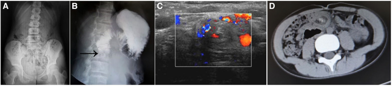
In the clinical image, compare panels A through D before reading the caption. Which panel examines contrast passage, and which examines vessels? The published panel B demonstrates duodenal obstruction; panel C is the authors' ultrasound example of a whirlpool. Panel A shows scant intestinal gas, and panel D shows a CT whirlpool. These are teaching examples from a published series, not an instruction to obtain all four tests. The image alone cannot determine bowel viability. [4]
Figure 1 from Yang et al., Frontiers in Pediatrics (2024), doi:10.3389/fped.2024.1390856, CC BY 4.0. A: scant bowel gas is nonspecific. B: the source arrow identifies duodenal obstruction, not a separately established corkscrew. C: Doppler whirlpool. D: CT whirlpool (asterisk). Compare which structure each modality depicts. This limited panel cannot exclude disease in an individual patient. Unmodified source image. Image: Xiaofeng Yang, Wei Wang, Kun Wang, Jingquan Zhao, Liandong Sun, Shuai Jiang, Yewen Wang, Wenyu Feng, Guojian Ding, Tingliang Fu, Aiwu Li and Lei Geng; original source; CC BY 4.0. [4].Check the imaging comparison
Panel B follows contrast through the duodenum; panel C examines mesenteric structures with ultrasound. A lumen study and a vascular study provide different evidence.
Transfer this to an equivocal report: if the vein is reversed but the duodenum was not seen and no whirlpool was established, say what remains unknown. Do not convert vessel orientation into proof of infarction or permission to dismiss persistent bilious vomiting. [1] [3]
Does a normal abdominal film end the evaluation?
Green, bile-stained emesis in an infant warrants immediate assessment for obstruction. Bile enters the second portion of the duodenum at the ampulla. In an obstructive presentation, bile in vomit points to a problem beyond that entry site rather than an isolated pyloric obstruction. It does not identify a unique cause: duodenal abnormalities, distal obstruction and other illnesses remain alternatives. Absence of green emesis also does not exclude malrotation. [4] [5] [7]
A stable 10-day-old has green emesis, a soft abdomen and a nondiagnostic abdominal radiograph. Predict whether an oral feeding trial or urgent anatomical evaluation is safer. Gas already beyond an intermittent or partial obstruction can remain visible. A normal or nonspecific film cannot exclude malrotation or early volvulus. [1] [2]
Check the next-test prediction
Continue urgent anatomical evaluation with surgical involvement. Distal gas is not proof of normal rotation.
For a stable infant with suspected malrotation, an urgent upper GI contrast series is usually appropriate under the ACR criteria. It establishes the duodenal course and junction position and may show obstruction or a corkscrew. At centers with an established pediatric ultrasound pathway, promptly available expert ultrasound can be the first examination. A systematic review supports high diagnostic accuracy but also reports heterogeneity and operator dependence. This is not a contest between modalities: choose the reliable study that the team can obtain promptly. [2] [3]
If ultrasound is nondiagnostic or clinical concern remains despite an apparently reassuring result, obtain urgent expert reassessment and usually upper GI imaging while the child remains stable. Review a discordant upper GI study with the radiologist rather than treating its report as infallible. Intermittent torsion and technical limitations can produce false reassurance. Deterioration changes the priority to operative management, not a longer sequence of tests. [1] [3]
Apply test selection to a different pattern. A neonate with multiple dilated distal loops and failure to pass meconium may need contrast enema to investigate distal obstruction. In suspected malrotation, however, enema only indirectly assesses the cecum and is not the preferred initial confirmatory test. Routine CT or MRI should not delay infant evaluation; CT may be useful in selected older patients, but radiation and local expertise matter. Nonbilious progressive projectile vomiting in a young infant instead supports a pyloric ultrasound pathway when the clinical findings fit. [1] [2]
When does another image become the wrong priority?
Compare two infants with green emesis. One is alert, warm, and has a soft abdomen. The other is lethargic with poor perfusion and a rigid abdomen. Both need urgent evaluation and early pediatric surgical contact, but the second has evidence of an ischemic abdominal emergency. Resuscitation and operative preparation occur together; treatment should not wait for an elective imaging slot or normalization of every laboratory result. [1] [4]
For suspected obstructive volvulus, keep the patient without oral intake, obtain vascular access, restore circulation with appropriately reassessed isotonic fluid, and decompress the stomach with a nasogastric or orogastric tube. Monitor physiology, glucose, electrolytes and acid-base status. Give broad-spectrum antibiotics when ischemic or perforated bowel or sepsis is suspected, alongside appropriate perioperative prophylaxis. These measures support the patient but do not untwist the mesentery. [8]
Predict what changes if the first infant's expert ultrasound demonstrates a convincing whirlpool. A soft abdomen does not make confirmed volvulus an observation problem. Arrange urgent operative treatment rather than waiting for hematochezia or shock. Conversely, tachycardia or a single abnormal lactate value alone does not diagnose volvulus; integrate the abdominal findings, trajectory and available imaging with the surgeon. [1] [3] [4]
Check the urgency comparison
Convincing volvulus warrants urgent surgery before the child becomes unstable. Stability permits diagnosis; it does not justify delaying treatment of a demonstrated twist.
Now consider a hospital without pediatric surgery. Early communication, resuscitation and emergency transfer planning should begin when volvulus is suspected. Local imaging is useful only if it can answer the question without materially delaying definitive care. A temporary improvement after decompression or fluids does not prove that the vascular threat has resolved. [1] [4]
Why does a Ladd procedure leave an unusual bowel arrangement?
The goal is functional safety, not reconstruction of embryonic rotation. When volvulus is present, counterclockwise detorsion restores the vascular orientation, followed by assessment of bowel viability. Reperfusing bowel should be preserved when possible. Clearly nonviable bowel requires resection; questionable viability may justify a planned second-look operation rather than unnecessarily sacrificing a long segment. Decisions depend on operative findings and physiology, not a fixed waiting interval. [4] [5]
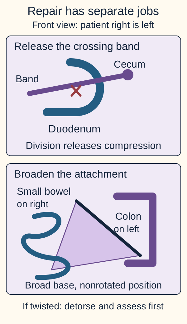
Trace the before-and-after repair diagram. Detorsion addresses the immediate twist. Division of Ladd bands releases extrinsic duodenal compression. Broadening the mesenteric base separates vulnerable attachment points and reduces future twisting. The small bowel is placed on the patient's right and the colon on the left. Appendectomy is commonly included to avoid later diagnostic confusion from an atypically located appendix; operative documentation should state whether it was performed. [1] [5] [7]
Band division releases extrinsic compression. A broadened base with small bowel on the patient’s right and colon on the left reduces twisting risk. If torsion exists, detorsion and viability assessment come first. [1][5][8]
Predict what remains wrong if the surgeon divides an obstructing band but leaves a narrow base. Food may pass more freely, yet the bowel remains able to twist. Predict what remains wrong if the surgeon only untwists the bowel. The initial blood-flow problem may improve while the predisposing anatomy persists.
Check the repair prediction
Band release, detorsion and base widening solve different problems. Relieving today's obstruction is not the same as reducing tomorrow's torsion risk.
After repair, adhesive small-bowel obstruction can occur, and recurrent volvulus is uncommon but possible. Recurrent green emesis, severe pain, distention or systemic illness requires urgent assessment. An expected right-sided small bowel on later imaging is not, by itself, proof of a failed operation. Loss of extensive viable bowel can cause short-bowel syndrome and prolonged nutritional dependence, which is why early recognition and careful viability assessment matter. [4] [5]
Apply the distinction to an incidental finding in an asymptomatic older patient. This is not the same decision as obstructive symptoms or volvulus. Prophylactic surgery versus observation remains individualized according to anatomy, age, comorbidities and reliable follow-up. The APSA systematic review found limited quality evidence; selected broad-based rotational abnormalities can be observed, and severe cardiac disease may affect timing. Observation requires explicit education about urgent symptoms and specialist follow-up, not dismissal of the finding. [6]
Before practice, explain the whole relationship in one sentence: abnormal fixation may create bands and a narrow base; a twist threatens venous drainage and then perfusion; diagnosis is urgent, and repair treats both the event and the anatomy.
Apply the lesson
Case 1
Show answer and explanations for case 1
A. Repeat abdominal radiography now to reassess the gas pattern (Why this does not fit)
A repeat film can identify an evolving obstruction or free air. It cannot establish normal rotational anatomy after a limited ultrasound, so it does not answer the unresolved question here. Define the duodenal course promptly.
Reasoning steps for option A
What can another abdominal radiograph reveal after this infant's green emesis?
It may reveal evolving obstruction or free air, even though the current film shows distal gas and no free air.
Why would repeating the film leave the limited ultrasound unresolved?
Neither distal gas nor another plain film establishes the duodenal course or excludes malrotation when ultrasound could not show it.
Which anatomic question should replace another gas-pattern check?
Urgently define the duodenal course and duodenojejunal junction with the available upper GI study.
B. Upper GI contrast series now (Best answer)
The infant is stable but has bilious symptoms. The limited ultrasound did not establish normal anatomy, and an upper GI series can evaluate the duodenal course and junction. A nondiagnostic scan should not end urgent evaluation.
Reasoning steps for option B
Why do normal perfusion and rectal gas not settle this infant's bilious vomiting?
The infant is presently stable, but green emesis still raises concern for malrotation or proximal obstruction; distal gas cannot rule either out.
What does immediate fluoroscopy add after bowel gas obscures the duodenum on ultrasound?
An upper GI contrast series directly traces the duodenal course and duodenojejunal junction that the limited ultrasound failed to establish.
If the ultrasound is nondiagnostic, should the soft abdomen end evaluation?
No. Stable examination and a nondiagnostic scan warrant prompt upper GI assessment, not reassurance that rotational anatomy is normal.
C. Contrast enema to locate the cecum (Why this does not fit)
An enema can show cecal malposition and investigate distal obstruction. No distal obstructive pattern is established here, and normal cecal position would not exclude malrotation. The upper GI series answers the proximal anatomical question more directly.
Reasoning steps for option C
What might a contrast enema show in this infant?
It can locate an abnormally placed cecum or help assess suspected distal obstruction.
Why is cecal localization insufficient despite green emesis and distal gas?
The question is proximal rotational anatomy; a normal cecal position would not exclude malrotation, and the film does not establish distal obstruction.
Which contrast examination directly tests the unvisualized duodenum?
An urgent upper GI series defines the duodenal course and junction more directly than a contrast enema.
D. Contrast-enhanced abdominal CT now (Why this does not fit)
CT can demonstrate rotational abnormalities. Immediately available fluoroscopy can answer this infant anatomical question without the additional CT radiation burden. Routine CT is not the preferred next study.
Reasoning steps for option D
What could contrast-enhanced CT contribute to a malrotation evaluation?
CT can depict abnormal rotational anatomy, but it is not needed merely because the ultrasound could not show the duodenum.
Why choose available fluoroscopy over CT in this stable 9-day-old?
An upper GI series can answer the unresolved duodenal-anatomy question promptly without the additional radiation burden of CT.
When fluoroscopy is immediately available, what should follow the limited ultrasound?
Obtain the upper GI study rather than routine abdominal CT to define the infant's duodenal course.
Takeaway: A limited ultrasound and distal gas do not exclude malrotation.
A. Complete the upper GI series during continued fluid resuscitation (Why this does not fit)
Concurrent resuscitation is appropriate. New rigidity and poor perfusion make further diagnostic delay unsafe when ischemic bowel is strongly suspected. Physiology can supersede the imaging plan.
Reasoning steps for option A
What part of completing the upper GI plan remains appropriate?
Fluid resuscitation should continue while treating this infant's deteriorating circulation.
Why is finishing the upper GI unsafe after refill reaches 5 seconds and the abdomen becomes rigid?
Rigidity, poor perfusion and lactate rising to 6.1 mmol/L suggest threatened bowel; diagnostic imaging would delay urgent exploration.
What overrides the previously planned fluoroscopy?
New peritoneal signs and shock physiology shift the priority to operative assessment alongside resuscitation.
B. Obtain CT angiography while continuing fluid resuscitation (Why this does not fit)
CT may depict vascular compromise. Shock and peritoneal signs already demand urgent operative assessment. Another imaging study should not delay treatment.
Reasoning steps for option B
What might CT angiography show in suspected midgut volvulus?
It might depict vascular compromise, but visual confirmation is not the immediate priority in this deteriorating infant.
Why not send an infant with rigidity, refill of 5 seconds and rising lactate to CT?
Those findings already support possible ischemic bowel requiring urgent surgical assessment, so CT would add delay.
What should replace further vascular imaging in this setting?
Proceed toward exploration while continuing circulatory support rather than waiting for CT angiography.
C. Continue resuscitation and serial examinations before committing to exploration (Why this does not fit)
Reassessment guides fluid therapy. It should accompany urgent operative preparation rather than postpone treatment of likely ischemic bowel. Support circulation and address the surgical cause together.
Reasoning steps for option C
Why are fluids and repeated examinations relevant during deterioration?
They support circulation and track response, but neither treats a possible strangulated bowel.
What is lost by waiting for serial examinations with rigidity and lactate 6.1 mmol/L?
Waiting postpones operative assessment despite signs of bowel ischemia and worsening perfusion.
How should reassessment be integrated with definitive care?
Continue resuscitation and reassessment during urgent operative preparation, not as a prerequisite to exploration.
D. Proceed toward exploration with resuscitation and antibiotics (Best answer)
Rigidity, worsening perfusion and rising lactate suggest threatened or infarcted bowel. Urgent operative management is needed alongside supportive treatment rather than after more imaging. Treat the anatomical cause while restoring circulation.
Reasoning steps for option D
Which changes make this more than stable bilious vomiting awaiting imaging?
New rigidity, capillary refill of 5 seconds and lactate rising from 1.8 to 6.1 mmol/L signal possible ischemic bowel.
Why combine exploration with resuscitation and antibiotics now?
An operation addresses the suspected mechanical and ischemic cause while fluids, decompression and antibiotics support the infant; imaging must not postpone treatment.
What determines the timing once peritonitis and poor perfusion appear?
The deteriorating physiology and suspected bowel threat make urgent exploration the priority even before the planned upper GI series.
Takeaway: Peritonitis and shock change the priority to urgent source control.
A. Peritoneal attachments from the cecum crossing the duodenum (Best answer)
The abnormal junction and high cecum support malrotation. Expansion when the attachments are lifted demonstrates their external compressive effect, favoring Ladd bands without current volvulus. Obstruction need not include torsion.
Reasoning steps for option A
What do the low right-sided junction and high cecum indicate?
They support abnormal intestinal rotation, which can place cecal peritoneal attachments across the duodenum.
Why does lifting the attachments implicate Ladd bands rather than a fixed intraluminal lesion?
The second duodenal portion expands when the external cecal attachments are raised, demonstrating reversible extrinsic compression.
Does absence of a mesenteric twist rule out band-related obstruction?
No. Ladd bands can obstruct the duodenum in malrotation without a current volvulus.
B. A mucosal diaphragm within the duodenal lumen (Why this does not fit)
A fenestrated duodenal web can permit passage and cause partial obstruction. However, lifting external cecal attachments would not relieve an intrinsic mucosal narrowing. Patency alone does not exclude a web; the dynamic response is the discriminator.
Reasoning steps for option B
How could a duodenal mucosal diaphragm cause intermittent symptoms?
A fenestrated web can allow some passage while partially obstructing the lumen and causing episodic vomiting.
Would lifting cecal attachments relieve a mucosal web?
No. An intrinsic diaphragm stays within the duodenal lumen, whereas this narrowed segment expands with an external maneuver.
What observation distinguishes a web from cecal bands here?
The reversible narrowing on lifting the attachments, not mere partial patency, points to extrinsic Ladd bands.
C. Pancreatic tissue encircling the duodenal wall (Why this does not fit)
Annular pancreas can narrow the duodenum and may coexist with other abnormalities. Relief specifically on lifting cecal attachments favors external bands as the observed cause here. The maneuver, not the presence of malrotation alone, identifies the mechanism.
Reasoning steps for option C
Why consider pancreatic tissue around the duodenum?
Annular pancreas can narrow the duodenal wall and cause obstruction, potentially alongside malrotation.
Why does expansion on lifting cecal attachments argue against the pancreas as this obstruction's cause?
Relief precisely with manipulation of those external attachments identifies band compression rather than persistent encircling pancreatic tissue.
Which evidence should decide between annular pancreas and Ladd bands?
Use the dynamic relief of narrowing when cecal attachments are lifted, not malrotation alone, to identify the operative mechanism.
D. Compression of the third duodenal portion beneath the artery (Why this does not fit)
Arterial compression can obstruct the third duodenal portion. This narrowing is in the second portion and accompanies a high cecum. Localize before choosing the mechanism.
Reasoning steps for option D
Where would compression beneath the mesenteric artery usually narrow the duodenum?
It typically affects the third duodenal portion, so it is a possible but anatomically different obstruction mechanism.
What location contradicts arterial compression in this operation?
The observed narrowing is in the second portion and expands when high-cecal attachments are lifted.
How should the narrowed segment's level guide diagnosis?
Localize the second-portion narrowing and its response to lifting bands before attributing it to arterial compression of the third portion.
Takeaway: Ladd bands can obstruct the duodenum without a current twist.
A. Primary occlusion of the proximal mesenteric artery (Why this does not fit)
Arterial interruption threatens bowel survival. Preserved pulsation with congestion better fits impaired drainage before complete inflow failure. Proximal pulsation does not establish tissue safety.
Reasoning steps for option A
Why would proximal mesenteric arterial occlusion be dangerous?
It can deprive bowel of inflow and cause ischemic injury or infarction.
Why does detectable arterial pulsation with a congested wall not chiefly indicate primary proximal occlusion?
Pulsation persists while twisted mesentery impairs venous drainage, producing swelling before complete arterial inflow failure.
Does a detectable supplying-artery pulse mean the twisted bowel is safe?
No. Venous outflow can already be obstructed and bowel threatened despite proximal arterial pulsation.
B. Reduced arterial inflow without impaired venous drainage (Why this does not fit)
Reduced inflow can cause ischemic injury. The early congested, swollen bowel with a local twist is better explained by restricted venous drainage while inflow continues. Distinguish congestion from isolated inadequate delivery.
Reasoning steps for option B
What consequence can reduced arterial inflow eventually produce?
Inadequate arterial delivery can cause ischemic injury, including as volvulus progresses.
Why does isolated low inflow fail to explain the first swollen, congested wall?
Early local torsion more readily compresses thin-walled veins while arterial inflow persists, causing blood pooling and edema.
What vascular mechanism better explains congestion before loss of the pulse?
Impaired venous return with continued arterial delivery, rather than reduced arterial inflow alone, causes the initial wall swelling.
C. Impaired venous drainage with continuing arterial inflow (Best answer)
Thin-walled veins are compressed early. Continuing inflow with restricted outflow produces congestion and edema before arterial perfusion is completely lost. Outflow failure can precede inflow failure.
Reasoning steps for option C
Which vessels are vulnerable first when the short mesenteric attachment twists?
Thin-walled mesenteric veins can be compressed before the supplying artery loses its detectable pulse.
How does ongoing arterial inflow produce a swollen, congested wall?
Blood enters through the still-pulsatile artery but drains poorly through compressed veins, increasing congestion and edema.
What does preserved arterial pulsation mean for the sequence of volvulus injury?
It does not exclude serious vascular compromise: venous outflow failure can precede arterial inflow failure.
D. Systemic hypotension without local outflow obstruction (Why this does not fit)
Systemic hypotension can impair bowel perfusion. The observed twist and local congestion identify a local vascular problem, not isolated low systemic pressure. Explain physiology from the supplied anatomy.
Reasoning steps for option D
Could systemic hypotension impair intestinal perfusion?
Yes, low systemic pressure can reduce bowel perfusion, but it does not specifically account for the local twisted attachment.
Why does isolated hypotension not explain the congestion seen at exploration?
The bowel is twisted around a short mesentery and locally swollen despite a detectable arterial pulse, pointing to impeded local venous return.
Which supplied anatomical finding should control the vascular explanation?
The local mesenteric twist explains outflow obstruction and congestion better than systemic hypotension without local obstruction.
Takeaway: Venous obstruction can coexist with arterial pulsation.
A. A has demonstrated torsion and needs urgent detorsion; B has malrotation for planned assessment (Why this does not fit)
A has abnormal rotational anatomy with obstruction. Smooth narrowing without a demonstrated spiral does not establish an active twist, whereas B has the corkscrew evidence of torsion. Match operative urgency to what the examination actually shows.
Reasoning steps for option A
Why might A's symptoms and abnormal junction prompt surgical assessment?
Weeks of green emesis, a low right-sided junction and smooth second-portion narrowing suggest symptomatic malrotation with obstruction.
Why is assigning urgent detorsion to A but planned care to B reversed?
A has no demonstrated spiral on this study; B's corkscrew distal duodenum and proximal jejunum demonstrate current torsion.
Which imaging pattern determines which infant needs urgent surgery?
B's corkscrew requires urgent operative treatment, whereas A needs assessment for symptomatic malrotation without demonstrated active twist.
B. Both have demonstrated torsion and need detorsion regardless of their different contrast patterns (Why this does not fit)
Both require prompt surgical involvement for abnormal rotation and symptoms. Only B has the supplied contrast evidence of current torsion; A may have band obstruction or intermittent disease requiring assessment. Do not equate abnormal junction position with a demonstrated twist.
Reasoning steps for option B
Why do both infants merit surgical attention?
Both have bilious symptoms and abnormal duodenojejunal junction position consistent with clinically important malrotation.
Why cannot the two studies both be called demonstrated torsion?
Only B has a corkscrew course; A's smooth narrowing may reflect bands or intermittent disease, not a twist shown now.
What must be present before claiming detorsion is indicated by current imaging?
A demonstrated spiral or corkscrew supports active torsion; a low junction alone does not prove it.
C. Both have malrotation for planned assessment because their perfusion is currently preserved (Why this does not fit)
Both infants remain clinically stable. B nevertheless has imaging evidence of volvulus, for which preserved perfusion is not a reason to defer urgent operative treatment. Interpret the anatomy before using stability to assign timing.
Reasoning steps for option C
What clinical feature makes planned assessment seem tempting for both?
Both infants currently have good perfusion and no peritoneal signs.
Why is preserved perfusion insufficient to defer B's treatment?
B's acute emesis and corkscrew course show volvulus despite current stability, so delaying urgent surgery risks bowel ischemia.
How should stability and the contrast patterns be weighed together?
A requires assessment for symptomatic malrotation; B's demonstrated torsion warrants urgent surgery even before perfusion worsens.
D. A needs assessment for symptomatic malrotation; B has demonstrated torsion requiring urgent surgery (Best answer)
A has abnormal rotation and obstruction without a twist demonstrated on this study. B has a corkscrew consistent with active volvulus and needs urgent treatment despite preserved perfusion. Absence of a demonstrated spiral in A does not exclude intermittent torsion or make the symptoms unimportant.
Reasoning steps for option D
How do A's weeks of symptoms and smooth narrowing differ from B's abrupt symptoms and corkscrew?
A has symptomatic abnormal rotation without a twist shown on this examination; B has contrast evidence of active volvulus.
Why do their findings call for different operative urgency despite normal perfusion?
A needs surgical assessment for malrotation and obstruction, while B's demonstrated torsion warrants urgent operation before vascular compromise develops.
Does the lack of a spiral in A make intermittent torsion impossible?
No. The current study does not demonstrate a twist in A, but intermittent torsion remains possible and A's symptoms still require assessment.
Takeaway: Abnormal junction position and a corkscrew carry different implications; preserved perfusion does not justify delay after torsion is demonstrated.
A. Possible active torsion; a fixed rightward junction would confirm the ongoing twist (Why this does not fit)
An inverted vessel relationship can raise concern but does not establish an active twist. A rightward junction supports abnormal rotation rather than directly showing spiraling mesentery. Positional abnormalities should not be relabeled as demonstrated torsion.
Reasoning steps for option A
Does the vein lying left of the artery establish active torsion?
No. Vessel inversion in this vomiting neonate raises suspicion for malrotation, but does not itself show mesenteric twisting.
Would a fixed rightward duodenojejunal junction confirm the proposed ongoing twist?
No. A rightward junction adds evidence of abnormal rotation, not direct evidence that bowel and vessels are currently winding around the artery.
What finding would distinguish torsion from these positional abnormalities?
Look for a true whirlpool of mesentery and vein around the superior mesenteric artery rather than calling two abnormal positions proof of volvulus.
B. Possible malrotation; mesentery and vein wrapping around the artery would support torsion (Best answer)
Vessel inversion raises concern for malrotation without independently diagnosing it. A true whirlpool adds evidence of active mesenteric twisting, which changes urgency in this symptomatic infant. Distinguish orientation from an actual spiral.
Reasoning steps for option B
What does a vein left of the artery imply when the distal duodenum was not seen?
It raises concern for malrotation in a 5-day-old with bilious vomiting, but the unvisualized duodenum and isolated inversion do not establish malrotation or active torsion.
What would wrapping of vein and mesentery around the artery add?
A true whirlpool on repeat expert scanning directly supports an active mesenteric twist, unlike vessel orientation alone.
Why does this distinction matter despite a soft abdomen?
A symptomatic infant with evidence of current volvulus needs urgent surgical assessment; a soft examination cannot downgrade a demonstrated twist.
C. Possible malrotation; a rightward junction would identify an active vascular twist (Why this does not fit)
A rightward junction supports abnormal rotation. It does not itself show an active vascular twist, which requires other evidence such as a whirlpool. Two positional abnormalities are not the same as demonstrated torsion.
Reasoning steps for option C
What would a rightward duodenojejunal junction show in this infant?
It would support abnormal intestinal rotation, complementing the inverted vein-artery relationship.
Does junction position identify an active vascular twist?
No. A displaced junction is a fixed anatomical position, whereas ongoing torsion requires evidence of spiraling mesentery or vessels.
Which observation resolves the specific question on repeat scanning?
Identify or exclude a whirlpool around the superior mesenteric artery rather than treating two malposition signs as direct proof of torsion.
D. Possible active torsion; preserved proximal arterial flow would identify spontaneous resolution (Why this does not fit)
Proximal arterial flow can remain present during torsion. Its detection does not show that the vein and bowel have untwisted or that tissue perfusion is adequate. Resolution cannot be inferred from this signal alone.
Reasoning steps for option D
Can arterial Doppler flow persist during midgut torsion?
Yes. Flow in the proximal superior mesenteric artery may still be detectable while the bowel and mesenteric vein are twisted.
Would preserved proximal flow prove spontaneous untwisting?
No. It cannot demonstrate that the vein and bowel have unwrapped or that downstream bowel perfusion is adequate.
What would support resolution of the suspected twist?
Reassess the actual mesenteric configuration rather than equating a proximal arterial signal with resolved volvulus.
Takeaway: Vessel inversion and junction position concern rotation; a whirlpool adds evidence of torsion.
A. Urgent exploration with concurrent circulatory support (Best answer)
Abnormal bowel position and vascular spiraling indicate malrotation complicated by torsion. Reduced enhancement with guarding and hypotension raises concern for ischemia. Adult presentation does not reduce the urgency.
Reasoning steps for option A
What do right-sided jejunum, an abnormal junction, and spiraling mesenteric vessels indicate?
They indicate malrotation with mechanical midgut volvulus in this 29-year-old, not merely an atypical bowel position.
Why are reduced loop enhancement, guarding, and hypotension alarming?
Together they suggest threatened or established bowel ischemia and systemic deterioration requiring immediate source control.
What treatment addresses this adult's CT-demonstrated twist with guarding and hypotension?
Arrange urgent operative exploration while providing circulatory resuscitation; adult age does not make this vascular twist less urgent.
B. Confirm the duodenal course with fluoroscopy before exploration (Why this does not fit)
Fluoroscopy can define rotational anatomy. CT already shows a vascular twist with reduced enhancement in a deteriorating patient. Another anatomical study should not delay definitive care.
Reasoning steps for option B
When could fluoroscopy help clarify this patient's anatomy?
An upper GI study can delineate the duodenal course when rotational anatomy remains uncertain in a stable patient.
Is that uncertainty still worth a fluoroscopy delay here?
No. CT already shows an abnormal junction and mesenteric spiral, with reduced enhancement, guarding, and hypotension.
What should take priority over fluoroscopy after CT shows a twist and poorly enhancing loops?
Urgent exploration with concurrent resuscitation, rather than postponing treatment of possible ischemic bowel for confirmation.
C. Therapeutic anticoagulation with serial abdominal examinations (Why this does not fit)
Anticoagulation can treat selected thrombotic disease. The supplied spiral and malposition identify mechanical torsion, which anticoagulation does not correct. Treat the cause of ischemia.
Reasoning steps for option C
Why might anticoagulation initially seem relevant to reduced bowel enhancement?
Thrombotic mesenteric disease can impair perfusion and sometimes warrants therapeutic anticoagulation.
What identifies a different mechanism in this CT examination?
Right-sided jejunum, an abnormal junction, and vessels spiraling around the artery point to mechanical midgut torsion rather than an isolated clot.
Why are anticoagulation and serial examinations insufficient?
They cannot unwind the mesentery or relieve the vascular obstruction; guarding and hypotension call for urgent operative assessment.
D. Endoscopic decompression followed by elective fixation (Why this does not fit)
Endoscopy can decompress selected colonic volvulus cases. This is a midgut vascular twist with possible ischemia, not an uncomplicated accessible colonic lesion. Operative assessment is required.
Reasoning steps for option D
Where can endoscopic decompression have a role in volvulus?
Selected uncomplicated colonic volvulus may be accessible to endoscopic decompression.
Why is this adult's twist not that situation?
CT localizes the spiral to midgut mesenteric vessels and shows poorly enhancing loops, while guarding and hypotension suggest ischemia.
What is required instead of elective fixation after endoscopy?
Urgent exploration and circulatory support to assess and treat the threatened bowel.
Takeaway: Midgut volvulus remains an emergency in adulthood.
A. It excludes the need for urgent operative treatment (Why this does not fit)
Arterial flow can persist early in torsion. The symptomatic whirlpool is not neutralized by a proximal signal. Treatment should precede late perfusion failure.
Reasoning steps for option A
What does Doppler flow in the proximal artery actually establish?
It establishes a detectable arterial signal at the sampled site, even though torsion may persist.
Can that signal eliminate the significance of green emesis and a whirlpool?
No. Ultrasound directly shows clockwise wrapping of vein and bowel around the artery in a symptomatic infant.
Why should operative treatment not await loss of the signal?
Venous congestion and downstream ischemia can develop before proximal arterial flow disappears or guarding and lactate abnormalities appear.
B. It indicates adequate oxygen delivery throughout the involved bowel (Why this does not fit)
A proximal arterial signal shows flow at the sampled location. It does not establish venous drainage, microcirculatory perfusion or adequate oxygen delivery throughout the twisted segment. Interpret a local measurement within the anatomical problem.
Reasoning steps for option B
Where was arterial flow measured in this six-week-old?
Doppler detected flow proximally in the superior mesenteric artery, not oxygen delivery in every involved bowel loop.
What does preserved proximal arterial Doppler fail to establish in the bowel wrapped around the artery?
It does not establish unobstructed venous drainage, intact microcirculation, or adequate tissue oxygenation distal to the twist.
How should the measured signal be weighed against the whirlpool?
Treat the symptomatic mesenteric wrapping as urgent rather than declaring the entire bowel adequately perfused from one proximal measurement.
C. It does not exclude impaired drainage or evolving bowel ischemia (Best answer)
A proximal signal can coexist with venous compression and impaired downstream perfusion. The whirlpool remains urgent despite normal lactate and absence of guarding. One reassuring measurement cannot establish bowel safety.
Reasoning steps for option C
What does clockwise wrapping of bowel and mesenteric vein show?
The expert ultrasound demonstrates a current whirlpool compatible with midgut torsion in an infant with green emesis.
Does proximal arterial flow rule out impaired drainage?
No. Venous compression may coexist with arterial inflow, and downstream bowel ischemia can evolve while the proximal signal persists.
Do normal lactate and no guarding establish bowel safety?
No. Those early reassuring findings do not negate the symptomatic whirlpool or justify waiting for later ischemic signs.
D. It establishes a chronic rotational variant without a current twist (Why this does not fit)
A variant can change orientation without spiraling bowel. The examination directly describes wrapping, which the flow signal does not negate. Distinguish malposition from active torsion.
Reasoning steps for option D
What could a chronic rotational variant explain on imaging?
It might explain an abnormal positional relationship without active wrapping of bowel around the artery.
What finding contradicts a variant without current twist?
Expert ultrasound explicitly shows clockwise wrapping of mesenteric vein and bowel around the superior mesenteric artery.
Can preserved arterial flow reclassify that wrapping as a benign variant?
No. A proximal flow signal neither untwists the mesentery nor excludes threatened bowel in this vomiting infant.
Takeaway: Normal lactate and proximal flow do not cancel a symptomatic whirlpool.
A. Wait for transfer and obtain fluoroscopy before considering targeted ultrasound (Why this does not fit)
Upper GI fluoroscopy is usually appropriate for suspected malrotation. In this stable infant, an immediately available expert ultrasound pathway can assess the relevant anatomy while escalation is arranged if needed. The prior untargeted scan is not a reason to bypass available expert evaluation.
Reasoning steps for option A
Why is upper GI fluoroscopy a reasonable consideration for green emesis?
It can define the duodenal course and duodenojejunal junction when malrotation is suspected.
What does waiting for transfer sacrifice in this particular stable infant?
An expert pediatric team can immediately perform targeted ultrasound, whereas fluoroscopy requires transfer and the earlier scan never assessed rotation.
How should fluoroscopy fit into the available pathway?
Use prompt targeted expert ultrasound now, with upper GI assessment if the relevant findings remain unresolved or discordant rather than waiting before scanning.
B. Repeat plain abdominal films first and use a normal gas pattern to defer further imaging (Why this does not fit)
Films can detect obstruction or perforation. Normal gas does not demonstrate normal rotation, so it cannot close the diagnostic gap left by the general ultrasound. Evaluate the duodenum and mesenteric structures.
Reasoning steps for option B
What can repeat abdominal radiographs reveal?
They may show an obstructive gas pattern or free air in a vomiting infant.
Would a normal gas pattern resolve the question left by the organ ultrasound?
No. Neither normal gas nor normal liver, kidneys, and gallbladder establishes the course of the duodenum or rotational anatomy.
Which structures should the immediate study interrogate instead?
Have the expert team assess duodenal course and mesenteric vessels, escalating if ultrasound is nondiagnostic or conflicts with symptoms.
C. Obtain local CT angiography first to resolve the unreported vascular anatomy (Why this does not fit)
CT can show mesenteric abnormalities. An immediately available pediatric ultrasound team offers the preferred targeted initial approach in this established pathway without CT radiation. Reserve other modalities for a justified unresolved question.
Reasoning steps for option C
What could CT angiography depict that the general ultrasound omitted?
CT can depict mesenteric vascular relationships and other abdominal anatomy.
Why is CT angiography not the best first response here?
This well-perfused infant has immediate access to an established targeted pediatric ultrasound pathway that can address the missing anatomy without CT radiation.
Which targeted ultrasound results would require escalation beyond the immediately available expert scan?
Escalate unresolved or discordant findings, including to upper GI evaluation, rather than substituting local CT for available expert ultrasound.
D. Obtain targeted expert ultrasound now, escalating unresolved or discordant findings (Best answer)
The first study did not evaluate the relevant structures. Expert ultrasound can promptly assess the duodenal course and vascular relationship in the stated pathway, with upper GI evaluation if findings remain unresolved. Match test scope and availability to the clinical question.
Reasoning steps for option D
Did the reported normal liver, kidneys, and gallbladder evaluate malrotation?
No. The general ultrasound omitted the distal duodenal course and mesenteric vessels despite persistent green emesis.
Why perform expert ultrasound now rather than wait for transferred fluoroscopy?
The infant is currently well perfused without peritoneal signs and the experienced pediatric team can immediately target the relevant anatomy while surgical involvement continues.
What if the targeted result is incomplete or discordant with symptoms?
Do not declare malrotation excluded; escalate promptly to upper GI evaluation or further surgical assessment as indicated.
Takeaway: Use a promptly available expert pathway to answer the actual anatomical question, with escalation when uncertainty persists.
A. Continue decompression and serial examinations before deciding on surgery (Why this does not fit)
Decompression and serial examination support care. They do not justify deferring treatment after the corkscrew has demonstrated torsion. Intervene before tenderness or shock develops.
Reasoning steps for option A
What can decompression and serial examinations contribute?
They can reduce vomiting and track clinical change while the 17-day-old is stabilized.
Why can they not be used to defer the operative decision?
The low rightward junction and corkscrew duodenum and jejunum already demonstrate malrotation with torsion, even with a soft abdomen.
Should surgery await tenderness or shock after a corkscrew has been demonstrated?
Arrange urgent surgery now, before tenderness, poor perfusion, or shock develops rather than waiting for serial examinations to deteriorate.
B. Arrange urgent operative treatment with continued stabilization (Best answer)
The abnormal junction suggests malrotation and the corkscrew indicates associated torsion. Maintained perfusion is an opportunity to intervene before ischemia progresses. Confirmed volvulus needs urgent surgery.
Reasoning steps for option B
What does the low rightward duodenojejunal junction indicate?
It supports abnormal intestinal rotation in this infant with green emesis.
What additional diagnosis does the corkscrew proximal bowel course support?
The corkscrew pattern indicates associated midgut volvulus, a current twist that threatens mesenteric perfusion.
How should normal perfusion and a soft abdomen affect the next step?
They allow intervention before ischemia advances, not observation; arrange urgent operative treatment with continued stabilization.
C. Obtain a contrast enema to confirm cecal displacement (Why this does not fit)
Cecal position can support a rotational diagnosis. It adds no necessary information after this demonstration of torsion, and normal position would not exclude the problem. Avoid redundant diagnostic delay.
Reasoning steps for option C
What could a contrast enema contribute to a rotation workup?
An ectopic cecum could lend support to malrotation in a patient whose anatomy is still uncertain.
Would cecal position change management after this upper GI series?
No. The low rightward junction and corkscrew already indicate volvulus, and even a normally positioned cecum would not exclude malrotation.
What should replace cecal localization after the upper GI series demonstrates a corkscrew?
Avoid redundant imaging delay and proceed to urgent operative management with ongoing stabilization.
D. Repeat fluoroscopy after decompression to document resolution (Why this does not fit)
Decompression can lessen vomiting and distention. It does not reliably correct a mesenteric twist. Do not postpone treatment to seek spontaneous resolution.
Reasoning steps for option D
What might gastric decompression accomplish before repeat fluoroscopy?
It may relieve vomiting and distention during supportive care.
Would it reliably straighten the corkscrew mesenteric twist?
No. Decompression does not correct the demonstrated volvulus or guarantee spontaneous resolution.
What response to the corkscrew avoids waiting for decompression to resolve a vascular twist?
Arrange urgent operative treatment rather than repeat contrast imaging to await apparent improvement in a currently stable infant.
Takeaway: Treat demonstrated volvulus before shock develops.
A. Complete the local contrast study before transfer (Why this does not fit)
Imaging can define rotation in a stable child when promptly available. This delay in a poorly perfused infant would postpone definitive care. Stabilization and transfer should proceed without waiting for the study.
Reasoning steps for option A
When would local contrast imaging help define this infant's rotation?
A promptly available study can define rotational anatomy in a stable infant with bilious vomiting.
Why is waiting 90 minutes for that study unsafe here?
This infant is poorly perfused and increasingly distended, and local surgery is unavailable; waiting delays transfer to definitive care.
What should proceed while the poorly perfused infant waits for transport rather than 90-minute-delayed imaging?
Continue stabilization and emergency transfer to the accepting surgeon without waiting for contrast imaging.
B. Give a single intravenous fluid bolus and defer gastric decompression until arrival (Why this does not fit)
An initial bolus may be needed to restore circulation. Ongoing reassessment and gastric decompression remain important during preparation and transport. A single intervention should not replace continued stabilization.
Reasoning steps for option B
What benefit could an initial fluid bolus provide in this poorly perfused infant?
Intravenous fluid may improve circulating volume and peripheral perfusion.
Why is one bolus with deferred decompression insufficient during transfer?
Persistent green emesis and distention require gastric decompression, while circulation needs reassessment and further support as indicated.
How should support continue en route to pediatric surgery?
Reassess response to resuscitation and maintain decompression rather than treating a single bolus as complete stabilization.
C. Keep NPO, decompress the stomach, and reassess intravenous resuscitation (Best answer)
Poor perfusion with possible obstruction requires vascular support and gastric decompression while definitive care is organized. Reassess the response to fluids rather than treating a fixed bolus as sufficient. Supportive care accompanies, not replaces, emergency transfer.
Reasoning steps for option C
Which threats require simultaneous care before this infant reaches the receiving surgeon?
Poor perfusion needs reassessed intravenous resuscitation, while persistent bilious emesis and distention suggest obstruction needing NPO status and gastric decompression.
Why do these measures belong alongside emergency transfer rather than after it?
With no local surgeon and imaging delayed 90 minutes, circulatory support and decompression can proceed during transport arrangements without postponing definitive care.
Does improvement after fluids replace the need for transfer?
No. Supportive measures can improve physiology but cannot exclude or correct an obstructing twist or threatened bowel.
D. Pause transfer after a transient fluid response and repeat the abdominal examination (Why this does not fit)
A transient improvement can follow restoration of circulating volume. It does not establish that the obstruction or vascular threat has resolved. Continue stabilization and definitive-care planning together.
Reasoning steps for option D
Why might a repeat examination after fluids seem reassuring?
Restored circulating volume can temporarily improve perfusion and the infant's appearance.
What remains unresolved if this bilious infant improves transiently?
Neither obstruction nor a mesenteric vascular threat is excluded by a temporary fluid response.
Should reassessment pause the accepted emergency transfer?
No. Repeat examinations accompany stabilization and transfer, not a delay in access to pediatric surgery.
Takeaway: Resuscitation and transfer planning occur together when local definitive care is unavailable.
A. Detorse counterclockwise, restore physiology, then reassess viability (Best answer)
The twist can cause potentially reversible congestion. Detorsion with continued resuscitation permits evaluation after circulation is restored before finalizing resection extent. Initial color alone can underestimate viability.
Reasoning steps for option A
What must be undone before judging the dusky bowel around the superior mesenteric artery?
The clockwise volvulus must be detorsed counterclockwise to relieve vascular twisting.
Why reassess after detorsion and resuscitation before setting resection margins?
Unperforated dusky bowel may reflect reversible venous congestion and can recover as circulation and physiology improve.
What does initial discoloration alone establish about bowel survival?
It does not establish irreversible injury across the entire discolored length; viability needs assessment after flow restoration.
B. Divide duodenal bands, resect dusky bowel, then correct vascular orientation (Why this does not fit)
Band division relieves external compression. Resection before relief of the twist may sacrifice bowel that would recover after flow restoration. Prioritize the vascular problem before deciding the full extent of irreversible injury.
Reasoning steps for option B
What does division of duodenal bands treat in malrotation?
It relieves extrinsic compression of the duodenum, not the acute mesenteric vascular twist.
Why is resection before correcting this infant's clockwise twist premature?
Some dusky unperforated bowel could reperfuse after counterclockwise detorsion and would otherwise be sacrificed.
Which operative problem should precede the resection decision?
Relieve torsion and restore circulation, then assess which bowel remains nonviable.
C. Resect the full discolored segment, then detorse the remaining mesentery (Why this does not fit)
Clearly necrotic tissue requires resection. Dusky appearance before detorsion does not establish that the entire segment is irreversibly injured. Restore flow before defining the final resection boundary.
Reasoning steps for option C
When does bowel resection become necessary after volvulus?
Clearly nonviable bowel requires removal after its viability has been assessed.
Why not remove every discolored loop before detorsion here?
Dusky color without perforation can reflect reversible congestion from the still-twisted mesentery.
What should determine the resection boundary in dusky bowel once the clockwise twist is relieved?
Detorse and resuscitate first, then reassess perfusion and tissue viability rather than using the initial color border.
D. Broaden the mesenteric base, then use initial color to determine resection (Why this does not fit)
Widening reduces future torsion risk. It does not replace immediate detorsion or assessment of the response to reperfusion. Different components address the acute vascular event and the predisposing anatomy.
Reasoning steps for option D
What future problem does broadening the mesenteric base address?
It reduces susceptibility to another twist by widening the narrow attachment.
Why can widening alone not determine survival of this dusky bowel?
The active clockwise twist still impairs circulation; initial color cannot substitute for reassessment after detorsion.
Why must detorsion and viability assessment precede relying on mesenteric widening in this acute clockwise volvulus?
Relieve torsion and reassess viability before using base widening to address the underlying predisposition.
Takeaway: Detorse the acute twist before defining the final resection extent.
A. Resect the originally purple length despite its improving perfusion (Why this does not fit)
Initial discoloration can represent reversible congestion. The observed reperfusion argues against using the original color boundary as the resection boundary. Avoid unnecessary loss of absorptive bowel.
Reasoning steps for option A
What might have caused the long segment's original purple color?
Venous congestion during volvulus may be reversible once the mesentery is untwisted.
What does pinking up with restored pulsation say about the original color boundary?
The bowel is reperfusing, so its earlier purple extent is not a reliable margin for resection.
What is at stake if all originally purple bowel is removed?
A long length of potentially viable absorptive bowel could be lost unnecessarily.
B. Preserve the bowel and close without correcting the narrow attachment (Why this does not fit)
Improving color favors preservation. It does not eliminate the predisposing mesenteric anatomy or the need to complete repair. Recovery of perfusion is not the entire operation.
Reasoning steps for option B
Which observation supports preserving this bowel after detorsion?
Its color improves with warming and arterial pulsation returns, without a clearly necrotic segment.
What dangerous anatomy persists if the surgeon simply closes?
The narrow mesenteric base remains a short axis around which bowel can twist again.
What must accompany preservation to complete treatment?
Continue viability assessment and repair the predisposing malrotation, including broadening the mesenteric base.
C. Preserve bowel based on restored pulsation without reassessing its wall (Why this does not fit)
Restored pulsation is encouraging. A proximal or mesenteric arterial signal alone does not guarantee viable bowel tissue, so wall appearance and other operative findings still require assessment. Combine observations rather than relying on a single signal.
Reasoning steps for option C
What does restored mesenteric pulsation suggest after detorsion?
Arterial inflow has improved, supporting possible recovery of the purple bowel.
Why is a pulse alone not proof the entire bowel wall will survive?
A detectable arterial signal does not establish adequate tissue-level perfusion or exclude residual injury.
Which findings should be integrated before preserving the full segment?
Reassess wall color and other operative evidence of viability along with pulsation, while completing the anatomical repair.
D. Preserve recovering bowel, reassess viability, and complete repair (Best answer)
Improving color and pulsation support reversible injury. Continued assessment can preserve useful bowel while Ladd repair addresses the anatomy. Reserve resection for tissue determined to be nonviable.
Reasoning steps for option D
How should improving color and pulsation change the resection decision?
These changes favor recovering bowel rather than automatic removal of the originally purple length.
What else must be done given no necrotic demarcation but a narrow base?
Continue direct viability reassessment and complete Ladd repair to address the persistent torsion risk.
What finding would justify resection despite improving color and pulsation after detorsion?
Resect tissue established as nonviable, not bowel merely discolored before detorsion.
Takeaway: Recovery after detorsion favors preservation with reassessment, not indiscriminate resection.
A. Complete the extensive resection based on initial discoloration (Why this does not fit)
Clearly gangrenous tissue must be resected. The remaining segment is explicitly uncertain, and extensive resection risks intestinal failure. Initial color alone should not settle that decision.
Reasoning steps for option A
Which bowel has already justified resection at this operation?
The short frankly gangrenous segment has been removed because it is clearly nonviable.
Why not extend resection across the much longer questionable segment now?
Its survival remains uncertain after flow restoration, and losing it all would leave very little small bowel.
What should determine further removal rather than its initial color?
Further assessment of actual viability should guide resection to avoid preventable intestinal failure.
B. Consider a planned second-look operation after further reassessment (Best answer)
Uncertain viability after restoring flow differs from obvious gangrene. A second look can clarify recovery while avoiding premature extensive bowel loss. Timing and interim management depend on operative findings and physiology.
Reasoning steps for option B
What uncertainty remains after detorsion and removal of frank gangrene?
The viability of a long additional bowel segment remains indeterminate despite restored flow.
How does a planned second look protect this child?
Direct reassessment may identify recovery before committing to a resection that would leave minimal small bowel.
Is second-look timing fixed solely by the bowel's first appearance?
No. Operative findings, interval physiology and the child's ability to receive intensive-care support guide timing and interim care.
C. Rely on intensive-care laboratory monitoring without planned operative reassessment (Why this does not fit)
Intensive-care monitoring is essential. Systemic measurements alone may not resolve uncertainty about a long questionable bowel segment. A planned second look can provide direct reassessment before deterioration declares irreversible injury.
Reasoning steps for option C
What can intensive-care monitoring contribute after detorsion leaves a long segment of uncertain viability?
Laboratory trends and physiology help track the supported child after the first operation.
Why cannot those measurements alone settle this long segment's fate?
Systemic markers do not directly establish whether each questionable bowel region recovers or becomes nonviable.
What adds direct evidence before a potentially devastating resection?
A planned operative second look can reassess bowel viability after additional time and support.
D. Use postoperative contrast passage to decide whether the questionable bowel survives (Why this does not fit)
Contrast passage can assess luminal transit. It cannot establish the viability of a long segment that remains doubtful after restoring flow. Tissue survival and an open lumen are different questions.
Reasoning steps for option D
What question can postoperative contrast passage answer?
It can show whether contrast traverses the intestinal lumen.
Why does contrast transit not resolve the surgical uncertainty?
An open lumen does not prove the long questionable bowel segment is viable after volvulus.
What test of tissue survival is more direct in this setting?
Planned operative reassessment of the segment addresses viability rather than only luminal patency.
Takeaway: A second look can resolve uncertain viability without premature extensive resection.
A. Place a feeding tube beyond the duodenal narrowing (Why this does not fit)
A distal feeding tube can facilitate nutrition. It does not change the closely spaced mesenteric attachments. Nutritional access does not correct torsion susceptibility.
Reasoning steps for option A
What could a tube placed beyond this child's duodenal narrowing accomplish?
It could provide distal nutritional access while bypassing a narrowed region.
Why does that not reduce future volvulus after band division?
The duodenojejunal and ileocecal attachment points remain close, leaving a narrow axis for twisting.
What anatomic change, unlike feeding access, addresses that axis?
Broaden the mesenteric base to separate the fixation points.
B. Perform appendectomy without altering the mesenteric attachment (Why this does not fit)
Appendectomy can prevent later diagnostic confusion. It does not widen the stalk around which bowel can twist. Each operative component has a separate purpose.
Reasoning steps for option B
Why might appendectomy be included in a malrotation repair?
An atypically located appendix could otherwise cause later diagnostic confusion.
What risk remains if only the appendix is removed here?
The closely spaced mesenteric attachments still permit the midgut to twist around a short stalk.
Which separate repair step targets torsion susceptibility?
Widen the mesenteric base; appendectomy does not perform that function.
C. Broaden the mesenteric base (Best answer)
Close fixation points leave a short axis for torsion. Widening the base addresses this risk even after band division has relieved current obstruction. Patent lumen and safer fixation are different goals.
Reasoning steps for option C
What has division of the band from the high cecum already fixed?
It has relieved extrinsic duodenal compression and restored passage, without treating the narrow root.
Why broaden the base when no volvulus is currently present?
Closely spaced duodenojejunal and ileocecal attachments still create a short axis for a future twist.
Which goals must be distinguished in this operation?
D. Fix the cecum in the right lower quadrant without widening the mesentery (Why this does not fit)
Cecal fixation may appear to restore a normal positional landmark. Without widening the short mesenteric attachment, it does not directly address the supplied torsion axis. The goal of Ladd repair is safer geometry rather than normal position alone.
Reasoning steps for option D
Why might moving the cecum to the right lower quadrant seem attractive?
It appears to recreate a familiar normal positional landmark.
Why is cecal fixation without widening inadequate for this child?
The close mesenteric attachment points remain, so the short axis for torsion is not directly corrected.
What matters more than restoring cecal position alone?
Broaden the mesenteric root to reduce twisting risk after relief of band obstruction.
Takeaway: Band division relieves compression; widening the base reduces torsion susceptibility.
A. The position is consistent with repair, but does not document adequate mesenteric widening (Best answer)
Ladd repair commonly leaves this nonrotated arrangement. Bowel position alone does not reveal whether the mesenteric base was adequately widened, and this asymptomatic scan does not diagnose a complication. Distinguish an expected landmark from proof that every operative objective was achieved.
Reasoning steps for option A
Why is right-sided small bowel with left-sided colon unsurprising years after Ladd repair?
The operation intentionally places small bowel on the right and colon on the left rather than restoring embryologic rotation.
Can the incidental renal scan establish how widely the mesenteric root was opened?
No. Bowel location does not measure the width of mesenteric attachment, and the operative report is unavailable.
What does the absence of pain, vomiting, obstruction or vascular twist establish here?
There is no demonstrated current complication; expected position neither proves adequate widening nor recurrent disease.
B. The position indicates recurrent malrotation, but cannot distinguish whether torsion is present (Why this does not fit)
The arrangement may be intentionally created during repair. It does not establish recurrent disease merely because it differs from normal developmental position. Acute symptoms and evidence of torsion need separate assessment.
Reasoning steps for option B
Does right-sided small bowel after this recorded Ladd procedure necessarily represent recurrent malrotation?
No. That nonrotated arrangement can be the intended postoperative result.
What evidence would be needed to suggest recurrent torsion instead of merely abnormal position?
New obstructive symptoms or direct evidence of twisting would require separate assessment; neither appears on this asymptomatic scan.
Why is calling the position recurrent disease an error?
It confuses the deliberate postoperative bowel layout with a new pathological event.
C. The position documents a broad base, but cannot establish whether bands were divided (Why this does not fit)
A broad base is a goal of repair. Right-sided small bowel and left-sided colon alone do not measure attachment width, so they cannot document that goal. Operative details and actual fixation anatomy matter.
Reasoning steps for option C
Was mesenteric base widening an objective of the child's Ladd procedure?
Yes. Separating attachment points reduces the risk of subsequent twisting.
Do right-sided small bowel and left-sided colon prove that the attachment was widened?
No. Loop locations do not quantify mesenteric root width, particularly without the operative report.
Can those loop locations establish whether obstructing Ladd bands were divided?
No. Band division is another operative step whose completion cannot be reconstructed from this incidental positioning alone.
D. The position indicates incomplete repair, but cannot establish whether obstruction is adhesive (Why this does not fit)
An unusual arrangement after Ladd repair is often expected. Without symptoms or obstructive findings, it does not establish either incomplete treatment or an adhesive complication. Do not infer failure solely from position.
Reasoning steps for option D
Could left-sided colon and right-sided small bowel signify incomplete repair in this well child?
Not by themselves: this arrangement is typically left intentionally after a Ladd procedure.
Does the renal scan suggest an adhesive bowel obstruction?
No. The child has no pain, vomiting or distention, and imaging shows no obstruction.
What would prompt evaluation for postoperative failure or adhesions?
New obstructive symptoms and relevant imaging findings, not the expected bowel position alone.
Takeaway: Expected postoperative position neither proves recurrent disease nor documents every component of the repair.
A. To release the extrinsic compression of the duodenum (Why this does not fit)
Duodenal compression is relieved by dividing Ladd bands. Appendectomy does not perform that task. Connect the action to the structure it actually treats.
Reasoning steps for option A
Which Ladd maneuver relieves bands compressing the duodenum?
Division of the obstructing Ladd bands releases their extrinsic pressure.
Does removal of a nondiseased appendix divide those duodenal bands?
No. Appendectomy removes the appendix, not the peritoneal bands crossing the duodenum.
Why was appendectomy documented in this teenager's operation instead?
With the colon placed leftward, removing an atypically located appendix can prevent future diagnostic confusion.
B. To increase the separation of the mesenteric attachment points (Why this does not fit)
Broadening the mesentery reduces torsion risk. Appendectomy is not what establishes the wider base. Do not attribute every operative benefit to one component.
Reasoning steps for option B
What operative change reduces the risk from closely spaced mesenteric attachment points?
Broadening the mesenteric base separates the attachments and reduces susceptibility to torsion.
Can excision of the appendix widen that narrow mesenteric root?
No. Appendectomy does not spread the mesenteric attachments.
What distinct purpose fits appendectomy with left-sided colonic placement?
It avoids a potentially misleading location for future appendicitis, rather than mechanically preventing volvulus.
C. To restore flow through the twisted mesenteric vein (Why this does not fit)
Detorsion addresses the vascular orientation. Appendix excision does not untwist the mesentery. Immediate flow restoration and later diagnostic clarity are different objectives.
Reasoning steps for option C
If mesenteric vessels are twisted, which maneuver addresses their orientation?
Detorsion of the midgut addresses the twist and allows assessment of bowel perfusion.
Would excising the appendix restore flow through a twisted mesenteric vein?
No. Appendix removal does not untwist the vessels or treat venous compression.
What concern does appendix removal address in a left-sided colon?
It reduces later diagnostic confusion if appendicitis would otherwise present in an unusual location.
D. To avoid diagnostic confusion from an atypically located appendix (Best answer)
The left-sided colon places the appendix outside its usual location. Appendectomy commonly prevents future confusion if appendicitis were to occur there. The operative record, not an assumption about all Ladd procedures, establishes whether it was performed.
Reasoning steps for option D
Where might the appendix be after left-sided colonic placement during Ladd repair?
Its location can be atypical rather than in the expected right lower abdomen.
Why remove an otherwise healthy appendix at that operation?
A later episode of appendicitis in an atypical location could be harder to recognize, so appendectomy commonly prevents that confusion.
Does the old operative note matter when deciding whether this teenager has an appendix?
Yes. It documents appendectomy; the procedure is commonly included but should not be assumed without operative documentation.
Takeaway: Appendectomy commonly addresses later diagnostic confusion; band division and base widening have different purposes.
A. Recurrent symptoms are most consistent with reflux because rotation was treated (Why this does not fit)
Reflux does not explain this obstructive pattern. Prior repair reduces specific risks but does not exclude later obstruction. Evaluate the new symptoms urgently.
Reasoning steps for option A
Does reflux adequately explain green emesis with progressive distention and dilated small-bowel loops?
No. This combination suggests mechanical obstruction rather than uncomplicated reflux.
Does neonatal Ladd repair rule out a new obstruction at age five?
No. Postoperative adhesions can obstruct bowel, and recurrent torsion remains possible despite repair.
What should happen rather than attributing these acute symptoms to reflux?
The child needs urgent surgical evaluation of the new obstructive presentation.
B. Consider adhesions without excluding recurrent volvulus (Best answer)
Adhesions can obstruct bowel after abdominal surgery, and recurrent volvulus is uncommon but possible. New bilious emesis with dilated loops needs urgent surgical evaluation. Prior repair does not confer immunity to obstruction.
Reasoning steps for option B
What postoperative mechanism could cause dilated loops and bilious vomiting years after Ladd repair?
Adhesive small-bowel obstruction is possible following abdominal surgery.
Why must recurrent volvulus remain in the differential?
It is uncommon after repair but not impossible, and bilious vomiting with distention cannot distinguish it from adhesions alone.
What response is needed for new bilious emesis and dilated loops years after Ladd repair?
Seek urgent surgical reassessment of the obstruction rather than treating the prior operation as protection from all future threats.
C. Assume adhesions are sufficiently likely to omit assessment for recurrent torsion (Why this does not fit)
Adhesions are a plausible postoperative cause. Prior repair does not make recurrent volvulus irrelevant in a child with acute obstructive symptoms. Evaluate the current presentation instead of assuming the mechanism.
Reasoning steps for option C
Why do adhesions seem plausible in a child with prior abdominal surgery?
They can form after surgery and cause small-bowel obstruction with pain, distention and bilious emesis.
Does that plausible mechanism exclude a recurrent mesenteric twist?
No. A Ladd procedure reduces but does not eliminate the possibility of recurrent volvulus.
Why is omitting evaluation for torsion unsafe here?
The acute obstructive findings require urgent assessment before assigning a single postoperative mechanism.
D. The symptoms establish short-bowel syndrome from the previous procedure (Why this does not fit)
Short-bowel syndrome reflects insufficient absorptive bowel and often causes nutritional or fluid problems. The acute distention, pain and dilated loops suggest obstruction, and no prior resection is given. Do not infer bowel loss from the procedure name.
Reasoning steps for option D
What would short-bowel syndrome require that the stem does not report?
Loss of substantial absorptive bowel, often after extensive resection; a prior Ladd procedure alone does not establish that loss.
Do crampy pain, green emesis, distention and dilated loops point to chronic malabsorption?
No. Together they indicate an acute obstructive process instead.
What diagnoses remain urgent despite the prior operation?
Adhesive obstruction and possible recurrent volvulus need evaluation, not an unsupported diagnosis of short-bowel syndrome.
Takeaway: New obstructive symptoms after Ladd repair require fresh urgent evaluation.
A. Perform emergency Ladd repair before assessing the current cardiac physiology (Why this does not fit)
An ischemic volvulus would demand emergency management. No obstruction or torsion is demonstrated, and severe cardiac disease materially affects prophylactic surgical risk. Assess the clinical setting before assigning emergency timing.
Reasoning steps for option A
Would demonstrated ischemic volvulus in this infant justify emergency treatment?
Yes. An active vascular twist with threatened bowel would require emergency surgical management.
Is that emergency demonstrated by an incidental rotational abnormality with tolerated feeds?
No. There is no obstruction or volvulus, while severe cardiac disease makes prophylactic operative risk important.
Why not demand surgery before evaluating cardiac physiology?
Timing of an elective preventive operation should be individualized with surgical and cardiac teams rather than assigned emergency status.
B. Observe without surgical follow-up unless obstruction appears on routine cardiac imaging (Why this does not fit)
Observation can be appropriate in selected patients. It requires an explicit specialist plan and caregiver education, not reliance on unrelated surveillance to detect intestinal danger. Asymptomatic status does not establish zero future risk.
Reasoning steps for option B
Can observation ever be reasonable for an asymptomatic rotational abnormality with severe cardiac disease?
Yes. Selected patients may have surgery deferred while cardiac risks and benefits are weighed.
Can routine cardiac imaging substitute for intestinal surgical follow-up?
No. It does not provide a dedicated plan to detect evolving obstruction or volvulus.
What must accompany any decision to defer Ladd repair?
Explicit specialist follow-up and caregiver instructions to seek urgent care for bilious emesis or other obstructive symptoms.
C. Individualize timing with surgery and cardiology, including possible deferral (Best answer)
There is limited quality evidence for managing asymptomatic rotational abnormalities. Severe cardiac disease may justify delaying prophylactic surgery until after palliation with an explicit safety plan. This reasoning does not apply to a child with acute volvulus.
Reasoning steps for option C
What makes immediate prophylactic surgery a different decision from treatment of volvulus here?
This infant feeds and grows without demonstrated obstruction or torsion, so there is no established intestinal emergency.
How does severe congenital heart disease awaiting palliation affect the choice?
It can increase surgical risk and make postponement of prophylactic repair until after cardiac palliation reasonable.
What plan appropriately handles uncertainty about future risk?
Surgery and cardiology should individualize timing and provide clear safety instructions and follow-up; deferral is not dismissal.
D. Schedule prophylactic Ladd repair before palliation as the standard sequence (Why this does not fit)
Preventive repair may benefit selected patients. Available evidence does not support a uniform sequence for every asymptomatic infant with severe heart disease, and deferral can be reasonable. Individualize timing rather than applying an unconditional rule.
Reasoning steps for option D
Could prophylactic Ladd repair benefit some asymptomatic patients?
Yes. Preventive repair can be considered because rotational abnormalities may predispose to volvulus.
Does that create a universal rule to operate before cardiac palliation?
No. Evidence is limited, and this infant's severe cardiac disease can change the balance and sequencing.
How should this infant's operative timing be set instead?
Make a multidisciplinary individualized plan, potentially deferring repair with safety education, unless acute obstruction or volvulus develops.
Takeaway: An asymptomatic rotational abnormality is a different decision from obstructive volvulus.
A. Urgent upper GI contrast examination (Best answer)
Distal gas means this is not a classic complete double-bubble pattern. It does not exclude malrotation or partial obstruction, and upper GI imaging can define the duodenal course while the infant is stable. Match the study to the unresolved anatomical question.
Reasoning steps for option A
What do green emesis and a dilated stomach and proximal duodenum suggest in this 30-hour-old?
They raise concern for obstruction beyond bile entry in the duodenum, including malrotation-related obstruction.
Does gas in multiple distal loops exclude malrotation or partial obstruction?
No. It makes a classic complete double-bubble pattern less likely, but gas may pass beyond partial or intermittent obstruction.
Which study answers the unresolved question while the neonate is stable?
An urgent upper GI contrast examination traces the duodenal course and junction to evaluate malrotation.
B. Contrast enema to define cecal and colonic position (Why this does not fit)
An enema is useful for a distal obstructive pattern. The dominant finding here is proximal obstruction, and cecal position alone cannot exclude malrotation. Examine the duodenal course.
Reasoning steps for option B
Which radiographic localization would make contrast enema more useful than in this neonate's proximal dilation?
It can investigate a predominantly distal obstructive pattern or colonic anatomy.
Does cecal position on enema resolve this proximal duodenal dilation with bilious emesis?
No. Cecal location is an indirect marker and cannot reliably exclude malrotation.
What anatomical course should be examined first here?
Urgent upper GI imaging should evaluate the duodenum and duodenojejunal junction while the infant remains stable.
C. Pyloric ultrasound to assess gastric outlet anatomy (Why this does not fit)
Pyloric ultrasound is useful for an appropriate nonbilious syndrome. Green emesis and proximal duodenal dilation point beyond the pylorus. A pylorus-directed examination does not answer the rotational question.
Reasoning steps for option C
What presentation typically prompts pyloric ultrasound?
A fitting gastric outlet syndrome, classically progressive nonbilious vomiting, prompts examination of the pylorus.
Why do green vomit and a dilated proximal duodenum argue against a pylorus-only workup?
Bile in the emesis points beyond its entry into the duodenum, so isolated pyloric obstruction does not explain the pattern.
Which test addresses the rotational question instead?
Urgent upper GI contrast imaging can define the duodenal course and junction.
D. Repeat plain radiographs after gastric decompression (Why this does not fit)
Repeat films can show changes in gas pattern. They still do not demonstrate the duodenal course or fixation. Decompression and distal gas should not replace the anatomical evaluation.
Reasoning steps for option D
Could repeat radiographs after decompression show a changed bowel gas pattern?
Yes. They could document changing distention but would remain indirect.
Would another plain film establish the duodenal course or mesenteric fixation?
No. Distal gas and a decompressed stomach cannot exclude malrotation or intermittent proximal obstruction.
What should not be delayed by repeating films in this stable bilious neonate?
Urgent upper GI anatomical assessment of the duodenum and its junction.
Takeaway: A nonclassic double bubble with distal gas still warrants urgent evaluation for malrotation.
A. Contrast enema to assess the distal colon (Why this does not fit)
An enema evaluates distal obstruction. The nonbilious postfeeding pattern and hypochloremic alkalosis favor gastric outlet obstruction instead. Localize the problem before choosing the study.
Reasoning steps for option A
What question would a contrast enema answer in this five-week-old?
It would assess distal colonic obstruction, not directly examine the suspected gastric outlet.
Why do milk-colored postfeeding emesis and chloride 88 argue against that target?
Progressive nonbilious vomiting with hypochloremic alkalosis and no distention localizes the likely problem proximally, favoring pyloric obstruction over distal colonic disease.
Which anatomy should the initial study target instead?
Image the pylorus with ultrasound while correcting dehydration and electrolyte deficits.
B. Reassessment after rehydration before selecting anatomical imaging (Why this does not fit)
Rehydration is necessary and may improve nonspecific symptoms. The progressive vomiting with weight loss and chloride-depletion alkalosis already warrants targeted assessment for gastric outlet obstruction. Correction of deficits and diagnostic evaluation can proceed together.
Reasoning steps for option B
Why begin rehydration in this infant with weight loss and alkalosis?
Vomiting has depleted fluid and chloride; replacement and reassessment are necessary.
Why not wait for rehydration to finish before choosing imaging?
Progressively forceful nonbilious vomiting after feeds at five weeks, with chloride-depletion alkalosis, already provides a targeted gastric-outlet question.
How should pyloric evaluation proceed while correcting this infant's chloride-depletion alkalosis?
Correct fluid and electrolyte losses while arranging pyloric ultrasound, rather than deferring anatomic assessment until symptoms are reassessed.
C. Upper GI contrast examination (Why this does not fit)
Upper GI imaging is useful for suspected malrotation and selected unresolved gastric outlet questions. The supplied age, progressive nonbilious vomiting and electrolyte pattern make pyloric ultrasound the appropriate initial targeted test here. Use the combined clinical pattern rather than absence of bile alone.
Reasoning steps for option C
When could an upper GI contrast study be useful for vomiting?
It can define duodenal rotation when malrotation is suspected or address selected unresolved gastric-outlet questions.
What makes it less appropriate as the first study here?
At five weeks, progressive milk-colored postfeeding emesis, weight loss and hypochloremic alkalosis together favor hypertrophic pyloric stenosis; there is no green emesis or distal obstruction pattern.
Which initial test follows from that combined pattern?
A targeted pyloric ultrasound, with simultaneous correction of dehydration and electrolytes.
D. Ultrasound directed at the pylorus (Best answer)
Age, progressive nonbilious postfeeding vomiting and chloride-depletion alkalosis support hypertrophic pyloric stenosis. Ultrasound is the appropriate initial targeted study while fluid and electrolyte deficits are corrected. Test selection follows the combined clinical pattern.
Reasoning steps for option D
Which features point to hypertrophic pyloric stenosis in this infant?
Progressively forceful nonbilious vomiting after feeds at five weeks, weight loss, chloride 88 mmol/L and bicarbonate 34 mmol/L form a gastric-outlet obstruction pattern.
Why is pyloric ultrasound the appropriate next diagnostic step?
It directly assesses the suspected pyloric narrowing, whereas a distal enema or initial malrotation-focused upper GI study targets a less likely level of obstruction.
Does choosing ultrasound mean postponing fluid treatment?
No. Continue correcting dehydration and chloride-depletion alkalosis alongside the targeted examination.
Takeaway: Bilious and progressive nonbilious vomiting can require different initial imaging pathways.
Pyloric disease causes a proximal, usually nonbilious syndrome. The failure to pass meconium and distal dilation instead require evaluation of distal obstruction. Follow the supplied localization.
Reasoning steps for option A
What syndrome does pyloric ultrasound investigate?
It evaluates proximal pyloric obstruction, classically presenting with progressive nonbilious vomiting.
Why does that miss the localization in this 36-hour-old?
Green emesis, absent meconium, abdominal distention and multiple dilated distal loops point to distal bowel obstruction rather than an isolated pyloric outlet problem.
Which study better addresses the distal pattern while perfusion is normal?
After decompression and surgical consultation, contrast enema can define distal colonic caliber and obstructive anatomy.
B. Contrast enema (Best answer)
The radiographic pattern and absent meconium indicate distal obstruction. A contrast enema can assess colonic caliber and the distal obstructive anatomy while the child is stable. This is a different indication from using cecal position alone to exclude malrotation.
Reasoning steps for option B
Which findings localize the obstruction in this neonate?
Failure to pass meconium, distention and radiographs showing multiple dilated distal loops instead of an isolated stomach-duodenum pattern suggest distal obstruction.
Why choose contrast enema after decompression and surgical consultation?
With normal perfusion and no free air, it can assess colonic caliber and distal anatomy among several possible causes; it is not being used merely to infer rotation from cecal position.
Does bilious emesis alone require an upper GI study first?
No. Green emesis demands urgent assessment, but the distal radiographic pattern guides this stable infant toward contrast enema.
C. Rectal suction biopsy (Why this does not fit)
Rectal biopsy can establish Hirschsprung disease in the appropriate evaluation. The supplied distal obstruction pattern has several possible causes, making contrast enema the usually appropriate initial anatomical study. Do not prematurely commit to one distal diagnosis.
Reasoning steps for option C
When would rectal suction biopsy clarify neonatal obstruction?
It can establish Hirschsprung disease when that diagnosis is indicated by the subsequent evaluation.
Why is biopsy premature as the first anatomical study here?
No meconium and distal loop dilation identify a level of obstruction but do not establish Hirschsprung disease over other distal causes.
Which distal anatomy should be characterized before attributing absent meconium and dilated loops to Hirschsprung disease?
Use contrast enema to evaluate distal bowel and colonic caliber while the stable neonate remains under surgical care.
D. Upper GI series to define the duodenal junction (Why this does not fit)
Upper GI imaging is preferred when malrotation is the main anatomical question. Here radiographs indicate distal obstruction, for which contrast enema is usually appropriate. Bilious color does not override the rest of the localization evidence.
Reasoning steps for option D
What would an upper GI series define if malrotation were the main concern?
It would evaluate the duodenal course and duodenojejunal junction.
Why is that not the usually appropriate first study for this radiograph?
Multiple dilated distal loops, absent meconium and distention favor a distal obstruction rather than an isolated proximal duodenal pattern.
How should the green emesis influence, but not override, test selection?
Maintain urgent evaluation and surgical involvement, then investigate the supplied distal localization with contrast enema while stable.
Takeaway: Bilious vomiting prompts urgent assessment, but the radiographic pattern helps select the study.
A. Arrange emergency exploration because the narrow attachment implies current ischemia (Why this does not fit)
A narrow attachment predisposes to torsion. It does not itself establish current ischemia in this stable patient without a twist or perfusion abnormality. Distinguish susceptibility from an active emergency.
Reasoning steps for option A
What does a narrow mesenteric attachment imply on this adult's CT?
It creates susceptibility to future midgut torsion.
Why does it not by itself justify emergency exploration for current ischemia?
The patient is well between episodes, without whirl, bowel dilation, perfusion abnormality, tenderness or systemic illness.
What response accounts for the risk without inventing an acute twist?
Arrange surgical assessment of recurrent symptoms and abnormal fixation, with discussion of possible definitive repair.
B. Defer surgical referral unless imaging captures a whirl during symptoms (Why this does not fit)
A captured whirl would support current torsion. Its absence between episodes does not erase the relevant anatomical abnormality or recurrent symptoms. Surgical assessment need not wait for a documented acute twist.
Reasoning steps for option B
What would a mesenteric whirl during pain establish?
It would support an active twist and change the urgency of evaluation.
Why is a whirl-free CT between episodes insufficient reason to withhold referral?
It still shows abnormal duodenal course and a narrow base in a patient with years of recurrent postprandial pain and vomiting; intermittent symptoms need not coincide with captured torsion.
What should prompt surgical discussion now?
Correlate the recurrent symptoms with malrotation anatomy and consider repair without waiting for imaging of a future acute volvulus.
C. Seek surgical assessment of symptom attribution and possible definitive repair (Best answer)
Recurrent symptoms with an abnormal duodenal course and narrow base warrant assessment for symptomatic malrotation. The absence of acute torsion or ischemia allows planned evaluation and discussion of repair. Correlate symptoms with anatomy without assuming either an emergency or irrelevance.
Reasoning steps for option C
Which facts support symptomatic malrotation in this 24-year-old?
Years of postprandial pain and intermittent vomiting accompany an abnormal duodenal course and narrow mesenteric attachment.
How does the symptom-free CT affect the timing of management?
No whirl, dilation or perfusion abnormality and no current tenderness or systemic illness argue against an immediate ischemic emergency, not against clinically relevant anatomy.
What plan fits recurrent adult symptoms and a narrow mesenteric base without current torsion or ischemia?
Seek planned surgical assessment of symptom attribution and possible definitive repair, with urgent reassessment if acute obstructive or ischemic symptoms develop.
D. Use serial CT surveillance to decide when the attachment requires correction (Why this does not fit)
Imaging can evaluate a new acute presentation. A schedule of repeated CT scans does not reliably predict a future twist or replace assessment of recurrent symptoms. Balance radiation exposure against a defined clinical question.
Reasoning steps for option D
When might CT be useful for this patient's symptoms?
New acute symptoms may justify imaging directed at a specific concern such as obstruction or torsion.
Why not schedule serial CT to decide when the narrow base needs repair?
Repeated scans cannot reliably predict the next twist, add radiation and do not resolve the existing recurrent symptoms or abnormal fixation.
What should guide a repair decision instead of serial CT in this symptomatic adult with abnormal fixation?
Surgical assessment should weigh whether symptoms arise from malrotation and whether definitive repair is appropriate.
Takeaway: No current twist does not make recurrent symptoms with malrotation irrelevant.
A. Insufficient remaining functional small-bowel surface after extensive resection (Best answer)
Extensive resection reduces nutrient and fluid absorption. Unobstructed continuity with high stool losses and poor growth fits intestinal failure from short bowel. Restored passage does not guarantee sufficient absorptive capacity.
Reasoning steps for option A
What anatomical capacity was lost when extensive small bowel was resected?
A large portion of functional surface for absorbing nutrients and fluid.
Why does patent continuity not solve the feeding problem?
Contents can pass without obstruction, yet the shortened bowel cannot absorb enough, producing high stool losses, dehydration and poor weight gain.
What explains the ongoing parenteral nutrition requirement?
Short-bowel intestinal failure after extensive resection; preserving viable bowel at surgery protects later absorptive capacity.
B. Prolonged postoperative ileus with failure of intestinal contents to advance (Why this does not fit)
Ileus can impair early postoperative feeding and cause distention or vomiting. The supplied continuing high stool output with unobstructed continuity after major bowel loss instead points to inadequate absorption. Distinguish impaired transit from insufficient absorptive capacity.
Reasoning steps for option B
What symptoms might prolonged postoperative ileus cause?
Impaired transit can cause distention, vomiting and early difficulty advancing feeds.
Why does ileus not best explain weeks of high stool output here?
Intestinal contents are advancing through patent continuity after major small-bowel loss, but fluid and nutrients are being lost rather than absorbed.
Which mechanism better accounts for poor growth and hydration?
Insufficient remaining small-bowel absorptive surface, not failure of contents to move forward.
C. Recurrent partial duodenal obstruction limiting nutrient delivery to distal bowel (Why this does not fit)
Partial obstruction can cause feeding intolerance. The stem supplies no obstruction and emphasizes high stool losses after extensive small-bowel resection. Reduced absorptive surface better explains the ongoing fluid and nutritional losses.
Reasoning steps for option C
How could recurrent partial duodenal obstruction impair feeding?
It could limit delivery of intestinal contents past the duodenum and cause intolerance or vomiting.
What supplied observations argue against it here?
Continuity has been restored with no obstruction, while feeds produce high stool losses after extensive small-bowel resection.
What mechanism explains losses despite unobstructed passage?
D. Primary pancreatic enzyme deficiency unrelated to the intestinal resection (Why this does not fit)
Pancreatic insufficiency can cause malabsorption and poor growth. No pancreatic disease is supplied, while major intestinal loss directly explains both nutrient and fluid deficits. Use the established anatomical injury before invoking an unrelated cause.
Reasoning steps for option D
Why might pancreatic enzyme deficiency enter the differential for poor growth?
Inadequate digestive enzymes can cause nutrient malabsorption.
Why is an unrelated primary pancreatic disorder less supported here?
No pancreatic disease is given; extensive bowel resection directly precedes high stool output, poor weight gain and difficulty maintaining hydration.
Which established injury most directly explains parenteral dependence?
Loss of functional small-bowel absorptive surface after volvulus-related resection.
Takeaway: Preserving viable bowel protects later nutrient and fluid absorption.
A. Duodenal bypass with cecal fixation in the right lower quadrant (Why this does not fit)
Bypass and cecal fixation can appear to restore passage and position. They leave the described band and narrow mesenteric attachment insufficiently addressed compared with Ladd repair. Treat the supplied extrinsic and fixation problems directly.
Reasoning steps for option A
What might duodenal bypass and right-lower-quadrant cecal fixation seem to accomplish?
Bypass may restore passage and fixation may appear to normalize cecal position.
Which two operative findings remain inadequately addressed?
The compressing cecal band is not divided and the narrow mesenteric base, the axis of future torsion, is not broadened.
What repair directly treats both problems in this viable, untwisted bowel?
Divide the band and widen the mesenteric base as part of Ladd repair, leaving bowel in nonrotated positioning.
B. Band division with cecal fixation but no mesenteric widening (Why this does not fit)
Band division can relieve the current obstruction. Restoring cecal position without widening the mesenteric base leaves the torsion axis untreated. Luminal relief and reduced torsion susceptibility are different objectives.
Reasoning steps for option B
What would dividing the cecal band accomplish for this child?
It releases external duodenal compression causing partial obstruction and recurrent bilious vomiting.
Why does fixing the cecum without widening the mesentery fall short?
Returning the cecum to the right lower quadrant does not remove the narrow vascular stalk around which viable bowel could later twist.
Which additional component reduces the future torsion risk?
Broaden the mesenteric base and position small bowel on the right and colon on the left rather than aiming for normal cecal position.
C. Duodenal bypass with mesenteric widening but no band division (Why this does not fit)
Widening addresses the narrow base. The duodenum is viable and its external compression can be released by dividing the band, making bypass an unnecessary substitute here. Correct the removable external cause rather than bypassing healthy bowel.
Reasoning steps for option C
Which risk does mesenteric widening address when a cecal band and narrow base coexist?
It reduces susceptibility to future volvulus from the narrow attachment.
Why is duodenal bypass without band division an unnecessary substitute?
The duodenum is viable and compressed externally by an identified band that can be divided directly; bypass does not remove that cause.
How should the present obstruction be relieved while preserving bowel?
Divide the Ladd band as well as widening the base, without routine bypass or resection of viable bowel.
D. Band division with mesenteric widening and nonrotated positioning (Best answer)
The band explains current obstruction and the narrow base confers future torsion risk. Ladd repair addresses both without an active volvulus or routine bowel resection. Viable bowel should be preserved.
Reasoning steps for option D
What separate problems were found at operation in this nine-year-old?
A cecal band compresses the duodenum now, and a narrow mesenteric base creates future torsion risk; bowel remains viable with no active twist.
Why does band division with mesenteric widening address both?
Band division relieves partial duodenal obstruction, while base widening reduces the ability of the midgut to rotate around a short vascular axis.
What completes the appropriate repair without treating a nonexistent volvulus?
Leave small bowel on the patient's right and colon on the left in nonrotated positioning; no detorsion or routine resection is indicated for untwisted viable bowel.
Takeaway: A Ladd procedure can be indicated for symptomatic malrotation even without active volvulus.