
GI
Jaundice: Build the Differential
Use clinical urgency, bilirubin fractions, liver-test patterns, and targeted imaging to distinguish excess production, liver injury, and impaired bile flow.
Jaundice tells you that bilirubin has accumulated. It does not tell you whether the patient is stable, which handling step failed, or whether a duct needs urgent drainage.
Check danger before sorting the bilirubin fraction
Begin with the patient, not the laboratory category. Assess vital signs, mental status, hydration, pain, fever, bleeding, and the time course. Jaundice with shock or altered consciousness demands immediate assessment and stabilization. A neat differential diagnosis is not a substitute for treating a physiologic emergency while tests are being obtained. [1] [4]
 Enlarge the whole image
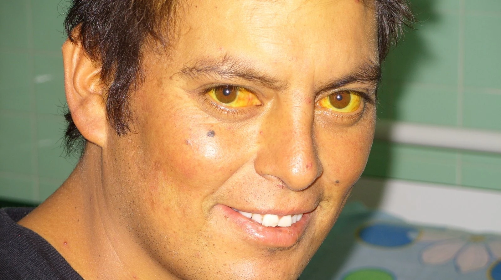
Published scleral-icterus example, originally documented in acute hepatitis. This is not a photograph of Gilbert syndrome. Yellow sclera does not establish the cause or bilirubin fraction. Whole image retained; WebP optimization only.
Sheila J. Toro (2011). CC BY 4.0. Source and provenance

Fever or systemic illness with evidence of biliary obstruction raises concern for acute cholangitis, an infected drainage system. Hypotension or organ dysfunction increases urgency. The full combination of fever, right upper abdominal pain, and jaundice is not required before treatment begins. Antibiotics and resuscitation address infection and circulation, while appropriate biliary drainage provides source control. [4]
New encephalopathy, meaning altered brain function, together with acute liver injury and impaired coagulation raises concern for acute liver failure, particularly without established cirrhosis. Urgent specialist and transplant-center discussion may be necessary. Investigate exposures such as medicines and supplements, but do not wait for every viral or autoimmune test before obtaining advanced help. [1]
An infant with jaundice and pale stools belongs on another time-sensitive pathway. Measure total and direct bilirubin and arrange cholestasis evaluation even when the infant is feeding and growing. A reassuring appearance or an earlier diagnosis of ordinary newborn jaundice does not exclude progressive biliary disease. [5]
Infection with obstruction
Resuscitate, give appropriate antibiotics, and arrange source control. [4]
Acute injury with encephalopathy
Assess urgently for liver failure and the need for transplant-center care. [1]
Infant pale stools
Investigate cholestasis promptly rather than borrowing adult reassurance. [5]
Locate the accumulation in bilirubin handling
Macrophages convert heme from aging red cells and other heme proteins into bilirubin. Unconjugated bilirubin travels through blood mainly bound to albumin. Hepatocytes take it up and use UGT1A1 to attach glucuronic acid. Conjugated bilirubin then requires transport toward bile and delivery through the biliary tree into the intestine. [2]
 Enlarge the whole image
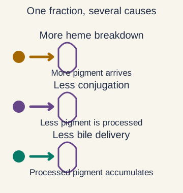
A similar laboratory fraction can have more than one explanation. Locate the candidate process, then look for evidence that distinguishes the causes.
Bone Wizardry. Original educational schematic. Source and provenance
An indirect-predominant increase can reflect increased production, impaired uptake, or reduced conjugation. Hemolysis increases production. Gilbert and Crigler-Najjar syndromes reduce conjugating capacity. Drugs can also alter uptake or conjugation. The fraction narrows the failed steps; it does not choose among them without further evidence. [1] [2] [6]
A direct-predominant increase raises a different set of possibilities. These include hepatocyte injury, impaired canalicular transport, or reduced bile flow. Cholestasis means impaired formation or flow of bile and may occur inside the liver or in larger extrahepatic ducts. Benign Dubin-Johnson and Rotor syndromes are important exceptions to the assumption that every direct increase means destructive disease. [1] [2] [6]
Direct and indirect are assay terms rather than perfect chemical compartments. Indirect bilirubin is usually calculated as total minus direct. A total of 5.6 mg/dL and direct of 0.8 mg/dL gives an indirect value of 4.8 mg/dL in a fictional adult example. Delta bilirubin, conjugated pigment bound to albumin, also contributes to the direct result and can persist after obstruction improves. [2]
- More arriving upstream Increased heme breakdown can outpace clearance. [2]
- Less processed inside the cell Reduced uptake or conjugation increases indirect pigment. [2] [6]
- Less delivered toward the bowel Transport or flow problems can increase conjugated pigment. [2]
Use pigment findings without turning them into absolute rules
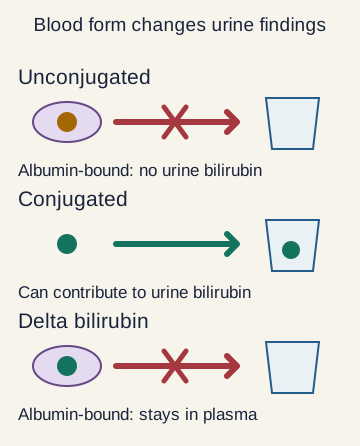
Albumin-bound unconjugated bilirubin is not freely filtered by the kidney. A patient with isolated indirect hyperbilirubinemia therefore usually has no bilirubin in urine. Some circulating conjugated bilirubin is filterable, so bilirubinuria points toward a conjugated component. It does not tell you whether the cause is an obstructed duct, hepatocyte injury, or a benign transporter disorder. [2] [6]
 Enlarge the whole image
Urine bilirubin reflects the filterable conjugated fraction. Delta bilirubin can contribute to a direct blood result without being freely filtered. Urine urobilinogen is a different test.
Bone Wizardry. Original educational schematic. Source and provenance
Bilirubin delivered to the bowel is modified by intestinal bacteria. Downstream pigments contribute to brown stool, while some urobilinogen is absorbed and a small amount reaches urine. The urine bilirubin and urobilinogen assays measure different substances. A negative bilirubin test can coexist with measurable urobilinogen without either result being contradictory. [2]
Complete obstruction can reduce intestinal pigment, producing pale stool and low urobilinogen, while conjugated pigment in blood darkens urine. Partial or intermittent obstruction is less predictable. Hepatocyte injury can affect uptake, processing, and secretion at the same time. Do not require a textbook pair of urine results before investigating a concerning clinical presentation. [2]
Dark urine is a description, not a diagnosis. Concentrated urine, blood, or other pigments can also alter its appearance. Confirm what the urine actually contains and compare with the serum fractions. Delta bilirubin provides another caveat. Because this direct-reacting fraction is albumin-bound, a persistent serum direct result need not produce equivalent urinary bilirubin excretion. [2]
Urine urobilinogen
Reflects a downstream intestinal product and its subsequent handling. [2]
Stool pigment
Helps assess delivery to the bowel, with important limits in partial obstruction. [2]
Ask what each blood test actually measures
ALT and AST are mainly used as injury markers rather than direct measurements of remaining liver function. ALT is more liver-specific; AST is also present in muscle and other tissues. Their magnitude and relative elevation help characterize a hepatocellular pattern. A large elevation may be clinically important, but the number alone does not measure the amount of functioning liver left. [1]
 Enlarge the whole image
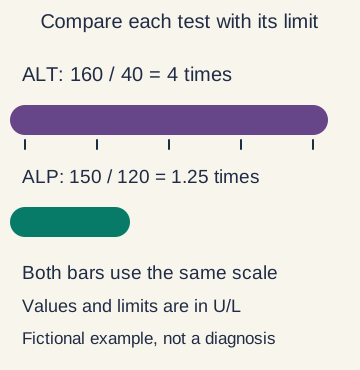
Fictional values: ALT 160/40 equals 4 times its upper limit; ALP 150/120 equals 1.25 times its upper limit. Compare normalized values, not raw heights or diagnostic labels.
Bone Wizardry. Original educational schematic. Source and provenance
Alkaline phosphatase, or ALP, often rises with cholestasis, but bone and other tissues can contribute. GGT helps support a hepatic source when ALP is elevated. Compare results with each test own upper reference limit rather than comparing raw numbers with different normal ranges. A mixed pattern can occur, and drug-induced injury may be hepatocellular, cholestatic, or mixed. [1]
Albumin and PT or INR add information about synthetic function, but each has confounders. Albumin can fall with inflammation, malnutrition, renal loss, or fluid excess. INR may change with vitamin K deficiency, anticoagulants, or disseminated coagulation as well as reduced hepatic synthesis. In established cirrhosis, INR is not a reliable stand-alone estimate of bleeding risk. [1]
Interpret the trajectory and clinical state together. Falling aminotransferases do not necessarily mean recovery if mental status, glucose, or coagulation is worsening. Conversely, slowly resolving direct bilirubin after drainage can reflect delta bilirubin rather than a persisting blockage. The right question is which process each result supports and whether the overall patient is improving. [1] [2]
Injury
ALT and AST describe cell injury, with tissue-specific caveats. [1]
Cholestatic pattern
ALP and GGT support impaired bile handling but do not locate it alone. [1]
Function and context
Albumin and INR require their own competing explanations. [1]