Identify the origin of liver deposits, interpret imaging, choose useful tissue, and connect complete treatment to liver reserve and whole-patient disease control.
A liver mass has two separate questions: where did these cells originate, and can all important disease be treated while leaving a functioning liver? Identify the likely origin, map the full disease distribution, then judge local treatment with a multidisciplinary team. Metastatic disease is not automatically untreatable, but a small lesion is not automatically safe to destroy.
How do cells from another organ reach the liver?
A patient with a sigmoid tumor has several new liver nodules. These deposits are not renamed primary liver cancer: a colorectal metastasis retains colorectal tumor identity, which helps determine systemic treatment. A recurrence can also appear years after treatment of the original cancer. The time interval changes probability and prognosis, not the definition of metastasis. [1]
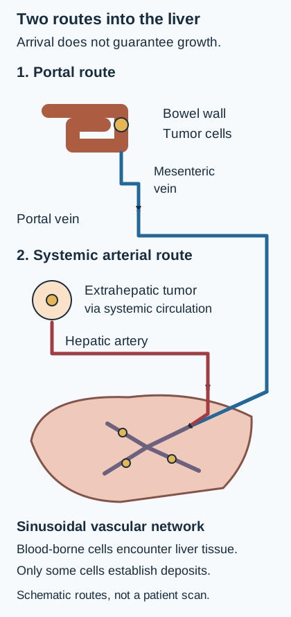
The liver receives blood through both the portal vein and the hepatic artery. Venous blood from much of the gastrointestinal tract passes through the portal circulation before returning to the heart. Tumor cells that enter this drainage can encounter the liver's sinusoidal microvasculature. The sinusoids provide an extensive vascular interface, but arrival does not guarantee survival or growth into a deposit. This anatomical exposure helps explain the importance of colorectal, pancreatic, gastric and other gastrointestinal primaries. [2]
Trace the portal route in the accompanying diagram with a finger or your eyes: bowel wall, mesenteric venous drainage, portal vein, then hepatic sinusoids. Now trace the arterial route. Breast, lung, melanoma, renal, thyroid and other tumors, including some sarcomas, can reach the liver through the systemic circulation. Gastrointestinal primaries are important, not exclusive. Neuroendocrine tumors can also produce hepatic metastases. Rectal venous drainage includes systemic as well as portal pathways. [1][2]
Trace the two delivery routes. Portal drainage makes gastrointestinal primaries important, but does not exclude systemic arterial sources. The diagram concerns initial delivery, not the contrast enhancement of an established deposit. Not a literal anatomical map. [1][2][1][2]
Keep arrival separate from enhancement. A colorectal cell can arrive through portal blood even though an established metastasis develops predominantly arterial tumor perfusion. Contrast enhancement describes present blood delivery to tissue; it does not reconstruct the original route taken by every cell. [2]
Predict before checking: a patient without a bowel mass has a lung primary and new liver deposits. Which route still provides a plausible connection, and does a normal portal vein exclude metastasis?
Check the route prediction
Systemic arterial delivery provides a connection. A patent portal vein does not exclude metastases, because it is neither the sole route of spread nor a test of whether tumor cells previously passed through the liver. Apply the same distinction to a remote renal primary. [1][2]
Which pattern supports metastasis, and what could imitate it?
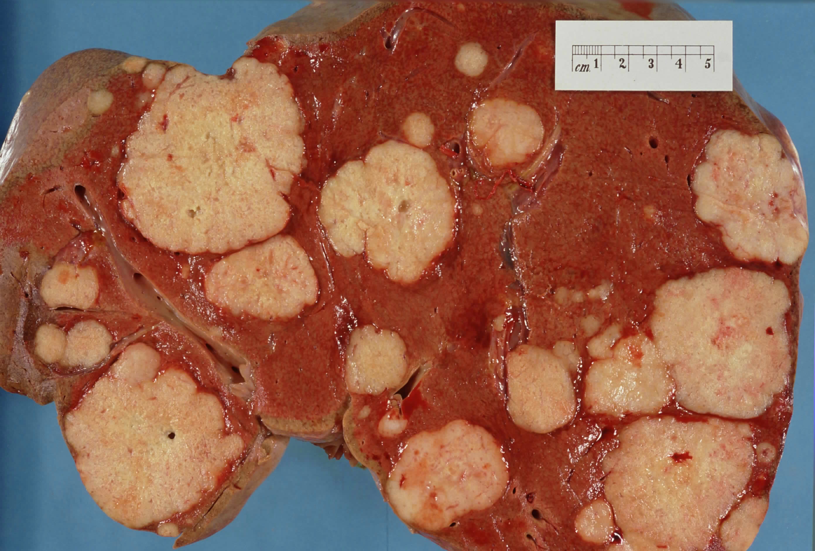
Several masses of different sizes in both lobes, particularly in someone with an established extrahepatic malignancy, favor metastases. A single lesion can still be metastatic. Conversely, a person with cancer can also have an unrelated benign nodule or a second primary cancer. Metastases are a major category of malignant liver tumors; their frequency relative to primary liver cancer varies with the population and background liver disease. [1][2]
The gross specimen accompanying this section shows pale deposits of different sizes interrupting darker liver tissue. Identify two separate deposits and the tissue between them. The contributor documents pancreatic adenocarcinoma as the source, but the photograph alone cannot identify that primary, establish current resectability, or substitute for imaging. It is a real specimen, not a diagnostic calculation. [12]
Identify separate deposits and the intervening liver. The contributor identifies pancreatic adenocarcinoma as the primary source, but the photograph alone cannot determine origin, staging or resectability. Image: [12] Haymanj; original source; Public domain, released by the contributor.
Most colorectal and many other adenocarcinoma metastases are relatively hypovascular: they enhance less than surrounding liver, often becoming conspicuous in the portal venous phase. Viable tumor at the edge with central necrosis can create a target or rim appearance. Restricted diffusion supports a cellular lesion, but neither diffusion restriction nor a rim is specific for metastasis; infection and other malignancies can overlap. Interpret the entire examination and clinical setting. [2]
Renal cell, neuroendocrine, thyroid and melanoma metastases may instead be hypervascular. Arterial brightness therefore does not establish hepatocellular carcinoma (HCC). Calcification can occur in mucinous colorectal or ovarian metastases and after treatment. Calcium is not proof of benignity, and its presence does not identify a primary by itself. [2]
Contrast behavior is interpreted with the background liver and cancer history
Candidate
Helpful pattern
Boundary on the inference
CandidateMetastatic deposit
Helpful patternMultiplicity, growth, targetoid morphology, restricted diffusion, or enhancement resembling a known primary
Boundary on the inferenceNot every metastasis is multiple, and none of these findings alone is diagnostic. [2]
CandidateHCC
Helpful patternIn an appropriate at-risk patient, qualifying nonrim arterial enhancement with nonperipheral washout and other size-dependent imaging criteria
Boundary on the inferenceApply a validated HCC framework; do not extend it automatically to every noncirrhotic patient or targetoid mass. [10]
CandidateHemangioma
Helpful patternVery bright T2 signal and discontinuous peripheral nodules of enhancement that progressively fill toward the center
Boundary on the inferenceThis is different from a continuous malignant rim. Atypical or incomplete patterns need characterization. [2][9]
CandidateFocal nodular hyperplasia
Helpful patternOften homogeneous arterial enhancement, sometimes a central scar, and commonly retention of hepatobiliary contrast
Boundary on the inferenceA scar is not required. Contrast uptake is contextual, not a stand-alone guarantee. Estrogen exposure alone does not establish FNH. [2][9]
Compare two lesions in the same patient: one has remained unchanged for years and shows nodular peripheral filling; the other is new, has a continuous rim, and restricts diffusion. Assign a working category to each before deciding whether they should share the same staging label.
Compare the two working categories
The stable, nodular-filling lesion fits a hemangioma pattern. The new rim-enhancing lesion deserves malignant and infectious differential assessment in context. Do not classify both as metastases just because they occupy the same organ. In a new patient, repeat this lesion-by-lesion comparison rather than relying on the cancer history alone. [2][9]
What information would change the next decision?
Before ordering tissue, name the uncertainty: is this a metastasis, where is the primary, what is the full disease distribution, or which treatment fits the tumor biology? These are different questions. Review the previous cancer pathology, disease-free interval, cirrhosis or other chronic liver disease, prior chemotherapy, symptoms, liver tests and earlier scans. A new abnormality becomes more informative when its time course is known. [1][3][9]
Contrast-enhanced CT usually provides broad staging, including extrahepatic disease. Liver MRI with diffusion-weighted sequences and, when appropriate, hepatobiliary contrast can characterize indeterminate findings and reveal small deposits important for local planning. Hepatobiliary contrast highlights functioning background hepatocytes; most metastases lack this uptake and stand out against the liver. Darkness in that phase is not specific for metastasis. Intraoperative ultrasound can add information about deep or small lesions and their relationship to vessels. [2][3]
Imagine CT shows three right-lobe deposits and a proposed operation would preserve the left liver. MRI finds an additional small deposit in the planned remnant. The consequence is not automatically abandonment of curative intent. The team must revise the map and assess whether a limited resection or ablation can clear that deposit while preserving functional tissue. The extra image matters because it changes the proposed treatment geometry. [3][5]
Biopsy should answer a management-changing question. It is often useful when the primary is unknown, imaging is indeterminate, a second malignancy is plausible, or current tumor tissue is needed to select therapy. Select a safely accessible, representative site with sufficient viable tissue; that site need not be the liver. Discuss bleeding risk, specimen adequacy and the planned definitive treatment with the relevant specialists. [3][8][9]
In suspected recurrent breast cancer, accessible metastatic tissue can confirm the process and reassess estrogen receptor, progesterone receptor and HER2 status. These can differ from the original tumor. In contrast, a classic, technically treatable colorectal metastatic pattern with established primary pathology may not require routine percutaneous liver biopsy before hepatobiliary review. Avoid an unnecessary procedure, but do not turn concern about bleeding or needle-tract dissemination into a universal prohibition on useful biopsy. [3][8]
If the primary is unknown, use the history, examination, imaging distribution and pathology to direct further evaluation. A gastrointestinal pattern may lead to endoscopic investigation; a breast pattern requires a different assessment. Serum markers such as CEA can support monitoring in context but cannot independently establish the primary, prove a liver lesion malignant, or declare treatment failure. [2][4][8]
A patient previously treated for breast cancer now has liver lesions and a superficial node suitable for core sampling. Explain which uncertainty the node biopsy could resolve that repeating a serum marker cannot.
Check what tissue adds
The node may provide histology and current receptor information that can change systemic treatment. A marker trend cannot establish those features. For another patient with already-secure, resectable colorectal disease, reconsider whether biopsy would add useful information before requesting the same procedure. [3][8]
Can disease be cleared and a working liver remain?
Four deposits do not automatically prevent surgery, and one deposit does not automatically permit it. Resectability combines tumor biology, total disease distribution, patient fitness, and the anatomy of a complete treatment plan. The useful question is whether the team can clear the planned disease with adequate margins while preserving enough functional liver, rather than whether a fixed lesion count has been exceeded. [3][5]
A future liver remnant is the tissue intended to remain after resection. It needs adequate portal and arterial inflow, hepatic venous outflow and biliary drainage. Its volume matters, but so does its quality: steatosis, chronic liver disease and chemotherapy-associated injury can reduce the reserve supplied by a given volume. A large remnant with obstructed venous drainage is not equivalent to an equally large, well-drained remnant. There is no single percentage that certifies safety for every liver. [3][5]
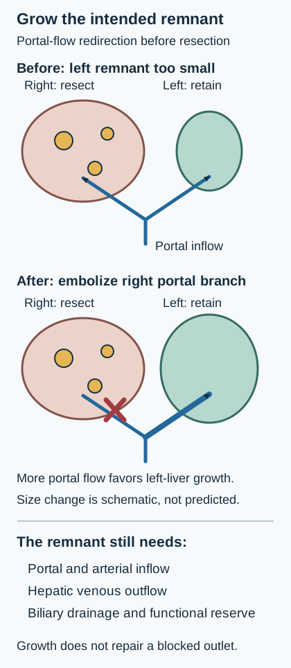
Use the remnant diagram as a planning exercise. Cover the proposed resection side. Trace portal blood entering the retained tissue. Then name the arterial inflow, hepatic venous outflow and bile drainage routes that still need confirmation on clinical imaging; the schematic does not display their full anatomy. If one route is interrupted, describe the predicted problem before counting how much tissue remains. Venous obstruction can produce congestion; interrupted biliary drainage can create a different postoperative problem. Preserving tissue and preserving function must be assessed together. [3][5]
Predict which branch should be targeted before uncovering or comparing the second state. Right portal embolization can support growth of an intended left remnant. The functional territories and size change are schematic; actual growth, function and disease control require reassessment. [3][5][3][5]
Bilobar disease can sometimes be addressed with parenchymal-sparing resections, combined resection and ablation, or a staged strategy. When the limiting issue is an inadequate future remnant, portal vein embolization can redirect portal flow away from the part intended for resection toward the retained liver and promote remnant growth. It is not a substitute for confirming function, safe anatomy or cancer control after the interval. [3][5]
Predict the response to changing the flow. In the diagram, the right liver is scheduled for resection and the left liver is the intended remnant. Choose which portal branch would be embolized to support growth of the left remnant. Then compare the before-and-after drawings. Remnant growth varies by patient and requires reassessment. [3][5]
Check the branch and its consequence
The right portal branch supplies the planned resection side. Embolizing that branch redirects portal flow toward the left liver and can stimulate its growth. Embolizing the left branch would target the intended remnant instead. If the left hepatic venous outflow were also compromised, extra portal flow would not repair that drainage problem. [3][5]
Include the primary tumor and extrahepatic sites in the assessment. Extensive uncontrolled extrahepatic disease usually prevents a useful complete local strategy. Selected, limited extrahepatic disease that can also be controlled is not automatically equivalent to diffuse progression. Synchronous deposits are recognized with the primary; metachronous deposits appear later. Either timing requires assessment, and a liver-limited recurrence can still be considered for local treatment. [3][13]
Why does a small tumor still need a safety margin?
Local treatment must address both the visible tumor and the risk of residual disease at its edge. Margin-negative resection is an established potentially curative option for selected colorectal liver metastases. Thermal ablation destroys tissue in place and can preserve more liver, especially for a small deep deposit that would otherwise require a disproportionate resection. These are tools to compare, not automatic choices determined by one diameter. [3][5]
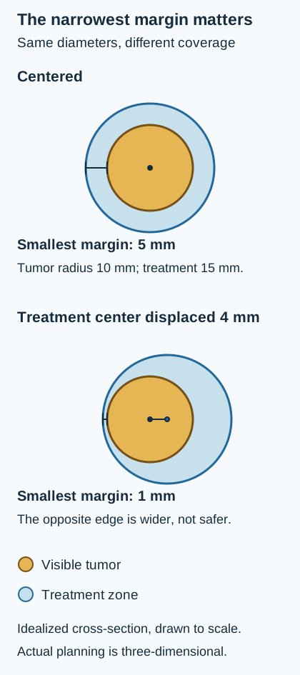
The ablation diagram distinguishes the tumor boundary from the treatment boundary. A treatment zone that merely touches the visible tumor edge leaves no surrounding margin. With an off-center treatment zone, an apparently generous margin on one side can conceal a very narrow margin on the other. Assess the minimum margin around the whole three-dimensional target, not just the largest diameter on a convenient image. [5]
Calculate the smallest margin, then compare the two scaled states. The treatment-zone diameter stays constant while the narrowest edge changes. Idealized two-dimensional geometry explains the concept; it is not a clinical treatment prescription. [5][5]
Use a simplified cross-section: a circular tumor has a 1 cm radius and a centered treatment zone has a 1.5 cm radius. Calculate the surrounding margin. Then shift the treatment center 4 mm away from the tumor center and calculate the narrowest margin. These ideal circles explain geometry; they are not a clinical ablation prescription.
Check the margin calculation
The centered margin is 0.5 cm, or 5 mm. A 4 mm displacement leaves only 1 mm at the nearest edge, even though the treatment-zone diameter has not changed. In another patient, compare the minimum margin with the team's required target and the nearby structures rather than assuming the same diameter guarantees complete treatment. [5]
Small lesions, often 3 cm or less, are favorable candidates when complete coverage and a safe margin are feasible. Flowing blood in a large adjacent vessel can dissipate heat, particularly during radiofrequency ablation, and reduce effective heating at that edge. Nearby central bile ducts or bowel can be injured by thermal treatment. A technically possible needle path therefore does not establish safe, complete ablation. Review the modality, protection options and alternatives with specialists. [5]
The 2025 COLLISION trial supports reconsidering the idea that ablation is restricted to unresectable disease. It enrolled selected patients with up to ten colorectal liver deposits, each 3 cm or less, without extrahepatic metastases. Its interim findings support individualized comparison of ablation and resection within that setting. They do not establish identical lifetime survival, justify treating much larger tumors the same way, or eliminate anatomical safety assessment. [6]
Which treatment goal fits the whole patient now?
A multidisciplinary discussion should state the goal explicitly: complete local treatment now, treatment intended to make local clearance feasible, or systemic disease control with symptom support. Include hepatobiliary surgery, medical oncology, diagnostic and interventional radiology, and other specialists as appropriate. The patient participates in weighing expected benefit, recovery, uncertainty and preferences. [4][5]
For clearly treatable colorectal liver disease, surgery alone or a perioperative chemotherapy approach can be considered according to disease burden, recurrence risk and patient factors. Perioperative chemotherapy is not an obligatory prerequisite for every small resectable deposit. Nor should treatment continue indefinitely just because another cycle might shrink an already treatable lesion. [4][13]
Conversion treatment has a defined purpose: reduce tumor burden or improve the anatomical situation enough to permit complete local treatment. Review response and resectability early and repeatedly. Shrinkage near a critical vessel may create a useful window, while new extrahepatic disease may close it. Prolonged treatment can also injure the liver. An initially unsuitable plan deserves reassessment, not a permanent label based only on the first scan. [3][4][5]
A metastasis that disappears radiologically after chemotherapy is not proven sterilized. Preserve the pretreatment lesion map and review original sites with appropriate MRI and intraoperative assessment. The plan for an undetectable site is individualized; neither declaring cure from invisibility nor performing an unplanned major resection is a substitute for that review. [3]
Systemic therapy follows the primary cancer and its biology. For colorectal cancer, mismatch-repair or microsatellite-instability status, RAS and BRAF findings, primary location, prior therapy, fitness and organ function can change selection. Deficient mismatch repair can favor immune checkpoint-based therapy. Left-sided location alone is not enough to select conventional anti-EGFR-based therapy when RAS is mutated. This lesson establishes the selection principles, not an exhaustive regimen or dosing reference. [4]
When surgery or ablation is unsuitable, selected liver-limited disease may be discussed for stereotactic body radiotherapy. Arterial therapies have specialized roles and are not interchangeable with one another; radioembolization is not a routine replacement for systemic therapy in every colorectal liver-metastasis presentation. Decisions depend on distribution, reserve, prior treatment and specialist expertise. [4][13]
The TransMet trial also supports liver transplantation plus chemotherapy as an option for a narrowly selected group with permanently unresectable, liver-only colorectal metastases and favorable treatment-controlled biology. Expert transplant assessment and organ-allocation considerations are essential. This is not routine initial treatment for a resectable deposit, nor a pathway for uncontrolled extrahepatic disease or metastatic pancreatic cancer. [7]
Compare the direction of two findings after treatment: the liver deposits have shrunk, but previously absent peritoneal deposits are now biopsy-confirmed and cannot be completely treated locally. Does the better liver image alone support major liver resection?
Check the whole-patient interpretation
No. The new uncontrolled extrahepatic disease changes the overall goal even though liver measurements improved. Reassess systemic options and symptom needs rather than treating liver shrinkage as complete disease control. By contrast, newly feasible clearance of every relevant site can justify renewed local-treatment review. [3][4]
Diffuse progressive liver and extrahepatic disease generally calls for primary-specific systemic treatment when suitable, together with pain control, nutrition, management of obstruction or other complications, and supportive or palliative care. These services can accompany active anticancer treatment. In widespread pancreatic adenocarcinoma, treating each liver nodule separately does not address the systemic disease. [1][11]
Apply the full assessment
For each patient, decide what the findings imply, identify the information that changes treatment, and compare the closest alternatives before checking the reasoning.
Case 1
Show answer and explanations for case 1
A. Direct extension of the sigmoid tumor into liver (Why this does not fit)
Contiguous spread requires a continuous anatomical path from the primary into adjacent liver. The separated sigmoid mass and bilobar deposits do not form a contiguous extension pattern. Use the primary-site evidence and the anatomical route together; a liver deposit retains the identity of its source cancer.
Reasoning steps for option A
What would direct extension require?
Contiguous spread requires a continuous anatomical path from the primary into adjacent liver.
Does the scan describe such continuity?
The separated sigmoid mass and bilobar deposits do not form a contiguous extension pattern.
What anatomical separation rules out direct invasion of the liver by this sigmoid mass?
The sigmoid tumor is remote from the liver, while deposits occupy separate sites in both lobes; direct invasion requires contiguous tissue spread.
B. Multicentric primary hepatocellular carcinoma (Why this does not fit)
Cirrhosis or another established HCC risk context and qualifying imaging would increase that possibility. The symptomatic sigmoid mass and separate hypovascular deposits are better explained by a colorectal primary with metastases. Use the primary-site evidence and the anatomical route together; a liver deposit retains the identity of its source cancer.
Reasoning steps for option B
What context would support primary HCC?
Cirrhosis or another established HCC risk context and qualifying imaging would increase that possibility.
Which competing explanation better unifies this presentation?
The symptomatic sigmoid mass and separate hypovascular deposits are better explained by a colorectal primary with metastases.
Why do the bowel mass and hypovascular bilobar nodules favor metastatic colorectal cancer over multicentric HCC?
A symptomatic sigmoid primary provides a source for multiple hepatic deposits, whereas no HCC-risk background or qualifying HCC imaging pattern is described.
C. Portal venous dissemination from a colorectal primary (Best answer)
Mesenteric venous drainage connects the sigmoid circulation to the portal system. A sigmoid malignancy can seed separate hepatic sites through portal blood, fitting the multiple hypovascular deposits. Use the primary-site evidence and the anatomical route together; a liver deposit retains the identity of its source cancer.
Reasoning steps for option C
Where does venous blood from the sigmoid commonly drain?
Mesenteric venous drainage connects the sigmoid circulation to the portal system.
How does that anatomy fit the observed lesions?
A sigmoid malignancy can seed separate hepatic sites through portal blood, fitting the multiple hypovascular deposits.
Which vascular route delivers sigmoid tumor cells to multiple hepatic sites?
Sigmoid venous drainage reaches the inferior mesenteric and portal venous circulation, allowing hematogenous seeding of both liver lobes.
D. Peritoneal implantation onto the liver surface (Why this does not fit)
Peritoneal spread typically produces deposits on serosal surfaces and may accompany other peritoneal abnormalities. The multiple intraparenchymal deposits fit blood-borne dissemination better than surface implantation. Use the primary-site evidence and the anatomical route together; a liver deposit retains the identity of its source cancer.
Reasoning steps for option D
Where would peritoneal implants be expected?
Peritoneal spread typically produces deposits on serosal surfaces and may accompany other peritoneal abnormalities.
How do the reported hepatic lesions differ?
The multiple intraparenchymal deposits fit blood-borne dissemination better than surface implantation.
What lesion location distinguishes portal-borne deposits from peritoneal seeding here?
The deposits lie within liver parenchyma rather than on its serosal surface, favoring blood-borne metastases over peritoneal implants.
Takeaway: Use the primary-site evidence and the anatomical route together; a liver deposit retains the identity of its source cancer.
A. Sample a suitable lesion to establish tumor origin (Best answer)
Renal cell carcinoma can produce arterial-enhancing metastases and recur after a long interval. The new liver and lung findings suggest recurrence, but histology can distinguish renal recurrence from a second malignancy before treatment. Match imaging to the cancer history and validated diagnostic population; hypervascularity is not a synonym for HCC.
Reasoning steps for option A
Can renal carcinoma recur with hypervascular liver deposits?
Renal cell carcinoma can produce arterial-enhancing metastases and recur after a long interval.
Why obtain representative tissue here?
The new liver and lung findings suggest recurrence, but histology can distinguish renal recurrence from a second malignancy before treatment.
Why is biopsy informative despite a remote history of renal carcinoma and arterial-enhancing liver lesions?
Renal carcinoma can recur late with hypervascular liver and lung metastases, but tissue establishes whether the new disease is renal recurrence or another malignancy.
B. Assign HCC from arterial enhancement and stage it (Why this does not fit)
Noninvasive HCC criteria are used within defined risk populations and require more than arterial brightness alone. This patient lacks the specified background and has a plausible renal source with new disease in another organ. Match imaging to the cancer history and validated diagnostic population; hypervascularity is not a synonym for HCC.
Reasoning steps for option B
When can imaging establish HCC without routine biopsy?
Noninvasive HCC criteria are used within defined risk populations and require more than arterial brightness alone.
Why is that shortcut unsuitable here?
This patient lacks the specified background and has a plausible renal source with new disease in another organ.
What missing prerequisite prevents diagnosing HCC from arterial enhancement alone?
There is no qualifying HCC-risk background, and arterial enhancement alone is insufficient for the noninvasive HCC imaging pathway.
C. Classify the lesions as benign vascular nodules (Why this does not fit)
Some benign vascular lesions enhance avidly and must be considered during characterization. New liver lesions with enlarging pulmonary nodules after renal cancer require investigation rather than a benign label. Match imaging to the cancer history and validated diagnostic population; hypervascularity is not a synonym for HCC.
Reasoning steps for option C
What benign lesion can enhance strongly?
Some benign vascular lesions enhance avidly and must be considered during characterization.
What prevents a benign conclusion from enhancement alone?
New liver lesions with enlarging pulmonary nodules after renal cancer require investigation rather than a benign label.
Which concurrent finding makes incidental benign vascular nodules an unsafe explanation?
Enlarging pulmonary nodules alongside new liver lesions after renal cancer raise concern for systemic malignant recurrence, not simply benign enhancing nodules.
D. Use the normal alpha-fetoprotein to exclude malignancy (Why this does not fit)
Alpha-fetoprotein can contribute to selected liver-cancer assessments but is not a universal cancer detector. It excludes neither metastatic renal carcinoma nor all HCC, so it cannot settle the new lesions. Match imaging to the cancer history and validated diagnostic population; hypervascularity is not a synonym for HCC.
Reasoning steps for option D
What role can alpha-fetoprotein play?
Alpha-fetoprotein can contribute to selected liver-cancer assessments but is not a universal cancer detector.
What does its normal value establish here?
It excludes neither metastatic renal carcinoma nor all HCC, so it cannot settle the new lesions.
Why does normal alpha-fetoprotein fail to resolve the liver-lesion differential?
Normal AFP does not exclude either metastatic renal carcinoma or every HCC; it cannot replace lesion characterization and tissue diagnosis here.
Takeaway: Match imaging to the cancer history and validated diagnostic population; hypervascularity is not a synonym for HCC.
A. Metastasis and cavernous hemangioma (Why this does not fit)
The colorectal history and targetoid lesion are compatible with metastatic disease. A continuous rim is not the characteristic discontinuous nodular filling pattern, and this pair fails to account for the infectious presentation. A target pattern does not identify its cause; combine imaging with the circumstances that change the differential and urgency.
Reasoning steps for option A
Why does a metastasis remain plausible?
The colorectal history and targetoid lesion are compatible with metastatic disease.
Why is hemangioma not the strongest competing process here?
A continuous rim is not the characteristic discontinuous nodular filling pattern, and this pair fails to account for the infectious presentation.
Why must a colorectal metastasis remain in the differential despite the fever?
Prior colorectal cancer and a targetoid hepatic lesion support metastatic tumor, but post-biliary-instrumentation fever and leukocytosis also require evaluation for abscess.
B. HCC and intrahepatic cholangiocarcinoma (Why this does not fit)
A targetoid mass can represent a primary hepatic malignancy, particularly a non-HCC malignancy. It omits both the relevant established colorectal source and the post-instrumentation infectious context; no qualifying HCC-risk pattern is supplied. A target pattern does not identify its cause; combine imaging with the circumstances that change the differential and urgency.
Reasoning steps for option B
Can hepatic primary malignancies enter a rim-lesion differential?
A targetoid mass can represent a primary hepatic malignancy, particularly a non-HCC malignancy.
Why is this pair not the best synthesis of this presentation?
It omits both the relevant established colorectal source and the post-instrumentation infectious context; no qualifying HCC-risk pattern is supplied.
Which two stem features are missed by pairing hepatic primary tumors as the leading diagnoses?
The established colorectal source supports metastasis, while recent biliary instrumentation with fever supports infection; neither is captured by a primary-tumor pair.
C. Hemangioma and focal nodular hyperplasia (Why this does not fit)
A cancer history does not make every liver nodule metastatic. The described rim and diffusion pattern is not the characteristic combination for either lesion, and the fever after biliary instrumentation requires an infectious differential. A target pattern does not identify its cause; combine imaging with the circumstances that change the differential and urgency.
Reasoning steps for option C
Why are benign focal lesions considered in patients with cancer?
A cancer history does not make every liver nodule metastatic.
Why does this benign pair fit poorly here?
The described rim and diffusion pattern is not the characteristic combination for either lesion, and the fever after biliary instrumentation requires an infectious differential.
Why do benign focal nodules fail to account for the lesion and acute illness together?
The rim and diffusion findings are not typical benign-nodule signatures, and fever with leukocytosis after biliary instrumentation raises a specific abscess concern.
D. Metastasis and hepatic abscess (Best answer)
Yes. A colorectal deposit can have peripheral viable tumor around a poorly enhancing center. Recent biliary instrumentation, fever and leukocytosis raise concern for an abscess, whose rim and diffusion findings can overlap. A target pattern does not identify its cause; combine imaging with the circumstances that change the differential and urgency.
Reasoning steps for option D
Can viable tumor around necrosis produce this pattern?
Yes. A colorectal deposit can have peripheral viable tumor around a poorly enhancing center.
Why does the clinical setting preserve another leading process?
Recent biliary instrumentation, fever and leukocytosis raise concern for an abscess, whose rim and diffusion findings can overlap.
What makes abscess a genuine competitor to necrotic colorectal metastasis in this targetoid lesion?
Both can exhibit a peripheral rim and central abnormality, but instrumentation followed by fever and leukocytosis specifically increases the likelihood of infection.
Takeaway: A target pattern does not identify its cause; combine imaging with the circumstances that change the differential and urgency.
A. Calcification establishes a completed response to prior therapy (Why this does not fit)
Calcification can occur after treatment and must be interpreted alongside the full response assessment. The lesions grew and new nodules appeared without intervening therapy, which does not support that response conclusion. Judge calcium with lesion growth, treatment history and primary histology; calcification is not a benignity test.
Reasoning steps for option A
Can treated deposits sometimes calcify?
Calcification can occur after treatment and must be interpreted alongside the full response assessment.
Does that explain these findings as a completed response?
The lesions grew and new nodules appeared without intervening therapy, which does not support that response conclusion.
Which temporal finding contradicts calling the calcifications a completed treatment response?
The lesions enlarged and additional calcified nodules arose without intervening therapy, raising concern for progression rather than supporting a completed treatment response.
B. Growth remains concerning for mucinous metastatic disease (Best answer)
Mucinous colorectal metastases can calcify, so calcium does not make a lesion benign. Growth and additional nodules raise concern for progression and require appropriate evaluation rather than reassurance from the calcium. Judge calcium with lesion growth, treatment history and primary histology; calcification is not a benignity test.
Reasoning steps for option B
Can mucinous metastases contain calcium?
Mucinous colorectal metastases can calcify, so calcium does not make a lesion benign.
How should the interval change affect interpretation?
Growth and additional nodules raise concern for progression and require appropriate evaluation rather than reassurance from the calcium.
Why does calcium in enlarging liver nodules not reassure in mucinous colorectal cancer?
Mucinous colorectal metastases can calcify, and interval growth plus new nodules without therapy supports active metastatic progression.
C. Growth represents benign enlargement of old granulomas (Why this does not fit)
Healed granulomatous lesions can contain calcium and are a benign consideration. Substantial interval growth and new nodules in a patient with mucinous cancer require investigation for active disease. Judge calcium with lesion growth, treatment history and primary histology; calcification is not a benignity test.
Reasoning steps for option C
Why might calcified granulomas enter the differential?
Healed granulomatous lesions can contain calcium and are a benign consideration.
What prevents assuming healed granulomas here?
Substantial interval growth and new nodules in a patient with mucinous cancer require investigation for active disease.
What behavior separates these nodules from healed calcified granulomas?
Healed granulomas would not ordinarily show substantial interval growth with new nodules in this oncologic setting; active disease needs evaluation.
D. Calcification establishes a new hepatocellular primary tumor (Why this does not fit)
Calcification is not exclusive to metastatic tumors and can appear in several hepatic lesions. It does not supply a specific HCC diagnosis and cannot outweigh the relevant history and progressive metastatic-compatible pattern. Judge calcium with lesion growth, treatment history and primary histology; calcification is not a benignity test.
Reasoning steps for option D
Can a primary hepatic tumor contain calcium?
Calcification is not exclusive to metastatic tumors and can appear in several hepatic lesions.
Does calcium identify HCC in this patient?
It does not supply a specific HCC diagnosis and cannot outweigh the relevant history and progressive metastatic-compatible pattern.
Why can calcification not establish a primary HCC diagnosis in this case?
Calcium is nonspecific, whereas mucinous colorectal histology and enlarging new hepatic nodules supply a coherent metastatic explanation.
Takeaway: Judge calcium with lesion growth, treatment history and primary histology; calcification is not a benignity test.
A. A new HCC supported by at-risk imaging criteria (Best answer)
Cirrhosis places this patient within a population where a validated HCC imaging framework can establish a noninvasive diagnosis. The size and nonrim arterial enhancement with nonperipheral washout support HCC-specific assessment rather than automatic attribution to the remote colorectal cancer. A cancer history does not prevent a second primary; use both the background liver and the complete enhancement pattern.
Reasoning steps for option A
Why does the background liver matter?
Cirrhosis places this patient within a population where a validated HCC imaging framework can establish a noninvasive diagnosis.
How do the lesion features fit that framework?
The size and nonrim arterial enhancement with nonperipheral washout support HCC-specific assessment rather than automatic attribution to the remote colorectal cancer.
Which combined features permit an HCC-specific imaging assessment rather than automatic metastatic staging?
Cirrhosis places the patient in an at-risk population, and a lesion over 2 cm with nonrim arterial enhancement, nonperipheral washout, and capsule supports HCC assessment.
B. Colorectal recurrence established by the cancer history (Why this does not fit)
A late metastatic recurrence is possible after treatment of colorectal cancer. The cirrhotic background and qualifying HCC pattern provide a strong competing diagnosis of a second primary. A cancer history does not prevent a second primary; use both the background liver and the complete enhancement pattern.
Reasoning steps for option B
Why remains a colorectal metastasis conceivable?
A late metastatic recurrence is possible after treatment of colorectal cancer.
Why does the history not settle this lesion?
The cirrhotic background and qualifying HCC pattern provide a strong competing diagnosis of a second primary.
Why should the remote colorectal history not override this lesion's HCC pattern?
A late colorectal metastasis remains possible, but cirrhosis plus qualifying enhancement, washout, and capsule favor a possible independent HCC primary.
C. Focal nodular hyperplasia supported by arterial enhancement (Why this does not fit)
FNH can enhance avidly in the arterial phase. The at-risk liver and combined washout and capsule pattern require HCC assessment, not a conclusion from arterial enhancement alone. A cancer history does not prevent a second primary; use both the background liver and the complete enhancement pattern.
Reasoning steps for option C
Why can FNH be considered in an enhancing lesion?
FNH can enhance avidly in the arterial phase.
Which additional findings argue against stopping at FNH?
The at-risk liver and combined washout and capsule pattern require HCC assessment, not a conclusion from arterial enhancement alone.
What finding beyond arterial enhancement makes FNH an inadequate endpoint here?
In a cirrhotic liver, nonperipheral washout and an enhancing capsule in a lesion over 2 cm require HCC-specific assessment rather than an FNH label.
D. Cavernous hemangioma supported by delayed contrast retention (Why this does not fit)
A hemangioma commonly shows progressive centripetal filling from discontinuous peripheral nodules. The reported pattern is nonperipheral washout rather than progressive filling, in a liver already at risk for HCC. A cancer history does not prevent a second primary; use both the background liver and the complete enhancement pattern.
Reasoning steps for option D
What delayed behavior supports a typical hemangioma?
A hemangioma commonly shows progressive centripetal filling from discontinuous peripheral nodules.
Is that the described enhancement behavior?
The reported pattern is nonperipheral washout rather than progressive filling, in a liver already at risk for HCC.
How does the delayed phase distinguish this lesion from a typical hemangioma?
The lesion shows relative washout with a capsule, not persistent discontinuous peripheral nodules that progressively fill inward.
Takeaway: A cancer history does not prevent a second primary; use both the background liver and the complete enhancement pattern.
A. Count the nodule as synchronous metastatic disease (Why this does not fit)
Colorectal cancer can spread to the liver, making focal lesions important to assess. The characteristic benign pattern and stability years before diagnosis argue against classifying this nodule as a new metastasis. Characterize each lesion before assigning stage; a known cancer does not convert a typical benign nodule into metastatic disease.
Reasoning steps for option A
Why must a staging scan examine the liver carefully?
Colorectal cancer can spread to the liver, making focal lesions important to assess.
What information weighs against metastasis here?
The characteristic benign pattern and stability years before diagnosis argue against classifying this nodule as a new metastasis.
Which prediagnosis imaging evidence argues against counting this nodule as a new colorectal metastasis?
Four years of stability and hepatobiliary contrast retention with an FNH-type pattern predate the cancer and argue against metastatic upstaging.
B. Count the nodule as a separate HCC primary (Why this does not fit)
HCC may show arterial enhancement and must be considered in the appropriate clinical setting. The stem supplies characteristic FNH findings and longstanding stability, not an HCC-risk context with a qualifying malignant pattern. Characterize each lesion before assigning stage; a known cancer does not convert a typical benign nodule into metastatic disease.
Reasoning steps for option B
Why can HCC enter a liver-lesion differential?
HCC may show arterial enhancement and must be considered in the appropriate clinical setting.
What prevents that assignment from this scan?
The stem supplies characteristic FNH findings and longstanding stability, not an HCC-risk context with a qualifying malignant pattern.
Why is arterial enhancement alone insufficient to reclassify the stable nodule as HCC?
The supplied pattern includes hepatobiliary retention and prolonged stability consistent with FNH, without a stated HCC-risk setting or qualifying washout pattern.
C. Treat the nodule as a benign staging finding (Best answer)
Homogeneous enhancement, hepatobiliary contrast retention and longstanding stability support the reported FNH diagnosis. This characterized nodule should not be counted as a colorectal metastasis solely because the patient now has colon cancer. Characterize each lesion before assigning stage; a known cancer does not convert a typical benign nodule into metastatic disease.
Reasoning steps for option C
What does the combined pattern support?
Homogeneous enhancement, hepatobiliary contrast retention and longstanding stability support the reported FNH diagnosis.
What staging conclusion follows from that characterization?
This characterized nodule should not be counted as a colorectal metastasis solely because the patient now has colon cancer.
Which two observations justify leaving this nodule out of the metastatic burden?
Characteristic FNH hepatobiliary retention and stability for four years support a benign lesion rather than a new colorectal deposit.
D. Count the nodule as a mucinous tumor deposit (Why this does not fit)
Mucinous metastases can have unusual imaging features, including calcification or cystic components. The actual combination is characteristic FNH with sustained stability, which is not displaced by merely proposing a mucinous deposit. Characterize each lesion before assigning stage; a known cancer does not convert a typical benign nodule into metastatic disease.
Reasoning steps for option D
What can make a mucinous deposit atypical?
Mucinous metastases can have unusual imaging features, including calcification or cystic components.
Are those possibilities stronger than the supplied characterization?
The actual combination is characteristic FNH with sustained stability, which is not displaced by merely proposing a mucinous deposit.
Why does invoking an unusual mucinous metastasis not defeat the FNH interpretation?
No proposed atypical metastatic feature outweighs the actual hepatobiliary retention and four-year stability characteristic of FNH.
Takeaway: Characterize each lesion before assigning stage; a known cancer does not convert a typical benign nodule into metastatic disease.
A. Peripheral viable tumor around a necrotic metastasis (Why this does not fit)
Viable peripheral tumor around a poorly enhancing center can form a continuous rim. The stem describes discontinuous nodules with progressive inward filling, not a persistent rim around a necrotic center. Read the enhancement sequence, not just the fact that the edge enhances.
Reasoning steps for option A
How can a metastasis produce a target pattern?
Viable peripheral tumor around a poorly enhancing center can form a continuous rim.
How does this sequence differ from that pattern?
The stem describes discontinuous nodules with progressive inward filling, not a persistent rim around a necrotic center.
What enhancement geometry separates this nodule from a necrotic metastasis?
A hemangioma forms separate peripheral enhancement nodules that fill centripetally; necrotic metastasis more often has a continuous viable-tumor rim.
B. Progressive filling of vascular spaces in a hemangioma (Best answer)
Contrast enters vascular spaces in peripheral nodules before filling more central portions. Progressive centripetal filling, very high T2 signal and stability fit a hemangioma better than an actively growing metastatic rim. Read the enhancement sequence, not just the fact that the edge enhances.
Reasoning steps for option B
What does nodular peripheral enhancement represent in a typical hemangioma?
Contrast enters vascular spaces in peripheral nodules before filling more central portions.
Which observations support that interpretation here?
Progressive centripetal filling, very high T2 signal and stability fit a hemangioma better than an actively growing metastatic rim.
How do T2 signal and serial enhancement support a vascular-space mechanism?
Very high T2 signal and discontinuous peripheral nodules filling inward over time, with stability, fit blood pooling in a hemangioma.
C. Hepatocyte contrast retention in focal nodular hyperplasia (Why this does not fit)
Hepatobiliary-phase retention can support FNH when the full lesion pattern is concordant. The sequential nodular vascular filling is a hemangioma-type contrast pattern, not a description of hepatocyte uptake. Read the enhancement sequence, not just the fact that the edge enhances.
Reasoning steps for option C
What phase helps assess functioning-hepatocyte uptake?
Hepatobiliary-phase retention can support FNH when the full lesion pattern is concordant.
What process is described in this question instead?
The sequential nodular vascular filling is a hemangioma-type contrast pattern, not a description of hepatocyte uptake.
Why is progressive centripetal filling not evidence of functioning hepatocytes in FNH?
It tracks contrast entering vascular spaces from peripheral nodules toward the center rather than hepatobiliary-phase uptake by hepatocytes.
D. Nonperipheral washout in a hepatocellular carcinoma (Why this does not fit)
A lesion becomes relatively less enhanced than the liver in a later phase after arterial enhancement. No. The reported filling pattern and stability support a different mechanism from HCC-type relative washout. Read the enhancement sequence, not just the fact that the edge enhances.
Reasoning steps for option D
What does washout mean in the appropriate HCC assessment?
A lesion becomes relatively less enhanced than the liver in a later phase after arterial enhancement.
Does progressive inward filling represent washout?
No. The reported filling pattern and stability support a different mechanism from HCC-type relative washout.
What direction of enhancement change distinguishes this lesion from HCC washout?
The lesion progressively fills inward instead of becoming relatively less enhanced than surrounding liver on later phases.
Takeaway: Read the enhancement sequence, not just the fact that the edge enhances.
A. Proceed with the original right-sided plan without modification (Why this does not fit)
CT placed all known lesions within the proposed resection. MRI now shows disease in tissue intended to remain, requiring reassessment of the map. A new lesion changes the treatment map; it does not independently establish unresectability.
Reasoning steps for option A
Why was the original plan reasonable?
CT placed all known lesions within the proposed resection.
Which finding makes it incomplete?
MRI now shows disease in tissue intended to remain, requiring reassessment of the map.
Why must the right-hepatectomy map change after the MRI finding?
MRI identifies a new deposit in the planned left-liver remnant, so the original right-only operation would leave known tumor behind.
B. Replace local treatment with an exclusively systemic strategy (Why this does not fit)
Disease not amenable to complete safe local treatment often needs a systemic strategy. One small accessible contralateral deposit does not establish that a combined local plan is impossible. A new lesion changes the treatment map; it does not independently establish unresectability.
Reasoning steps for option B
When is a systemic approach appropriate?
Disease not amenable to complete safe local treatment often needs a systemic strategy.
Does this finding establish that limitation?
One small accessible contralateral deposit does not establish that a combined local plan is impossible.
Why does one small accessible contralateral deposit not automatically mandate systemic therapy alone?
With no extrahepatic disease, combined local treatment may still clear all visible deposits while preserving adequate functional remnant.
C. Extend resection into the left liver without remnant reassessment (Why this does not fit)
Additional resection can sometimes clear another deposit. The left liver is the intended remnant, so further treatment must preserve its function and anatomy. A new lesion changes the treatment map; it does not independently establish unresectability.
Reasoning steps for option C
Why might added resection be considered?
Additional resection can sometimes clear another deposit.
Why must reserve be reassessed first?
The left liver is the intended remnant, so further treatment must preserve its function and anatomy.
What safety constraint governs any added resection of the left-sided lesion?
The left liver is the intended remnant; further resection must leave sufficient functional liver volume and viable inflow, outflow, and biliary drainage.
D. Reassess a combined plan that treats the left-sided deposit (Best answer)
Right hepatectomy alone would leave the new left-liver deposit. A limited resection or ablation may address that deposit as part of a complete plan preserving functional liver. A new lesion changes the treatment map; it does not independently establish unresectability.
Reasoning steps for option D
What would the original operation leave?
Right hepatectomy alone would leave the new left-liver deposit.
What does its location permit consideration of?
A limited resection or ablation may address that deposit as part of a complete plan preserving functional liver.
How can the remnant lesion be treated without abandoning a complete local approach?
A limited left-sided resection or ablation can be considered alongside right hepatectomy if all disease can be cleared with adequate liver reserve.
Takeaway: A new lesion changes the treatment map; it does not independently establish unresectability.
A. Select therapy from the original receptor results without new tissue (Why this does not fit)
The original pathology is a useful starting point for assessing possible recurrence. The lesions are not yet confirmed and their current treatment-relevant biology could differ. Choose a safe, adequate, representative tissue site for information that can change care; that site need not be the liver.
Reasoning steps for option A
Why are the old results relevant?
The original pathology is a useful starting point for assessing possible recurrence.
What remains uncertain without new tissue?
The lesions are not yet confirmed and their current treatment-relevant biology could differ.
Why can archived breast pathology not alone determine therapy for the new lesions?
The current process needs histologic confirmation, and metastatic ER, PR, or HER2 status may differ from the original tumor.
B. Biopsy the node and reassess ER, PR and HER2 (Best answer)
The receptor profile may differ from the original tumor, and the new disease process needs confirmation. It is a representative accessible site expected to provide sufficient tissue for histology and receptor testing. Choose a safe, adequate, representative tissue site for information that can change care; that site need not be the liver.
Reasoning steps for option B
What can differ in a later metastasis?
The receptor profile may differ from the original tumor, and the new disease process needs confirmation.
Why is the node useful here?
It is a representative accessible site expected to provide sufficient tissue for histology and receptor testing.
Which sample can both verify recurrence and guide receptor-directed treatment?
The accessible representative node can supply histology plus current ER, PR, and HER2 testing without requiring liver sampling.
C. Sample the liver because receptor testing requires hepatic tissue (Why this does not fit)
A liver deposit may be a suitable representative biopsy target. No. The accessible node can provide the needed histology and receptor assessment. Choose a safe, adequate, representative tissue site for information that can change care; that site need not be the liver.
Reasoning steps for option C
When can liver sampling be appropriate?
A liver deposit may be a suitable representative biopsy target.
Is liver tissue required in this patient?
No. The accessible node can provide the needed histology and receptor assessment.
Why choose the accessible node instead of insisting on a liver biopsy?
The node is a low-risk target that can provide sufficient representative tissue for diagnosis and receptor testing, so sampling the liver is not obligatory.
D. Use serum markers to establish the present receptor profile (Why this does not fit)
Selected serum markers can complement clinical and imaging assessment. They cannot supply the tissue diagnosis or current ER, PR and HER2 findings. Choose a safe, adequate, representative tissue site for information that can change care; that site need not be the liver.
Reasoning steps for option D
How can markers help follow-up?
Selected serum markers can complement clinical and imaging assessment.
What cannot they establish here?
They cannot supply the tissue diagnosis or current ER, PR and HER2 findings.
Which treatment-defining information cannot be obtained from serum markers alone?
Markers cannot establish that the lesions are breast metastases or determine current ER, PR, and HER2 expression on tumor tissue.
Takeaway: Choose a safe, adequate, representative tissue site for information that can change care; that site need not be the liver.
A. Dedicated liver MRI with diffusion and contrast sequences (Best answer)
The small left-sided focus has not been characterized and may affect complete local treatment. Diffusion and contrast assessment can improve characterization and detection of small liver lesions before planning treatment. Use the test that resolves the specific planning uncertainty rather than one that repeats established information.
Reasoning steps for option A
What is missing from the current map?
The small left-sided focus has not been characterized and may affect complete local treatment.
Why does MRI address that uncertainty?
Diffusion and contrast assessment can improve characterization and detection of small liver lesions before planning treatment.
Which feature of the 6 mm left-liver focus could change the local-treatment map?
If diffusion and contrast MRI characterize it as another metastasis, treatment must account for disease in the left liver as well as the 2.4 cm right-sided deposit.
B. Repeat colonoscopy to establish the documented primary (Why this does not fit)
It is valuable when colorectal primary evaluation is needed. The primary is already documented; the unresolved finding is the small liver focus. Use the test that resolves the specific planning uncertainty rather than one that repeats established information.
Reasoning steps for option B
When would colonoscopy help?
It is valuable when colorectal primary evaluation is needed.
What question remains here?
The primary is already documented; the unresolved finding is the small liver focus.
Why would repeating colonoscopy leave the operative uncertainty unresolved?
The colon primary has already been resected; colonoscopy cannot determine whether the indeterminate 6 mm left-liver focus is metastatic.
C. Serial CEA measurements to classify the liver focus (Why this does not fit)
It can supplement surveillance and response assessment. A serum trend cannot distinguish a benign liver nodule from a deposit or provide its treatment anatomy. Use the test that resolves the specific planning uncertainty rather than one that repeats established information.
Reasoning steps for option C
Why might CEA be measured?
It can supplement surveillance and response assessment.
Can it characterize this particular focus?
A serum trend cannot distinguish a benign liver nodule from a deposit or provide its treatment anatomy.
What information about the 6 mm focus cannot a CEA trend supply?
CEA reflects possible tumor activity but cannot localize or characterize the left-sided focus needed for complete liver treatment planning.
D. Noncontrast CT to determine the enhancement pattern (Why this does not fit)
Baseline attenuation and findings such as calcification can be assessed. It cannot supply contrast behavior or diffusion information for this indeterminate focus. Use the test that resolves the specific planning uncertainty rather than one that repeats established information.
Reasoning steps for option D
What can unenhanced CT reveal?
Baseline attenuation and findings such as calcification can be assessed.
Why does it not answer this question as well?
It cannot supply contrast behavior or diffusion information for this indeterminate focus.
Which missing properties make noncontrast CT inadequate for the left-liver focus?
Without enhancement behavior or diffusion data, noncontrast CT is unlikely to characterize the 6 mm nodule sufficiently to plan treatment.
Takeaway: Use the test that resolves the specific planning uncertainty rather than one that repeats established information.
A. Sample all three liver lesions before hepatobiliary review (Why this does not fit)
It can resolve diagnostic uncertainty or supply information that changes treatment. The concordant, technically treatable pattern and secure colorectal pathology support specialist review before unnecessary procedures. Potentially curative treatment depends on complete clearance, disease biology and functional reserve rather than metastasis alone.
Reasoning steps for option A
When does biopsy add value?
It can resolve diagnostic uncertainty or supply information that changes treatment.
Why is sampling every lesion not the routine next step?
The concordant, technically treatable pattern and secure colorectal pathology support specialist review before unnecessary procedures.
What finding makes routine sampling of all three lesions unnecessary before specialist review?
Established colorectal pathology and concordant CT and MRI appearances support review of a complete resection plan; biopsy is reserved for uncertainty that changes management.
B. Begin exclusively palliative treatment because three lesions exist (Why this does not fit)
Higher burden can complicate complete treatment and influence prognosis. All lesions appear removable with adequate functional liver and no extrahepatic disease. Potentially curative treatment depends on complete clearance, disease biology and functional reserve rather than metastasis alone.
Reasoning steps for option B
Why does multiplicity matter?
Higher burden can complicate complete treatment and influence prognosis.
Why does the count not determine intent here?
All lesions appear removable with adequate functional liver and no extrahepatic disease.
Why do three liver deposits not automatically require palliative intent?
All three can be removed while preserving adequate functional liver and drainage, and staging finds no extrahepatic disease.
C. Arrange hepatobiliary planning for complete local treatment (Best answer)
Selected colorectal liver metastases can be treated with curative intent when complete safe clearance is feasible. Specialist planning is appropriate without making percutaneous sampling of every lesion a routine prerequisite. Potentially curative treatment depends on complete clearance, disease biology and functional reserve rather than metastasis alone.
Reasoning steps for option C
What treatment goal is plausible?
Selected colorectal liver metastases can be treated with curative intent when complete safe clearance is feasible.
What follows from the established diagnosis and anatomy?
Specialist planning is appropriate without making percutaneous sampling of every lesion a routine prerequisite.
Which remnant findings support a potentially curative hepatobiliary plan?
Imaging shows complete removal is possible while preserving sufficient functional liver, arterial and portal inflow, venous outflow and bile drainage.
D. Refer directly for transplant listing instead of resection review (Why this does not fit)
Specialist pathways concern selected permanently unresectable liver-only colorectal disease. The liver deposits appear resectable, so that pathway does not replace review of standard local options. Potentially curative treatment depends on complete clearance, disease biology and functional reserve rather than metastasis alone.
The liver deposits appear resectable, so that pathway does not replace review of standard local options.
What feature makes transplant listing a poor substitute for resection review here?
These three liver lesions are already considered removable with adequate reserve; transplant pathways concern selected permanently unresectable liver-only disease.
Takeaway: Potentially curative treatment depends on complete clearance, disease biology and functional reserve rather than metastasis alone.
A. Change systemic treatment on the basis of the CEA trend (Why this does not fit)
A rising CEA can accompany increasing colorectal tumor burden. The trend does not show the current distribution or establish the whole clinical response needed for this decision. A marker can trigger reassessment but cannot replace a current map of disease when treatment intent is being reconsidered.
Reasoning steps for option A
Why is progression a reasonable concern?
A rising CEA can accompany increasing colorectal tumor burden.
Why is a marker-only regimen change insufficiently supported?
The trend does not show the current distribution or establish the whole clinical response needed for this decision.
Why does a CEA rise from 9 to 27 ng/mL not by itself justify changing the regimen?
The serial rise raises concern but supplies neither a current disease distribution nor proof of radiological progression after a scan three months ago.
B. Proceed with liver treatment using the previous staging scan (Why this does not fit)
A previously limited liver distribution can support complete local treatment. The new concern requires current assessment for additional hepatic or extrahepatic disease before committing to local treatment. A marker can trigger reassessment but cannot replace a current map of disease when treatment intent is being reconsidered.
Reasoning steps for option B
Why might local treatment remain an option?
A previously limited liver distribution can support complete local treatment.
Why is the earlier map inadequate for this decision?
The new concern requires current assessment for additional hepatic or extrahepatic disease before committing to local treatment.
What could a three-month-old staging scan miss before committing to liver-directed treatment?
New hepatic or extrahepatic deposits could make the former limited-disease map and its local-treatment plan obsolete.
C. Ignore the rise because symptoms and examination are stable (Why this does not fit)
Symptoms, examination and tolerance are important components of response assessment. They do not exclude radiological progression, so the repeated rise deserves investigation rather than dismissal. A marker can trigger reassessment but cannot replace a current map of disease when treatment intent is being reconsidered.
Reasoning steps for option C
Why are clinical findings relevant?
Symptoms, examination and tolerance are important components of response assessment.
Do unchanged findings settle the marker discrepancy?
They do not exclude radiological progression, so the repeated rise deserves investigation rather than dismissal.
Why do stable symptoms and examination not dismiss the repeated CEA rise?
Radiological progression can precede symptoms or palpable findings, so the rise merits updated staging despite an unchanged clinical examination.
D. Restage disease and integrate the trend with the clinical assessment (Best answer)
The repeated rise warrants evaluation for progression but does not establish its location or full extent. Current staging and clinical assessment can determine whether disease has progressed and whether a local strategy remains feasible. A marker can trigger reassessment but cannot replace a current map of disease when treatment intent is being reconsidered.
Reasoning steps for option D
What does the marker trajectory contribute?
The repeated rise warrants evaluation for progression but does not establish its location or full extent.
What information is needed before altering the treatment plan?
Current staging and clinical assessment can determine whether disease has progressed and whether a local strategy remains feasible.
Which decision does updated staging resolve after the repeated CEA increase?
A current hepatic and extrahepatic map, interpreted with treatment tolerance and clinical findings, helps determine whether disease has progressed and whether a local strategy remains feasible.
Takeaway: A marker can trigger reassessment but cannot replace a current map of disease when treatment intent is being reconsidered.
A. It avoids outflow obstruction in the retained liver (Best answer)
The retained tissue can become congested despite having an apparently adequate volume. It preserves venous drainage while retaining a volume already judged adequate in this patient. Evaluate remnant inflow, outflow and bile drainage alongside volume and liver quality.
Reasoning steps for option A
What happens when the only draining hepatic vein is lost?
The retained tissue can become congested despite having an apparently adequate volume.
Why does plan B address the decisive difference?
It preserves venous drainage while retaining a volume already judged adequate in this patient.
Why is a 40% remnant unsafe if its only patent hepatic vein is sacrificed?
Without reconstructable venous outflow, retained liver becomes congested; plan B preserves outflow despite its smaller but adequate 30% volume.
B. It compensates for interrupted bile drainage (Why this does not fit)
The remnant needs an effective route for bile to leave it. Both plans preserve bile drainage; the stated failure in plan A is hepatic venous outflow. Evaluate remnant inflow, outflow and bile drainage alongside volume and liver quality.
Reasoning steps for option B
Why is biliary drainage necessary?
The remnant needs an effective route for bile to leave it.
Is bile drainage the difference between the plans?
Both plans preserve bile drainage; the stated failure in plan A is hepatic venous outflow.
Which shared feature of plans A and B rules out biliary drainage as the deciding factor?
Both plans maintain bile drainage, whereas only plan A sacrifices the sole patent vein draining the remnant.
C. It meets a universal 30% remnant safety threshold (Why this does not fit)
Volume helps estimate postoperative reserve. Thresholds depend on liver quality and function, and plan A illustrates that adequate volume cannot compensate for lost outflow. Evaluate remnant inflow, outflow and bile drainage alongside volume and liver quality.
Reasoning steps for option C
Why are remnant percentages measured?
Volume helps estimate postoperative reserve.
Why does the number alone not establish safety?
Thresholds depend on liver quality and function, and plan A illustrates that adequate volume cannot compensate for lost outflow.
What does the apparently generous 40% remnant in plan A fail to guarantee?
Volume does not ensure function when its sole hepatic venous drainage is lost; the team already considers both 40% and 30% adequate by volume.
D. It reduces the number of lesions left untreated (Why this does not fit)
A complete local plan must account for the disease intended to be cleared. Both clear the same disease; their crucial difference is the functional anatomy of the retained liver. Evaluate remnant inflow, outflow and bile drainage alongside volume and liver quality.
Reasoning steps for option D
Why must residual tumor be considered?
A complete local plan must account for the disease intended to be cleared.
Do these plans differ in stated clearance?
Both clear the same disease; their crucial difference is the functional anatomy of the retained liver.
Why does tumor clearance not distinguish the two operative plans?
Both plans remove the same colorectal liver disease; the difference is whether the retained liver has patent venous outflow.
Takeaway: Evaluate remnant inflow, outflow and bile drainage alongside volume and liver quality.
A. Left portal branch; increased growth of the left remnant (Why this does not fit)
It would reduce portal delivery to the intended remnant. No. The purpose is to favor portal flow to the left liver, not deprive it of that supply. Name the intended remnant before choosing a vascular target, then reassess actual growth, function and disease control.
Reasoning steps for option A
What would left portal embolization target?
It would reduce portal delivery to the intended remnant.
Does that direction serve the stated goal?
No. The purpose is to favor portal flow to the left liver, not deprive it of that supply.
Why would embolizing the left portal branch work against left-remnant growth?
The left lobe is the future remnant; blocking its portal supply diverts flow away from rather than toward the tissue needing hypertrophy.
B. Right hepatic vein; restoration of left biliary drainage (Why this does not fit)
It drains blood from the liver toward the systemic venous circulation. The proposed intervention redirects portal inflow; it does not restore bile drainage, which is already intact. Name the intended remnant before choosing a vascular target, then reassess actual growth, function and disease control.
Reasoning steps for option B
What does a hepatic vein conduct?
It drains blood from the liver toward the systemic venous circulation.
Why does this not describe the proposed strategy?
The proposed intervention redirects portal inflow; it does not restore bile drainage, which is already intact.
What physiological mismatch rules out right hepatic vein embolization for the stated goal?
A hepatic vein carries outflow, whereas portal vein embolization redirects inflow from the resected right liver toward the left remnant; its bile drainage is intact.
C. Left hepatic artery; increased perfusion of the left remnant (Why this does not fit)
It provides arterial inflow to the retained left liver. Interrupting remnant arterial supply does not accomplish the intended portal-flow redirection and would threaten a preserved inflow route. Name the intended remnant before choosing a vascular target, then reassess actual growth, function and disease control.
Reasoning steps for option C
What does the left hepatic artery supply?
It provides arterial inflow to the retained left liver.
Why is that not the appropriate target here?
Interrupting remnant arterial supply does not accomplish the intended portal-flow redirection and would threaten a preserved inflow route.
Why should the left hepatic artery be preserved rather than embolized?
It supplies arterial inflow to the intended left remnant, and its interruption does not redirect right portal blood to stimulate remnant growth.
D. Right portal branch; increased growth of the left remnant (Best answer)
The right liver, not the left remnant, is scheduled for resection. Right portal embolization redirects portal flow toward the left liver and can promote remnant growth before reassessment. Name the intended remnant before choosing a vascular target, then reassess actual growth, function and disease control.
Reasoning steps for option D
Which territory is scheduled for removal?
The right liver, not the left remnant, is scheduled for resection.
How can targeting its portal supply help?
Right portal embolization redirects portal flow toward the left liver and can promote remnant growth before reassessment.
How does right portal branch embolization favor the too-small left remnant?
Occluding portal inflow to the right liver scheduled for removal shifts portal flow toward the retained left liver, allowing hypertrophy before functional and oncologic reassessment.
Takeaway: Name the intended remnant before choosing a vascular target, then reassess actual growth, function and disease control.
A. Patient B has a feasible resection plan; patient A does not (Why this does not fit)
A smaller lesion count often reduces the apparent amount of disease to address. The single central lesion compromises essential remnant anatomy, while four peripheral deposits have a feasible clearance plan. Assess the actual clearance and remnant plan; neither bilobar disease nor a solitary lesion decides resectability by itself.
Reasoning steps for option A
Why may a single lesion seem simpler?
A smaller lesion count often reduces the apparent amount of disease to address.
Why does that shortcut fail in this comparison?
The single central lesion compromises essential remnant anatomy, while four peripheral deposits have a feasible clearance plan.
Which anatomical fact defeats a lesion-count argument favoring patient B?
B has one central lesion but it involves essential inflow and outflow that cannot be preserved or reconstructed, while all four peripheral lesions in A can be cleared safely.
B. Both patients have feasible plans because neither has extrahepatic disease (Why this does not fit)
It helps determine whether a complete useful local strategy is plausible. No. Patient B still lacks a safe remnant vascular plan. Assess the actual clearance and remnant plan; neither bilobar disease nor a solitary lesion decides resectability by itself.
Reasoning steps for option B
Why is extrahepatic staging important?
It helps determine whether a complete useful local strategy is plausible.
Does liver-only distribution establish technical safety?
No. Patient B still lacks a safe remnant vascular plan.
Why does absence of extrahepatic disease not make patient B technically resectable?
Liver-only staging does not solve the central lesion involvement of essential remnant inflow and outflow pathways.
C. Patient A has a feasible resection plan; patient B does not (Best answer)
All deposits can be cleared while preserving adequate functional liver. Its central relationship prevents preservation or reconstruction of essential remnant vessels. Assess the actual clearance and remnant plan; neither bilobar disease nor a solitary lesion decides resectability by itself.
Reasoning steps for option C
What makes patient A technically treatable?
All deposits can be cleared while preserving adequate functional liver.
Why does a single lesion not establish feasibility in patient B?
Its central relationship prevents preservation or reconstruction of essential remnant vessels.
What makes four bilobar lesions in patient A more resectable than one central lesion in B?
Limited resections can clear all four while retaining adequate functional liver in A; B cannot retain or reconstruct essential remnant vessels.
D. Neither patient has a feasible plan because each has an adverse feature (Why this does not fit)
Either can make complete safe treatment more difficult. The stated limited resections solve patient A's anatomical problem, whereas patient B's essential vascular problem remains unresolved. Assess the actual clearance and remnant plan; neither bilobar disease nor a solitary lesion decides resectability by itself.
Reasoning steps for option D
Why do bilobar or central distributions require scrutiny?
Either can make complete safe treatment more difficult.
Why cannot both be rejected from those labels alone?
The stated limited resections solve patient A's anatomical problem, whereas patient B's essential vascular problem remains unresolved.
Which specific finding prevents treating bilobar distribution as an absolute contraindication?
Patient A has a defined limited-resection plan that clears both lobes while preserving an adequate functioning remnant, unlike B with unreconstructable vessel involvement.
Takeaway: Assess the actual clearance and remnant plan; neither bilobar disease nor a solitary lesion decides resectability by itself.
A. Proceed with major hepatectomy before reassessing vascular relationships (Why this does not fit)
Complete liver treatment may become possible in this liver-limited presentation. Essential remnant vascular relationships currently prevent a safe complete plan. Conversion treatment aims to create a complete local-treatment opportunity; reassess the opportunity rather than treating indefinitely.
Reasoning steps for option A
Why might surgery eventually be appropriate?
Complete liver treatment may become possible in this liver-limited presentation.
Why is immediate major resection unsuitable on the supplied assessment?
Essential remnant vascular relationships currently prevent a safe complete plan.
Which preoperative finding bars immediate major hepatectomy despite liver-only disease?
Current deposits involve vessels required by the future liver remnant, so a safe complete resection cannot yet be achieved.
B. Begin biomarker-guided conversion therapy with early resectability review (Best answer)
The tumor-vessel relationships prevent a safe complete liver plan now. Appropriate systemic treatment may reduce the burden, with early repeated review to identify whether complete local treatment becomes feasible. Conversion treatment aims to create a complete local-treatment opportunity; reassess the opportunity rather than treating indefinitely.
Reasoning steps for option B
What is the present barrier to local treatment?
The tumor-vessel relationships prevent a safe complete liver plan now.
How does the proposed strategy address that barrier?
Appropriate systemic treatment may reduce the burden, with early repeated review to identify whether complete local treatment becomes feasible.
What change should early review after biomarker-guided systemic therapy look for?
Sufficient shrinkage or separation from critical remnant vessels could convert bulky bilobar metastases into a safely clearable liver-only distribution.
C. Resect the asymptomatic primary and defer metastatic treatment review (Why this does not fit)
Obstruction, perforation or significant bleeding can alter sequencing. The primary is nonobstructing, while the management goal is to address the metastatic anatomical barrier through coordinated therapy. Conversion treatment aims to create a complete local-treatment opportunity; reassess the opportunity rather than treating indefinitely.
Reasoning steps for option C
When can primary-tumor treatment have priority?
Obstruction, perforation or significant bleeding can alter sequencing.
What makes that not the proposed solution here?
The primary is nonobstructing, while the management goal is to address the metastatic anatomical barrier through coordinated therapy.
Why is resection of the rectal primary not the immediate solution to the liver barrier?
The rectal primary is nonobstructing; removing it does not free the remnant vessels currently involved by bilobar liver metastases.
D. Ablate the largest deposits and observe the untreated smaller lesions (Why this does not fit)
It can be part of a complete local plan for suitably located lesions. Bulky vascularly involved disease is not solved by leaving other known metastases outside the treatment plan. Conversion treatment aims to create a complete local-treatment opportunity; reassess the opportunity rather than treating indefinitely.
Reasoning steps for option D
What role can ablation have?
It can be part of a complete local plan for suitably located lesions.
Why is selective treatment of the largest deposits inadequate here?
Bulky vascularly involved disease is not solved by leaving other known metastases outside the treatment plan.
What makes ablating only the largest deposits an incomplete conversion strategy?
Known smaller metastases would remain untreated, while bulky vascular involvement still prevents a safe complete liver-treatment plan.
Takeaway: Conversion treatment aims to create a complete local-treatment opportunity; reassess the opportunity rather than treating indefinitely.
A. Continue the same chemotherapy until every lesion becomes invisible (Why this does not fit)
Smaller visible deposits can seem easier to treat. Radiological disappearance does not establish sterilization, and complete local treatment is already feasible on the current assessment. Reassess conversion treatment against its intended anatomical goal rather than await an arbitrary number of cycles or invisible lesions.
Reasoning steps for option A
Why might further shrinkage appear attractive?
Smaller visible deposits can seem easier to treat.
Why is disappearance not the required endpoint?
Radiological disappearance does not establish sterilization, and complete local treatment is already feasible on the current assessment.
Why not wait for every metastasis to vanish after four chemotherapy cycles?
Vascular separation and adequate remnant already allow complete local clearance; imaging disappearance is neither necessary nor proof that viable tumor is gone.
B. Retain the original unresectable classification despite the response (Why this does not fit)
The pretreatment anatomy prevented complete safe local treatment. Treatment has changed the anatomical constraint, so the original classification is not a permanent property of this patient. Reassess conversion treatment against its intended anatomical goal rather than await an arbitrary number of cycles or invisible lesions.
Reasoning steps for option B
Why was the original classification appropriate?
The pretreatment anatomy prevented complete safe local treatment.
Why must that judgment be revisited?
Treatment has changed the anatomical constraint, so the original classification is not a permanent property of this patient.
What new finding overturns the original unresectable designation?
After conversion chemotherapy the deposits have separated from critical vessels, allowing complete clearance with adequate functional liver.
C. Stop cancer-directed planning and begin observation alone (Why this does not fit)
It demonstrates treatment sensitivity and an improvement in the local-treatment opportunity. It does not prove that viable disease is absent or that observation alone achieves complete disease control. Reassess conversion treatment against its intended anatomical goal rather than await an arbitrary number of cycles or invisible lesions.
Reasoning steps for option C
What does the response demonstrate?
It demonstrates treatment sensitivity and an improvement in the local-treatment opportunity.
What does it not demonstrate?
It does not prove that viable disease is absent or that observation alone achieves complete disease control.
Why is observation alone insufficient after this radiological response?
Improved anatomy does not prove tumor eradication; viable disease may remain even though local clearance is now feasible.
D. Plan local treatment now through multidisciplinary reassessment (Best answer)
The lesions no longer prevent the vascular preservation required for complete local treatment. The stated conversion goal has become feasible; further chemotherapy should be weighed against delay and treatment-associated liver injury. Reassess conversion treatment against its intended anatomical goal rather than await an arbitrary number of cycles or invisible lesions.
Reasoning steps for option D
What changed beyond the measured tumor diameter?
The lesions no longer prevent the vascular preservation required for complete local treatment.
Why reassess local treatment now?
The stated conversion goal has become feasible; further chemotherapy should be weighed against delay and treatment-associated liver injury.
Which anatomical endpoint signals that conversion chemotherapy has achieved its goal?
Repeat multidisciplinary review finds all deposits can be locally cleared while preserving adequate functional liver and critical vessels, without new extrahepatic disease.
Takeaway: Reassess conversion treatment against its intended anatomical goal rather than await an arbitrary number of cycles or invisible lesions.
A. Exclude the site from further consideration because MRI is negative (Why this does not fit)
It documents a substantial radiological response and improves assessment of the current visible burden. An undetectable treated deposit can still contain viable tumor, particularly relevant when its site will remain untreated. Preserve the pretreatment map: radiological disappearance changes detectability, not proof of eradication.
Reasoning steps for option A
Why is the negative MRI useful?
It documents a substantial radiological response and improves assessment of the current visible burden.
Why cannot it alone exclude residual disease?
An undetectable treated deposit can still contain viable tumor, particularly relevant when its site will remain untreated.
Why does a negative post-treatment MRI not justify omitting the third site?
The mapped pretreatment deposit lies outside the planned resection field and may retain viable tumor despite becoming undetectable on CT and MRI.
B. Review its pretreatment location and assess it during definitive planning (Best answer)
The lesion is below current imaging detectability, not necessarily free of viable tumor. It lies outside the proposed field, so the original map and appropriate intraoperative assessment help the team decide how to manage the site. Preserve the pretreatment map: radiological disappearance changes detectability, not proof of eradication.
Reasoning steps for option B
What does disappearance establish?
The lesion is below current imaging detectability, not necessarily free of viable tumor.
Why is the old location important?
It lies outside the proposed field, so the original map and appropriate intraoperative assessment help the team decide how to manage the site.
What does the pretreatment map add when the third lesion is no longer visible?
It identifies the original location outside the field so the team can assess and, where appropriate, address the site during definitive local planning.
C. Expand to a major resection without reassessing the original location (Why this does not fit)
Residual disease can remain despite radiological disappearance. Localization, remnant reserve and safer local options require individualized specialist assessment. Preserve the pretreatment map: radiological disappearance changes detectability, not proof of eradication.
Reasoning steps for option C
Why may treatment of an original site be considered?
Residual disease can remain despite radiological disappearance.
Why is an unplanned major resection not the necessary conclusion?
Localization, remnant reserve and safer local options require individualized specialist assessment.
Why does disappearance of the third lesion not mandate an unplanned major resection?
Residual tumor is possible, but localization, liver reserve and more limited treatment options must be assessed before enlarging the resection.
D. Delay planning until the site becomes visible again (Why this does not fit)
Later imaging may reveal changes that help localization or follow-up. The team already has a pretreatment map and a current local-treatment opportunity that should be assessed rather than deferred solely for regrowth. Preserve the pretreatment map: radiological disappearance changes detectability, not proof of eradication.
Reasoning steps for option D
Why could repeat imaging sometimes provide useful information?
Later imaging may reveal changes that help localization or follow-up.
Why is waiting for recurrence not the best routine response here?
The team already has a pretreatment map and a current local-treatment opportunity that should be assessed rather than deferred solely for regrowth.
Why not wait until the third deposit reappears before planning its management?
Its pretreatment location is already documented and a local-treatment opportunity exists now; waiting for visible regrowth could forfeit that opportunity.
Takeaway: Preserve the pretreatment map: radiological disappearance changes detectability, not proof of eradication.
A. Discuss thermal ablation as a complete local-treatment option (Best answer)
The lesion is small and can be covered with a safe margin away from vulnerable structures. Ablation may clear the deposit while preserving liver that a larger resection would remove. Compare complete coverage, anatomical safety and preserved liver rather than treating diameter or patient fitness as the only criterion.
Reasoning steps for option A
Which features support ablation feasibility?
The lesion is small and can be covered with a safe margin away from vulnerable structures.
What advantage matters in this location?
Ablation may clear the deposit while preserving liver that a larger resection would remove.
Why is parenchymal preservation especially relevant for this small deep deposit?
A safe complete ablation zone can clear the deep metastasis while avoiding the substantial normal-liver loss required by resection.
B. Choose major resection because a fit patient cannot receive ablation (Why this does not fit)
It affects procedural risk and the ability to tolerate surgery or other treatments. No. Suitability depends on the lesion, safe complete treatment and comparative benefit, not a rule that ablation is limited to unfit patients. Compare complete coverage, anatomical safety and preserved liver rather than treating diameter or patient fitness as the only criterion.
Reasoning steps for option B
Why does fitness matter in local-treatment planning?
It affects procedural risk and the ability to tolerate surgery or other treatments.
Does fitness exclude ablation?
No. Suitability depends on the lesion, safe complete treatment and comparative benefit, not a rule that ablation is limited to unfit patients.
Does being fit for surgery make major resection preferable when ablation can safely clear this deposit?
No. Fitness permits surgery but does not exclude a feasible complete ablation; the substantial difference in preserved liver warrants comparing both options.
C. Continue systemic treatment because deep lesions cannot be treated locally (Why this does not fit)
A deep deposit may require more parenchymal sacrifice or a more complex surgical approach. The stem explicitly establishes a safe complete ablation plan. Compare complete coverage, anatomical safety and preserved liver rather than treating diameter or patient fitness as the only criterion.
Reasoning steps for option C
Why can depth complicate resection?
A deep deposit may require more parenchymal sacrifice or a more complex surgical approach.
Why does depth not prevent all local treatment here?
The stem explicitly establishes a safe complete ablation plan.
What finding defeats the claim that a deep deposit cannot receive local therapy?
The planned ablation can completely cover this deep lesion with a safe margin away from vulnerable structures.
D. Observe the deposit because its diameter is below 3 cm (Why this does not fit)
Small size can improve the feasibility of complete ablation. No. It supports a treatment option rather than a reason to leave established disease untreated. Compare complete coverage, anatomical safety and preserved liver rather than treating diameter or patient fitness as the only criterion.
Reasoning steps for option D
Why is the diameter relevant?
Small size can improve the feasibility of complete ablation.
Does that size make a known metastasis harmless?
No. It supports a treatment option rather than a reason to leave established disease untreated.
Does a diameter below 3 cm justify surveillance of an established metastasis?
No. Small size makes complete ablation more feasible; it does not turn a known metastatic deposit into harmless disease.
Takeaway: Compare complete coverage, anatomical safety and preserved liver rather than treating diameter or patient fitness as the only criterion.
A. Proceed with thermal ablation because the lesion is below 3 cm (Why this does not fit)
It often makes complete tumor coverage easier. The treatment margin still encompasses a vulnerable central duct that cannot be protected. Ablation feasibility includes a safe complete margin and neighboring structures, not only tumor size.
Reasoning steps for option A
Why is small size favorable?
It often makes complete tumor coverage easier.
Why does that not settle safety here?
The treatment margin still encompasses a vulnerable central duct that cannot be protected.
What structure makes thermal treatment unsafe despite the small tumor diameter?
The necessary treatment margin would heat a central bile duct that cannot be separated or protected.
B. Reduce the treatment zone to the visible tumor boundary (Why this does not fit)
It could appear to reduce exposure of adjacent structures. Stopping at the visible edge sacrifices the surrounding margin rather than establishing safe complete treatment. Ablation feasibility includes a safe complete margin and neighboring structures, not only tumor size.
Reasoning steps for option B
Why might a smaller zone seem attractive?
It could appear to reduce exposure of adjacent structures.
What oncological problem would remain?
Stopping at the visible edge sacrifices the surrounding margin rather than establishing safe complete treatment.
Why is heating only to the visible tumor edge not a safe workaround?
Reducing the zone may reduce duct exposure, but stopping at the visible tumor edge abandons the surrounding margin needed for complete local treatment.
C. Reassess resection or another suitable local modality (Best answer)
The necessary thermal margin includes an unprotected central bile duct. A different local strategy may treat the lesion without accepting the specified thermal injury risk; a small diameter does not resolve that problem. Ablation feasibility includes a safe complete margin and neighboring structures, not only tumor size.
Reasoning steps for option C
What is the limiting issue?
The necessary thermal margin includes an unprotected central bile duct.
Why reconsider the modality?
A different local strategy may treat the lesion without accepting the specified thermal injury risk; a small diameter does not resolve that problem.
Which anatomic constraint warrants a different local strategy here?
A central bile duct lies within the required thermal margin and cannot be protected, so complete heating and duct safety cannot both be assured.
D. Increase thermal exposure to compensate for the duct proximity (Why this does not fit)
Insufficient treatment can leave viable tumor at an inadequately covered edge. The stated problem is unavoidable exposure of a vulnerable duct, which greater thermal treatment would not protect. Ablation feasibility includes a safe complete margin and neighboring structures, not only tumor size.
Reasoning steps for option D
Why is complete heating important?
Insufficient treatment can leave viable tumor at an inadequately covered edge.
Why does more exposure not solve this case?
The stated problem is unavoidable exposure of a vulnerable duct, which greater thermal treatment would not protect.
Would increasing thermal exposure spare the central bile duct or worsen the conflict?
It would worsen exposure of the unprotectable duct within the required zone rather than solve the safety problem.
Takeaway: Ablation feasibility includes a safe complete margin and neighboring structures, not only tumor size.
A. 6 mm, then 6 mm; both plans meet the stated target (Why this does not fit)
Concentric circles leave the same 6 mm difference at every edge. Displacement makes the margin asymmetric; the nearest edge loses 4 mm. Assess the narrowest surrounding margin, not the largest diameter or the most generous edge; this idealized geometry is not a clinical prescription.
Reasoning steps for option A
Why does the first margin equal 6 mm?
Concentric circles leave the same 6 mm difference at every edge.
Why does the second margin not remain 6 mm?
Displacement makes the margin asymmetric; the nearest edge loses 4 mm.
How does a 4 mm center shift alter the minimum of a centered 6 mm margin?
The nearest edge falls from 6 mm to 2 mm, below the hypothetical 5 mm target despite unchanged radii.
B. 6 mm, then 2 mm; the displaced plan misses the stated target (Best answer)
The difference between the radii is 18 minus 12, or 6 mm. It becomes 6 minus 4, or 2 mm, below the hypothetical 5 mm target despite the unchanged treatment diameter. Assess the narrowest surrounding margin, not the largest diameter or the most generous edge; this idealized geometry is not a clinical prescription.
Reasoning steps for option B
What is the centered margin?
The difference between the radii is 18 minus 12, or 6 mm.
What happens to the narrowest edge after displacement?
It becomes 6 minus 4, or 2 mm, below the hypothetical 5 mm target despite the unchanged treatment diameter.
Which calculation identifies the limiting edge after the 4 mm displacement?
The centered margin is 18 minus 12 = 6 mm; subtracting the 4 mm shift yields a 2 mm narrowest margin.
C. 12 mm, then 8 mm; both plans meet the stated target (Why this does not fit)
The difference in the two diameters is 36 minus 24, or 12 mm. The concentric diameter difference is shared across two edges, so the single-edge margin starts at 6 mm rather than 12 mm. Assess the narrowest surrounding margin, not the largest diameter or the most generous edge; this idealized geometry is not a clinical prescription.
Reasoning steps for option C
What measurement gives a difference of 12 mm?
The difference in the two diameters is 36 minus 24, or 12 mm.
Why is that not the margin around one edge?
The concentric diameter difference is shared across two edges, so the single-edge margin starts at 6 mm rather than 12 mm.
Why does a 12 mm difference between diameters overstate the single-edge margin?
The 36 mm treatment diameter minus the 24 mm tumor diameter spans both sides, leaving 6 mm per edge when centered and 2 mm at the shifted nearest edge.
D. 6 mm, then 10 mm; both plans meet the stated target (Why this does not fit)
The far edge gains 4 mm, increasing its margin from 6 to 10 mm. The relevant minimum is on the opposite edge, where the margin has fallen to 2 mm. Assess the narrowest surrounding margin, not the largest diameter or the most generous edge; this idealized geometry is not a clinical prescription.
Reasoning steps for option D
Where does displacement create a 10 mm margin?
The far edge gains 4 mm, increasing its margin from 6 to 10 mm.
Why does that not determine complete coverage?
The relevant minimum is on the opposite edge, where the margin has fallen to 2 mm.
Why does the 10 mm far-side margin not show adequate circumferential coverage?
The 4 mm shift increases the far margin to 10 mm but decreases the opposite margin to 2 mm, which misses the hypothetical 5 mm minimum.
Takeaway: Assess the narrowest surrounding margin, not the largest diameter or the most generous edge; this idealized geometry is not a clinical prescription.
A. Three 2 cm colorectal deposits, liver-only, against unprotectable central ducts (Why this does not fit)
Origin, size, count and liver-only distribution fit the stated evidence. Unprotectable central ducts limit safe thermal margins despite favorable lesion diameter. Apply comparative evidence to the population studied and then verify safe complete treatment in the individual anatomy.
Reasoning steps for option A
Which features resemble the trial population?
Origin, size, count and liver-only distribution fit the stated evidence.
Which feature prevents a straightforward modality comparison?
Which anatomic feature defeats extrapolation to three 2 cm colorectal deposits here?
Although origin, number, size and liver-only status match, unprotectable central ducts preclude safe complete thermal margins.
B. Three 2 cm breast-cancer deposits, liver-only, with safe margins feasible (Why this does not fit)
Small lesions with safe margins may be technically suitable for a local approach. The trial evaluated colorectal metastases, so its disease-specific comparative results cannot simply be assigned to breast-cancer metastases. Apply comparative evidence to the population studied and then verify safe complete treatment in the individual anatomy.
Reasoning steps for option B
Which anatomical features favor local treatment assessment?
Small lesions with safe margins may be technically suitable for a local approach.
Why is this not the closest application of the named trial?
The trial evaluated colorectal metastases, so its disease-specific comparative results cannot simply be assigned to breast-cancer metastases.
Why cannot the named colorectal trial establish comparative benefit for these breast-cancer deposits?
Safe margins and liver-only disease do not change the different primary: the trial studied colorectal metastases, not breast-cancer metastases.
C. Three 2 cm colorectal deposits, liver-only, with safe margins feasible (Best answer)
It matches colorectal origin, a small number of deposits, size below 3 cm and absence of extrahepatic disease. Safe complete ablation margins are feasible, making a meaningful comparison with resection possible. Apply comparative evidence to the population studied and then verify safe complete treatment in the individual anatomy.
Reasoning steps for option C
Which trial features does this profile match?
It matches colorectal origin, a small number of deposits, size below 3 cm and absence of extrahepatic disease.
What additional technical fact supports comparing the modalities?
Safe complete ablation margins are feasible, making a meaningful comparison with resection possible.
Which combination makes the three 2 cm colorectal deposits the closest evidence match?
Colorectal origin, three deposits below 3 cm, liver-only distribution and feasible safe complete margins align both trial population and anatomy.
D. Three 5 cm colorectal deposits, liver-only, with safe margins feasible (Why this does not fit)
The colorectal origin, count and liver-only distribution resemble its population. The 5 cm deposits exceed the specified 3 cm size boundary, where complete ablation and outcomes may differ. Apply comparative evidence to the population studied and then verify safe complete treatment in the individual anatomy.
Reasoning steps for option D
Which features match the named trial?
The colorectal origin, count and liver-only distribution resemble its population.
What key difference limits extrapolation?
The 5 cm deposits exceed the specified 3 cm size boundary, where complete ablation and outcomes may differ.
What size difference limits applying the small-deposit trial to these colorectal lesions?
Each lesion is 5 cm, above the stated 3 cm boundary, even though the origin, count and liver-only distribution match.
Takeaway: Apply comparative evidence to the population studied and then verify safe complete treatment in the individual anatomy.
A. Assessment by a colorectal-metastasis transplant program (Best answer)
The patient has a resected colorectal primary, permanently unresectable liver-only disease, favorable treatment control and good performance status. Specialist assessment can evaluate eligibility, expected benefit and organ-allocation considerations; it does not guarantee listing. Transplantation is a specialist option for a narrowly selected population, not the default response to metastatic liver disease.
Reasoning steps for option A
Which features resemble the TransMet population?
The patient has a resected colorectal primary, permanently unresectable liver-only disease, favorable treatment control and good performance status.
What is justified without promising a transplant?
Specialist assessment can evaluate eligibility, expected benefit and organ-allocation considerations; it does not guarantee listing.
Why is transplant assessment reasonable without guaranteeing listing?
The resected primary, permanently unresectable liver-only deposits, controlled biology and good performance warrant specialist selection and allocation review.
B. Routine transplant listing without further selection assessment (Why this does not fit)
A randomized trial supports benefit in a strictly selected liver-only colorectal population. Program-specific assessment, biological selection, recipient risks and organ availability remain essential. Transplantation is a specialist option for a narrowly selected population, not the default response to metastatic liver disease.
Reasoning steps for option B
Why does transplant enter this discussion?
A randomized trial supports benefit in a strictly selected liver-only colorectal population.
Why is routine listing not the conclusion?
Program-specific assessment, biological selection, recipient risks and organ availability remain essential.
Which unresolved constraints prevent automatic transplant listing?
Biological selection, recipient risk, program criteria and organ availability still require review even in controlled liver-only colorectal disease.
C. Immediate resection despite the established anatomical assessment (Why this does not fit)
It is an established complete local option for selected colorectal liver metastases. Independent review has found permanent unresectability, so this is not a missed routine resection candidate. Transplantation is a specialist option for a narrowly selected population, not the default response to metastatic liver disease.
Reasoning steps for option C
Why is resection usually considered first when feasible?
It is an established complete local option for selected colorectal liver metastases.
What supplied evidence prevents simply choosing it here?
Independent review has found permanent unresectability, so this is not a missed routine resection candidate.
What finding rules out immediate conventional resection in this candidate?
Independent anatomical review has established permanent unresectability, rather than merely overlooking a feasible liver resection.
D. Thermal treatment of selected lesions as complete disease clearance (Why this does not fit)
It can provide complete local treatment for appropriately selected deposits. The stated distribution is not amenable to complete ablation, so treating only some lesions would leave known disease. Transplantation is a specialist option for a narrowly selected population, not the default response to metastatic liver disease.
Reasoning steps for option D
Why can ablation be useful in liver metastases?
It can provide complete local treatment for appropriately selected deposits.
Why cannot selective ablation be called complete treatment here?
The stated distribution is not amenable to complete ablation, so treating only some lesions would leave known disease.
Why would ablating only accessible deposits fail the intended disease clearance?
The distribution cannot be completely ablated, so selective treatment would leave known colorectal liver metastases behind.
Takeaway: Transplantation is a specialist option for a narrowly selected population, not the default response to metastatic liver disease.
A. Proceed with liver resection because its anatomical barrier has resolved (Why this does not fit)
The liver deposits are technically removable after treatment response. New uncontrolled peritoneal disease means hepatic clearance alone would not address the whole disease burden. Judge treatment benefit across all disease sites; local technical success is not the same as control of systemic progression.
Reasoning steps for option A
Why might resection now seem attractive?
The liver deposits are technically removable after treatment response.
What competing finding changes the decision?
New uncontrolled peritoneal disease means hepatic clearance alone would not address the whole disease burden.
Why does newly confirmed peritoneal spread outweigh improved liver resectability?
Removing the shrinking liver lesions would leave new uncontrolled peritoneal metastases untreated and cannot provide whole-disease control.
B. Continue the same plan because shrinking liver lesions establish global response (Why this does not fit)
The liver lesions have decreased enough to improve local feasibility. New biopsy-confirmed metastases elsewhere demonstrate a different and clinically important trajectory. Judge treatment benefit across all disease sites; local technical success is not the same as control of systemic progression.
Reasoning steps for option B
What observation supports response at one site?
The liver lesions have decreased enough to improve local feasibility.
Why is global control not established?
New biopsy-confirmed metastases elsewhere demonstrate a different and clinically important trajectory.
What demonstrates that hepatic shrinkage is not a global response?
New biopsy-confirmed peritoneal metastases show extrahepatic progression despite the improved hepatic lesions.
C. Substitute liver ablation for resection to address the peritoneal progression (Why this does not fit)
It can preserve parenchyma while treating selected hepatic deposits. Neither hepatic modality treats the extensive peritoneal disease driving reassessment of the overall goal. Judge treatment benefit across all disease sites; local technical success is not the same as control of systemic progression.
Reasoning steps for option C
What advantage can ablation offer within the liver?
It can preserve parenchyma while treating selected hepatic deposits.
Would switching hepatic modalities solve the new problem?
Neither hepatic modality treats the extensive peritoneal disease driving reassessment of the overall goal.
Would exchanging liver resection for ablation control the new progression site?
No. Either liver-directed modality leaves the extensive peritoneal disease driving systemic reassessment untouched.
D. Reassess systemic treatment because hepatic clearance would leave uncontrolled disease (Best answer)
It improves the technical possibility of clearing the liver lesions. The new extensive untreated peritoneal disease changes the overall disease-control problem and requires systemic reassessment. Judge treatment benefit across all disease sites; local technical success is not the same as control of systemic progression.
Reasoning steps for option D
What benefit does hepatic shrinkage provide?
It improves the technical possibility of clearing the liver lesions.
Why does that not establish a complete local strategy?
The new extensive untreated peritoneal disease changes the overall disease-control problem and requires systemic reassessment.
What does the discordant liver and peritoneal response require before local treatment?
Despite technically removable liver deposits, the new extensive peritoneal disease requires reassessment of systemic control and treatment goal.
Takeaway: Judge treatment benefit across all disease sites; local technical success is not the same as control of systemic progression.
A. Systemic treatment with concurrent pain and nutritional support (Best answer)
Diffuse hepatic and peritoneal disease requires a systemic strategy when appropriate for the patient. Pain management, nutrition and supportive care can accompany anticancer treatment rather than await its completion. Supportive care and suitable systemic treatment can proceed together when a local-only approach cannot address the distribution.
Reasoning steps for option A
What treatment scope matches the distribution?
Diffuse hepatic and peritoneal disease requires a systemic strategy when appropriate for the patient.
How should symptom needs be handled?
Pain management, nutrition and supportive care can accompany anticancer treatment rather than await its completion.
Why pair systemic treatment with symptom support from the outset?
Innumerable hepatic and peritoneal pancreatic metastases require systemic therapy, while current pain and nutritional problems merit concurrent care.
B. Serial liver ablation followed by systemic treatment for residual disease (Why this does not fit)
It can control selected individual deposits in an appropriate complete local strategy. Innumerable liver deposits and peritoneal disease are not addressed by serial treatment of selected hepatic lesions. Supportive care and suitable systemic treatment can proceed together when a local-only approach cannot address the distribution.
Reasoning steps for option B
Why might local ablation be considered in some patients?
It can control selected individual deposits in an appropriate complete local strategy.
Why is it not the initial solution here?
Innumerable liver deposits and peritoneal disease are not addressed by serial treatment of selected hepatic lesions.
Why cannot serial liver ablation address this pancreatic cancer distribution?
Treating selected hepatic deposits leaves innumerable other liver metastases and peritoneal disease beyond the ablation fields.
C. Systemic treatment with supportive care deferred until treatment failure (Why this does not fit)
It addresses the metastatic pancreatic cancer beyond individual liver lesions. Pain and nutritional problems already affect the patient and can be treated concurrently with active therapy. Supportive care and suitable systemic treatment can proceed together when a local-only approach cannot address the distribution.
Reasoning steps for option C
Why is systemic treatment relevant?
It addresses the metastatic pancreatic cancer beyond individual liver lesions.
Why should support not be postponed?
Pain and nutritional problems already affect the patient and can be treated concurrently with active therapy.
What current problems make delayed supportive care inappropriate?
Pain and nutritional impairment are already present and can be treated while eligible systemic anticancer therapy proceeds.
D. Nutritional rehabilitation alone with cancer treatment deferred indefinitely (Why this does not fit)
Weight loss can affect well-being and treatment tolerance. The patient remains eligible for systemic therapy and has no supplied reason for indefinite deferral of cancer-directed treatment. Supportive care and suitable systemic treatment can proceed together when a local-only approach cannot address the distribution.
Reasoning steps for option D
Why is nutritional assessment important?
Weight loss can affect well-being and treatment tolerance.
Why is support alone not the stated plan for this patient?
The patient remains eligible for systemic therapy and has no supplied reason for indefinite deferral of cancer-directed treatment.
Why is nutritional rehabilitation alone insufficient as an indefinite plan?
Weight loss needs support, but the patient remains eligible for systemic treatment of diffuse pancreatic cancer without a stated reason to defer it indefinitely.
Takeaway: Supportive care and suitable systemic treatment can proceed together when a local-only approach cannot address the distribution.
A. Cytotoxic therapy with an anti-EGFR-based approach (Why this does not fit)
Left-sided RAS-wild-type disease can favor an anti-EGFR-based strategy. This tumor has a KRAS G12D mutation, so left-sided location does not supply the required molecular context. Primary location informs treatment but does not replace tumor identity and molecular selection; this is a class-level exercise, not a dosing reference.
Reasoning steps for option A
Why does left-sided location enter this discussion?
Left-sided RAS-wild-type disease can favor an anti-EGFR-based strategy.
Why is that distinction decisive here?
This tumor has a KRAS G12D mutation, so left-sided location does not supply the required molecular context.
Which molecular finding overrides left-sided location as an anti-EGFR selection cue?
KRAS G12D mutation argues against a conventional anti-EGFR approach even though the colorectal primary is left-sided.
B. Checkpoint inhibitor monotherapy based on mismatch-repair status (Why this does not fit)
Deficient mismatch repair or high microsatellite instability supports checkpoint-based therapy in appropriate patients. It is pMMR/MSS, so the offered mismatch-repair rationale does not fit. Primary location informs treatment but does not replace tumor identity and molecular selection; this is a class-level exercise, not a dosing reference.
Reasoning steps for option B
Which mismatch-repair finding supports first-line checkpoint treatment?
Deficient mismatch repair or high microsatellite instability supports checkpoint-based therapy in appropriate patients.
Does this tumor have that finding?
It is pMMR/MSS, so the offered mismatch-repair rationale does not fit.
Why does mismatch-repair status not support checkpoint monotherapy here?
The tumor is pMMR/MSS rather than dMMR/MSI-high, so the proposed mismatch-repair indication is absent.
C. Cytotoxic therapy with an anti-VEGF-based approach (Best answer)
KRAS mutation argues against conventional anti-EGFR selection simply because the primary is left-sided. A suitable cytotoxic and anti-VEGF-based strategy is consistent with the pMMR/MSS, RAS-mutated setting. Primary location informs treatment but does not replace tumor identity and molecular selection; this is a class-level exercise, not a dosing reference.
Reasoning steps for option C
What does the RAS result change?
KRAS mutation argues against conventional anti-EGFR selection simply because the primary is left-sided.
Which offered approach fits the supplied eligibility and biology?
A suitable cytotoxic and anti-VEGF-based strategy is consistent with the pMMR/MSS, RAS-mutated setting.
Which supplied biology and eligibility support the cytotoxic plus anti-VEGF class?
KRAS G12D excludes conventional anti-EGFR selection based on sidedness; pMMR/MSS and eligibility support the offered cytotoxic and anti-VEGF strategy.
D. HCC-directed systemic treatment based on the dominant liver burden (Why this does not fit)
It retains the identity of the primary cancer rather than becoming a new hepatic primary. The established disease is metastatic colorectal cancer, whose molecular context should guide treatment. Primary location informs treatment but does not replace tumor identity and molecular selection; this is a class-level exercise, not a dosing reference.
Reasoning steps for option D
What determines the identity of a metastasis?
It retains the identity of the primary cancer rather than becoming a new hepatic primary.
Why is an HCC regimen not selected from this location?
The established disease is metastatic colorectal cancer, whose molecular context should guide treatment.
Why does dominant liver burden not make this an HCC treatment decision?
These liver lesions retain their established colorectal origin, and colorectal molecular findings rather than hepatic location guide systemic treatment.
Takeaway: Primary location informs treatment but does not replace tumor identity and molecular selection; this is a class-level exercise, not a dosing reference.
A. An anti-EGFR-only strategy selected from microsatellite instability (Why this does not fit)
RAS status, primary location and the broader clinical setting are important to that choice. No. The provided mismatch-repair finding directly supports discussion of checkpoint-based treatment instead. Match tumor biology to the treatment task and the patient, without treating a biomarker as a substitute for staging.
Reasoning steps for option A
What information helps select anti-EGFR therapy?
RAS status, primary location and the broader clinical setting are important to that choice.
Does MSI-high itself establish that selection?
No. The provided mismatch-repair finding directly supports discussion of checkpoint-based treatment instead.
Why is MSI-high insufficient to select anti-EGFR treatment alone?
MSI-high/dMMR directly supports checkpoint discussion; anti-EGFR selection requires other context such as RAS status and sidedness.
B. An HCC treatment strategy selected from the hepatic disease burden (Why this does not fit)
Liver burden affects organ reserve, symptoms and local-treatment feasibility. No. These are colorectal metastases, so HCC treatment is not selected solely because the liver is involved. Match tumor biology to the treatment task and the patient, without treating a biomarker as a substitute for staging.
Reasoning steps for option B
Why does the involved organ matter clinically?
Liver burden affects organ reserve, symptoms and local-treatment feasibility.
Does it change the established tumor identity?
No. These are colorectal metastases, so HCC treatment is not selected solely because the liver is involved.
Why is HCC-directed therapy inappropriate despite substantial hepatic disease?
The lesions are metastases from colorectal cancer, not a newly established primary HCC; organ burden does not change tumor identity.
C. A liver-only local strategy selected from the mismatch-repair result (Why this does not fit)
It can clear suitably limited deposits when a complete plan is feasible. No. Distant nodal and liver disease are not amenable to complete local treatment here; the biomarker informs systemic selection. Match tumor biology to the treatment task and the patient, without treating a biomarker as a substitute for staging.
Reasoning steps for option C
What can local treatment accomplish in selected disease?
It can clear suitably limited deposits when a complete plan is feasible.
Does the biomarker remove the supplied distribution barrier?
No. Distant nodal and liver disease are not amenable to complete local treatment here; the biomarker informs systemic selection.
Why cannot dMMR justify a liver-only local plan in this distribution?
The hepatic and distant nodal disease has been assessed as not amenable to complete local treatment; dMMR informs systemic checkpoint selection rather than removing that distribution barrier.
D. An immune checkpoint-based colorectal treatment strategy (Best answer)
Deficient mismatch repair and high microsatellite instability identify a setting with established checkpoint-treatment benefit. The patient needs systemic treatment and has no supplied contraindication, making a checkpoint-based strategy an appropriate initial direction. Match tumor biology to the treatment task and the patient, without treating a biomarker as a substitute for staging.
Reasoning steps for option D
Which tumor feature changes the treatment discussion?
Deficient mismatch repair and high microsatellite instability identify a setting with established checkpoint-treatment benefit.
Why does it apply to this clinical decision?
The patient needs systemic treatment and has no supplied contraindication, making a checkpoint-based strategy an appropriate initial direction.
What makes checkpoint treatment an appropriate initial systemic direction?
The colorectal tumor is dMMR/MSI-high, systemic treatment is needed for the distributed disease, and no checkpoint contraindication is supplied.
Takeaway: Match tumor biology to the treatment task and the patient, without treating a biomarker as a substitute for staging.