Liver tissue architecture: follow the path, predict the injury
Trace blood and bile through liver tissue, distinguish sinusoidal cells, and use oxygen gradients and fibrosis to predict where injury occurs.
Why can two neighboring liver cells receive the same blood yet have different risks of injury? Start with a map, not a list of names. By the end, you should be able to identify an inlet and an outlet on a tissue section, trace blood separately from bile, locate an injured cell, and predict how a damaged boundary changes liver function.
Find the neighborhood before naming the vessel
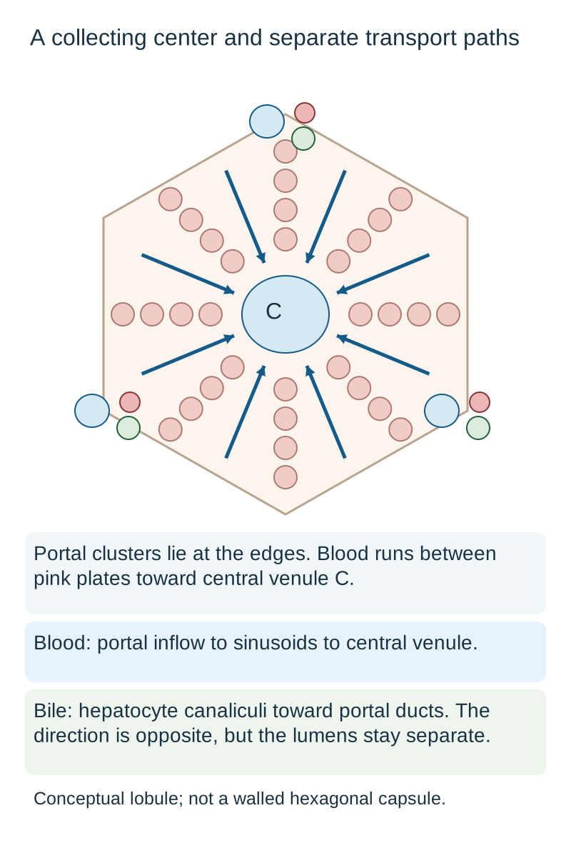
A round empty space is not automatically a central vein. First inspect its wall, lining and neighbors. In the classical lobule, a terminal hepatic venule, usually called the central vein, collects blood near the center. Portal tracts lie around the periphery. The familiar hexagon is an organizing diagram, not a capsule surrounding every lobule in a human biopsy. [1]
Locate the central collector before tracing blood. The hexagon organizes the view; it does not imply a capsule. Portal arterial and venous contributions are represented together in the incoming sinusoidal path. [1]
A different map, the portal lobule, groups cells around a shared bile duct. Its triangular territory can send blood toward central venules at different corners while bile converges on one portal duct. Changing the function used to draw the boundary does not create a new capsule. [1]
A portal tract contains branches of the portal vein and hepatic artery alongside an interlobular bile duct. The portal venule usually has the largest lumen and a relatively thin wall. The arteriole has a muscular wall around a smaller lumen. The duct has a ring of cuboidal epithelial cells rather than a thin endothelial lining. Section angle and vessel collapse change apparent size, so size alone is insufficient. Connective tissue, lymphatics and nerves also belong to the tract; the word triad names its three principal structures, not everything present. [1]
Consider a biopsy field containing one thin-walled blood vessel, a small muscular vessel and a cuboidal-lined tube together in connective tissue. Decide whether this is an inlet neighborhood or an isolated outlet. The combined pattern identifies a portal tract: its blood vessels supply the nearby sinusoids, while its duct receives bile. An isolated thin endothelial channel into which several sinusoids converge instead supports a central vein. [1]
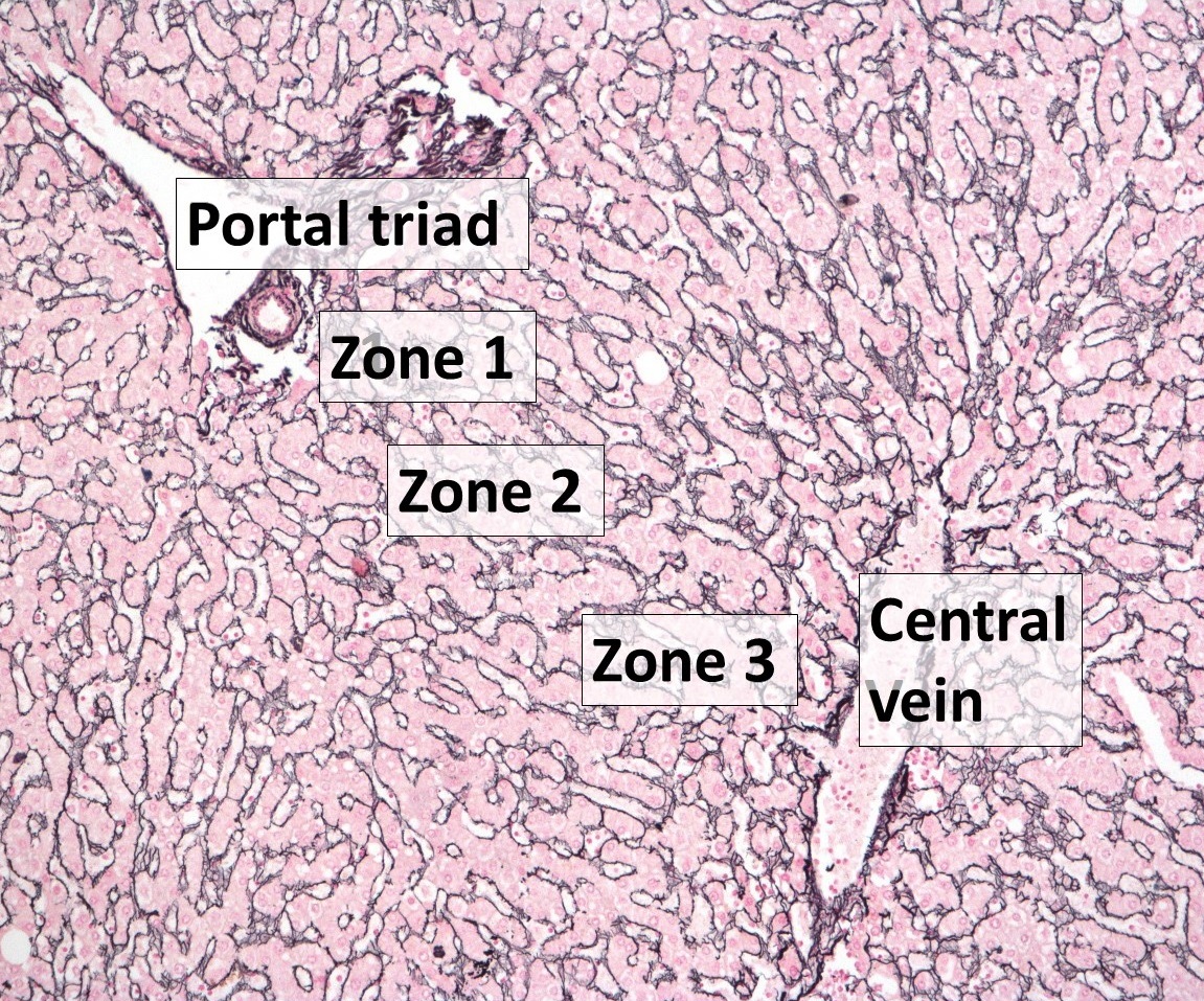
Hepatocytes form plates, generally one cell thick in normal adult liver. Blood-containing sinusoids run between those plates, not through the hepatocyte cytoplasm. The original map separates the vascular spaces from the cellular plates. The accompanying reticulin-stained clinical image makes a second point: real plates curve and branch, and a section does not look like a perfect geometric tile. [1]
Use this actual tissue image to compare branching plates and regional landmarks with the schematic. The pre-existing zonal labels are an orientation aid, not proof of a disease. Image: Alexander Boyd, Owen Cain, Abhishek Chauhan and Gwilym James Webb; labeled Commons rendition by Mikael Häggström. CC BY 4.0. Image: Alexander Boyd, Owen Cain, Abhishek Chauhan and Gwilym James Webb; Commons rendition by Mikael Häggström; original source; CC BY 4.0.
Transfer: a thin-walled vessel looks large, but serial sections show a duct and arteriole beside it. Which identification survives those additional observations?
Check the identification
A portal venule. Its neighborhood outweighs its isolated size. A central vein would be supported by converging sinusoids without an accompanying portal duct and arteriole.
Trace two paths without joining them
Does blood leave a portal tract through the nearby bile duct? No. The two systems share a neighborhood but remain separate. Portal venous blood brings material absorbed from the intestine and also carries oxygen. Hepatic arterial blood adds an oxygen-rich supply. Their terminal branches deliver blood into the sinusoidal network, where it flows between hepatocyte plates toward central veins. Collecting veins lead onward to hepatic veins and the inferior vena cava. The portal vein is an inlet despite being called a vein. [1]
A portosystemic connection can divert portal blood away from sinusoidal processing. Less contact with functioning hepatocytes can reduce ammonia handling without a new wave of cell death. Confusion still requires clinical assessment: a raised ammonia concentration alone does not diagnose hepatic encephalopathy. [11]
Bile starts with secretion by hepatocytes into canaliculi between adjacent cells. It passes through canals of Hering and ductules to interlobular ducts in portal tracts, then through larger ducts. It does not originate in the central vein. Across a classical lobule, blood is directed inward toward the central venule, whereas bile is directed outward toward portal ducts. A canaliculus is not simply a tiny blood vessel. [1]
There is also an interstitial drainage route. Fluid filtered from sinusoids into the space of Disse can pass toward portal lymphatics through the portal interstitium; other hepatic lymphatic routes exist as well. A tracer recovered from lymph rather than bile has followed a different collection system. [12]
Two-path trace
Use these five landmarks: A is a portal venule; B is a sinusoid; C is a central venule; D is a canaliculus; E is a portal bile duct. Trace an intravascular particle beginning at A, then trace bile beginning at D. State each destination before checking the worked paths below. These are different substances in different connected spaces, not two colors of liquid in one pipe.
The blood path is A to B to C. The bile path is D toward E through the intervening ductular system. Blood leaving C continues toward the hepatic veins. Neither path normally crosses directly into the other.
Now change only the lesion: a small portal bile duct becomes obstructed while nearby vessels remain patent. Predict which path loses drainage. Bile secretion upstream can encounter impaired outflow, while sinusoidal blood can still reach the central vein. Conversely, hepatic venous obstruction raises pressure downstream of sinusoids without turning the portal bile duct into a vascular bypass. [1][5]
Does a red blood cell normally enter the space between two apical hepatocyte surfaces?
No. That intercellular lumen is a bile canaliculus. Red blood cells remain in the blood pathway; they do not enter canaliculi as part of normal perfusion.
Transfer: a tracer has already reached a central venule. Predict its next larger drainage system without retracing the portal inlet. The answer is the hepatic venous system, not the bile ducts.
A cell has a blood-facing side and a bile-facing side
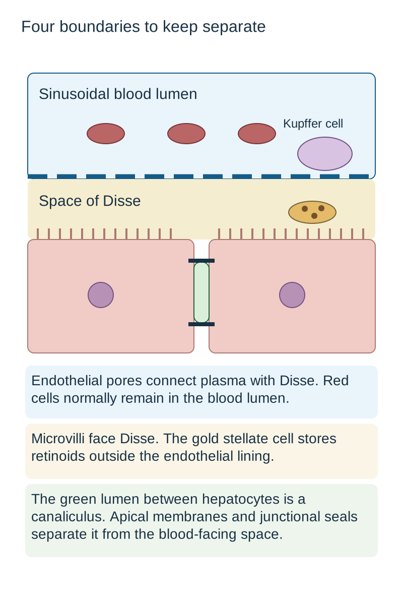
How can a hepatocyte send albumin toward blood and bile toward a duct without mixing their routes? Its membrane has distinct domains. The sinusoidal, or basolateral, surface faces the space of Disse and bears microvilli for exchange. The apical surfaces of adjacent hepatocytes form a canaliculus. Tight junctions help seal the canalicular compartment from the neighboring intercellular and blood-facing spaces. [1]
Read the compartment drawing from the blood inward: sinusoidal lumen, fenestrated endothelial lining, space of Disse, then the hepatocyte's basolateral surface. The space of Disse is outside the vascular lumen and inside the exchange boundary beside hepatocytes. Plasma constituents can pass through endothelial fenestrae, while red blood cells normally remain in the lumen. Normal sinusoidal endothelium lacks a continuous basement membrane; adding one would increase the exchange barrier. [1][4]
Read from blood to endothelium to Disse to the hepatocyte basolateral surface. The canaliculus occupies a different, apical compartment. The drawing is enlarged and simplified to separate the boundaries. [1][3][4]
Place a newly secreted albumin molecule on the blood-facing route. It enters the perisinusoidal exchange space and then the circulation. Place a bile constituent on the apical route. It enters a canaliculus and proceeds toward portal ducts. This is a prediction from polarity, not a claim that every molecule uses the same transporter. Hepatocytes also perform intermediary metabolism, detoxification, urea production and bilirubin processing. [1][2]
Location helps distinguish neighboring cells
Cell
Position
Function to predict
CellSinusoidal endothelial cell
PositionBlood-space lining
Function to predictFenestrated exchange boundary
CellKupffer cell
PositionResident macrophage exposed to sinusoidal blood
Function to predictPhagocytosis of microbes, debris and red-cell breakdown products; inflammatory signaling
CellStellate cell
PositionSpace of Disse
Function to predictRetinoid storage when quiescent; fibrogenic response when activated
CellCholangiocyte
PositionBile duct epithelium
Function to predictBile conduction and modification
A Kupffer cell can ingest blood-borne material and signal to a stellate cell without itself becoming the dominant collagen producer. Likewise, a cholangiocyte lines a duct; it does not form the hepatocyte-to-hepatocyte canaliculus. These distinctions prevent a correctly recognized liver disorder from leading to the wrong cell answer. [1][3][5]
Transfer: an electron micrograph shows a lipid-droplet-containing cell between endothelium and hepatocytes, not inside the blood lumen. Predict its resting function before naming it.
Check the location and function
The location supports a stellate cell, whose quiescent state stores vitamin A as retinoids. A sinusoidal macrophage containing phagocytosed debris would support a different identity and function.
Use the acinus to ask who receives blood first
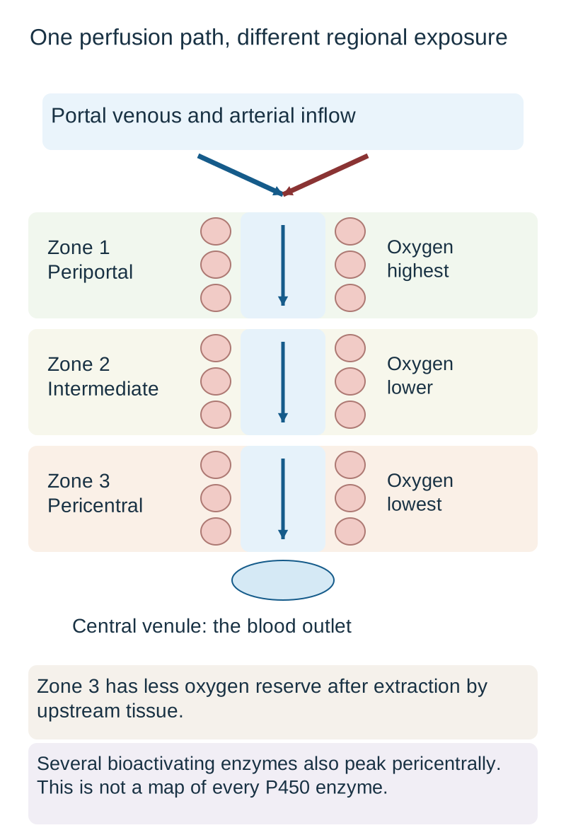
Is zone 1 closest to the central vein because one comes first? The numbering follows access to incoming blood, not proximity to the center of a drawn lobule. Zone 1 is periportal, zone 2 is intermediate, and zone 3 is pericentral. The hepatic acinus is a perfusion model spanning tissue between portal inflow branches and terminal hepatic venules; it is not an additional walled-off organ inside the lobule. [1]
As blood passes through functioning tissue, hepatocytes extract oxygen. Zone 1 therefore has the highest oxygen availability along the usual inlet-to-outlet path, while zone 3 has the lowest reserve. Both inflow vessels contribute oxygen. Their contributions depend on flow multiplied by oxygen content; calling portal blood venous does not make its oxygen contribution zero. The gradient diagram is qualitative, not a set of fixed oxygen thresholds for individual patients. [1]
Follow one inlet-to-outlet path rather than interpreting this strip as an entire walled acinus. Oxygen labels are qualitative. Low oxygen reserve and enzyme-dependent toxic bioactivation can localize injury to the same region for different reasons. [1][2][7][10]
Metabolic tasks are also distributed unevenly. Periportal hepatocytes are associated with oxidative metabolism, gluconeogenesis and urea-cycle activity. Pericentral hepatocytes have greater activity of several xenobiotic-metabolizing enzymes, including CYP2E1. This does not mean every P450 enzyme, every detoxification reaction or every metabolic process peaks in zone 3. Enzyme abundance, substrate availability and injury all matter. [2][7][10]
Compare two possible explanations for an injured pericentral ring. Reduced oxygen delivery makes downstream cells vulnerable because blood has already supplied upstream cells. A compound that becomes toxic after CYP-dependent metabolism can preferentially damage a region rich in the relevant enzyme even without systemic hypotension. The same location does not imply the same initiating mechanism.
Zone 2 is not biologically inactive filler. Spatial gene-expression experiments show profiles with maxima in intermediate tissue as well as periportal and pericentral patterns. The cited reconstruction used mouse liver; it supports the principle of gene-specific spatial specialization, not a universal numerical map for every human liver. [2]
Transfer: In a three-region assay, enzyme X reads 20, 90 and 30 units from inlet to outlet. Name the peak region, then decide whether the result reverses the direction of normal blood flow.
Check the assay interpretation
The peak is intermediate, corresponding to zone 2 in this simplified ordering. A gene-specific peak does not reverse blood flow or imply that oxygen is highest there.
Explain the injury, not just its zone number
A patient has severe hemorrhage followed by acute hepatocellular injury. The important sequence is reduced effective perfusion, reduced oxygen delivery, then vulnerability of downstream pericentral hepatocytes. That predicts a centrilobular pattern in hypoxic injury. It does not mean every case of hypotension causes necrosis or that all liver injury begins in zone 3. [1]
Now consider right-sided heart failure. Pressure backs up through hepatic veins toward central venules and sinusoids. A biopsy may show pericentral sinusoidal dilatation, congestion and, with chronic injury, fibrosis. Arterial and portal vessels can remain patent. The problem is impaired drainage and a reduced perfusion gradient, not necessarily an occluded arterial inlet. Sinusoidal dilatation alone is nonspecific; the distribution and clinical setting make the interpretation stronger. [5][9]
In acetaminophen toxicity, the CYP-generated reactive metabolite NAPQI can exceed available glutathione detoxification capacity and injure hepatocytes, characteristically in a centrilobular distribution. Systemic hypoxemia is not required for this mechanism. Acetylcysteine helps maintain or restore glutathione and detoxify reactive metabolites. A suspected toxic ingestion needs prompt clinical assessment and treatment decisions; the architecture lesson is not a reason to wait for biopsy, symptoms or established necrosis. Dosing and exposure assessment belong to a dedicated toxicity protocol. [6][10]
Carbon tetrachloride is another classic example of metabolic bioactivation associated with pericentral injury. Experiments in CYP2E1-deficient mice found protection compared with exposed wild-type mice. That finding supports enzyme-dependent susceptibility, not use of a research inhibitor as established human treatment. Alcohol-associated steatohepatitis often has pericentral injury and pericellular fibrosis, but alcohol exposure is not the only cause of this pattern. [7][9]
Ask what produces the distribution
Setting
Useful localization
Additional reasoning
SettingLow-flow or hypoxic injury
Useful localizationOften centrilobular
Additional reasoningDownstream oxygen reserve is lower
Additional reasoningClinical and virologic evidence are still needed
Human fatal yellow-fever specimens support prominent zone 2 injury, but injury can involve other regions. An apoptotic hepatocyte or a midzonal pattern alone does not establish yellow fever. Likewise, chronic viral interface activity or bile-duct-centered inflammation can be portal-based. Location narrows the mechanism; it does not replace exposure history, hemodynamics, laboratory testing or pathology context. [8][9]
Transfer: two specimens both show pericentral damage. One patient had sustained hemorrhagic shock; the other has a toxic acetaminophen exposure without hypotension. State the shared location and the different reasons for susceptibility.
Compare the mechanisms
Both can involve zone 3. The shock case is explained by inadequate oxygen delivery to downstream cells; the acetaminophen case is explained by toxic bioactivation and insufficient glutathione detoxification. Neither observation requires the other mechanism.
Fibrosis changes the exchange space and the resistance
How does a vitamin-storage cell contribute to portal hypertension? A quiescent hepatic stellate cell, also called an Ito cell, stores retinoids in lipid droplets in the space of Disse. Persistent injury and signals from neighboring cells can activate it into a contractile, matrix-producing myofibroblast-like state. Its identity is determined by its location and lineage, not just the star-like shape suggested by its name. [1][3]
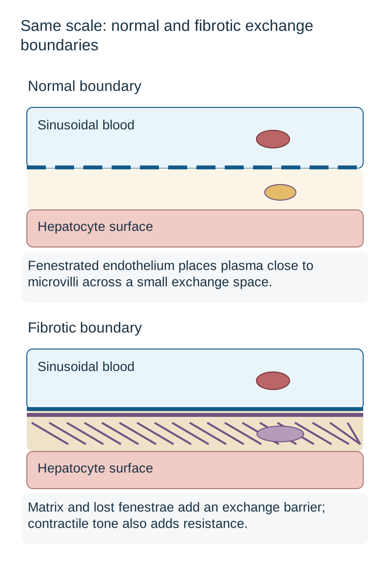
Compare the normal and fibrotic drawings at the same scale. In normal tissue, a thin fenestrated lining and a small exchange space place plasma close to hepatocyte microvilli. During chronic injury, excess extracellular matrix accumulates, endothelial fenestrae can be lost, and a more continuous basement-membrane-like barrier can develop. This process, called sinusoidal capillarization, impairs exchange. Contractile cells and structural distortion also increase resistance to blood flow. It is not simply extra collagen inside the blood itself. [3][4]
Compare the actual boundary, not just the cell name. Matrix accumulation and sinusoidal capillarization hinder exchange; activated cell contraction can add a dynamic resistance component. Neither panel depicts a measured patient specimen. [1][3][4]
Predict two consequences separately. A thicker exchange barrier can reduce access of circulating substances to hepatocytes even when those hepatocytes remain viable. Greater intrahepatic vascular resistance can raise portal pressure and reduce effective perfusion. Portal hypertension is not caused by the stellate cell secreting bile, and a pressure change does not by itself measure the amount of scar. Dynamic contractile tone and fixed matrix are different contributors. [3][4]
Stellate cells are major, not exclusive, sources of fibrogenic myofibroblasts. Other stromal populations, including portal fibroblasts, can contribute, particularly in biliary injury. Lineage-tracing results from animal models should not be presented as a fixed human percentage. Kupffer macrophages can promote inflammatory signaling without being the principal collagen-producing population. Hepatocytes and cholangiocytes have different primary roles. [3]
For a concrete perturbation, suppose an experimental intervention reduces contractile activity while the collagen stain is unchanged. At a fixed pressure difference, better flow can follow reduced resistance without proving that established scar has disappeared. Conversely, a lower enzyme concentration in blood does not prove that a fibrotic exchange barrier has returned to normal. Experiments showing endothelial-stellate interactions or reversibility in rodents do not establish an approved clinical regimen. [4]
Transfer: a biopsy has both portal duct injury and perisinusoidal collagen. Identify the bile-conducting epithelial cell and the major perisinusoidal matrix-producing lineage separately. The duct is lined by cholangiocytes; activated stellate cells are a major contributor to the perisinusoidal scar.
What finding would argue against calling every lipid-containing cell a stellate cell?
A large lipid vacuole inside a polygonal cell in a hepatocyte plate indicates hepatocellular steatosis. Stellate retinoid droplets occur in a separate perisinusoidal cell. First locate the cell, then interpret its contents.
Apply the architecture
Use the stem to identify the relevant compartment, then predict its function or response to injury. The cases contain the findings needed for their decisions; a nearby section heading is not diagnostic evidence.
Case 1
Show answer and explanations for case 1
A. Hepatic arteriole, then sinusoids (Why this does not fit)
An arteriole supplies the sinusoidal network. The marked vessel is large and thin-walled; the smaller muscular companion is the arteriole. Combine wall structure with neighboring anatomy before tracing flow.
Reasoning steps for option A
Which of the paired portal-tract vessels actually has an arterial wall?
The small muscular companion is the hepatic arteriole, not the injected large thin-walled vessel.
Could arterial blood nevertheless enter the channels between hepatocyte plates?
Yes. Arteriolar blood enters sinusoids, so the proposed downstream segment is plausible.
Why does that plausible downstream route not identify the injected vessel?
The injection starts in the large thin-walled portal venule; the muscular arterial companion is a separate inlet.
B. Hepatic venule, then sinusoids (Why this does not fit)
A hepatic venule collects sinusoidal outflow. The injected vessel has a duct and arteriole beside it, supporting a portal inlet rather than hepatic venous outflow. The word vein does not establish flow direction within an organ.
Reasoning steps for option B
Where would a collecting hepatic venule sit relative to converging sinusoids?
At their downstream confluence, exemplified by the solitary endothelial lumen in the second field.
What makes the injected large vessel an unlikely hepatic venule?
It lies beside a muscular vessel and a cuboidal-lined duct in portal connective tissue, marking the portal inlet.
Does calling both structures veins make their flow roles equivalent?
No. Blood travels from the portal venule through sinusoids to the solitary central venule, not from that central collector into sinusoids.
C. Portal venule, then sinusoids (Best answer)
A portal venule is identified by its association with an arteriole and a duct. The solitary collecting lumen in the second field is a central venule, reached through sinusoids. Use both neighborhood and connectivity to distinguish an inlet from an outlet.
Reasoning steps for option C
What establishes the injected large lumen as a portal venule?
Its thin wall and association with a muscular arteriole and cuboidal bile duct identify a portal tract.
What must the intravascular marker traverse between this inlet and the isolated collector?
It enters blood-filled sinusoids between hepatocyte plates before reaching the solitary central venule.
How does the second field verify the direction of this route?
Sinusoids converge on its unaccompanied endothelial lumen, identifying downstream central venous collection.
D. Portal venule, then canaliculi (Why this does not fit)
A portal venule carries incoming blood, whereas canaliculi carry secreted bile. An intravascular marker does not normally pass through the cuboidal tube or the canalicular system. Adjacent blood and bile structures are not directly connected lumens.
Reasoning steps for option D
Which half of portal venule then canaliculi fits the injection site?
Portal venule fits the large thin-walled portal-tract vessel where the intravascular marker begins.
What does a canaliculus carry and where is it located?
It carries hepatocyte-secreted bile between adjacent apical hepatocyte surfaces toward portal ducts.
Why cannot canaliculi deliver this blood marker to the solitary lumen?
Blood stays within sinusoids before central venous collection; the canalicular bile compartment is separate.
E. Portal bile duct, then ductules (Why this does not fit)
A duct is lined by cuboidal epithelium and receives bile from ductules. The marker enters a thin-walled blood vessel, not the epithelial tube. Epithelial lining distinguishes a bile duct from its vascular companions.
Reasoning steps for option E
Which nearby structure would justify the label portal bile duct?
The cuboidal-lined tube, rather than the large thin-walled injected vessel, is the portal bile duct.
In which direction does bile reach that duct relative to ductules?
Hepatocyte canaliculi drain via ductules toward the portal duct, not from the portal duct toward ductules.
What path must a marker injected into the neighboring vessel follow?
The blood marker enters sinusoids and then the isolated central venule, without entering the epithelial duct.
Takeaway: Use both neighborhood and connectivity to distinguish an inlet from an outlet.
A. The perisinusoidal space of Disse (Best answer)
The space of Disse lies between sinusoidal endothelium and hepatocyte basolateral surfaces. The marker has crossed the endothelial pores but has not entered hepatocytes; the red cells remain intravascular. Plasma access to hepatocytes occurs through an extravascular exchange space, not through bile.
Reasoning steps for option A
What lining has the soluble marker just crossed?
Fenestrated sinusoidal endothelium lining blood channels between hepatocyte plates.
Where is the first extravascular space on the way to hepatocyte microvilli?
The space of Disse sits between that endothelium and the hepatocyte basolateral surface.
Why can the soluble marker reach that space while labeled red cells stay in the channels?
Plasma solutes can pass endothelial fenestrae, whereas red cells ordinarily remain in the sinusoidal lumen.
B. The canalicular lumen between hepatocytes (Why this does not fit)
Canaliculi are intercellular lumens at hepatocyte apical surfaces. The observed microvilli face the sinusoidal exchange side and bile is collected through a separate route. Hepatocyte polarity separates the blood-facing and bile-facing compartments.
Reasoning steps for option B
Which hepatocyte membrane domain encloses a canaliculus?
Adjacent apical hepatocyte surfaces enclose the bile canaliculus.
Which domain do the observed microvilli contact after the marker crosses endothelial pores?
The blood-facing basolateral domain borders the perisinusoidal space of Disse.
What observation rules out immediate entry into the canaliculus?
Bile is collected separately, and the marker crosses sinusoidal endothelium rather than the sealed apical bile boundary.
C. The lumen of a portal lymphatic (Why this does not fit)
Portal lymphatics drain interstitial fluid from the liver. The first space reached across sinusoidal endothelium is adjacent to hepatocytes, before lymphatic drainage. An eventual fluid destination is not necessarily the immediately adjacent compartment.
Reasoning steps for option C
Could hepatic interstitial fluid eventually enter a portal lymphatic?
Yes. Fluid from the space of Disse may drain through portal interstitium to lymphatics.
What space is reached immediately on the hepatocyte side of fenestrated sinusoidal endothelium?
The perisinusoidal space of Disse, where the marker encounters hepatocyte microvilli.
Why is a portal lymphatic not the first compartment after crossing?
Its lumen is a downstream collection route, not the space immediately between sinusoidal endothelium and hepatocytes.
D. The lumen of an interlobular duct (Why this does not fit)
An interlobular duct is an epithelial-lined structure in a portal tract. The marker is next to hepatocyte microvilli after crossing endothelium, not inside ductal epithelium. Portal ducts conduct bile rather than serving as the sinusoidal filtration space.
Reasoning steps for option D
What does an interlobular duct normally conduct?
It carries bile through cuboidal epithelium in the portal tract.
Where is the marker when it first contacts hepatocyte microvilli?
Outside sinusoidal endothelium in the perisinusoidal exchange space, not in a portal epithelial tube.
Why does separately collected bile matter for this proposed duct route?
It confirms that plasma-side exchange and ductal bile drainage are distinct pathways.
E. The lumen of a central venule (Why this does not fit)
A central venule receives sinusoidal blood. Crossing out of the channel lining is a lateral exchange step, not continued intravascular drainage. Distinguish exchange across a vessel wall from transport along its lumen.
Reasoning steps for option E
How would a red blood cell approach a central venule from these plate-bounded channels?
It would remain in sinusoidal blood and flow toward the central venule.
Does crossing pores in the channel lining represent that intravascular downstream movement?
No. The soluble marker leaves the sinusoidal lumen laterally and enters the space of Disse.
What spatial clue excludes the central venule as its immediate destination?
The marker reaches adjacent hepatocyte microvilli just beyond endothelium, before any central venous collection.
Takeaway: Plasma access to hepatocytes occurs through an extravascular exchange space, not through bile.
A. Pericentral tissue; reduced systemic oxygen delivery (Why this does not fit)
Downstream cells are vulnerable when perfusion or oxygen delivery falls. Sustained normal pressure and oxygenation make that mechanism less consistent than the documented toxic exposure. The same zone can be damaged by different initiating processes.
Zone 3 lies downstream of oxygen extraction and has comparatively low oxygen reserve.
Which supplied measurements weaken a systemic hypoxic explanation here?
Blood pressure and oxygen saturation stayed normal, and Doppler shows patent hepatic vessels.
What instead connects the repeated medication exposure to the same region?
Acetaminophen bioactivation generates NAPQI, whose burden can exceed glutathione detoxification in susceptible pericentral tissue.
B. Periportal tissue; loss of arterial inflow (Why this does not fit)
Periportal cells are closest to incoming arterial and portal blood. The hepatic vessels are patent, and the exposure supplies a toxic mechanism without an arterial occlusion. Anatomic proximity to an inlet is different from susceptibility to a metabolically activated toxin.
Reasoning steps for option B
What blood supply defines the periportal region?
Zone 1 lies closest to portal venous and hepatic arterial inflow.
Does the Doppler study support loss of arterial inflow?
No. Hepatic vessels are patent, so an arterial blockage is not demonstrated.
Why does repeated acetaminophen use point away from periportal ischemia?
It supports CYP-mediated reactive metabolite injury, typically pericentral, despite maintained systemic perfusion.
C. Periportal tissue; obstructed interlobular ducts (Why this does not fit)
Bile-duct obstruction can produce portal-based injury and cholestasis. The acute exposure-related hepatocellular injury is not explained by a supplied duct obstruction. A hepatocellular toxic mechanism should not be replaced with a ductal mechanism without evidence.
Reasoning steps for option C
Which structure would an obstructed interlobular duct affect first?
Bile outflow through a portal-tract duct, potentially producing cholestatic or portal-based injury.
Is bile-duct obstruction supplied as the cause of the acute enzyme rise?
No. The documented trigger is multiple acetaminophen-containing products, without a stated obstructed duct.
What injury mechanism better matches this hepatocellular exposure?
D. Midacinar tissue; direct viral cytopathic injury (Why this does not fit)
Some infections can prominently involve intermediate acinar tissue. No virologic finding is supplied, while repeated acetaminophen exposure directly supports a different mechanism. A zonal association requires a compatible cause rather than a familiar location alone.
Reasoning steps for option D
What region does midacinar designate?
Zone 2, intermediate between periportal inflow and pericentral outflow.
What evidence in this vignette would be needed to favor viral cytopathic injury?
A compatible infectious or virologic finding; none is provided.
Why does the exposure favor another cause and distribution?
Repeated acetaminophen intake supports NAPQI-related pericentral injury rather than a presumed midzonal viral process.
E. Pericentral tissue; toxic bioactivation (Best answer)
Several CYP enzymes generate the reactive acetaminophen metabolite NAPQI. The exposure with preserved systemic perfusion supports bioactivation and insufficient glutathione detoxification rather than shock. A pericentral injury pattern can reflect enzyme-dependent toxicity without systemic hypoxemia.
Reasoning steps for option E
Which metabolite links acetaminophen exposure to hepatocyte damage?
CYP-mediated metabolism generates reactive NAPQI, which glutathione normally detoxifies.
Why is pericentral tissue especially plausible despite normal oxygenation?
Relevant xenobiotic-metabolizing activity is enriched pericentrally, so toxicity need not originate with systemic hypoxia.
What makes this the favored initiating mechanism in this patient?
Repeated acetaminophen-containing products precede the aminotransferase rise while pressure, oxygenation and vessel patency do not indicate low flow.
Takeaway: A pericentral injury pattern can reflect enzyme-dependent toxicity without systemic hypoxemia.
A. Toxic bioactivation exhausts intracellular glutathione (Why this does not fit)
Acetaminophen toxicity can produce reactive metabolites and centrilobular damage. There is no hepatotoxic exposure, while a substantial preceding low-flow event is documented. Prefer the supported initiating insult when several mechanisms can injure the same region.
Reasoning steps for option A
How can glutathione depletion produce damage around central venules?
A bioactivated toxin such as acetaminophen can overwhelm glutathione defenses and injure zone 3 hepatocytes.
Is that toxic trigger documented in this bleeding patient?
No hepatotoxic exposure is identified; prolonged hypotension after hemorrhage is documented.
What better accounts for injury with spared portal-adjacent cells?
B. Oxygen extraction upstream limits reserve downstream (Best answer)
Pericentral hepatocytes receive blood after upstream tissue has extracted oxygen. Hypotension followed by injury around central venules, with periportal preservation, fits inadequate oxygen delivery. Identify the flow direction before deciding which region has the smallest oxygen reserve.
Reasoning steps for option B
Which biopsy landmark identifies the injured downstream neighborhood?
Solitary thin-walled central venules lack the duct and arteriole that accompany portal inlets.
What happens to oxygen before blood reaches hepatocytes around those venules?
Blood passes from portal inflow through sinusoids while upstream tissue extracts oxygen.
Why does prolonged hemorrhagic hypotension accentuate this gradient?
Reduced delivery leaves pericentral cells with the least reserve, matching their injury and the relative sparing beside portal tracts.
C. Portal inflammation damages adjacent hepatocytes (Why this does not fit)
Interface inflammation can produce portal-based hepatocellular injury. The cells beside duct-and-arteriole clusters are relatively preserved rather than maximally injured. Use the spared region as well as the injured region to test a proposed mechanism.
Reasoning steps for option C
Where would portal interface inflammation produce the greatest local injury?
Near portal tracts, beside the duct and arteriole clusters.
Are those cells the principal injured population in this specimen?
No. They are relatively preserved, while injury surrounds solitary central venules.
What supplied event explains this inverse distribution?
Prolonged blood-loss hypotension reduces oxygen delivery to downstream pericentral tissue.
D. Hepatic venous pressure impairs sinusoidal drainage (Why this does not fit)
High hepatic venous pressure can produce pericentral congestion and injury. The specified initiating event is severe blood loss with systemic hypotension, without evidence of new right-sided pressure elevation. Shared pericentral involvement does not make low inflow and high outflow pressure the same process.
Reasoning steps for option D
What would elevated hepatic venous pressure do to central venules and sinusoids?
It could impede drainage and produce pericentral congestion.
Which hemodynamic change is actually stated?
Major bleeding caused prolonged systemic hypotension, not documented right-sided venous backpressure.
Why is the shared pericentral location insufficient to infer congestion?
Low inflow lowers downstream oxygen delivery, whereas venous congestion reflects high outflow pressure; this case documents the former.
E. Ductal obstruction retains injurious bile constituents (Why this does not fit)
Biliary obstruction can injure hepatocytes as well as ducts. The injury follows hemorrhagic hypotension and is centered on central venules rather than a demonstrated obstructed duct. Distinguish a low-flow event from impaired bile drainage.
Reasoning steps for option E
Which anatomic path would a blocked duct interrupt?
Bile drainage from canaliculi and ductules into a portal bile duct.
Does the specimen localize maximal injury to that portal duct neighborhood?
No. Hepatocytes beside duct-and-arteriole clusters are relatively spared; injury surrounds central venules.
Which documented insult predicts that central distribution?
Hemorrhagic hypotension reduces oxygen delivery after upstream extraction along the sinusoidal blood route.
Takeaway: Identify the flow direction before deciding which region has the smallest oxygen reserve.
A. Pericellular scar around ballooned lipid-laden hepatocytes (Why this does not fit)
Steatohepatitis can produce pericentral ballooning and pericellular fibrosis. The supplied findings specifically demonstrate increased hepatic venous pressure rather than a steatohepatitic process. A pericentral location alone does not distinguish fat-associated injury from congestion.
Reasoning steps for option A
What histologic process produces ballooning and pericellular scar around fatty hepatocytes?
Steatohepatitis can cause lipid-associated hepatocyte injury and pericellular fibrosis, sometimes pericentrally.
Which findings instead identify a venous pressure problem?
Severe tricuspid regurgitation, a dilated inferior vena cava and increased hepatic venous pulsatility indicate backpressure.
What biopsy change follows transmission of that pressure into liver tissue?
Central venules and nearby sinusoids become congested and dilated; ballooning with fat-associated scar is not the direct pressure signature.
B. Confluent necrosis confined to periportal hepatocytes (Why this does not fit)
Periportal damage can accompany selected inflammatory or toxic processes. Venous pressure first affects the central outflow neighborhood rather than exclusively the portal inlet. Map where the abnormal pressure is transmitted before assigning a zone.
Reasoning steps for option B
Where is periportal tissue along the sinusoidal blood path?
It is near portal venous and arterial inlet branches, upstream of central venous drainage.
Where does tricuspid regurgitation transmit pressure first in this pathway?
Backward through hepatic veins to central venules and neighboring pericentral sinusoids.
Why is exclusive periportal necrosis an unlikely direct result?
Patent inflow and demonstrated venous backpressure favor central congestion rather than damage confined to the inlet zone.
C. Inflammation centered on interlobular bile ducts (Why this does not fit)
Duct-centered inflammation supports a biliary injury pattern. The supplied abnormality is venous pressure rather than evidence of duct destruction. Biliary and vascular outflow problems involve different compartments.
Reasoning steps for option C
Which compartment is implicated by inflammation around interlobular ducts?
Portal bile-duct epithelium and its surrounding biliary tissue.
Does tricuspid regurgitation obstruct the portal bile ducts?
No. The abnormality is elevated hepatic venous pressure with increased venous pulsatility, not documented duct inflammation.
Which structures should instead bear the immediate pressure effect?
Central venules and adjoining sinusoids on the hepatic venous outflow side.
D. Dilated congested sinusoids around central venules (Best answer)
High hepatic venous pressure is transmitted toward central venules and adjacent sinusoids. The cardiac and venous findings indicate impaired outflow despite patent inflow vessels. Raised downstream pressure can produce pericentral congestion without arterial obstruction.
Reasoning steps for option D
How do the cardiac and caval findings connect to the hepatic microcirculation?
Tricuspid regurgitation and caval dilation signal right-sided pressure transmitted backward through hepatic veins.
Which microscopic structures receive that raised downstream pressure?
Central venules and surrounding pericentral sinusoids, which can dilate with blood congestion.
Why does arterial and portal patency not contradict this biopsy pattern?
The flow impairment lies at venous outflow, not the patent inflow vessels.
E. Thrombi filling branches of the hepatic artery (Why this does not fit)
Arterial thrombosis compromises an inflow route. Doppler documents a patent hepatic artery and the major abnormality is right-sided venous pressure. Patent inflow does not exclude hepatic injury from impaired drainage.
Reasoning steps for option E
What would hepatic arterial branch thrombi represent?
Obstruction of an inflow vessel rather than hepatic venous outflow congestion.
Which imaging fact contradicts those thrombi?
The hepatic artery remains patent, as does the portal vein.
Which vascular change is actually supported by the clinical findings?
Tricuspid regurgitation with caval dilation and increased hepatic venous pulsatility supports pericentral sinusoidal congestion.
Takeaway: Raised downstream pressure can produce pericentral congestion without arterial obstruction.
A. It falls from 4 to 1 because the resistance has doubled (Why this does not fit)
Doubling resistance can halve flow at an unchanged pressure difference. The model explicitly holds resistance at 2; applying a second reduction invents another change. Calculate from the variables that changed rather than adding an unstated lesion.
Reasoning steps for option A
If resistance had doubled from 2, what alternative cause would this option invoke?
Doubling resistance could lower flow at a fixed gradient, but no doubling occurs in this model.
Why does the fixed resistance of 2 rule out an added resistance-based fall?
Resistance remains 2 before and after the outlet-pressure rise; another resistance change is not supplied.
What flows follow from gradients of 8 and 4 divided by 2?
The gradients are 12 - 4 = 8 and 12 - 8 = 4, giving flows of 4 and 2, not 4 and 1.
B. It falls from 4 to 2 because the pressure difference narrows (Best answer)
Flow depends on the pressure difference across a vascular bed. The difference falls from 8 to 4 units while resistance stays 2, so calculated flow halves. A higher venous outlet pressure can reduce flow without an obstructed inlet.
Reasoning steps for option B
How does raising outlet pressure from 4 to 8 alter the driving gradient?
The gradient decreases from 12 - 4 = 8 to 12 - 8 = 4 units.
What are the two flows when each gradient is divided by the fixed resistance of 2?
Initial flow is 8/2 = 4; final flow is 4/2 = 2, a halving.
Why can right-sided venous congestion reduce flow despite a patent portal inlet?
The patent inlet remains at 12, but higher venous outlet pressure reduces the driving gradient.
C. It falls from 6 to 4 because inlet pressure alone sets flow (Why this does not fit)
Dividing inlet pressure by resistance omits the downstream pressure. Both outlet values are supplied and must be subtracted, producing 4 and 2 rather than 6 and 4. Use a pressure difference rather than a single pressure in the flow relation.
Reasoning steps for option C
What downstream term is missing when 12/2 yields the proposed initial flow of 6?
The proposed 6 comes from 12/2, which omits the original outlet pressure of 4.
What results when 4 and then 8 are subtracted from the inlet pressure of 12?
Subtracting outlet pressures yields 8/2 = 4 initially and 4/2 = 2 afterward, not 6 and 4.
Why must downstream pressure enter both flow calculations?
The flow formula uses inlet minus outlet pressure; neither 4 nor 8 can be discarded.
D. It stays at 4 because vascular resistance is unchanged (Why this does not fit)
Unchanged resistance preserves flow only if the driving pressure also stays constant. The inlet is fixed but the outlet pressure rises, so the driving difference does not stay constant. A resistance measurement alone cannot determine flow.
Reasoning steps for option D
Does holding resistance at 2 hold the inlet-to-outlet gradient constant?
No. Resistance is 2 throughout, but the driving gradient changes as outlet pressure rises.
What happens to the gradient when the fixed inlet of 12 faces an outlet rising from 4 to 8?
The gradient falls from 8 to 4 units despite the inlet remaining at 12.
Why does unchanged resistance not imply unchanged sinusoidal perfusion?
At resistance 2, halving the gradient halves flow from 4 to 2.
E. It rises from 4 to 6 because the outlet pressure increases (Why this does not fit)
Increasing inlet pressure can increase flow when other variables are fixed. Here the pressure that rises is the outlet pressure, which subtracts from rather than adds to the driving difference. Identify which end of the vascular bed has the pressure change.
Reasoning steps for option E
Would a rise in inlet or outlet pressure increase flow with the other quantities fixed?
A higher inlet could increase the gradient; a higher outlet reduces it when the inlet is fixed.
Does the outlet rise from 4 to 8 add to or subtract from the gradient?
Raising outlet pressure from 4 to 8 reduces 12 minus outlet pressure from 8 to 4.
What direction of flow change follows from venous backpressure rather than added inflow pressure?
Venous backpressure decreases model flow from 4 to 2 rather than increasing it to 6.
Takeaway: A higher venous outlet pressure can reduce flow without an obstructed inlet.
A. Kupffer cells acquiring greater phagocytic activity (Why this does not fit)
Kupffer macrophages respond to injury and ingest material from sinusoidal blood. The described cells are outside the endothelial lining and change from retinoid storage to collagen production. Inflammatory signaling and the principal matrix-producing response belong to different populations.
Reasoning steps for option A
What function makes Kupffer cells plausible in chronic hepatitis B injury?
Kupffer cells are injury-responsive macrophages that phagocytose material in sinusoidal blood.
Do blood-facing phagocytes explain retinoid droplet loss outside the endothelial lining and new collagen around plates?
The observed cells are outside endothelium and lose retinoids while producing collagen, not a phagocytic Kupffer-cell pattern.
Which perisinusoidal lineage, rather than the macrophage, supplies this matrix-producing transition?
A stellate cell in the space of Disse can acquire the matrix-producing phenotype in this hepatitis B fibrosis.
B. Endothelial cells increasing sinusoidal fenestration (Why this does not fit)
Differentiated sinusoidal endothelium supports exchange through fenestrae. The affected cells lie outside that lining and are losing retinoid stores while depositing collagen. Distinguish the exchange lining from the neighboring fibrogenic cell.
Reasoning steps for option B
What role do sinusoidal endothelial fenestrae serve?
Fenestrae of the sinusoidal endothelial lining support blood-to-hepatocyte exchange.
Are the retinoid-depleted collagen-producing cells within the endothelial lining or outside it?
The changing cells are outside that lining and lose retinoid droplets while synthesizing collagen.
Why does a change in exchange fenestrae fail to explain contractile fibrogenic cells?
More endothelial fenestration does not account for an extraluminal, contractile, fibrogenic cell.
C. Cholangiocytes increasing ductal fluid secretion (Why this does not fit)
Cholangiocytes line ducts and modify bile. The cells are around hepatocyte plates rather than forming ductal epithelium. A cell in the space of Disse is not identified by a bile-duct function.
Reasoning steps for option C
Where would cholangiocytes need to be located to account for increased ductal secretion?
Cholangiocytes line bile ducts, where they modify ductal fluid.
Does the observed position around hepatocyte plates identify ductal epithelium?
The observed cells lie around hepatocyte plates, outside the sinusoidal endothelium, not in ducts.
What does the perisinusoidal position imply instead of a bile-duct response?
Their location and retinoid loss identify a perisinusoidal stellate transition rather than ductal secretion.
D. Hepatocytes increasing intracellular lipid storage (Why this does not fit)
Hepatocytes may accumulate lipid during steatosis. The cells are separate from hepatocyte plates, lose storage droplets and acquire matrix synthesis. Hepatocellular fat storage differs from activation of a perisinusoidal stromal cell.
Reasoning steps for option D
What would increasing hepatocyte lipid storage predict about intracellular droplets?
Hepatocyte steatosis would involve greater lipid accumulation within hepatocytes.
Are the observed cells hepatocytes accumulating lipid or separate cells losing retinoid droplets?
These cells are separate from the plates and have fewer retinoid droplets rather than more stored lipid.
Why do contraction and collagen synthesis favor stellate activation over steatosis?
New contractility and collagen synthesis fit activated stellate cells, not fat-filled hepatocytes.
E. Stellate cells becoming contractile and fibrogenic (Best answer)
Quiescent stellate cells store retinoids in the space of Disse and can become myofibroblast-like. The perisinusoidal location, declining retinoid stores and new collagen synthesis identify this transition. Use location plus the change in function to identify the fibrogenic lineage.
Reasoning steps for option E
Which resting cell stores retinoids outside the sinusoidal endothelium?
Quiescent stellate cells store retinoids in the space of Disse outside sinusoidal endothelium.
What do fewer retinoid droplets together with contractile proteins and collagen show about its state?
Retinoid depletion with acquired contractile proteins and collagen synthesis marks myofibroblast-like activation.
How does this transition account for fibrosis surrounding hepatocyte plates?
Activated stellate cells deposit perisinusoidal collagen around plates during progressive fibrosis.
Takeaway: Use location plus the change in function to identify the fibrogenic lineage.
A. New vascular bypasses avoiding fibrotic sinusoids (Why this does not fit)
A bypass can increase flow through an alternative connection. Vascular connections were fixed while perisinusoidal contraction was reduced. Do not infer a new anatomical route from a change in contractile tone.
Reasoning steps for option A
How could a new vascular bypass raise perfusion independently of sinusoidal tone?
A bypass would provide a new route around resistant fibrotic sinusoids.
Were vascular connections allowed to change during the minutes after myofibroblast relaxation?
Connections are held constant, and only perisinusoidal contraction is reduced.
Why does fixed connectivity make a new anatomical bypass unnecessary here?
The prompt supports altered tone in existing vessels rather than creation of collateral connections within minutes.
B. Higher driving pressure through the sinusoidal network (Why this does not fit)
A larger inlet-to-outlet pressure difference can raise flow. Both inlet and outlet pressures were held constant during the experiment. Use the measured pressure conditions before attributing a flow change to pressure.
Reasoning steps for option B
What would need to happen to the inlet-to-outlet gradient for pressure alone to raise flow?
A larger inlet-to-outlet pressure difference would be required for a pressure-driven flow rise.
Did either measured boundary pressure change during the intervention?
Both inlet and outlet pressures remain fixed, so their difference cannot rise.
At fixed pressure difference, what resistance change accounts for increased flow?
Increased flow at that fixed gradient implies reduced effective resistance, consistent with relaxation.
C. Reduced tone without demonstrated scar regression (Best answer)
Contractile tone can contribute to resistance in addition to fixed matrix. Flow rises at the same pressure difference before any change in collagen is demonstrated. An immediate hemodynamic benefit does not establish removal of established fibrosis.
Reasoning steps for option C
What resistance component can fall when perisinusoidal myofibroblasts relax?
Relaxation lowers the dynamic component of sinusoidal resistance imposed by contractile perisinusoidal cells.
What does greater flow at fixed pressure and viscosity imply while collagen staining stays unchanged?
At the same pressure difference, greater flow implies lower effective resistance. Unchanged collagen staining favors a dynamic reduction in contractile tone rather than demonstrated scar removal.
Why does the rapid functional response not demonstrate scar regression?
The minutes-long hemodynamic gain demonstrates reduced tone, not loss of established collagen scar.
D. Dissolution of scar with restored lobular architecture (Why this does not fit)
Removing matrix can reduce fixed structural resistance. Collagen staining is unchanged over the brief observation period, so matrix removal is not the demonstrated explanation. Separate a functional response from evidence of structural repair.
Reasoning steps for option D
What tissue evidence would support dissolution of fixed collagen scar?
Scar dissolution would require evidence of reduced matrix or restored architecture.
Did collagen staining or lobular structure demonstrably change over several minutes?
Collagen staining remains unchanged over the short observation; restored lobular structure is not shown.
Why is lower contractile tone a better immediate explanation than matrix removal?
Relaxation of myofibroblasts explains immediate flow improvement without claiming structural repair.
E. Lower perfusate viscosity within the existing vessels (Why this does not fit)
Lower viscosity can reduce resistance without changing vessel structure. The experiment explicitly holds perfusate viscosity constant. Controlled variables help distinguish vascular tone from fluid properties.
Reasoning steps for option E
How would reducing perfusate viscosity affect resistance at a fixed gradient?
Lower viscosity could reduce flow resistance independently of anatomy.
Was viscosity changed along with perisinusoidal contraction?
Perfusate viscosity is explicitly held constant while myofibroblast contraction is reduced.
Which controlled property excludes fluid thinning as the source of increased flow?
Because fluid viscosity is controlled, altered perisinusoidal tone is the supported variable.
Takeaway: An immediate hemodynamic benefit does not establish removal of established fibrosis.
A. A greater barrier to sinusoidal plasma exchange (Best answer)
Sinusoidal fenestrae and the normally discontinuous basement-membrane boundary facilitate access to hepatocytes. Uptake is preserved after the cells are isolated, while the intact tissue contains an added exchange barrier. A functioning hepatocyte can have impaired access to circulating substrates.
Reasoning steps for option A
What normal sinusoidal features permit blood-borne substance to reach hepatocytes?
Sinusoidal endothelial fenestrae and the normally discontinuous basement-membrane boundary facilitate plasma access to hepatocytes.
Why does normal uptake after isolation localize the intact-tissue defect outside the hepatocyte?
Isolated viable hepatocytes still take up the substance, so their intrinsic uptake is preserved.
How do lost fenestrae and a continuous basement-membrane-like layer impair exchange?
Loss of fenestrae plus a continuous basement-membrane-like layer adds a blood-to-hepatocyte exchange barrier in intact tissue.
B. Obstruction of the downstream interlobular bile ducts (Why this does not fit)
Duct obstruction impairs bile drainage and can cause cholestasis. The demonstrated abnormality is between sinusoidal blood and hepatocytes rather than inside the duct system. Blood-to-cell exchange and bile outflow are different steps.
Reasoning steps for option B
Which direction of transport is disrupted by an interlobular duct obstruction?
Interlobular duct obstruction would impair downstream bile drainage and cause cholestasis.
Does the observed lesion lie in bile ducts or between sinusoidal blood and hepatocytes?
The observed barrier lies between sinusoidal blood and hepatocytes, not in the bile ducts.
Why cannot impaired bile drainage explain preserved isolated-cell uptake but poor intact-tissue access?
The experiment compares uptake from blood in intact tissue with isolated cells, not downstream bile outflow.
C. Increased phagocytosis by sinusoidal macrophages (Why this does not fit)
Macrophage uptake can affect clearance of particulate material. The supplied observations demonstrate an endothelial exchange barrier and normal isolated hepatocyte uptake, not increased macrophage ingestion. Use the observed compartmental lesion rather than assigning every clearance change to macrophages.
Reasoning steps for option C
What substrate clearance might sinusoidal macrophage phagocytosis alter?
Sinusoidal macrophages can phagocytose blood-borne particles.
Is enhanced macrophage ingestion shown, or is an endothelial barrier directly visualized?
No increased macrophage ingestion is reported; electron microscopy instead demonstrates lost fenestrae and an added barrier.
Why does normal isolated hepatocyte uptake point to delivery rather than macrophage competition?
Normal hepatocyte uptake after isolation makes impaired access across the sinusoidal boundary the direct explanation.
D. Loss of the apical hepatocyte secretory membrane (Why this does not fit)
Apical membrane injury can interfere with canalicular secretion. The measured defect is access from blood across a remodeled endothelial boundary, not a demonstrated canalicular membrane loss. Localize the failed step before naming a membrane domain.
Reasoning steps for option D
Which hepatocyte surface faces canalicular bile rather than sinusoidal plasma?
The apical hepatocyte membrane faces bile canaliculi and participates in secretion into bile.
Is apical membrane loss documented in the tissue with lost fenestrae?
There is no demonstrated apical membrane loss; the imaged lesion is on the blood-facing endothelial boundary.
Why is blood-to-cell access the affected step instead of canalicular secretion?
The discrepancy concerns uptake from blood before canalicular secretion, so an apical defect is misplaced.
E. Reduced activity of the hepatocyte uptake transporter (Why this does not fit)
A transporter defect can reduce uptake despite adequate substrate delivery. The same hepatocytes take up the substance normally once isolated from the altered tissue boundary. Compare intrinsic cell function with delivery through surrounding tissue.
Reasoning steps for option E
What would an intrinsic uptake-transporter defect do to isolated hepatocytes?
A defective hepatocyte uptake transporter should also reduce uptake when those cells are isolated.
How does preserved uptake after isolation contradict reduced transporter activity?
The isolated hepatocytes take up the same substance normally, contradicting an intrinsic transporter defect.
Which intact-tissue boundary can limit substrate delivery despite functioning transporters?
The intact-tissue fenestrae loss and continuous basement-membrane-like layer limit delivery to functioning cells.
Takeaway: A functioning hepatocyte can have impaired access to circulating substrates.
A. Less bilirubin secretion across the apical membrane (Why this does not fit)
Hepatocytes secrete bile constituents through the canalicular surface. Hepatocyte viability and biliary transport are explicitly unchanged. Macrophage particle clearance is not equivalent to hepatocyte bile transport.
Reasoning steps for option A
Which cell membrane normally secretes bilirubin into bile?
Hepatocytes secrete bilirubin at their canalicular apical membrane.
Was hepatocyte biliary transport changed when macrophage phagocytosis was inhibited?
Hepatocyte viability and biliary transport remain unchanged during selective macrophage inhibition.
Why does reduced particle capture not predict reduced canalicular bilirubin secretion?
Blocking Kupffer-cell phagocytosis affects bacterial-particle removal, not demonstrated bilirubin secretion.
B. More retinoid droplets in sinusoidal endothelial cells (Why this does not fit)
Retinoid storage is a quiescent stellate-cell function. The intervention targets blood-facing macrophage phagocytosis, not the lineage or storage function of endothelial cells. Identify the affected cell before predicting a change in storage.
Reasoning steps for option B
Which perisinusoidal cell stores retinoid droplets, and is it endothelial?
Quiescent stellate cells store retinoids in the perisinusoidal space; sinusoidal endothelial cells do not serve that storage role.
Does selective inhibition of blood-facing macrophages alter endothelial identity or stellate storage?
The manipulation selectively inhibits blood-facing resident macrophage phagocytosis, not endothelial or stellate function.
Why does the intervention not predict retinoid accumulation in sinusoidal endothelium?
No retinoid-storage increase in endothelial cells follows from reduced Kupffer-cell bacterial uptake.
C. Less collagen produced by a lost stellate population (Why this does not fit)
Stellate cells are major matrix-producing cells after activation. No loss of the perisinusoidal stellate population is described; the immediate target is phagocytosis. A selective functional intervention does not establish loss of another lineage.
Reasoning steps for option C
Which activated perisinusoidal cell can synthesize collagen?
Activated stellate cells, not blood-facing Kupffer macrophages, produce much of the perisinusoidal collagen.
Is loss of stellate cells reported when only macrophage phagocytosis is blocked?
No stellate population is lost; only resident macrophage phagocytosis is inhibited.
Why cannot a selective clearance defect establish less collagen from a missing stellate population?
Reduced particle capture alone cannot establish loss of stellate cells or reduced collagen synthesis.
D. More bacterial particles in hepatic venous effluent (Best answer)
Kupffer macrophages remove blood-borne microbes and particulate material from sinusoidal blood. Their uptake is selectively inhibited while incoming particle concentration and flow are unchanged. Loss of sinusoidal clearance can increase material escaping through hepatic venous outflow.
Reasoning steps for option D
Where do Kupffer macrophages encounter particles entering from the portal inflow?
Kupffer macrophages contact portal-borne particles on the blood-facing sinusoidal surface.
With input concentration and flow fixed, what happens to removal when their phagocytosis is inhibited?
With particle input and blood flow unchanged, inhibiting their phagocytosis leaves more particles unremoved.
Where will the additional unremoved bacterial particles appear after traversing the sinusoids?
More bacterial particles therefore escape the sinusoids into hepatic venous effluent.
Takeaway: Loss of sinusoidal clearance can increase material escaping through hepatic venous outflow.
A. The canaliculus between neighboring hepatocytes (Best answer)
Canaliculi are formed by the apical surfaces of adjacent hepatocytes. The defective export step is explicitly apical, upstream of the patent duct system. A patent bile duct does not exclude a defect in canalicular secretion.
Reasoning steps for option A
Where does an apical hepatocyte bilirubin exporter discharge?
Into the canaliculus bounded by neighboring hepatocyte apical surfaces.
Why do patent interlobular ducts not rescue this child's export?
The block occurs before bilirubin reaches ductules or interlobular ducts.
Which compartment therefore receives less conjugated bilirubin directly?
The canalicular lumen, despite preserved basolateral membrane and tight junctions.
B. The blood lumen within a hepatic sinusoid (Why this does not fit)
Sinusoids carry blood between hepatocyte plates. The affected membrane secretes into the biliary rather than the vascular compartment. A canalicular export defect is not a failure to secrete bilirubin directly into blood.
Reasoning steps for option B
Does sinusoidal blood directly receive hepatocyte apical bilirubin export?
No. The sinusoidal lumen contacts the blood-facing side across endothelium and Disse.
What in this child separates the sinusoid from the blocked route?
Basolateral function is preserved; the defective transporter is apical.
Would less canalicular export imply less direct secretion into sinusoidal blood?
No. The specified immediate loss is on the bile side, not direct vascular secretion.
C. The lumen of a portal lymphatic vessel (Why this does not fit)
Portal lymphatics drain interstitial fluid. The defect involves direct hepatocyte apical secretion, not drainage from an interstitial space. The first recipient of apical secretion is a canaliculus.
Reasoning steps for option C
What normally enters a portal lymphatic vessel?
Interstitial fluid draining toward portal lymphatics, not bile directly exported by hepatocytes.
Does an apical bilirubin transporter empty into portal interstitium?
No. Adjacent hepatocyte apical surfaces enclose the canalicular lumen.
Why is lymphatic drainage not the immediate affected recipient here?
The defect precedes patent ducts at the canalicular secretion step, rather than involving interstitial collection.
D. The space beside hepatocyte basolateral microvilli (Why this does not fit)
That perisinusoidal space participates in exchange with blood. Basolateral function is preserved, whereas the specified defect is on the apical membrane. Use membrane polarity to separate secretion into bile from exchange with blood.
Reasoning steps for option D
Which hepatocyte surface faces the space beside basolateral microvilli?
The blood-facing surface bordering the perisinusoidal space of Disse.
Is that surface identified as defective in the child?
No. Basolateral membrane function is preserved while apical export fails.
Where does conjugated bilirubin normally go at the affected apical step instead?
Into a canaliculus, not into the perisinusoidal exchange space.
E. The lumen of a terminal hepatic venule (Why this does not fit)
Terminal hepatic venules collect sinusoidal blood. They are downstream vascular collectors, not the immediate recipient of apical export. The central venule and canaliculus serve different transport systems.
Reasoning steps for option E
What does a terminal hepatic venule collect?
Blood arriving through sinusoids, downstream of the portal inflow.
Can a central venule be the immediate recipient of apical bilirubin export?
No. It is a vascular outlet, whereas the apical hepatocyte surface borders a canaliculus.
Which observation makes the venular choice especially misplaced?
The reported transport lesion is apical with preserved blood-facing membrane function.
Takeaway: A patent bile duct does not exclude a defect in canalicular secretion.
A. The signal preserves collagen by preventing cell death (Why this does not fit)
Improved cell survival can increase the number of cells available to produce matrix. Neither population loses viability after signal blockade, yet synthesis falls. A viability control helps distinguish activation from cell-number effects.
Reasoning steps for option A
Could fewer surviving matrix-producing cells explain the blockade effect?
Cell loss could reduce total collagen synthesis in principle.
What does the viability control show after macrophage-signal blockade?
Neither separated population dies, although synthesis by perisinusoidal cells falls.
Does the signal primarily preserve cells or stimulate their output here?
The evidence supports stimulation of collagen production without a detectable survival effect.
B. Perisinusoidal cells remove collagen through phagocytosis (Why this does not fit)
Matrix degradation and synthesis are distinct processes that can affect net scar. The measured response is increased collagen synthesis, not removal of existing collagen. Identify which matrix process the experiment actually measures.
Reasoning steps for option B
Which measured process changes after conditioned-medium exposure?
Collagen synthesis by the retinoid-storing perisinusoidal population increases.
Was collagen removal by phagocytosis measured?
No. The experiment reports new synthesis, not degradation or uptake of existing matrix.
Production and removal are different fluxes; the observed direction concerns production.
C. Macrophages stimulate stellate-cell collagen synthesis (Best answer)
Signals from inflammatory cells can promote activation of fibrogenic stellate cells. Collagen production changes in the retinoid-storing perisinusoidal population after exposure to macrophage-conditioned medium. The cell providing an activating signal need not be the cell producing the matrix.
Reasoning steps for option C
Which separated cells supply the active medium?
Activated blood-facing macrophages release a signal into the collected medium.
Which cells increase collagen synthesis in response?
The retinoid-storing perisinusoidal cells, consistent with stellate cells, produce more collagen.
What does selective signal blockade establish without cell death?
Macrophage-derived signaling drives stellate-cell collagen synthesis rather than merely changing viable cell number.
D. Macrophages become the measured collagen-producing population (Why this does not fit)
A lineage transition would require evidence that the original macrophages acquired the new phenotype. The populations remain separated, and collagen is measured in the perisinusoidal cells. Do not infer transdifferentiation from an effect transmitted through culture medium.
Reasoning steps for option D
What would demonstrate macrophages becoming collagen-producing cells?
Lineage evidence that the original macrophages acquired collagen-producing identity would be needed.
Where was increased collagen synthesis actually observed?
In the separate retinoid-storing perisinusoidal population exposed to macrophage-conditioned medium.
Why does a medium-transfer effect not prove macrophage conversion?
A soluble signal crosses between populations without transferring macrophage lineage or identity.
Takeaway: The cell providing an activating signal need not be the cell producing the matrix.
A. Cholangiocyte bile modification; hepatocyte protein secretion (Why this does not fit)
Cholangiocytes modify bile, and hepatocytes secrete proteins toward blood. The first cells form plates rather than a ductal ring, and the second population lies outside those plates. Match both members of a functional pairing to their architectural positions.
Reasoning steps for option A
Do the large-vacuole cells occupy a ductal epithelial ring?
No. They are polygonal cells arranged in hepatocyte plates, not cholangiocytes lining ducts.
Are the smaller droplets in plate-forming protein-secreting hepatocytes?
No. That separate population sits between the plates and sinusoidal endothelial lining.
Why does this cholangiocyte-hepatocyte pairing fail?
Its first identity mislabels plate cells and its second mislabels perisinusoidal storage cells.
B. Hepatocyte fat storage; stellate-cell retinoid storage (Best answer)
Hepatocellular steatosis and quiescent stellate-cell vitamin A storage occur in different cells. The large vacuoles occupy cells in plates, while the smaller storage cells are perisinusoidal. Cell location separates hepatocyte steatosis from the retinoid-storage role of stellate cells.
Reasoning steps for option B
What do large clear vacuoles inside polygonal plates indicate?
Fat accumulation within hepatocytes in this steatohepatitis biopsy.
Where are the separate small lipid-droplet cells situated?
In the perisinusoidal space between hepatocyte plates and endothelial lining, where stellate cells reside.
What is the contrasting storage function of quiescent stellate cells?
They store vitamin A as retinoids, distinct from hepatocellular fat accumulation.
C. Macrophage debris uptake; endothelial plasma exchange (Why this does not fit)
Macrophages ingest debris, and sinusoidal endothelial cells form an exchange lining. The first population forms polygonal hepatocyte plates, while the second lies outside rather than forming the endothelial lining. Both named functions are real, but they do not match the observed cell locations.
Reasoning steps for option C
Are the vacuolated plate-forming cells Kupffer macrophages?
No. Kupffer cells are sinusoidal macrophages that ingest blood-borne debris, not polygonal plate-forming hepatocytes.
Is the second droplet-containing population the endothelial exchange lining?
No. It lies between that lining and the hepatocyte plates, rather than constituting the lining.
What does this pairing overlook about both observed populations?
It assigns true macrophage and endothelial functions to cells with hepatocyte and stellate-cell locations.
D. Endothelial plasma exchange; cholangiocyte bile modification (Why this does not fit)
Sinusoidal endothelium supports exchange, and cholangiocytes line bile ducts. Neither a vascular lining nor a ductal ring matches the described plate cells and separate perisinusoidal storage cells. Use the tissue architecture to distinguish otherwise valid cell-function pairings.
Reasoning steps for option D
Does the first population form a sinusoidal endothelial sheet?
No. Large vacuoles occur inside polygonal plate cells, not the vascular lining.
Does the second population line a portal bile duct?
No. Small droplet-bearing cells are perisinusoidal, not a cuboidal ductal ring.
Why are exchange and bile modification the wrong paired roles?
Neither the endothelial boundary nor a portal cholangiocyte matches the two observed cellular positions.
E. Stellate-cell retinoid storage; macrophage debris uptake (Why this does not fit)
Quiescent stellate cells store retinoids, and Kupffer macrophages ingest blood-borne debris. Plate-forming cells are not stellate cells, and the second population lies outside the blood-facing macrophage compartment. Recognizing real functions is insufficient when their assigned locations are reversed.
Reasoning steps for option E
Can a stellate cell account for the large vacuoles inside hepatocyte plates?
No. Stellate retinoid stores are in separate perisinusoidal cells, not within polygonal plate-forming hepatocytes.
Is the second droplet-bearing cell a sinusoidal debris-ingesting Kupffer cell?
Its location between endothelial lining and plates instead supports a quiescent stellate cell.
What specifically is reversed by this option?
It assigns stellate storage to hepatocyte steatosis and macrophage clearance to the separate retinoid-storing population.
Takeaway: Cell location separates hepatocyte steatosis from the retinoid-storage role of stellate cells.
A. Predominantly zone 1 injury with involvement beyond that zone (Why this does not fit)
Zone 1 is the tissue nearest portal inflow. The maximal injury is described midway between portal and central structures rather than nearest the portal tract. Define a zone by its position relative to inflow.
Reasoning steps for option A
Where is acinar zone 1 relative to portal tracts?
It lies nearest the portal blood inlet, not halfway toward the central venule.
Where is the greatest injury in this confirmed yellow-fever specimen?
Midway between portal tracts and central venules, away from the periportal zone 1 maximum.
Does damage outside the peak make this a zone 1 predominance?
No. Other regions can be involved while the dominant injury remains midzonal.
B. Predominantly ductal injury with secondary acinar extension (Why this does not fit)
A duct-centered lesion is localized to the epithelial structures in portal tracts. The described maximum lies in intermediate parenchyma rather than the portal duct epithelium. Distinguish a zonal parenchymal pattern from injury centered on a duct.
Reasoning steps for option B
Where would a primarily ductal lesion be centered?
On the cuboidal-lined bile ducts in portal tracts.
What tissue is identified as the peak of injury here?
Intermediate acinar parenchyma between portal and central landmarks, not duct epithelium.
Why is ductal extension not the best description?
The distribution is defined by hepatocellular zone, with no portal duct-centered maximum specified.
C. Uniform acinar injury without a regional predominance (Why this does not fit)
Diffuse damage may involve multiple zones. The specimen explicitly has a region of greater injury despite additional damage elsewhere. Multiple involved zones can coexist with a dominant zonal pattern.
Reasoning steps for option C
Does injury in adjacent regions imply an even acinar distribution?
No. Spread beyond the peak does not erase a regional maximum.
Which stem finding rules out uniform injury?
The greatest damage is explicitly midway between portal tracts and central venules.
How should the extra damage be described?
As extension beyond a predominant zone 2 pattern, not absence of zonation.
D. Predominantly zone 3 injury with involvement beyond that zone (Why this does not fit)
Zone 3 is closest to central venules. The greatest injury is intermediate, not immediately around the venular outlet. Do not assign every severe hepatic injury to zone 3.
Reasoning steps for option D
Where is zone 3 in relation to the central venule?
It is pericentral, nearest the venular outlet.
Does this biopsy put its injury maximum around central venules?
No. Its peak is midway from portal inlet to central outlet.
Can yellow fever severity alone justify a zone 3 label?
No. The observed midzonal maximum determines the predominant zone, even with other zones affected.
E. Predominantly zone 2 injury with involvement beyond that zone (Best answer)
Intermediate acinar tissue corresponds to zone 2. The maximal damage is between inlet and outlet, but the specimen also shows injury outside that region. A predominant distribution does not mean disease is confined to one zone.
Reasoning steps for option E
Which zone occupies intermediate tissue along portal-to-central flow?
Zone 2 lies between periportal zone 1 and pericentral zone 3.
What feature identifies the predominant zone in this specimen?
Injury is greatest midway between portal tracts and central venules.
How does injury in adjacent regions qualify the answer?
Zone 2 predominates but the damage is not confined to zone 2; virologic confirmation supplies the disease context.
Takeaway: A predominant distribution does not mean disease is confined to one zone.
A. Hepatocyte loss impairing canalicular bile secretion (Why this does not fit)
Hepatocytes form plates and secrete bile constituents into canaliculi. The injured cells form ductal rings in portal tracts rather than plates or venous collectors. A portal duct and a central venule have different linings and drainage roles.
Reasoning steps for option A
Where do hepatocytes secrete bile before it reaches a portal duct?
Hepatocytes in plates export bile into canaliculi between adjacent cells.
Which cells are actually attacked by lymphocytes in this biopsy?
Cuboidal epithelial cells enclosing small portal-tract lumens, rather than hepatocytes in plates.
Why is impaired canalicular secretion not the direct loss?
The observed lesion targets downstream duct lining and bile conduction, not the hepatocyte secretion site.
B. Endothelial loss impairing arterial oxygen delivery (Why this does not fit)
Endothelium lines the hepatic vascular system. The target cells are cuboidal epithelium, while vascular lumens remain patent. Identify the cell lining before attributing a drainage defect to a vessel.
Reasoning steps for option B
What type of lining would indicate arterial vascular injury?
Do the damaged small lumens have an endothelial lining?
No. They are cuboidal-lined ducts beside portal venules and arterioles, whose lumens stay open.
Why would endothelial loss not explain the cholestatic drainage problem?
The attacked epithelium conducts bile, whereas the adjacent vessels remain patent.
C. Kupffer-cell loss impairing portal particle clearance (Why this does not fit)
Kupffer cells clear material from sinusoidal blood. They do not form the cuboidal epithelial rings damaged in this biopsy. Phagocytic clearance and ductal bile conduction require different cell identities.
Reasoning steps for option C
Where do Kupffer cells perform particle clearance?
They phagocytose blood-borne material in sinusoids, not inside portal bile-duct epithelial rings.
What morphology identifies the injured cells instead?
Cuboidal cells encircle small lumens beside the portal venule and arteriole.
Why is impaired particle clearance not the local drainage defect?
The cholestatic pattern and portal duct epithelial injury indicate bile outflow impairment.
D. Cholangiocyte loss impairing bile conduction (Best answer)
Cholangiocytes form the epithelial lining of bile ducts. The damaged cuboidal-lined lumens lie beside portal vessels, identifying small portal bile ducts. Portal tract damage can impair bile drainage without closing its adjacent blood vessels.
Reasoning steps for option D
What structure is lined by cuboidal cells beside portal venules and arterioles?
A small interlobular bile duct in the portal tract, lined by cholangiocytes.
What does lymphocytic destruction of that lining impair?
Local bile conduction from ductules toward larger ducts.
Why can bile drainage fail while neighboring vessels remain open?
The damaged compartment is duct epithelium, not the patent portal venule or arteriole.
E. Stellate-cell loss impairing interstitial collagen synthesis (Why this does not fit)
Activated stellate cells can contribute matrix to hepatic fibrosis. The observed injury destroys ductal epithelium rather than the perisinusoidal matrix-producing population. Scar production is different from the immediate role of the injured duct lining.
Reasoning steps for option E
Where do hepatic stellate cells reside when they contribute to fibrosis?
In the perisinusoidal space, where activation can promote collagen matrix deposition.
Are the lymphocyte-damaged cells perisinusoidal matrix producers?
No. They are cuboidal epithelial cells surrounding portal-tract duct lumens.
Which function is lost more directly than collagen synthesis?
Cholangiocyte-mediated bile conduction is impaired by injury to the small portal ducts.
Takeaway: Portal tract damage can impair bile drainage without closing its adjacent blood vessels.
A. It stays at 40 mL O2/min throughout (Why this does not fit)
The arterial contribution alone is 2 dL/min times 20 mL O2/dL. The portal inflow also contains oxygen and contributes 72, then 36 mL O2/min. The word venous does not mean oxygen content is zero.
Reasoning steps for option A
What does a constant 40 mL O2/min count from the two inflows?
It counts only arterial delivery: 200 mL/min is 2 dL/min, multiplied by 20 mL O2/dL.
How much oxygen does the supposedly ignored portal blood supply before and after its flow falls?
At 6 then 3 dL/min and 12 mL O2/dL, portal delivery is 72 then 36 mL O2/min.
Why cannot total delivery stay at 40 even after the portal reduction?
The remaining portal delivery of 36 adds to the arterial 40, giving 76; portal venous blood is not oxygen-free.
B. It decreases from 112 to 56 mL O2/min (Why this does not fit)
Halving both inflows would halve their combined oxygen delivery. Only portal flow is halved; the arterial contribution remains 40 mL O2/min. Keep the unchanged inflow contribution in the total.
Reasoning steps for option B
What change in inflow would make 112 fall to 56?
Halving both portal delivery of 72 and arterial delivery of 40 would halve the initial total of 112.
Which of the two stated flows actually falls by half?
Only portal flow falls from 600 to 300 mL/min; arterial flow stays at 200 mL/min.
What total results when only the 72-unit portal contribution becomes 36?
Add the unchanged arterial 40: delivery falls from 112 to 76 mL O2/min, not 56.
C. It decreases from 72 to 36 mL O2/min (Why this does not fit)
Those values describe the portal contribution before and after its flow reduction. The total must also include the unchanged 40 mL O2/min arterial contribution. A change in one component is not the complete dual-inflow total.
Reasoning steps for option C
Which vessel supplies the proposed 72 to 36 mL O2/min values?
Those are portal contributions: 6 times 12 before and 3 times 12 after the flow reduction.
What unchanged quantity must be added to both portal values?
Arterial delivery remains 2 dL/min times 20 mL O2/dL, or 40 mL O2/min.
What paired totals follow after including the arterial inflow?
The dual-inflow totals are 72 + 40 = 112 and 36 + 40 = 76 mL O2/min.
D. It decreases from 1120 to 760 mL O2/min (Why this does not fit)
Multiplying flow by content requires compatible volume units. Oxygen content is given per deciliter, so milliliters of flow must be divided by 100 rather than by 10. Check units before interpreting an oxygen-delivery calculation.
Reasoning steps for option D
Why does 1120 mL O2/min signal a flow-unit error?
Oxygen content is per dL, but a flow of 600 mL/min is 6 dL/min, not 60.
What portal and arterial deliveries result after converting milliliters to deciliters?
Initially portal delivery is 6 times 12 = 72 and arterial delivery is 2 times 20 = 40 mL O2/min.
Does the proposed final value of 760 survive the same conversion?
No. After portal flow becomes 3 dL/min, 3 times 12 + 2 times 20 = 76 mL O2/min, tenfold lower than 760.
E. It decreases from 112 to 76 mL O2/min (Best answer)
Both portal and arterial blood contribute oxygen delivery. Converting flow to dL/min gives 6 times 12 plus 2 times 20 initially, then 3 times 12 plus 2 times 20. Venous portal blood can make a substantial oxygen contribution despite lower oxygen content.
Reasoning steps for option E
What are the two baseline oxygen deliveries at 600 and 200 mL/min?
Portal blood supplies 6 times 12 = 72; arterial blood supplies 2 times 20 = 40 mL O2/min.
What changes after portal flow alone becomes 300 mL/min?
Portal delivery falls to 3 times 12 = 36, while arterial delivery remains 40 mL O2/min.
How do those component values support 112 to 76 rather than halving the whole total?
The sums are 72 + 40 = 112 and 36 + 40 = 76; only one component has been halved.
Takeaway: Venous portal blood can make a substantial oxygen contribution despite lower oxygen content.
A. Obstruction of the portal ducts preventing bilirubin secretion (Why this does not fit)
Duct obstruction primarily disrupts bile drainage. The measured substance is ammonia, and the intervention changes the vascular route rather than duct patency. Match the biochemical change to the relevant processing and transport system.
Reasoning steps for option A
Which route would a blocked portal bile duct interrupt?
It would interrupt bile drainage from canaliculi toward larger ducts, not portal-blood delivery to hepatocytes.
Which measured change follows the patent shunt in this patient?
Circulating ammonia rises as more portal blood enters systemic veins before sinusoidal passage.
Why is bilirubin secretion through an obstructed duct the wrong link?
Neither duct obstruction nor a bilirubin change is supplied; bypass of the ammonia-processing exchange bed is supplied.
B. Loss of hepatic artery patency causing new widespread necrosis (Why this does not fit)
Arterial inflow failure can cause ischemic injury in a vulnerable liver. The described change is portal diversion, without evidence of arterial occlusion or a new major enzyme rise. Prefer the demonstrated routing change over an unobserved new vascular occlusion.
Reasoning steps for option B
What would loss of hepatic arterial patency threaten?
It could reduce oxygen delivery and produce ischemic hepatocellular injury, rather than specifically explain shunted portal ammonia.
Is an arterial blockage or new major injury reported after shunt placement?
No arterial blockage is described, and there is no new marked aminotransferase increase.
Which observed vascular alteration better explains ammonia rising without new widespread necrosis?
A larger fraction of portal blood bypasses sinusoids and the viable hepatocytes that handle ammonia.
C. Increased delivery of portal ammonia through hepatocyte plates (Why this does not fit)
Greater sinusoidal exposure can increase access to functioning metabolic cells. The supplied route change reduces rather than increases passage through the hepatic exchange bed. Trace where blood actually travels before assigning a delivery effect.
Reasoning steps for option C
Where would portal ammonia travel if delivery through hepatocyte plates increased?
More portal blood would traverse sinusoids beside hepatocyte plates and encounter metabolic processing.
Does the patent shunt increase or decrease that sinusoidal passage?
It diverts a greater fraction directly into systemic veins before the sinusoids, decreasing hepatic exposure.
What direction of ammonia change fits reduced rather than increased hepatic contact?
Less processing can raise circulating ammonia, whereas increased contact would not explain the stated route change.
D. Reduced exposure of portal ammonia to hepatocyte metabolism (Best answer)
Hepatocytes participate in ammonia handling, including urea formation. The new connection diverts more portal blood away from sinusoidal contact without evidence of a new large hepatocellular injury. Metabolic clearance can fall because blood bypasses viable processing tissue.
Reasoning steps for option D
What normal route lets portal ammonia reach hepatic metabolism?
Portal blood passes through sinusoids alongside hepatocytes, which participate in ammonia handling and urea production.
How does the newly placed shunt alter that route?
It carries a greater fraction of portal blood into systemic veins before sinusoidal contact.
Why can ammonia rise despite no new marked aminotransferase increase?
Reduced access to viable processing hepatocytes can impair ammonia clearance without requiring new widespread cell death.
Takeaway: Metabolic clearance can fall because blood bypasses viable processing tissue.
A. Urea formation from nitrogenous substrates (Best answer)
Urea-cycle activity has an important periportal distribution. The damaged cells lie beside portal structures, whereas the CYP2E1-rich pericentral population remains relatively preserved. Zonal metabolism predicts relative vulnerability of functions, not complete exclusivity to one region.
Reasoning steps for option A
Which acinar region lies beside the duct, venule and arteriole clusters?
Those are portal tracts, so the selectively injured hepatocytes are periportal, or zone 1.
Which hepatocyte task has an important contribution from that region?
Periportal hepatocytes have substantial urea-cycle activity, contributing to urea formation from nitrogenous substrates.
Does preservation of pericentral CYP2E1 rule out a disproportionate urea-cycle loss?
No. The injury preferentially removes a periportal contribution; zonation is relative rather than absolute.
B. Stellate-cell storage of vitamin A droplets (Why this does not fit)
Quiescent stellate cells store retinoids in the perisinusoidal space. The experiment targets plate-forming hepatocytes rather than perisinusoidal storage cells. Separate the hepatocyte metabolic gradient from the functions of neighboring cell types.
Reasoning steps for option B
Where are the vitamin A droplets of a quiescent stellate cell found?
They reside in stellate cells in the space of Disse, not within hepatocyte plates.
Which cells does the selective injury actually target near the portal tracts?
It targets hepatocytes immediately beside portal clusters, not the perisinusoidal stellate lineage.
Why does a periportal hepatocyte lesion not establish loss of retinoid storage?
Retinoid storage belongs to a different cell population, whereas the regional metabolic loss concerns hepatocytes.
C. CYP2E1-dependent reactive metabolite formation (Why this does not fit)
CYP2E1 activity is prominent in pericentral hepatocytes. The pericentral region and its measured CYP2E1 activity are preserved in this preparation. A toxin-metabolizing function need not decline in parallel with a periportal metabolic function.
Reasoning steps for option C
Where is CYP2E1 activity especially prominent along the acinus?
It is prominent in pericentral hepatocytes near solitary collecting venules.
What does the specimen say about that pericentral population?
Hepatocytes around the collecting venules are preserved and remaining cells retain substantial CYP2E1 activity.
Which proposed regional function is therefore less directly impaired than periportal urea formation?
CYP2E1-dependent reactive-metabolite formation is not the disproportionately lost function described here.
D. Cholangiocyte modification of ductal bile (Why this does not fit)
Cholangiocytes form the bile duct epithelium. The injured cells are hepatocytes beside portal structures, not the ductal epithelial cells themselves. Proximity to a bile duct does not make a hepatocyte a cholangiocyte.
Reasoning steps for option D
Which cells modify bile inside a portal duct?
Cholangiocytes line the duct and participate in bile conduction and modification.
Does being next to a duct make the injured plate-forming cell duct epithelium?
No. The selectively injured cells are hepatocytes beside portal structures, not cholangiocytes lining the duct.
What function instead tracks injury of those periportal hepatocytes?
Their important urea-cycle contribution is at risk, without evidence of selective ductal epithelial damage.
E. Macrophage removal of sinusoidal bacterial particles (Why this does not fit)
Kupffer macrophages phagocytose material exposed to sinusoidal blood. The stated selective injury affects hepatocytes rather than the resident macrophage population. A hepatocyte zonal lesion does not directly establish loss of a separate immune lineage.
Reasoning steps for option E
Which liver cell removes bacterial particles from sinusoidal blood?
Kupffer macrophages phagocytose blood-borne particles within the sinusoidal compartment.
Are those macrophages identified as the injured cells in this preparation?
No. The targeted population is hepatocytes beside portal clusters; macrophage injury is not described.
Why is loss of particle uptake less justified than loss of urea formation?
A zonal hepatocyte lesion implicates periportal metabolism, not a separate sinusoidal immune-cell function.
Takeaway: Zonal metabolism predicts relative vulnerability of functions, not complete exclusivity to one region.
A. Loss of pores in the sinusoidal endothelial lining (Why this does not fit)
Loss of fenestrae can limit exchange between blood and the space of Disse. The endothelial pores are unchanged, while the observed leak begins at a disrupted canalicular seal. Locate where the tracer first leaves its expected compartment.
Reasoning steps for option A
What exchange step would loss of endothelial fenestrae impair?
It would hinder plasma exchange between sinusoidal blood and the space of Disse.
Are endothelial fenestrae actually lost where the biliary tracer escapes?
No. Fenestrae are unchanged, while junctional seals around the tracer's first lumen are disrupted.
Why does the tracer's sequence implicate a different boundary?
It first enters a canaliculus between hepatocytes and then appears beside basolateral microvilli, crossing an apical seal rather than an endothelial pore.
B. Obstruction of a terminal hepatic venule (Why this does not fit)
Venous outflow obstruction can raise sinusoidal pressure and cause congestion. The described structural defect is at the junctions bordering an apical lumen, not in a venous collector. Biliary barrier leakage and venous congestion have different immediate boundaries.
Reasoning steps for option B
Where would a terminal hepatic venule obstruction act?
It would impair blood outflow downstream of sinusoids and could produce venous congestion.
Which structure is abnormal in the tracer preparation instead?
Junctional seals bordering tiny interhepatocyte lumens are disrupted; the described abnormality is not a blocked venous collector.
Why cannot venous backpressure account directly for this tracer path?
The newly secreted biliary tracer leaves an apical canaliculus for the basolateral space, a local barrier failure rather than an outflow blockage.
C. Loss of the canalicular junctional seal (Best answer)
Tight junctions help separate canalicular bile from neighboring intercellular and blood-facing spaces. The tracer leaks after apical secretion across disrupted junctions into the basolateral exchange neighborhood. A patent downstream duct does not compensate for failure of the local canalicular barrier.
Reasoning steps for option C
What is the first tiny lumen reached by the newly secreted biliary tracer?
The lumen between adjacent hepatocytes is a bile canaliculus at their apical surfaces.
Which observed lesion permits tracer to reach the basolateral microvilli afterward?
Disruption of junctional seals bordering the canaliculus permits leakage toward the blood-facing exchange space.
Why do patent interlobular ducts not prevent this abnormal escape?
Duct patency concerns downstream drainage, whereas the local canalicular tight-junction barrier has already failed.
D. Loss of macrophage uptake within sinusoidal blood (Why this does not fit)
Reduced Kupffer-cell uptake can let more particulate material remain in blood. The tracer has already been secreted into a canaliculus and leaks beside hepatocytes before entering blood. The behavior of a newly secreted biliary tracer is not explained by failed blood-particle ingestion.
Reasoning steps for option D
What material does a Kupffer macrophage normally ingest?
It phagocytoses material in sinusoidal blood, including microbes and debris.
Where is the labeled tracer before it appears near basolateral microvilli?
It has been secreted into a canaliculus between hepatocytes, outside the sinusoidal blood lumen.
Why is deficient macrophage uptake not the cause of its abnormal route?
Failure to ingest blood particles does not create the disrupted apical junction through which biliary tracer leaks.
Takeaway: A patent downstream duct does not compensate for failure of the local canalicular barrier.
A. A classical lobule organized by shared venous drainage (Why this does not fit)
The classical lobule is organized around a central venule receiving sinusoidal blood. This territory has three different central venous destinations rather than one central collector. Use the shared destination to distinguish a biliary map from a classical blood-drainage map.
Reasoning steps for option A
What common destination defines a classical lobule?
Its sinusoids deliver blood toward one central venule, the classical unit's collecting vessel.
How many venous collectors serve the outlined territory here?
Blood reaches three different central venules at its corners, so there is no single shared venous collector.
What destination is actually shared by the mapped hepatocytes?
Their bile converges on one portal duct, making this a bile-drainage rather than classical venous-drainage map.
B. A portal lobule organized by shared bile drainage (Best answer)
The portal lobule groups parenchyma around its common portal bile-duct drainage. Its cells share a biliary destination despite contributing blood to different central venules. A functional map can change when the transport system used to define it changes.
Reasoning steps for option B
Which transport route did the pathologist use to outline the territory?
The outlined hepatocytes all send bile toward the same small portal duct.
Does their blood have to converge on that portal duct or one central venule?
No. Blood flows to three central venules, while bile converges on the shared duct.
Why does this identify a portal lobule rather than a new physical boundary?
A portal lobule groups cells by a common biliary destination; it does not require a capsule around the territory.
C. A vascular shunt connecting portal and hepatic venules (Why this does not fit)
A shunt diverts blood through an alternative vascular connection. The map describes normal hepatocyte transport territories without an abnormal bypass vessel. Different boundaries on a diagram do not establish a new anatomical connection.
Reasoning steps for option C
What anatomical finding would support a portal-to-systemic vascular shunt?
A bypass vessel diverting blood from normal sinusoidal processing toward systemic venous drainage would support a shunt.
Is any bypass connection shown by the three central venules and shared duct?
No. They are ordinary blood outflow and bile drainage destinations, not a direct portal-to-hepatic venous connection.
Why cannot changing the outline on a tissue map establish a shunt?
Grouping hepatocytes by their ductal destination changes the functional map without changing vascular anatomy.
D. An acinus organized by shared terminal inflow branches (Why this does not fit)
The acinus emphasizes perfusion-related differences from terminal inflow toward outflow. The described territory instead groups cells by a common bile duct despite differing venous destinations. A perfusion gradient is not interchangeable with a biliary drainage territory.
Reasoning steps for option D
What relationship does a hepatic acinus principally map?
It follows perfusion from terminal portal and arterial inflow toward venous outflow, highlighting zone-dependent exposure.
Is terminal inflow or a common portal duct used to select these cells?
The described selection is bile draining to one duct despite blood going to three separate central venules.
Why is the perfusion-based acinus not the best label for this outline?
The criterion is shared bile drainage, which defines a portal lobule rather than an inlet-to-outlet gradient.
E. A fibrous nodule separated from surrounding sinusoids (Why this does not fit)
Established fibrosis can reshape tissue into regenerative nodules. The outline follows a shared bile destination rather than a demonstrated fibrous boundary. A conceptual unit need not correspond to a capsule or scarred nodule.
Reasoning steps for option E
What would distinguish a fibrous nodule from a conceptual drainage territory?
A fibrous nodule would have a demonstrated scarred boundary and distorted surrounding architecture.
What feature does the pathologist actually use to draw this outline?
Hepatocytes are included because their bile drains to one portal duct, not because collagen separates them.
Why do three venous destinations not imply surrounding fibrosis?
Different blood collectors can coexist within a shared biliary territory without forming a capsule or regenerative nodule.
Takeaway: A functional map can change when the transport system used to define it changes.
A. Albumin remains intracellular; canalicular tracer secretion rises (Why this does not fit)
This combination would require separate defects affecting albumin release and increased bile export. The experiment instead preserves albumin synthesis and basolateral release while reducing apical export. A proposed paired result must match both supplied transport observations.
Reasoning steps for option A
Which two secretory changes does this pair claim?
It claims intracellular albumin retention and an increase in canalicular export of the bile tracer.
Does the specified apical lesion explain either claimed direction?
What compartments would have to change to produce this pair?
Retention of newly synthesized albumin would require impaired blood-facing release, which is preserved; increased tracer secretion contradicts the stipulated apical export defect.
B. Albumin reaches blood; canalicular tracer secretion falls (Best answer)
Albumin is directed toward the blood-facing route, whereas bile secretion uses the apical compartment. The apical export defect occurs with preserved basolateral secretion and perfusion. A selective membrane-domain defect need not impair both secretory routes.
Reasoning steps for option B
Which hepatocyte surface exports labeled albumin, and where should it appear?
Preserved basolateral secretion directs newly synthesized albumin into the sinusoidal blood.
What happens to the separately tracked bile constituent at the defective surface?
Its apical export into canaliculi is reduced, so canalicular tracer secretion falls.
Why do these two tracer results coexist despite the same hepatocyte being tested?
Albumin and the bile constituent use distinct blood-facing and canalicular routes; only the apical route is impaired.
C. Albumin delivery falls; canalicular tracer secretion stays constant (Why this does not fit)
Reduced perfusion or basolateral function could limit albumin delivery despite intact bile export. Perfusion and basolateral secretion are intact, while the apical route is the one affected. Do not interchange the vascular and canalicular consequences of a selective defect.
Reasoning steps for option C
What would reduced albumin delivery imply about the blood-facing route?
It would suggest impaired basolateral secretion or delivery through the perfused sinusoid.
Which preserved findings oppose that albumin prediction?
Albumin synthesis, basolateral secretion and sinusoidal perfusion are all intact.
Can the canalicular tracer stay constant when its apical exporter is defective?
No. The supplied export defect reduces bile-constituent secretion rather than sparing it.
D. Albumin reaches blood; canalicular tracer secretion rises (Why this does not fit)
Preserved basolateral secretion supports albumin reaching the circulation. The measured apical defect reduces rather than increases secretion of the test bile constituent. Interpret each tracer using the direction of its measured transport change.
Reasoning steps for option D
Which half of this paired observation follows from preserved basolateral secretion?
Albumin can still leave the hepatocyte toward blood.
Does the same observation give the bile tracer the correct direction of change?
No. Apical export is reduced, so canalicular secretion should fall, not rise.
Why does normal sinusoidal perfusion not rescue the rising-tracer claim?
Perfusion preserves access to the blood-facing route but does not reverse the apical export defect.
E. Albumin remains intracellular; canalicular tracer secretion falls (Why this does not fit)
Loss of protein synthesis or basolateral secretion could reduce albumin delivery to blood. Those functions remain intact, so the apical defect does not establish intracellular albumin retention. Do not extend a selective export defect to a preserved membrane domain.
Reasoning steps for option E
Why might reduced canalicular tracer secretion be plausible here?
The defect directly impairs apical export of the test bile constituent.
What would be needed to keep labeled albumin intracellular as this pair proposes?
Impaired basolateral release would be needed to retain newly synthesized albumin, but that release remains intact.
Which selective compartment lesion separates these otherwise mixed predictions?
An apical lesion reduces bile export without establishing retention of blood-directed albumin.
Takeaway: A selective membrane-domain defect need not impair both secretory routes.
A. Sinusoid to Disse to portal lymphatics (Best answer)
Perisinusoidal interstitial fluid can contribute to hepatic lymph formation. The marker leaves blood through endothelial pores, reaches the hepatocyte exchange space and later appears in lymph rather than bile. Interstitial drainage is distinct from canalicular bile secretion.
Reasoning steps for option A
Which space receives marker immediately after it crosses sinusoidal endothelial pores?
The marker enters the perisinusoidal space of Disse beside hepatocyte microvilli.
Which downstream collection identifies the drainage of that interstitial space?
Recovery in portal-tract lymphatics identifies lymphatic drainage from the exchange space.
Why is a canalicular leg unnecessary in this route?
Separately collected bile lacks the marker; crossing endothelium does not place it in the apical bile lumen.
B. Portal lymphatic to sinusoid to canaliculus (Why this does not fit)
Portal lymphatics drain interstitial fluid toward larger lymphatic channels. The observed first step is from blood across endothelium, followed by lymphatic recovery without biliary recovery. Use tracer timing to distinguish the source from the downstream collection.
Reasoning steps for option B
What is the starting compartment in the proposed lymphatic-first sequence?
It starts in a portal lymphatic, although the observed tracer initially leaves sinusoidal blood.
Does later recovery from lymph mean the tracer traveled from lymph into a sinusoid?
No. It was seen crossing endothelial pores first and recovered in portal lymph only later.
What observation rejects the final canalicular leg?
The marker is absent from separately collected bile, so a bile-canalicular destination is unsupported.
C. Sinusoid to canaliculus to portal lymphatics (Why this does not fit)
Canaliculi form the first hepatocyte bile-collecting compartment. The marker appears beside basolateral microvilli and is absent from bile. The space beside the sinusoidal surface is not a canalicular lumen.
Reasoning steps for option C
Where does the sinusoid-to-canaliculus proposal misplace the marker?
It treats the space beside basolateral hepatocyte microvilli as if it were an apical canalicular lumen.
Which measured collection would reveal a canalicular passage?
Bile should contain the marker if the proposed canalicular passage occurred, but it does not.
What route actually links the observed perisinusoidal location to portal recovery?
Interstitial fluid in the space of Disse contributes to lymph draining toward portal-tract lymphatics.
D. Central venule to sinusoid to portal duct (Why this does not fit)
Normal sinusoidal blood drains toward central venules. This choice reverses the normal vascular path and ends in a separate bile system. Vascular outflow does not normally empty into a portal duct.
Reasoning steps for option D
Does blood normally move from central venules back through sinusoids?
No. Sinusoidal blood flows toward the central venule, not outward from it.
Could a portal duct receive the vascular tracer by the stated normal route?
No. A portal duct belongs to the bile drainage system, and the collected bile lacks tracer.
Which measured intermediate compartment rules out a purely central-venular path?
The marker appears beside hepatocyte microvilli after crossing sinusoidal endothelial pores, in the space of Disse.
E. Portal duct to space of Disse to central venule (Why this does not fit)
A portal duct conducts bile toward larger ducts. The marker is introduced into blood and no entry into the bile collection is demonstrated. Trace the known starting compartment before choosing a drainage sequence.
Reasoning steps for option E
Is a portal duct the source of the injected plasma marker?
No. Portal ducts carry bile; this soluble marker begins in perfused sinusoidal blood.
Which direction is supported by finding the marker beside hepatocyte microvilli?
It crosses outward from sinusoidal blood into the space of Disse, not inward from a bile duct.
Why is central venular recovery not the reported endpoint?
The measured later destination is portal-tract lymphatic drainage, with no marker in collected bile.
Takeaway: Interstitial drainage is distinct from canalicular bile secretion.
A. Greater oxygen delivery prevents pericentral hypoxia (Why this does not fit)
Improved oxygen delivery could reduce an oxygen-limited injury. Oxygen delivery is matched between the two groups, so it does not explain their different injury severity. Use controlled variables to distinguish toxic bioactivation from hypoxic injury.
Reasoning steps for option A
How could improved oxygen delivery ordinarily change pericentral injury?
It could mitigate hypoxic injury in downstream hepatocytes.
Was oxygen delivery higher in CYP2E1-deficient mice?
No. Hepatic oxygen delivery was matched between deficient and wild-type mice.
What changed despite the oxygen control?
CYP2E1 expression differed and necrosis declined in deficient mice, supporting an enzyme-dependent toxic mechanism instead of increased oxygen supply.
B. Lower administered exposure reduces hepatic toxin uptake (Why this does not fit)
A smaller exposure can reduce toxicity. The study matches exposure and instead changes CYP2E1 expression. An exposure control directs attention to metabolism rather than dose differences.
Reasoning steps for option B
Would a lower carbon tetrachloride dose plausibly reduce injury by itself?
Yes, a lower administered exposure could reduce hepatic toxin burden.
Does this comparison actually vary administered exposure?
No. Toxin exposure was matched in the CYP2E1-deficient and wild-type groups.
Which experimental difference remains relevant after the dose control?
Loss of CYP2E1 is associated with less necrosis, consistent with reduced metabolic bioactivation of the matched toxin exposure.
C. Faster portal flow bypasses the injured liver tissue (Why this does not fit)
A vascular bypass can reduce tissue exposure to circulating compounds. No altered route or flow is demonstrated; the assigned difference is enzyme expression. Do not infer a vascular bypass from protection after a metabolic perturbation.
Reasoning steps for option C
What vascular change would the bypass explanation require?
It would require evidence that circulating toxin avoids hepatic tissue through altered portal flow or routing.
Does the study report faster portal flow or a hepatic bypass?
No. Exposure and oxygen delivery are controlled, and the identified perturbation is CYP2E1 deletion.
Why does protection under this genetic perturbation favor metabolism over shunting?
With no demonstrated flow diversion, reduced necrosis after CYP2E1 loss points to enzyme-dependent toxic activation.
D. Reduced enzymatic bioactivation lowers toxic injury (Best answer)
CYP2E1 can participate in generating damaging metabolites from carbon tetrachloride. Removing that enzyme reduces injury despite matched exposure and oxygen delivery. An enzyme-dependent susceptibility result does not establish a safe human treatment.
Reasoning steps for option D
What role can CYP2E1 have in carbon tetrachloride injury?
It can bioactivate carbon tetrachloride into damaging metabolites that contribute to hepatocellular necrosis.
What does lower necrosis after CYP2E1 deletion show under matched exposure and oxygen delivery?
It supports a contribution from CYP2E1-dependent bioactivation rather than differences in dose or oxygen supply.
What clinical claim cannot be drawn from this mouse comparison?
It does not establish that CYP2E1 inhibition is safe or effective treatment in humans.
Takeaway: An enzyme-dependent susceptibility result does not establish a safe human treatment.
A. Blood must flow from the central sample toward the portal sample (Why this does not fit)
Reversing flow would alter which tissue encounters incoming blood first. The sampling order and perfusion direction are verified, and a gene-specific peak does not establish reversal. Regional enzyme abundance and the direction of blood flow are different observations.
Reasoning steps for option A
What verified direction does the sampling axis follow?
Samples run from portal inflow toward a central venule, with oxygen decreasing in that direction.
Does a 20, 90, 30 gene X profile require reversal of blood flow?
No. A middle-region expression maximum can coexist with the verified portal-to-central perfusion direction.
Which measured quantity should set flow direction rather than gene X abundance?
The verified perfusion and oxygen gradient set direction; gene expression describes a separate spatial property.
B. Gene X must be absent from the portal and central samples (Why this does not fit)
An exclusive expression pattern would require no meaningful expression outside one region. The outer samples have measured values of 20 and 30 rather than absent expression. A regional maximum does not mean the gene is expressed nowhere else.
Reasoning steps for option B
What does absence of gene X at both ends predict numerically?
Portal and central samples would each have no detected expression, rather than nonzero values.
What are gene X values at the portal and central positions?
They are 20 and 30 arbitrary units, respectively, even though the middle reaches 90.
How should the 90-unit middle value be described without erasing the outer signals?
It is an intermediate maximum, not exclusive expression in the middle sample.
C. All hepatic metabolic genes should peak in the same sample (Why this does not fit)
A shared spatial profile would require supporting measurements for those other genes. Only gene X is measured, and hepatic genes can have different regional profiles. One spatial assay does not define the entire liver metabolic program.
Reasoning steps for option C
What other genes were assayed alongside gene X?
None are reported; the 20, 90, 30 measurements describe gene X alone.
Does the oxygen decline force all hepatic metabolic genes to share gene X's middle peak?
No. Oxygen decreases across the samples, while distinct genes may have different spatial regulation.
What scope of inference is justified by this single expression profile?
Only gene X is shown to peak in intermediate tissue; other genes' maxima cannot be assigned.
D. The middle sample must receive the most oxygen (Why this does not fit)
A regional expression maximum can reflect specialized metabolic regulation. Direct oxygen measurements show a decreasing gradient, so gene X abundance does not identify the oxygen maximum. A gene-expression measurement is not a substitute for an oxygen measurement.
Reasoning steps for option D
Which sample actually has the greatest measured oxygen availability?
The portal-end sample, since oxygen decreases toward the central venule.
Why might the middle sample be mistaken for the oxygen maximum?
Gene X expression is highest there at 90 units, versus 20 and 30 at the outer positions.
Can gene X abundance replace the oxygen measurements?
No. Its nonmonotonic expression profile does not override the separately observed decreasing oxygen gradient.
E. Gene X peaks in intermediate tissue despite the oxygen gradient (Best answer)
Different hepatic genes can have different spatial expression profiles. The middle sample has the largest gene X value even though oxygen is highest at the portal end. Do not assume every metabolic gene follows a single monotonic oxygen-related pattern.
Reasoning steps for option E
Where is gene X expression maximal along the verified portal-to-central axis?
It peaks at the intermediate sample: 90 units versus 20 portal and 30 central.
How does oxygen change across those same three positions?
Oxygen availability decreases from portal to central despite gene X's middle peak.
What inference reconciles the nonmonotonic gene pattern with the oxygen gradient?
Gene-specific spatial expression need not follow a monotonic oxygen profile or imply reversed perfusion.
Takeaway: Do not assume every metabolic gene follows a single monotonic oxygen-related pattern.
A. Centrilobular hypoxic injury caused by reduced systemic perfusion (Why this does not fit)
Low-flow injury often disproportionately affects downstream pericentral hepatocytes. There is no recent hypotension, and the lesion is centered on portal tracts rather than solitary central venules. A known oxygen gradient does not determine the location of every disease process.
Reasoning steps for option A
Where would reduced systemic perfusion most strongly threaten hepatocytes?
Downstream pericentral hepatocytes near solitary central venules are vulnerable to low-flow hypoxia.
Does this biopsy center injury on those downstream hepatocytes?
No. Inflammation extends from duct-and-vessel-containing portal tracts into adjacent hepatocytes, while central areas are relatively preserved.
What clinical observation further weakens a recent low-flow cause?
There has been no recent hypotension, so portal interface activity should not be relabeled centrilobular ischemia.
B. Hepatic venous congestion caused by elevated right-sided pressure (Why this does not fit)
Outflow impairment can produce pericentral sinusoidal congestion. The supplied lesion is portal-interface inflammation with relative central preservation, not a congestive distribution. Distinguish inflammatory boundaries from dilated blood-filled sinusoids.
Reasoning steps for option B
Which histologic region typically shows venous outflow congestion?
Impaired hepatic venous drainage tends to congest pericentral sinusoids around central venules.
What distribution is described instead of pericentral congestion?
Inflammatory extension occurs at portal tract borders, with relatively preserved hepatocytes around solitary collecting venules.
Why do portal duct-and-vessel clusters not establish elevated right-sided pressure?
They locate the inflammatory interface rather than demonstrate dilated, blood-filled pericentral sinusoids from outflow impairment.
C. Portal interface injury, not centrilobular hypoxia (Best answer)
Interface activity involves inflammation at the boundary between portal tracts and adjacent parenchyma. The inflamed neighborhoods contain ductal and vascular companions, while central venular regions are relatively preserved. Use architectural landmarks and the clinical setting instead of assigning every hepatocellular injury to zone 3.
Reasoning steps for option C
What identifies the inflamed connective-tissue tracts as portal rather than central?
They contain a cuboidal-lined bile duct and two different blood vessels, the portal tract companions.
Which boundary does inflammation cross in this chronic viral hepatitis biopsy?
It extends from portal connective tissue into adjacent hepatocytes, defining portal interface activity.
What makes centrilobular hypoxia a worse fit?
Hepatocytes around solitary central venules are relatively spared and no recent hypotension is reported.
D. Canalicular obstruction caused by an apical export defect (Why this does not fit)
Canalicular transport defects can impair bile secretion despite patent ducts. The biopsy demonstrates inflammatory extension across the portal-parenchymal boundary, not an isolated secretory defect. Inflammatory localization and membrane transport failure answer different diagnostic questions.
Reasoning steps for option D
Which hepatocyte function would an apical export defect primarily disturb?
It would impair canalicular bile secretion rather than by itself explain a portal-parenchymal inflammatory front.
Does seeing a cuboidal-lined duct within the involved tract prove canalicular obstruction?
No. The duct is a portal landmark, while the described lesion is inflammation crossing into neighboring hepatocytes.
Which feature distinguishes this biopsy from an isolated transport defect?
Portal interface inflammation in chronic viral hepatitis is a spatial inflammatory pattern, not evidence of selective apical export failure.
E. Midzonal necrosis caused by a newly acquired systemic infection (Why this does not fit)
Selected infections can produce prominent intermediate-zone damage. The described maximum is adjacent to portal tracts in chronic viral hepatitis rather than between portal and central landmarks. Use the observed distribution rather than borrowing a different infection-associated pattern.
Reasoning steps for option E
Where should a midzonal lesion peak relative to portal and central landmarks?
It should be most prominent between the portal tracts and the central venules.
Where is the reported inflammatory maximum instead?
It lies beside portal tracts with a bile duct and two vessels, not in the intervening midzone.
How does the clinical history constrain the infection claim?
Chronic viral hepatitis is supplied; a newly acquired systemic infection causing midzonal necrosis is not demonstrated.
Takeaway: Use architectural landmarks and the clinical setting instead of assigning every hepatocellular injury to zone 3.