Locate gastric and intestinal secretory cells, distinguish hormone signals from digestive products, and predict responses when a pathway is interrupted.
A cell's name is useful only when it helps you predict what a patient will lose, what a biopsy should contain, or which response survives a drug. Start with the region, locate the cell within the wall, and then follow its product to the target. The central distinction is simple: the cell that orders a secretion is not necessarily the cell that supplies it.
By the end, you should be able to localize acid and intrinsic-factor loss, separate intestinal nutrient sensing from acid sensing, and explain why a high gastrin concentration can coexist with very little gastric acid. Regional distributions are patterns of enrichment rather than absolute boundaries.
Where is the factory, and where is the regulator?
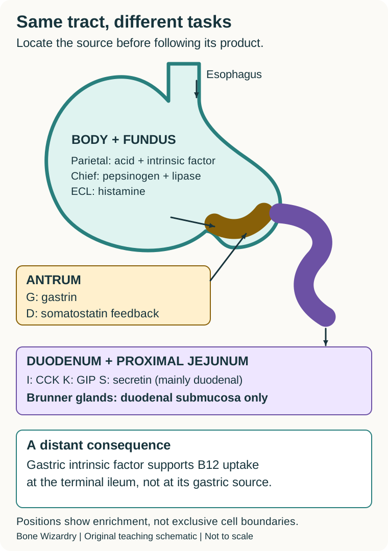
Can damage to the body of the stomach impair a process completed in the ileum? Yes. Parietal cells in body and fundus supply intrinsic factor; the intrinsic-factor and vitamin B12 complex is later absorbed in the terminal ileum. Production and absorption therefore occur in different organs. Acid is the other major parietal product, but acid and intrinsic factor perform different jobs. [3][6]
The oxyntic mucosa of the body and fundus contains acid-producing parietal cells, pepsinogen-producing chief cells, histamine-producing enterochromaffin-like cells, and mucous neck cells. Oxyntic means acid-producing. The antrum has mainly mucus-secreting glands and many gastrin-producing G cells. Its D cells provide somatostatin feedback. D cells also occur in the body and other gastrointestinal sites; their pancreatic counterpart is the islet delta cell. [1][3][9][16]
Trace the regional map from stomach to bowel. The duodenal and proximal jejunal mucosa contains nutrient-responsive I and K cells; S cells are especially associated with the duodenum. These endocrine cells signal to other cells. Goblet cells instead supply mucus to the intestinal lumen, and absorptive enterocytes carry brush-border enzymes and transporters. Brunner glands belong to the duodenal submucosa, not a shared duodenum-and-jejunum compartment. [2][4][5][10][11]
Locate a source region before predicting which downstream function will fail. Regional labels indicate enrichment, not exclusive boundaries. [1][2][3][4][6][9][10][11][15]
The first practical habit is to name both the location and the function. Saying "a mucus cell" is insufficient: gastric surface cells defend the stomach, intestinal goblet cells lubricate the bowel, and duodenal Brunner glands contribute alkaline mucus. Similar products do not establish a common anatomical address. [1][2][3]
Try a regional prediction. In a simplified comparison, body and fundus glands are damaged while the antrum is preserved. Predict acid output, intrinsic-factor availability, and the direction of the gastrin response before checking.
Check the regional prediction
Acid and intrinsic-factor production fall. With less acid reaching the antrum, acid-dependent inhibitory feedback weakens and surviving G cells can increase gastrin release. The extra signal does not replace the missing parietal cells. [6][9]
Apply it elsewhere: a later vitamin B12 deficiency does not, by itself, prove ileal disease. Ask whether the upstream binding partner was lost. Conversely, ileal disease can impair uptake despite intact gastric production. [3][6]
Read the gland from its opening to its base
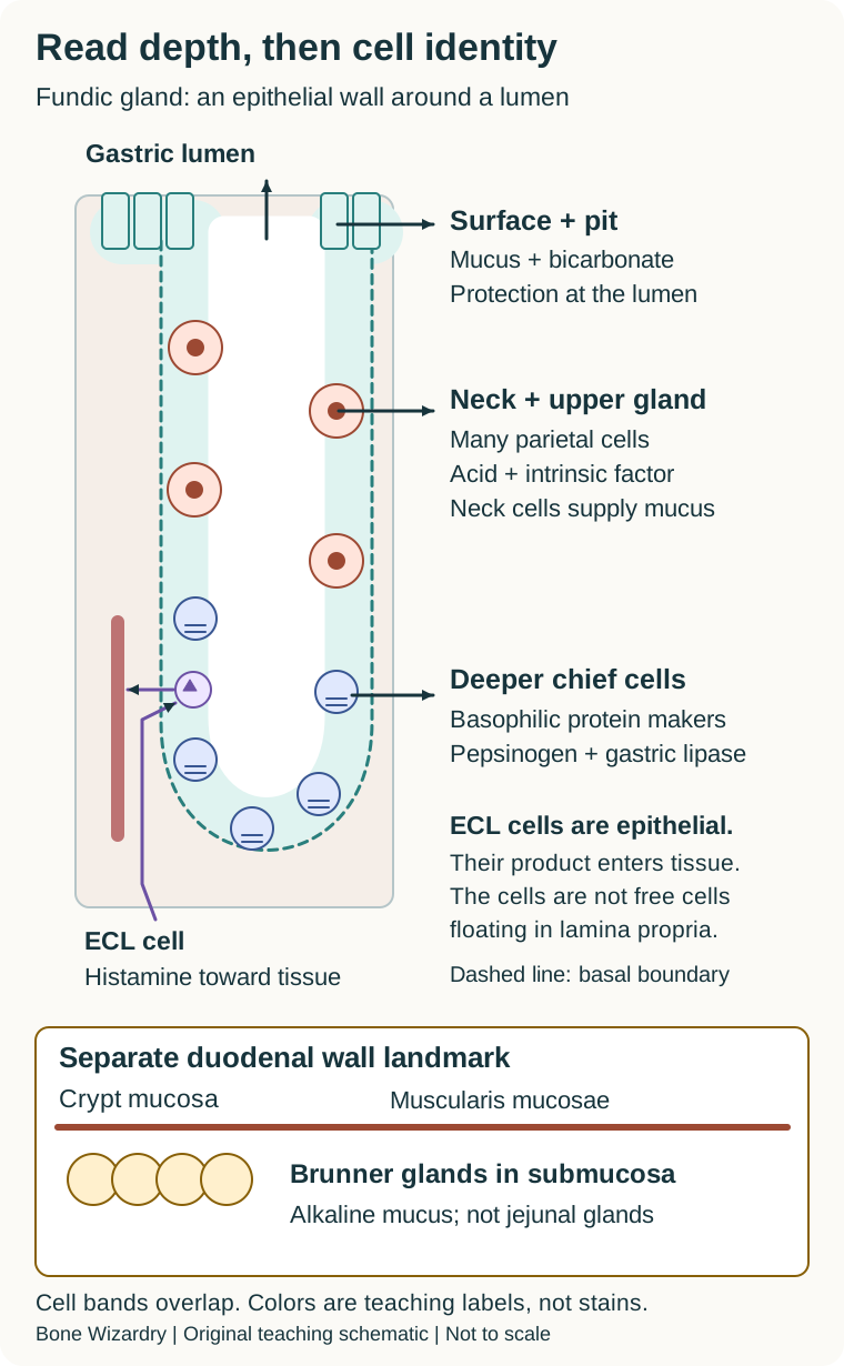
Why should the most exposed cells differ from those deep in the gland? The surface faces acid and proteases. Surface and pit, or foveolar, cells produce the mucus-bicarbonate barrier. Tight epithelial junctions and renewal also protect the mucosa. Normal gastric surface cells are not called goblet cells; true intestinal-type goblet cells in a gastric biopsy suggest intestinal metaplasia. [1][3][6]
The gland-depth drawing separates the narrow gland lumen from its epithelial wall and surrounding lamina propria. The isthmus contains renewal cells. Mucous neck cells sit in the neck among many large parietal cells. Parietal cells are typically eosinophilic on hematoxylin-and-eosin staining, partly reflecting their abundant mitochondria and energy-intensive acid secretion. Their distribution extends beyond one sharply bounded horizontal band. [1][6]
Separate surface protection, glandular secretion, epithelial endocrine position, and duodenal submucosal glands. [1][2][3][6][15]
Chief cells are concentrated deeper in oxyntic glands. Their basophilic basal cytoplasm reflects protein-synthetic machinery. They release pepsinogen, an inactive precursor, and gastric lipase. [15] Acid supports conversion of pepsinogen to pepsin. A normal supply of precursor therefore does not guarantee normal gastric proteolysis when acidity is markedly reduced. This is a secretion-versus-activation distinction, not proof that chief cells stopped working. [1][3]
Gastric endocrine cells occupy the gland epithelium, commonly toward its basal portions, and release products basolaterally toward interstitial fluid and capillaries. ECL histamine acts locally near parietal cells. Do not place normal ECL cells as free cells in the lamina propria just because their secretions enter that compartment. Routine staining also cannot reliably distinguish every endocrine subtype; anatomical context and appropriate immunostains may be required. [3][6][7]
At the duodenal wall, identify the thin muscularis mucosae before naming the glands. Crypt epithelium lies above it; Brunner glands lie below it in submucosa. This landmark distinguishes two glands that can look similarly pale in a small field. [2]
Try the depth test. A specimen contains intestinal villi and clusters of pale glands beneath the muscularis mucosae. Name the secretion and the region.
Check the depth test
These are duodenal Brunner glands producing alkaline mucus. Intestinal endocrine cells are in the mucosal epithelium; they do not form the described submucosal clusters. [2]
Transfer: with pale surface cells and intact deep chief cells in a gastric sample, an injured luminal barrier can still coexist with continued enzyme-precursor production. Protection, secretion, and activation must be evaluated separately. [1][3]
Separate a nerve ending from a circulating hormone
Does vagal stimulation mean acetylcholine acts on every secretory cell? No. Vagal preganglionic fibers signal through enteric neurons. At the final gastric targets, acetylcholine supports parietal acid secretion and chief-cell secretion, whereas gastrin-releasing peptide, or GRP, is the important neural signal to antral G cells. GRP is not gastrin. It stimulates another cell to release gastrin into the circulation. [3][7][8][9]
Follow the acid circuit across two regions. Neural GRP reaches the antral G cell. Gastrin then travels through blood to the oxyntic mucosa. It activates CCK2 receptors, also called CCK-B receptors, especially on ECL cells. ECL histamine crosses a short local distance to parietal H2 receptors. Finally, the parietal cell secretes hydrogen ions into the gland lumen. These are neural, endocrine, paracrine, and exocrine steps in one integrated response. [7][8][9]
Acetylcholine can stimulate parietal M3 receptors directly. Avoid adding a universal "M3 on ECL" rule: experimental ECL preparations have shown important peptide-mediated neural signaling, including PACAP, rather than a uniform direct cholinergic response. Those animal and isolated-cell findings refine the diagram but do not establish identical receptor contributions in every human setting. [7]
Feedback gives the circuit direction. As the antral lumen becomes more acidic, D-cell somatostatin inhibits G-cell gastrin output. Somatostatin also restrains ECL and parietal secretion. When a meal buffers acid, that inhibitory influence is reduced while peptides and distention support gastrin release. Later, increasing acidity restrains the response. Somatostatin is an inhibitor, not a weaker acid stimulant. [7][8][9]
Try a transmitter distinction. In an isolated preparation, a muscarinic antagonist blocks the parietal response to acetylcholine. Would that alone establish that antral cells cannot respond to directly supplied GRP?
Check the transmitter distinction
No. GRP uses a different receptor pathway. A response to directly supplied GRP can remain even when muscarinic signaling is blocked. Whole-organ output is more complex because feedback and several neural targets change together. [8][9]
Transfer: distinguish loss of a neural stimulus from loss of its target cell. A preserved response to an externally supplied hormone can help place a defect upstream of that hormone's receptor.
Interrupt one connection and predict what survives
Why can blocking histamine blunt a response initiated by gastrin? Gastrin's major acid-stimulating route recruits ECL histamine. Gastrin does not need to bind the same receptor as histamine for H2 blockade to interrupt much of its downstream effect. Direct parietal gastrin signaling also exists; the relative contribution depends on the preparation and stimulus. The useful rule is the importance of the relay, not a universal percentage. [7][9]
At the parietal cell, H2 receptors promote cyclic AMP signaling. M3 and CCK2 pathways engage calcium-dependent signaling. These inputs cooperate in activation of the apical hydrogen-potassium ATPase. The pump transfers hydrogen ions into the secretory lumen; receptor stimulation and actual acid transport are distinct events. [7][9][14]
Use the circuit as an interruption exercise. First point to the affected connection on the drawing. Then trace every remaining route to the acid pump. Finally separate the immediate cellular response from the later feedback response. The comparison below stays visible so the exercise can be used without scripting.
Trace which response survives a G-cell, ECL, H2, or pump interruption. Distinguish a measured upstream signal from the final acid output. Direct parietal gastrin signaling is retained in text rather than assigned a universal percentage. [7][8][9][14][16]
Interruption at G-cell stimulation
Less endogenous gastrin reaches the oxyntic mucosa. Directly supplied gastrin or histamine can bypass an upstream deficit if the downstream cells are intact.
Interruption at parietal H2 receptors
The histamine-dependent acid response is reduced. Histamine synthesis is not the blocked process, and a direct muscarinic pathway is not the same receptor.
Interruption at the acid pump
Different receptor inputs converge on an impaired final transport step. More upstream stimulation need not restore acid while the pump remains inhibited.
Stronger somatostatin feedback
Gastrin and histamine signaling are restrained. This is inhibitory regulation, not another route supplying acid.
Predict before checking. Gastrin still causes histamine release in a model, but histamine no longer produces its usual parietal cyclic AMP response. Which connection failed?
Check the interrupted connection
The observation places the problem at the histamine-to-parietal signaling step, consistent with H2 blockade. Failure of the G cell or ECL histamine production would not explain preserved histamine release after directly supplied gastrin. [7][9]
Change one condition: cyclic AMP now rises normally, yet acid transport is reduced after pump inhibition. The target is farther downstream. A measured signal is evidence that one step worked, not evidence that the entire secretory pathway worked. [14]
Ask the intestine what arrived
Do acid, fat, and glucose request the same response? They reach overlapping intestinal territory but create different demands. Acid requires buffering, fat and protein require digestive secretions, and absorbed nutrients require metabolic coordination. Identify the arriving material before retrieving a cell letter. [4][5]
S cell: predominantly duodenal acid sensing
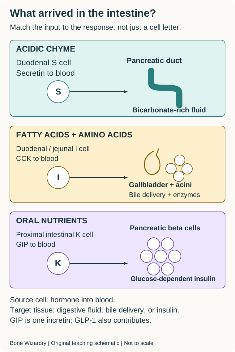
Acidic chyme stimulates secretin release toward blood. Pancreatic ductal cells respond with bicarbonate-rich fluid, helping the duodenal lumen become suitable for pancreatic enzyme activity. The S cell supplies the hormone, not the pancreatic bicarbonate itself. [5][10]
I cell: duodenum and jejunum, nutrient sensing
Fatty acids and amino acids stimulate cholecystokinin, or CCK. It promotes gallbladder contraction and pancreatic enzyme secretion and helps slow gastric emptying. These responses coordinate delivery of bile and enzymes rather than making gastric acid the principal output. [4][5][13]
K cell: greatest abundance in duodenum and proximal jejunum
Ingested nutrients stimulate glucose-dependent insulinotropic polypeptide, or GIP. It supports glucose-dependent insulin secretion from pancreatic beta cells. GIP and GLP-1 contribute to the incretin effect, the greater insulin response to oral than intravenous glucose at matched plasma glucose. That comparison alone does not uniquely identify GIP. [4][11][12]
The intestinal sensor drawing separates three destinations: pancreatic ducts for bicarbonate, acini and gallbladder for digestive delivery, and islet beta cells for insulin. A hormone in blood is not an enzyme secreted into the lumen. A normal hormone rise with a poor target response should direct attention to the target or its delivery pathway rather than automatically blaming the sensor.
Match controlled inputs to source and target. Separate acid buffering, digestive delivery, and metabolic coordination; GIP is not the only incretin. [4][5][10][11][12][13]
G cells respond to peptides, amino acids, gastric distention, and neural GRP mainly in the antrum. Ghrelin provides a different contrast: its principal gastric source is in fundic mucosa and its circulating level commonly rises before meals. G cells also occur in the proximal duodenum; the antrum is their principal gastric location. D-cell somatostatin restrains pancreatic and intestinal secretory pathways as well as gastric signaling. These are additional regional patterns, not extra names for S, I, or K cells. [3][9][16]
Try a matched experiment. An acidic, nutrient-free duodenal infusion raises pancreatic fluid bicarbonate. After neutralization, a fatty nutrient infusion at unchanged pH instead increases gallbladder contraction. Identify the dominant sensor for each observation.
Check the matched experiment
The acid-associated bicarbonate response points to S-cell secretin. The nutrient-associated gallbladder response points to I-cell CCK. Keeping pH separate from nutrient delivery prevents calling every intestinal response a secretin effect. Real meals can activate both systems. [5][10][13]
Transfer: after oral glucose, a selective GIP-receptor intervention can test GIP's contribution more specifically than an oral-versus-intravenous comparison by itself. Residual insulin secretion does not prove the intervention failed; other stimuli remain. [11][12]
Use two measurements to locate a failure
Does a high gastrin concentration prove excess acid? No. Interpret the signal beside its output. A patient with body-predominant atrophic gastritis can have low acid, reduced intrinsic factor, and compensatory hypergastrinemia. Sustained gastrin stimulation can promote ECL-cell hyperplasia even though the remaining parietal-cell output is poor. The messenger and the final product can change in opposite directions. [6]
Compare three situations. In parietal-cell loss, the secretory machinery and intrinsic-factor source are reduced. In acid-pump inhibition, the cells may remain present while hydrogen transport falls; feedback can still increase gastrin. In H2 blockade, one important receptor input is interrupted rather than all secretory machinery being absent. These patterns are not interchangeable, and a drug response is not a histologic diagnosis. [6][7][9][14]
The clinical safety boundary is equally important: elevated gastrin alone does not establish gastrinoma. Gastric acidity, acid-suppressive medicines, tissue findings, and the clinical setting matter. Do not stop prescribed acid suppression just to reproduce a physiology comparison. Testing and any medication changes require clinician-directed planning. The cases here assess mechanisms, not a self-testing or treatment protocol. [6][9][14]
Biopsy interpretation also needs paired information. In suspected atrophic gastritis, separately labeled body and antral/incisural specimens preserve the regional map. Metaplastic body glands can resemble pyloric glands. Appearance alone can therefore mislabel the site; documented sampling and appropriate stains can resolve the distinction. A pale gland is not automatically an antral gland. [6]
Try the signal-output pair. A patient with biopsy-proven corpus atrophy has high fasting gastrin and a nonacidic stomach. Predict the cell population exposed to trophic stimulation, and explain why adding more gastrin would not replace intrinsic factor.
Check the signal-output pair
ECL cells receive gastrin's trophic signal. Intrinsic factor comes from parietal cells, not ECL or G cells, so more gastrin is not a substitute for lost parietal-cell mass. [6]
Carry forward three questions: Where is the source cell? Which side receives its product? Does the observed failure involve the signal, the target cell, or the final secretion? Answering these is more reliable than memorizing a cell letter without context.
Practice localization and response prediction
Commit to an answer before reading feedback. Numerical examples are hypothetical, not results from the cited publications. Each question has one best answer under its stated conditions.
Case 1
Show answer and explanations for case 1
A. Chief cells producing pepsinogen (Why this does not fit)
Chief cells supply the precursor for gastric protein digestion. No. Reduced pepsinogen may accompany body atrophy but does not account for loss of intrinsic-factor-dependent uptake in an intact ileum.
Reasoning steps for option A
What do chief cells supply?
Chief cells supply the precursor for gastric protein digestion.
Does pepsinogen replace the B12 binding partner?
No. Reduced pepsinogen may accompany body atrophy but does not account for loss of intrinsic-factor-dependent uptake in an intact ileum.
B. Parietal cells producing intrinsic factor (Best answer)
Parietal cells in oxyntic mucosa produce intrinsic factor. The preserved ileum still needs intrinsic factor for efficient receptor-mediated vitamin B12 uptake, so loss of its upstream gastric source explains this combination.
Reasoning steps for option B
Which region has lost its glands?
The oxyntic body mucosa is damaged while the antrum is relatively preserved.
Which missing product is needed downstream?
Parietal-cell intrinsic factor supports efficient vitamin B12 uptake in the terminal ileum.
Why does an intact ileum not exclude this mechanism?
An intact uptake site cannot fully compensate for a missing upstream binding partner.
C. G cells producing gastrin (Why this does not fit)
Antral G cells release the acid-regulating hormone gastrin. No. The antrum is relatively preserved and G cells do not provide intrinsic factor.
Reasoning steps for option C
What is the major G-cell product?
Antral G cells release the acid-regulating hormone gastrin.
Does antral preservation suggest the missing B12 source?
No. The antrum is relatively preserved and G cells do not provide intrinsic factor.
D. ECL cells producing histamine (Why this does not fit)
ECL histamine stimulates nearby parietal cells. No. It is a paracrine signal, not the binding partner required for vitamin B12 uptake.
Reasoning steps for option D
What is the ECL contribution to digestion?
ECL histamine stimulates nearby parietal cells.
Can histamine serve as the B12 binding protein?
No. It is a paracrine signal, not the binding partner required for vitamin B12 uptake.
E. D cells producing somatostatin (Why this does not fit)
D-cell somatostatin restrains secretory signaling. Not directly. The body-gland lesion and preserved ileum identify a missing parietal product rather than failure of an inhibitory hormone.
A. Impaired synthesis of the deep-gland enzyme precursor (Why this does not fit)
Deep-gland chief cells synthesize and release pepsinogen. No. Release is unchanged, and changing only the fluid acidity restores digestion without supplying additional precursor.
Reasoning steps for option A
Which cells synthesize pepsinogen?
Deep-gland chief cells synthesize and release pepsinogen.
Would a synthesis deficit fit the measured release?
No. Release is unchanged, and changing only the fluid acidity restores digestion without supplying additional precursor.
B. Impaired release of the antral trophic hormone (Why this does not fit)
Gastrin contributes to stimulation of acid secretion. No. The observations identify the missing activation environment, not a specific upstream cause of the reduced acid.
Reasoning steps for option B
Which antral hormone supports acid output?
Gastrin contributes to stimulation of acid secretion.
Does the fluid rescue identify an antral secretory defect?
No. The observations identify the missing activation environment, not a specific upstream cause of the reduced acid.
C. Impaired secretion of the gastric B12 binding protein (Why this does not fit)
Intrinsic factor supports subsequent ileal uptake of vitamin B12. No. Intrinsic factor is not the enzyme precursor responsible for the observed gastric protein digestion.
Reasoning steps for option C
Which parietal product binds vitamin B12?
Intrinsic factor supports subsequent ileal uptake of vitamin B12.
Would its loss be corrected by acidifying protein substrate?
No. Intrinsic factor is not the enzyme precursor responsible for the observed gastric protein digestion.
D. Impaired activation of a preserved enzyme precursor (Best answer)
Pepsinogen requires an acidic environment for effective conversion to active pepsin. The precursor was available; the impaired gastric environment, rather than lack of precursor secretion, limited protein digestion.
Reasoning steps for option D
Was enzyme-precursor release lost?
No. The measured pepsinogen release is unchanged.
Which experimental change restored digestion?
Acidifying the collected gastric fluid restored the activation environment.
Where does that place the functional defect?
After precursor secretion, at acid-dependent enzyme activation and activity.
E. Impaired secretion of the surface protective gel (Why this does not fit)
Mucus and bicarbonate limit exposure of gastric epithelial cells to luminal acid and enzymes. No. The assay concerns enzyme activation outside the epithelium, and the rescue depends on acidity.
Reasoning steps for option E
What does surface mucus protect?
Mucus and bicarbonate limit exposure of gastric epithelial cells to luminal acid and enzymes.
Does that barrier explain digestion in collected fluid?
No. The assay concerns enzyme activation outside the epithelium, and the rescue depends on acidity.
Takeaway: Normal precursor release can coexist with poor enzyme activity when the activating environment is missing.
A. Reduced acid feedback with gastrin-driven ECL growth (Best answer)
Reduced acidity weakens inhibitory feedback to surviving G cells. Sustained gastrin stimulation promotes ECL growth even when parietal-cell loss prevents a strong acid response.
Reasoning steps for option A
Does the pH indicate an acidic gastric environment?
No. A pH of 6.8 is inconsistent with strong gastric acid output in this assessment.
What does corpus atrophy do to feedback?
Loss of parietal acid output weakens the acid-dependent brake on gastrin release.
Why can ECL cells expand in that setting?
Gastrin retains trophic activity at ECL cells despite poor final acid output.
B. Increased acid output with acid-driven ECL growth (Why this does not fit)
Histamine stimulates acid output from intact parietal cells. No. The nonacidic gastric pH and corpus atrophy indicate poor output rather than an acid-driven growth mechanism.
Reasoning steps for option B
What can ECL histamine ordinarily promote?
Histamine stimulates acid output from intact parietal cells.
Do the pH and histology establish increased output here?
No. The nonacidic gastric pH and corpus atrophy indicate poor output rather than an acid-driven growth mechanism.
C. Reduced acid output with histamine-driven G-cell loss (Why this does not fit)
ECL cells provide the local histamine signal in oxyntic mucosa. No. Loss of the gastrin source would oppose the observed hormone increase; gastrin is the trophic signal implicated in ECL growth.
Reasoning steps for option C
Which cells produce histamine?
ECL cells provide the local histamine signal in oxyntic mucosa.
Would loss of G cells explain high gastrin?
No. Loss of the gastrin source would oppose the observed hormone increase; gastrin is the trophic signal implicated in ECL growth.
D. Increased gastrin clearance with reduced ECL turnover (Why this does not fit)
Secretion and clearance both influence the measured concentration. Not without additional changes, and it does not explain the characteristic feedback response to documented corpus atrophy.
Reasoning steps for option D
What determines a circulating hormone concentration?
Secretion and clearance both influence the measured concentration.
Does increased clearance explain hypergastrinemia?
Not without additional changes, and it does not explain the characteristic feedback response to documented corpus atrophy.
E. Reduced somatostatin feedback with restored parietal mass (Why this does not fit)
Less somatostatin restraint can increase gastrin signaling. No. Persistent corpus atrophy and gastric pH 6.8 show that the increased signal has not restored effective parietal output.
Reasoning steps for option E
What can reduced inhibition do?
Less somatostatin restraint can increase gastrin signaling.
Has the high gastrin restored the missing acid-producing tissue?
No. Persistent corpus atrophy and gastric pH 6.8 show that the increased signal has not restored effective parietal output.
Takeaway: A high upstream signal can stimulate growth without restoring the lost final secretion.
A. Jejunum; bicarbonate-rich mucus (Why this does not fit)
Yes, the jejunum has secretory epithelial cells and goblet cells. No. The characteristic submucosal gland clusters are Brunner glands of the duodenum.
Reasoning steps for option A
Can jejunal epithelium contribute mucus and fluid?
Yes, the jejunum has secretory epithelial cells and goblet cells.
Does it normally contain the described submucosal glands?
No. The characteristic submucosal gland clusters are Brunner glands of the duodenum.
B. Ileum; mucosal antimicrobial peptides (Why this does not fit)
Paneth cells at crypt bases provide antimicrobial products. No. Crypt-base cells remain in the mucosa rather than the described submucosal gland clusters.
Reasoning steps for option B
Where is intestinal antimicrobial secretion associated?
Paneth cells at crypt bases provide antimicrobial products.
Do crypt-base cells match glands below the muscle boundary?
No. Crypt-base cells remain in the mucosa rather than the described submucosal gland clusters.
C. Gastric body; surface protective mucus (Why this does not fit)
Gastric foveolar cells form a protective mucus-bicarbonate barrier. No. Normal gastric mucosa lacks villi, and its surface cells do not form duodenal-type submucosal glands.
Reasoning steps for option C
Where is gastric surface protection supplied?
Gastric foveolar cells form a protective mucus-bicarbonate barrier.
Do villi and submucosal glands fit gastric body mucosa?
No. Normal gastric mucosa lacks villi, and its surface cells do not form duodenal-type submucosal glands.
D. Colon; goblet-cell protective mucus (Why this does not fit)
Goblet cells in the mucosal epithelium provide colonic mucus. No. Villi and characteristic submucosal glands do not match normal colonic architecture.
Reasoning steps for option D
Which cells provide much colonic mucus?
Goblet cells in the mucosal epithelium provide colonic mucus.
Does the specimen have the colonic arrangement?
No. Villi and characteristic submucosal glands do not match normal colonic architecture.
E. Duodenum; bicarbonate-rich mucus (Best answer)
Brunner glands lie in duodenal submucosa and secrete alkaline mucus. Villi establish intestinal mucosa, while glands below the muscularis mucosae identify the duodenal submucosal compartment.
Reasoning steps for option E
What do the villi establish?
The specimen contains small-intestinal mucosa rather than normal gastric or colonic mucosa.
Which compartment contains the pale glands?
They lie below the muscularis mucosae, in the submucosa.
Which regional secretion fits both observations?
Duodenal Brunner glands provide bicarbonate-rich mucus.
Takeaway: Determine the wall compartment before equating two mucus-producing structures.
A. Normal foveolar differentiation within fundic mucosa (Why this does not fit)
Surface mucous, or foveolar, cells normally line gastric pits. No. Calling them normal foveolar cells would miss the metaplastic phenotype and accompanying native-gland loss.
Reasoning steps for option A
What normally lines gastric pits?
Surface mucous, or foveolar, cells normally line gastric pits.
Are true intestinal-type goblet cells simply another name for those cells?
No. Calling them normal foveolar cells would miss the metaplastic phenotype and accompanying native-gland loss.
B. Normal pyloric differentiation within antral mucosa (Why this does not fit)
Antral glands are rich in mucus-producing cells and G cells. No. The separately labeled sites and intestinal-type cells require a metaplastic interpretation, not automatic relabeling as normal antrum.
Reasoning steps for option B
What is expected in normal antral glands?
Antral glands are rich in mucus-producing cells and G cells.
Does mucus-rich appearance override a documented body biopsy site?
No. The separately labeled sites and intestinal-type cells require a metaplastic interpretation, not automatic relabeling as normal antrum.
C. Intestinal metaplasia within atrophic gastric mucosa (Best answer)
They represent an intestinal epithelial phenotype rather than normal gastric foveolar cells. In a confirmed gastric-body sample, goblet-cell metaplasia with reduced native glands supports underlying atrophic gastritis.
Reasoning steps for option C
Is the sampling location uncertain?
No. The body and antral samples were correctly identified separately.
Are intestinal-type goblet cells normal gastric surface cells?
No. Their presence represents intestinal metaplasia in this location.
What does the accompanying native-gland reduction support?
Underlying gastric atrophy rather than normal regional variation.
D. Normal jejunal mucosa within a mislabeled specimen (Why this does not fit)
Intestinal mucosa, including jejunum, normally contains goblet cells. The specimen is confirmed as gastric body and retains adjacent oxyntic glands; normal intestinal identity does not explain that combination.
Reasoning steps for option D
Where are goblet cells normally found?
Intestinal mucosa, including jejunum, normally contains goblet cells.
What conflicts with assuming a mislabeled jejunal sample?
The specimen is confirmed as gastric body and retains adjacent oxyntic glands; normal intestinal identity does not explain that combination.
E. Brunner-gland expansion within duodenal submucosa (Why this does not fit)
Brunner glands are pale mucus-secreting structures in duodenal submucosa. No. The stated site and epithelial metaplasia are different from expansion of duodenal submucosal glands.
Reasoning steps for option E
What can Brunner glands look like?
Brunner glands are pale mucus-secreting structures in duodenal submucosa.
Do they replace gastric glands with goblet epithelium?
No. The stated site and epithelial metaplasia are different from expansion of duodenal submucosal glands.
Takeaway: A normally intestinal cell can signify metaplasia when found at a confirmed gastric site.
A. Lower ECL-cell output; preserved downstream GRP response (Why this does not fit)
ECL cells provide histamine in response to gastrin stimulation. No. Direct gastrin still raises histamine, whereas the blocked observation concerns the antral response to GRP.
Reasoning steps for option A
Which product do ECL cells provide?
ECL cells provide histamine in response to gastrin stimulation.
Would an ECL failure permit the stated histamine response?
No. Direct gastrin still raises histamine, whereas the blocked observation concerns the antral response to GRP.
B. Lower parietal output; preserved downstream histamine response (Why this does not fit)
Parietal cells supply hydrogen ions to the gastric lumen. No. The downstream acid response remains demonstrable, placing the tested interruption upstream.
Reasoning steps for option B
Where is gastric acid actually secreted?
Parietal cells supply hydrogen ions to the gastric lumen.
Would loss of their output fit the preserved gastrin-induced acid?
No. The downstream acid response remains demonstrable, placing the tested interruption upstream.
C. Lower chief-cell output; preserved downstream pepsinogen response (Why this does not fit)
Chief cells release pepsinogen into gastric glands. No. The experiment measures GRP-induced gastrin and its downstream acid pathway, not chief-cell precursor secretion.
Reasoning steps for option C
Which product is secreted by chief cells?
Chief cells release pepsinogen into gastric glands.
Does pepsinogen explain a blocked antral hormone response?
No. The experiment measures GRP-induced gastrin and its downstream acid pathway, not chief-cell precursor secretion.
D. Lower D-cell output; preserved downstream somatostatin response (Why this does not fit)
It reduces inhibitory somatostatin signaling. Not in this controlled setting. Somatostatin exposure is held constant, and the failure is at G-cell stimulation.
Reasoning steps for option D
What does reduced D-cell output usually change?
It reduces inhibitory somatostatin signaling.
Would that explain selective loss of the GRP-stimulated hormone rise?
Not in this controlled setting. Somatostatin exposure is held constant, and the failure is at G-cell stimulation.
E. Lower G-cell output; preserved downstream gastrin response (Best answer)
GRP stimulates G-cell gastrin release. It bypasses the inhibited G-cell stimulation step and demonstrates preserved downstream ECL and parietal function.
Reasoning steps for option E
Which stimulus fails first?
Directly supplied GRP no longer elicits the antral hormone response.
Which supplied substance bypasses the failure?
Gastrin still elicits histamine and acid secretion.
What remains functional downstream?
ECL and parietal responses remain intact, localizing the tested effect to G-cell stimulation.
Takeaway: A preserved downstream response can identify an upstream signaling interruption.
A. Neural delivery before G-cell stimulation (Best answer)
Sensory cues can initiate a vagally mediated cephalic response. The antral cells can still respond when their signal is supplied, so the documented neural injury lies upstream of their secretory response.
Reasoning steps for option A
Which physiological input has been interrupted?
Gastric vagal efferent signaling was injured.
Are the antral cells able to release gastrin?
Yes. They respond to directly supplied GRP.
What location explains failure only with the natural stimulus?
The neural delivery pathway before G-cell stimulation is impaired.
B. Gastrin synthesis within antral G cells (Why this does not fit)
It would prevent gastrin release even when an appropriate stimulus reaches the cells. No. The cells demonstrably release gastrin when GRP is supplied.
Reasoning steps for option B
What would complete loss of G-cell gastrin synthesis cause?
It would prevent gastrin release even when an appropriate stimulus reaches the cells.
Does the direct GRP response fit that defect?
No. The cells demonstrably release gastrin when GRP is supplied.
C. Histamine synthesis within gastric ECL cells (Why this does not fit)
It supplies an important local stimulus for parietal acid output. No. Gastrin release is upstream of the ECL relay, and the history identifies interrupted neural input.
Reasoning steps for option C
Why is histamine relevant downstream?
It supplies an important local stimulus for parietal acid output.
Would an isolated ECL defect explain the missing sham-feeding gastrin rise?
No. Gastrin release is upstream of the ECL relay, and the history identifies interrupted neural input.
D. Acid transport within oxyntic parietal cells (Why this does not fit)
It requires functional parietal signaling and acid-secretory machinery. No. Oxyntic tissue retains its response to histamine.
Reasoning steps for option D
What does an intact histamine response require?
It requires functional parietal signaling and acid-secretory machinery.
Do the tissue findings support failure of that final step?
No. Oxyntic tissue retains its response to histamine.
E. Somatostatin synthesis within antral D cells (Why this does not fit)
It would reduce inhibition of gastrin release. No. The response is diminished, while direct G-cell stimulation remains effective.
Reasoning steps for option E
What would reduced antral somatostatin tend to do?
It would reduce inhibition of gastrin release.
Is that the direction observed with sham feeding?
No. The response is diminished, while direct G-cell stimulation remains effective.
Takeaway: A failed physiological stimulus does not establish failure of a cell that still responds to its supplied agonist.
Antral G cells respond to GRP by releasing gastrin. No. Histamine is supplied downstream, and gastrin-induced ECL histamine release is already preserved.
Reasoning steps for option A
Which cell is normally stimulated through a GRP receptor?
Antral G cells respond to GRP by releasing gastrin.
Can that upstream target explain failure of directly added histamine?
No. Histamine is supplied downstream, and gastrin-induced ECL histamine release is already preserved.
B. ECL CCK2 receptor (Why this does not fit)
CCK2 receptors mediate gastrin stimulation of ECL cells. No. The upstream ECL response is measured and remains present.
Reasoning steps for option B
Which ECL receptor responds to gastrin?
CCK2 receptors mediate gastrin stimulation of ECL cells.
Would their blockade fit continued gastrin-induced histamine release?
No. The upstream ECL response is measured and remains present.
C. Parietal M3 receptor (Why this does not fit)
M3 signaling engages calcium-dependent mechanisms. No. That response is preserved while the histamine-induced cyclic AMP response is lost.
Reasoning steps for option C
Which parietal pathway is stimulated by acetylcholine?
Does the retained acetylcholine-induced calcium rise fit this target?
No. That response is preserved while the histamine-induced cyclic AMP response is lost.
D. Parietal H2 receptor (Best answer)
The H2 receptor mediates the principal histamine-dependent cyclic AMP response. It shows that the muscarinic pathway can still signal, while the observed interruption specifically affects histamine reception downstream of ECL release.
Reasoning steps for option D
Is gastrin able to recruit ECL secretion?
Yes. Histamine release still rises.
Which parietal signaling response is selectively absent?
Histamine no longer increases cyclic AMP.
Which intact response narrows the target?
Preserved acetylcholine-induced calcium signaling favors interruption of H2 rather than M3 or general cell failure.
E. Parietal acid pump (Why this does not fit)
The hydrogen-potassium ATPase transports hydrogen ions at the luminal secretory membrane. Not as its defining direct effect. It acts downstream of the measured receptor signal.
Reasoning steps for option E
Where does the acid pump act?
The hydrogen-potassium ATPase transports hydrogen ions at the luminal secretory membrane.
Would pump inhibition specifically prevent histamine from raising cyclic AMP?
Not as its defining direct effect. It acts downstream of the measured receptor signal.
Takeaway: Preserved transmitter release with selective loss of its target-cell signal places the defect at reception.
A. GRP signaling in G cells (Why this does not fit)
The antrum contains the principal gastrin-producing G-cell population. No. It lacks antral tissue and receives gastrin directly.
Reasoning steps for option A
Which region supplies most G cells?
The antrum contains the principal gastrin-producing G-cell population.
Does this preparation depend on antral GRP stimulation?
No. It lacks antral tissue and receives gastrin directly.
B. CCK2 signaling in ECL cells (Best answer)
Gastrin activates ECL CCK2 signaling and promotes histamine release. It bypasses the inhibited gastrin-to-ECL step and demonstrates that parietal H2 signaling and acid transport remain functional.
Reasoning steps for option B
Which input is supplied without needing G cells?
The experiment supplies gastrin directly to oxyntic tissue.
Which response to that input fails?
ECL histamine release no longer rises.
What does the successful histamine challenge establish?
Parietal signaling and transport are intact, placing the interruption at gastrin-dependent ECL activation.
C. H2 signaling in parietal cells (Why this does not fit)
It promotes parietal cyclic AMP signaling and acid secretion. The stated preserved response argues against H2 as the inhibited target.
Reasoning steps for option C
What does H2 stimulation normally produce?
It promotes parietal cyclic AMP signaling and acid secretion.
Would the supplied histamine response survive effective H2 blockade?
The stated preserved response argues against H2 as the inhibited target.
D. M3 signaling in parietal cells (Why this does not fit)
Acetylcholine activates the muscarinic calcium-dependent pathway. No. The measured failure occurs at the gastrin-stimulated ECL response, upstream of parietal muscarinic signaling.
Reasoning steps for option D
Which transmitter directly uses parietal M3 receptors?
Acetylcholine activates the muscarinic calcium-dependent pathway.
Does blocking that pathway explain absent ECL histamine release after gastrin?
No. The measured failure occurs at the gastrin-stimulated ECL response, upstream of parietal muscarinic signaling.
E. Hydrogen transport at parietal pumps (Why this does not fit)
The parietal acid pump secretes hydrogen ions into the lumen. No. Preserved acid output to histamine demonstrates a usable final transport step.
Reasoning steps for option E
What is the final transport event in acid secretion?
The parietal acid pump secretes hydrogen ions into the lumen.
Would failure there permit normal histamine-induced acid output?
No. Preserved acid output to histamine demonstrates a usable final transport step.
Takeaway: A downstream agonist can bypass failure of the cell that normally supplies it.
Does the recorded calcium response fit loss of that receptor?
No. Calcium still rises, and histamine-induced output is impaired as well.
C. Luminal hydrogen-potassium ATPase (Best answer)
They converge on activation of the luminal hydrogen-potassium ATPase and associated acid-secretory machinery. They show that receptor signaling occurred; the common failure of acid output lies downstream at the final transport stage.
Reasoning steps for option C
Have the two parietal receptors generated their signals?
Yes. Cyclic AMP and calcium responses remain present.
What output fails despite those signals?
Luminal acid secretion is reduced after either stimulus.
Which common downstream target fits?
The hydrogen-potassium ATPase is the final transport target shared by the stimulatory routes.
D. Gastrin-responsive ECL CCK2 receptor (Why this does not fit)
An increase in histamine release reports a downstream response to gastrin. No. Histamine release remains unchanged, placing the shared defect farther downstream.
Reasoning steps for option D
Which secretion reports ECL activation?
An increase in histamine release reports a downstream response to gastrin.
E. Neural GRP receptor on G cells (Why this does not fit)
It would impair GRP-stimulated gastrin release from G cells. No. Both stimuli bypass G-cell gastrin release and still activate their intracellular signals.
Reasoning steps for option E
Which secretion would a GRP-receptor defect first impair?
It would impair GRP-stimulated gastrin release from G cells.
Would that explain failure of directly supplied histamine and acetylcholine?
No. Both stimuli bypass G-cell gastrin release and still activate their intracellular signals.
Takeaway: Normal intracellular signaling does not guarantee a normal final secretory product.
Does that stimulatory oxyntic relay explain the antral result?
No. The measured response is suppressed gastrin release mediated by somatostatin, not histamine-stimulated acid output.
B. Chief-cell activation of luminal pepsinogen (Why this does not fit)
It supports conversion of pepsinogen to pepsin in the lumen. No. The endpoint is hormone release from viable antral tissue rather than luminal protease activation.
Reasoning steps for option B
What does acid do to secreted pepsinogen?
It supports conversion of pepsinogen to pepsin in the lumen.
Would somatostatin-receptor blockade identify that chemical activation?
No. The endpoint is hormone release from viable antral tissue rather than luminal protease activation.
C. D-cell inhibition of neighboring G cells (Best answer)
D cells release somatostatin as a local inhibitory signal. Preventing somatostatin signaling weakens the acid-associated fall in gastrin, while the preserved GRP response shows viable G-cell secretory capacity.
Reasoning steps for option C
How does increased luminal acidity change gastrin?
Gastrin falls when antral pH is reduced.
Which intervention prevents much of that fall?
Blocking somatostatin receptors weakens the response.
Are that sensor and target present in this antral experiment?
No. The isolated antral preparation tests a local gastric feedback mechanism.
E. K-cell stimulation of pancreatic beta cells (Why this does not fit)
K-cell GIP supports glucose-dependent insulin secretion. No. The site, stimulus, measured hormone, and receptor intervention identify a different circuit.
Reasoning steps for option E
What is the principal nutrient-responsive K-cell action?
A. Chief-cell pepsinogen entering the blood-facing compartment (Why this does not fit)
It is secreted toward the gastric gland lumen. No. The source region, release direction, and receptor-specific histamine response do not match pepsinogen.
Reasoning steps for option A
Where is chief-cell pepsinogen normally delivered?
It is secreted toward the gastric gland lumen.
Does it fit an antral vascular signal acting through CCK2?
No. The source region, release direction, and receptor-specific histamine response do not match pepsinogen.
B. Parietal-cell acid entering the shared luminal compartment (Why this does not fit)
It delivers them toward the gastric lumen. No. Luminal fluids are never mixed, and the transfer comes from antral blood-facing effluent.
Reasoning steps for option B
Where does the parietal acid pump deliver hydrogen ions?
It delivers them toward the gastric lumen.
Could mixed acid account for this experiment?
No. Luminal fluids are never mixed, and the transfer comes from antral blood-facing effluent.
C. D-cell somatostatin entering the blood-facing compartment (Why this does not fit)
Somatostatin restrains gastric secretory signaling. No. Its inhibitory action and receptor identity differ from the measured gastrin-responsive stimulation.
Reasoning steps for option C
What direction does somatostatin generally impose on secretion?
Does it account for increased histamine through CCK2?
No. Its inhibitory action and receptor identity differ from the measured gastrin-responsive stimulation.
D. ECL-cell histamine entering the shared luminal compartment (Why this does not fit)
It signals locally toward nearby parietal cells through H2 receptors. No. The experiment identifies stimulation of histamine release by a transferred antral signal, not luminal mixing of histamine.
Reasoning steps for option D
How does gastric ECL histamine normally act?
It signals locally toward nearby parietal cells through H2 receptors.
Does it explain a CCK2-dependent histamine release response?
No. The experiment identifies stimulation of histamine release by a transferred antral signal, not luminal mixing of histamine.
E. G-cell gastrin entering the blood-facing compartment (Best answer)
It is released basolaterally toward interstitial fluid and the circulation. It carries gastrin to CCK2-responsive ECL cells, consistent with a blood-borne hormone rather than a luminal digestive secretion.
Reasoning steps for option E
Which side of the antral tissue supplies the transferred material?
The blood-facing side, not the lumen.
Which receptor identifies the receiving response?
CCK2 dependence identifies a gastrin-responsive ECL pathway.
Which cell-product route joins those observations?
Antral G-cell gastrin reaches body ECL cells through the vascular-facing compartment.
Takeaway: Track both the release direction and the regional address of a cell.
A. I cell signaling to pancreatic acinar cells (Why this does not fit)
Fatty acids and amino acids promote I-cell CCK release and enzyme delivery. No. The solution lacks nutrients, and the measured change is bicarbonate-rich fluid rather than primarily enzyme secretion.
Reasoning steps for option A
Which nutrients promote CCK release?
Fatty acids and amino acids promote I-cell CCK release and enzyme delivery.
Do those stimuli or the acinar endpoint dominate here?
No. The solution lacks nutrients, and the measured change is bicarbonate-rich fluid rather than primarily enzyme secretion.
B. S cell signaling to pancreatic duct cells (Best answer)
Duodenal S-cell secretin is the principal acid-associated stimulus for pancreatic bicarbonate-rich fluid. They isolate acidity from distention and nutrient delivery, favoring a secretin-to-duct response.
Reasoning steps for option B
Which variable differs between the infusions?
Acidity changes while volume, distention, and absence of nutrients are controlled.
Which pancreatic compartment supplies bicarbonate-rich fluid?
Ductal cells supply that fluid.
Which intestinal hormone connects acid to that compartment?
S-cell secretin supplies the endocrine signal.
C. K cell signaling to pancreatic beta cells (Why this does not fit)
Pancreatic beta cells respond with enhanced glucose-dependent insulin secretion. No. An insulinotropic target is different from the ductal fluid endpoint in this acid-controlled comparison.
Reasoning steps for option C
Which target responds to K-cell GIP?
Pancreatic beta cells respond with enhanced glucose-dependent insulin secretion.
Does that explain the bicarbonate-rich exocrine output?
No. An insulinotropic target is different from the ductal fluid endpoint in this acid-controlled comparison.
D. G cell signaling to gastric ECL cells (Why this does not fit)
Gastrin promotes histamine release and downstream gastric acid secretion. No. The experiment measures an intestinal acid response in pancreatic fluid rather than increased gastric acid output.
Reasoning steps for option D
What does gastrin recruit from ECL cells?
Gastrin promotes histamine release and downstream gastric acid secretion.
Does that match the infusion site and measured fluid?
No. The experiment measures an intestinal acid response in pancreatic fluid rather than increased gastric acid output.
E. D cell signaling to gastric parietal cells (Why this does not fit)
It inhibits acid-related secretory signaling. No. Inhibition of gastric parietal output is not the principal sensor-target route identified by this experiment.
Reasoning steps for option E
What does somatostatin usually do to gastric output?
It inhibits acid-related secretory signaling.
Does that directly explain increased pancreatic ductal bicarbonate?
No. Inhibition of gastric parietal output is not the principal sensor-target route identified by this experiment.
Takeaway: Acid sensing by S cells and bicarbonate secretion by pancreatic ducts are different steps.
A. Pancreatic bicarbonate-secreting ductal epithelium (Best answer)
Pancreatic ductal epithelium supplies the bicarbonate-rich fluid response. It bypasses the intestinal hormone source, so the normal hormone rise, patent duct, and preserved acinar secretion support a selective ductal response defect.
Reasoning steps for option A
Did the duodenal sensor release its hormone?
Yes. Secretin rises normally after acid.
Can bypassing the sensor restore output?
No. Directly supplied secretin also fails.
Which additional observations narrow the site?
Preserved acinar secretion and a patent collecting duct favor dysfunction of bicarbonate-secreting ductal epithelium.
B. Duodenal secretin-producing S-cell population (Why this does not fit)
It would impair the secretin rise after duodenal acid exposure. No. Secretin rises normally, and supplying additional hormone does not rescue output.
Reasoning steps for option B
What would loss of S-cell secretion first impair?
It would impair the secretin rise after duodenal acid exposure.
Is that source response impaired in this preparation?
No. Secretin rises normally, and supplying additional hormone does not rescue output.
C. Duodenal CCK-producing I-cell population (Why this does not fit)
I-cell CCK coordinates pancreatic enzyme delivery and gallbladder contraction after nutrients. No. The experiment directly tests a ductal response and shows preserved acinar secretion.
Reasoning steps for option C
Which secretory demand is associated with I cells?
I-cell CCK coordinates pancreatic enzyme delivery and gallbladder contraction after nutrients.
Does that explain failure of a supplied-secretin bicarbonate response?
No. The experiment directly tests a ductal response and shows preserved acinar secretion.
D. Pancreatic enzyme-secreting acinar epithelium (Why this does not fit)
Acinar cells supply enzyme-rich secretion. It argues against the acinar compartment as the primary source of the selective bicarbonate deficit.
Reasoning steps for option D
Which pancreatic cells provide digestive enzymes?
Acinar cells supply enzyme-rich secretion.
What does preserved acinar enzyme release tell you?
It argues against the acinar compartment as the primary source of the selective bicarbonate deficit.
E. Gastric histamine-producing ECL-cell population (Why this does not fit)
ECL cells supply histamine to the parietal acid circuit. No. The measured intestinal signal is intact and the failed endpoint is pancreatic bicarbonate secretion.
Reasoning steps for option E
Which gastric signal is supplied by ECL cells?
ECL cells supply histamine to the parietal acid circuit.
Can that explain failure with directly supplied secretin?
No. The measured intestinal signal is intact and the failed endpoint is pancreatic bicarbonate secretion.
Takeaway: A normal hormone concentration with failed agonist rescue can localize dysfunction to the responding tissue.
A. S cells in duodenal mucosa (Why this does not fit)
S cells release secretin, which promotes bicarbonate-rich pancreatic fluid. No. The measured responses and receptor dependence favor CCK rather than an acid-triggered secretin effect.
Reasoning steps for option A
What is the main acid-responsive S-cell product?
S cells release secretin, which promotes bicarbonate-rich pancreatic fluid.
Does the neutral-pH nutrient and CCK1-dependent pattern fit that signal?
No. The measured responses and receptor dependence favor CCK rather than an acid-triggered secretin effect.
B. K cells in proximal jejunal mucosa (Why this does not fit)
K cells release GIP to support nutrient-associated insulin secretion. No. GIP does not provide the named receptor signal coordinating gallbladder and pancreatic enzyme responses.
Reasoning steps for option B
What is the principal K-cell product?
K cells release GIP to support nutrient-associated insulin secretion.
Would that explain both CCK1-dependent exocrine responses?
No. GIP does not provide the named receptor signal coordinating gallbladder and pancreatic enzyme responses.
C. G cells in pyloric antral mucosa (Why this does not fit)
It supports gastric acid secretion through the ECL-parietal circuit. No. The source, paired digestive responses, and CCK1 dependence point to intestinal I-cell signaling.
Reasoning steps for option C
What does antral gastrin primarily regulate?
It supports gastric acid secretion through the ECL-parietal circuit.
Is gastrin the best explanation for the intestinal nutrient response?
No. The source, paired digestive responses, and CCK1 dependence point to intestinal I-cell signaling.
D. I cells in duodenal and jejunal mucosa (Best answer)
I cells in proximal small-intestinal mucosa release CCK in response to fatty acids and amino acids. CCK links those nutrients to both gallbladder contraction and pancreatic enzyme secretion; the receptor intervention supports that common signal.
Reasoning steps for option D
What has been separated from nutrient delivery?
Luminal acidity is controlled near neutral.
Which two outputs rise together?
Gallbladder contraction and pancreatic enzyme output increase.
Which endocrine source fits the CCK1 dependence?
Proximal intestinal I-cell CCK links the stimulus and both outputs.
E. D cells in gastric gland mucosa (Why this does not fit)
It restrains gastric and other digestive secretions. No. The observed direction and receptor-specific attenuation identify a stimulatory CCK signal.
Reasoning steps for option E
What is the usual secretory effect of D-cell somatostatin?
It restrains gastric and other digestive secretions.
Does that fit two increased CCK1-dependent responses?
No. The observed direction and receptor-specific attenuation identify a stimulatory CCK signal.
Takeaway: The nutrient sensor and the affected target receptor should agree with both measured outputs.
A. Intestinal CCK release falls; pancreatic enzyme secretion falls (Why this does not fit)
No. Intestinal I cells, rather than gallbladder cells, provide the meal-associated hormone. No. That interpretation mistakes one target organ for the hormone source and ignores the intact pancreas.
Reasoning steps for option A
Is the gallbladder required to synthesize intestinal CCK?
No. Intestinal I cells, rather than gallbladder cells, provide the meal-associated hormone.
Does gallbladder absence justify loss of both responses?
No. That interpretation mistakes one target organ for the hormone source and ignores the intact pancreas.
I cells in proximal intestinal mucosa release CCK in response to the nutrients. No. The source cells and pancreatic target remain, so the meal can still raise CCK and support enzyme secretion.
Reasoning steps for option B
Which organ was absent before the meal?
The gallbladder, a target of CCK, was removed.
Are the source cells still exposed to a stimulus?
Yes. Intact proximal intestinal I cells receive fatty acids and amino acids.
Which separate digestive target can still respond?
The intact pancreas can increase enzyme secretion in the nutrient-associated CCK response.
C. Intestinal CCK release rises; pancreatic enzyme secretion falls (Why this does not fit)
Yes. Intestinal CCK secretion can persist after gallbladder removal. The pancreas and intestinal nutrient stimulus are intact; loss of the gallbladder does not by itself eliminate the pancreatic enzyme response.
Reasoning steps for option C
Can a source remain active when one target is absent?
Yes. Intestinal CCK secretion can persist after gallbladder removal.
Why is the predicted pancreatic fall unsupported here?
The pancreas and intestinal nutrient stimulus are intact; loss of the gallbladder does not by itself eliminate the pancreatic enzyme response.
D. Intestinal CCK release falls; pancreatic enzyme secretion rises (Why this does not fit)
Neural and other inputs also influence pancreatic secretion. The source I cells remain exposed to nutrients, and the case asks about that preserved meal-associated CCK response.
Reasoning steps for option D
Can other stimuli contribute to pancreatic secretion?
Neural and other inputs also influence pancreatic secretion.
Why is a fall in the CCK signal not the expected result of target removal?
The source I cells remain exposed to nutrients, and the case asks about that preserved meal-associated CCK response.
Takeaway: Removing a target organ does not automatically eliminate the hormone made elsewhere.
A. Oral delivery raises the systemic glucose stimulus more strongly (Why this does not fit)
Yes. Higher glucose can provide a stronger direct beta-cell stimulus. No. The intravenous infusion is adjusted to reproduce the oral plasma glucose profile.
Reasoning steps for option A
Can a larger plasma glucose rise increase insulin secretion?
Yes. Higher glucose can provide a stronger direct beta-cell stimulus.
Is a larger systemic rise present in this comparison?
No. The intravenous infusion is adjusted to reproduce the oral plasma glucose profile.
B. Intravenous delivery increases insulin clearance more strongly (Why this does not fit)
Yes. Concentration depends on both secretion and clearance. No. The case specifies greater secretion after oral delivery and similar clearance between days.
Reasoning steps for option B
Could clearance change a circulating insulin concentration?
Yes. Concentration depends on both secretion and clearance.
Does clearance account for the stated secretion difference?
No. The case specifies greater secretion after oral delivery and similar clearance between days.
C. Gastric acid directly activates insulin within the circulation (Why this does not fit)
It supports activation of pepsinogen in the gastric lumen. No. Luminal protease activation is distinct from endocrine stimulation of beta-cell insulin secretion.
Reasoning steps for option C
What does gastric acid activate during digestion?
It supports activation of pepsinogen in the gastric lumen.
Does that chemistry activate circulating insulin?
No. Luminal protease activation is distinct from endocrine stimulation of beta-cell insulin secretion.
D. Gut-derived signals augment glucose-stimulated insulin secretion (Best answer)
It limits the difference in the direct glucose stimulus reaching pancreatic beta cells. It supports an incretin contribution from gut-derived signals such as GIP and GLP-1, without identifying one peptide as the sole cause.
Reasoning steps for option D
Has systemic glucose exposure been matched?
Yes. The intravenous challenge reproduces the oral plasma glucose profile.
Can clearance explain the greater response?
No. Insulin secretion is compared and clearance is similar.
What additional route remains plausible?
Oral nutrients recruit gut-derived incretin signals, including GIP and GLP-1, that augment beta-cell secretion.
E. Intestinal absorption provides the only trigger for beta cells (Why this does not fit)
Yes. They respond directly to increased plasma glucose. No. The comparison demonstrates an added gut contribution rather than a requirement that glucose first cross intestinal mucosa.
Reasoning steps for option E
Can beta cells respond to intravenously supplied glucose?
Yes. They respond directly to increased plasma glucose.
Does a smaller intravenous response show an absent direct pathway?
No. The comparison demonstrates an added gut contribution rather than a requirement that glucose first cross intestinal mucosa.
Takeaway: Matched glucose reveals a gut contribution but does not by itself isolate a single incretin.
A. Antral G cells; other insulin stimuli remain (Why this does not fit)
Antral G cells supply gastrin to the gastric acid-regulating circuit. No. The blocked signal is GIP, which is supplied predominantly by proximal intestinal K cells.
Reasoning steps for option A
What is the principal G-cell hormone?
Antral G cells supply gastrin to the gastric acid-regulating circuit.
Are they the anatomical source of the selectively blocked incretin?
No. The blocked signal is GIP, which is supplied predominantly by proximal intestinal K cells.
B. Duodenal S cells; insulin production has stopped (Why this does not fit)
They release secretin to support pancreatic bicarbonate-rich fluid. No. Insulin secretion persists, and the selectively blocked hormone is not secretin.
Reasoning steps for option B
What do S cells release after acid exposure?
They release secretin to support pancreatic bicarbonate-rich fluid.
Does the residual insulin response support this interpretation?
No. Insulin secretion persists, and the selectively blocked hormone is not secretin.
C. Proximal K cells; other insulin inputs remain (Best answer)
K cells are most abundant in the duodenum and proximal jejunum. Glucose and other signals, including intact GLP-1 signaling, can still stimulate beta cells; residual secretion does not negate a GIP contribution.
Reasoning steps for option C
Which receptor intervention identifies the blocked peptide?
The antagonist selectively targets the GIP receptor.
Where is that peptide principally produced?
K cells enriched in duodenum and proximal jejunum supply GIP.
What explains a partial rather than complete insulin reduction?
Direct glucose stimulation and intact GLP-1 signaling remain.
D. Oxyntic ECL cells; insulin production has stopped (Why this does not fit)
Histamine acts locally on parietal H2 receptors. No. The source and receptor pathway are different, and insulin secretion has not stopped.
Reasoning steps for option D
What is the principal gastric ECL product?
Histamine acts locally on parietal H2 receptors.
Can ECL loss identify the blocked GIP signal?
No. The source and receptor pathway are different, and insulin secretion has not stopped.
E. Gastric D cells; the direct glucose response is absent (Why this does not fit)
D cells provide somatostatin rather than GIP. No. The experiment controls plasma glucose and leaves other insulin-stimulating pathways available.
Reasoning steps for option E
What inhibitory hormone is supplied by gastric D cells?
D cells provide somatostatin rather than GIP.
Does GIP-selective blockade abolish glucose sensing by itself?
No. The experiment controls plasma glucose and leaves other insulin-stimulating pathways available.
Takeaway: A partial response to selective blockade is compatible with several parallel physiological inputs.
A. GIP synthesis in proximal intestinal K cells (Why this does not fit)
It would reduce the supply of GIP released by nutrient-exposed intestinal tissue. No. GIP release is normal, placing the measured failure downstream of the source.
Reasoning steps for option A
What would a K-cell synthesis defect change?
It would reduce the supply of GIP released by nutrient-exposed intestinal tissue.
Is that source deficit observed here?
No. GIP release is normal, placing the measured failure downstream of the source.
B. Excessive GIP degradation in vascular perfusate (Why this does not fit)
Excess degradation could lower the active hormone available to islet receptors. No. The case states that degradation is normal and the failed response is selective at the receiving islets.
Reasoning steps for option B
Could rapid peptide degradation reduce target exposure?
Excess degradation could lower the active hormone available to islet receptors.
Does that fit the controlled observation?
No. The case states that degradation is normal and the failed response is selective at the receiving islets.
C. Global cyclic AMP generation in pancreatic beta cells (Why this does not fit)
It would impair multiple receptor pathways that use the same intracellular signaling machinery. The GLP-1-receptor agonist produces a normal response, so the entire cyclic AMP apparatus is not demonstrably lost.
Reasoning steps for option C
What would a global cyclic AMP defect affect?
It would impair multiple receptor pathways that use the same intracellular signaling machinery.
What argues against that broad defect?
The GLP-1-receptor agonist produces a normal response, so the entire cyclic AMP apparatus is not demonstrably lost.
D. Insulin synthesis in pancreatic beta cells (Why this does not fit)
It would limit secretion across different stimulatory pathways. No. Those preserved responses show that the cells can supply insulin under other stimulation.
Reasoning steps for option D
What would complete insulin-synthesis failure limit?
It would limit secretion across different stimulatory pathways.
Can it explain normal responses to glucose and GLP-1?
No. Those preserved responses show that the cells can supply insulin under other stimulation.
E. GIP-receptor signaling in pancreatic beta cells (Best answer)
The beta-cell GIP receptor normally couples the gut hormone to intracellular signaling that enhances insulin secretion. Normal glucose and GLP-1 responses support viable insulin-secretory and cyclic AMP machinery, while failure is selective for GIP reception.
Reasoning steps for option E
Is the intestinal hormone source functioning?
Yes. Nutrients elicit normal GIP release.
Is the beta cell generally unable to signal or secrete?
No. Glucose and GLP-1-receptor stimulation produce normal responses.
Which selective reception step remains implicated?
GIP-receptor signaling at the beta cell is the best-supported defect.
Takeaway: Normal hormone delivery plus preserved parallel responses can isolate a target-receptor pathway.
A. Foveolar-cell maintenance of surface defense (Best answer)
Foveolar cells support the mucus-bicarbonate barrier at the luminal surface. The injury can be explained by diminished protection against an unchanged acid load, without postulating parietal hypersecretion or failed chief-cell secretion.
Reasoning steps for option A
Has the luminal acid challenge increased?
No. Acid output is unchanged.
Which protective feature has decreased?
Surface mucus and bicarbonate retention are reduced.
Which cells occupy the affected location?
Foveolar cells line the surface and pits and support that protective barrier.
B. Chief-cell supply of the luminal enzyme precursor (Why this does not fit)
Pepsinogen release reflects a major chief-cell secretory function. No. It remains normal and does not explain the selective loss of surface protection.
Reasoning steps for option B
Which output reports chief-cell function in this case?
Pepsinogen release reflects a major chief-cell secretory function.
Is that output reduced in the injury model?
No. It remains normal and does not explain the selective loss of surface protection.
C. Parietal-cell control of the luminal acid load (Why this does not fit)
Parietal cells provide the acid that can damage inadequately protected epithelium. No. Luminal acid output is unchanged; the defective component is protection against it.
Reasoning steps for option C
Which cells provide gastric acid?
Parietal cells provide the acid that can damage inadequately protected epithelium.
Does the case show increased parietal output?
No. Luminal acid output is unchanged; the defective component is protection against it.
D. G-cell stimulation of the circulating acid signal (Why this does not fit)
G cells release gastrin, an upstream stimulant of the acid circuit. Not directly. The case documents reduced surface protection without an increased final acid load.
Reasoning steps for option D
What is the G-cell contribution to gastric secretion?
G cells release gastrin, an upstream stimulant of the acid circuit.
Does increased gastrin explain the measured barrier deficit?
Not directly. The case documents reduced surface protection without an increased final acid load.
E. ECL-cell support of the local histamine signal (Why this does not fit)
ECL histamine stimulates parietal acid secretion. No. The direct abnormality is the surface mucus-bicarbonate barrier, not deficient local acid stimulation.
Reasoning steps for option E
Which local signal comes from ECL cells?
ECL histamine stimulates parietal acid secretion.
Would reduced histamine explain greater penetration at unchanged acid output?
No. The direct abnormality is the surface mucus-bicarbonate barrier, not deficient local acid stimulation.
Takeaway: An unchanged secretion can become injurious when protection against it fails.
A. Ileal uptake of intrinsic-factor-bound B12 (Best answer)
Its efficient receptor-mediated uptake occurs in the terminal ileum. The upstream binding partner is supplied, but the resection removes the downstream uptake site.
Reasoning steps for option A
Is the gastric binding partner available?
Yes. Intrinsic-factor secretion is normal.
Which anatomical site was removed?
A substantial portion of terminal ileum was resected.
What step fails despite intact upstream secretion?
Receptor-mediated uptake of the intrinsic-factor and B12 complex is impaired.
B. Production of intrinsic factor in gastric parietal cells (Why this does not fit)
Parietal cells provide the binding protein needed for efficient ileal B12 uptake. No. Both preserved oxyntic glands and measured normal secretion argue against loss of that source.
Reasoning steps for option B
Which gastric cells produce intrinsic factor?
Parietal cells provide the binding protein needed for efficient ileal B12 uptake.
Is their production reduced in this patient?
No. Both preserved oxyntic glands and measured normal secretion argue against loss of that source.
C. Production of pepsinogen in gastric chief cells (Why this does not fit)
It provides the precursor for gastric protein digestion. No. The documented loss involves the distal uptake site while gastric secretory function is preserved.
Reasoning steps for option C
What does chief-cell pepsinogen contribute?
It provides the precursor for gastric protein digestion.
Does that locate the defect after terminal ileal resection?
No. The documented loss involves the distal uptake site while gastric secretory function is preserved.
D. Release of gastrin from gastric antral cells (Why this does not fit)
G cells release an upstream regulator of gastric acid secretion. No. The supplied findings specifically identify a downstream absorption problem rather than a missing gastric hormone.
Reasoning steps for option D
What is the principal antral G-cell function?
G cells release an upstream regulator of gastric acid secretion.
Does loss of antral stimulation explain normal gastric secretion and ileal resection?
No. The supplied findings specifically identify a downstream absorption problem rather than a missing gastric hormone.
E. Release of bicarbonate from pancreatic duct cells (Why this does not fit)
It helps neutralize acidic chyme in the duodenum. No. The relevant anatomical loss is terminal ileum, and gastric intrinsic-factor production is demonstrated to be normal.
Reasoning steps for option E
What does pancreatic bicarbonate do?
It helps neutralize acidic chyme in the duodenum.
Would reduced bicarbonate identify the removed B12 uptake site?
No. The relevant anatomical loss is terminal ileum, and gastric intrinsic-factor production is demonstrated to be normal.
Takeaway: The site that supplies a binding protein and the site that absorbs its cargo can fail independently.
A. Reduced hydrogen transport by parietal cells (Why this does not fit)
Parietal cells use an apical hydrogen-potassium ATPase to supply hydrogen ions. No. Acid output is preserved, the assay pH is standardized, and supplying precursor rather than more acid restores digestion.
Reasoning steps for option A
Which cells generate gastric acid?
Parietal cells use an apical hydrogen-potassium ATPase to supply hydrogen ions.
Does acid failure explain this assay?
No. Acid output is preserved, the assay pH is standardized, and supplying precursor rather than more acid restores digestion.
B. Reduced protective mucus from surface cells (Why this does not fit)
It helps maintain the barrier between luminal acid and the gastric epithelium. No. The measured rescue occurs in collected fluid and identifies enzyme-precursor supply, not epithelial protection.
Reasoning steps for option B
What does surface mucus protect?
It helps maintain the barrier between luminal acid and the gastric epithelium.
Would replacing pepsinogen restore a missing surface barrier?
No. The measured rescue occurs in collected fluid and identifies enzyme-precursor supply, not epithelial protection.
C. Reduced precursor supply from chief cells (Best answer)
Chief cells synthesize and secrete the protein-digesting enzyme precursor. The activating environment is already supplied; restoration by added pepsinogen identifies deficient precursor availability.
Reasoning steps for option C
Which population is selectively reduced?
The deep basophilic chief-cell population is diminished.
Has insufficient acidity been controlled?
Yes. Native acid output is preserved and the assay uses standardized acidic pH.
What does the pepsinogen rescue establish?
Insufficient precursor supply, rather than failed acid activation, limits digestion.
D. Reduced circulating gastrin from G cells (Why this does not fit)
It supports gastric acid output through ECL and parietal signaling. No. The regional cell loss and direct precursor rescue better identify a chief-cell secretory deficit.
Reasoning steps for option D
What does gastrin principally stimulate?
It supports gastric acid output through ECL and parietal signaling.
Does a missing gastric acid stimulus fit the preserved acid output?
No. The regional cell loss and direct precursor rescue better identify a chief-cell secretory deficit.
E. Reduced local histamine from ECL cells (Why this does not fit)
ECL histamine stimulates the parietal acid response. No. Gastric acid output is preserved, and restoring pepsinogen specifically corrects the digestion deficit.
Reasoning steps for option E
What is the principal local ECL function?
ECL histamine stimulates the parietal acid response.
Does loss of that signal explain the fixed-pH rescue?
No. Gastric acid output is preserved, and restoring pepsinogen specifically corrects the digestion deficit.
Takeaway: Control the activating environment before deciding whether an enzyme deficit reflects secretion or activation.
A. Combine all gastric fragments and increase their number (Why this does not fit)
Additional tissue can reveal abnormalities missed by a smaller sample. No. Increased volume alone does not recover the regional information lost when sites are pooled.
Reasoning steps for option A
Can more tissue improve sampling yield?
Additional tissue can reveal abnormalities missed by a smaller sample.
Does combining more fragments restore their anatomical origin?
No. Increased volume alone does not recover the regional information lost when sites are pooled.
B. Label each fragment according to its dominant gland appearance (Why this does not fit)
Oxyntic and antral glands usually have different appearances. Metaplastic corpus glands can resemble pyloric glands, so morphology cannot substitute for the documented sampling site.
Reasoning steps for option B
Can gland architecture suggest a normal region?
Oxyntic and antral glands usually have different appearances.
Why is appearance-based labeling unreliable in this case?
Metaplastic corpus glands can resemble pyloric glands, so morphology cannot substitute for the documented sampling site.
C. Use serum gastrin to assign every fragment to a region (Why this does not fit)
Serum gastrin can contribute to interpretation when paired with acidity and clinical context. No. A systemic hormone measurement cannot assign anatomical labels to pooled biopsy fragments.
Reasoning steps for option C
Can gastrin help interpret gastric physiology?
Serum gastrin can contribute to interpretation when paired with acidity and clinical context.
Can that concentration identify the origin of each tissue fragment?
No. A systemic hormone measurement cannot assign anatomical labels to pooled biopsy fragments.
D. Sample the antrum alone to represent the entire stomach (Why this does not fit)
An antral biopsy can establish abnormalities at that sampled site. No. Sampling only the antrum risks missing the very regional pattern that needs clarification.
Reasoning steps for option D
Can antral tissue describe local pathology?
An antral biopsy can establish abnormalities at that sampled site.
Would it characterize possible body-predominant gland loss?
No. Sampling only the antrum risks missing the very regional pattern that needs clarification.
E. Separately label body and antral/incisural specimens (Best answer)
The distribution of gland loss helps distinguish patterns of atrophic gastritis. It preserves site identity even when metaplastic body glands resemble pyloric glands, avoiding the ambiguity created by a combined jar.
Reasoning steps for option E
Which essential information is missing?
The original fragments cannot be assigned to body or antral/incisural sites.
Why can morphology fail to reconstruct the location?
Metaplastic corpus glands can resemble normal pyloric-type glands.
Which sampling change preserves the distinction?
Collect and separately label body and antral/incisural specimens.
Takeaway: Preserve anatomical labels when disease can make one region resemble another.
A. Antral G cells; duodenal and jejunal I cells (Why this does not fit)
G cells supply gastrin rather than the principal premeal ghrelin signal. The CCK location is appropriate, but the first source confuses the antral gastrin circuit with fundic ghrelin production.
Reasoning steps for option A
Which hormone is supplied by antral G cells?
G cells supply gastrin rather than the principal premeal ghrelin signal.
Which half of this pairing is inconsistent?
The CCK location is appropriate, but the first source confuses the antral gastrin circuit with fundic ghrelin production.
B. Fundic endocrine cells; duodenal S cells (Why this does not fit)
Duodenal S cells release secretin after acid exposure. No. The second pattern supports CCK, linked to fatty nutrients and gallbladder contraction at controlled pH.
Reasoning steps for option B
What is the principal S-cell response?
Duodenal S cells release secretin after acid exposure.
Does that identify the second hormone in this experiment?
No. The second pattern supports CCK, linked to fatty nutrients and gallbladder contraction at controlled pH.
C. Oxyntic ECL cells; proximal intestinal K cells (Why this does not fit)
ECL cells supply histamine, while K cells supply GIP. No. The patterns support ghrelin and CCK, not the local gastric histamine signal and intestinal incretin GIP.
Reasoning steps for option C
Which products identify these populations?
ECL cells supply histamine, while K cells supply GIP.
Do those products fit the two measured peptides?
No. The patterns support ghrelin and CCK, not the local gastric histamine signal and intestinal incretin GIP.
D. Fundic endocrine cells; proximal intestinal I cells (Best answer)
Ghrelin-producing endocrine cells are enriched in the fundic stomach. Proximal intestinal I cells release CCK, separating the premeal gastric signal from the meal-associated intestinal digestive signal.
Reasoning steps for option D
Which signal is primarily gastric and rises before a meal?
The inferred ghrelin signal is associated with fundic endocrine cells.
Which digestive response identifies the second intestinal system?
Fatty nutrients and gallbladder contraction fit CCK signaling.
Where is that CCK source enriched?
I cells in duodenal and jejunal mucosa provide the intestinal signal.
E. Antral D cells; pancreatic islet beta cells (Why this does not fit)
D cells supply somatostatin, while beta cells supply insulin. No. The peptides and their meal-associated patterns identify gastric ghrelin and intestinal CCK sources.
Reasoning steps for option E
Which products identify these populations?
D cells supply somatostatin, while beta cells supply insulin.
Does their location or product match the measured pair?
No. The peptides and their meal-associated patterns identify gastric ghrelin and intestinal CCK sources.
Takeaway: Different meal-related signals can originate in different regions and serve different functions.
A. Progressive parietal-cell destruction with loss of intrinsic factor (Why this does not fit)
Parietal-cell loss can lower acid and increase feedback-driven gastrin release. Parietal-cell mass and intrinsic-factor secretion are preserved, favoring functional pump inhibition rather than destructive gland loss.
Reasoning steps for option A
What can corpus atrophy do to gastrin?
Parietal-cell loss can lower acid and increase feedback-driven gastrin release.
Which supplied findings distinguish this patient?
Parietal-cell mass and intrinsic-factor secretion are preserved, favoring functional pump inhibition rather than destructive gland loss.
B. Reduced acid transport with compensatory gastrin release (Best answer)
It suppresses hydrogen transport by the parietal hydrogen-potassium ATPase. Reduced luminal acidity weakens inhibitory feedback, allowing more gastrin release without requiring destruction of the cells or loss of intrinsic factor.
Reasoning steps for option B
Which final secretory function is inhibited?
The prescribed medicine reduces parietal hydrogen transport.
What does the higher gastric pH indicate?
The gastric environment is less acidic during this assessment.
Why can the upstream hormone increase?
Weaker acid-dependent feedback permits compensatory gastrin release while the source cells remain intact.
C. Loss of G-cell secretion with reduced acid stimulation (Why this does not fit)
Less gastrin would reduce one source of stimulation for the acid circuit. No. Serum gastrin rises rather than falls after treatment.
Reasoning steps for option C
Would reduced G-cell secretion lower upstream stimulation?
Less gastrin would reduce one source of stimulation for the acid circuit.
Does that match the measured hormone direction?
No. Serum gastrin rises rather than falls after treatment.
D. Increased parietal acid transport with reduced D-cell inhibition (Why this does not fit)
It can permit an increase in gastrin signaling. No. The higher gastric pH indicates less acidity rather than the proposed increase in acid transport.
Reasoning steps for option D
What can reduced inhibitory feedback permit?
It can permit an increase in gastrin signaling.
Does increased acid transport fit the measured pH change?
No. The higher gastric pH indicates less acidity rather than the proposed increase in acid transport.
E. Reduced ECL growth with increased histamine-driven acidity (Why this does not fit)
It can support ECL-cell growth and histamine-related signaling. It reverses the expected trophic direction and predicts increased acidity despite a measured rise in gastric pH.
Reasoning steps for option E
What can sustained gastrin stimulation do to ECL cells?
It can support ECL-cell growth and histamine-related signaling.
Why does this proposed pattern fail?
It reverses the expected trophic direction and predicts increased acidity despite a measured rise in gastric pH.
Takeaway: A drug can reduce a final product while preserving the cells and increasing their upstream stimulus.