Connect postviral liver dysfunction to brain injury, distinguish toxic and inherited mimics, and prioritize safe pediatric stabilization.
A child seems to be recovering from influenza, then starts vomiting again and stops answering normally. Does this require yellow skin, an aspirin overdose or an abnormal brain scan to be dangerous? No. Learn to connect impaired liver metabolism with brain dysfunction, identify competing explanations and treat reversible threats before the diagnosis is settled.
When apparent recovery becomes a new illness
Reye syndrome is an uncommon clinical pattern of acute noninflammatory encephalopathy with hepatic dysfunction, after a better explanation has been sought. Influenza and varicella are classic preceding illnesses. Aspirin exposure is an important association, not a required test result or proof of causation. A postviral child with confusion also needs assessment for infection, toxic exposure and inherited metabolic disease. [1][2]
Compare two bedside reports. One child vomits but converses normally; another vomits, cannot sustain attention and becomes harder to wake. Point to what changes the immediate level of concern: the second report adds evolving brain dysfunction. The first still warrants appropriate clinical assessment; it does not, by itself, establish encephalopathy or cerebral edema.
Persistent vomiting can precede irritability, disorientation, lethargy, seizures, abnormal posturing and coma. Rapid change matters more than memorizing a rigid stage number. Posturing, impaired airway protection or abnormal pupillary responses demand emergency escalation. Being alert at one examination does not guarantee that the next examination will be reassuring. [4][7]
Check your interpretation of the two reports
Vomiting is shared. A new failure of attention or wakefulness distinguishes the second report. Neither a recent virus nor the absence of jaundice explains away that change.
Apply it elsewhere. A teenager with no reported aspirin use still develops confusion and hypoglycemia after an illness. Continue the emergency evaluation. Absence of a reported exposure neither excludes Reye syndrome nor excludes an unrecognized medication or metabolic cause. [1][4]
One liver, three metabolic jobs
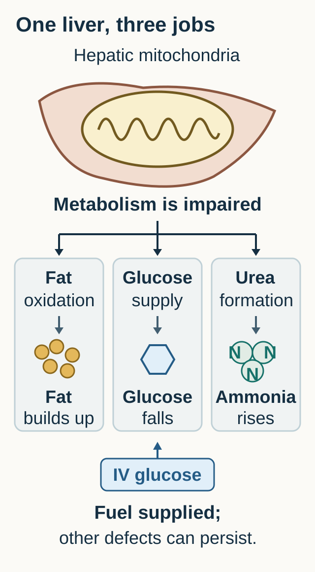
Why can one hepatic problem produce fat accumulation, low glucose and high ammonia? Mitochondria help oxidize fatty acids, generate usable energy and support other energy-dependent pathways. In Reye syndrome, mitochondrial dysfunction can impair fat use, gluconeogenesis and ureogenesis together. The exact initiating injury is not fully established; a useful physiologic model is not proof of a single molecular cause. [3]
Follow the three routes in the metabolic diagram. Poor fatty acid oxidation favors triglyceride storage in hepatocytes and can limit ketone production. Impaired glucose production makes fasting dangerous, especially in a young child with limited reserves. Impaired conversion of nitrogen to urea allows ammonia to accumulate. These are related failures, not three names for the same measurement. [3][4]
Trace each branch, then cover the glucose branch and predict what IV glucose will and will not correct. The initiating molecular lesion remains uncertain. [3][4][3][4]
Trace one correction. Imagine covering the glucose route with a card labeled "IV dextrose." Predict which laboratory value can improve immediately and which routes remain unresolved. Glucose replacement supplies circulating fuel; it does not establish recovery of hepatic fat oxidation or nitrogen disposal. Ammonia-associated brain dysfunction may therefore persist despite a normal repeat glucose. [4]
Check the prediction
The glucose concentration can rise while ammonia remains high and the child remains encephalopathic. If you also predict immediate disappearance of hepatic fat or correction of INR, you have treated one laboratory response as proof that the whole liver recovered.
Ammonia can enter the brain and contribute to astrocyte dysfunction and swelling. Glutamine formation and osmotic stress help explain this relationship, but inflammation and other metabolic disturbances also matter. Hypoglycemia adds an independent threat to cerebral energy supply. Neither a single ammonia concentration nor a corrected glucose replaces repeated neurologic examinations. Experimental astrocyte studies support glutamine involvement; they do not establish an exclusive mechanism or a treatment for children. [4][13]
Transfer the model. A fasting toddler has recurrent episodes of low glucose with very little ketone production. The same blocked-fat-use route now raises suspicion for an inherited fatty acid oxidation disorder rather than proving an acquired postviral syndrome. [4]
Read liver function, not just liver enzymes
Does a lower aminotransferase value prove recovery? AST and ALT reflect hepatocellular injury, not the full ability to sustain glucose, process nitrogen or synthesize coagulation proteins. Read mental status, glucose, ammonia and PT/INR together. Bilirubin may be relatively modest in the classic Reye pattern; a child need not be visibly jaundiced to have dangerous dysfunction. [1][3][4]
Different measurements answer different questions
Finding
What it tests
What it does not prove
FindingLow glucose
What it testsInsufficient circulating fuel
What it does not proveThat all altered consciousness is due to glucose alone
FindingHigh ammonia
What it testsImpaired nitrogen handling or excess production
What it does not proveA unique diagnosis or a complete neurologic grade
FindingPersistent high INR
What it testsImpaired synthesis, after considering vitamin K deficiency and other causes
What it does not proveThe probability of bleeding from the number alone
FindingAST and ALT
What it testsHepatocyte injury
What it does not proveHow much functional reserve remains
Compare the trajectory. At noon, a child answers questions and has INR 1.6. Later, INR is 2.8 and the child is increasingly somnolent, even though ALT has fallen. Choose which direction matters: functional and neurologic deterioration outweigh the superficially reassuring enzyme decrease. These INR examples are abnormal relative to the laboratory reference of 0.9 to 1.1; they are not a Reye-specific diagnostic threshold. [4]
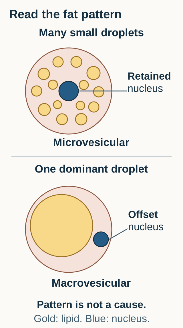
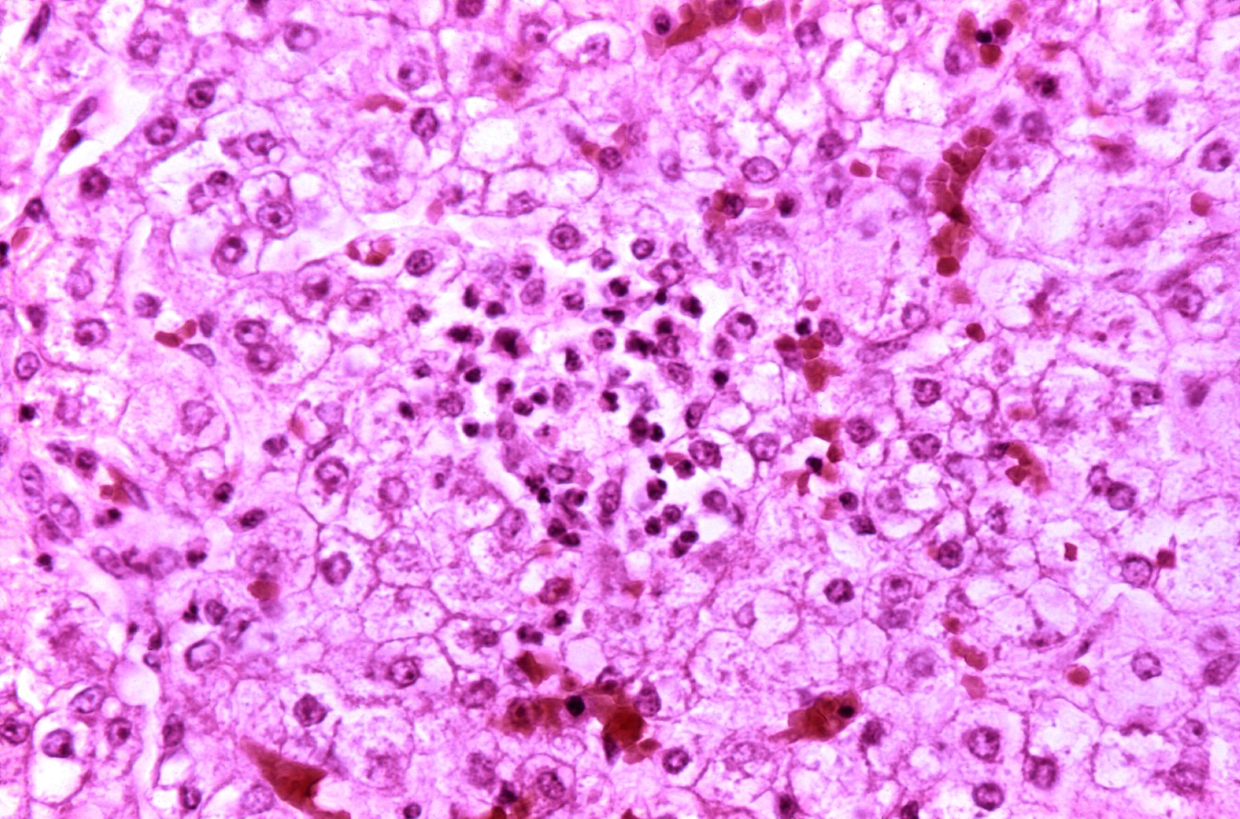
Inspect real tissue. In the accompanying CDC autopsy photograph, look for pale, vacuolated hepatocyte cytoplasm. The image shows intracellular fat droplets. Compare the diagram's many small droplets around a retained nucleus with one large droplet displacing a nucleus. The classic Reye-associated pattern is microvesicular steatosis with relatively little inflammation, not extensive inflammatory hepatitis. The real photograph is an example, not a stand-alone etiologic test. [3][10]
Locate the nucleus before naming the fat pattern. The first example is microvesicular, the second macrovesicular. Compare it with the separate CDC photograph. [3][10][3][10]
Find the pale, vacuolated hepatocyte cytoplasm and compare the small-droplet pattern. Histology alone cannot determine the cause or exclude mimics. [10]Image: CDC / Dr. Edwin P. Ewing, Jr. (source); public domain in the United States.Why not diagnose Reye syndrome from fat alone?
Small intracellular fat droplets can also accompany metabolic or drug-related mitochondrial injury. Clinical context and competing explanations remain necessary. A single tissue field cannot show that every alternative has been excluded.
Do not delay stabilization for biopsy. Histology is considered selectively when it can change the diagnostic or treatment plan; bleeding risk and sampling route require specialist judgment. Likewise, an unexpected ammonia result deserves prompt checking of specimen quality. Use the laboratory's validated collection and transport process, and repeat a discordant result while continuing bedside assessment. [4]
Apply the distinction. A falling ALT with improving alertness, stable glucose without escalating support and recovering INR is different from falling ALT with progressive coma. The direction of a single injury marker cannot distinguish those patients.
Find the better explanation
A recent virus is context, not a diagnosis. The most useful alternative is the one that explains the otherwise awkward findings: respiratory and acid-base changes, focal neurologic signs, recurrence after fasting or a medication change. Obtain an exact timeline and review prescription drugs, fever remedies, stomach medicines and possible household exposures. [4][5]
Choose the finding that redirects the investigation
Pattern
Reason to redirect
Focused assessment
PatternTinnitus, tachypnea, metabolic acidosis plus respiratory alkalosis
Reason to redirectSalicylate toxicity
Focused assessmentSerial salicylate concentrations, blood gas, electrolytes and toxicology consultation
PatternFever, focal seizures, meningism or inflammatory CSF
Reason to redirectMeningitis or encephalitis
Focused assessmentUrgent infection evaluation and appropriate empiric treatment
PatternRecurrent fasting intolerance with hypoketotic hypoglycemia
Reason to redirectFatty acid oxidation disorder
Focused assessmentAcylcarnitine profile and urine organic acids with metabolic consultation
PatternSevere hyperammonemia with preserved glucose, sometimes respiratory alkalosis
Reason to redirectUrea-cycle disorder
Focused assessmentPlasma amino acids and urine orotic acid within a broader metabolic evaluation
PatternHyperammonemia with substantial anion-gap acidosis and ketosis
Reason to redirectOrganic acidemia among the important alternatives
Focused assessmentUrine organic acids and acylcarnitines while treating the acute crisis
These patterns prioritize tests; none reliably excludes every competing disorder. Urea-cycle disease can also involve the liver. Organic acidemias and fatty acid oxidation defects can both mimic Reye syndrome. Early onset, repeated episodes or unexplained sibling deaths strengthen concern for inherited disease, but a negative family history does not exclude it. Collect time-sensitive specimens when feasible without delaying glucose or other emergency treatment. [4][8]
Reclassify a case. A teenager recovering from a viral illness has vomiting, ringing ears and rapid breathing. With an anion gap of 25 mmol/L (reference 8 to 12), bicarbonate 15 mmol/L and arterial carbon dioxide 20 mmHg, there is more ventilation than expected for metabolic acidosis alone. The competing respiratory alkalosis supports a toxicology assessment. Reference ranges are bicarbonate 22 to 28 mmol/L and PaCO2 35 to 45 mmHg. A nearly normal pH would not erase two opposing disturbances. [5]
What does a falling salicylate concentration tell you?
It may indicate declining circulating concentration, but worsening acidemia can favor salicylate entry into tissues. Clinical deterioration despite a falling concentration is not clearance for discharge. Reassess the entire patient with a toxicologist.
A low CSF white-cell count or an early negative herpes simplex PCR does not exclude encephalitis. Temporal-lobe abnormalities or focal seizures can justify continued empiric acyclovir and repeat testing when safe. Conversely, marked CSF inflammation weighs against the classic noninflammatory Reye pattern. Do not perform lumbar puncture merely to satisfy a surveillance definition. [1][6][7]
Apply it to the medication list. Valproate can produce symptomatic hyperammonemia even with normal hepatic tests. New lethargy and vomiting after a dose change require urgent assessment of the drug and possible metabolic susceptibility, not automatic attribution to a coincidental viral illness. [12]
Protect fuel, airway and brain perfusion together
Which task comes first: prove the cause or prevent the next injury? Airway, breathing and circulation are assessed immediately, with a bedside glucose obtained in parallel. Symptomatic hypoglycemia requires prompt glucose treatment and reassessment. Inability to protect the airway, seizures or declining consciousness requires urgent pediatric critical-care involvement; investigations must not delay stabilization. [4]
Match each threat to a response. A glucose of 32 mg/dL (reference 70 to 100) needs prompt correction. Recurrent hypoglycemia needs continuing glucose delivery rather than confidence in one normal recheck. Abnormal posturing with sluggish pupils needs emergency airway and intracranial-pressure assessment, even if the glucose is now normal. Neither response replaces the other.
Send liver injury and function tests, PT/INR, ammonia, electrolytes, a blood gas and targeted toxicology, infection and metabolic studies. Consult pediatric intensive care, hepatology and the relevant metabolic, toxicology or infectious-disease service early. Preserve acute blood or urine specimens if they can be obtained without delaying treatment. [4][8]
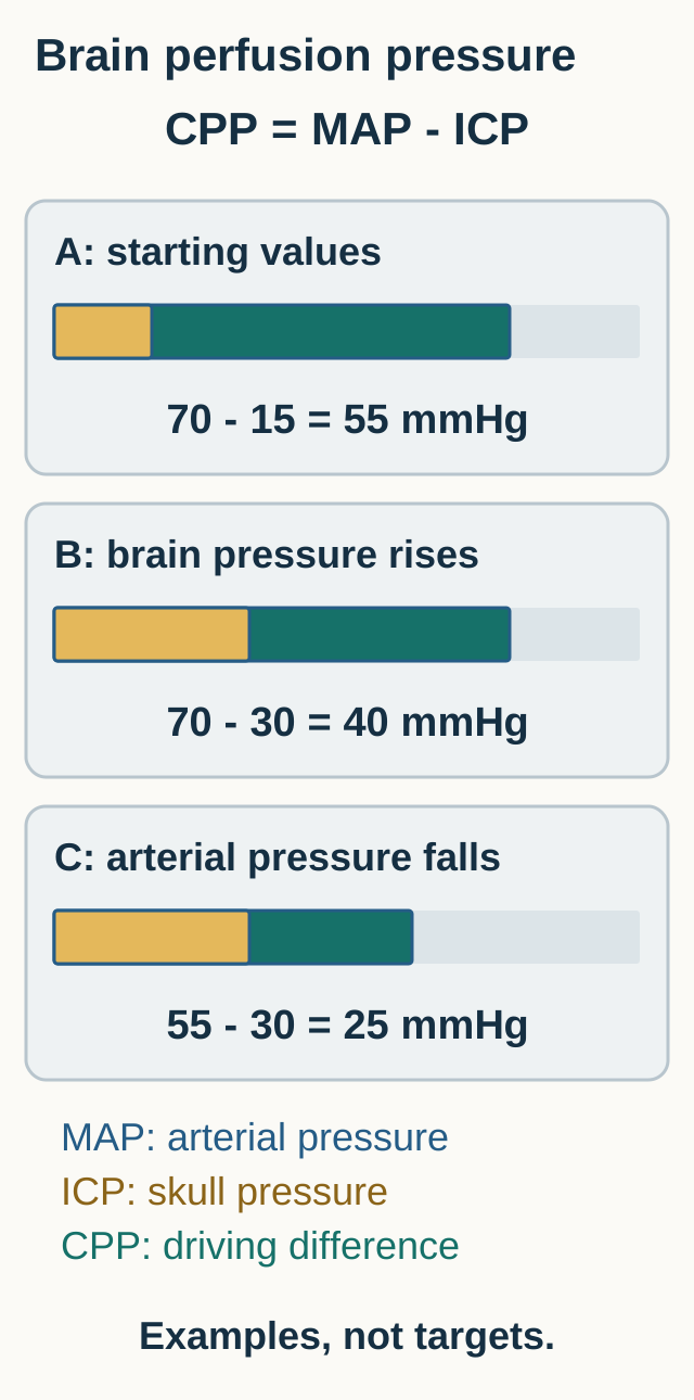
The brain-perfusion diagram compares two different threats. Rising intracranial pressure reduces the pressure difference driving cerebral blood flow. Falling arterial pressure can do the same. For the simplified relationship, cerebral perfusion pressure equals mean arterial pressure minus intracranial pressure. Protect oxygenation and systemic perfusion while the specialist team treats suspected cerebral edema; fluid restriction that causes shock is not brain protection. [4]
Subtract the orange intracranial-pressure segment from arterial pressure; the green remainder is cerebral perfusion pressure. Either rising intracranial pressure or falling arterial pressure can reduce the difference. Cerebral perfusion goals and the need for invasive monitoring depend on clinical assessment. [4][4]
Critical care includes avoiding hypoxia, hypotension and unnecessary fluid excess, maintaining appropriate glucose and electrolytes, controlling fever and seizures, and careful positioning with the head neutral. Fluid composition and amount, ventilation, osmotherapy and renal support are individualized. Invasive pressure monitoring is not automatic because benefit is uncertain and bleeding risk matters. There is no established antidote that reverses Reye syndrome. [4]
What changes if the CT report is reassuring?
A normal CT does not exclude raised intracranial pressure. Deteriorating consciousness, abnormal pupils, posturing or important coagulopathy still require reassessment before lumbar puncture. Treat suspected infection without waiting for an unsafe procedure. [7]
Transfer to a different result. INR remains 3.1 after vitamin K, but there is no bleeding or planned invasive procedure. This supports serious synthetic dysfunction, not routine plasma transfusion solely to normalize the result. Plasma can obscure the trend and add fluid; bleeding or a necessary procedure changes the risk-benefit assessment. Similarly, ammonia-lowering treatment does not substitute for glucose, airway or cerebral-perfusion support, and outcome benefit of routine lactulose or rifaximin in pediatric acute liver failure is not established. [4]
Follow the trajectory and prevent the next exposure
Can one better number justify less monitoring? Improvement is a combined trend: alertness and airway protection, glucose stability with the support documented, ammonia in clinical context, INR and overall organ function. New subtle twitching or unexplained failure to awaken can warrant EEG assessment for ongoing seizures. Neither the current ammonia value nor the original diagnosis explains every later change. [4][6]
Read a medicine label. A 15-year-old recovering from influenza asks for a stomach remedy. Its general directions include people aged 12 and older, but its active ingredient is bismuth subsalicylate. The illness-specific warning takes precedence: children and teenagers with or recovering from flu-like symptoms or chickenpox should not use that product. Age eligibility is not permission in every clinical context. [9]
Check the label decision
The hidden exposure is salicylate, even though the product is sold for stomach symptoms rather than fever. Review active ingredients rather than relying on a brand name or the front of the package.
Avoid nonprescribed aspirin or salicylate-containing remedies for children and adolescents during viral illness. This is not a direction to independently terminate medically necessary antiplatelet therapy: some children with Kawasaki disease receive aspirin under specialist supervision. Those families need a specific plan for intercurrent illness, medication changes and immunization timing from their treating team. [2][9][11]
Apply the prevention rule without overgeneralizing. One family needs help recognizing an over-the-counter salicylate; another needs prompt advice from the clinician managing a coronary aneurysm. Both need an accurate medication list, but the same blanket medication instruction is not appropriate for both.
After an apparent Reye-like event, diagnostic follow-through still matters. Unexplained or recurrent fasting-related episodes warrant metabolic evaluation and an individualized illness plan. Inherited disease can change counseling and prevention for relatives. Do not label every recurrence as a new viral complication. [4][8]
Apply the liver and brain model
Use the findings that distinguish competing explanations. Treat the immediate threat while keeping the cause open when evidence is incomplete.
Case 1
Show answer and explanations for case 1
A. Canalicular transport of conjugated bilirubin (Why this does not fit)
Impaired canalicular excretion favors conjugated hyperbilirubinemia and cholestasis. Bilirubin is normal, while hypoglycemia and hyperammonemia require an explanation involving glucose production and nitrogen disposal.
Reasoning steps for option A
What abnormality would dominate with impaired canalicular excretion?
Impaired canalicular excretion favors conjugated hyperbilirubinemia and cholestasis.
Does that account for these paired metabolic failures?
Bilirubin is normal, while hypoglycemia and hyperammonemia require an explanation involving glucose production and nitrogen disposal.
B. Lysosomal degradation of complex sphingolipids (Why this does not fit)
Progressive organ and neurologic involvement can occur in inherited lysosomal storage disease. The abrupt postinfectious hepatic metabolic failure does not establish a chronic storage disorder; the paired glucose and ammonia abnormalities fit mitochondrial dysfunction better.
Reasoning steps for option B
What course often accompanies a sphingolipid storage disorder?
Progressive organ and neurologic involvement can occur in inherited lysosomal storage disease.
Which supplied time course is less compatible?
The abrupt postinfectious hepatic metabolic failure does not establish a chronic storage disorder; the paired glucose and ammonia abnormalities fit mitochondrial dysfunction better.
C. Mitochondrial energy-dependent metabolism (Best answer)
Glucose production and conversion of nitrogen into urea are inadequate for current needs. Mitochondrial dysfunction can limit hepatic energy metabolism and ureogenesis together, fitting a Reye-like presentation while alternatives are investigated.
Reasoning steps for option C
Which two liver functions are failing simultaneously?
Glucose production and conversion of nitrogen into urea are inadequate for current needs.
What shared cellular problem can impair both?
Mitochondrial dysfunction can limit hepatic energy metabolism and ureogenesis together, fitting a Reye-like presentation while alternatives are investigated.
D. Microsomal conjugation of unconjugated bilirubin (Why this does not fit)
Unconjugated bilirubin accumulates when conjugation is impaired. The normal bilirubin does not support a conjugation disorder, which would not unify marked ammonia accumulation with hypoglycemia.
Reasoning steps for option D
What is the principal consequence of impaired bilirubin conjugation?
Unconjugated bilirubin accumulates when conjugation is impaired.
Which result argues against that as the unifying process?
The normal bilirubin does not support a conjugation disorder, which would not unify marked ammonia accumulation with hypoglycemia.
E. Immune destruction of intrahepatic bile ducts (Why this does not fit)
Cholestatic liver tests and bile-duct injury would favor immune destruction of ducts. The acute failure of glucose and nitrogen handling, without hyperbilirubinemia, points toward hepatocyte metabolic dysfunction rather than duct-centered disease.
Reasoning steps for option E
Which pattern would suggest an immune cholangiopathy?
Cholestatic liver tests and bile-duct injury would favor immune destruction of ducts.
Which functions instead dominate this presentation?
The acute failure of glucose and nitrogen handling, without hyperbilirubinemia, points toward hepatocyte metabolic dysfunction rather than duct-centered disease.
Takeaway: Connect simultaneous glucose and nitrogen failure to hepatic metabolism; exposure history alone does not establish the cause. [1] [3] [4]
A. Reduced fatty acid oxidation; reduced gluconeogenesis (Best answer)
Fatty acid oxidation supplies energy and substrate supporting ketone production and fasting metabolism. Impaired fat oxidation limits the ketone response, while impaired gluconeogenesis contributes to hypoglycemia.
Reasoning steps for option A
What should fat use normally provide during fasting?
Fatty acid oxidation supplies energy and substrate supporting ketone production and fasting metabolism.
What combined failure fits low ketones and low glucose?
Impaired fat oxidation limits the ketone response, while impaired gluconeogenesis contributes to hypoglycemia.
B. Increased fatty acid oxidation; reduced gluconeogenesis (Why this does not fit)
Yes, inadequate glucose production can explain fasting hypoglycemia. It argues against an intact, increased fat-oxidation response and supports impairment of that pathway as well.
Reasoning steps for option B
Could low glucose follow impaired gluconeogenesis?
Yes, inadequate glucose production can explain fasting hypoglycemia.
What does the very small ketone response add?
It argues against an intact, increased fat-oxidation response and supports impairment of that pathway as well.
C. Reduced fatty acid oxidation; increased gluconeogenesis (Why this does not fit)
Reduced fatty acid oxidation can limit fasting ketone production. Increased effective gluconeogenesis would support circulating glucose, not explain its marked fall during fasting.
Reasoning steps for option C
Which half explains the limited ketone response?
Reduced fatty acid oxidation can limit fasting ketone production.
Why is the proposed glucose response less consistent?
Increased effective gluconeogenesis would support circulating glucose, not explain its marked fall during fasting.
D. Increased fatty acid oxidation; increased gluconeogenesis (Why this does not fit)
They would support fuel availability during fasting through glucose and ketone production. Both glucose and the ketone response are inadequate, so effective increases in both pathways do not explain this episode.
Reasoning steps for option D
What would coordinated increases in both pathways accomplish?
They would support fuel availability during fasting through glucose and ketone production.
Does the observed fuel pattern show that compensation?
Both glucose and the ketone response are inadequate, so effective increases in both pathways do not explain this episode.
Takeaway: Hypoketotic hypoglycemia indicates an inadequate fasting response; it does not by itself distinguish Reye syndrome from inherited disease. [3] [4]
A. Unconjugated bilirubin deposition in basal ganglia (Why this does not fit)
Marked unconjugated hyperbilirubinemia is the relevant exposure. Bilirubin remains normal; persistent hyperammonemia is the unresolved metabolic abnormality.
Reasoning steps for option A
What biochemical setting supports bilirubin neurotoxicity?
Marked unconjugated hyperbilirubinemia is the relevant exposure.
Is that exposure present here?
Bilirubin remains normal; persistent hyperammonemia is the unresolved metabolic abnormality.
B. Neutrophil-mediated destruction of the meninges (Why this does not fit)
Meningism, inflammatory CSF or a compatible microbiologic result would strengthen that diagnosis. The rise in ammonia during hepatic dysfunction supplies a metabolic explanation; the stem does not provide evidence of neutrophilic meningitis.
Reasoning steps for option B
Which findings would strengthen bacterial meningeal inflammation?
Meningism, inflammatory CSF or a compatible microbiologic result would strengthen that diagnosis.
What directly supports the proposed metabolic brain injury?
The rise in ammonia during hepatic dysfunction supplies a metabolic explanation; the stem does not provide evidence of neutrophilic meningitis.
C. Continued glucose deprivation from a low plasma concentration (Why this does not fit)
The original concentration of 35 mg/dL was low enough to contribute to brain dysfunction. Serial glucose is now normal, so ongoing low circulating glucose does not account for the persistent threat as well as the rising ammonia does.
Reasoning steps for option C
Why was glucose deprivation initially plausible?
The original concentration of 35 mg/dL was low enough to contribute to brain dysfunction.
What has changed while consciousness worsens?
Serial glucose is now normal, so ongoing low circulating glucose does not account for the persistent threat as well as the rising ammonia does.
D. Astrocyte glutamine accumulation with osmotic stress (Best answer)
Ammonia remains markedly increased despite correction of circulating glucose. Astrocytic conversion of ammonia into glutamine contributes to osmotic and cellular stress; this can sustain brain injury independently of hypoglycemia.
Reasoning steps for option D
What unresolved metabolite can affect astrocytes?
Ammonia remains markedly increased despite correction of circulating glucose.
How can its handling contribute to swelling?
Astrocytic conversion of ammonia into glutamine contributes to osmotic and cellular stress; this can sustain brain injury independently of hypoglycemia.
Takeaway: Correcting glucose addresses one cerebral threat, not all causes of encephalopathy. [4] [13]
A. Macrovesicular steatosis; increased bilirubin conjugation (Why this does not fit)
A dominant large fat vacuole often displaces the nucleus. The described droplets are small and numerous, and increased bilirubin conjugation does not explain the paired glucose and ammonia abnormalities.
Reasoning steps for option A
What morphology supports macrovesicular steatosis?
A dominant large fat vacuole often displaces the nucleus.
Does either half fit these findings?
The described droplets are small and numerous, and increased bilirubin conjugation does not explain the paired glucose and ammonia abnormalities.
B. Microvesicular steatosis; impaired mitochondrial fat use (Best answer)
Small lipid droplets with relatively preserved nuclear position support microvesicular steatosis. Impaired mitochondrial fat use can accompany defective glucose and nitrogen handling, although the tissue pattern is not specific to Reye syndrome.
Reasoning steps for option B
What pattern is suggested by many small fat droplets?
Small lipid droplets with relatively preserved nuclear position support microvesicular steatosis.
How does the clinical pattern fit?
Impaired mitochondrial fat use can accompany defective glucose and nitrogen handling, although the tissue pattern is not specific to Reye syndrome.
C. Interface hepatitis; immune-mediated hepatocyte injury (Why this does not fit)
Inflammatory injury extends across the limiting plate from portal areas into adjacent hepatocytes. Small lipid droplets with little inflammation are emphasized, not interface inflammatory destruction.
Reasoning steps for option C
What tissue feature defines interface hepatitis?
Inflammatory injury extends across the limiting plate from portal areas into adjacent hepatocytes.
What is actually prominent in this specimen?
Small lipid droplets with little inflammation are emphasized, not interface inflammatory destruction.
D. Canalicular cholestasis; impaired bile secretion (Why this does not fit)
Bile plugs within canaliculi would support impaired bile secretion. The reported material is intracellular fat, and its small-droplet pattern is microvesicular rather than a bile-plug pattern.
Reasoning steps for option D
What would support canalicular cholestasis?
Bile plugs within canaliculi would support impaired bile secretion.
What feature must be classified here instead?
The reported material is intracellular fat, and its small-droplet pattern is microvesicular rather than a bile-plug pattern.
E. Glycogen accumulation; increased glycogen synthesis (Why this does not fit)
Yes, glycogen can alter hepatocyte appearance and requires appropriate histologic assessment. The pathologist explicitly identifies fat droplets; the metabolic syndrome therefore cannot be attributed to increased glycogen synthesis from pallor alone.
Reasoning steps for option E
Can stored glycogen also make hepatocytes appear pale?
Yes, glycogen can alter hepatocyte appearance and requires appropriate histologic assessment.
Why is that not the reported storage material?
The pathologist explicitly identifies fat droplets; the metabolic syndrome therefore cannot be attributed to increased glycogen synthesis from pallor alone.
Takeaway: Classify the storage pattern, then relate it to physiology; a pattern is not a unique etiologic diagnosis. [3] [10]
A. Preserved synthesis with selective bilirubin transport failure (Why this does not fit)
It would tend to increase bilirubin rather than cause this isolated lack of jaundice. Persistent INR prolongation after vitamin K supports impaired synthesis in this context, with additional metabolic dysfunction.
Reasoning steps for option A
What would selective bilirubin transport failure tend to produce?
It would tend to increase bilirubin rather than cause this isolated lack of jaundice.
Which result contradicts preserved synthesis?
Persistent INR prolongation after vitamin K supports impaired synthesis in this context, with additional metabolic dysfunction.
B. Vitamin K deficiency with preserved hepatic fuel production (Why this does not fit)
Improvement of the coagulation abnormality after adequate replacement would support deficiency. INR remains prolonged and glucose is low, so neither a simple corrected vitamin deficiency nor preserved glucose support is demonstrated.
Reasoning steps for option B
What response would favor vitamin K deficiency?
Improvement of the coagulation abnormality after adequate replacement would support deficiency.
What argues against both parts of this explanation?
INR remains prolonged and glucose is low, so neither a simple corrected vitamin deficiency nor preserved glucose support is demonstrated.
C. Impaired synthesis without marked bilirubin accumulation (Best answer)
After vitamin K has been addressed, it supports impaired hepatic production of coagulation proteins. Bilirubin handling and protein synthesis are different functions; low glucose and high ammonia further demonstrate metabolic failure without requiring jaundice.
Reasoning steps for option C
What does persistent INR prolongation suggest here?
After vitamin K has been addressed, it supports impaired hepatic production of coagulation proteins.
Why does normal bilirubin not cancel that concern?
Bilirubin handling and protein synthesis are different functions; low glucose and high ammonia further demonstrate metabolic failure without requiring jaundice.
D. Peripheral hemolysis with secondary bilirubin overproduction (Why this does not fit)
Excess heme breakdown can increase unconjugated bilirubin. Normal bilirubin and combined coagulation, glucose and ammonia abnormalities are not explained by isolated peripheral red-cell destruction.
Reasoning steps for option D
What bilirubin pattern often accompanies hemolysis?
Excess heme breakdown can increase unconjugated bilirubin.
Does hemolysis unify the supplied findings?
Normal bilirubin and combined coagulation, glucose and ammonia abnormalities are not explained by isolated peripheral red-cell destruction.
Takeaway: Absence of jaundice is not evidence of preserved hepatic metabolic or synthetic function. [1] [4]
A. Worsening function despite a lower injury-marker concentration (Best answer)
Increasing INR, greater glucose support and reduced responsiveness all indicate worsening functional or neurologic status. A lower injury-marker concentration cannot override those deteriorating functional measures or establish recovery.
Reasoning steps for option A
Which observations assess function and clinical severity?
Increasing INR, greater glucose support and reduced responsiveness all indicate worsening functional or neurologic status.
How should the falling ALT be weighted?
A lower injury-marker concentration cannot override those deteriorating functional measures or establish recovery.
B. Improving function demonstrated by reduced hepatocyte enzyme release (Why this does not fit)
It can accompany less ongoing hepatocyte injury during recovery. Synthetic, metabolic and neurologic measures are simultaneously worsening, so the enzyme change does not demonstrate improved function.
Reasoning steps for option B
Why might a lower ALT initially look reassuring?
It can accompany less ongoing hepatocyte injury during recovery.
What prevents that inference in this child?
Synthetic, metabolic and neurologic measures are simultaneously worsening, so the enzyme change does not demonstrate improved function.
C. Stable function with isolated deterioration of laboratory precision (Why this does not fit)
Discordant or technically questionable results can require repeat measurement. No. Independent clinical decline and increasing glucose requirements accompany the INR change, making isolated laboratory imprecision an inadequate explanation.
Reasoning steps for option C
Could an unexpected laboratory change justify verification?
Discordant or technically questionable results can require repeat measurement.
Are the changes isolated to one uncertain assay?
No. Independent clinical decline and increasing glucose requirements accompany the INR change, making isolated laboratory imprecision an inadequate explanation.
D. Resolved liver dysfunction with a delayed bilirubin-related encephalopathy (Why this does not fit)
A compatible marked unconjugated bilirubin burden and clinical context would be needed. Persistent synthetic failure and increasing need for glucose support show that hepatic dysfunction has not resolved.
Reasoning steps for option D
What evidence would support bilirubin as the current neurologic insult?
A compatible marked unconjugated bilirubin burden and clinical context would be needed.
What instead remains active in this record?
Persistent synthetic failure and increasing need for glucose support show that hepatic dysfunction has not resolved.
Takeaway: Trend injury markers alongside function and the bedside examination, not instead of them. [4]
A. Gastroenterology ward for intermittent metabolic measurements (Why this does not fit)
The child has evidence of acute hepatic dysfunction requiring specialist input. Progressive disorientation despite glucose correction requires frequent neurologic assessment and the ability to provide rapid organ support, rather than intermittent ward measurements.
Reasoning steps for option A
Why is a gastroenterology service relevant?
The child has evidence of acute hepatic dysfunction requiring specialist input.
Why is this ward insufficient as described?
Progressive disorientation despite glucose correction requires frequent neurologic assessment and the ability to provide rapid organ support, rather than intermittent ward measurements.
B. Neurology ward for an EEG before deciding the care setting (Why this does not fit)
Yes, seizures or unexplained altered responsiveness may warrant EEG. No. The supplied deterioration and hepatic dysfunction already require close monitoring while the cause is evaluated.
Reasoning steps for option B
Could EEG contribute to this child's evaluation?
Yes, seizures or unexplained altered responsiveness may warrant EEG.
Should that test delay the higher-acuity setting?
No. The supplied deterioration and hepatic dysfunction already require close monitoring while the cause is evaluated.
C. Radiology observation until cerebral edema is demonstrated (Why this does not fit)
Imaging can help identify complications or competing diagnoses when the child is stable enough. Clinical deterioration warrants escalation before imaging confirmation, and normal CT cannot exclude raised intracranial pressure.
Reasoning steps for option C
Can imaging help evaluate neurologic deterioration?
Imaging can help identify complications or competing diagnoses when the child is stable enough.
Why not make imaging the admission gate?
Clinical deterioration warrants escalation before imaging confirmation, and normal CT cannot exclude raised intracranial pressure.
D. Pediatric intensive care for neurologic and metabolic surveillance (Best answer)
New disorientation persists after glucose has normalized. Pediatric intensive-care assessment matches the need for repeated neurologic assessment, metabolic support and rapid escalation before coma develops.
Reasoning steps for option D
Which finding marks progression beyond isolated vomiting?
New disorientation persists after glucose has normalized.
Which available setting matches the trajectory?
Pediatric intensive-care assessment matches the need for repeated neurologic assessment, metabolic support and rapid escalation before coma develops.
Takeaway: Escalate according to trajectory and available monitoring capability, not only after coma or imaging-confirmed edema. [4] [7]
A. Complete the metabolic specimen set before giving glucose (Why this does not fit)
They can preserve an abnormal biochemical pattern that becomes less apparent after glucose treatment. Severe symptomatic hypoglycemia with unreliable swallowing poses an immediate cerebral threat; preserving a complete sample set does not justify a 15-minute delay.
Reasoning steps for option A
Why are pretreatment metabolic samples valuable?
They can preserve an abnormal biochemical pattern that becomes less apparent after glucose treatment.
What makes waiting inappropriate here?
Severe symptomatic hypoglycemia with unreliable swallowing poses an immediate cerebral threat; preserving a complete sample set does not justify a 15-minute delay.
B. Give IV dextrose now and obtain available specimens in parallel (Best answer)
The symptomatic glucose concentration of 27 mg/dL requires prompt treatment. Collect feasible acute specimens alongside treatment, documenting timing, rather than withholding glucose to complete every test.
Reasoning steps for option B
Which current abnormality requires immediate correction?
The symptomatic glucose concentration of 27 mg/dL requires prompt treatment.
How can diagnostic yield be preserved safely?
Collect feasible acute specimens alongside treatment, documenting timing, rather than withholding glucose to complete every test.
C. Give oral carbohydrate and repeat glucose before using the IV (Why this does not fit)
It can be used when the patient is sufficiently alert and can swallow safely. Unreliable swallowing creates aspiration risk; established IV access provides an appropriate immediate route.
Reasoning steps for option C
When can oral carbohydrate be appropriate?
It can be used when the patient is sufficiently alert and can swallow safely.
Which finding changes the route in this child?
Unreliable swallowing creates aspiration risk; established IV access provides an appropriate immediate route.
D. Wait for ammonia to determine whether glucose treatment is necessary (Why this does not fit)
Hyperammonemia would help identify impaired nitrogen handling and another potential brain threat. No. The documented severe hypoglycemia already warrants treatment regardless of the ammonia result.
Reasoning steps for option D
Why is ammonia relevant to the differential?
Hyperammonemia would help identify impaired nitrogen handling and another potential brain threat.
Does its pending value alter the need to treat glucose?
No. The documented severe hypoglycemia already warrants treatment regardless of the ammonia result.
Takeaway: Obtain critical specimens without delaying treatment of severe symptomatic hypoglycemia. [4] [8]
A. Administer lactulose through a nasogastric tube (Why this does not fit)
Ammonia is substantially increased and can contribute to encephalopathy. Pooled secretions and inadequate breathing indicate airway and ventilatory failure; enteral treatment does not secure either.
Reasoning steps for option A
Why might ammonia-lowering treatment be considered?
Ammonia is substantially increased and can contribute to encephalopathy.
What immediate problem would this leave untreated?
Pooled secretions and inadequate breathing indicate airway and ventilatory failure; enteral treatment does not secure either.
B. Obtain computed tomography to characterize cerebral swelling (Why this does not fit)
Imaging can help assess complications and competing structural disease. The child cannot protect the airway and is breathing inadequately; those threats require treatment without waiting for a scan.
Reasoning steps for option B
What could brain imaging add?
Imaging can help assess complications and competing structural disease.
Why must resuscitation precede transport for imaging?
The child cannot protect the airway and is breathing inadequately; those threats require treatment without waiting for a scan.
C. Give mannitol while arranging later airway reassessment (Why this does not fit)
Posturing and severe neurologic decline raise concern for dangerous intracranial pressure. Osmotherapy may be considered by the team, but it does not replace immediate treatment of the already failing airway and ventilation.
Reasoning steps for option C
Why might osmotherapy enter the management plan?
Posturing and severe neurologic decline raise concern for dangerous intracranial pressure.
Why is the proposed order insufficient?
Osmotherapy may be considered by the team, but it does not replace immediate treatment of the already failing airway and ventilation.
D. Arrange an EEG to characterize persistent seizure activity (Why this does not fit)
Yes, ongoing electrographic seizures can contribute to depressed consciousness. Immediate airway and ventilatory support is required because secretions are pooling and breathing is irregular.
Reasoning steps for option D
Can seizures persist without large convulsions?
Yes, ongoing electrographic seizures can contribute to depressed consciousness.
What must occur before that investigation is prioritized?
Immediate airway and ventilatory support is required because secretions are pooling and breathing is irregular.
E. Secure the airway with expert controlled ventilation (Best answer)
Deeply reduced consciousness, pooled secretions and inadequate breathing indicate failure of airway protection and ventilation. An experienced team should provide neuroprotective resuscitation while addressing suspected cerebral edema, perfusion and other metabolic threats.
Reasoning steps for option E
Which findings establish the resuscitation priority?
Deeply reduced consciousness, pooled secretions and inadequate breathing indicate failure of airway protection and ventilation.
How should the neurologic context shape support?
An experienced team should provide neuroprotective resuscitation while addressing suspected cerebral edema, perfusion and other metabolic threats.
Takeaway: A corrected glucose does not postpone airway support when protection and ventilation fail. [4] [7]
A. Proceed with lumbar puncture because the recent CT was normal (Why this does not fit)
It may appear reassuring about a large structural lesion at the time of imaging. Normal CT does not exclude raised intracranial pressure, and the child has new pupillary abnormalities, deteriorating consciousness and important coagulopathy.
Reasoning steps for option A
What might a normal scan appear to offer?
It may appear reassuring about a large structural lesion at the time of imaging.
Why does it not establish procedural safety here?
Normal CT does not exclude raised intracranial pressure, and the child has new pupillary abnormalities, deteriorating consciousness and important coagulopathy.
B. Correct the INR and perform lumbar puncture before reassessing pupils (Why this does not fit)
Important coagulopathy is one factor in the risk of an invasive procedure. No. Worsening consciousness and abnormal pupils still require urgent neurologic reassessment and stabilization.
Reasoning steps for option B
Why does coagulation status matter?
Important coagulopathy is one factor in the risk of an invasive procedure.
Would correcting it address every contraindication?
No. Worsening consciousness and abnormal pupils still require urgent neurologic reassessment and stabilization.
C. Defer lumbar puncture and stabilize the neurologic deterioration (Best answer)
Progressive reduced consciousness and abnormal pupils raise concern for dangerous intracranial pressure. Continue appropriate empiric assessment and treatment while deferring an unsafe procedure; coagulopathy adds another reason for caution.
Reasoning steps for option C
Which bedside findings override the reassuring report?
Progressive reduced consciousness and abnormal pupils raise concern for dangerous intracranial pressure.
How can the infectious possibility be handled meanwhile?
Continue appropriate empiric assessment and treatment while deferring an unsafe procedure; coagulopathy adds another reason for caution.
D. Repeat CT and use a second normal report as permission to proceed (Why this does not fit)
Yes, new neurologic findings can justify repeat imaging after stabilization. Clinical contraindications and coagulopathy must be resolved or appropriately addressed; imaging alone does not certify lumbar-puncture safety.
Reasoning steps for option D
Can repeat imaging sometimes add useful information?
Yes, new neurologic findings can justify repeat imaging after stabilization.
Why is a second normal report not sufficient permission?
Clinical contraindications and coagulopathy must be resolved or appropriately addressed; imaging alone does not certify lumbar-puncture safety.
Takeaway: Lumbar-puncture safety depends on the current clinical state, not simply a normal CT report. [7]
A. Perfusion pressure falls from 55 to 40 despite preserved oxygen saturation (Best answer)
They are 75 minus 20 = 55 mmHg and 75 minus 35 = 40 mmHg. No. Oxygen saturation does not measure the pressure gradient supplying the brain; rising intracranial pressure reduces that gradient at fixed arterial pressure.
Reasoning steps for option A
What are the two pressure differences?
They are 75 minus 20 = 55 mmHg and 75 minus 35 = 40 mmHg.
Does preserved saturation offset the pressure change?
No. Oxygen saturation does not measure the pressure gradient supplying the brain; rising intracranial pressure reduces that gradient at fixed arterial pressure.
B. Perfusion pressure rises from 95 to 110 because the pressures add (Why this does not fit)
Adding the pressures would give 95 and 110 mmHg, but their sum is not cerebral perfusion pressure. Intracranial pressure is subtracted, so its rise lowers rather than increases perfusion pressure.
Reasoning steps for option B
When would the sum of the pressures rise?
Adding the pressures would give 95 and 110 mmHg, but their sum is not cerebral perfusion pressure.
What relationship should be used instead?
Intracranial pressure is subtracted, so its rise lowers rather than increases perfusion pressure.
C. Perfusion pressure stays at 75 because mean arterial pressure is unchanged (Why this does not fit)
It is the maintained mean arterial pressure, not the pressure difference across the cerebral circulation. Intracranial pressure rose by 15 mmHg, so cerebral perfusion pressure falls by 15 mmHg.
Reasoning steps for option C
What quantity does 75 mmHg represent?
It is the maintained mean arterial pressure, not the pressure difference across the cerebral circulation.
Which other pressure changed?
Intracranial pressure rose by 15 mmHg, so cerebral perfusion pressure falls by 15 mmHg.
D. Perfusion pressure stays at 55 because oxygen saturation remains normal (Why this does not fit)
It describes hemoglobin oxygen saturation, a component of oxygen delivery rather than a pressure difference. No. With unchanged arterial pressure and higher intracranial pressure, the pressure difference falls to 40 mmHg.
Reasoning steps for option D
What does normal oxygen saturation describe?
It describes hemoglobin oxygen saturation, a component of oxygen delivery rather than a pressure difference.
Can it preserve the original pressure gradient?
No. With unchanged arterial pressure and higher intracranial pressure, the pressure difference falls to 40 mmHg.
Takeaway: Systemic oxygenation and cerebral perfusion pressure are related to brain safety but are not interchangeable measurements. [4]
A. Anion-gap metabolic acidosis with appropriate respiratory compensation (Why this does not fit)
The anion gap is 140 minus 100 minus 15 = 25 mmol/L, which is increased. Expected PaCO2 is about 30.5 mmHg, not 20; the extra fall indicates an additional respiratory alkalosis.
Reasoning steps for option A
Is there an anion-gap metabolic acidosis?
The anion gap is 140 minus 100 minus 15 = 25 mmol/L, which is increased.
Is the measured PaCO2 appropriate compensation?
Expected PaCO2 is about 30.5 mmHg, not 20; the extra fall indicates an additional respiratory alkalosis.
B. Anion-gap metabolic acidosis with an additional respiratory alkalosis (Best answer)
A gap of 25 mmol/L with low bicarbonate supports an anion-gap metabolic acidosis. PaCO2 of 20 is substantially below the expected 30.5, showing an added respiratory alkalosis consistent with the salicylate-toxicology concern.
Reasoning steps for option B
What does the calculated anion gap establish?
A gap of 25 mmol/L with low bicarbonate supports an anion-gap metabolic acidosis.
What does the PaCO2 comparison establish?
PaCO2 of 20 is substantially below the expected 30.5, showing an added respiratory alkalosis consistent with the salicylate-toxicology concern.
C. Respiratory alkalosis with an appropriate renal bicarbonate response (Why this does not fit)
The low PaCO2 and alkalemic pH support a respiratory alkalosis component. The increased anion gap identifies a concurrent metabolic acid load rather than merely renal adaptation to hyperventilation.
Reasoning steps for option C
Why is a respiratory alkalosis present?
The low PaCO2 and alkalemic pH support a respiratory alkalosis component.
Why is compensation alone an incomplete explanation?
The increased anion gap identifies a concurrent metabolic acid load rather than merely renal adaptation to hyperventilation.
D. Anion-gap metabolic acidosis with an additional respiratory acidosis (Why this does not fit)
It would make PaCO2 higher than expected for the metabolic acidosis. PaCO2 is lower than expected, indicating added respiratory alkalosis rather than respiratory acidosis.
Reasoning steps for option D
What would an added respiratory acidosis do to PaCO2?
It would make PaCO2 higher than expected for the metabolic acidosis.
Does the measured value show that direction?
PaCO2 is lower than expected, indicating added respiratory alkalosis rather than respiratory acidosis.
E. Metabolic alkalosis with an additional respiratory alkalosis (Why this does not fit)
Gastric acid loss can produce a metabolic alkalosis. Here bicarbonate is low and the anion gap is high, establishing a metabolic acidosis despite the history of vomiting.
Reasoning steps for option E
Could vomiting contribute to metabolic alkalosis in some patients?
Gastric acid loss can produce a metabolic alkalosis.
Why is that not the principal metabolic process demonstrated?
Here bicarbonate is low and the anion gap is high, establishing a metabolic acidosis despite the history of vomiting.
Takeaway: Calculate the gap and expected compensation before attributing all postviral vomiting and confusion to Reye syndrome. [5]
A. Reduced circulating concentration demonstrates reduced cerebral exposure (Why this does not fit)
Serial decline can help document that circulating concentrations are falling and ongoing absorption is no longer dominant. Acidemia and worsening consciousness indicate ongoing danger; the serum concentration alone does not establish reduced tissue exposure.
Reasoning steps for option A
Why is a declining concentration useful to follow?
Serial decline can help document that circulating concentrations are falling and ongoing absorption is no longer dominant.
Why does it not prove neurologic improvement?
Acidemia and worsening consciousness indicate ongoing danger; the serum concentration alone does not establish reduced tissue exposure.
B. Renal elimination has become adequate because the concentration declined (Why this does not fit)
Yes, elimination can lower circulating drug concentration. The patient is clinically worsening and becoming acidemic, so adequacy of treatment cannot be inferred from that decline alone.
Reasoning steps for option B
Can elimination contribute to a falling serum value?
Yes, elimination can lower circulating drug concentration.
What prevents a conclusion of adequate clearance?
The patient is clinically worsening and becoming acidemic, so adequacy of treatment cannot be inferred from that decline alone.
C. An unrelated postviral process is established by the declining drug level (Why this does not fit)
Yes, the differential may need reassessment when findings are not explained. No. The fall in pH itself can worsen salicylate distribution into tissues, so the observed decline does not establish a separate diagnosis.
Reasoning steps for option C
Can another neurologic process coexist with drug exposure?
Yes, the differential may need reassessment when findings are not explained.
Does this trend establish an unrelated process?
No. The fall in pH itself can worsen salicylate distribution into tissues, so the observed decline does not establish a separate diagnosis.
D. A larger nonionized fraction can increase salicylate entry into tissues (Best answer)
It increases the fraction in the nonionized form. Greater tissue and CNS entry can accompany a lower serum concentration; worsening consciousness demands urgent toxicologic reassessment rather than reassurance.
Reasoning steps for option D
What does decreasing blood pH do to salicylate ionization?
It increases the fraction in the nonionized form.
How does that affect interpretation of the neurologic decline?
Greater tissue and CNS entry can accompany a lower serum concentration; worsening consciousness demands urgent toxicologic reassessment rather than reassurance.
Takeaway: Interpret salicylate concentration together with pH and clinical status, especially when those trends disagree. [5]
A. A postviral metabolic encephalopathy explains the CSF findings (Why this does not fit)
The recent illness and aspirin exposure can suggest a Reye-like syndrome. Marked neutrophilic CSF inflammation, low CSF glucose and meningism demand prioritization of a CNS infection rather than a classic noninflammatory syndrome.
Reasoning steps for option A
Why does a postviral metabolic process initially enter the differential?
The recent illness and aspirin exposure can suggest a Reye-like syndrome.
Which findings are poorly explained by that label?
Marked neutrophilic CSF inflammation, low CSF glucose and meningism demand prioritization of a CNS infection rather than a classic noninflammatory syndrome.
B. Isolated salicylate toxicity explains the CSF findings (Why this does not fit)
Aspirin use requires assessment of dose, timing, acid-base status and possible salicylate toxicity. A toxic exposure alone does not account for this highly inflammatory, glucose-depleted CSF pattern with meningism.
Reasoning steps for option B
Why is the exposure relevant to toxicology?
Aspirin use requires assessment of dose, timing, acid-base status and possible salicylate toxicity.
Why is intoxication not the best explanation of this sample?
A toxic exposure alone does not account for this highly inflammatory, glucose-depleted CSF pattern with meningism.
C. CNS infection takes priority despite the aspirin exposure (Best answer)
A ratio of 24 to 96 = 0.25 with substantial neutrophilic pleocytosis strongly favors an inflammatory infectious process. It remains relevant, but it must not override the meningeal findings; urgent appropriate antimicrobial treatment and neurologic stabilization are required.
Reasoning steps for option C
What does the paired CSF result indicate?
A ratio of 24 to 96 = 0.25 with substantial neutrophilic pleocytosis strongly favors an inflammatory infectious process.
How should the medication history be weighted?
It remains relevant, but it must not override the meningeal findings; urgent appropriate antimicrobial treatment and neurologic stabilization are required.
D. An inherited nitrogen-disposal defect explains the CSF findings (Why this does not fit)
Yes, metabolic decompensation can be triggered by intercurrent illness. The marked inflammatory CSF response and low glucose ratio, combined with neck stiffness, are not explained by isolated impaired nitrogen disposal.
Reasoning steps for option D
Could inherited disease produce encephalopathy during illness?
Yes, metabolic decompensation can be triggered by intercurrent illness.
Which evidence instead points to a meningeal process?
The marked inflammatory CSF response and low glucose ratio, combined with neck stiffness, are not explained by isolated impaired nitrogen disposal.
Takeaway: A familiar exposure cannot outweigh a competing diagnosis supported by a different compartment and test pattern. [1] [6]
A. Continue IV acyclovir and arrange repeat CSF HSV PCR (Best answer)
Focal seizures and temporal localization remain compatible with HSV encephalitis, and early CSF testing can be falsely negative. Continue empiric IV acyclovir and plan appropriately timed repeat CSF PCR when safe, guided by the treating team.
Reasoning steps for option A
Why does suspicion remain despite the first CSF result?
Focal seizures and temporal localization remain compatible with HSV encephalitis, and early CSF testing can be falsely negative.
What approach avoids prematurely excluding a treatable infection?
Continue empiric IV acyclovir and plan appropriately timed repeat CSF PCR when safe, guided by the treating team.
B. Stop IV acyclovir and classify the illness as Reye syndrome (Why this does not fit)
It lacks the pleocytosis often expected with CNS infection. Early encephalitis can have normal CSF, while temporal localization persists and hepatic dysfunction has not been demonstrated.
Reasoning steps for option B
Why might a normal CSF cell count tempt a noninflammatory label?
It lacks the pleocytosis often expected with CNS infection.
Why does that label not fit the complete record?
Early encephalitis can have normal CSF, while temporal localization persists and hepatic dysfunction has not been demonstrated.
C. Replace IV acyclovir with oral therapy after the negative PCR (Why this does not fit)
Oral regimens are useful for selected non-CNS herpes infections and specific clinical contexts. No. A possible acute encephalitis remains, so the early negative test does not justify reducing treatment intensity on that basis.
Reasoning steps for option C
When can an oral antiviral regimen be useful?
Oral regimens are useful for selected non-CNS herpes infections and specific clinical contexts.
Does this first result establish a less serious non-CNS illness?
No. A possible acute encephalitis remains, so the early negative test does not justify reducing treatment intensity on that basis.
D. Use corticosteroids alone while awaiting repeat brain imaging (Why this does not fit)
An autoimmune or postinfectious inflammatory disorder may enter the differential. A treatable HSV infection has not been adequately excluded; the early negative PCR does not justify abandoning empiric antiviral coverage.
Reasoning steps for option D
What alternative can prompt consideration of immunotherapy?
An autoimmune or postinfectious inflammatory disorder may enter the differential.
What unresolved risk makes steroids alone inappropriate here?
A treatable HSV infection has not been adequately excluded; the early negative PCR does not justify abandoning empiric antiviral coverage.
Takeaway: Early negative CSF testing must be interpreted with timing and localization, not treated as universal exclusion. [6]
A. Plasma amino acids and urine orotic acid (Why this does not fit)
It helps evaluate defects of nitrogen disposal in the urea cycle. Fasting hypoglycemia with little ketosis despite available fatty acids particularly implicates fat oxidation, rather than an isolated nitrogen-disposal defect.
Reasoning steps for option A
Which class of disorders does this pair help characterize?
It helps evaluate defects of nitrogen disposal in the urea cycle.
Which additional findings prioritize a different pathway here?
Fasting hypoglycemia with little ketosis despite available fatty acids particularly implicates fat oxidation, rather than an isolated nitrogen-disposal defect.
B. Blood cultures and viral testing of cerebrospinal fluid (Why this does not fit)
Intercurrent infection can trigger deterioration and can itself cause altered consciousness. Repeated fasting-related episodes, a similarly affected sibling and the fuel pattern are not explained by a single CNS infection.
Reasoning steps for option B
Why can infection still deserve assessment?
Intercurrent infection can trigger deterioration and can itself cause altered consciousness.
What argues for an inherited metabolic investigation?
Repeated fasting-related episodes, a similarly affected sibling and the fuel pattern are not explained by a single CNS infection.
C. Paired insulin and C-peptide during hypoglycemia (Why this does not fit)
Insulin suppresses ketogenesis and can produce hypoketotic hypoglycemia. Insulin is suppressed and free fatty acids are increased, showing availability of fat without the expected ketone response.
Reasoning steps for option C
Why can insulin excess enter the differential?
Insulin suppresses ketogenesis and can produce hypoketotic hypoglycemia.
Which supplied response argues against it here?
Insulin is suppressed and free fatty acids are increased, showing availability of fat without the expected ketone response.
D. Autoimmune liver serology and serum immunoglobulin G (Why this does not fit)
It can support investigation for autoimmune hepatitis in acute liver disease. Reproducible fasting intolerance and failure of ketone production with a family history more directly suggest an inherited energy-metabolism disorder.
Reasoning steps for option D
What clinical process can this evaluation support?
It can support investigation for autoimmune hepatitis in acute liver disease.
Which recurring physiologic pattern is less well explained?
Reproducible fasting intolerance and failure of ketone production with a family history more directly suggest an inherited energy-metabolism disorder.
E. Plasma acylcarnitines and urine organic acids (Best answer)
The child releases fatty acids but does not produce the expected ketone supply during hypoglycemia. Acylcarnitine and urine organic-acid profiles help characterize the suspected pathway; specialist confirmation and an illness plan are then needed.
Reasoning steps for option E
What does the fasting fuel pattern imply?
The child releases fatty acids but does not produce the expected ketone supply during hypoglycemia.
Which tests investigate impaired fatty acid oxidation?
Acylcarnitine and urine organic-acid profiles help characterize the suspected pathway; specialist confirmation and an illness plan are then needed.
Takeaway: Recurrence, family history and an abnormal fasting response are reasons to investigate inherited disease rather than repeatedly assigning a postviral label. [4] [8]
A. Plasma acylcarnitines and free carnitine (Why this does not fit)
It helps assess fatty acid oxidation and related transport abnormalities. Marked hyperammonemia with low urea, preserved glucose and respiratory alkalosis points first toward a urea-cycle disorder.
Reasoning steps for option A
Which pathway does this testing help investigate?
It helps assess fatty acid oxidation and related transport abnormalities.
Which combination more strongly prioritizes nitrogen disposal?
Marked hyperammonemia with low urea, preserved glucose and respiratory alkalosis points first toward a urea-cycle disorder.
B. Plasma amino acids and urine orotic acid (Best answer)
It suggests failure to convert nitrogen effectively into urea. The amino-acid pattern and urine orotic acid help distinguish sites of urea-cycle dysfunction while emergency treatment proceeds.
Reasoning steps for option B
What does high ammonia with low urea suggest?
It suggests failure to convert nitrogen effectively into urea.
How does this test pair help?
The amino-acid pattern and urine orotic acid help distinguish sites of urea-cycle dysfunction while emergency treatment proceeds.
C. Urine organic acids and blood lactate (Why this does not fit)
Substantial metabolic acidosis, ketosis and a characteristic organic-acid pattern would support it. Respiratory alkalosis with relatively preserved glucose and low urea is more characteristic of a primary nitrogen-disposal problem.
Reasoning steps for option C
What would strengthen an organic-acidemia explanation?
Substantial metabolic acidosis, ketosis and a characteristic organic-acid pattern would support it.
Which supplied acid-base response favors another leading mechanism?
Respiratory alkalosis with relatively preserved glucose and low urea is more characteristic of a primary nitrogen-disposal problem.
D. Blood cultures and serial inflammatory markers (Why this does not fit)
Neonatal sepsis can be clinically nonspecific and requires prompt evaluation and treatment. Cultures do not localize the nitrogen-metabolism defect suggested by the ammonia, urea and respiratory pattern.
Reasoning steps for option D
Why is infectious testing still justified in a sick newborn?
Neonatal sepsis can be clinically nonspecific and requires prompt evaluation and treatment.
Why is it not the requested inherited-disease characterization?
Cultures do not localize the nitrogen-metabolism defect suggested by the ammonia, urea and respiratory pattern.
Takeaway: Severe neonatal hyperammonemia warrants immediate treatment and metabolic characterization even while sepsis is evaluated. [4] [8]
A. A fatty acid oxidation defect with failure of ketone production (Why this does not fit)
It can cause hypoglycemia, hepatic dysfunction and hyperammonemia during illness. The pronounced ketosis differs from the inadequate ketone production usually emphasized in a fat-oxidation defect.
Reasoning steps for option A
Why could a fat-oxidation disorder initially be considered?
It can cause hypoglycemia, hepatic dysfunction and hyperammonemia during illness.
Which finding weakens this particular explanation?
The pronounced ketosis differs from the inadequate ketone production usually emphasized in a fat-oxidation defect.
B. A primary urea-cycle defect with an isolated nitrogen-handling failure (Why this does not fit)
Primary urea-cycle disorders can produce severe hyperammonemia. The marked anion-gap acidosis and substantial ketosis indicate an additional organic-acid burden, prioritizing an organic acidemia.
Reasoning steps for option B
Why does the ammonia concentration raise this possibility?
Primary urea-cycle disorders can produce severe hyperammonemia.
Why is an isolated nitrogen defect not the best fit?
The marked anion-gap acidosis and substantial ketosis indicate an additional organic-acid burden, prioritizing an organic acidemia.
C. A salicylate exposure with predominant respiratory stimulation (Why this does not fit)
Respiratory alkalosis and metabolic acidosis can coexist in salicylate toxicity. No exposure or respiratory-alkalosis component is provided, while neonatal onset with marked ketosis and anion-gap acidosis fits an organic acidemia.
Reasoning steps for option C
What acid-base pattern can salicylates produce?
Respiratory alkalosis and metabolic acidosis can coexist in salicylate toxicity.
What makes it less supported than the inherited alternative here?
No exposure or respiratory-alkalosis component is provided, while neonatal onset with marked ketosis and anion-gap acidosis fits an organic acidemia.
D. An organic acidemia with secondary impairment of nitrogen disposal (Best answer)
Accumulating organic acids in a neonatal metabolic disorder can produce this strongly ketotic metabolic acidosis. No. Organic acidemias can secondarily impair nitrogen disposal and produce hyperammonemia; the full biochemical pattern guides urgent testing.
Reasoning steps for option D
What does the marked gap plus ketosis suggest?
Accumulating organic acids in a neonatal metabolic disorder can produce this strongly ketotic metabolic acidosis.
Must high ammonia therefore indicate a primary urea-cycle defect?
No. Organic acidemias can secondarily impair nitrogen disposal and produce hyperammonemia; the full biochemical pattern guides urgent testing.
Takeaway: Do not let one high ammonia result erase the diagnostic information in acid-base status and ketosis. [8]
A. Hold valproate and urgently revise antiseizure treatment (Best answer)
No. Valproate-associated hyperammonemia can occur despite a therapeutic concentration and normal hepatic tests. Symptomatic hyperammonemia warrants discontinuation of the implicated drug, urgent management and supervised planning of seizure therapy, with investigation for metabolic susceptibility.
Reasoning steps for option A
Does a therapeutic concentration exclude this adverse effect?
No. Valproate-associated hyperammonemia can occur despite a therapeutic concentration and normal hepatic tests.
What should the treating team address next?
Symptomatic hyperammonemia warrants discontinuation of the implicated drug, urgent management and supervised planning of seizure therapy, with investigation for metabolic susceptibility.
B. Increase valproate to treat presumed unrecognized seizures (Why this does not fit)
Yes, EEG may be needed if ongoing seizure activity is suspected. The temporal medication change and symptomatic hyperammonemia raise concern that valproate is contributing; increasing it could worsen the suspected adverse effect.
Reasoning steps for option B
Could seizures contribute to altered responsiveness?
Yes, EEG may be needed if ongoing seizure activity is suspected.
Why does that not justify increasing this drug now?
The temporal medication change and symptomatic hyperammonemia raise concern that valproate is contributing; increasing it could worsen the suspected adverse effect.
C. Continue valproate and follow only the aminotransferase trend (Why this does not fit)
They do not demonstrate hepatocellular injury at this measurement. Valproate-related hyperammonemic encephalopathy can occur with normal hepatic tests, so monitoring enzymes alone misses the current abnormality.
Reasoning steps for option C
Why might normal aminotransferases seem reassuring?
They do not demonstrate hepatocellular injury at this measurement.
What important drug effect can they fail to detect?
Valproate-related hyperammonemic encephalopathy can occur with normal hepatic tests, so monitoring enzymes alone misses the current abnormality.
D. Continue valproate and add lactulose pending metabolic results (Why this does not fit)
The child has clinically important hyperammonemia requiring treatment. Treating the ammonia while continuing a suspected provoking drug does not address the recognized adverse-effect pathway; the drug itself needs urgent reassessment.
Reasoning steps for option D
Why might ammonia-lowering treatment be considered?
The child has clinically important hyperammonemia requiring treatment.
Why is the medication plan incomplete?
Treating the ammonia while continuing a suspected provoking drug does not address the recognized adverse-effect pathway; the drug itself needs urgent reassessment.
Takeaway: Normal hepatic tests and a therapeutic drug concentration do not exclude valproate-associated hyperammonemic encephalopathy. [12]
A. Repeat rescue boluses without providing ongoing glucose delivery (Why this does not fit)
It temporarily corrected the circulating glucose deficit and improved a related symptom. The child cannot sustain glucose between boluses, so intermittent rescue alone does not address the continuing need for fuel.
Reasoning steps for option A
What has the first bolus accomplished?
It temporarily corrected the circulating glucose deficit and improved a related symptom.
What does the recurrence demonstrate?
The child cannot sustain glucose between boluses, so intermittent rescue alone does not address the continuing need for fuel.
B. Use a glucagon dose in place of further IV glucose support (Why this does not fit)
It promotes hepatic glucose release, including mobilization of glycogen. Fasting and hepatic dysfunction can limit the response, while established IV access and a documented recurrent deficit support controlled glucose delivery.
Reasoning steps for option B
How does glucagon raise glucose when hepatic reserves are adequate?
It promotes hepatic glucose release, including mobilization of glycogen.
Why is relying on that response less appropriate here?
Fasting and hepatic dysfunction can limit the response, while established IV access and a documented recurrent deficit support controlled glucose delivery.
C. Provide titrated IV glucose with frequent concentration checks (Best answer)
Replacement corrected the immediate deficit without demonstrating recovery of sustained hepatic glucose support. Continue monitored glucose delivery and adjust it to repeated measurements, accounting for fluid balance and the broader liver illness.
Reasoning steps for option C
What does the brief response followed by recurrence show?
Replacement corrected the immediate deficit without demonstrating recovery of sustained hepatic glucose support.
Which ongoing plan fits the persistent inability to eat?
Continue monitored glucose delivery and adjust it to repeated measurements, accounting for fluid balance and the broader liver illness.
D. Replace glucose support with a fat-predominant enteral feeding plan (Why this does not fit)
Sustained nutrition is important when endogenous fuel production is inadequate. Vomiting prevents reliable enteral delivery, and impaired fat use may be part of the hepatic disorder; this plan does not reliably correct the current hypoglycemia.
Reasoning steps for option D
Why might alternative caloric supply be considered?
Sustained nutrition is important when endogenous fuel production is inadequate.
Which findings make this the wrong immediate substitute?
Vomiting prevents reliable enteral delivery, and impaired fat use may be part of the hepatic disorder; this plan does not reliably correct the current hypoglycemia.
Takeaway: A transient treatment response does not demonstrate sustained metabolic recovery. [4]
A. Give plasma now to return the INR to its reference interval (Why this does not fit)
It indicates an abnormal coagulation profile and can reflect impaired hepatic synthesis. Without bleeding or a planned procedure, prophylactic plasma can add volume and obscure the functional trend without an established benefit from correcting the number alone.
Reasoning steps for option A
Why might a prolonged INR prompt concern?
It indicates an abnormal coagulation profile and can reflect impaired hepatic synthesis.
Why not transfuse solely to normalize it in this setting?
Without bleeding or a planned procedure, prophylactic plasma can add volume and obscure the functional trend without an established benefit from correcting the number alone.
B. Follow the coagulation trend without prophylactic plasma (Best answer)
No. Acute liver failure affects both procoagulant and anticoagulant proteins, so INR is not a complete measure of bleeding risk. There is no bleeding or planned invasive procedure; continued assessment preserves the trend and allows treatment if the clinical situation changes.
Reasoning steps for option B
Does INR alone quantify this child's bleeding probability?
No. Acute liver failure affects both procoagulant and anticoagulant proteins, so INR is not a complete measure of bleeding risk.
Which supplied conditions support observation rather than prophylactic transfusion?
There is no bleeding or planned invasive procedure; continued assessment preserves the trend and allows treatment if the clinical situation changes.
C. Give prothrombin complex concentrate to obtain a normal INR (Why this does not fit)
Replacement of selected coagulation factors can lower the measured INR. The child has no stated bleeding or procedural indication, and correcting the assay alone does not establish a clinical benefit or restore hepatic function.
Reasoning steps for option C
What laboratory effect could factor concentrate produce?
Replacement of selected coagulation factors can lower the measured INR.
Why is normalization not the goal by itself here?
The child has no stated bleeding or procedural indication, and correcting the assay alone does not establish a clinical benefit or restore hepatic function.
D. Give platelets to compensate for the prolonged INR (Why this does not fit)
Thrombocytopenia or selected procedural and bleeding circumstances can justify platelet support. The platelet count is normal, and transfusing platelets does not specifically address an isolated prolonged INR in a nonbleeding child.
Reasoning steps for option D
When can platelet replacement be relevant?
Thrombocytopenia or selected procedural and bleeding circumstances can justify platelet support.
Which supplied result argues against this proposed response?
The platelet count is normal, and transfusing platelets does not specifically address an isolated prolonged INR in a nonbleeding child.
Takeaway: Manage coagulation according to the clinical bleeding and procedural context, not an isolated goal of normalizing INR. [4]
A. Repeat the plasma ammonia measurement immediately (Why this does not fit)
It remains abnormal and may contribute to encephalopathy. The recurrent stereotyped eye and facial events require assessment for seizure activity rather than attribution to the ammonia trend alone.
Reasoning steps for option A
Why is following ammonia still relevant?
It remains abnormal and may contribute to encephalopathy.
What feature does another concentration not characterize?
The recurrent stereotyped eye and facial events require assessment for seizure activity rather than attribution to the ammonia trend alone.
B. Repeat the salicylate concentration immediately (Why this does not fit)
It helps evaluate a suspected exposure in the appropriate toxicologic context. The stereotyped intermittent motor and ocular events specifically raise concern for ongoing cerebral electrical seizures.
Reasoning steps for option B
When would a salicylate concentration help?
It helps evaluate a suspected exposure in the appropriate toxicologic context.
Which current finding is more directly addressed by another test?
The stereotyped intermittent motor and ocular events specifically raise concern for ongoing cerebral electrical seizures.
C. Obtain magnetic resonance imaging as the first seizure assessment (Why this does not fit)
It can identify structural, inflammatory or other anatomic abnormalities when safe and appropriate. Imaging does not establish whether the recurring events represent ongoing electrographic seizures; EEG evaluates that possibility directly.
Reasoning steps for option C
What can MRI contribute to neurologic evaluation?
It can identify structural, inflammatory or other anatomic abnormalities when safe and appropriate.
Why does it not directly answer the immediate electrical question?
Imaging does not establish whether the recurring events represent ongoing electrographic seizures; EEG evaluates that possibility directly.
D. Obtain EEG monitoring to assess ongoing seizure activity (Best answer)
Their recurrent, stereotyped ocular deviation and rhythmic facial movements raise concern for seizure activity. Improving glucose and ammonia do not exclude an additional electrical cause of impaired responsiveness, and EEG can identify ongoing seizures requiring treatment.
Reasoning steps for option D
What distinguishes these events from nonspecific reduced alertness?
Their recurrent, stereotyped ocular deviation and rhythmic facial movements raise concern for seizure activity.
Improving glucose and ammonia do not exclude an additional electrical cause of impaired responsiveness, and EEG can identify ongoing seizures requiring treatment.
Takeaway: A known metabolic encephalopathy does not explain every later neurologic event; reassess new stereotyped findings. [4] [6]
A. Avoid this product during recovery because its salicylate warning applies (Best answer)
Bismuth subsalicylate is a salicylate-containing stomach remedy, even though it is not sold as aspirin for fever. No. The label warns against use by children and teenagers with or recovering from flu-like symptoms or chickenpox, including this teenager.
Reasoning steps for option A
Which ingredient creates the relevant exposure?
Bismuth subsalicylate is a salicylate-containing stomach remedy, even though it is not sold as aspirin for fever.
Does the general dosing age override the illness-specific warning?
No. The label warns against use by children and teenagers with or recovering from flu-like symptoms or chickenpox, including this teenager.
B. Use the listed dose because the general age criterion is satisfied (Why this does not fit)
The teenager falls within the general age group described in the dosing directions. The specific warning for children and teenagers recovering from flu-like illness still applies and takes precedence over general dosing eligibility.
Reasoning steps for option B
Which part of the label has the caregiver correctly read?
The teenager falls within the general age group described in the dosing directions.
What additional label condition changes the decision?
The specific warning for children and teenagers recovering from flu-like illness still applies and takes precedence over general dosing eligibility.
C. Use the product because the salicylate restriction concerns fever treatment (Why this does not fit)
It is marketed for gastrointestinal symptoms rather than primarily as an antipyretic. No. The warning applies to the salicylate-containing product in this illness context, not only to its use for fever.
Reasoning steps for option C
Why might the product seem different from aspirin?
It is marketed for gastrointestinal symptoms rather than primarily as an antipyretic.
Does the intended symptom eliminate the exposure warning?
No. The warning applies to the salicylate-containing product in this illness context, not only to its use for fever.
D. Use half the listed dose because reduced dosing avoids the warning (Why this does not fit)
A smaller dose changes the amount administered. No. The illness-specific warning does not authorize a half-dose exception for children or teenagers recovering from influenza-like illness.
Reasoning steps for option D
Can dose affect drug exposure?
A smaller dose changes the amount administered.
Does the label provide this dose reduction as an exception?
No. The illness-specific warning does not authorize a half-dose exception for children or teenagers recovering from influenza-like illness.
Takeaway: Read illness-specific warnings and active ingredients, not only the age printed beside a dose. [9]
A. Replace the aspirin with an OTC stomach salicylate without consultation (Why this does not fit)
It is part of an individualized cardiovascular treatment plan, whereas an OTC stomach salicylate has a different indication and formulation. No. It introduces another salicylate exposure without providing an appropriate supervised replacement for the prescribed plan.
Reasoning steps for option A
Why is the original prescription not interchangeable with a stomach remedy?
It is part of an individualized cardiovascular treatment plan, whereas an OTC stomach salicylate has a different indication and formulation.
Would the substitution avoid the salicylate concern?
No. It introduces another salicylate exposure without providing an appropriate supervised replacement for the prescribed plan.
B. Permanently discontinue the prescription because the warning forbids pediatric use (Why this does not fit)
It discourages unsupervised salicylate use in children and adolescents during relevant viral illnesses. Some children with Kawasaki-related coronary disease receive aspirin under specialist supervision; abruptly discarding that plan ignores its distinct cardiovascular purpose.
Reasoning steps for option B
What does the Reye-related warning appropriately discourage?
It discourages unsupervised salicylate use in children and adolescents during relevant viral illnesses.
Why is this proposed interpretation too broad?
Some children with Kawasaki-related coronary disease receive aspirin under specialist supervision; abruptly discarding that plan ignores its distinct cardiovascular purpose.
C. Discuss the concern and an illness plan with the prescribing team (Best answer)
The aspirin is prescribed for an identified cardiovascular indication with specialist follow-up. The family should discuss treatment and intercurrent-illness instructions with the prescribing team rather than independently stopping or substituting the medicine.
Reasoning steps for option C
Why is this different from an OTC fever remedy?
The aspirin is prescribed for an identified cardiovascular indication with specialist follow-up.
What plan addresses both sets of risks?
The family should discuss treatment and intercurrent-illness instructions with the prescribing team rather than independently stopping or substituting the medicine.
D. Continue without an illness plan because prescribed aspirin has no Reye-related concern (Why this does not fit)
No. A supervised indication changes the benefit-risk decision, not the identity of the drug. They need a clinician-directed plan for illness and medication questions rather than a claim that prescription use eliminates every concern.
Reasoning steps for option D
Does medical supervision make the drug a different chemical exposure?
No. A supervised indication changes the benefit-risk decision, not the identity of the drug.
What does the family still need?
They need a clinician-directed plan for illness and medication questions rather than a claim that prescription use eliminates every concern.
Takeaway: Avoiding unsupervised salicylates is not the same instruction as independently terminating a necessary specialist prescription. [9] [11]
A. Diagnose Reye syndrome because ammonia exceeds three times the reference limit (Why this does not fit)
It is substantially abnormal and could be clinically important if confirmed. A single potentially compromised specimen does not establish noninflammatory encephalopathy, and the current examination does not demonstrate encephalopathy.
Reasoning steps for option A
Why does the degree of elevation require attention?
It is substantially abnormal and could be clinically important if confirmed.
Why does it not establish the syndrome?
A single potentially compromised specimen does not establish noninflammatory encephalopathy, and the current examination does not demonstrate encephalopathy.
B. Promptly repeat a properly handled specimen while continuing clinical assessment (Best answer)
The difficult collection and documented handling deviation can compromise the interpretation of an ammonia sample. Obtain a prompt correctly collected and processed repeat, maintain clinical surveillance and pursue confirmed hyperammonemia rather than assuming either disease or harmless artifact.
Reasoning steps for option B
What creates uncertainty about the first concentration?
The difficult collection and documented handling deviation can compromise the interpretation of an ammonia sample.
How can that uncertainty be resolved without dismissing a dangerous result?
Obtain a prompt correctly collected and processed repeat, maintain clinical surveillance and pursue confirmed hyperammonemia rather than assuming either disease or harmless artifact.
C. Attribute the value to collection error and end the metabolic evaluation (Why this does not fit)
The collection and processing did not follow the laboratory's validated method. A plausible artifact is not proof of artifact; a marked result still needs prompt confirmation and ongoing assessment.
Reasoning steps for option C
Why is a specimen problem plausible?
The collection and processing did not follow the laboratory's validated method.
Why is dismissal without verification insufficient?
A plausible artifact is not proof of artifact; a marked result still needs prompt confirmation and ongoing assessment.
D. Arrange liver biopsy before deciding whether the result is reproducible (Why this does not fit)
Yes, selected unresolved diagnostic questions may justify specialist-directed biopsy. The immediate question is whether the ammonia result is valid in a clinically stable child without other demonstrated hepatic dysfunction, which a properly handled repeat can assess.
Reasoning steps for option D
Could tissue assessment sometimes help in unexplained liver disease?
Yes, selected unresolved diagnostic questions may justify specialist-directed biopsy.
What simpler uncertainty should be addressed first here?
The immediate question is whether the ammonia result is valid in a clinically stable child without other demonstrated hepatic dysfunction, which a properly handled repeat can assess.
Takeaway: Verify a discordant ammonia result promptly without mistaking a surveillance threshold for a bedside diagnosis. [1] [4]