Interpret isolated prolonged PT, distinguish inherited VII deficiency from acquired causes, and plan bleeding care using phenotype, activity and procedure.
A prolonged prothrombin time with a normal activated partial thromboplastin time narrows the investigation, but it does not establish an inherited disorder. The central question is: is tissue-factor-driven clot initiation impaired, why is it impaired, and what does this patient need now? By the end, you should be able to interpret the test pattern, select the next investigation, and explain why a bleeding plan cannot be calculated from one factor activity result.
Read from test localization through confirmation to treatment, or return directly to a relevant section. The worked examples are educational, not individual prescribing instructions.
One abnormal test identifies a starting point, not a diagnosis
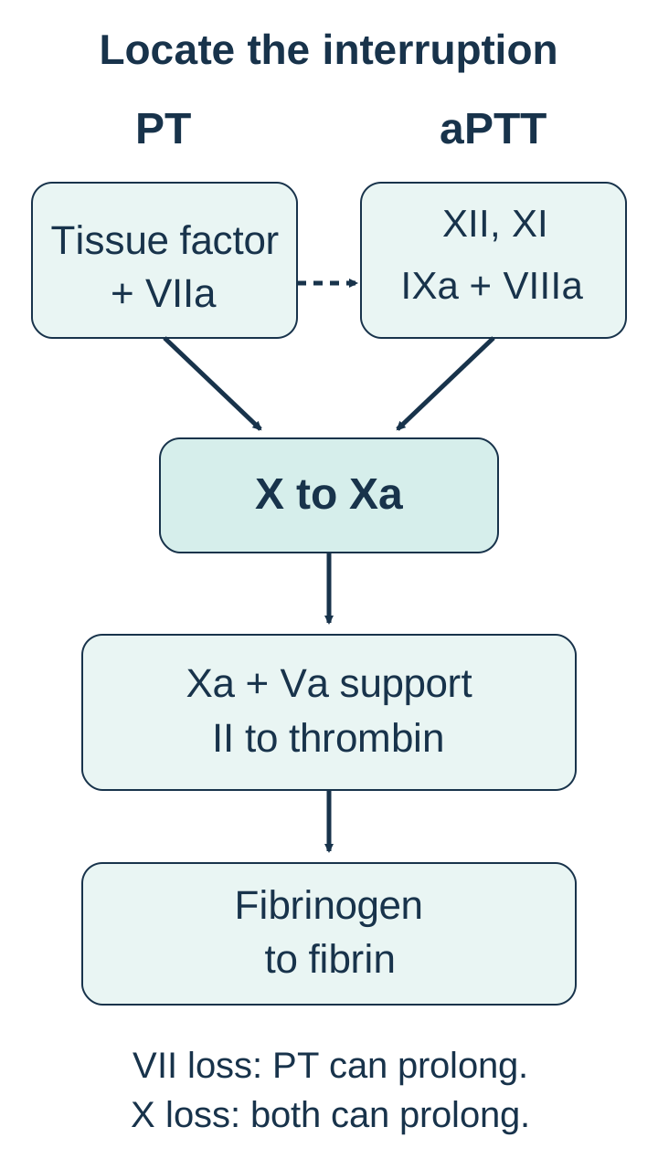
Why can PT be prolonged while aPTT is normal? Factor VII is a liver-synthesized, vitamin K-dependent protein. At vascular injury, exposed tissue factor binds activated factor VII, written VIIa. This complex activates factors X and IX. Activated X, with factor V, supports conversion of prothrombin to thrombin; thrombin then converts fibrinogen to fibrin. Initiation and amplification cooperate in the body rather than operating as two independent circulation systems. [1]
The laboratory begins these reactions in different ways. PT uses a tissue-factor-containing reagent and is particularly sensitive to factor VII. The aPTT begins through contact activation and assesses intrinsic and common-pathway components. A severe isolated VII defect can therefore prolong PT while leaving aPTT within its reference interval. A normal aPTT does not mean that physiological clot initiation is adequate. [1][2]
Localize the pattern before naming its cause
Pattern
Factor location to consider
Important qualification
PatternPT prolonged; aPTT normal
Factor location to considerVII in tissue-factor-driven initiation
Important qualificationAcquired deficiencies, drugs and assay interference can produce this pattern.
PatternPT normal; aPTT prolonged
Factor location to considerVIII, IX or XI in a bleeding patient
Important qualificationContact-factor deficiencies and lupus anticoagulant can prolong aPTT without the same bleeding phenotype.
PatternBoth prolonged
Factor location to considerCommon-pathway II, V, X or fibrinogen; multiple factors
Important qualificationSeverity and reagent sensitivity affect which tests become abnormal.
Trace the pathway diagram from each assay's starting point. Predict what happens when VII is deficient, then repeat the tracing with X deficient. The first interruption affects the PT route; the second affects the shared route and can prolong both tests. [2][3]
Trace each assay route, first with VII deficient and then with X deficient. The dashed connection also represents physiological activation of IX by tissue factor and VIIa. This is a simplified assay-location model, not a complete coagulation network. [1][2][3]Why does a normal platelet count not settle this question?
The count measures platelet number, not factor VII function. A patient can have normal platelet numbers and defective tissue-factor-driven coagulation. [2]
Apply the distinction: recurrent nosebleeds might suggest a platelet or von Willebrand disorder, but a reproducible isolated prolonged PT calls for a coagulation-factor investigation. Conversely, persistent bleeding with normal PT and aPTT still deserves investigation; these screening tests do not detect every hemostatic defect. [2][3]
Ask what changed before diagnosing what was inherited
Does a low factor VII result prove congenital deficiency? No. First assess active bleeding and specimen validity, then medication exposure, nutrition, intestinal absorption and liver disease. A repeat sample may resolve a collection-related or transient abnormality. Severe bleeding requires stabilization and urgent specialist care in parallel with this investigation, not after its completion. [2][5]
Factor VII has a short circulating half-life, approximately 3 to 6 hours. Reduced availability of vitamin K, vitamin K antagonism and acute loss of liver synthetic function may affect PT early, before aPTT becomes abnormal. Vitamin K supports functional production of factors II, VII, IX and X. Warfarin and vitamin K deficiency therefore affect a family of proteins rather than selectively creating an inherited VII defect. [1][4][8]
Ask specifically about warfarin, direct oral anticoagulants, prolonged antibiotic exposure, reduced dietary intake, cholestasis and malabsorption. Assess liver findings and selected additional factor activities when needed. Factor V is not vitamin K dependent: preserved V with reduced vitamin K-dependent factors favors a vitamin K-related pattern, whereas reduced V can support broader hepatic synthesis failure or consumption. No single factor result independently excludes liver disease or disseminated intravascular coagulation. Platelets, fibrinogen, fibrin-related markers and clinical context remain important. [2][3]
Compare two patients with the same PT. In one, the abnormality began after warfarin and II, VII, IX and X activities fell. In the other, repeated testing off interfering drugs shows only VII reduced. Predict whether giving a name to the low VII result explains the whole pattern.
Worked comparison: the first patient has a medication-associated multiple-factor pattern; the second warrants investigation of a selective VII abnormality. Correction after clinician-directed vitamin K replacement supports a vitamin K-responsive cause, but lack of correction is not by itself proof of a genetic disorder. [2][3][4]
Can a normal aPTT exclude a direct factor Xa inhibitor?
No. The effect depends on the drug, concentration and reagent. Direct Xa inhibitors can prolong PT, imitate inhibition in mixing studies and underestimate clot-based factor activities. Medication timing and laboratory advice are needed before interpreting such results. [4]
Transfer: a patient with new jaundice and an abnormal PT should not receive an inherited diagnosis from a single low VII assay. The new illness and the broader factor pattern must be explained first.
A mixing study asks whether normal plasma supplies what is missing
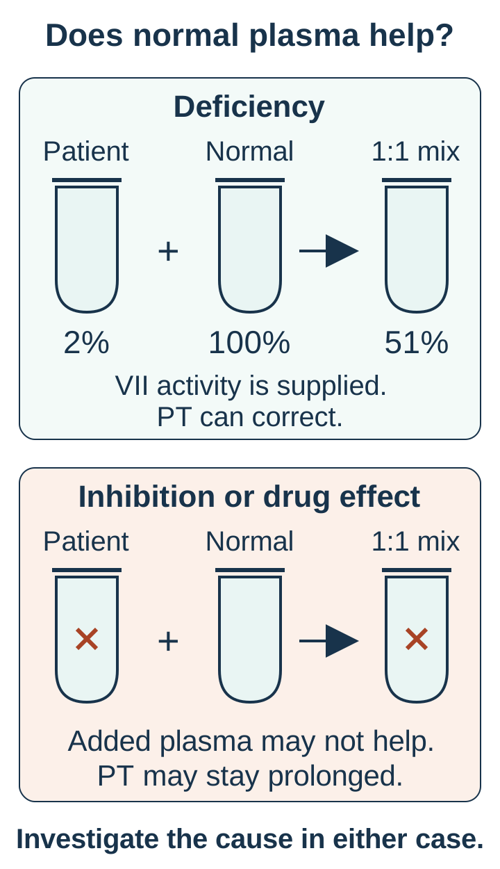
What does correction actually establish? In a 1:1 mixing study, the laboratory combines equal volumes of patient plasma and normal plasma, then measures the clotting time. Substantial correction by the laboratory's validated criteria supports a factor deficiency. Failure to correct suggests an inhibitor or interfering anticoagulant. Neither result, by itself, identifies the cause. Weak inhibitors, multiple deficiencies and drug effects can complicate interpretation. [3][4]
The mixing diagram separates two mechanisms. In a simple deficiency, the normal plasma contributes working protein. In inhibition, the added protein may also be impaired. Assume a sample has PT 30 seconds, normal plasma PT 12 seconds, and mixed PT 13 seconds, with a local reference interval of 11 to 14 seconds. The result supports deficiency. It is not the arithmetic average of the two original clotting times.
Compare how adding normal plasma changes the available factor concentration. Supplying normal plasma distinguishes broad patterns; it does not establish the cause of the deficiency or prove a particular inhibitor. [3][4]
Predict the result before reading the explanation: a sample contains 2% VII activity, normal plasma contains 100%, and equal volumes are mixed with no inhibitor or interference. The estimated activity becomes (2 + 100) / 2 = 51%. Supplying activity can restore the assay endpoint despite the original patient's deficiency. This simplified concentration calculation is not a formula for PT seconds or bleeding risk.
What changes when the mixture remains prolonged?
Supplying normal plasma has not restored the expected test response. Review interfering drugs and arrange specialist inhibitor investigation rather than interpreting the result as uncomplicated inherited deficiency. [3][4]
A rare specific VII inhibitor is considered when the persistent laboratory pattern fits and drug interference has been addressed. Lupus anticoagulant more often affects aPTT-based testing and is not the default explanation for isolated PT prolongation. The specialist laboratory may select immediate and incubated testing when clinically appropriate; incubation is not a universal detector of every inhibitor. [2][3][4]
After confirming a persistent, otherwise unexplained isolated PT abnormality, measure VII coagulant activity, or VII:C, and repeat the measurement for confirmation. Selected other factor assays distinguish an isolated defect from a broader deficiency. Antigen testing asks how much protein is present; activity asks how effectively it works. An inherited qualitative defect can leave protein quantity relatively preserved. F7 testing can help resolve selected diagnostic questions or support family counseling, especially in severe disease. It is not required before treating an urgent bleed. [1][3]
Assay details matter when the history and result disagree. Some VII variants react differently to thromboplastin from different species. The Nordic guideline favors human-tissue or recombinant tissue factor for activity testing. A strikingly low result with one reagent, but a mild phenotype, warrants laboratory clarification rather than a prediction of inevitable severe bleeding. In young children, use age-appropriate reference intervals, repeat low results and exclude vitamin K deficiency and liver disease. [1][8]
Transfer: two correcting mixtures can represent different diseases. One patient may have a reversible acquired deficiency, while another has persistently isolated low VII activity. Mixing identifies the broad mechanism; context and specific assays determine the diagnosis.
The activity result and the bleeding history answer different questions
Does 2% activity mean the same risk for every patient? It does not. Markedly low activity increases concern, particularly in infants, but individual bleeding severity and VII:C correlate imperfectly. Some patients with very low activity have little bleeding; others bleed during menstruation, trauma or procedures despite measurable activity. Activity remains useful information, not a stand-alone forecast. [1][8]
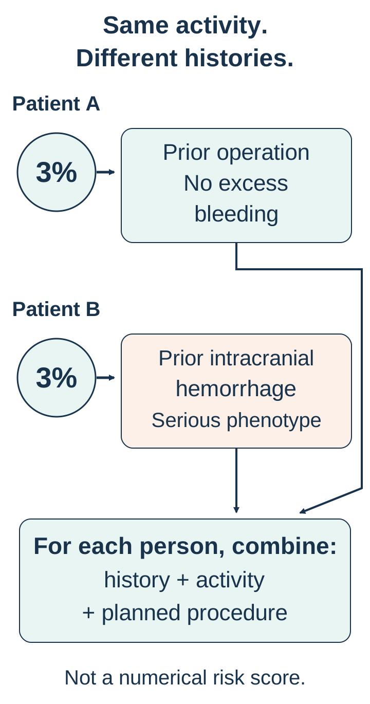
In the comparison diagram, the same activity is paired with two different histories. One patient has completed substantial hemostatic challenges without excess bleeding. The other has had spontaneous intracranial hemorrhage. Their assessment should differ even before considering the planned procedure. A lack of prior symptoms is less reassuring in a young child who has never had surgery than in an adult who has already tolerated comparable challenges.
These are constructed examples, not a risk score or clinical records. Identical activity does not establish identical observed bleeding severity; a favorable history also does not guarantee future safety. [1][6][10]
Classify each observation before making a plan: 2% VII:C is a laboratory measurement; occasional self-limited epistaxis is a mucosal bleeding history; recurrent joint bleeding or intracranial hemorrhage establishes a serious clinical phenotype. These are not interchangeable descriptions.
Worked consequence: major bleeding history increases the need for specialist prevention planning, while an isolated laboratory result cannot establish that such bleeding has occurred. Assessment combines activity, personal and family history, age, the anatomical site and extent of the procedure, other hemostatic abnormalities, and thrombotic risk. [1][6]
Does an uncomplicated earlier operation guarantee the next operation is safe?
No. The next procedure may involve a different site or greater tissue injury, and the patient's health may have changed. Prior challenges inform the assessment but do not replace procedure-specific planning. [6]
Prior surgical bleeding is clinically important to document, but calling it a universally strong predictor overstates the evidence. The Nordic guideline describes a retrospective surgical cohort in which previous surgical bleeding did not predict later bleeding, although frequent epistaxis and excessive trauma-related bleeding did. Use the whole phenotype instead of assigning all predictive weight to one previous operation. [6]
Transfer: an asymptomatic person with very low activity and a planned minor skin procedure needs confirmation, counseling and an individualized plan, not an automatic lifelong replacement schedule. A person with prior intracranial or recurrent joint bleeding requires a different level of preventive assessment, even if the activity value is higher. [1][6][10]
Recognize the phenotype across childhood and reproductive life
Can an inherited coagulation disorder present as nosebleeds rather than joint bleeding? Yes. Congenital VII deficiency is usually autosomal recessive and can affect any sex. Epistaxis, gum bleeding, bruising, heavy menstrual bleeding and bleeding after dental work or surgery are common presentations. Severe disease can include gastrointestinal, joint, muscle or intracranial bleeding. A normal platelet count does not contradict any of these factor-related presentations. [1][8]
Autosomal recessive inheritance also explains why an affected child may have no previously diagnosed relatives. When each parent carries one pathogenic F7 variant, each pregnancy has a 1 in 4 chance of a child inheriting both affected alleles. This probability is independent of the child's sex and of earlier pregnancy outcomes. It predicts inheritance, not the precise bleeding severity. Genotype and activity do not fully determine the phenotype. [1]
Consider two siblings with the same familial variants. One has heavy menstrual bleeding and iron deficiency; the other has not yet faced surgery or major trauma. Predict whether the second history proves that the first person's bleeding must have another cause.
Worked answer: different hemostatic challenges and variable expression can produce different observed histories. Investigate other causes when appropriate, but do not dismiss an inherited factor disorder because a relative has fewer symptoms. In the patient with heavy menstrual bleeding, assess iron status and address ongoing blood loss as well as iron replacement. [1][7]
Why is infancy a special diagnostic setting?
Vitamin K-dependent factor levels are normally lower than adult levels early in life. Vitamin K deficiency and liver disease can further reduce them. Age-adjusted interpretation and repeat testing help prevent an acquired or developmental pattern from becoming an unsupported congenital diagnosis. [8]
For pregnancy, involve hematology, obstetrics and the delivery team early. Assess bleeding history, prior postpartum hemorrhage, current VII activity and the intended delivery or procedure. VII activity may rise during pregnancy; this does not erase an established inherited diagnosis. A third-trimester measurement helps plan delivery, whereas a pregnancy-associated value may not represent the nonpregnant baseline. [1][7]
The Nordic guidance reserves prophylactic replacement at delivery for high-risk patients and calls for hemostatic treatment to be readily available. This is a specialist plan, not a reason to prohibit pregnancy or assume postpartum hemorrhage is inevitable. Heavy menstrual bleeding and fertility goals also merit coordinated gynecologic and hematologic care. [7]
Transfer: a first presentation after dental extraction and a first presentation with heavy menstrual bleeding can share the same coagulation mechanism. The site and consequences of bleeding determine what else must be assessed.
Treat the clinical bleed and prepare for the next challenge
What is the treatment target: a normal PT or effective hemostasis? Effective hemostasis. For life-threatening bleeding in confirmed VII deficiency, arrange urgent factor-directed therapy with hematology, resuscitation and site-specific emergency care. Do not wait for F7 sequencing. For elective procedures, agree on the preventive plan before the procedure rather than responding only after hemorrhage begins. [5][6][9]
Recombinant activated VII, or rFVIIa, is a preferred replacement option when available. The United States NovoSeven RT label lists 15 to 30 micrograms/kg intravenously every 4 to 6 hours for bleeding in congenital VII deficiency, individualized to response; perioperative treatment begins before surgery. This is distinct from the higher schedules used for hemophilia with inhibitors. Repeat dosing may be necessary because the drug is short acting. The prescribing team determines the schedule, duration and monitoring. [5][9]
Monitor clinical blood loss, vital signs, hemoglobin when appropriate, the affected site and laboratory response. The label calls for PT and VII activity monitoring in deficient patients and consideration of antibodies when expected response is absent. It also warns that coagulation-test normalization does not reliably establish successful hemostasis. Continued wound bleeding after a shorter PT requires reassessment of local bleeding, replacement response and other contributors. [9]
Plasma-derived VII, prothrombin complex concentrate and plasma are alternatives in selected settings, not interchangeable default choices. Availability varies. Plasma carries a volume burden. PCC contains other vitamin K-dependent factors with longer half-lives, which can accumulate with repeated dosing and increase thrombotic risk. Both inherited VII deficiency and a prolonged PT can coexist with thrombosis; neither provides reliable protection from it. [1][5]
Local pressure, appropriate procedural hemostasis and antifibrinolytic therapy can help selected mucosal or dental bleeding. Tranexamic acid limits fibrin breakdown rather than supplying VII, so symptom improvement does not require PT normalization. It can be used alone in selected mild situations or with replacement when indicated. The Nordic guidance cautions against its use in renal-tract bleeding; site, renal function, contraindications and thrombotic history must be considered by the treating team. [5]
Compare treatment mechanisms: factor replacement improves clot initiation; tranexamic acid helps preserve formed fibrin; iron replacement treats the consequence of chronic blood loss. Predict which intervention could improve anemia without controlling the next menstrual bleed.
Worked consequence: iron alone can replenish depleted stores but does not supply a hemostatic plan. Conversely, antifibrinolytic benefit does not mean that the inherited factor deficiency has disappeared. Selected mild dental or menstrual presentations may be managed without routine replacement, while major surgery or serious spontaneous bleeding may require it. [5][6][7]
When does recurrent bleeding justify discussing long-term prevention?
Recurrent serious bleeding, especially intracranial, gastrointestinal or joint bleeding, warrants specialist consideration of prophylaxis. STER supports this approach in selected severe phenotypes; it does not establish that every asymptomatic low activity result requires long-term treatment. [10]
The balance includes thrombosis, treatment burden and access, not only bleeding. rFVIIa carries a warning for serious arterial and venous thrombosis. It should not be combined casually with other procoagulant concentrates; the label specifically advises avoiding simultaneous activated PCC. A new suspected thrombotic event needs prompt assessment, not dismissal because the person has a bleeding disorder. For postoperative leg symptoms with a high clinical likelihood of DVT, venous imaging is appropriate; an increased D-dimer after surgery is not by itself diagnostic. [9][11]
For the next encounter: confirm the diagnosis, document the individual's bleeding history and treatment response, and provide a written plan for procedures, trauma and emergency contact. A stable asymptomatic patient needs counseling and follow-up rather than treatment solely to improve a laboratory display. [1][5][6]
Apply the lesson to a new patient
Case 1
Show answer and explanations for case 1
A. Measure von Willebrand factor activity (Why this does not fit)
Epistaxis and dental bleeding can occur in von Willebrand disease. A reproducible isolated prolonged PT that corrects on mixing points to a factor in tissue-factor-driven coagulation, not the usual von Willebrand pattern.
Reasoning steps for option A
Why could this test initially seem relevant?
Epistaxis and dental bleeding can occur in von Willebrand disease.
Which laboratory finding requires a different initial target?
A reproducible isolated prolonged PT that corrects on mixing points to a factor in tissue-factor-driven coagulation, not the usual von Willebrand pattern.
B. Measure factor VII coagulant activity (Best answer)
Normal aPTT with a correcting PT supports deficiency in the tissue-factor-dependent assay route. VII activity directly tests the factor suggested by that route after acquired causes have been assessed.
Reasoning steps for option B
What does the correcting isolated PT localize?
Normal aPTT with a correcting PT supports deficiency in the tissue-factor-dependent assay route.
Which assay identifies the missing function?
VII activity directly tests the factor suggested by that route after acquired causes have been assessed.
C. Measure factor VIII coagulant activity (Why this does not fit)
A sufficiently severe VIII deficiency usually prolongs aPTT rather than PT. The child's aPTT is normal while PT is reproducibly prolonged, making VIII a poor first localization.
Reasoning steps for option C
Which screening assay is usually affected by VIII deficiency?
A sufficiently severe VIII deficiency usually prolongs aPTT rather than PT.
How does that compare with this child?
The child's aPTT is normal while PT is reproducibly prolonged, making VIII a poor first localization.
D. Measure factor X coagulant activity (Why this does not fit)
Factor X contributes to the common route used by both PT and aPTT. Marked isolated PT prolongation with normal aPTT favors the tissue-factor-specific component over a severe common-pathway defect.
Reasoning steps for option D
Where does X participate in the assay pathways?
Factor X contributes to the common route used by both PT and aPTT.
Why does the supplied pattern favor VII?
Marked isolated PT prolongation with normal aPTT favors the tissue-factor-specific component over a severe common-pathway defect.
Takeaway: A correcting isolated PT directs functional testing toward VII, but acquired causes still determine whether the disorder is inherited.
A. Selective neutralization of circulating VII by an antibody (Why this does not fit)
A VII inhibitor can produce a low VII activity and an abnormal PT. Concordant decreases in II, IX and X after warfarin indicate a broader vitamin K-dependent effect rather than isolated neutralization of VII.
Reasoning steps for option A
What pattern could a selective VII antibody cause?
A VII inhibitor can produce a low VII activity and an abnormal PT.
Which supplied feature is not explained by selectivity?
Concordant decreases in II, IX and X after warfarin indicate a broader vitamin K-dependent effect rather than isolated neutralization of VII.
B. Reduced synthesis of vitamin K-dependent and independent factors alike (Why this does not fit)
Severe hepatic synthetic failure can reduce vitamin K-dependent factors and factor V. V is unchanged while the vitamin K-dependent group falls after warfarin and a documented normal baseline.
Reasoning steps for option B
Which illness could impair a broad set of hepatic factors?
Severe hepatic synthetic failure can reduce vitamin K-dependent factors and factor V.
Why is a selective drug effect more coherent here?
V is unchanged while the vitamin K-dependent group falls after warfarin and a documented normal baseline.
C. Consumption of coagulation proteins during systemic fibrin formation (Why this does not fit)
Disseminated consumption can lower multiple factors during systemic coagulation activation. The change follows warfarin and is concentrated in vitamin K-dependent factors with preserved V, fitting vitamin K antagonism more directly.
Reasoning steps for option C
What process can lower several coagulation factors?
Disseminated consumption can lower multiple factors during systemic coagulation activation.
Which supplied temporal and biochemical pattern favors another process?
The change follows warfarin and is concentrated in vitamin K-dependent factors with preserved V, fitting vitamin K antagonism more directly.
D. Impaired vitamin K recycling with early loss of functional VII (Best answer)
II, VII, IX and X require vitamin K for functional production. The short half-life of VII allows its functional level to fall early after vitamin K antagonism, making the tissue-factor-driven assay abnormal first.
Reasoning steps for option D
Which shared property links the factors that fell?
II, VII, IX and X require vitamin K for functional production.
Why can PT change before aPTT?
The short half-life of VII allows its functional level to fall early after vitamin K antagonism, making the tissue-factor-driven assay abnormal first.
Takeaway: Early isolated PT prolongation can be the first laboratory expression of a multiple-factor vitamin K antagonist effect.
A. The findings support acquired vitamin K-dependent factor deficiency (Best answer)
Several vitamin K-dependent factors are low despite preserved V, with nutritional and intestinal risk factors. Restoration of the factor activities and PT supports a reversible vitamin K-responsive deficiency rather than an isolated inherited VII defect.
Reasoning steps for option A
What does the initial factor pattern suggest?
Several vitamin K-dependent factors are low despite preserved V, with nutritional and intestinal risk factors.
What does recovery after vitamin K add?
Restoration of the factor activities and PT supports a reversible vitamin K-responsive deficiency rather than an isolated inherited VII defect.
B. The findings support inherited isolated factor VII deficiency (Why this does not fit)
VII deficiency can produce a correcting isolated prolonged PT. II and X were also reduced, and all abnormalities resolved after vitamin K, favoring an acquired multiple-factor process.
Reasoning steps for option B
Why was VII deficiency initially plausible?
VII deficiency can produce a correcting isolated prolonged PT.
What additional findings argue against it as the explanation?
II and X were also reduced, and all abnormalities resolved after vitamin K, favoring an acquired multiple-factor process.
C. The findings support an acquired factor VII neutralizing inhibitor (Why this does not fit)
It can prolong PT and lower measured VII activity. Correction on mixing and recovery of several vitamin K-dependent activities after vitamin K fit replacement of missing functional production rather than selective inhibition.
Reasoning steps for option C
How can a VII inhibitor affect the initial screen?
It can prolong PT and lower measured VII activity.
Why does the complete course favor deficiency instead?
Correction on mixing and recovery of several vitamin K-dependent activities after vitamin K fit replacement of missing functional production rather than selective inhibition.
D. The findings support primary hepatic failure of factor synthesis (Why this does not fit)
The liver produces most coagulation factors, and synthetic dysfunction can prolong PT. Preserved V and normalization of the affected activities after vitamin K better support vitamin K deficiency than primary persistent hepatic synthetic failure.
Reasoning steps for option D
Why does liver disease enter the differential?
The liver produces most coagulation factors, and synthetic dysfunction can prolong PT.
Which supplied findings favor a vitamin K-responsive process?
Preserved V and normalization of the affected activities after vitamin K better support vitamin K deficiency than primary persistent hepatic synthetic failure.
Takeaway: A correcting mixture identifies deficiency; the affected factor group and response to vitamin K help identify its cause.
A. An isolated inherited defect in VII function (Why this does not fit)
The initial isolated PT prolongation could resemble a VII-specific abnormality. Falling V and prolongation of both tests during acute liver illness are not explained by a persistent isolated VII defect.
Reasoning steps for option A
Which part of the course could resemble isolated VII deficiency?
The initial isolated PT prolongation could resemble a VII-specific abnormality.
Which later findings require a broader explanation?
Falling V and prolongation of both tests during acute liver illness are not explained by a persistent isolated VII defect.
B. Nutritional depletion of vitamin K alone (Why this does not fit)
Functional VII can decline before longer-lived vitamin K-dependent factors. Factor V is not vitamin K dependent, and its marked decline with failure to respond to vitamin K favors broader synthetic dysfunction.
Reasoning steps for option B
Why can vitamin K depletion initially prolong PT?
Functional VII can decline before longer-lived vitamin K-dependent factors.
What limits that explanation in this case?
Factor V is not vitamin K dependent, and its marked decline with failure to respond to vitamin K favors broader synthetic dysfunction.
C. Hepatic synthetic dysfunction affecting several coagulation factors (Best answer)
It shows that the defect extends beyond vitamin K-dependent functional production. Short-lived VII can affect PT early; subsequent broader factor reduction can prolong both PT and aPTT as hepatic synthesis deteriorates.
Reasoning steps for option C
What does low V add to the low VII result?
It shows that the defect extends beyond vitamin K-dependent functional production.
How does the time course fit the liver illness?
Short-lived VII can affect PT early; subsequent broader factor reduction can prolong both PT and aPTT as hepatic synthesis deteriorates.
D. Systemic consumption from disseminated intravascular coagulation (Why this does not fit)
Systemic coagulation activation can consume factors from several pathways. Stable platelets, preserved fibrinogen and no increase in fibrin-related markers favor the documented hepatic synthetic illness over an active consumptive pattern.
Reasoning steps for option D
Why could consumption produce both prolonged tests?
Systemic coagulation activation can consume factors from several pathways.
Which supplied findings make it less supported here?
Stable platelets, preserved fibrinogen and no increase in fibrin-related markers favor the documented hepatic synthetic illness over an active consumptive pattern.
Takeaway: Low V helps identify a process broader than isolated vitamin K-dependent factor impairment; interpret it with the whole illness.
A. Send F7 sequencing to identify a clinically silent inherited variant (Why this does not fit)
It can resolve selected confirmed or strongly suspected inherited cases and assist family counseling. The abnormality was confined to an unsuitable specimen and disappeared on a valid repeat, so a persistent VII phenotype has not been established.
Reasoning steps for option A
When can genetic testing contribute to a VII evaluation?
It can resolve selected confirmed or strongly suspected inherited cases and assist family counseling.
What evidence is missing here?
The abnormality was confined to an unsuitable specimen and disappeared on a valid repeat, so a persistent VII phenotype has not been established.
B. Obtain a factor VII inhibitor titer despite the normal repeat PT (Why this does not fit)
A persistent abnormal assay with an inhibitory mixing pattern raises that question. Both screening tests are normal on the suitable sample, with no clinical bleeding to support targeted inhibitor testing.
Reasoning steps for option B
What laboratory behavior usually prompts inhibitor investigation?
A persistent abnormal assay with an inhibitory mixing pattern raises that question.
Does the valid repeat supply that behavior?
Both screening tests are normal on the suitable sample, with no clinical bleeding to support targeted inhibitor testing.
C. Document the specimen issue and assess procedural bleeding history (Best answer)
The properly collected repeat sample shows no persistent screening-test abnormality. Do not label an inherited factor disorder from the unsuitable sample; retain ordinary history-based procedural assessment.
Reasoning steps for option C
Which measurement can be interpreted reliably?
The properly collected repeat sample shows no persistent screening-test abnormality.
What conclusion follows without ignoring clinical safety?
Do not label an inherited factor disorder from the unsuitable sample; retain ordinary history-based procedural assessment.
D. Measure the vitamin K-dependent factors to classify the original abnormality (Why this does not fit)
It is useful when a valid persistent abnormality requires explanation. The original result is not interpretable and the valid repeat is normal, so the proposed panel would be classifying a finding that has not been confirmed.
Reasoning steps for option D
When does a factor panel help classify prolonged PT?
It is useful when a valid persistent abnormality requires explanation.
Why is it not the best next action here?
The original result is not interpretable and the valid repeat is normal, so the proposed panel would be classifying a finding that has not been confirmed.
Takeaway: Confirm that a screening abnormality is real before using it to establish a lifelong diagnosis.
A. An acquired inhibitor impairing factor VII function (Best answer)
It suggests inhibition rather than an uncomplicated absence of a coagulation factor. The new isolated PT abnormality and selectively low VII activity, after drug interference is addressed, support testing for a specific VII inhibitor.
Reasoning steps for option A
What does failure of normal plasma to correct PT suggest?
It suggests inhibition rather than an uncomplicated absence of a coagulation factor.
What narrows the inhibitor investigation?
The new isolated PT abnormality and selectively low VII activity, after drug interference is addressed, support testing for a specific VII inhibitor.
B. An inherited reduction in production of factor VII (Why this does not fit)
Normal plasma usually supplies enough VII to substantially correct the PT. The mixture remains prolonged and PT was previously normal, favoring a newly acquired inhibitory mechanism.
Reasoning steps for option B
How should an uncomplicated inherited VII deficiency respond to mixing?
Normal plasma usually supplies enough VII to substantially correct the PT.
Which findings argue against that as the full explanation?
The mixture remains prolonged and PT was previously normal, favoring a newly acquired inhibitory mechanism.
C. An acquired impairment of vitamin K-dependent factor production (Why this does not fit)
It reduces functional production of several vitamin K-dependent proteins, including II, VII, IX and X. II and X are normal and normal plasma does not correct the PT, unlike the expected uncomplicated deficiency pattern.
Reasoning steps for option C
What factor pattern can vitamin K deficiency produce?
It reduces functional production of several vitamin K-dependent proteins, including II, VII, IX and X.
Why is it less explanatory here?
II and X are normal and normal plasma does not correct the PT, unlike the expected uncomplicated deficiency pattern.
D. A broad reduction in hepatic coagulation protein synthesis (Why this does not fit)
It can lower multiple factors, including vitamin K-independent V. Normal II, V and X with a selectively low VII and a noncorrecting mixture support targeted inhibition rather than broad synthesis failure.
Reasoning steps for option D
How does hepatic synthetic failure commonly affect coagulation?
It can lower multiple factors, including vitamin K-independent V.
What makes it a poor match to the supplied data?
Normal II, V and X with a selectively low VII and a noncorrecting mixture support targeted inhibition rather than broad synthesis failure.
Takeaway: A noncorrecting isolated PT with selective VII loss warrants inhibitor evaluation only after anticoagulant and specimen interference are addressed.
A. Factor VII antigen concentration (Why this does not fit)
It can compare the amount of VII protein with its measured function. It does not quantify the rivaroxaban present shortly after dosing, which can distort the clot-based activity and mixing results.
Reasoning steps for option A
What distinction can antigen testing help make?
It can compare the amount of VII protein with its measured function.
Why does it not resolve the immediate confounder?
It does not quantify the rivaroxaban present shortly after dosing, which can distort the clot-based activity and mixing results.
B. Factor VII inhibitor quantitation (Why this does not fit)
A noncorrecting mixture can prompt investigation for a factor inhibitor. A direct Xa inhibitor can imitate inhibition and distort inhibitor testing, so this sample cannot be classified confidently without addressing the drug.
Reasoning steps for option B
Why might the mixture suggest this test?
A noncorrecting mixture can prompt investigation for a factor inhibitor.
Why must drug interference be considered first?
A direct Xa inhibitor can imitate inhibition and distort inhibitor testing, so this sample cannot be classified confidently without addressing the drug.
C. Sequencing of the F7 coding region (Why this does not fit)
It investigates inherited variants rather than measuring a circulating anticoagulant. The abnormality appeared during rivaroxaban use and the sample was drawn near the dose, making drug-related assay interference the immediate concern.
Reasoning steps for option C
What question does sequencing answer?
It investigates inherited variants rather than measuring a circulating anticoagulant.
Which supplied facts make another test more direct?
The abnormality appeared during rivaroxaban use and the sample was drawn near the dose, making drug-related assay interference the immediate concern.
D. Rivaroxaban-calibrated anti-Xa assay (Best answer)
No. Its effect on routine coagulation assays varies with concentration and reagent. A drug-calibrated anti-Xa assay assesses rivaroxaban exposure and helps the laboratory interpret whether clot-based VII testing is reliable.
Reasoning steps for option D
Can a normal aPTT exclude rivaroxaban interference?
No. Its effect on routine coagulation assays varies with concentration and reagent.
Which assay targets the suspected confounder?
A drug-calibrated anti-Xa assay assesses rivaroxaban exposure and helps the laboratory interpret whether clot-based VII testing is reliable.
Takeaway: Address direct anticoagulant interference before interpreting a low clot-based factor activity as an inherited deficiency or inhibitor.
A. 2% activity; PT remains prolonged (Why this does not fit)
It adds functional VII to the patient's plasma. Equal-volume mixing with 100% plasma raises the estimated activity to 51%, rather than leaving it at the patient's original 2%.
Reasoning steps for option A
What does normal pooled plasma add?
It adds functional VII to the patient's plasma.
Why is 2% not the expected mixture activity?
Equal-volume mixing with 100% plasma raises the estimated activity to 51%, rather than leaving it at the patient's original 2%.
B. 51% activity; PT enters the reference interval (Best answer)
The mean of 2% and 100% is 51%. It exceeds the supplied 40% threshold, so PT is expected to enter the reference interval despite the patient's original deficiency.
Reasoning steps for option B
What is the equal-volume concentration estimate?
The mean of 2% and 100% is 51%.
How does that estimate relate to this assay's stated requirement?
It exceeds the supplied 40% threshold, so PT is expected to enter the reference interval despite the patient's original deficiency.
C. 51% activity; PT remains prolonged (Why this does not fit)
Yes. Equal volumes give an estimated VII activity of 51%. This assay is stated to be normal above 40% when other factors are adequate, and both conditions are met.
Reasoning steps for option C
Is the concentration estimate correct?
Yes. Equal volumes give an estimated VII activity of 51%.
Why does the PT prediction conflict with the stem?
This assay is stated to be normal above 40% when other factors are adequate, and both conditions are met.
D. 100% activity; PT enters the reference interval (Why this does not fit)
No. It is diluted by an equal volume of plasma with only 2% activity. The mixture reaches approximately 51%, enough for the stated assay; it does not need to restore activity to 100%.
Reasoning steps for option D
Does normal plasma retain its original concentration after mixing?
No. It is diluted by an equal volume of plasma with only 2% activity.
What does correction actually require in this example?
The mixture reaches approximately 51%, enough for the stated assay; it does not need to restore activity to 100%.
Takeaway: Mixing averages concentrations under simplified conditions, not PT seconds; partial restoration of activity can normalize the assay.
A. Loss of the tissue-factor-specific initiator delays PT alone (Why this does not fit)
An isolated VII deficiency affects tissue-factor-driven initiation. VII activity is normal and both screening tests are prolonged, while X is severely deficient.
Reasoning steps for option A
Which factor defect typically isolates the PT route?
An isolated VII deficiency affects tissue-factor-driven initiation.
Which supplied findings exclude that as the main explanation?
VII activity is normal and both screening tests are prolonged, while X is severely deficient.
B. Loss of an intrinsic-pathway cofactor delays aPTT alone (Why this does not fit)
A sufficiently severe VIII-related intrinsic-pathway defect could do so. PT is also prolonged and the assay identifies severe X deficiency, a component shared by both routes.
Reasoning steps for option B
Which kind of defect could selectively prolong aPTT?
A sufficiently severe VIII-related intrinsic-pathway defect could do so.
What additional result requires a common-pathway explanation?
PT is also prolonged and the assay identifies severe X deficiency, a component shared by both routes.
C. Loss of a common-pathway component delays both assay routes (Best answer)
Both PT and aPTT require factor X-dependent common-pathway thrombin generation. Severely reduced X can prolong both tests, while normal plasma supplies the missing activity and corrects them.
Reasoning steps for option C
Where does X participate after each assay is initiated?
Both PT and aPTT require factor X-dependent common-pathway thrombin generation.
How do the activity and mixing results fit?
Severely reduced X can prolong both tests, while normal plasma supplies the missing activity and corrects them.
D. Loss of fibrin cross-linking delays formation of the initial clot (Why this does not fit)
Factor XIII acts after the initial fibrin clot forms. PT and aPTT can remain normal in XIII deficiency; this patient's tests and direct assay instead identify a severe common-pathway X deficit.
Reasoning steps for option D
Which factor stabilizes fibrin by cross-linking?
Factor XIII acts after the initial fibrin clot forms.
Why is that not the explanation for these screening results?
PT and aPTT can remain normal in XIII deficiency; this patient's tests and direct assay instead identify a severe common-pathway X deficit.
Takeaway: A shared common-pathway defect can prolong both PT and aPTT; a VII-specific defect is not the answer to every prolonged PT.
A. Review thromboplastin sensitivity with the coagulation laboratory (Best answer)
The thromboplastin reagent changed while the specimen remained the same. Some VII variants have reagent-dependent activity results, so assay clarification and clinical correlation should precede severity assignment.
Reasoning steps for option A
What changed between the two activity measurements?
The thromboplastin reagent changed while the specimen remained the same.
What known feature of VII testing makes that important?
Some VII variants have reagent-dependent activity results, so assay clarification and clinical correlation should precede severity assignment.
B. Classify severity using the lower activity measurement alone (Why this does not fit)
It seems to provide a cautious estimate when a marked deficiency is reported. The same sample has a large reagent-dependent discrepancy and a mild observed phenotype, which require explanation rather than automatic reliance on one number.
Reasoning steps for option B
Why might the lowest result appear attractive?
It seems to provide a cautious estimate when a marked deficiency is reported.
Why is that not a defensible resolution?
The same sample has a large reagent-dependent discrepancy and a mild observed phenotype, which require explanation rather than automatic reliance on one number.
C. Average the two activities to set a procedural replacement dose (Why this does not fit)
It treats them as interchangeable estimates of the same assay behavior. Different tissue-factor reagents can interact differently with a VII variant, and no validated dose follows from averaging the discordant results.
Reasoning steps for option C
What does averaging assume about these two measurements?
It treats them as interchangeable estimates of the same assay behavior.
Why does that assumption fail here?
Different tissue-factor reagents can interact differently with a VII variant, and no validated dose follows from averaging the discordant results.
D. Use an incubated PT mixture to determine which activity is correct (Why this does not fit)
It helps distinguish a deficiency pattern from inhibition or interference. The discrepancy is between activity assays using different reagents on the same sample; reagent characterization is the more direct investigation.
Reasoning steps for option D
What is the usual purpose of a mixing study?
It helps distinguish a deficiency pattern from inhibition or interference.
Why does it not directly answer the current question?
The discrepancy is between activity assays using different reagents on the same sample; reagent characterization is the more direct investigation.
Takeaway: When VII activity depends strongly on the assay reagent, clarify the laboratory method before converting a result into a severity label.
A. Classify the result by the adult interval and then investigate the parents (Why this does not fit)
Vitamin K-dependent factor levels are normally lower early in life. An age-appropriate comparison and confirmation of vitamin K status are missing, so family investigation should not substitute for validating the infant's result.
Reasoning steps for option A
Why can an adult interval misclassify this infant?
Vitamin K-dependent factor levels are normally lower early in life.
Which unresolved issues prevent that classification?
An age-appropriate comparison and confirmation of vitamin K status are missing, so family investigation should not substitute for validating the infant's result.
B. Assess vitamin K and repeat testing with pediatric reference intervals (Best answer)
Developmental factor levels differ from adult levels, and vitamin K prophylaxis is undocumented. Address vitamin K deficiency and liver disease as appropriate, and confirm persistent abnormality using pediatric reference intervals and repeat activity testing.
Reasoning steps for option B
What two nonhereditary issues can affect this result?
Developmental factor levels differ from adult levels, and vitamin K prophylaxis is undocumented.
What must be clarified before an inherited diagnosis?
Address vitamin K deficiency and liver disease as appropriate, and confirm persistent abnormality using pediatric reference intervals and repeat activity testing.
C. Use normal aPTT to establish that the low VII result is inherited (Why this does not fit)
It helps localize the screening abnormality but does not determine its cause. Developmental levels and vitamin K deficiency can still affect VII and PT without proving an inherited disorder.
Reasoning steps for option C
What does a normal aPTT say about an isolated PT pattern?
It helps localize the screening abnormality but does not determine its cause.
Why is it insufficient in this infant?
Developmental levels and vitamin K deficiency can still affect VII and PT without proving an inherited disorder.
D. Use absence of bleeding to classify the low activity as clinically irrelevant (Why this does not fit)
Only three weeks, often without substantial procedural or traumatic challenges. The result needs age-adjusted confirmation and assessment for reversible causes; a short symptom-free history does not establish future safety.
Reasoning steps for option D
How much hemostatic history has this infant accumulated?
Only three weeks, often without substantial procedural or traumatic challenges.
Why is dismissal unsupported?
The result needs age-adjusted confirmation and assessment for reversible causes; a short symptom-free history does not establish future safety.
Takeaway: An infant's factor activity requires pediatric reference intervals and assessment for vitamin K deficiency before an inherited label.
A. Repeat VII activity with a different tissue-factor reagent (Why this does not fit)
They can clarify a discordant low activity result that does not fit the clinical history. VII activity and repeated PT are normal, while delayed rebleeding suggests a problem after initial fibrin formation.
Reasoning steps for option A
When can alternative VII reagents help?
They can clarify a discordant low activity result that does not fit the clinical history.
Why is another target more appropriate here?
VII activity and repeated PT are normal, while delayed rebleeding suggests a problem after initial fibrin formation.
B. Perform a PT mixing study after incubation (Why this does not fit)
An unexplained prolonged PT is needed to assess correction meaningfully. Repeated PT is normal, so mixing does not directly test the suspected defect in clot stabilization.
Reasoning steps for option B
What baseline abnormality makes a PT mixture informative?
An unexplained prolonged PT is needed to assess correction meaningfully.
Does this child have that abnormality?
Repeated PT is normal, so mixing does not directly test the suspected defect in clot stabilization.
C. Measure factor IX coagulant activity (Why this does not fit)
It usually prolongs aPTT by impairing an intrinsic-pathway component. The repeatedly normal aPTT and delayed rebleeding after initial closure favor investigation of fibrin stabilization rather than a severe IX deficit.
Reasoning steps for option C
How can severe IX deficiency appear on screening?
It usually prolongs aPTT by impairing an intrinsic-pathway component.
Which supplied findings favor a later step?
The repeatedly normal aPTT and delayed rebleeding after initial closure favor investigation of fibrin stabilization rather than a severe IX deficit.
D. Measure factor XIII functional activity (Best answer)
It raises concern about the stability of a clot that initially formed. XIII cross-links formed fibrin, while PT and aPTT end at initial clot formation; a functional XIII assay directly evaluates that later step.
Reasoning steps for option D
Which aspect of hemostasis does delayed rebleeding question?
It raises concern about the stability of a clot that initially formed.
Why can XIII deficiency escape these screens?
XIII cross-links formed fibrin, while PT and aPTT end at initial clot formation; a functional XIII assay directly evaluates that later step.
Takeaway: Normal PT and aPTT do not exclude a serious bleeding disorder when the defect acts after initial fibrin formation.
A. Superimposed disseminated intravascular coagulation (Best answer)
Both clotting times are now prolonged with thrombocytopenia, low fibrinogen and markedly increased fibrin breakdown products. Septic shock can trigger systemic coagulation activation and consumption, explaining the broad new pattern beyond the inherited isolated PT abnormality.
Reasoning steps for option A
What distinguishes the current results from baseline VII deficiency?
Both clotting times are now prolonged with thrombocytopenia, low fibrinogen and markedly increased fibrin breakdown products.
How does the new illness connect these abnormalities?
Septic shock can trigger systemic coagulation activation and consumption, explaining the broad new pattern beyond the inherited isolated PT abnormality.
B. Increasing expression of the inherited isolated VII defect (Why this does not fit)
It produced an isolated prolonged PT with preserved platelets and fibrinogen. It does not account for new consumption of platelets and fibrinogen, increased D-dimer and marked aPTT prolongation during sepsis.
Reasoning steps for option B
What pattern did the inherited defect previously produce?
It produced an isolated prolonged PT with preserved platelets and fibrinogen.
Why does it fail to explain the current change?
It does not account for new consumption of platelets and fibrinogen, increased D-dimer and marked aPTT prolongation during sepsis.
C. New selective neutralization of VII by a factor inhibitor (Why this does not fit)
It could further prolong PT and reduce VII function. Thrombocytopenia, fibrinogen depletion and increased fibrin breakdown with both tests prolonged indicate more than selective VII inhibition.
Reasoning steps for option C
What could a selective VII inhibitor add?
It could further prolong PT and reduce VII function.
Which new findings require a wider process?
Thrombocytopenia, fibrinogen depletion and increased fibrin breakdown with both tests prolonged indicate more than selective VII inhibition.
D. Superimposed isolated nutritional vitamin K deficiency (Why this does not fit)
It could reduce vitamin K-dependent factor function and prolong clotting times. The abrupt platelet and fibrinogen depletion with marked D-dimer increase during septic shock is not explained by isolated vitamin K deficiency.
Reasoning steps for option D
How could poor intake affect clotting factors?
It could reduce vitamin K-dependent factor function and prolong clotting times.
Why is consumption better supported here?
The abrupt platelet and fibrinogen depletion with marked D-dimer increase during septic shock is not explained by isolated vitamin K deficiency.
Takeaway: A known inherited abnormality must not obscure a new consumptive pattern during systemic illness.
No. F7 inheritance in this family is autosomal, not restricted to one sex. The mother transmits a pathogenic allele and the father can transmit his pathogenic allele, so the probability is not zero.
Reasoning steps for option A
Does being a son prevent inheritance of an F7 disorder?
No. F7 inheritance in this family is autosomal, not restricted to one sex.
What inheritance route remains possible?
The mother transmits a pathogenic allele and the father can transmit his pathogenic allele, so the probability is not zero.
B. 25% (Why this does not fit)
Two heterozygous carriers each transmit a pathogenic allele with probability one half. The mother has two pathogenic alleles, so every child receives a pathogenic maternal allele; only paternal transmission contributes a one-half probability.
Reasoning steps for option B
Which parental combination produces the familiar 25% probability?
Two heterozygous carriers each transmit a pathogenic allele with probability one half.
Why does this family differ?
The mother has two pathogenic alleles, so every child receives a pathogenic maternal allele; only paternal transmission contributes a one-half probability.
C. 50% (Best answer)
One pathogenic F7 allele, because both of her alleles are pathogenic. The father transmits his pathogenic allele in half of pregnancies, independently of the child's sex and the prior child's inheritance.
Reasoning steps for option C
What must the mother transmit?
One pathogenic F7 allele, because both of her alleles are pathogenic.
What is the probability of the required paternal allele?
The father transmits his pathogenic allele in half of pregnancies, independently of the child's sex and the prior child's inheritance.
D. 100% (Why this does not fit)
Yes. Every child receives one of her two pathogenic alleles. The father can transmit his normal allele in half of pregnancies, so only half of children inherit two pathogenic alleles.
Reasoning steps for option D
Does the mother's genotype ensure a pathogenic maternal allele?
Yes. Every child receives one of her two pathogenic alleles.
Why is biallelic inheritance not certain?
The father can transmit his normal allele in half of pregnancies, so only half of children inherit two pathogenic alleles.
Takeaway: Calculate inheritance from both parental genotypes; sex and previous pregnancy outcomes do not change this autosomal probability.
A. The lower activity in A establishes a more severe observed bleeding phenotype (Why this does not fit)
It measures very low VII activity in the assay. No. B has documented serious spontaneous bleeding, while A's laboratory result alone does not establish that such bleeding has occurred.
Reasoning steps for option A
What does A's 3% result actually measure?
It measures very low VII activity in the assay.
Does it describe a more severe observed phenotype than B's?
No. B has documented serious spontaneous bleeding, while A's laboratory result alone does not establish that such bleeding has occurred.
B. The uneventful operation in A supplies the most relevant history for both patients (Why this does not fit)
It informs A's own assessment for future hemostatic challenges. B has a distinct history of intracranial and joint bleeding that cannot be replaced by another patient's favorable experience.
Reasoning steps for option B
Whose risk assessment can A's prior operation inform?
It informs A's own assessment for future hemostatic challenges.
Why can it not determine B's plan?
B has a distinct history of intracranial and joint bleeding that cannot be replaced by another patient's favorable experience.
C. Comparable procedures make the factor percentages sufficient for selecting coverage (Why this does not fit)
It reduces one difference in the planned hemostatic challenge. The patients' observed bleeding phenotypes differ greatly, so equal procedure type does not make VII activity a complete treatment rule.
Reasoning steps for option C
What uncertainty does comparing similar procedures reduce?
It reduces one difference in the planned hemostatic challenge.
Which clinically important difference remains?
The patients' observed bleeding phenotypes differ greatly, so equal procedure type does not make VII activity a complete treatment rule.
D. B has a serious bleeding phenotype despite having the higher measured activity (Best answer)
Patient B has intracranial hemorrhage and recurrent hemarthroses. The higher VII activity does not negate B's serious phenotype; both patients need individualized procedural assessment rather than ranking by activity alone.
Reasoning steps for option D
Which patient has documented severe spontaneous bleeding?
Patient B has intracranial hemorrhage and recurrent hemarthroses.
What does that show about activity-based ranking?
The higher VII activity does not negate B's serious phenotype; both patients need individualized procedural assessment rather than ranking by activity alone.
Takeaway: A severe bleeding history remains important at a measurable VII activity; very low activity in another patient is not a complete comparison.
A. The biopsy should be treated as equivalent to the severe spontaneous-bleeding phenotype (Why this does not fit)
No. A planned procedure is a future challenge, not an observed severe bleeding event. The activity result warrants care, but it does not transform a minor planned procedure into evidence supporting long-term prophylaxis for recurrent serious bleeding.
Reasoning steps for option A
Does the planned biopsy establish a history of serious spontaneous bleeding?
No. A planned procedure is a future challenge, not an observed severe bleeding event.
Why does the distinction matter?
The activity result warrants care, but it does not transform a minor planned procedure into evidence supporting long-term prophylaxis for recurrent serious bleeding.
B. The biopsy needs an individual plan, while long-term prophylaxis needs a separate indication (Best answer)
They document a mild observed phenotype despite markedly low activity. Plan the biopsy with hematology, including whether local measures or replacement are appropriate, without treating the laboratory percentage alone as an indication for chronic prophylaxis.
Reasoning steps for option B
What do her previous challenges contribute?
They document a mild observed phenotype despite markedly low activity.
How should that affect the request for long-term treatment?
Plan the biopsy with hematology, including whether local measures or replacement are appropriate, without treating the laboratory percentage alone as an indication for chronic prophylaxis.
C. The low activity should determine chronic prophylaxis before the biopsy is considered (Why this does not fit)
It represents markedly reduced VII activity and requires specialist consideration. Her observed phenotype lacks recurrent serious bleeding, and chronic prophylaxis is individualized rather than determined solely by a percentage.
Reasoning steps for option C
Why does 2% merit attention?
It represents markedly reduced VII activity and requires specialist consideration.
Why does it not settle chronic treatment by itself?
Her observed phenotype lacks recurrent serious bleeding, and chronic prophylaxis is individualized rather than determined solely by a percentage.
D. The prior appendectomy should supply the same hemostatic plan for the biopsy (Why this does not fit)
It is evidence of a tolerated prior hemostatic challenge. The new procedure and current risks still require assessment; a previous outcome is informative but does not specify the appropriate present coverage.
Reasoning steps for option D
Why is the uncomplicated appendectomy relevant?
It is evidence of a tolerated prior hemostatic challenge.
Why is copying a prior plan insufficient?
The new procedure and current risks still require assessment; a previous outcome is informative but does not specify the appropriate present coverage.
Takeaway: Separate the plan for one procedure from the decision to provide long-term prophylaxis.
A. Plasma replacement with repeated large-volume infusions (Why this does not fit)
Yes. Plasma can supply VII when specific treatment is unavailable or unsuitable. The patient previously developed fluid overload and rFVIIa is available, favoring a lower-volume factor-directed option.
Reasoning steps for option A
Can plasma supply VII?
Yes. Plasma can supply VII when specific treatment is unavailable or unsuitable.
Why is it not preferred in this patient?
The patient previously developed fluid overload and rFVIIa is available, favoring a lower-volume factor-directed option.
B. Planned rFVIIa dosing with clinical and laboratory monitoring (Best answer)
Major surgery, activity below 10% and a significant bleeding history warrant a prearranged specialist hemostatic plan. rFVIIa is a preferred option for congenital VII deficiency and avoids the large plasma volume, with dosing and monitoring individualized to the operation and response.
Reasoning steps for option B
Which findings support planned coverage rather than waiting for bleeding?
Major surgery, activity below 10% and a significant bleeding history warrant a prearranged specialist hemostatic plan.
Which available treatment best addresses the supplied constraints?
rFVIIa is a preferred option for congenital VII deficiency and avoids the large plasma volume, with dosing and monitoring individualized to the operation and response.
C. PCC replacement with repeated doses directed only by the PT (Why this does not fit)
It contains VII together with other vitamin K-dependent factors. Repeated PCC can accumulate longer-lived factors, and PT alone is not a sufficient treatment endpoint when a preferred specific option is available.
Reasoning steps for option C
Why can PCC be considered as an alternative?
It contains VII together with other vitamin K-dependent factors.
Why is this not the preferred schedule?
Repeated PCC can accumulate longer-lived factors, and PT alone is not a sufficient treatment endpoint when a preferred specific option is available.
D. Tranexamic acid alone with postoperative rescue replacement (Why this does not fit)
It can preserve formed fibrin and serve as an adjunct to replacement. The major procedure and significant deficiency justify planned factor-directed coverage rather than antifibrinolytic treatment alone until bleeding occurs.
Reasoning steps for option D
What useful role can tranexamic acid have?
It can preserve formed fibrin and serve as an adjunct to replacement.
Why is waiting for rescue less appropriate here?
The major procedure and significant deficiency justify planned factor-directed coverage rather than antifibrinolytic treatment alone until bleeding occurs.
Takeaway: For a high-risk major procedure, choose coverage in advance and account for product availability, volume burden and thrombotic risk.
A. Start urgent rFVIIa under the hematology emergency protocol (Best answer)
There is confirmed intracranial bleeding in a patient with a known factor deficiency. Urgent factor-directed treatment should proceed with emergency care rather than waiting for additional genetic or elective laboratory confirmation.
Reasoning steps for option A
What is the time-critical problem?
There is confirmed intracranial bleeding in a patient with a known factor deficiency.
What must not delay the hemostatic response?
Urgent factor-directed treatment should proceed with emergency care rather than waiting for additional genetic or elective laboratory confirmation.
B. Give vitamin K first and reassess the next coagulation sample (Why this does not fit)
It can correct impaired functional production due to vitamin K deficiency. The child has established severe congenital deficiency and active intracranial hemorrhage, requiring urgent factor-directed treatment rather than a trial of vitamin K alone.
Reasoning steps for option B
When can vitamin K address a low VII level?
It can correct impaired functional production due to vitamin K deficiency.
Why is it insufficient as the initial hemostatic plan here?
The child has established severe congenital deficiency and active intracranial hemorrhage, requiring urgent factor-directed treatment rather than a trial of vitamin K alone.
C. Use tranexamic acid alone until the hemorrhage is reimaged (Why this does not fit)
It reduces fibrin breakdown but does not replace deficient VII activity. Life-threatening intracranial bleeding in a known severe factor deficiency requires urgent factor-directed treatment alongside specialist emergency care.
Reasoning steps for option C
What does tranexamic acid contribute?
It reduces fibrin breakdown but does not replace deficient VII activity.
Why is monotherapy inadequate for this decision?
Life-threatening intracranial bleeding in a known severe factor deficiency requires urgent factor-directed treatment alongside specialist emergency care.
D. Obtain F7 sequencing before selecting factor replacement (Why this does not fit)
It may assist genetic characterization and family counseling. The diagnosis and active critical-site hemorrhage are already established, so waiting for genotype would delay necessary hemostasis.
Reasoning steps for option D
What can sequencing add to established disease?
It may assist genetic characterization and family counseling.
Why should it not determine the immediate timing?
The diagnosis and active critical-site hemorrhage are already established, so waiting for genotype would delay necessary hemostasis.
Takeaway: Known VII deficiency with a critical-site bleed requires urgent hemostatic treatment in parallel with emergency care.
A. Restoration of hepatic vitamin K-dependent VII production (Why this does not fit)
Vitamin K replacement can do so when vitamin K deficiency is present. Tranexamic acid does not supply vitamin K or repair inherited VII production, and VII activity remains unchanged.
Reasoning steps for option A
Which intervention addresses vitamin K-dependent production?
Vitamin K replacement can do so when vitamin K deficiency is present.
Why does that not describe this response?
Tranexamic acid does not supply vitamin K or repair inherited VII production, and VII activity remains unchanged.
B. Direct replacement of VII in the tissue-factor complex (Why this does not fit)
It would supply factor function for coagulation initiation. Tranexamic acid is not a VII replacement, and the unchanged activity confirms that this response does not require added VII.
Reasoning steps for option B
What would VII replacement directly address?
It would supply factor function for coagulation initiation.
What distinguishes this treatment?
Tranexamic acid is not a VII replacement, and the unchanged activity confirms that this response does not require added VII.
C. Reduced fibrin breakdown at the treated bleeding site (Best answer)
It limits breakdown of already formed fibrin. Preserving the clot at the socket can improve hemostasis without correcting the inherited initiation defect measured by PT.
Reasoning steps for option C
What process does an antifibrinolytic target?
It limits breakdown of already formed fibrin.
Why can the local bleeding improve while PT stays prolonged?
Preserving the clot at the socket can improve hemostasis without correcting the inherited initiation defect measured by PT.
D. Enhanced platelet adhesion to exposed vascular collagen (Why this does not fit)
It helps form the primary platelet plug at a damaged vessel. Tranexamic acid limits fibrin breakdown rather than directly increasing platelet adhesion; its benefit can therefore occur without replacing VII or changing PT.
Reasoning steps for option D
Which hemostatic process does platelet adhesion initiate?
It helps form the primary platelet plug at a damaged vessel.
Why does it not identify the stated drug action?
Tranexamic acid limits fibrin breakdown rather than directly increasing platelet adhesion; its benefit can therefore occur without replacing VII or changing PT.
Takeaway: Improved local hemostasis does not require PT normalization when treatment preserves fibrin rather than replaces VII.
A. Iron replacement with observation of subsequent menstrual blood loss (Why this does not fit)
It treats iron depletion and the resulting anemia. The ongoing heavy menstrual blood loss still needs a hemostatic strategy rather than observation alone.
Reasoning steps for option A
Which consequence does iron replacement treat?
It treats iron depletion and the resulting anemia.
What problem remains untreated by that plan alone?
The ongoing heavy menstrual blood loss still needs a hemostatic strategy rather than observation alone.
B. Combined hormonal contraception with iron replacement (Why this does not fit)
It can reduce menstrual blood loss in patients for whom that approach is appropriate. The patient wants to attempt conception soon, so a contraceptive strategy conflicts with her immediate goal.
Reasoning steps for option B
Why can hormonal therapy be useful in heavy menstrual bleeding?
It can reduce menstrual blood loss in patients for whom that approach is appropriate.
Which stated goal makes it a poorer initial fit here?
The patient wants to attempt conception soon, so a contraceptive strategy conflicts with her immediate goal.
C. Regular rFVIIa prophylaxis with iron replacement (Why this does not fit)
It can be considered for selected severe or refractory bleeding phenotypes. A stable mucosal presentation with a suitable nonhormonal option does not by itself establish a need for regular factor prophylaxis.
Reasoning steps for option C
When can factor prophylaxis be appropriate?
It can be considered for selected severe or refractory bleeding phenotypes.
Why is it not the best initial default in this scenario?
A stable mucosal presentation with a suitable nonhormonal option does not by itself establish a need for regular factor prophylaxis.
D. Clinician-directed menstrual tranexamic acid with iron replacement (Best answer)
It can reduce menstrual blood loss without using contraception. Iron replacement treats the documented deficiency while hematology and gynecology coordinate bleeding control and reassess response.
Reasoning steps for option D
What does a nonhormonal antifibrinolytic strategy address?
It can reduce menstrual blood loss without using contraception.
What second component is needed and why?
Iron replacement treats the documented deficiency while hematology and gynecology coordinate bleeding control and reassess response.
Takeaway: Treat both chronic blood loss and iron deficiency, with the hemostatic plan aligned to reproductive goals.
A. Arrange a multidisciplinary delivery plan using current activity and prior bleeding (Best answer)
Yes. VII activity may increase during pregnancy without eliminating the inherited disorder. The serious obstetric history remains relevant, so hematology and obstetrics should plan hemostatic availability and any indicated coverage using the current assessment.
Reasoning steps for option A
Can pregnancy change measured VII activity?
Yes. VII activity may increase during pregnancy without eliminating the inherited disorder.
How should the prior hemorrhage affect planning?
The serious obstetric history remains relevant, so hematology and obstetrics should plan hemostatic availability and any indicated coverage using the current assessment.
B. Use the higher activity to return to routine delivery planning without factor assessment (Why this does not fit)
It describes current factor function and helps plan delivery. The activity increase does not erase the confirmed diagnosis or the prior postpartum hemorrhage requiring transfusion.
Reasoning steps for option B
What useful information does the third-trimester activity provide?
It describes current factor function and helps plan delivery.
Why does it not justify disregarding the disorder?
The activity increase does not erase the confirmed diagnosis or the prior postpartum hemorrhage requiring transfusion.
C. Select cesarean delivery as the primary means of avoiding hemostatic treatment (Why this does not fit)
No. Cesarean delivery is itself a surgical hemostatic challenge. Factor-related bleeding risk and access to appropriate hemostatic treatment require multidisciplinary preparation.
Reasoning steps for option C
Does changing the delivery route eliminate a hemostatic challenge?
No. Cesarean delivery is itself a surgical hemostatic challenge.
What still needs planning whichever route is chosen?
Factor-related bleeding risk and access to appropriate hemostatic treatment require multidisciplinary preparation.
D. Postpone replacement planning until nonpregnant activity can be measured again (Why this does not fit)
It helps characterize the disorder outside pregnancy-associated physiological changes. The upcoming delivery requires a plan now using the third-trimester result and clinical history, not a later postpartum baseline.
Reasoning steps for option D
When is a nonpregnant baseline useful?
It helps characterize the disorder outside pregnancy-associated physiological changes.
Why is waiting inappropriate for delivery preparation?
The upcoming delivery requires a plan now using the third-trimester result and clinical history, not a later postpartum baseline.
Takeaway: A pregnancy-associated activity increase informs delivery planning but does not cancel an inherited diagnosis or serious obstetric bleeding history.
A. Reduce hemostatic treatment because the PT endpoint has been reached (Why this does not fit)
It demonstrates an effect on the laboratory coagulation endpoint. Fresh blood loss and falling hemoglobin indicate unresolved clinical bleeding despite the normalized assay.
Reasoning steps for option A
What does the shortened PT demonstrate?
It demonstrates an effect on the laboratory coagulation endpoint.
Why is reducing treatment on that basis alone unsafe?
Fresh blood loss and falling hemoglobin indicate unresolved clinical bleeding despite the normalized assay.
B. Increase factor dosing until PT becomes shorter than the reference interval (Why this does not fit)
No. More marked shortening is not a validated substitute for clinical hemostasis. The ongoing blood loss requires evaluation of the bleeding source and treatment response, not pursuit of a supranormal assay endpoint.
Reasoning steps for option B
Would a still shorter PT establish control of the operative bleed?
No. More marked shortening is not a validated substitute for clinical hemostasis.
What should direct reassessment instead?
The ongoing blood loss requires evaluation of the bleeding source and treatment response, not pursuit of a supranormal assay endpoint.
C. Reassess the bleeding source and response with surgery and hematology (Best answer)
Increasing fresh drain output and a substantial hemoglobin fall indicate continued bleeding. It does not establish successful hemostasis; the teams should assess local bleeding and replacement response and adjust care using the whole picture.
Reasoning steps for option C
Which findings indicate the clinical endpoint has not been achieved?
Increasing fresh drain output and a substantial hemoglobin fall indicate continued bleeding.
How should the normal PT be interpreted?
It does not establish successful hemostasis; the teams should assess local bleeding and replacement response and adjust care using the whole picture.
D. Observe until the next PT prolongation before investigating the drain output (Why this does not fit)
Yes. The label notes that coagulation parameters do not reliably establish treatment effectiveness. The clinical evidence already shows fresh blood loss, so a later abnormal PT is not needed to justify reassessment.
Reasoning steps for option D
Can ongoing bleeding coexist with a normal PT after rFVIIa?
Yes. The label notes that coagulation parameters do not reliably establish treatment effectiveness.
Why should investigation not wait?
The clinical evidence already shows fresh blood loss, so a later abnormal PT is not needed to justify reassessment.
Takeaway: Clinical bleeding after rFVIIa overrides reassurance from a normalized PT and requires source-specific reassessment.
A. VII persists longer than II and X, so repeated dosing preferentially accumulates VII (Why this does not fit)
II and X rise progressively, whereas VII falls again before the next dose. The observed pattern fits shorter persistence of VII, not preferential accumulation of VII.
Reasoning steps for option A
Which measured activities actually accumulate here?
II and X rise progressively, whereas VII falls again before the next dose.
Why does the proposed half-life relation conflict with the data?
The observed pattern fits shorter persistence of VII, not preferential accumulation of VII.
B. II and X persist longer than VII, so repeated dosing can increase thrombotic risk (Best answer)
VII is shorter lived than several other factors supplied in PCC. Longer-lived procoagulant factors can accumulate across doses and increase thrombotic risk, requiring specialist reassessment of product and schedule.
Reasoning steps for option B
Why can VII fall while accompanying PCC factors remain increased?
VII is shorter lived than several other factors supplied in PCC.
What risk follows from chasing the VII trough with the same mixture?
Longer-lived procoagulant factors can accumulate across doses and increase thrombotic risk, requiring specialist reassessment of product and schedule.
C. Low VII neutralizes the excess II and X, so their accumulation offsets bleeding safely (Why this does not fit)
No. Low VII activity is not an inhibitor of II or X. A patient with VII deficiency can still thrombose, particularly during replacement and other risk exposures; the increased factors are not made harmless by the low VII trough.
Reasoning steps for option C
Does a VII deficit directly neutralize other procoagulant factors?
No. Low VII activity is not an inhibitor of II or X.
Why is accumulation still concerning?
A patient with VII deficiency can still thrombose, particularly during replacement and other risk exposures; the increased factors are not made harmless by the low VII trough.
D. An increased PT indicates low total coagulation activity, so the excess factors are incidental (Why this does not fit)
No. It is an assay endpoint sensitive to selected procoagulant components. Persistently low VII can coexist with accumulation of other factors, so the PT or VII result alone cannot establish the safety of repeated PCC.
Reasoning steps for option D
Does PT measure the total balance of bleeding and thrombosis risk?
No. It is an assay endpoint sensitive to selected procoagulant components.
Why must the additional activities be considered?
Persistently low VII can coexist with accumulation of other factors, so the PT or VII result alone cannot establish the safety of repeated PCC.
Takeaway: A mixed-factor product can accumulate longer-lived procoagulants while VII falls again; do not dose solely to one trough result.
A. Continue the same on-demand plan and reassess after additional joint injury (Why this does not fit)
It has controlled individual episodes after they occur. Recurrent bleeds and worsening function show that episode treatment alone is not adequately preventing cumulative injury.
Reasoning steps for option A
What has the current plan achieved?
It has controlled individual episodes after they occur.
What limitation is already demonstrated?
Recurrent bleeds and worsening function show that episode treatment alone is not adequately preventing cumulative injury.
B. Substitute vitamin K maintenance for on-demand factor treatment (Why this does not fit)
It can address vitamin K-dependent production when vitamin K is deficient. The child has established severe congenital deficiency with serious recurrent bleeding, not a demonstrated reversible vitamin K deficit.
Reasoning steps for option B
What type of VII impairment can vitamin K correct?
It can address vitamin K-dependent production when vitamin K is deficient.
Why is substitution not supported here?
The child has established severe congenital deficiency with serious recurrent bleeding, not a demonstrated reversible vitamin K deficit.
C. Use local antifibrinolytic treatment as the primary joint-bleed prevention plan (Why this does not fit)
They are useful in selected mucosal and dental bleeding situations. Recurrent hemarthroses and prior gastrointestinal bleeding require assessment for systemic preventive treatment rather than a local mucosal strategy.
Reasoning steps for option C
Where are local and antifibrinolytic measures especially useful?
They are useful in selected mucosal and dental bleeding situations.
Why is this insufficient for the supplied pattern?
Recurrent hemarthroses and prior gastrointestinal bleeding require assessment for systemic preventive treatment rather than a local mucosal strategy.
D. Discuss specialist factor prophylaxis to prevent further serious bleeding (Best answer)
Repeated joint bleeding, prior gastrointestinal bleeding and progressive functional impairment do so. Specialist factor prophylaxis can be considered for this severe recurrent phenotype, with regimen and response assessed individually.
Reasoning steps for option D
Which findings establish a serious recurrent phenotype?
Repeated joint bleeding, prior gastrointestinal bleeding and progressive functional impairment do so.
Which strategy targets prevention rather than only episode control?
Specialist factor prophylaxis can be considered for this severe recurrent phenotype, with regimen and response assessed individually.
Takeaway: Recurrent serious bleeding despite successful treatment of individual episodes can justify a separate prophylaxis plan.
A. Ultrasonography of the operative wound for a local hematoma (Why this does not fit)
A congenital factor deficiency and recent surgery can predispose to a postoperative hematoma. Whole-leg edema and deep calf tenderness after immobility, with stable hemoglobin and a dry incision, favor venous obstruction over a local wound collection.
Reasoning steps for option A
Why is postoperative bleeding a plausible concern in this patient?
A congenital factor deficiency and recent surgery can predispose to a postoperative hematoma.
Which supplied pattern directs the first investigation elsewhere?
Whole-leg edema and deep calf tenderness after immobility, with stable hemoglobin and a dry incision, favor venous obstruction over a local wound collection.
B. Image-guided aspiration of the hip joint for bacterial culture (Why this does not fit)
It would evaluate a suspected infected joint or periarticular process. The patient is afebrile with a dry incision, while asymmetric calf enlargement and whole-leg edema during immobility point more directly to venous thrombosis.
Reasoning steps for option B
What complication would aspiration evaluate?
It would evaluate a suspected infected joint or periarticular process.
Why is that a poorer first target here?
The patient is afebrile with a dry incision, while asymmetric calf enlargement and whole-leg edema during immobility point more directly to venous thrombosis.
C. Arterial CT angiography of the leg for acute limb ischemia (Why this does not fit)
It would assess arterial obstruction compromising limb perfusion. The foot is warm with palpable pulses, while diffuse swelling and pitting edema suggest impaired venous drainage rather than acute arterial occlusion.
Reasoning steps for option C
Which vascular complication would this study assess?
It would assess arterial obstruction compromising limb perfusion.
How do the supplied findings favor a venous study?
The foot is warm with palpable pulses, while diffuse swelling and pitting edema suggest impaired venous drainage rather than acute arterial occlusion.
D. Proximal leg venous ultrasonography for deep venous thrombosis (Best answer)
Deep venous thrombosis is an important possibility. VII deficiency does not protect reliably from thrombosis, and venous ultrasonography evaluates the suspected anatomical complication directly.
Reasoning steps for option D
What new complication is supported by surgery, immobility and unilateral swelling?
Deep venous thrombosis is an important possibility.
Why does the prolonged PT not end that investigation?
VII deficiency does not protect reliably from thrombosis, and venous ultrasonography evaluates the suspected anatomical complication directly.
Takeaway: A prolonged PT in VII deficiency does not provide protection against postoperative venous thrombosis.