
Choose an answer, then open any option to work its reasoning.
MSK · Pediatric Orthopedics
Pediatric Hip and Foot: DDH, Clubfoot, and the Limping Child
The newborn hip clunks or it does not, the clubfoot points down and in or it does not, and the limping child names the diagnosis by age and acuity. Barlow provokes the dislocation, Ortolani reduces it, the ultrasound sees cartilage only before the ossific nucleus appears, and the Pavlik harness wins before 6 months or the spica takes over. In the older child, the toddler after a URI has transient synovitis, the 4 to 8 year old with a painless limp has Perthes, and the obese adolescent with knee pain has a slipped capital femoral epiphysis until the Klein line says otherwise.
What this page makes you able to do
- Run the newborn hip exam: Barlow the provoker, Ortolani the reducer, and the 3 month window where the clunk lives
- Pick the imaging by ossification: ultrasound at 6 weeks, radiograph after the ossific nucleus at 4 to 6 months, and read Hilgenreiner, Perkin, the acetabular index, and Shenton
- Route the limping child by age plus acuity: Kocher criteria for the toddler, the frog-leg film for the school-age child, the Klein line for the adolescent
- Sequence the Ponseti method for clubfoot: cast, cut, brace, and the compliance that keeps the correction
- Dr. Fatima Ali, DOPsychiatry residentPrimary reviewer
Last reviewed
The pearl
Age plus acuity routes every pediatric hip and foot story: the newborn clunk goes to ultrasound at 6 weeks, the afebrile toddler after a URI observes with ibuprofen, the febrile non-weight-bearer aspirates at 3 or 4 Kocher criteria, and the chronic hip of the 6 year old and the obese adolescent lives on the frog-leg film and the Klein line.
Prove it
Opening question
Answer before you read anything, then keep age plus acuity in mind through every section.
A 3-year-old boy is brought to the office because of a limp for the past 2 days. He had a runny nose and cough last week. Temperature is 37.2 C. He is playing and bears weight without complaint. Examination shows a mild limp with full hip range of motion and no effusion. Laboratory studies show an ESR of 18 mm/hr and a WBC count of 9,200/mm3.Which of the following is the most appropriate next step?
- Why this is rightAn afebrile toddler after a URI who still bears weight with a normal ESR and WBC has transient (toxic) synovitis, the most common cause of a limp at this age. That is zero Kocher criteria, about a 0.2 percent probability of septic arthritis, so ibuprofen plus observation with clear return precautions is the management. Rule: age plus acuity plus the Kocher count route the limp; the afebrile toddler after a URI observes.
- Why this failsThe tap belongs to the febrile, non-weight-bearing hip at 3 to 4 Kocher criteria. This child has none of the septic profile, and an unnecessary aspiration carries sedation and procedural risk for a hip that will recover on its own. Rule: the 0.2 percent hip does not earn a needle.
- Why this failsContainment treats Perthes, the chronic painless limp with a sclerotic femoral head in the 4 to 8 year old. A 2 day post-viral limp in a toddler is acute and self-limited, not the months-long AVN story. Rule: containment is for the chronic hip; this is the acute viral one.
- Why this failsPinning belongs to slipped capital femoral epiphysis: the obese adolescent with knee pain, obligate external rotation, and a Klein line miss. A 3 year old has no adolescent physis to pin. Rule: SCFE is the teenage physis failing under load, not a toddler with a viral limp.
- Why this failsAdmission and IV antibiotics treat the septic hip: high fever, refusal to bear weight, ESR above 40, WBC above 12,000. This child is afebrile, weight-bearing, and inflammation-free. Rule: admission treats infection, and there is no infection to treat here.
Work the reasoning
Transient synovitis is the joint reaction to a viral illness: the most common cause of a pediatric limp, benign, and self-limited within about a week.
The four criteria are fever above 38.5 C, refusal to bear weight, ESR above 40 mm/hr, and WBC above 12,000. Zero of four is about a 0.2 percent probability of septic arthritis; four of four is about 99.6 percent.
Fever, refusal to bear weight, a rising ESR or WBC, or a limp that worsens brings the child back for reassessment and, at 3 to 4 criteria, arthrocentesis with cultures and empiric IV antibiotics.
The answer is A: ibuprofen and observation with return precautions. The benign limp is diagnosed by its negatives: no fever, no refusal, no inflammation.
THE NEWBORN HIP
The Newborn Hip: Barlow Provokes, Ortolani Reduces
Developmental dysplasia of the hip is an underdeveloped acetabulum that lets the femoral head slide. The board gives you two maneuvers, a window, and a timeline: Barlow pushes the head out, Ortolani puts it back, the clunk is reliable only before 3 months, and imaging follows ossification.
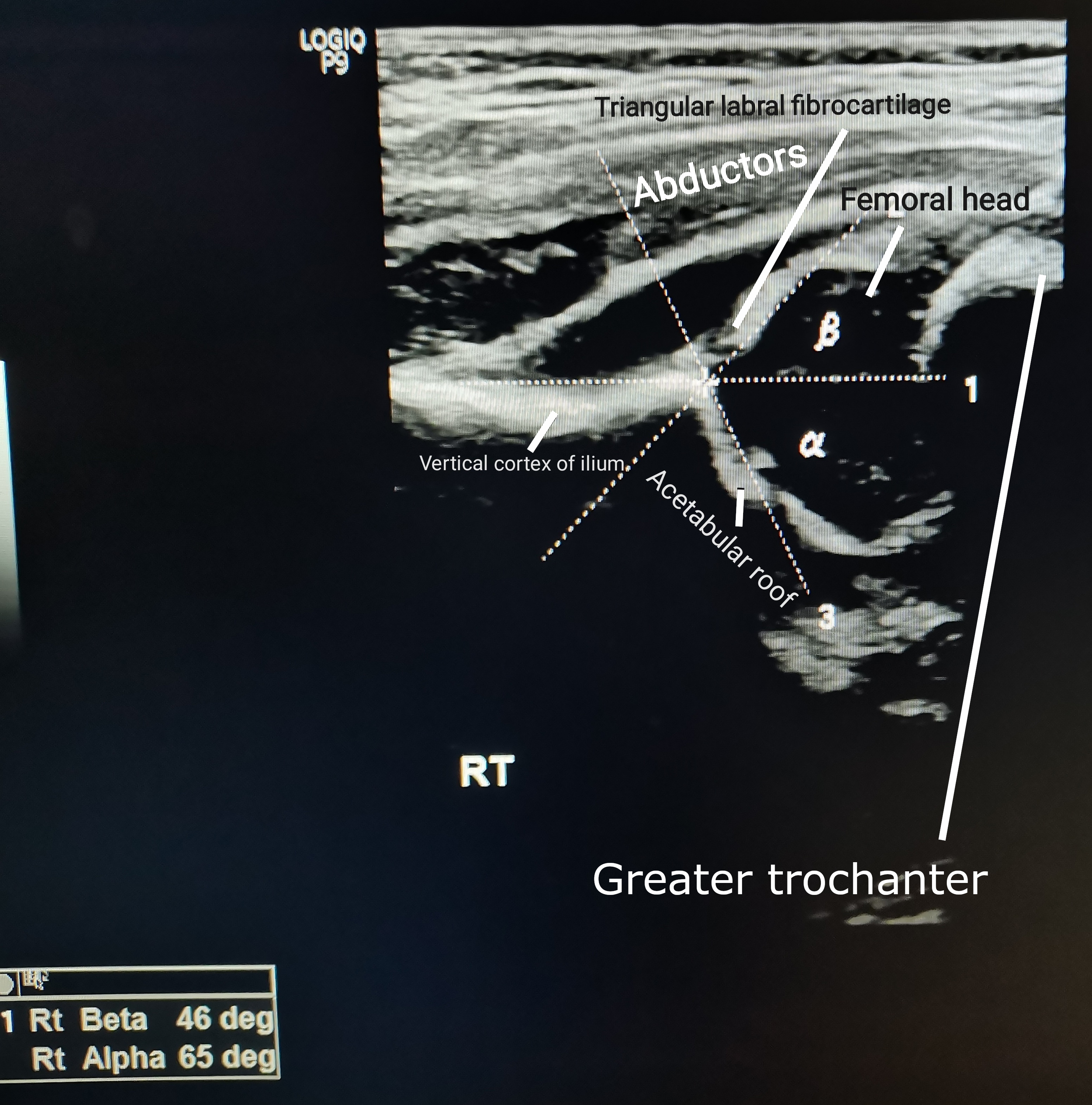
Developmental dysplasia of the hip (DDH) is a spectrum: a shallow, underdeveloped acetabulum that cannot hold the femoral head, from mild instability to frank dislocation. The newborn examination is the screen, and the sign boards test is the clunk, a palpable sense of the head translating across the socket rim. The two maneuvers are direction opposites. Barlow is the provoker: with the hips and knees flexed, adduct the hip and apply gentle posteriorly directed pressure, and a clunk as the head slides out of the acetabulum means the hip is dislocatable. Ortolani is the reducer: abduct the hip with an anteriorly directed lift, and a clunk as the head re-enters means the hip was out and is relocatable. Both are most reliable before 3 months: after that the capsule tightens and the ossific nucleus forms, and the clunk disappears while the dysplasia does not.
The soft signs carry the older infant: asymmetric thigh folds, limited abduction, and the Galeazzi sign, apparent shortening seen when the hips and knees are flexed and one knee sits lower. Screening runs on a fixed clock: every newborn is examined, an abnormal exam or risk factors trigger ultrasound at about 6 weeks (the study that sees cartilage), and radiographs take over after 4 to 6 months once the ossific nucleus blocks ultrasound. Never let a quiet exam after 3 months reassure you; the film is the truth then.
Flip between the two maneuvers until the direction is automatic.
RISK AND THE HARNESS
DDH Risk Factors and the Pavlik Decision
The board hands you the risk factors in the first sentence, then grades the treatment clock: Pavlik before 6 months, closed reduction and spica after harness failure, open reduction for the late hip, and two complications to name on sight.
The DDH risk set is small and board-fixed: breech presentation (the headline, especially frank breech), female sex, first-born, family history, oligohydramnios, and swaddling with the hips extended and adducted. Breech plus a positive Barlow means ultrasound at 6 weeks, exam normal or not; risk factors alone trigger imaging. Swaddling is the modifiable one: hips held extended and adducted push the head out of a shallow socket, so the teaching is to swaddle with the hips flexed and free to abduct.
Treatment is a clock. Pavlik harness before 6 months holds the hip flexed with gentle abduction while the socket remodels; it is checked weekly, and the checks exist because the harness has two named complications. Hold too much flexion and the femoral nerve is compressed: weak knee extension, diminished patellar reflex, anterior thigh numbness, fixed by repositioning with less flexion. Force abduction and the vessels along the femoral neck are compressed: avascular necrosis of the femoral head, the feared complication, so the harness flexes but never forces. After 6 months the harness loses its window: the next rung is closed reduction under anesthesia plus a spica cast, and the late, high dislocation in the walking child goes to open reduction. Missed DDH ends in AVN and early osteoarthritis.
Pick the risk factor set that earns the 6 week ultrasound.
A 6-week-old girl is brought for a well-child visit. She was born at term in frank breech position, and the newborn examination was normal. Which of the following best explains why a hip ultrasound is indicated despite the normal examination?
A. Breech means ultrasound at 6 weeks, exam normal or not. Breech, female, first-born, family history, and oligohydramnios all trigger screening imaging; the normal exam does not clear the risk, and ultrasound sees the cartilaginous head that radiographs cannot. B. Normal exam clears the hip. False: the newborn exam misses many dysplasias, which is why risk factors earn imaging. C. Radiograph at 6 weeks. No ossific nucleus yet, so the radiograph is blind; ultrasound is the 6 week study. D. Clunk required. Risk factors alone are an indication; the clunk is just one trigger. E. Harness first. Imaging confirms the diagnosis before treatment; the harness never precedes the ultrasound. Rule: breech means ultrasound at 6 weeks, exam normal or not.
THE FOUR LINES
The Pelvis Lines: Hilgenreiner, Perkin, the Index, and Shenton
Once the ossific nucleus appears at 4 to 6 months, the radiograph owns the diagnosis. Four reference lines divide the hip into a story: where the head sits, how steep the socket is, and whether the arc of the joint is intact.
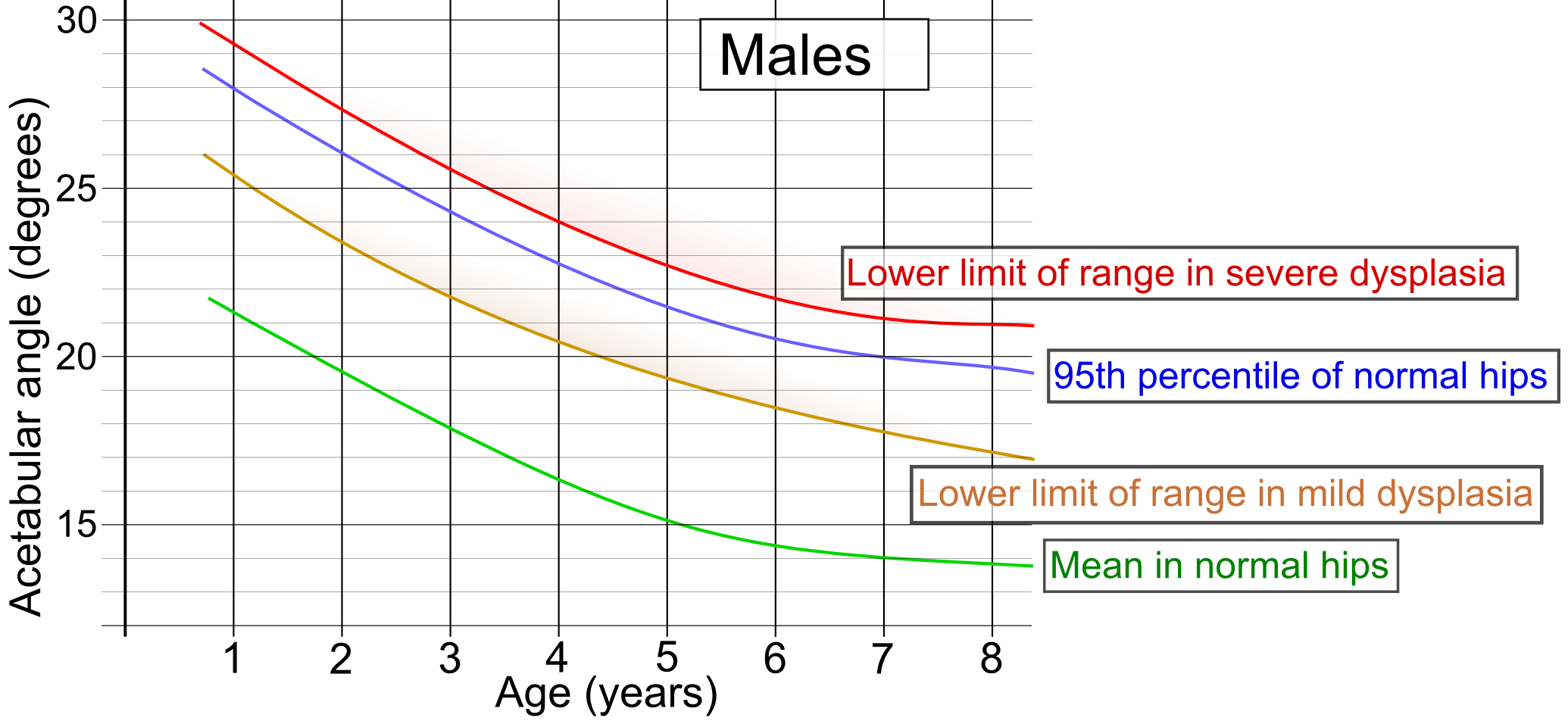
The AP pelvis radiograph is read with four landmarks, and each answers one question. Hilgenreiner line: the horizontal line through the triradiate cartilages. Perkin line: the vertical line from the lateral acetabular margin, perpendicular to Hilgenreiner. Together they divide each hip into four quadrants, and the rule is positional: the ossified femoral head belongs in the inferomedial quadrant, and a head sitting superolateral is dislocated. Acetabular index: the angle between Hilgenreiner and the line from the triradiate cartilage to the lateral acetabular rim. It measures the socket slope, normally under about 30 degrees in the infant and falling with age; a high, non-falling index is the dysplastic signature. Shenton line: the continuous arc from the inferior border of the femoral neck to the superior border of the obturator foramen. When the head rides up, the arc breaks, and the break is the dislocation in one glance.
Read them in order: quadrants for position, the index for the socket, Shenton for the arc. The same four lines answer the follow-up question too, because treatment success is judged by the head staying inferomedial and the index falling over time.
Tap each line on the diagram and read what it tells you.
Tap a line.
THE FOOT THAT POINTS DOWN
Clubfoot: CAVE Anatomy, Ponseti Sequence
Talipes equinovarus has four components, one acronym, and one treatment order. The rigid foot is clubfoot; the flexible foot is positional. The Ponseti method runs a fixed sequence, and the brace at the end is what keeps the correction.
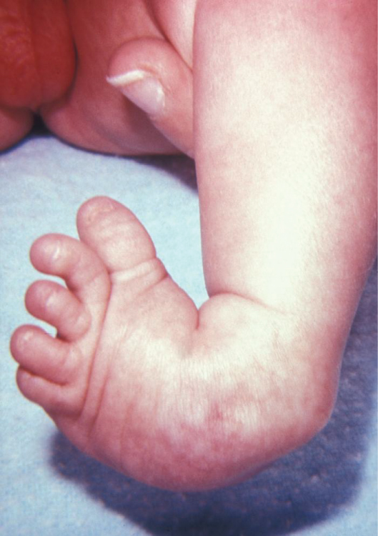
Clubfoot is the foot frozen in a fixed posture, and the board names it with CAVE: cavus (the high medial arch), adductus (the forefoot turned in), varus (the hindfoot turned in), and equinus (the ankle plantarflexed, pointing down). It is more common in boys, about 2 to 1, and bilateral in about half of cases. The discriminator on exam is rigidity: the positional foot is flexible and corrects with gentle stretching, while the teratologic foot is rigid, rides with other contractures (arthrogryposis, myelomeningocele), and carries a higher recurrence risk. Clubfoot is a clinical diagnosis; the board does not make you order films to name it.
The Ponseti method is a fixed sequence and the sequence is the treatment. Weekly long-leg casts start in the first weeks of life, each cast correcting the components from the ground up: cavus first, then adductus, then varus, and the equinus last. When only equinus remains, a percutaneous Achilles tenotomy releases the tight tendon, and casting continues in dorsiflexion. Then comes the foot abduction brace (the Dennis Browne bar and boots): 23 hours a day for about 3 months, then at night and nap time until about age 4. The brace outlasts the casts because the foot wants to relapse, and noncompliance is the leading cause of recurrence.
Put the Ponseti sequence in order.
AGE PLUS ACUITY
The Limping Child: Kocher Criteria and the Age Split
The limp is a routing problem. The toddler after a URI with no fever has transient synovitis. Fever, refusal to bear weight, and a high ESR or WBC push toward septic arthritis, where the Kocher count sets the probability and the aspiration decides.
Transient (toxic) synovitis is the most common cause of a pediatric limp: a benign, self-limited joint effusion that follows a viral illness, classically in the toddler, with little or no fever and a child who still bears weight. It resolves in days with ibuprofen. Its entire differential matters because of what it is not: the septic hip, the emergency that destroys cartilage in days. The Kocher criteria are the probability engine: fever above 38.5 C, refusal to bear weight, ESR above 40 mm/hr, and WBC above 12,000/mm3. The septic arthritis probability climbs with the count: about 0.2 percent at zero, 3 percent at one, 40 percent at two, 93 percent at three, and 99.6 percent at all four. Three or four criteria means arthrocentesis, blood cultures, and empiric IV antibiotics now, with surgical drainage for the hip, whose intracapsular femoral head and fragile blood supply make pus under pressure an epiphyseal threat. The child with a septic hip holds it flexed and externally rotated and may show pseudoparalysis, refusing to move the limb at all.
The age split routes the chronic limps: the 4 to 8 year old with a painless limp goes to the frog-leg lateral film for Perthes, and the obese adolescent with knee pain goes to the Klein line for SCFE.
Check the criteria, watch the probability climb, then commit.
Criteria met: 0
Route the limping child by age.
Which door fits which presentation?
THE CHRONIC HIP
Perthes and SCFE: Two Hips That Fail Slowly
The chronic hip is an age ladder with a film at each rung: the frog-leg lateral for the school-age head that is dying, and the AP pelvis with the Klein line for the adolescent physis that is slipping.
Legg-Calve-Perthes disease is idiopathic avascular necrosis of the femoral head: the blood supply to the head fails, the bone dies, and the head collapses. The classic patient is a boy 4 to 8 years old with an insidious, painless limp who keeps playing, and examination shows limited internal rotation and abduction. The frog-leg lateral radiograph shows the signature: increased density, then flattening and collapse of the femoral head. It is bilateral in about 10 percent. The course is prolonged, measured in years, and management is containment: keeping the softened head inside the socket while it revascularizes and remodels, with activity modification and follow-up films.
Slipped capital femoral epiphysis (SCFE) is the proximal femoral epiphysis slipping off the neck through a physis failing under load. The classic patient is the obese adolescent, 10 to 16, whose presenting complaint is often referred knee or thigh pain with a normal knee exam, the classic trap. The exam signature is obligate external rotation with flexion: the slipped head drags the leg outward as the hip bends, and internal rotation is limited. On the AP radiograph, the Klein line drawn along the superior femoral neck misses the epiphysis, and the frog-leg lateral shows the slip best. It is bilateral in about 20 to 40 percent, so both hips are filmed, and management is pinning in situ: a screw holds the epiphysis while the physis closes, and it is urgent because an unstable slip can progress suddenly and raise the AVN risk. Obesity is the mechanical driver; hypothyroidism and growth hormone deficiency are the endocrine risks worth remembering.
Open each treatment ladder and hold the timing facts.
Pavlik harness before 6 months with weekly checks; reposition with less flexion for femoral nerve palsy and never force abduction, which risks avascular necrosis. After the window, closed reduction under anesthesia plus a spica cast; open reduction for the late, high dislocation.
Ultrasound at about 6 weeks sees the cartilaginous head; once the ossific nucleus appears around 4 to 6 months, the AP pelvis radiograph takes over with Hilgenreiner, Perkin, the acetabular index, and Shenton.
Weekly long-leg casts from the first weeks correct cavus, adductus, and varus; percutaneous Achilles tenotomy finishes the equinus; then the foot abduction brace 23 hours a day for about 3 months and at night until about age 4. Brace noncompliance is the leading cause of recurrence.
Keep the necrotic femoral head contained in the socket with activity modification and, when needed, bracing or surgery, with years of radiographic follow-up; the outcome tracks how much the head collapses and remodels.
Bilateral hip films because 20 to 40 percent are bilateral, then pinning in situ of the slipped epiphysis; discuss prophylactic pinning of the contralateral hip. The slip is urgent: progression and AVN are the risks of waiting.
At 3 or 4 Kocher criteria, arthrocentesis for cell count, Gram stain, and culture, blood cultures, empiric IV antibiotics, and surgical drainage of the hip, whose intracapsular head cannot survive pus under pressure.
THE BOWED TIBIA
Blount Disease: Physiologic Bowing or the Beak That Progresses
Bowlegs are normal until they are not. The beak at the proximal tibial physis separates the toddler who outgrows it from the one who needs a brace.
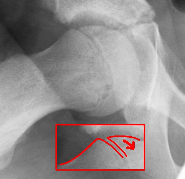
Physiologic genu varum is symmetric, appears with the first steps, and resolves by age 3 as the medial tibial physis outgrows the lateral. Reassurance is the treatment. Blount disease is the pathologic version: progressive, often asymmetric tibia vara in an obese early walker, driven by growth arrest of the medial proximal tibial physis. The X-ray shows the tell, a medial beaking of the proximal tibial metaphysis, and Langenskiold staging follows it over time.
Early Blount (Langenskiold I to II, under age 4) is braced with a chance of correction; late disease needs an osteotomy. Genu valgum runs the mirror course: physiologic knock-knees peak around age 4 and settle by 7, while asymmetric or worsening valgus points to renal osteodystrophy, trauma, or tumor (cross-ref metabolic-bone-trio). The one-question split is symmetry and direction of progression.
Name the bow: physiologic or physis.
THE DISCRIMINATOR
The One-Screen Discriminator
Every pediatric hip and foot story on boards is one of these doors, and the door is set by age, acuity, and the Kocher count.
Run the doors in order. Newborn with a clunk: Barlow provokes, Ortolani reduces, and breech or a positive exam earns ultrasound at 6 weeks, never a radiograph before the ossific nucleus. Toddler after a URI, afebrile, bearing weight: transient synovitis, zero Kocher criteria, observe with ibuprofen. Toddler with fever and refusal to bear weight: count the Kocher criteria, and at 3 or 4, aspirate, culture, and treat now. School-age child with a chronic painless limp: the frog-leg lateral and the Perthes story, containment and time. Obese adolescent with knee pain: the Klein line, obligate external rotation, and pinning in situ, with the contralateral hip in the same films. Newborn with a rigid foot: CAVE anatomy, the Ponseti sequence, and the brace that outlasts the casts. The acronyms are the anchors: CAVE for the foot, Kocher for the acute hip, Klein for the slipped epiphysis.
Tap each letter of the CAVE acronym.
CAVE
CCavus: the high medial arch, corrected first
AAdductus: the forefoot turned in
VVarus: the hindfoot turned in
EEquinus: the ankle pointing down, last to yield, finished by the tenotomy
Prove it
Walkthrough: race the age ladder
Original practice scenarios, one at a time. Choose an answer, then open any option to work its reasoning.
Clinical walkthrough
