Connect patellar restraint and groove engagement to acute injury assessment, recurrence anatomy, rehabilitation and individualized treatment decisions.
Why can a kneecap slip out again after the first injury has healed? Stability depends on a medial soft-tissue restraint, timely entry into a containing groove, and the direction of force across the knee. Repeated lateral displacement means that this combination is not working adequately.
By the end, you should be able to explain the vulnerable part of knee bending, recognize an associated injury that cannot wait, interpret the anatomy without prescribing from one number, and match a treatment discussion to the patient. Follow restraint, acute assessment, imaging, and treatment selection, then try the independent cases.
Why is early knee bending vulnerable?
A 17-year-old reports that the patella slips outward near the beginning of a squat but feels more secure after the knee bends further. The important question is not simply which ligament was torn. It is which restraint should be working at that part of the arc.
The medial patellofemoral ligament (MPFL) connects the upper medial patella to the medial distal femur. It resists lateral translation, especially near extension and in early flexion, when trochlear engagement is limited. It is a passive restraint, not a muscle that actively pulls the patella inward. Cadaveric testing at 20 degrees supports its major restraining contribution; that experiment does not assign a fixed percentage to every living knee. [2]
The trochlea is the groove on the anterior distal femur. As the patella enters it during bending, the articular geometry supplies more containment. This is a gradual change, not a switch at exactly 30 degrees. Patellar height, trochlear shape, load and knee position affect engagement. Soft tissue and bone cooperate rather than taking perfectly separate turns. [1]
Think of three questions: is the medial restraint competent, is the patella inside a containing groove, and where does the extensor mechanism direct force?
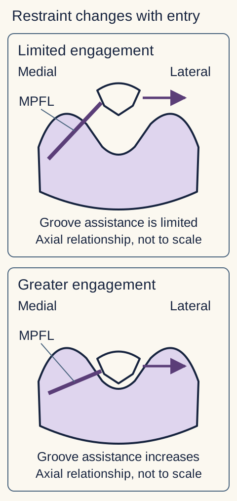
Trace and predict. In the paired axial schematic, trace the lateral force arrow, then the medial restraint. Compare limited engagement with deeper engagement. Now imagine that only the medial restraint is deficient. Predict which position becomes most vulnerable before reading on.
Trace the medial band and lateral arrow, then compare groove engagement. Soft tissue and bony geometry cooperate; the drawing does not assign an exact flexion threshold or measured force. The band represents the MPFL, with simplified attachments. [1][2]
The visible comparison shows why early flexion can become unstable while a well-shaped groove still provides useful restraint later. A deficient groove changes that prediction: greater flexion cannot provide normal containment if the containing surface itself is abnormal. This model explains symptom timing; it does not prove a diagnosis from the knee angle alone. [1][2]
Apply the model to a different knee
A patient with lateral escape continuing well into flexion needs assessment of trochlear containment and tracking, not just an assumption of an isolated MPFL tear. The later instability suggests that the expected bony assistance is inadequate. Confirm the pattern clinically and with appropriate imaging. [1]
Which part of the stabilizing system is failing?
Two patients have damaged medial restraints after dislocation. One has a normally positioned patella and a deep groove; the other has a high patella and a shallow proximal groove. Why need their recurrence risks and treatment discussions differ?
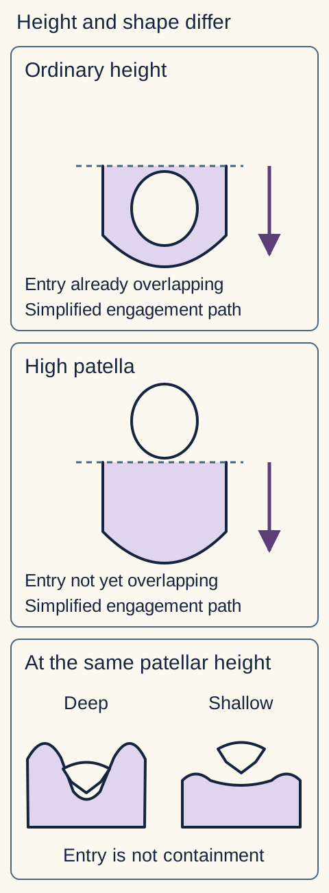
Patella alta means an abnormally high patella. It can delay entry into the trochlea, extending the portion of bending during which soft tissue carries more of the stabilizing demand. Trochlear dysplasia means abnormal groove geometry, often with reduced concavity or a prominent proximal contour. It reduces the benefit of engagement itself. Height and shape are different problems even when they coexist. [1][3]
A relatively lateral tibial tubercle can alter the patellar tendon line of pull. Valgus alignment and abnormal femoral or tibial rotation can also affect the relationship between the extensor mechanism and the groove. These are three-dimensional relationships: a large tubercle-to-groove distance is not proof that the tubercle alone is abnormal. Generalized laxity reduces passive restraint, while deficient hip and quadriceps control can impair dynamic tracking. Strengthening can improve control; it does not reshape a dysplastic trochlea. [1]
Compare one change at a time. In the height-and-containment schematic, first compare ordinary height with a high patella over the same groove. Then compare deep and shallow grooves at the same patellar height. Predict whether the problem is delayed entry or inadequate containment after entry. The first comparison changes timing; the second changes the containing geometry.
The upper comparisons hold the groove constant and show delayed entry with greater height. The lower axial comparison holds patellar height conceptually constant and changes containment. Compare patellar height and groove containment separately; clinical indices require measured imaging. The downward arrow represents the direction of entry in the upper model. [1][3]
Record the age at first event, number of dislocations, low-energy episodes, opposite-knee symptoms, family history and response to rehabilitation. Young age and anatomic predisposition increase concern, but a painless flexible joint is not automatically an unstable joint. Laxity is a finding; symptomatic escape is the clinical problem. A low-energy recurrence can justify a more detailed structural assessment than its apparently minor mechanism suggests. [1][3][4]
Test the distinction without naming an operation
With patella alta and a normal groove, earlier engagement could reduce the vulnerable arc. With a normally positioned patella and severe dysplasia, earlier entry alone does not create a deeper containing surface. Neither observation by itself is a surgical prescription. [1]
What must be assessed before a recurrence plan?
A reduced patella can look reassuring while the joint still contains an injured cartilage surface or a loose fragment. Reduction restores position; it does not certify that the rest of the knee is intact.
In an acute episode, assess the whole injury, document distal perfusion and neurologic findings, confirm reduction, and evaluate active extension. A patella that remains displaced, impaired circulation, new neurologic deficit, or suspected major knee injury needs emergency assessment. Do not confuse a displaced kneecap with a tibiofemoral dislocation. This lesson is not a guide to attempting reduction without clinical supervision.
Look for rapid or tense swelling, hemarthrosis, mechanical locking and inability to extend. A lateral dislocation can injure the MPFL and medial retinacular tissues while shearing cartilage or an osteochondral fragment. A fragment may obstruct extension even after reduction. Failure of active extension can also reflect pain inhibition or extensor injury, so it requires examination rather than a single presumed diagnosis. [1][3]
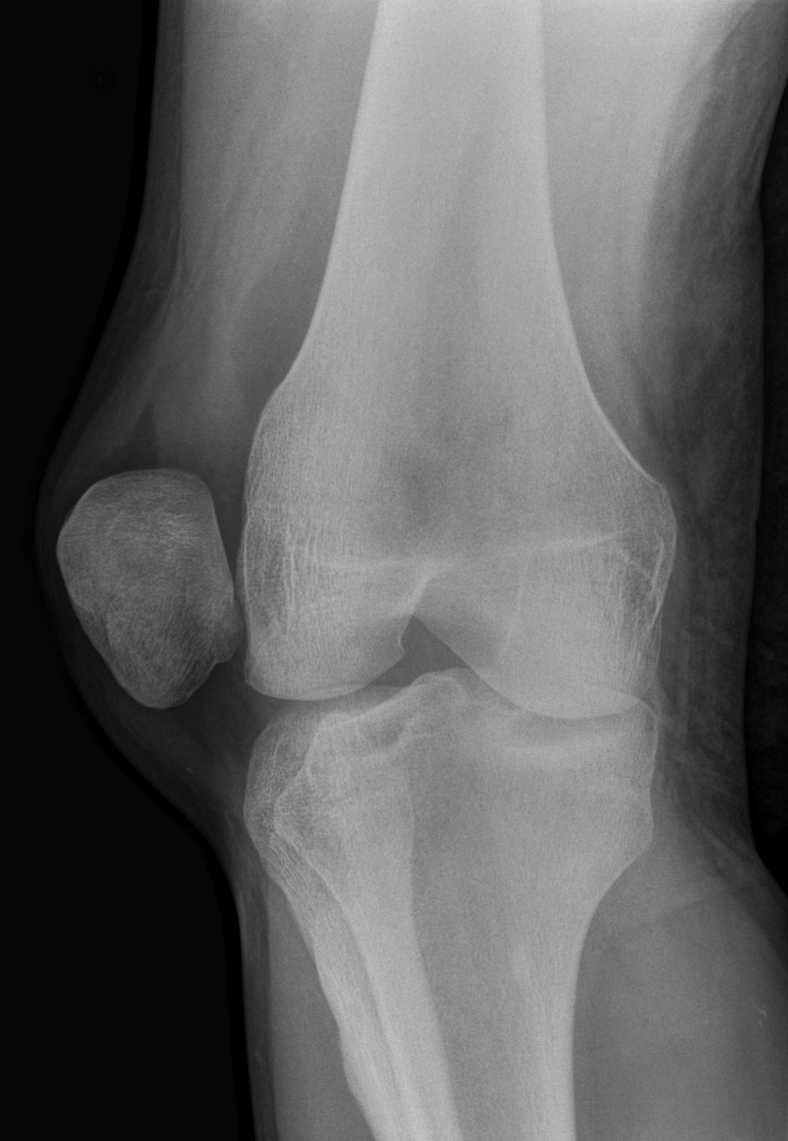
Locate before interpreting. In the unaltered clinical radiograph, identify the patella relative to the femur and use the fibula to orient the lateral side. Then ask what an ordinary radiograph cannot establish: cartilage integrity, all loose fragments, and the complete medial soft-tissue injury. A normal-appearing post-reduction film cannot answer all of those questions.
Identify the patella outside the lateral distal femoral contour, using the fibula to orient the lateral side. This unaltered image shows displacement, but cannot establish the full cartilage injury or medial soft-tissue damage. It does not illustrate every case in this lesson. Image: Hellerhoff; original source; CC BY-SA 3.0.
Obtain prompt appropriate imaging rather than using absence of a visible fracture as clearance for rehabilitation. The ESSKA consensus recommends prompt MRI with radiographs, or immediately available MRI alone, after first-time dislocation; an asymptomatic late presentation with a normal examination is a possible exception. Hemarthrosis or locking particularly strengthens the need for timely MRI and urgent orthopedic assessment. These are consensus recommendations with evidence limitations, not a reason to delay emergency care while awaiting a scan. [3]
Once urgent associated injuries are addressed, reassess tracking and risk anatomy when pain and swelling permit. If a repairable osteochondral injury is found, specialist planning considers preservation or restoration of the joint surface and whether stabilization should accompany treatment. Not every effusion requires an operation, and not every loose fragment should simply be discarded. [3][4]
Change one finding: what changes the priority?
Compare a comfortable reduced knee with a reduced knee that remains mechanically locked. The latter needs an associated-injury explanation before exercise progression. A normal distal examination is reassuring about circulation and nerve function, not about intra-articular cartilage. [1][3]
What do the examination and measurements actually establish?
A patient reports three visible lateral dislocations, yet one imaging measurement is only borderline. Another has a large measurement but pain without any displacement history. The numbers cannot substitute for identifying what is happening to each knee.
Document true displacement episodes and distinguish apprehension from tenderness. Apprehension is the patient's fear or protective response when lateral displacement reproduces instability, assessed gently when tolerated. Compare patellar glide and its endpoint with the opposite side. Observe active tracking, including a J-sign, in which the patella deviates laterally near terminal extension. Examine alignment, generalized laxity and other knee structures. Pain, ACL instability and meniscal symptoms can overlap, and an acute guarded examination may need repeating. [1][3]
Imaging answers several different questions: is the cartilage injured; is the groove dysplastic; is the patella high or poorly engaged; is the tubercle position contributing; are valgus or rotational abnormalities present; and are the growth plates open? Radiographs, MRI and selected alignment or rotational studies are complementary, not interchangeable labels for the same information. [1][3]
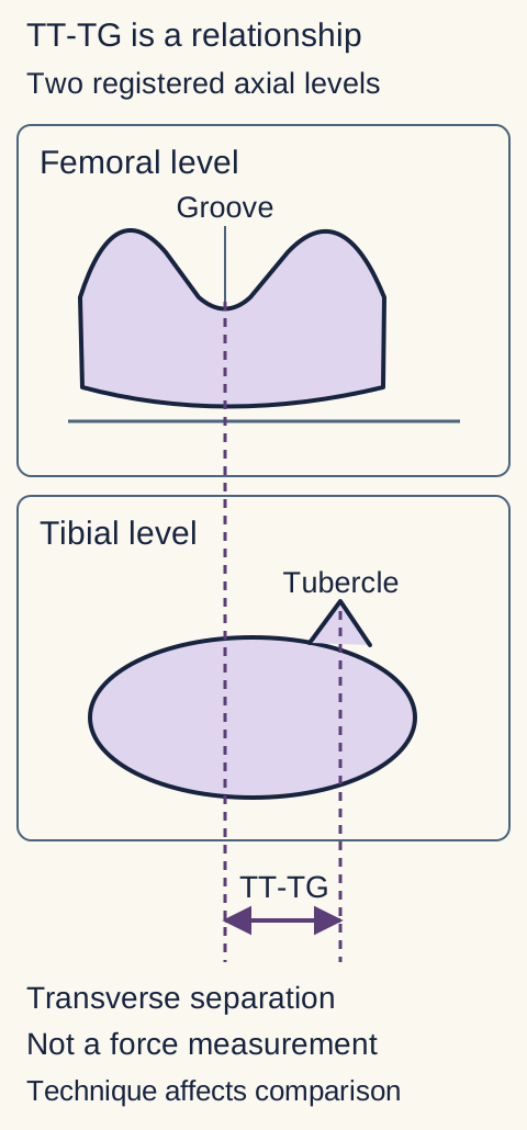
Tibial tubercle-to-trochlear groove distance (TT-TG) describes the transverse separation between landmarks projected from different axial levels using a common reference. Trace the two reference lines in the geometry schematic before estimating the separation. This is a positional measurement, not a force reading. Acquisition, knee position, landmark selection and modality can affect it. Compare like with like before interpreting a discrepancy. [1]
Trace each dashed projection from its own level. The lower double arrow depicts transverse separation in a shared reference, with a simplified posterior femoral reference line above. Slice registration and knee position matter. This diagram is not a measurement from a patient and gives no surgical threshold. [1][3]
A patellar height index and an overlap measure also describe different relationships. The former characterizes height relative to an anatomic reference; the latter asks how much patellar cartilage engages the trochlea in the imaged position. Neither alone establishes which operation is necessary. Current consensus does not provide one universally decisive cutoff for the many anatomical contributors. [1][3][4]
Combine symptoms, objective instability, complete anatomy, maturity, treatment response and patient goals. Borderline TT-TG does not negate repeated observed dislocations. Conversely, isolated anterior pain with no displacement or apprehension merits a broader patellofemoral assessment, not a reconstruction justified by a scan value. [1]
Interpret a discordant pair of reports
If one report used CT in extension and another used MRI with some knee flexion, first verify acquisition and landmarks. A different TT-TG value is not evidence that the tubercle migrated between visits. Repeat or additional imaging should answer a specific unresolved clinical question. [1][3]
Which outcome matters when comparing treatments?
One patient wants fewer dislocations; another is most concerned about stair pain and confidence at work. These goals overlap, but a treatment that improves stability does not necessarily improve every measure of function.
A skeletally mature patient with a fully assessed, uncomplicated first event and low recurrence risk can often begin nonoperative care. This includes symptom-guided protection, restoration of motion, quadriceps and hip rehabilitation, and follow-up. High-risk anatomy, ongoing symptoms, skeletal immaturity or a significant osteochondral injury changes the discussion. First-time status is not an instruction to ignore those findings or to delay specialist advice until another injury occurs. [3][4]
Rehabilitation addresses strength, gait, coordination and confidence as well as motion. Progress depends on the examination and functional control, not simply elapsed weeks or a reassuring radiograph. A brace may provide short-term support, but prolonged restriction can impede recovery. A randomized comparison found more early stiffness and quadriceps atrophy with four weeks of restricted motion, without establishing a recurrence advantage over a nonhinged brace. Its imprecise recurrence estimate does not prove equivalence. [4][7]
Separate the endpoints. A 2026 randomized trial in selected patients with recurrent dislocations compared MPFL reconstruction plus rehabilitation with rehabilitation without reconstruction. Both groups underwent diagnostic arthroscopy. Participants were aged 12 to 30, had at least two dislocations, and did not have Dejour D dysplasia or CT TT-TG above 20 mm. At three years, persistent subjective instability was reported by 16.7% of the reconstruction group and 53.6% of the comparison group. This is an instability symptom endpoint, not a count of radiographically confirmed redislocations. [5]
Patient-reported functional scores improved in both groups, without a statistically significant between-group difference. That is not proof of equivalent function. The trial also does not determine whether isolated MPFL reconstruction adequately treats a patient with more severe excluded bony abnormalities. A separate pooled analysis of randomized trials favored operative care for recurrence but found incomplete information for a firm universal conclusion and no clear pooled functional-score advantage. The pooled interventions and patients were not identical to those in the recurrent-dislocation trial. [5][6]
Compare two columns on paper: frequency of instability and everyday function. Place each reported outcome in its own column, then add recovery burden and possible complications. A recommendation should address the patient's actual goals rather than use a favorable result in one column to fill every other column.
Apply the evidence to someone outside the trial
A patient with severe excluded dysplasia still deserves treatment assessment. The trial's selection criteria limit how directly its isolated-reconstruction results apply; they do not prove that surgery is ineffective in that patient. Anatomy-specific assessment and a discussion of uncertainty are needed. [5]
How does the proposed treatment address the demonstrated problem?
Before naming a procedure, complete the sentence: the patella escapes because this particular restraint or relationship is inadequate. Then ask what the proposed procedure changes and what it leaves unchanged.
MPFL reconstruction restores a deficient medial passive restraint. It is an important option for symptomatic recurrent instability, particularly when major bony contributors are absent or have been appropriately addressed. Reconstruction does not lower a high patella, deepen a dysplastic groove or correct femoral rotation. Persistent dislocations despite appropriate rehabilitation warrant specialist evaluation, not indefinite repetition of an unchanged exercise plan. [1]
Tibial tubercle procedures alter the relationship of the distal extensor mechanism to the joint. Medialization can address a relevant lateral vector; distalization can address selected clinically important patella alta. Those directions solve different problems. Trochlear procedures address selected substantial dysplasia, while alignment procedures address a relevant valgus or rotational deformity. The decision is not dictated by one numerical threshold, and the presence of several risk factors does not automatically require an operation for each. [1][4]
Growth plates change what can be done safely. An adult-style tibial tubercle osteotomy is generally avoided with wide-open physes. Pediatric specialists may consider growth-respecting stabilization or selected growth-related alignment treatment. This is neither permission to apply adult techniques to a child nor a rule that every unstable child must wait until adulthood for care. [4]
Isolated lateral release does not reconstruct the MPFL or correct alta, dysplasia or a lateralized extensor mechanism. Releasing lateral tissues can also produce medial instability. A genuinely tight lateral retinaculum may require an adjunctive procedure in selected surgical planning, but lateral tilt on an image alone does not establish that indication. Clinical examination must demonstrate the relevant tightness. [1]
Match and predict. Consider a mature patient with normal height and groove but deficient medial restraint, then a patient with that deficit plus marked alta. Predict which anatomic relationship an isolated reconstruction would change in each. The medial restraint can improve in both, but delayed engagement remains in the second. Whether that remaining factor needs correction depends on its clinical importance and the complete assessment.
Discuss possible persistent symptoms, recurrent instability, stiffness and procedure-specific complications alongside expected benefit. Rehabilitation remains part of surgical care. New pain or restricted flexion after stabilization is not automatically evidence that the graft is too loose; reassessment should include tracking, joint surfaces and possible overconstraint. A treatment label cannot replace an examination of the current problem. [1][3][4]
Return to the central question
First establish the acute injury, then identify the failing relationship. Restore the relevant restraint or alignment when indicated while protecting cartilage and respecting maturity. Fewer episodes, useful function and acceptable treatment burden all matter. [1][4][5]
Apply the clinical relationships
Case 1
Show answer and explanations for case 1
A. Arrange prompt MRI and urgent assessment for an intra-articular injury. (Best answer)
Cartilage or osteochondral fragments can remain after the patella is reduced. Locking and rapid tense swelling persist despite restored patellar position and normal distal findings. Evaluate the associated joint injury before advancing rehabilitation.
Reasoning steps for option A
What joint injury can remain despite a centered post-reduction patella and reassuring radiographs?
A cartilage or osteochondral fragment may remain in the joint even after reduction and without an obvious fracture on radiographs.
How do rapid tense swelling and blocked extension support an occult osteochondral fragment?
Rapid hemarthrosis suggests intra-articular injury, and persistent mechanical loss of extension raises concern for an obstructing fragment.
Why should prompt MRI and urgent assessment precede rehabilitation?
Prompt MRI and urgent orthopedic assessment can identify a repairable or obstructing joint injury before motion is advanced.
B. Repeat the tracking examination after several weeks of rehabilitation. (Why this does not fit)
Pain and swelling can limit an acute tracking examination. A mechanically blocked knee with rapid tense swelling raises a more immediate intra-articular concern than chronic tracking. Reassessment for recurrence anatomy should not defer evaluation of a possible obstructing fragment.
Reasoning steps for option B
Why might an acute tracking examination be unreliable in this swollen knee?
Swelling and pain can obscure accurate assessment of patellar tracking soon after dislocation.
Why does mechanical locking take priority over repeating tracking tests after weeks of rehabilitation?
Waiting weeks to repeat tracking tests would leave the present mechanical block and possible intra-articular fragment unresolved.
What risk would delayed tracking reassessment leave unevaluated?
Delayed reassessment could miss a loose osteochondral fragment responsible for locking.
C. Begin protected motion and schedule routine outpatient follow-up. (Why this does not fit)
An uncomplicated reduced first event can be managed with rehabilitation and surveillance. Mechanical locking and a tense effusion make this event potentially complicated even without a visible fracture. Routine rehabilitation alone does not address a suspected loose fragment.
Reasoning steps for option C
When would protected motion and routine follow-up be reasonable after a first dislocation?
Protected motion with routine surveillance fits a reduced first event without concerning signs of associated injury.
Why do the tense effusion and extension block make this event unlike an uncomplicated reduction?
A tense rapid effusion and mechanical extension block suggest hemarthrosis and an intra-articular injury despite reassuring plain films.
Why is rehabilitation alone insufficient for a possible loose fragment?
Motion exercises cannot remove or characterize a suspected obstructing fragment; imaging and urgent assessment are needed.
D. Obtain a rotational CT profile before selecting protected activity. (Why this does not fit)
Rotational anatomy can contribute to recurrent patellar instability. The unresolved problem is acute locking with an effusion, not an established torsional deformity. Image the acute intra-articular injury before elective rotational planning.
Reasoning steps for option D
What instability contributor would a rotational CT profile investigate?
A rotational CT profile examines torsional alignment that may predispose to recurrent patellar instability.
Why does acute locking with a tense effusion not establish a torsional deformity?
Neither a torsional abnormality nor recurrent maltracking has been established; the immediate problem is a swollen, locked joint.
Which injury evaluation must precede elective rotational planning?
The suspected acute osteochondral or loose-fragment injury warrants MRI and assessment before elective torsional workup.
Takeaway: A reduced patella and normal distal examination do not exclude an important intra-articular injury.
A. Reduced containment from a markedly dysplastic trochlea. (Why this does not fit)
Abnormal groove geometry can reduce bony restraint during engagement. The groove is well formed and restraint becomes stronger with greater flexion. The supplied pattern points to the restraint needed before substantial engagement.
Reasoning steps for option A
What restraint would a markedly dysplastic trochlea weaken as flexion increases?
A dysplastic trochlea would weaken bony containment when the patella engages the groove.
How do a well-formed groove and stronger restraint at 60 degrees argue against dysplasia?
The well-formed groove provides increasing restraint by 60 degrees, unlike substantial dysplasia.
Which earlier-flexion restraint better explains the soft endpoint at 20 degrees?
The medial soft-tissue restraint, especially the MPFL, is most important before substantial groove engagement at 20 degrees.
B. Deficient medial restraint before substantial groove engagement. (Best answer)
The MPFL is a major passive restraint to lateral translation in early flexion. A soft early-flexion endpoint with stronger later restraint fits deficient medial tissue assisted later by normal bony geometry. This pattern supports medial restraint assessment rather than automatic bony correction.
Reasoning steps for option B
Which passive medial structure normally limits lateral glide near 20 degrees of flexion?
The medial patellofemoral ligament (MPFL) is a principal passive check against lateral translation in early flexion.
Why does stronger restraint at 60 degrees with a normal groove implicate early medial restraint?
A soft endpoint at 20 degrees with stronger restraint at 60 degrees fits weak medial tissue followed by normal trochlear containment.
What tissue should be assessed before assuming bony correction is needed?
The MPFL and associated medial stabilizers merit assessment; normal height and groove do not automatically justify bony correction.
C. Loss of continuity in the quadriceps tendon above the patella. (Why this does not fit)
Quadriceps tendon disruption impairs the active extensor mechanism. The defining examination here is passive lateral laxity with flexion-dependent restraint, not loss of active extension. Localize passive restraint failure rather than infer an extensor rupture.
Reasoning steps for option C
What function would quadriceps tendon discontinuity impair?
Quadriceps tendon continuity permits active knee extension through the extensor apparatus.
Why is passive lateral translation with a soft early-flexion endpoint different from loss of active extension?
Passive lateral glide and its flexion-dependent endpoint test patellar restraint; quadriceps rupture instead impairs active extension.
Which deficit is better localized than quadriceps rupture here?
Early-flexion medial passive restraint deficiency better explains the findings than quadriceps tendon rupture.
D. Excessive proximal position of the patella relative to the groove. (Why this does not fit)
A high patella can delay trochlear entry and prolong early-flexion vulnerability. Imaging establishes ordinary patellar height in this patient. Delayed engagement from alta does not account for the stated anatomy.
Reasoning steps for option D
How could patella alta extend the arc without trochlear containment?
Patella alta can delay entry into the trochlea and extend dependence on medial restraint in early flexion.
Which imaging finding contradicts excessive proximal patellar position?
MRI shows ordinary patellar height, not excessive proximal position.
Why does ordinary patellar height weaken the delayed-entry explanation?
With normal height, alta-related delayed trochlear entry cannot account for this patient’s soft endpoint.
Takeaway: The flexion arc helps localize instability, but the examination must be interpreted with the actual anatomy.
A. A longer flexion arc depends heavily on medial soft-tissue restraint. (Best answer)
Patella alta can postpone entry into the trochlea. Later observed overlap leaves less bony assistance during a longer portion of early flexion. The height abnormality changes engagement timing rather than proving a primary tubercle-vector problem.
Reasoning steps for option A
How does a markedly high patella alter the timing of trochlear entry?
Patella alta delays the point in flexion when the patellar cartilage overlaps and engages the trochlea.
What does delayed cartilage overlap imply about bony restraint during early flexion?
The patella receives less groove containment across a longer early-flexion arc and relies more on medial soft tissue.
Why does this pattern favor prolonged medial soft-tissue dependence over a lateral tubercle vector?
Delayed overlap is documented with high patellar position, whereas substantial tubercle lateralization is absent.
B. A shallower groove supplies less restraint after engagement. (Why this does not fit)
Trochlear dysplasia can weaken bony containment after the patella enters the groove. The supplied groove is well formed and the observed abnormality is delayed entry. Separate the timing of engagement from the quality of the containing surface.
Reasoning steps for option B
How would a shallow trochlea affect containment once the patella engages?
A shallow groove would reduce bony containment even after the patella had entered it.
Which findings distinguish delayed entry from a poorly formed groove?
The groove is well formed; the abnormality is that overlap begins later, rather than inadequate depth after entry.
Why should engagement timing not be mistaken for groove dysplasia?
A timing defect from patella alta is distinct from deficient containment due to trochlear dysplasia.
C. An excessively tight medial restraint prevents normal tracking. (Why this does not fit)
Overconstraint can interfere with normal patellar motion and cause symptoms. This patient has outward escape with delayed engagement, not a stated tight medial endpoint. Do not replace the observed height-related vulnerability with an unmeasured tightness mechanism.
Reasoning steps for option C
What tracking problem can excessively tight medial tissue cause?
Excessively tight medial tissue could overconstrain motion and disturb normal tracking.
Which observed findings contradict a tight medial restraint as the explanation for outward escape?
Repeated outward escape and delayed trochlear overlap are documented; a tight medial endpoint is not.
Why is unmeasured medial overconstraint a weaker explanation than patella alta?
The measured high patella and late engagement explain early vulnerability without positing an unmeasured medial contracture.
D. A lateralized tendon insertion increases the transverse pull. (Why this does not fit)
A relatively lateral tubercle can increase the lateral component of the extensor-mechanism vector. The tubercle is not substantially lateralized, whereas height and overlap are abnormal. Prefer the demonstrated engagement problem over an unsupported insertion abnormality.
Reasoning steps for option D
How can a lateralized tubercle change the extensor-mechanism pull?
A lateralized tibial tubercle increases the lateral component of the extensor-mechanism pull.
Which comparison favors high patella over a lateral tendon insertion?
The tubercle is not substantially lateralized, while patellar height is increased and overlap delayed.
What demonstrated abnormality should guide the mechanism of instability?
The demonstrated delayed engagement from patella alta, not an unsupported lateral insertion, explains the prolonged vulnerable arc.
Takeaway: Patella alta can delay engagement even when the groove itself is well formed.
A. Repeating only the cartilage injury assessment at each recurrence. (Why this does not fit)
Repeated dislocation can damage cartilage and warrants attention to new injury. Acute fragment assessment is already reassuring, while bilateral and family features raise a separate structural-risk question. Associated injury assessment does not replace evaluation of why displacement recurs.
Each recurrent dislocation may cause new cartilage or osteochondral injury.
What do reassuring fragment imaging and bilateral familial episodes add beyond acute cartilage risk?
Imaging already excludes an acute loose fragment; bilateral low-energy episodes and maternal history additionally suggest an underlying predisposition.
Why does repeated cartilage assessment alone fail to explain recurrence?
Repeated injury checks cannot identify the anatomy or laxity that makes low-energy displacement recur.
B. Measuring the isolated medial ligament defect without alignment imaging. (Why this does not fit)
Medial restraint can be damaged during a lateral dislocation. An isolated ligament measurement would miss height, groove, alignment and growth-related contributors in this bilateral pattern. The injury to one restraint is not the complete recurrence assessment.
Reasoning steps for option B
What medial restraint can be injured during lateral patellar displacement?
The MPFL and other medial patellofemoral restraints can be injured when the patella dislocates laterally.
Which risk factors would isolated ligament measurement omit in this 14-year-old with bilateral episodes?
A single ligament measurement would overlook patellar height, trochlear shape, limb alignment, generalized laxity and skeletal maturity.
Why must the evaluation extend beyond one ligament defect?
Bilateral familial low-energy instability raises broader structural and soft-tissue risk beyond a unilateral medial ligament lesion.
C. Assessing bilateral anatomy, generalized laxity and skeletal maturity. (Best answer)
Early low-energy and familial instability can accompany underlying anatomic or soft-tissue predisposition. The recurrent, bilateral pattern makes a single high-energy accident an inadequate explanation. Define the predisposition and growth status before discussing longer-term treatment.
Reasoning steps for option C
What does low-energy bilateral instability with a maternal history suggest?
Low-energy recurrence, contralateral instability and a similar maternal history suggest inherited or developmental anatomic and laxity risks.
Why does this pattern warrant assessment of anatomy, generalized laxity and skeletal maturity?
Both knees slipping with ordinary activities warrants bilateral anatomic assessment, laxity examination and evaluation of remaining growth.
How should growth status and predisposition inform longer-term planning?
The contributors to recurrence and open versus mature growth plates affect the suitability of future stabilization procedures.
D. Quantifying recent training volume as the principal explanation. (Why this does not fit)
Repetitive loading can contribute to anterior knee symptoms in active adolescents. Bilateral displacement during low-energy activities and a family pattern are not well explained by a recent training increase. Evaluate predisposition rather than treating this history as isolated overuse.
Reasoning steps for option D
Why might training volume initially seem relevant to adolescent anterior knee symptoms?
Training changes can provoke anterior knee pain through repetitive loading.
Why do curb-step displacement, contralateral episodes and maternal history argue against isolated overuse?
True bilateral lateral displacement during ordinary movements with a family pattern is not adequately explained by overuse.
What should take priority over training-volume quantification?
Assess bilateral anatomy, generalized laxity and skeletal maturity to explain recurrent displacement.
Takeaway: Low-energy bilateral or familial instability warrants assessment of the whole stabilizing system.
A. Treat presumed fixed lateral retinacular tightness with isolated release. (Why this does not fit)
An objectively tight lateral retinaculum can contribute to selected patellofemoral problems. The stem supplies pain but no fixed lateral tilt or demonstrated retinacular contracture. A release indication requires the relevant examination finding, not pain location alone.
Reasoning steps for option A
What objective finding would support isolated lateral retinacular release?
A demonstrably fixed lateral tilt or lateral retinacular contracture would be needed to support selected release.
Why does stair pain without fixed tilt or contracture fail to establish lateral tightness?
Pain on stairs does not establish lateral retinacular tightness, and neither fixed tilt nor contracture is documented.
Why should pain location alone not trigger release?
Isolated release targets objective lateral tightness, not nonspecific anterior pain.
B. Plan medial patellofemoral ligament reconstruction for recurrent instability. (Why this does not fit)
Reconstruction can address deficient medial restraint in symptomatic instability. There is no displacement history or apprehension, and the glide endpoint is firm. Pain alone does not establish the restraint failure treated by reconstruction.
Reasoning steps for option B
What disorder does MPFL reconstruction aim to treat?
How do no displacement, no apprehension and a firm glide endpoint argue against medial restraint failure?
No patellar displacement or apprehension is reported, and a firm lateral glide endpoint argues against medial laxity.
Why is reconstruction not justified by stair pain alone?
Stair pain without objective instability does not establish an indication for MPFL reconstruction.
C. Plan tubercle realignment to correct the source of stair pain. (Why this does not fit)
Tubercle procedures can address a relevant extensor-mechanism position abnormality. Imaging does not show the proposed alignment target and the presentation lacks instability. Identify a coherent mechanical indication before considering realignment.
Reasoning steps for option C
What measurable mechanical target would justify tubercle realignment?
A relevant abnormal tubercle position or extensor-mechanism alignment would provide a mechanical target.
Why do normal alignment and absent displacement undermine this proposed cause of stair pain?
Imaging shows ordinary alignment and the patient reports pain rather than displacement.
What indication is missing before considering realignment?
There is no demonstrated alignment abnormality or instability mechanism for tubercle realignment to correct.
D. Evaluate patellofemoral pain contributors and begin targeted rehabilitation. (Best answer)
Patellofemoral pain can occur without objective patellar instability. Pain is reproduced, but neither the history nor the examination establishes recurrent escape. Assess the pain mechanism instead of selecting stabilization for an unconfirmed instability disorder.
Reasoning steps for option D
Can patellofemoral pain occur despite a firm lateral glide endpoint?
Yes. Patellofemoral pain can arise without objective lateral patellar instability.
How do squat-provoked pain and no escape history distinguish pain from recurrent instability?
Squatting reproduces pain, but there is no escape history or apprehension and lateral glide has a firm endpoint.
Why is targeted pain rehabilitation preferable to unindicated stabilization?
Targeted rehabilitation addresses the symptomatic pain contributors without operating on an unproven instability mechanism.
Takeaway: Anterior knee pain deserves evaluation even when recurrent instability is not established.
A. Lateral patellar dislocation with spontaneous reduction. (Best answer)
Lateral displacement and return can injure the medial restraint and the contacting patellar and femoral surfaces. The transient anterior deformity and combined MRI distribution fit that event despite a currently centered patella. A normal current position does not exclude a recent dislocation.
Reasoning steps for option A
What mechanism injures medial patellar tissues and opposing patellar-femoral surfaces?
Lateral patellar escape and reduction can bruise the medial patellar facet against the lateral femoral condyle and tear medial restraint.
How do transient anterior deformity and paired MRI bruises support spontaneous reduction?
A briefly deformed then centered patella, paired contact bruises and torn medial patellofemoral tissue fit a displacement-and-return event.
Why does a centered patella now not exclude recent lateral dislocation?
Spontaneous reduction restores patellar position before imaging but does not erase the characteristic bone and soft-tissue injuries.
B. A direct isolated blow to the anterior patellar surface. (Why this does not fit)
A direct impact can bruise the patella. The paired medial patellar and lateral femoral findings plus medial restraint disruption are more consistent with a displacement-and-return event. Integrate the paired contact pattern with the soft-tissue injury.
Reasoning steps for option B
What finding could an isolated anterior patellar blow explain?
A direct anterior impact could cause an isolated patellar contusion.
Why do lateral femoral bruising and medial restraint disruption favor displacement over a direct blow?
An isolated blow less well explains opposed medial patellar and lateral femoral bruises together with disrupted medial restraint.
Which combined injury pattern is not explained by patellar impact alone?
The paired contact injuries and medial tissue disruption indicate patellar displacement and reduction rather than impact alone.
C. An anterior tibiofemoral instability episode from ACL rupture. (Why this does not fit)
ACL injury can produce giving way after a pivot. The ACL is continuous, while the injury localizes to opposed patellar and lateral femoral surfaces with medial patellar tissue disruption. Use the tissue distribution rather than the nonspecific giving-way symptom.
Reasoning steps for option C
Why could an ACL tear initially be considered after pivot-related giving way?
Pivot-related giving way is also possible with ACL injury.
How do ACL continuity and patellofemoral bruising argue against ACL rupture?
The ACL remains continuous, while bruises and medial soft-tissue disruption localize to the patellofemoral joint.
Why should injury distribution outweigh the nonspecific giving-way report?
The specific patellar-femoral contact pattern identifies lateral patellar dislocation more reliably than the nonspecific symptom of giving way.
D. A transient posterior tibiofemoral displacement episode. (Why this does not fit)
Posterior tibiofemoral instability can produce a transient deformity. Posterior tibial translation is absent and the MRI abnormalities center on the patellofemoral articulation. Localize the observed injury to the affected articulation.
Reasoning steps for option D
How might posterior tibiofemoral displacement resemble this reported transient deformity?
A transient posterior tibiofemoral event could also produce a visible deformity that resolves.
What do absent posterior translation and patellofemoral MRI injuries show?
There is no posterior tibial translation; the MRI abnormalities affect the medial patella, lateral femoral condyle and medial patellar restraint.
Which articulation best localizes the transient event?
The patellofemoral articulation, not posterior tibiofemoral translation, localizes the injury.
Takeaway: Combine the distribution of bone injury with the damaged soft-tissue restraint to reconstruct the event.
A. An isolated patellar height variant requiring elective distalization. (Why this does not fit)
A height abnormality can affect patellar engagement. The immediate finding is loss of active extension with preserved passive extension after injury. Establish extensor function before elective correction of height.
Reasoning steps for option A
What role can a patellar height variant play in instability?
A high patella can delay trochlear engagement and predispose to lateral instability.
Why does inability to actively extend despite full passive extension demand a different assessment?
New loss of active extension despite full passive extension implicates extensor function rather than an elective height correction.
What must be established before elective distalization?
Assess active extensor apparatus continuity and function before considering distalization.
B. An obstructing loose fragment fixed in the intercondylar region. (Why this does not fit)
A displaced intra-articular fragment can create a mechanical extension block. The examiner can obtain full passive extension without a block in this case. The active-passive mismatch requires extensor assessment even though associated injury imaging may still be needed.
Reasoning steps for option B
How would an obstructing intra-articular fragment affect passive knee extension?
A lodged fragment would mechanically obstruct passive extension as well as patient-initiated movement.
Which examination finding argues against a fixed intercondylar loose fragment?
The examiner achieves full passive extension without any mechanical block.
What deficit does the active-passive extension mismatch prioritize?
The active-only deficit instead requires evaluation of the extensor mechanism and activation.
C. An acute extensor-mechanism deficit preventing active knee extension. (Best answer)
Active extension requires a functioning extensor apparatus and adequate activation. Passive extension is available but active extension remains absent despite analgesia. Assess the tendons and extensor mechanism rather than equating reduction with functional continuity.
Reasoning steps for option C
What structures are required for an active straight-leg raise?
An intact quadriceps tendon, patella and patellar tendon, with adequate activation, permit a straight-leg raise.
Why is failed active extension despite analgesia concerning when passive extension is full?
Persistent inability to raise the leg after analgesia despite full passive extension suggests an extensor apparatus deficit.
What acute extensor-mechanism problem must be assessed before rehabilitation?
Evaluate quadriceps and patellar tendon continuity and the rest of the extensor mechanism before routine rehabilitation.
D. A chronic rotational deformity producing ordinary terminal tracking. (Why this does not fit)
Rotational anatomy can affect tracking in recurrent instability. It does not directly explain the new post-injury loss of active extension with preserved passive range. Prioritize the acute functional deficit before rotational planning.
Reasoning steps for option D
How can rotational deformity affect chronic patellar tracking?
Rotational malalignment may alter chronic patellar tracking and recurrence risk.
Why cannot ordinary terminal tracking explain new inability to perform a straight-leg raise?
Tracking variation does not account for acute absent active extension when the joint can extend passively.
Which acute finding takes priority over a rotational profile?
The failed straight-leg raise after injury warrants immediate extensor-mechanism assessment before rotational planning.
Takeaway: Preserved passive extension with absent active extension requires assessment of extensor function.
A. Assess osteochondral preservation with maturity-appropriate stabilization. (Best answer)
Repairable osteochondral injury and instability can require coordinated treatment. The important contact-surface fragment and structural predisposition make both problems relevant. Plan cartilage preservation and stabilization with attention to skeletal maturity.
Reasoning steps for option A
Why might a bone-attached osteochondral fragment merit preservation?
Attached bone and a potentially repairable fragment from an important contact surface may permit fixation and preservation of joint cartilage.
How do locking and substantial predisposition create both articular and stability priorities?
The displaced fragment causes persistent locking, while substantial predisposition increases the risk of another dislocation and further surface injury.
Why should stabilization planning account for skeletal maturity in this 15-year-old?
Because skeletal growth may remain at 15 years, stabilization should be selected with the patient’s maturity and growth plates in mind.
B. Discard the fragment and postpone all stability assessment until another dislocation. (Why this does not fit)
Excision can be appropriate for some nonrepairable fragments. This fragment appears repairable, and the patient already has substantial instability predisposition. Evaluate preservation and recurrence risk rather than assuming the fragment has no reconstructive value.
Reasoning steps for option B
When might fragment excision rather than preservation be considered?
Excision may be reasonable if a fragment cannot be repaired or preserved.
Why do potential repairability and established predisposition argue against discarding and deferring stability assessment?
This bone-attached contact-surface fragment appears repairable, and the patient already has substantial recurrence risk.
What two risks would excision with postponed stability assessment neglect?
Discarding it may sacrifice salvageable joint surface; deferring stability assessment leaves the predisposition to repeat injury unaddressed.
C. Use rehabilitation alone while waiting for the free fragment to reattach. (Why this does not fit)
Rehabilitation is important after uncomplicated dislocation. The joint remains locked by a displaced osteochondral fragment. Exercise alone does not address this mechanical injury.
Reasoning steps for option C
When is rehabilitation alone reasonable after patellar reduction?
Rehabilitation alone suits an uncomplicated reduced dislocation without an obstructing joint lesion.
How does persistent locking from a displaced osteochondral fragment change that plan?
The displaced osteochondral fragment continues to block the joint after reduction and needs specialist assessment.
Why is rehabilitation alone insufficient for the displaced fragment?
Exercise does not address the displaced mechanical obstruction or assess whether its articular surface can be preserved.
D. Reconstruct the medial restraint and leave the locked joint surface untreated. (Why this does not fit)
Stabilization can address a deficient medial restraint. A displaced potentially repairable fragment is still causing a mechanical problem. Stabilization does not by itself restore the injured articular surface.
Reasoning steps for option D
What problem can medial restraint reconstruction address?
Medial restraint reconstruction can reduce lateral patellar instability when that restraint is deficient.
Which mechanically symptomatic articular injury remains untreated by reconstruction alone?
The displaced potentially repairable osteochondral fragment still locks an important patellofemoral contact surface.
Why must the repairable contact-surface fragment be considered alongside stabilization?
Stabilization alone does not restore or preserve that injured articular surface; the fragment also needs assessment.
Takeaway: A repairable osteochondral injury calls for preservation assessment as well as an instability plan.
A. Unrestricted pivoting sport once a post-reduction film appears normal. (Why this does not fit)
Restored alignment is a necessary part of injury recovery. A normal film does not establish recovered strength, control or symptom tolerance. Progress activity according to function rather than alignment alone.
Reasoning steps for option A
Does a normal post-reduction film establish readiness for pivoting sport?
No. It confirms alignment, not recovered strength, control or tolerance of cutting movements.
Which deficits could persist despite the reassuring radiograph?
Pain, swelling, quadriceps weakness and impaired movement control can persist after reduction.
What should determine return to pivoting activity?
Progress according to recovered function and symptoms, not the film alone.
B. Prolonged rigid immobilization until imaging demonstrates ligament normalization. (Why this does not fit)
Temporary protection can help acute symptoms. Prolonged restriction risks stiffness and weakness, and imaging normalization is not the stated functional goal. Use clinical recovery and rehabilitation rather than an indefinite imaging target.
Reasoning steps for option B
What role can immobilization have immediately after this first dislocation?
Brief protection may relieve acute symptoms while the reduced knee settles.
Why is waiting for ligament normalization on imaging a poor endpoint?
Prolonged rigid restriction risks stiffness and weakness; imaging normalization is not the functional recovery target.
What replaces prolonged immobilization in this improving knee?
Restore motion and strength through progressive rehabilitation, guided by clinical recovery.
C. Immediate MPFL reconstruction followed by routine postoperative rehabilitation. (Why this does not fit)
Reconstruction can reduce instability in selected patients. This is a low-risk first event without a demonstrated associated surgical injury. Initial nonoperative care remains appropriate after this complete assessment.
Reasoning steps for option C
When might MPFL reconstruction enter an instability discussion?
It can reduce recurrent instability in appropriately selected patients.
Does this first episode show a surgical injury or major predisposition?
No. MRI excludes an osteochondral injury and important anatomic risk, and symptoms are improving.
Should reconstruction precede rehabilitation here?
No. Begin nonoperative recovery after the reassuring complete assessment.
D. Protected recovery, progressive rehabilitation and scheduled reassessment. (Best answer)
Low-risk uncomplicated first events can often begin with nonoperative treatment. The injury workup is reassuring and symptoms are improving with follow-up available. Restore motion, strength and control while reassessing symptoms and instability.
Reasoning steps for option D
Which findings support protected nonoperative recovery?
This mature patient has a reduced, extending, unlocked knee, reassuring imaging and improving symptoms.
What must rehabilitation restore beyond radiographic alignment?
Motion, strength, control and activity tolerance need progressive restoration.
Why schedule reassessment rather than simply discharge?
Follow-up can detect persistent symptoms or new instability and adjust the recovery plan.
Takeaway: Low-risk first-event care requires rehabilitation and follow-up, not an automatic operation or automatic clearance.
A. Schedule standard adult tubercle osteotomy before beginning rehabilitation. (Why this does not fit)
A tubercle procedure can address selected height or vector abnormalities. Open growth plates make a routine adult bony procedure inappropriate without pediatric planning. A recognized risk factor is not permission to disregard skeletal maturity.
Reasoning steps for option A
What abnormality might tempt a tubercle operation in this child?
Patella alta may delay engagement, and a tubercle procedure can address selected height abnormalities.
What makes a routine adult tubercle osteotomy unsafe to assume?
The 13-year-old has open growth plates, requiring pediatric growth-respecting planning.
Does marked alta override the growth-plate constraint?
No. Its clinical importance deserves assessment, not automatic adult bony surgery.
B. Defer specialist evaluation until growth plates have completely closed. (Why this does not fit)
Maturity affects procedure selection and protects growing bone. It does not justify postponing assessment of substantial current instability risk. Growth-respecting options and close follow-up can be discussed before adulthood.
Reasoning steps for option B
Why might closure of the physes matter to procedure choice?
Growth plates constrain the safety and design of bony realignment.
Can evaluation wait despite walking-onset dislocation and bilateral slipping?
No. These findings and marked dysplasia and alta indicate substantial present recurrence risk.
What can be discussed before skeletal maturity?
Specialist assessment, rehabilitation, close follow-up and growth-respecting options.
C. Use the same low-risk pathway without discussing the imaging findings. (Why this does not fit)
Many first events can be treated nonoperatively. Very young age, low-energy onset, bilateral symptoms and abnormal anatomy distinguish this presentation from a low-risk first event. Explain risk and alternatives rather than making first-event status the sole decision.
Reasoning steps for option C
Is first-event status alone proof of low recurrence risk?
No. Many uncomplicated first events start nonoperatively, but this one has several risk factors.
Which findings make the usual low-risk pathway misleading?
Age 13, low-energy onset, opposite-knee slipping, patella alta and marked dysplasia.
How should the imaging affect the family discussion?
Explain recurrence risk and alternatives rather than ignoring anatomy until a second complete event.
D. Review recurrence risk and plan maturity-appropriate treatment together. (Best answer)
First-event management depends on anatomy, symptoms, maturity and associated injury. Several risk factors are present despite the absence of a loose fragment. Include rehabilitation, follow-up and specialist treatment options in a shared plan.
Reasoning steps for option D
What makes this first dislocation high risk despite no loose fragment?
Early age, walking-onset injury, bilateral symptoms, dysplasia and alta raise recurrence concern.
How do open physes change the treatment conversation?
They require maturity-appropriate choices rather than standard adult osteotomy or no specialist care.
What shared plan is appropriate now?
Discuss rehabilitation, follow-up and pediatric specialist options with the family.
Takeaway: High-risk first events deserve individualized discussion even before a second documented dislocation.
A. Distalizing tibial tubercle osteotomy. (Why this does not fit)
Distalization can address selected clinically important patella alta. Patellar height is ordinary in this patient. Lowering the patella does not directly replace the deficient medial restraint.
Reasoning steps for option A
What positional problem would tubercle distalization correct?
It can lower a clinically important high patella to improve engagement.
Is delayed engagement from patella alta documented here?
No. Patellar height is ordinary, and lateral glide has a soft medial endpoint.
Would lowering the patella replace the deficient restraint?
No. It misses the documented medial soft-tissue deficit.
B. Deepening trochleoplasty. (Why this does not fit)
Trochlear reshaping addresses selected substantial dysplasia. This patient has a well-formed groove. A bony containing-surface procedure lacks the corresponding anatomic target here.
Reasoning steps for option B
What structural defect is deepening trochleoplasty meant to address?
Selected substantial trochlear dysplasia with inadequate groove containment.
Does MRI identify an abnormal containing groove?
No. The trochlea is well formed.
What makes groove reshaping mismatched to these recurrent episodes?
The dominant abnormality is deficient medial restraint rather than deficient bony containment.
C. Anatomic MPFL reconstruction. (Best answer)
The MPFL supplies important passive medial restraint against lateral translation. The recurrent symptoms, soft endpoint and deficient medial tissue persist without a substantial bony target. Reconstructing the medial restraint addresses the dominant documented problem.
Reasoning steps for option C
What does the MPFL restrain near extension and early flexion?
It passively resists lateral patellar translation before strong groove engagement.
Which examination and history findings implicate medial restraint?
Three dislocations despite rehabilitation, apprehension, a soft medial endpoint and deficient medial tissue.
Why favor anatomic MPFL reconstruction over a bony correction?
It restores the demonstrated restraint while height, groove, tubercle position and alignment lack major abnormalities.
D. Isolated lateral retinacular release. (Why this does not fit)
Lateral tissue procedures can address selected objectively tight lateral restraints as an adjunct. The documented deficit is deficient medial restraint, not lateral contracture. Isolated release does not reconstruct the missing stabilizer.
Reasoning steps for option D
When might a lateral retinacular procedure be considered?
Selected objectively tight lateral tissues may justify an adjunctive procedure.
Does a soft medial endpoint demonstrate fixed lateral contracture?
No. It points to deficient medial restraint, not documented lateral tightness.
What would isolated lateral release leave untreated?
It would not reconstruct the missing medial stabilizer and could create medial instability.
Takeaway: Match reconstruction to the demonstrated medial restraint deficit rather than choose a familiar procedure.
A. Deepen the trochlea to increase the concavity of its surface. (Why this does not fit)
Deepening can improve containment in selected severe dysplasia. The groove is well formed, but the patella engages it late. Changing normal groove depth does not directly address the high patellar position.
Reasoning steps for option A
What does trochlear deepening change?
It improves containment in selected severely dysplastic grooves.
Is the receiving groove deficient in this patient?
No. The groove is well formed; the high patella enters it late.
Why does deepening miss the residual mechanical problem?
Changing normal groove depth does not bring the patella into the groove earlier.
B. Distalize the tubercle to address the documented engagement delay. (Best answer)
Selected distalization can reduce clinically important patella alta. The high patella and delayed entry are the remaining documented contributors. This is the relevant correction to consider, not a mandatory operation based on height alone.
Reasoning steps for option B
How can tubercle distalization affect patella alta?
Moving the tubercle distally can lower the patella and improve the timing of groove engagement.
Which findings support considering that direction here?
Marked alta and delayed engagement persist alongside medial restraint deficiency in a mature patient.
Is height alone an automatic surgical indication?
No. Consider distalization as a possible adjunct after full clinical and anatomic assessment.
C. Lengthen the lateral retinaculum to reduce fixed lateral tilt. (Why this does not fit)
Lateral tissue lengthening can address selected fixed lateral tightness. The stated contributor is a high patella with delayed entry, not demonstrated fixed lateral contracture. Lengthening does not lower the patella.
Reasoning steps for option C
What finding would support lateral retinacular lengthening?
Demonstrated fixed lateral tissue tightness could justify a selected adjunct.
Is fixed lateral contracture the supplied cause of late engagement?
No. The documented cause is a high patella; no fixed lateral tightness is described.
Can lateral lengthening lower this high patella?
No. It leaves the delayed groove entry from alta uncorrected.
D. Medialize the tubercle to reduce an excessive transverse offset. (Why this does not fit)
Medialization can alter a clinically important lateral extensor vector. The stated residual abnormality is patellar height, not material tubercle lateralization. Match the direction of correction to the actual positional problem.
Reasoning steps for option D
What vector does tubercle medialization modify?
It reduces a clinically relevant lateral extensor-mechanism pull.
Is substantial lateral tubercle offset present?
No. Tubercle position is not materially lateralized; patellar height is abnormal.
Which direction of tubercle correction fits delayed entry?
Consider distal rather than medial translation when clinically important alta is the target.
Takeaway: Medialization and distalization address different positional relationships.
A. Lateral retinacular lengthening to reduce fixed lateral tilt. (Why this does not fit)
Lateral tissue lengthening can address selected fixed lateral tightness. Reproducible tubercle offset and lateral tracking are supplied, but a fixed retinacular contracture is not. Lengthening does not correct tubercle position or reconstruct deficient medial restraint.
Reasoning steps for option A
What examination finding would justify lateral retinacular lengthening?
Fixed lateral retinacular tightness, rather than lateral tracking alone, would support it.
Do reproducible offset and lateral tracking prove fixed contracture?
No. They support a tubercle-related lateral vector without demonstrating lateral tissue tightness.
What would lengthening fail to correct?
It neither repositions the lateralized tubercle nor restores deficient medial restraint.
B. Trochlear deepening to correct a deficient containing groove. (Why this does not fit)
Trochlear procedures address selected significant dysplasia. The groove is not substantially dysplastic in this case. A groove operation would not directly correct the demonstrated tubercle offset.
Reasoning steps for option B
When does trochlear deepening address the source of instability?
When substantial groove dysplasia limits containment in a selected patient.
Is groove dysplasia the demonstrated abnormality?
No. The trochlea is not substantially dysplastic; lateral tubercle offset is reproducible.
Why would deepening miss the demonstrated lateral vector?
It changes groove shape, not the tubercle position driving the line of pull.
C. Medial tubercle translation to reduce the demonstrated lateral vector. (Best answer)
A relevant lateral tubercle position can contribute to the lateral line of pull. Both reproducible offset and dynamic tracking support the contributor rather than an isolated scan number. Assess medialization in the full plan without turning the measurement into an automatic indication.
Reasoning steps for option C
How can a lateralized tibial tubercle affect tracking?
It can direct the extensor mechanism laterally and contribute to patellar escape.
Why is the offset more than an isolated scan number here?
What should be evaluated alongside medial reconstruction?
Consider tubercle medialization within the full plan, without treating a measurement as an automatic indication.
D. Distal tubercle translation to correct excessive patellar height. (Why this does not fit)
Distalization can address selected patella alta. The patella has ordinary height, while offset and lateral tracking are abnormal. The proposed direction does not target the demonstrated positional contributor.
Reasoning steps for option D
What positional problem is distal tubercle translation designed for?
Selected clinically important patella alta and delayed engagement.
Is this patella abnormally high?
No. Height is ordinary, whereas lateral offset and tracking are abnormal.
Why is distalization the wrong directional adjunct?
Lowering an ordinarily positioned patella does not address the demonstrated lateral vector.
Takeaway: Combine an imaging relationship with dynamic findings before attributing clinical importance to it.
A. An excessively high patella delaying entry into an otherwise normal groove. (Why this does not fit)
Alta can delay the onset of useful bony containment. Patellar height is ordinary and the groove itself is abnormal. Distinguish delayed entry from inadequate containment at the receiving surface.
Reasoning steps for option A
How would patella alta cause delayed containment?
A high patella would enter the trochlear groove later in flexion.
Do height and groove imaging support that explanation?
No. Height is ordinary, while the proximal groove is convex and spurred.
What distinguishes this case from delayed entry into a normal groove?
Instability persists into flexion, and the abnormal receiving surface supports inadequate containment rather than delayed entry from alta.
B. Inadequate trochlear containment requiring specialist dysplasia-based planning. (Best answer)
Substantial dysplasia can limit bony guidance despite competent medial restraint. The J-sign, flexion-persistent instability and convex spurred trochlea support this residual contributor. Consider specialist groove-focused planning rather than automatically repeating the soft-tissue operation.
Reasoning steps for option B
Why can instability persist despite an intact MPFL graft?
Severe trochlear dysplasia can fail to provide bony guidance as flexion increases.
Which observations point to inadequate groove containment?
A large J-sign, flexion-persistent escape and a convex spurred proximal trochlea.
What should precede another operation?
Specialist reassessment of dysplasia and groove-focused planning, not automatic repeat reconstruction.
C. An uncorrected lateral tubercle position causing excessive transverse pull. (Why this does not fit)
Tubercle position can contribute to a lateral force vector. No major offset is present, whereas abnormal proximal groove geometry is demonstrated. Reassess the demonstrated containing-surface problem rather than presume an unmeasured offset.
Reasoning steps for option C
How might lateral tubercle offset otherwise promote dislocation?
It can increase the lateral pull of the extensor mechanism.
Is substantial offset documented in this postoperative knee?
No. Imaging finds no major tubercle offset but does show a dysplastic proximal groove.
Which anatomical contributor warrants attention instead?
The abnormal containing surface, rather than presumed tubercle lateralization.
D. A failed reconstruction requiring routine tightening without anatomic reassessment. (Why this does not fit)
Loss of medial restraint can cause recurrent lateral instability after reconstruction. The reconstruction is intact and appropriately positioned, and the abnormal groove remains untreated. Tightening alone is not a substitute for assessing the residual bony contributor.
Reasoning steps for option D
When would revision of the MPFL reconstruction be considered?
Graft failure or malposition could leave medial restraint deficient and explain recurrent lateral escape.
What does the assessment say about this graft?
It is intact and appropriately positioned, while severe groove dysplasia remains.
Why not tighten the graft without reassessing anatomy?
Tightening does not correct the demonstrated abnormal trochlear containment; that contributor needs reassessment.
Takeaway: Persistent instability after medial stabilization requires reassessment of remaining anatomy.
A. Use isolated lateral release as the growth-neutral equivalent of distalization. (Why this does not fit)
Soft-tissue procedures do not all affect the growth plate directly. Lateral release does not lower a high patella and can create another instability pattern. A less bony procedure is not automatically a mechanically equivalent substitute.
Reasoning steps for option A
Why might lateral release seem attractive with open tibial physes?
It is a soft-tissue procedure rather than an adult-style tubercle osteotomy.
Does releasing lateral tissue correct this child’s patella alta?
No. It does not lower the patella and may cause medial instability.
Can it substitute mechanically for distalization?
No. Avoid equating avoidance of a bony cut with correction of delayed engagement.
B. Use the adult osteotomy because the height abnormality defines the indication. (Why this does not fit)
Distalization can address selected alta in a mature skeleton. The growth plates are wide open, so the mature-skeleton procedure cannot simply be transferred. An anatomic target does not erase growth-related constraints.
Reasoning steps for option B
What mechanical target makes distalization tempting?
Patella alta can delay trochlear entry, and distalization can lower the patella in selected mature patients.
What do wide-open proximal tibial physes change?
They make routine adult-style tubercle osteotomy inappropriate without growth-respecting pediatric planning.
Does identifying alta alone authorize the adult osteotomy?
No. Growth-plate safety remains a separate constraint.
C. Choose a growth-respecting specialist plan before selecting a bony procedure. (Best answer)
Open physes affect the safety and design of stabilization and realignment. A routine adult tubercle osteotomy can endanger the growing tibia despite a relevant height abnormality. Evaluate pediatric options and monitoring without assuming either adult surgery or no care.
Reasoning steps for option C
Which two facts must a pediatric treatment plan reconcile?
Recurrent instability and patella alta need attention, while proximal tibial growth plates remain wide open.
Why not simply apply a standard adult tubercle osteotomy?
A routine bony procedure could endanger the growing tibia.
What is the next planning step?
Seek pediatric specialist assessment of growth-respecting stabilization, realignment options and monitoring.
D. Wait without specialist follow-up until skeletal growth has ended. (Why this does not fit)
Some bony corrections are deferred until maturity. Recurrent instability remains a current clinical problem even when a particular technique is unsuitable. Deferring one technique is not a reason to defer assessment and care.
Reasoning steps for option D
Can an adult-style tubercle operation be deferred?
Yes. Some bony corrections may need to await skeletal maturity.
Does deferring that technique resolve the current dislocations?
No. Instability persists despite rehabilitation, even without a loose body or mechanical block.
What care must continue during growth?
Specialist follow-up and consideration of suitable pediatric options rather than waiting without care.
Takeaway: Skeletal maturity changes the treatment plan, not the need to evaluate recurrent instability.
A. Tubercle distalization directed at delayed patellar engagement. (Why this does not fit)
Distalization addresses selected high patellar position. Patellar height is ordinary and the demonstrated abnormality is coronal limb alignment. A height correction does not directly target genu valgum.
Reasoning steps for option A
What does tubercle distalization address?
It can lower a high patella and improve delayed groove engagement.
Is patella alta present on this adolescent’s assessment?
No. Height is ordinary; standing films demonstrate substantial genu valgum.
Would lowering the patella correct the coronal deformity?
No. It does not directly target valgus limb alignment.
B. Growth-related alignment assessment directed at the valgus deformity. (Best answer)
Selected growing patients may have alignment options that use remaining growth. Standing valgus and open growth potential identify a relevant alignment question. Evaluate that contributor with pediatric specialists as part of the overall stability plan.
Reasoning steps for option B
What do standing long-leg films identify in this patient?
Substantial genu valgum, a coronal alignment contributor to lateral patellar displacement.
Why does meaningful remaining growth matter?
Selected alignment strategies can use growth, subject to pediatric specialist assessment.
How should the valgus finding enter the stability plan?
Assess growth-related alignment options alongside ongoing management of recurrent instability.
C. Trochlear reshaping directed at deficient groove concavity. (Why this does not fit)
Groove reshaping can address selected dysplastic containment. The groove is not materially abnormal, whereas standing alignment shows valgus. Localize the bony contributor before choosing a surface procedure.
Reasoning steps for option C
What pathology is trochlear reshaping designed to treat?
Selected significant groove dysplasia with deficient containment.
Do these images identify a deficient trochlear groove?
No. Groove morphology is not materially abnormal; the long-leg view shows valgus.
Why avoid a groove procedure as the alignment solution?
Reshaping the surface does not correct the demonstrated coronal limb deformity.
D. Tubercle medialization directed at an isolated lateral insertion. (Why this does not fit)
Medialization can reduce a relevant lateral tubercle vector. The tubercle is not substantially lateralized, and the abnormality is demonstrated along the limb alignment axis. Do not substitute insertion realignment for assessment of the actual deformity.
Reasoning steps for option D
What would tubercle medialization change?
It could reduce a clinically relevant lateralized insertion and lateral extensor vector.
Is the tubercle substantially lateralized here?
No. The abnormality is standing genu valgum, not marked tubercle offset.
What should be assessed instead of isolated insertion realignment?
The coronal limb alignment and growth-related options appropriate to it.
Takeaway: A coronal alignment problem and a tubercle-position problem are not interchangeable targets.
A. The MRI value rules out an anatomic contribution to the symptoms. (Why this does not fit)
A smaller offset may be reassuring about one positional relationship. The symptoms persist, and neither all risk anatomy nor standardized comparability is supplied. One measurement cannot exclude the other structural contributors.
Reasoning steps for option A
What does the 15 mm flexed MRI TT-TG suggest in isolation?
It suggests a smaller measured tubercle-to-groove offset.
Why can that MRI number not negate persistent symptoms or other anatomy?
Symptoms persist, and the flexed MRI is not a standardized comparison or survey of all risk anatomy.
Can this unmatched MRI exclude a structural contributor?
No. One TT-TG measurement cannot exclude other structural contributors.
B. Reconcile acquisition and landmark selection before comparing values. (Best answer)
TT-TG depends on how the patella-related landmarks are imaged and referenced. Different modalities and knee positions can influence the reported distance without true bony change. Compare technically comparable information within the full clinical assessment.
Reasoning steps for option B
Which acquisition differences separate the 22 mm CT and 15 mm MRI?
The CT was near extension at 22 mm; MRI was in flexion at 15 mm, with unmatched protocols.
How can knee flexion, modality and landmark selection alter TT-TG without bony change?
Position, modality and landmark references can change the measured distance without moving bone.
What must be reconciled before these TT-TG values guide treatment?
Reconcile acquisition and landmarks, then interpret comparable data with the clinical assessment.
C. The tubercle has shifted medially as the injured ligament healed. (Why this does not fit)
A smaller reported offset could appear consistent with improved alignment. No bony procedure or new event occurred, and acquisition conditions differ. Do not infer migration of a bone landmark from unmatched measurements.
Reasoning steps for option C
What apparent alignment change might the lower MRI TT-TG imply?
The lower MRI number might falsely suggest improved alignment.
What absent intervention argues against actual medial migration of the tubercle?
There was no new injury or bony procedure between scans.
Should ligament healing be credited with shifting a bony tubercle?
No. Ligament healing does not explain bony tubercle migration across unmatched scans.
D. The CT value establishes the need for a tubercle osteotomy. (Why this does not fit)
Marked offset can contribute to a surgical planning discussion. The reports are not directly comparable and the full anatomy and dynamic findings still matter. A single number does not establish an automatic operation.
Reasoning steps for option D
Why might a CT TT-TG of 22 mm enter osteotomy planning?
A marked offset can be relevant to tubercle realignment planning.
What prevents the unmatched CT value from acting as an automatic surgical threshold?
The scans are not comparable and a number alone does not capture the entire instability pattern.
What else must be assessed before selecting tubercle osteotomy?
Review comparable imaging, full anatomy and dynamic findings before choosing osteotomy.
Takeaway: Verify measurement comparability before interpreting a change in anatomy.
A. The normal distance makes trochlear reshaping the next indicated procedure. (Why this does not fit)
A groove procedure can be relevant to substantial dysplasia. The stated additional abnormality is height and overlap, not demonstrated severe groove dysplasia. Excluding one contributor does not automatically establish another surgical target.
Reasoning steps for option A
When can trochlear reshaping be considered for recurrent instability?
It may be considered when substantial trochlear dysplasia is demonstrated.
Does the MRI describe severe groove dysplasia or instead alta and poor early overlap?
The supplied abnormalities are patella alta and limited early overlap, not severe groove dysplasia.
Does normal TT-TG alone establish a trochlear procedure?
No. A normal TT-TG does not identify a trochlear surgical target.
Patellar stability depends on more than the transverse tubercle-to-groove relationship. Witnessed displacement and apprehension coexist with a high patella and poor early overlap. Assess the height-related contributor even when TT-TG is not abnormal.
Reasoning steps for option B
What do three witnessed dislocations and reproducible apprehension establish?
They establish objective recurrent patellar instability.
How can patella alta and poor early trochlear overlap matter despite normal TT-TG?
A high patella engages the trochlea late despite a normal transverse tubercle-to-groove distance.
Which height-related engagement problem warrants evaluation?
Evaluate delayed engagement attributable to patellar height.
C. Normal TT-TG makes the observed episodes more consistent with pain-related buckling. (Why this does not fit)
Pain-related giving way can mimic a complaint of instability. The episodes were witnessed patellar dislocations and the examination reproduces apprehension. A normal positional measurement does not erase direct clinical evidence of displacement.
Reasoning steps for option C
Why could pain-related buckling be confused with patellar instability?
Subjective giving way from pain can resemble instability.
Which witnessed events and examination finding contradict buckling here?
Three dislocations were witnessed, and lateral testing reproduces apprehension.
Can normal TT-TG erase directly observed lateral dislocations?
No. Normal TT-TG does not negate documented patellar displacement.
D. The high patella requires distalization regardless of symptoms or maturity. (Why this does not fit)
Selected clinically significant alta can prompt discussion of distalization. The assessment still requires the full anatomy, maturity, treatment response and goals. An anatomic risk factor informs planning but does not independently prescribe a procedure.
Reasoning steps for option D
When might clinically important patella alta prompt distalization discussion?
Selected significant alta may warrant distalization discussion.
Which maturity, anatomy, symptoms and treatment factors remain to be assessed?
Maturity, remaining anatomy, treatment response and patient goals still matter.
Does high patellar position alone mandate distalization?
No. Height alone does not prescribe surgery.
Takeaway: One normal measurement does not negate established instability or abnormalities of another relationship.
A. A 36.9 percentage-point difference in reported persistent instability. (Best answer)
An absolute risk difference subtracts event proportions measured for the same endpoint and interval. 53.6 minus 16.7 is 36.9 percentage points for the reported subjective-instability endpoint. Do not relabel that endpoint as confirmed redislocation or general functional recovery.
Reasoning steps for option A
Which three-year endpoint do the 53.6% and 16.7% proportions measure?
Persistent subjective patellar instability at three years.
What is 53.6 minus 16.7 in percentage points?
It equals 36.9 percentage points.
Does that absolute difference describe subjective instability rather than proven redislocation?
Yes. It is the absolute difference in reported subjective instability, not confirmed redislocation.
B. A 36.9 percent relative reduction in radiographically proven redislocation. (Why this does not fit)
A relative reduction divides the absolute difference by the comparison-group risk. The subtraction alone is an absolute difference, and the endpoint was subjective instability rather than radiographic confirmation. Keep both the effect measure and the measured outcome correctly named.
Reasoning steps for option B
How is relative reduction calculated from the comparison-group event rate?
Divide the absolute risk difference by the comparison-group rate.
Why is 36.9 the absolute percentage-point difference rather than a 36.9% relative reduction?
36.9 is obtained by subtraction; relative reduction would divide 36.9 by 53.6.
Was radiographically confirmed redislocation the trial endpoint?
No. The reported endpoint was subjective instability.
C. A 53.6 percentage-point increase in patient-reported functional improvement. (Why this does not fit)
Functional scores can change after rehabilitation and stabilization. 53.6% is the comparison-group instability proportion, not a functional improvement estimate. An instability proportion cannot be substituted for a separate functional score.
Reasoning steps for option C
What does the comparison-group figure of 53.6% actually count?
It is the comparison group’s persistent subjective-instability proportion.
Did subtracting instability proportions measure a functional-score improvement?
No functional score was subtracted; the two figures concern instability.
Can a reported instability rate be relabeled functional recovery?
No. Instability and functional improvement are distinct outcomes.
D. A 16.7 percent residual risk of persistent instability for any surgical patient. (Why this does not fit)
A treated-group event rate describes the observed study group. 16.7% applies to a selected trial group rather than every patient, and it is not the result of the subtraction. Separate group-specific observed risk from an individualized prediction.
Reasoning steps for option D
What does the 16.7% reconstructed-group rate represent?
It is the observed persistent-instability proportion in the selected reconstruction group.
Why does that observed rate not equal the subtraction or predict every surgical patient?
It is one group’s observed rate, not the between-group 36.9-point difference or an individual forecast.
To whom can the selected trial-group estimate reasonably apply?
It describes selected trial participants, not every surgical patient.
Takeaway: Name the endpoint and distinguish absolute percentage points from relative risk reduction.
A. Use it more directly for the first patient and assess the second anatomically. (Best answer)
Applicability depends on the population and procedure actually studied. The first patient resembles the stated eligibility profile, while the second has excluded bony findings. Use other relevant evidence and individualized planning for the second patient.
Reasoning steps for option A
Which patient matches the trial age, recurrent unilateral episodes and anatomical exclusions?
The 18-year-old with three unilateral events and TT-TG 16 mm, without major dysplasia.
How do Dejour D dysplasia and 27 mm TT-TG distinguish the second patient?
The 23-year-old has Dejour D dysplasia and TT-TG 27 mm, both outside stated eligibility.
For whom is the trial more directly applicable, and what does the other need?
Apply it more directly to the first; evaluate the second’s anatomy and other evidence individually.
B. Use the result only for the second patient because greater deformity predicts greater benefit. (Why this does not fit)
More abnormal anatomy may create a substantial need for treatment. The trial did not test whether those excluded deformities predict greater benefit from isolated reconstruction. Do not infer an untested treatment effect from a more severe presentation.
Reasoning steps for option B
Why might the second patient’s greater deformity suggest a substantial need for treatment?
Greater deformity could indicate a more complex treatment need.
Were Dejour D dysplasia and TT-TG above 20 mm included to test greater isolated-MPFL benefit?
No. The trial excluded Dejour D dysplasia and CT TT-TG above 20 mm.
Can severity in an excluded group establish a larger trial treatment effect?
No. An excluded subgroup’s effect cannot be inferred from this trial.
C. Use it for neither patient because a trial cannot inform an individual treatment decision. (Why this does not fit)
Trial averages are not exact predictions for a particular patient. That limitation does not eliminate their usefulness when eligibility and patient goals are considered. Combine evidence with clinical assessment rather than discard it entirely.
Reasoning steps for option C
Why is a trial average not an exact prediction for the 18-year-old?
An average treatment effect cannot guarantee one person’s outcome.
Does that limitation make the eligible 18-year-old’s evidence useless?
No. This patient resembles the eligible population, so the result remains informative.
How should trial results be combined with personal goals and anatomy?
Use trial evidence alongside anatomy, burden and goals.
D. Use the isolated-reconstruction result equally for both patients because both have recurrence. (Why this does not fit)
Both histories satisfy the recurrent-event feature of the trial. The second patient has two anatomical features specifically excluded from enrollment. Matching event count does not establish equal anatomical applicability.
Reasoning steps for option D
Which enrollment feature do both patients share?
Both have recurrent unilateral dislocations within the studied age range.
Which two excluded anatomical findings occur only in the 23-year-old?
The second has Dejour D dysplasia and TT-TG 27 mm, exceeding the 20 mm cutoff.
Does recurrence alone justify equal application of isolated-reconstruction results?
No. Shared recurrence does not overcome the trial’s anatomical exclusions.
Takeaway: Eligibility limits transportability; it does not establish lack of benefit outside a trial.
A. The treatments have equivalent effects on knee function despite different recurrence rates. (Why this does not fit)
Similar observed scores can suggest that differences may be small. An imprecise superiority comparison is not an equivalence study and may include clinically relevant differences. Do not convert lack of statistical significance into established equivalence.
Reasoning steps for option A
Why could similar observed functional scores seem reassuring?
Close point estimates may look like similar function.
Did the imprecise pooled comparison test equivalence or exclude important differences?
No. The imprecise comparison neither tested equivalence nor excluded clinically meaningful differences.
Can nonsignificant functional superiority establish equal function?
No. Failure to show superiority is not proof of equivalent function.
B. The uncertainty in function means the observed recurrence direction must be reversed. (Why this does not fit)
Uncertainty can limit confidence in a treatment recommendation. It does not reverse the direction of the separately measured recurrence finding. Interpret each outcome and its uncertainty rather than substitute one for another.
Reasoning steps for option B
Which outcome has uncertain pooled effects in this review?
Functional-score effects remain uncertain.
Does uncertainty about function reverse the separately observed recurrence direction?
No. The review separately found fewer recurrent instability events with surgery.
How should the two outcome findings be interpreted independently?
Keep the recurrence direction distinct from uncertainty about function.
C. The pooled recurrence result establishes the same overall benefit for every anatomy. (Why this does not fit)
Reduced recurrence is relevant to patients seeking better stability. An average recurrence effect does not establish uniform benefit across anatomy or all functional outcomes. Individual anatomy and patient goals remain necessary to the decision.
Reasoning steps for option C
What does the pooled reduction in recurrent instability support?
It supports a direction toward fewer recurrent events with operative care.
Did the review establish identical benefits across anatomy and functional outcomes?
No. Average recurrence findings do not prove uniform anatomical or functional benefit.
What patient-specific factors temper extrapolation of average recurrence benefit?
Consider anatomy, instability burden and the patient’s functional priorities.
D. Discuss recurrence benefit and unresolved functional effects as separate outcomes. (Best answer)
Treatment can affect instability frequency and functional experience differently. The review supports a recurrence direction while leaving functional and information-size uncertainty. Explain the outcome-specific evidence and weigh it with anatomy, burden and goals.
Reasoning steps for option D
Which outcome favors operative care in the review?
A. Restart prolonged rigid immobilization until the apprehension has disappeared. (Why this does not fit)
Protection can be useful during the painful acute phase. The current deficits involve strength and control after swelling has resolved. Further prolonged restriction may worsen the deficits that need rehabilitation.
Reasoning steps for option A
When is short-term protection useful after acute dislocation?
It can protect a painful, swollen knee in the acute period.
Why do resolved effusion and ongoing poor quadriceps control argue against prolonged rigidity?
Swelling has resolved, while weakness and movement control remain the limiting problems.
What deficit could renewed immobilization worsen?
Prolonged immobilization could further impair strength and control.
B. Proceed directly to reconstruction because six weeks of recovery has elapsed. (Why this does not fit)
Persistent symptomatic instability can prompt surgical assessment. Elapsed time and incomplete functional recovery after one uncomplicated event do not alone establish a surgical indication. Reassess rehabilitation progress and actual recurrence rather than operate on a date.
Reasoning steps for option B
What clinical pattern might prompt assessment for reconstruction?
Persistent symptomatic recurrent instability may merit surgical assessment.
Does six weeks after one uncomplicated event establish recurrence or failed rehabilitation?
No. Six weeks after one uncomplicated dislocation and incomplete recovery alone are insufficient.
What should be reassessed before surgery rather than relying on elapsed time?
Reassess rehabilitation progress and whether true recurrent instability develops.
C. Clear cutting sport because the radiograph confirms restored position. (Why this does not fit)
Restored position is reassuring about current alignment. The functional examination still shows poor control and apprehension during loading. A static image does not establish readiness for high-demand activity.
Reasoning steps for option C
What does the centered patella on follow-up radiograph confirm?
It confirms current patellar position on a static image.
What do inward knee collapse and pivoting apprehension reveal beyond the static image?
The step-down shows valgus collapse and weak control, with apprehension about pivoting.
Does normal radiographic position clear cutting sport?
No. Static alignment does not demonstrate cutting readiness.
D. Continue progressive strength and control training before reassessing activity. (Best answer)
Activity progression depends on symptoms and functional control as well as range of motion. Despite reduced swelling, control and confidence remain inadequate for pivoting demands. Target the demonstrated deficits and reassess rather than clear by calendar or image.
Reasoning steps for option D
Which step-down findings identify targets for progressive rehabilitation?
Poor quadriceps control, inward collapse and pivoting apprehension.
Why do nearly full motion and absent effusion not yet establish pivoting readiness?
Motion and swelling have improved, but loaded control and confidence remain deficient.
What should precede renewed high-demand activity?
Progress strength and control training, then reassess activity readiness.
Takeaway: Quiet swelling and normal alignment do not substitute for recovered functional control.
A. The smaller restricted-group percentage establishes protection sufficient to offset atrophy. (Why this does not fit)
A lower observed event proportion can favor a treatment in a point estimate. The uncertainty did not establish that advantage, and the early functional harms were measured separately. Do not treat the point estimate alone as a proven net benefit.
Reasoning steps for option A
Which group had the numerically lower redislocation rate, 34.4% or 37.5%?
The motion-restricting group had 34.4%, versus 37.5% with unrestricted motion.
How does the wide confidence interval affect claims of proven protection?
The wide interval crossing zero does not establish a recurrence advantage.
Can that point estimate alone outweigh measured atrophy and motion loss?
No. Early atrophy and reduced motion are separate measured costs.
B. The study establishes that avoiding every type of brace prevents recurrence. (Why this does not fit)
A nonrestricting strategy might reduce immobilization-related harm. Both study groups used braces, so it did not test a universal no-brace strategy. Keep the conclusion within the actual comparison.
Reasoning steps for option B
What nonrestricting comparator was actually used in the trial?
The comparator used an unrestricted-motion nonhinged brace.
Did either group avoid bracing altogether?
No. Both groups wore braces for four weeks.
Can this brace-versus-brace trial establish benefits of no brace?
No. A no-brace strategy was not tested.
C. Restriction did not establish a recurrence advantage and had early functional costs. (Best answer)
A small observed difference with wide uncertainty does not establish a preventive advantage. The study also demonstrated early atrophy and motion loss in the restricted group. Discuss those measured costs without claiming that modest recurrence differences were excluded.
Reasoning steps for option C
What does a confidence interval crossing zero imply for the observed recurrence difference?
A preventive advantage of restriction was not established, though modest differences remain possible.
Which early quadriceps and motion outcomes worsened with restriction?
The restricted group had more early quadriceps atrophy and less knee motion.
What qualified conclusion integrates recurrence uncertainty and functional costs?
Restriction lacked an established recurrence advantage and carried early functional costs.
D. The two braces were proven equivalent for preventing recurrent dislocation. (Why this does not fit)
The observed recurrence proportions were close. A wide confidence interval in a superiority comparison does not establish equivalence. A nonsignificant result does not prove identical prevention.
Reasoning steps for option D
Why might 34.4% and 37.5% appear similar?
The observed proportions differ by only 3.1 percentage points.
Does a wide superiority-trial interval establish equivalence of recurrence prevention?
No. A wide interval crossing zero cannot prove equivalence.
What claim about identical brace effects remains unsupported?
A. Persistent lateral instability from deficient medial restraint. (Why this does not fit)
Medial restraint deficiency can permit lateral patellar escape. The reproduced direction is medial and the lateral endpoint is firm. Identify the present direction rather than assume the previous symptom pattern persists.
Reasoning steps for option A
Which direction would deficient medial restraint tend to permit?
Deficient medial restraint would allow excessive lateral translation.
Which direction reproduces slipping, and how does lateral translation end?
Medial translation reproduces symptoms; lateral translation has a firm endpoint.
Does this examination support persistent lateral escape?
No. The current objective pattern points medially.
B. An isolated pain syndrome without objective patellar instability. (Why this does not fit)
Anterior knee pain can occur without true displacement. The examination reproduces the symptom with objectively excessive medial translation. Do not dismiss a directionally demonstrated instability finding as pain alone.
Is pain alone sufficient to explain the medial glide finding?
No. The directional examination demonstrates more than pain alone.
C. Symptomatic patella alta requiring correction of height. (Why this does not fit)
Patella alta can contribute to lateral instability through delayed engagement. The new medial glide abnormality follows a procedure affecting the lateral restraint. The temporal and directional findings favor the operated soft-tissue restraint.
Reasoning steps for option C
How does patella alta generally affect early engagement and instability direction?
Alta delays trochlear engagement and can contribute to lateral instability.
What recent lateral-release event better explains new medial slipping?
New medial slipping followed loss of lateral restraint through release.
Should an unreported height abnormality displace the directional examination?
No. The demonstrated post-release medial translation is more directly explanatory.
D. Medial patellar instability after lateral restraint loss. (Best answer)
The lateral retinacular tissues also restrain excessive medial translation. New inward slipping and excessive symptomatic medial glide follow lateral release. Assess medial instability rather than repeat a procedure that further reduces lateral restraint.
Reasoning steps for option D
What restraint against medial glide may be lost after lateral retinacular release?
A. A new displaced osteochondral fragment requiring excision. (Why this does not fit)
A loose fragment can restrict motion and cause pain. MRI identifies no loose fragment or new fracture in this case. Reassess the demonstrated restraint and tracking pattern instead of presuming an excluded obstruction.
Reasoning steps for option A
How could a displaced osteochondral fragment affect flexion?
A displaced fragment can mechanically restrict motion and cause pain.
What does MRI show about loose fragments and new fracture?
MRI shows no loose fragment or new fracture.
Should an excluded fragment drive the revision assessment?
No. Examine the documented tracking and restraint pattern instead.
B. Recurrent lateral laxity requiring additional graft tightening. (Why this does not fit)
An insufficient graft can permit recurrent lateral translation. The patient has no recurrent escape and lateral glide is reduced rather than excessive. Further tightening could aggravate a restriction instead of treating the present finding.
Reasoning steps for option B
What examination pattern would suggest recurrent lateral laxity after MPFL reconstruction?
Recurrent dislocation with excessive lateral translation would suggest laxity.
How do no further dislocations and markedly reduced lateral glide compare?
There are no recurrent dislocations, and lateral glide is reduced rather than excessive, arguing against recurrent lateral laxity.
Would more graft tightening address or worsen this restriction?
Tightening could worsen an already restricted patella.
C. Graft mechanics and joint surfaces, including overconstraint. (Best answer)
Excessive restraint can interfere with normal tracking and increase patellofemoral symptoms. Pain, restricted flexion and reduced glide despite an intact graft warrant that assessment. A stable patella can still have a postoperative mechanical problem requiring evaluation.
Reasoning steps for option C
How might an overconstraining MPFL graft alter tracking and joint loading?
Excess restraint can alter tracking and increase patellofemoral symptoms.
Why do medial pain, flexion loss and reduced glide matter despite graft continuity?
An intact graft may still be overconstraining when flexion and lateral glide are restricted with medial pain.
Which graft mechanics and articular surfaces merit evaluation before revision?
Assess graft tension/position mechanics and patellofemoral joint surfaces.
D. Persistent patella alta requiring routine distalizing osteotomy. (Why this does not fit)
Height can affect stability and engagement. No height abnormality is supplied, while reduced glide and an intact reconstruction accompany the new symptoms. Do not select a bony correction without a demonstrated target.
Reasoning steps for option D
What instability mechanism can patella alta create?
Alta can delay engagement and increase instability risk.
Is abnormal patellar height documented alongside reduced glide and intact graft?
No height abnormality is supplied; the salient finding is restricted glide after reconstruction.
Is routine distalizing osteotomy justified without a height target?
No. Distalization needs a demonstrated height-related target.
Takeaway: Persistent pain after stabilization is not synonymous with recurrent laxity.