
Choose an answer, then open any option to work its reasoning.
MSK · Shoulder Impingement
Rotator Cuff Disease and Subacromial Impingement
The mnemonic is SItS. Four muscles stabilize the humeral head in the glenoid, and one of them runs through a bony corridor that narrows during a specific arc of motion. That corridor is the board answer.
What this page makes you able to do
- Identify the four rotator cuff muscles, their actions, and their nerve supply
- Connect the painful arc of 60 to 120 degrees to supraspinatus impingement under the coracoacromial arch
- Separate impingement from full-thickness tear with the special tests and imaging
- Choose first-line treatment and know when a tear needs surgery
- Dr. Fatima Ali, DOPsychiatry residentPrimary reviewer
Last reviewed
The pearl
Every painful-arc question asks the same thing: supraspinatus tendon impingement between the acromion and the greater tuberosity of the humerus at 60-120 degrees of abduction. Know that sentence and the whole block falls into place.
Prove it
Opening question
Answer before you read anything, then keep the mechanism in mind through every section.
A patient has shoulder pain that starts when abducting the arm, peaks around 90 degrees, then resolves as the arm approaches full elevation. Passive glenohumeral range of motion is full and symmetric, and the drop arm test is negative.Which arc range defines the classic painful arc of supraspinatus impingement?
- Why this failsPain in this low range suggests acromioclavicular joint pathology or deltoid involvement, not supraspinatus tendon impingement. Below 60 degrees the subacromial corridor is wide open, so the tendon cannot be pinched yet.
- Why this is rightDuring this arc the greater tuberosity approaches the acromion most closely, compressing the supraspinatus tendon and subacromial bursa between them. The negative drop arm test tells you the tendon is still intact, so this is impingement, not a complete tear.
- Why this failsPast 120 degrees the greater tuberosity rotates posteriorly and clears the arch, so the corridor reopens. Pain isolated to the top of the arc is more consistent with acromioclavicular joint disease, reproduced by cross-arm adduction.
- Why this failsPain across the entire arc suggests global restriction from adhesive capsulitis or glenohumeral arthritis, and this patient has full, symmetric passive range of motion. A focal pattern that starts, peaks, and resolves is the signature of tendon impingement.
Work the reasoning
The greater tuberosity arcs upward and medially during abduction, coming closest to the acromion and coracoacromial arch at roughly 90 degrees.
The supraspinatus tendon with the subacromial bursa, between the acromion and coracoacromial ligament above and the greater tuberosity below.
The greater tuberosity rotates posteriorly and clears the arch, the corridor reopens, and the pain resolves.
The answer is 60 to 120 degrees: the bracket where the greater tuberosity squeezes the supraspinatus tendon against the acromion.
THE LINEUP
SItS: Four Muscles, Four Jobs
One of these four runs through the corridor that closes.
The shoulder is a ball in a shallow socket: the humeral head sits in the glenoid like a golf ball on a tee. Stability comes from the labrum and from four muscles whose tendons fuse into a cuff around the head. Learn them as a lineup, because every stem names one of their jobs and asks you to pick the muscle.
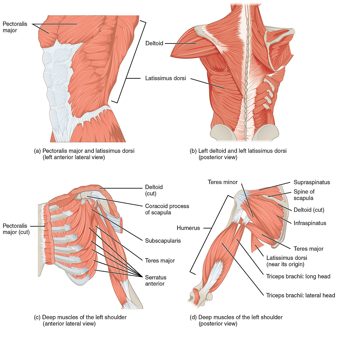
Supraspinatus. Initiates abduction from 0 to 15-30 degrees before the deltoid takes over, and it is the most commonly torn cuff tendon. Its tendon runs through the subacromial outlet, above the humeral head and below the acromion and coracoacromial ligament, which is why this is the muscle that impinges. Test it with the empty can (Jobe) maneuver.
Infraspinatus. The primary external rotator with the arm at the side, and the pitching muscle. It shares the suprascapular nerve with the supraspinatus, so a nerve lesion at the suprascapular notch weakens both. Atrophy of the infraspinatus fossa is a late sign of a chronic tear.
Teres minor. External rotation with the arm abducted, and a weak adductor. It rides the axillary nerve with the deltoid. Inability to externally rotate with the arm elevated, the hornblower's sign, marks a massive tear that has taken this muscle too.
Subscapularis. The only anterior cuff muscle, on the front of the scapula, inserting on the lesser tuberosity. It is the primary internal rotator and an anterior stabilizer. Test it with lift-off and belly-press; tears here are often traumatic, from forced external rotation.
Match each muscle to its job.
01Match: muscle to action
Tap a muscle, then its action.
Flip through the four-card deck until the lineup is automatic.
02Flashcards: the SItS lineup
Which muscle starts the arm moving?
03Recall: who starts abduction
Which rotator cuff muscle initiates abduction of the arm, and is the most commonly torn?
Supraspinatus. It fires first to start abduction from 0 to 15-30 degrees, then the deltoid takes over, and its tendon is the one that runs through the subacromial outlet. Infraspinatus. External rotation with the arm at the side, not abduction. Teres minor. External rotation with the arm abducted. Subscapularis. Internal rotation, the anterior cuff muscle.
SEE IT
See the Structures the Stem Is Describing
Tap each rotator cuff muscle on the illustration.
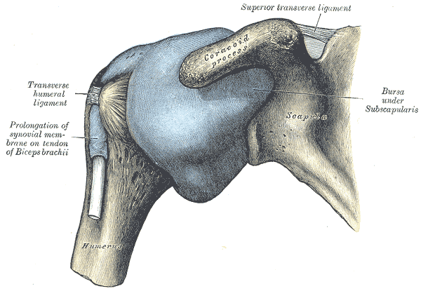
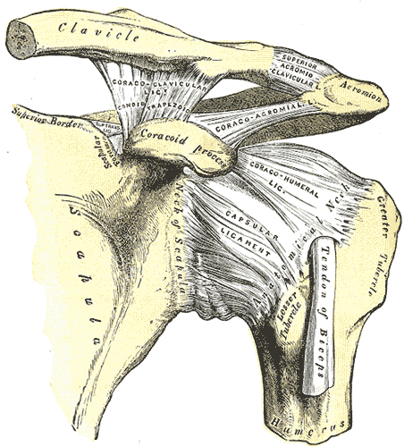
The subacromial space is a corridor with three contents: the supraspinatus tendon, the subacromial bursa, and the top of the humeral head. The ceiling is the coracoacromial arch, made of the acromion, the coracoid, and the coracoacromial ligament between them. The floor is the greater tuberosity.
When the arm elevates, the greater tuberosity and the supraspinatus tendon pass beneath that arch. Anything that narrows the corridor, a hooked acromion, an osteophyte, or a thickened bursa, pinches the tendon at the moment it is under the arch. That is why the same muscle keeps appearing in every impingement stem: it is the only cuff tendon that lives inside the outlet.
04Explore: the four muscles
Tap a region. Diagram: InjuryMap, CC BY-SA 4.0.
Two arc patterns, two different joints. Toggle between them.
05Compare: which arc means which joint
UPGRADE THE DIAGNOSIS
Impingement vs Full-Thickness Tear
Same tendon, different severity. One test changes the management.
The word impingement describes a pinch, not a rupture. Tendinopathy and impingement leave the tendon continuous: pain with overhead activity, a painful arc, night pain that wakes the patient, and preserved strength, though pain may limit effort. Neer and Hawkins-Kennedy tests are positive, and a subacromial injection typically quiets the pain.
A partial-thickness tear behaves like tendinopathy and may add subtle weakness. A full-thickness tear is different: true weakness rather than pain-limited effort, inability to initiate or sustain abduction, a positive drop-arm test, an external rotation lag sign, and eventually fossa atrophy. In a massive tear the patient may show pseudoparalysis, an arm that cannot be actively elevated at all.
The acute-on-chronic pattern is a favorite stem: a 60-year-old lifts something heavy, feels a pop, and the arm stops working. That is a full-thickness tear on top of years of silent tendinopathy, and it is the presentation that changes the plan from injection to imaging and surgical discussion.
Which finding makes it a complete tear?
06Recall: the test that upgrades
A 62-year-old man has both a positive empty-can test and a positive drop arm test. Which finding upgrades the diagnosis from impingement to a complete tear?
Positive drop arm test. The drop arm test is positive only when the supraspinatus tendon can no longer maintain the continuity needed to decelerate the lowering arm. Impingement leaves the tendon intact; a full-thickness tear does not. Drop arm positive = full tear; drop arm negative = impingement. That one test changes the management.
Route the three presentations to their next step.
07Branch: after the pop
Tap a presentation.
MECHANISM THEATER
The Corridor Closes at 90 Degrees
Drag the slider through abduction and watch the subacromial corridor narrow, then reopen.
Elevation is a door closing and reopening. As the arm rises, the greater tuberosity and the supraspinatus tendon move up and medially under the coracoacromial arch. The corridor is widest with the arm at the side, narrows through mid-elevation, and is tightest near 90 degrees, which is exactly where the pain peaks.
The geometry explains the whole exam. A hooked acromion, Bigliani type III, lowers the ceiling; subacromial osteophytes and a thickened bursa raise the floor. Each of those narrows the corridor and makes the same arc hurt. In throwers, a different pinch occurs: internal impingement, where the undersurface of the supraspinatus and infraspinatus tendons is compressed against the posterosuperior glenoid and labrum in the cocked position of abduction with maximum external rotation.
08Explore: the arc, step by step
Abduction
Why this arc, and no other?
09Recall: the reason for the arc
Why does the painful arc of impingement span 60 to 120 degrees of abduction?
The supraspinatus tendon passes under the coracoacromial arch only in that range. Below 60 degrees the tendon has not reached the arch; past 120 degrees the greater tuberosity rotates posteriorly and clears it. The pain brackets the moment the tendon is under the arch. The deltoid fatigues at mid-elevation. Fatigue is not a 60-120 degree phenomenon. The glenoid labrum is pinched at the top of the arc. Labral pinch is an internal impingement phenomenon of throwers, not a mid-arc painful arc. The AC joint compresses at exactly 90 degrees. AC joint pain is end-range and reproduced by cross-arm adduction.
THE SPECIAL TESTS
The Bedside Battery: What Each Test Proves
Six maneuvers, six answers. Match the test to the tendon it isolates.
The bedside battery separates the shoulder into its parts. Neer impingement sign: passive forward flexion with the scapula depressed, which jams the supraspinatus under the acromion; pain means impingement. Hawkins-Kennedy: arm forward flexed 90 degrees with the elbow bent, then forced internal rotation, which drives the greater tuberosity under the coracoacromial ligament; pain means impingement.
Empty can (Jobe): arms abducted 90 degrees in the scapular plane with thumbs down, resisting downward pressure; weakness names the supraspinatus. Drop-arm test: passively elevate to 90 degrees and ask the patient to lower slowly; an immediate drop means a full-thickness tear. External rotation lag sign: with the elbow at 90 degrees, passively externally rotate and ask the patient to hold; a lag means infraspinatus or teres minor tear. Lift-off and belly-press: internal rotation against resistance; failure names the subscapularis.
10Match: test to what it isolates
Tap a test, then what it isolates.
Run the shoulder exam in a reproducible order.
11Checklist: the exam sequence
IMAGING PATHWAY
X-ray First, Then Ultrasound or MRI
Plain films rule out bone; ultrasound and MRI look at the tendon.
Imaging starts with plain radiographs: AP, axillary, and outlet views. They show the acromial morphology, Bigliani type I flat, II curved, or III hooked, subacromial osteophytes, sclerosis of the greater tuberosity, calcific deposits, and, most importantly, the position of the humeral head. Superior migration of the head on an AP film means the cuff can no longer hold it down, which is the radiographic signature of a chronic massive tear with cuff arthropathy.
Ultrasound is cheap, dynamic, and excellent for full-thickness tears. MRI is the gold standard: it shows partial-thickness tears that ultrasound can miss, tendon retraction, muscle atrophy, and fatty infiltration, graded with the Goutallier classification, which drives the surgical decision. A tendon that is torn but healthy is repairable; a tendon that is torn, retracted, and fatty is not.
12Sequence: the workup order
Tap the steps in order.
The humeral head has moved up. What does that mean?
13Recall: the superior migration
Superior migration of the humeral head on a plain radiograph most strongly suggests what?
A chronic massive rotator cuff tear with cuff arthropathy. The cuff normally depresses the humeral head against the glenoid during deltoid contraction. When the tear is massive and chronic, the deltoid pulls the head upward against the acromion, and the joint degrades. Acute subacromial impingement. Impingement does not displace the head. AC joint separation. That moves the clavicle, not the humeral head. Calcific tendinitis. Calcium deposits do not migrate the head.
TREATMENT LADDER
PT and Injection First; Surgery for Tears That Matter
The ladder starts with the least invasive step that still changes the outcome.
Pure impingement and tendinopathy are managed without surgery. Physical therapy targets rotator cuff strengthening, scapular stabilization, and posterior capsule stretching; NSAIDs control the inflammation; and a subacromial corticosteroid injection is both diagnostic and therapeutic, since relief after injection confirms the subacromial space as the source. Subacromial decompression, an acromioplasty, is rarely needed for impingement alone.
Full-thickness tears change the conversation. An acute traumatic tear in a young, active patient is repaired early, ideally within about 3 months, before the tendon retracts and the muscle fattens. A chronic tear in an older patient starts with a nonoperative trial of therapy and injection. A massive tear with pseudoparalysis in an older patient, and cuff tear arthropathy, the destroyed, superiorly migrated joint of Milwaukee shoulder, are the domain of the reverse total shoulder arthroplasty, which swaps the ball and socket so the deltoid can elevate the arm without a cuff.
A 57-year-old active patient sustains an acute full-thickness supraspinatus tendon tear after a fall and now has pain with weakness of abduction.What is the most appropriate management?
14Hints: the acute tear in an active patient
A 57-year-old active patient with an acute full-thickness supraspinatus tear gets a different answer than the same tear in an 80-year-old with pseudoparalysis: one is repaired, the other may end in a reverse arthroplasty. The stem hands you age, activity, and the acuity of the tear. Read all three before you pick the rung.
Clinical walkthrough
