
Choose an answer, then open any option to work its reasoning.
MSK · Seronegative Spondyloarthropathies
The Spondyloarthropathy Quartet
Four seronegative arthritides: ankylosing spondylitis, psoriatic arthritis, reactive arthritis, and IBD-associated arthritis. One mechanism: enthesitis. One genetic marker: HLA-B27. Zero rheumatoid factor. Learn to split them before the test hits the one that fools everyone.
What this page makes you able to do
- Distinguish the four spondyloarthropathies by joint pattern, skin, and trigger history
- Rank the HLA-B27 associations: AS, reactive, IBD-axial, psoriatic
- Explain why enthesitis heals with new bone while RA synovitis erodes
- Read the imaging timeline from sacroiliitis to bamboo spine
- Dr. Fatima Ali, DOPsychiatry residentPrimary reviewer
Last reviewed
The pearl
Seronegative + enthesitis + HLA-B27 = the spondyloarthropathy family. The HLA-B27 ranking is locked: AS > reactive > IBD-axial > psoriatic, and axial IBD-arthritis looks exactly like AS.
Prove it
Opening question
A 24-year-old man has 3 months of low back pain and morning stiffness that improves with exercise. Exam shows limited lumbar flexion. He is HLA-B27 positive, and radiographs show bilateral sacroiliitis.Which spondyloarthropathy is most likely?
- Why this is rightA young man with inflammatory back pain (morning stiffness, better with activity), HLA-B27 positivity, and bilateral sacroiliitis is the prototype. HLA-B27 positive + sacroiliitis = ankylosing spondylitis until proven otherwise.
- Why this failsPsA needs psoriasis (skin first) and targets DIP joints with dactylitis and pencil-in-cup deformity. No skin disease, no DIP pattern here. DIP + psoriasis = PsA; axial + sacroiliitis = AS.
- Why this failsReactive arthritis follows a GU or GI infection by 1-4 weeks and gives the triad: can't see, can't pee, can't climb a tree. There is no infectious trigger in this story. The reactive triad needs a recent trigger.
- Why this failsAxial IBD-arthritis looks exactly like AS on imaging, but there is no Crohn or ulcerative colitis history here. Without bowel disease, the label is AS. Axial IBD-arthritis mimics AS: look for the bowel history.
- Why this failsRA is seropositive (RF and anti-CCP), targets the small joints of the hands and feet symmetrically, and spares the sacroiliac joints. This story is seronegative and axial. The quartet is RF negative. RA is not in it.
Work the reasoning
One mechanism (enthesitis, inflammation where tendon meets bone), plus the HLA-B27 link and seronegativity (RF negative). Enthesitis is the shared lesion of the quartet.
The sacroiliac joints: bilateral sacroiliitis appears long before spinal syndesmophytes. AS starts at the SI joints.
Anterior uveitis (sudden, unilateral, painful red eye), aortitis, and restrictive lung disease from a rigid chest. AS travels with uveitis, aortitis, and a stiff cage.
Inflammatory back pain + HLA-B27 + bilateral sacroiliitis = ankylosing spondylitis, the prototype of the seronegative quartet.
MEET THE QUARTET
Four seronegatives, one family
All four are RF negative, share the HLA-B27 link, and start at the enthesis. After that, they go four different directions.
Every member of the quartet is seronegative: rheumatoid factor and anti-CCP are absent. What they share is a target (the enthesis, where tendon, ligament, or capsule meets bone) and a genetic marker, HLA-B27, that tags a different proportion of each disease. The board test is not the family resemblance; it is the pattern that separates the four.
The Fuser. Young men 15-40; HLA-B27 in about 90%. Axial disease: sacroiliac joints first, then the spine. Pain is inflammatory: morning stiffness that improves with activity. X-ray: bamboo spine and bilateral sacroiliitis. Extra-articular: uveitis, aortitis, restrictive lung disease. Anterior uveitis is the most classic eye event: sudden, unilateral, painful, photophobic.
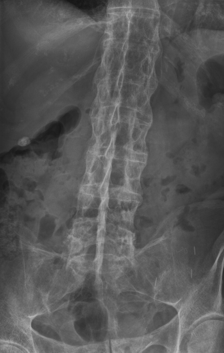
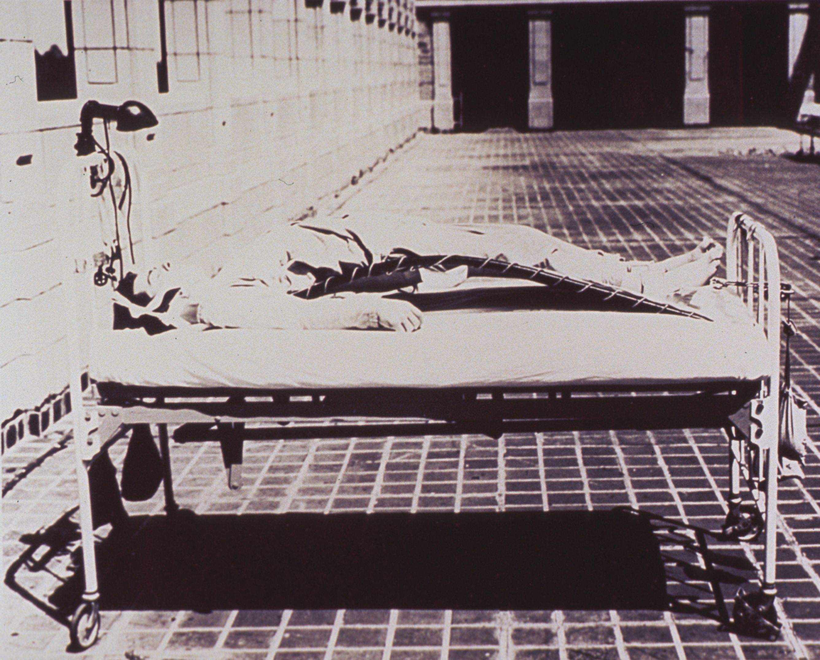
Bamboo spine = late-stage AS in clinical practice.
The Sausage Maker. A psoriasis patient (skin usually first) develops DIP joint arthritis with dactylitis (the sausage digit) and pencil-in-cup deformity at the DIP joints. Axial involvement is possible and asymmetric. HLA-B27 in about 40%, the weakest link of the four. Nail pitting and onycholysis are bedside clues; five clinical patterns exist, and asymmetric oligoarthritis is the most common.
DIP + psoriasis = PsA until proven otherwise.
The Triad. Triggered by Chlamydia (urogenital) or enteric pathogens (Salmonella, Shigella, Campylobacter, Yersinia); arthritis follows 1-4 weeks later. Classic triad: can't see (conjunctivitis), can't pee (urethritis), can't climb a tree (asymmetric large-joint, lower-limb arthritis). Skin: keratoderma blennorrhagica and circinate balanitis. HLA-B27 in 75-80%. Most cases resolve within 3-6 months once the trigger is treated.
Recent GU or GI infection + arthritis = think reactive.
The Bowel Watcher. Crohn or ulcerative colitis. Peripheral arthritis tracks bowel activity: the joints flare with the bowel, and treating the bowel treats the joint. Axial disease runs independently of the bowel and looks exactly like AS on imaging (HLA-B27 in about 60% of the axial type). Skin: erythema nodosum and pyoderma gangrenosum. NSAIDs can flare IBD, so they are used cautiously.
Peripheral IBD-arthritis follows the bowel; axial IBD-arthritis mimics AS.
Three questions separate the quartet: which joints, which skin, which trigger. DIP disease and dactylitis belong to psoriasis; a urogenital or enteric trigger belongs to reactive; the bowel belongs to IBD; and pure axial disease in a young adult belongs to AS until proven otherwise.
A 45-year-old with a 10-year history of psoriasis has a swollen right index finger involving the whole digit. Radiographs show pencil-in-cup erosions at the DIP joint. Which pattern is this?
A. DIP erosions + dactylitis + psoriasis is the PsA fingerprint. AS is axial, reactive arthritis needs a GU or GI trigger, and RA is symmetric, seropositive, and spares the DIP joints. DIP + sausage digit + psoriasis = PsA until proven otherwise.
One distinction runs underneath the whole family: enthesitis heals with new bone. That is why the quartet fuses while RA erodes.
THE SHARED MECHANISM
Enthesitis: one lesion, four endings
All four diseases attack the same anatomical target: the enthesis, where tendon meets bone. Slide through the cascade to fusion.
The enthesis is a specialized fibrocartilaginous junction that disperses the force of tendon pulling on bone. In the spondyloarthropathies it is the primary battlefield: macrophages, T cells, and IL-17- and TNF-driven inflammation ignite at this interface.
Here is the paradox that separates this family from RA. Synovial inflammation in RA ends in erosion: bone is eaten away. Entheseal inflammation in the quartet ends in the opposite: new bone formation. The inflamed enthesis lays down bone, syndesmophytes bridge the disc spaces, and the spine fuses. Inflammation that heals by building bone is the spondyloarthropathy signature.
Enthesitis cascade
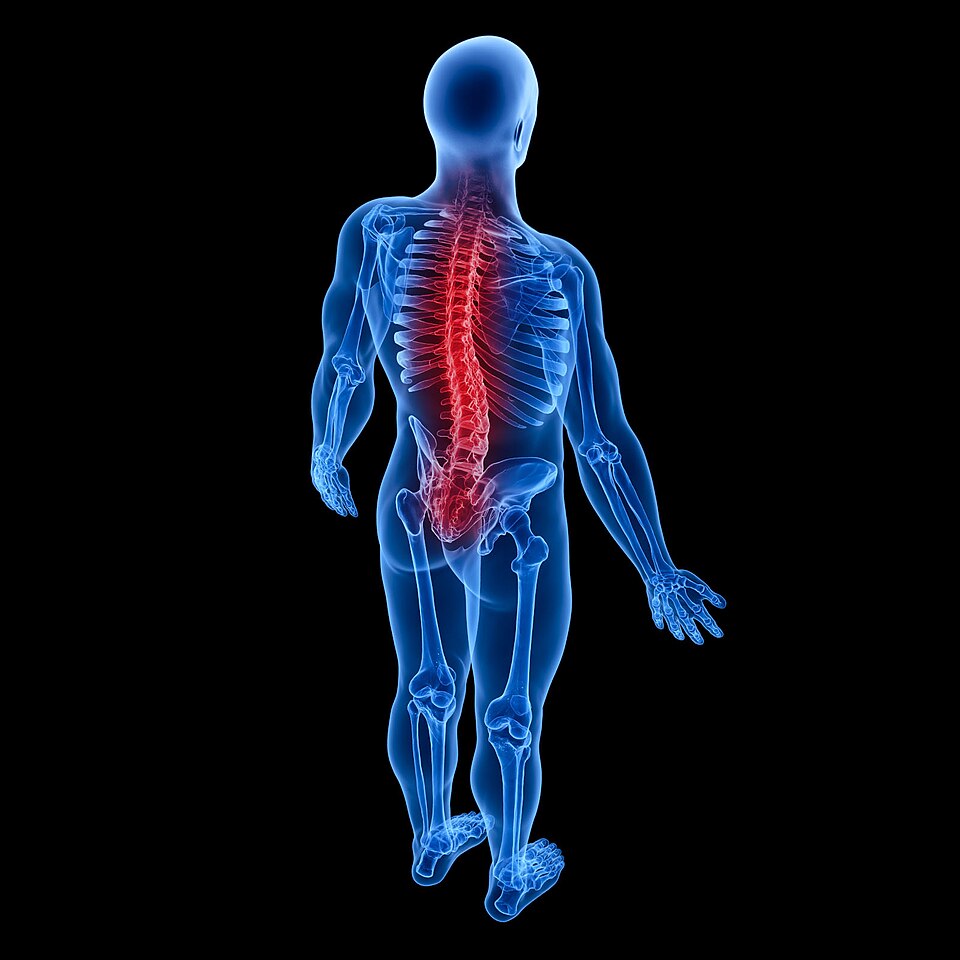
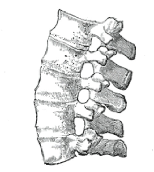
Cytokine biology explains the treatment targets: TNF and IL-17 both drive entheseal inflammation, which is why anti-TNF and IL-17 inhibitors are the biologic workhorses for axial disease.
THE HLA-B27 TRACKER
Association strength, ranked
The percentages are approximate, but the ranking is locked. Match each disease to its HLA-B27 link.
HLA-B27 is an MHC class I molecule that presents peptides to CD8 T cells. Its link to the quartet is one of the strongest known HLA-disease associations, and yet most B27 carriers are perfectly healthy. Association is not causation: roughly 90% of AS patients carry the gene, but only a small fraction of carriers ever develop the disease.
What the boards test is the ranking. Memorize the order and the approximate percentages, then let the clinical pattern, not the gene, make the diagnosis.
Tap a disease, then its HLA-B27 association.
ASk the Rheumatologist If Psoriasis
ASAnkylosing spondylitis: about 90%
RReactive arthritis: about 75-80%
IIBD-arthritis (axial): about 60%
PPsoriatic arthritis: about 40%
A positive HLA-B27 with bilateral sacroiliitis in a young adult is AS until proven otherwise. A negative HLA-B27 does not exclude the family, especially PsA, where the link is weakest.
WHICH SPONDYLOARTHROPATHY?
The discriminator: what happened before the arthritis
The clue that matters is often the history, not the joint exam. Work the branches, then lock the pattern with the question.
Four questions in the history separate the quartet. Is there a trigger, a urogenital or enteric infection? Is there skin disease, and which kind? Is there bowel disease, and does the joint track it? Is the pattern axial, and does it fit a young adult with inflammatory pain? The joint exam confirms; the history discriminates.
Gradual low back pain in a young man, morning stiffness improving with activity. Pelvis X-ray and HLA-B27: bilateral sacroiliitis with a positive HLA-B27?
Asymmetric large-joint arthritis with red eye and urethritis, 2-3 weeks after unprotected sex or a diarrheal illness. Is the classic triad present?
A 30-year-old man with Crohn disease has 4 months of inflammatory back pain. MRI shows sacroiliitis, and treating his bowel has not helped the back. Which statement is correct?
B. Axial IBD-arthritis runs independently of bowel activity and is radiographically indistinguishable from AS (HLA-B27 in about 60% of axial cases). Peripheral type tracks the bowel; NSAIDs can worsen IBD, so they are used cautiously. Axial IBD-arthritis = the AS lookalike that ignores the bowel.
A Crohn disease patient has new joint pain. Do the joints flare and settle together with the bowel?
BEYOND THE JOINTS
The quartet outside the joint
Uveitis, aortitis, skin, and nail: each member of the family has a signature beyond the enthesis.
Anterior uveitis is the shared eye event (sudden, unilateral, painful, photophobic), and it can occur in all four diseases, most classically in AS. Aortitis with aortic regurgitation and restrictive lung disease from a rigid chest complete the AS extra-articular set.
The skin is the fastest discriminator: keratoderma blennorrhagica and circinate balanitis belong to reactive arthritis; erythema nodosum and pyoderma gangrenosum belong to IBD; nail pitting and onycholysis belong to psoriasis. Dactylitis, the sausage digit, is enthesitis of the digital tendon sheaths and is the PsA (and reactive) fingerprint.
Tap a manifestation, then its disease.
Why the eye? The uveal tract and the enthesis sit on the same inflammatory axis: both are vascularized connective-tissue interfaces, which is why the same cytokine-driven disease lights up the eye, the aorta, and the tendon-bone junction.
THE IMAGING TIMELINE
From sacroiliitis to bamboo spine
The disease writes a predictable story on film, and the story is the test.
X-ray the SI joints first. Sacroiliitis begins with subchondral erosions and sclerosis; the joint appears to widen before it narrows, and end-stage disease fuses it to a thin line. In AS the sacroiliitis is bilateral and symmetric: unilateral sacroiliitis should make you think of reactive arthritis or infection instead.
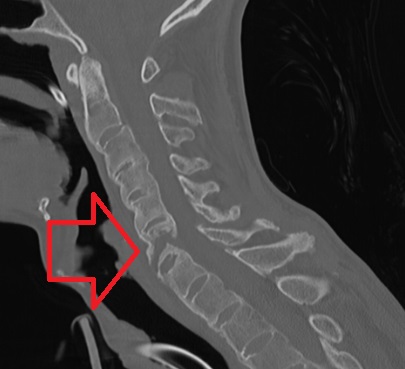
MRI is the earliest window: bone marrow edema at the SI joints on STIR sequences is active sacroiliitis before any X-ray change. Later the spine shows syndesmophytes (thin, vertical ossifications arising from the outer annulus), which differ from the horizontal traction osteophytes of osteoarthritis. When the bridges are complete the spine reads as bamboo, and ossification of the supraspinous ligament produces the dagger sign on the frontal view.
The fused spine is a liability: it behaves as one long lever, so minor trauma can produce a fracture through the fusion mass: an injury plain films undercall. New focal midline pain after trauma in a fused spine is a fracture until proven otherwise.
Tap the imaging timeline in order, earliest first.
THE TREATMENT LADDER
NSAIDs first, biologics for refractory axial disease
The spondyloarthropathies are managed by pattern: axial and peripheral disease are treated differently.
NSAIDs are first-line across the quartet: they relieve pain and stiffness but do not change the course of the disease. Physical therapy preserves posture and motion, especially in AS, where a rigid, kyphotic spine is the endpoint to avoid.
For axial disease that stays active despite NSAIDs, the answer is a biologic: anti-TNF agents (infliximab, adalimumab, etanercept) or IL-17 inhibitors (secukinumab), and in IBD, anti-TNF treats the bowel and the spine together. The key board fact: sulfasalazine and methotrexate work for peripheral arthritis but do not control axial disease.
Every AS patient is screened: uveitis and aortitis on history and exam, and DXA bone density, because the rigid spine and immobility drive bone loss. Reactive arthritis is managed by eradicating the trigger; peripheral IBD-arthritis by treating the bowel; PsA by NSAIDs first, then methotrexate or anti-TNF for peripheral disease.
Clinical walkthrough
