
Choose an answer, then open any option to work its reasoning.
MSK · Spine
Spondyl- Words: Itis, Osis, Lysis, Listhesis
Four suffixes, four diseases, one root. The words look identical and the mechanisms could not differ more; boards love this family.
What this page makes you able to do
- Decode the four spondyl- suffixes into four mechanisms
- Read the Scottie dog and recognize the pars collar
- Separate inflammatory spondylitis from degenerative spondylosis
- Screen any back pain history for red flags and cauda equina syndrome
- Dr. Fatima Ali, DOPsychiatry residentPrimary reviewer
Last reviewed
The pearl
-itis inflames, -osis wears, -lysis breaks, -listhesis slips. Name the mechanism and you have named the diagnosis.
Prove it
Opening question
A 19-year-old female gymnast has 3 months of low back pain that worsens when she extends her spine during tumbling. The lateral radiograph is normal, but the oblique view shows a defect through the pars interarticularis of L5.Which suffix names her pathology?
- Why this fails-itis means inflammation. Her imaging shows a fracture, not inflammation, and she has no morning stiffness, sacroiliitis, or HLA-B27 story.
- Why this fails-osis means degeneration: the wear-and-tear of the older spine (osteophytes, disc collapse). A teenage gymnast with a pars defect is the classic -lysis story.
- Why this is right-lysis is breakdown. Repetitive extension (tumbling, gymnastics, volleyball) fatigue-fractures the pars: the Scottie dog's collar. She has spondylolysis.
- Why this fails-listhesis is the slip: what happens after both pars break and the vertebra is free to move. Her defect is the cause; the slip is the consequence.
- Why this fails-malacia is softening (think osteomalacia). Nothing here is soft. The pars has broken.
Work the reasoning
The pars interarticularis (the bony bridge between the superior and inferior facets), plus the facet joints themselves. It is the weak point of the neural arch.
The vertebra loses its rear anchor and is free to slip forward: the listhesis. One collar is a lysis; two collars can become a listhesis.
Extension loads the posterior elements directly. The fractured pars impinges: pain on extension is the mechanical clue that separates this from discogenic pain.
The collar IS the lysis. Two collars = no rear anchor = the vertebra is free to slip.
TRANSLATE THE SUFFIX
Four suffixes, four mechanisms
The word tells you the mechanism. Match each suffix to its meaning: this is the whole page in one move.
Every word on this page is spondyl- plus a suffix; spondylos is Greek for vertebra, and the suffix carries the entire diagnosis. -itis is inflammation (spondylitis, the ankylosing family). -osis is a chronic condition without acute inflammation: in practice, degeneration (spondylosis, the wear-and-tear spine). -lysis is breakdown: a break in the pars interarticularis (spondylolysis). -listhesis is a slip: the vertebra slides forward (spondylolisthesis).
The classic confusion is the mimic pair: spondylosis is not inflammation: it is the osteophyte-and-stenosis spine of aging, and it never causes uveitis or sacroiliitis. Spondylitis is the inflammatory disease of young adults. The lysis-listhesis pair is mechanical: the pars fracture is the cause, and the slip is the consequence.
Tap a suffix, then its meaning.
Board question anatomy: when the stem says pars defect, answer lysis; when it says slip, answer listhesis; when it says morning stiffness, answer itis; when it says osteophytes and stenosis, answer osis.
SEE IT ON IMAGING
The Scottie dog and the four films
Tap the regions of the Scottie dog, the oblique view that puts spondylolysis on the map. Then read the four conditions side by side.
The oblique lumbar view turns each posterior element into a Scottie dog: the transverse process is the nose, the pedicle the eye, the superior articular process the ear, the inferior articular process the leg, and the pars interarticularis, the neck, carries the collar. A radiolucent break across the neck is the pars defect of spondylolysis.
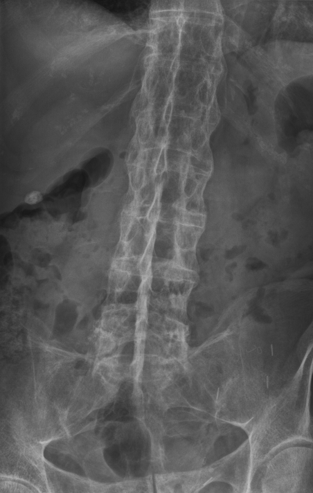
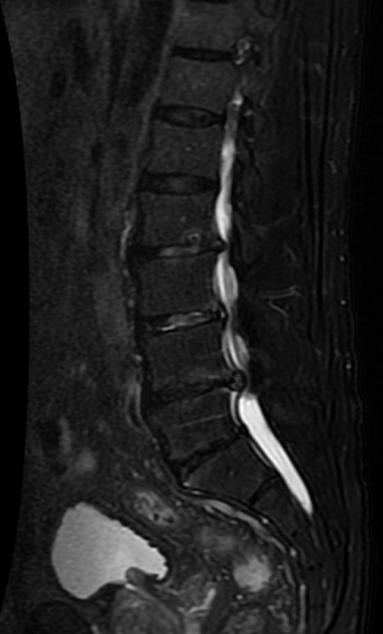
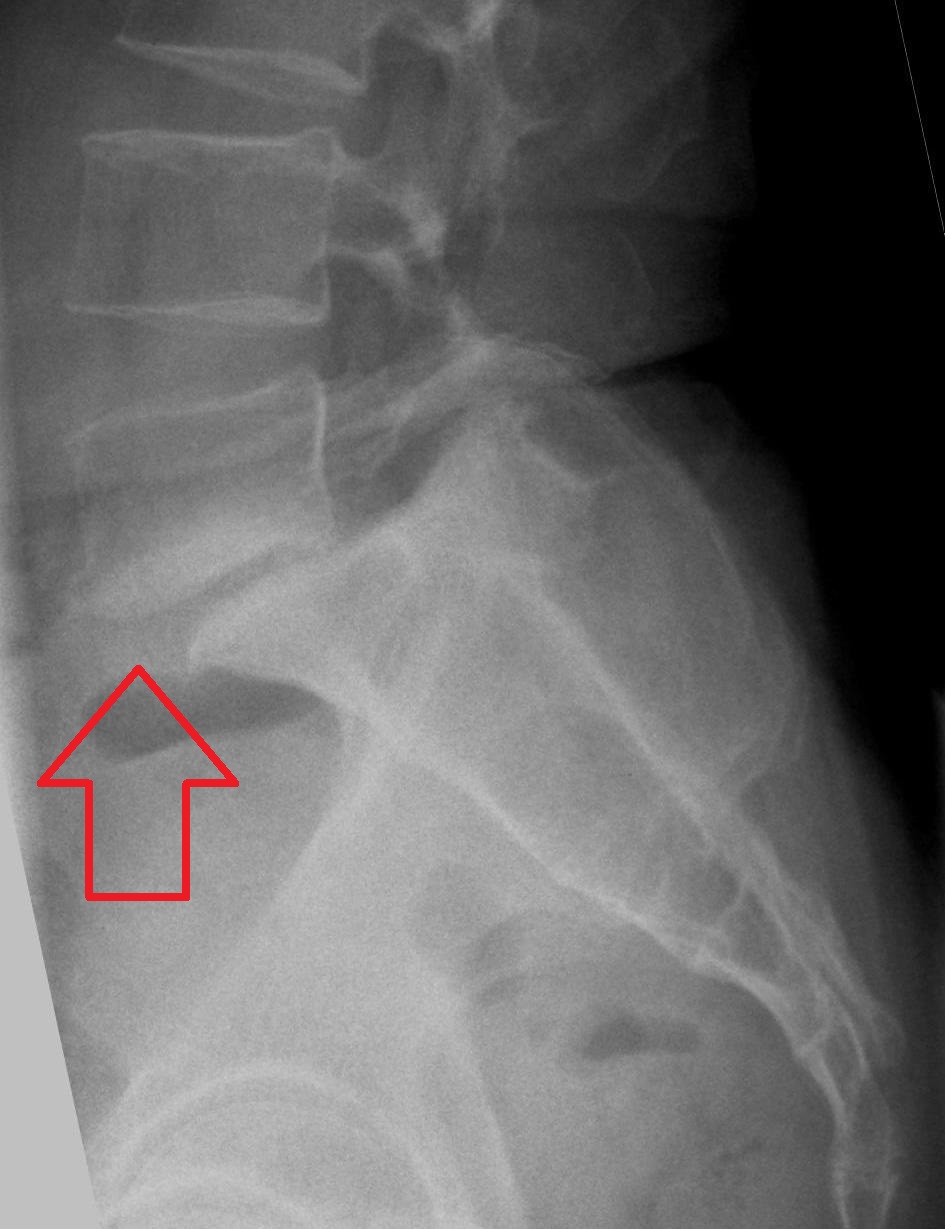
One defect is a lysis; two defects (bilateral) remove the rear anchor and the vertebra can slip: the listhesis you measure on the lateral view as a percentage of the vertebral body width. The four films below show the whole family: bamboo spine (itis), canal stenosis (osis), the pars collar (lysis), and the L5-on-S1 step (listhesis).
Tap a region of the Scottie dog.
THE DISCRIMINATOR
itis vs osis: the mimic pair
Both hurt the back. One is on fire, one is worn out: flip between them and watch the features that separate them.
Two patients, both with low back pain. The first is 25, stiff in the morning for over an hour, better with exercise, HLA-B27 positive, with sacroiliitis on MRI: inflammatory spondylitis. The second is 70, hurts when standing and walking, better when sitting, with osteophytes and stenosis on imaging: degenerative spondylosis. The history separates them before any test.
A 70-year-old has back pain that worsens with walking and standing and eases when she leans forward on a shopping cart. ESR is normal. Which diagnosis fits?
A. Positional pain relieved by forward flexion, normal ESR, no morning stiffness: that is the spondylosis/stenosis pattern (neurogenic claudication). AS is inflammatory and young; lysis is a pars fracture; cauda equina is a surgical emergency with sphincter dysfunction.
Back pain history: age, onset, morning stiffness, and response to activity?
THE CONSEQUENCE
Years of inflammation become bamboo
Slide across the untreated course of ankylosing spondylitis: from normal disc spaces to a spine that reads as one bone.
Untreated spondylitis writes itself on the spine. Sacroiliitis comes first; then thin syndesmophytes rise from the disc margins; the bridges spread up the column; and finally the disc spaces are gone and the spine reads as one bone: the bamboo spine, with the dagger sign from ossified supraspinous ligaments.
The fused spine is a danger, not just a deformity. It moves as a single lever, so a fall from standing can fracture it, often through the thoracolumbar junction, and plain films undercall these injuries. Cervical fusion adds neck stiffness and fracture risk; kyphosis can restrict chest expansion and produce a restrictive pattern on pulmonary function tests.
Ankylosing spondylitis over time
Tap the untreated course in order, earliest first.
THE PARS INTERARTICULARIS
The anatomy that makes lysis and listhesis possible
One small bony bridge carries the whole mechanical story.
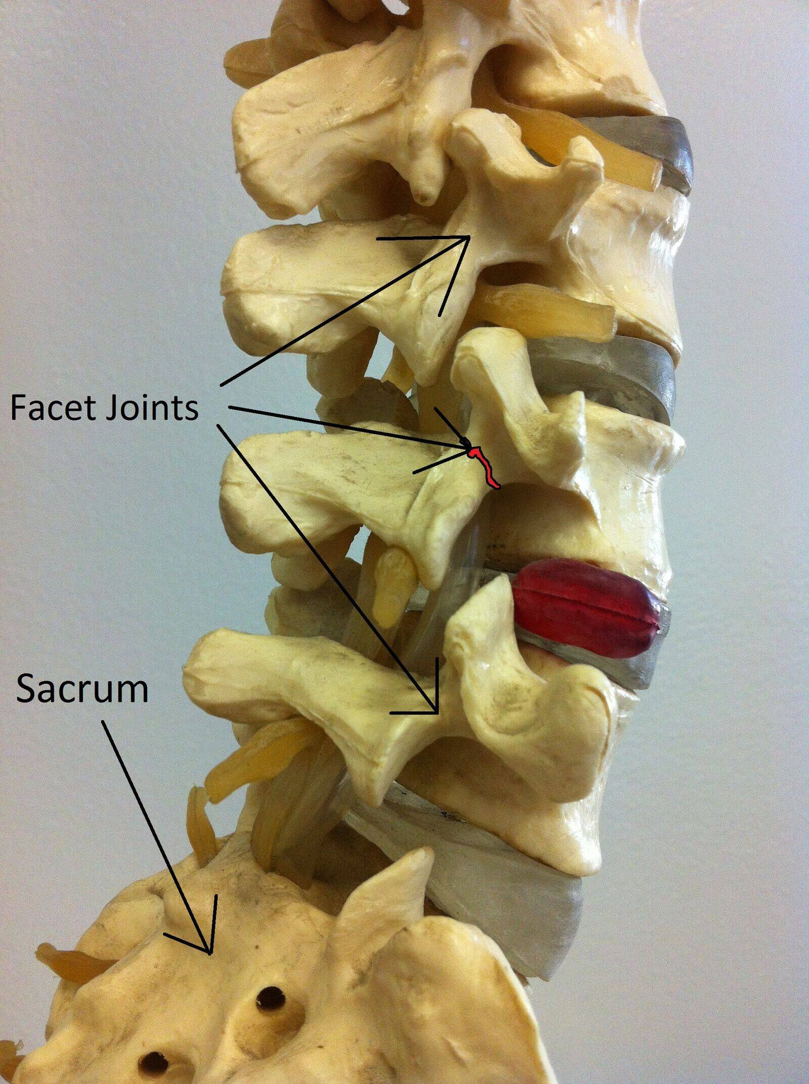
The pars interarticularis is the bony bridge between the superior and inferior articular processes, part of the neural arch, and the weakest link in the posterior column. It absorbs the shear of every extension, which is why repetitive hyperextension (gymnastics, football linemen, weightlifting, fast bowling) stress-fractures it. L5 is the most common site, then L4.
A unilateral defect usually stays a lysis. Bilateral defects remove the posterior anchor, and the vertebra is free to slip: that is the isthmic listhesis of adolescence. In older adults the same slip can happen with an intact pars, when arthritic facet joints fail to hold (degenerative listhesis, usually at L4).
The slip is graded on the lateral view by Meyerding: grade I is under 25% of the vertebral body width, grade II 25-50%, grade III 50-75%, grade IV 75-100%, and grade V (spondyloptosis) over 100%.
Checked 0
RED FLAGS
When back pain is not mechanical
Fever, night pain, sphincter dysfunction, and the claudication that bends forward.
Mechanical back pain is activity-related and self-limited. Anything else is a red flag. Fever with back pain raises discitis, vertebral osteomyelitis, or epidural abscess, especially in IV drug users or after procedures. Night pain and weight loss raise malignancy: metastases and multiple myeloma, particularly in patients over 50. Saddle anesthesia, bowel or bladder dysfunction, or progressive leg weakness is cauda equina syndrome, a surgical emergency.
Neurogenic claudication is the spondylosis calling card: bilateral leg aching with standing and walking, relieved by sitting or leaning forward, because flexion widens the canal. Vascular claudication mimics it but has diminished pulses, vascular risk factors, and relief from standing still, not from posture.
Back pain plus one of these features: what is the priority?
A 68-year-old has bilateral leg aching after two blocks of walking. He leans forward on a shopping cart to continue; femoral and pedal pulses are palpable. What is the best next step?
A. Normal pulses plus relief with forward flexion is neurogenic claudication from lumbar stenosis: posture changes the canal. Vascular claudication has diminished pulses and relieves with standing still; cauda equina has sphincter dysfunction; this picture is not inflammatory.
Clinical walkthrough
