
Choose an answer, then open any option to work its reasoning.
MSK · Trauma
Soft-Tissue Trauma: Shoulder and Hip Dislocations, Knee and Ankle Ligament Injuries
Dislocations and ligament tears obey one reflex: read the mechanism, read the limb position, then name the nerve at risk and the clock. Abduction plus external rotation with an anterior prominence is the anterior shoulder dislocation, about 95% of cases, and the axillary nerve is the structure to check before and after reduction. The leg that lies shortened, internally rotated, and adducted after a dashboard crash is the posterior hip, and the femoral head starves in about 6 hours. The popped knee and the rolled ankle are mechanism-and-timing puzzles: ACL versus meniscus, ATFL sprain grades, and the Ottawa rules that decide who gets a radiograph.
What this page makes you able to do
- Read the limb position and name the dislocation: anterior or posterior shoulder, luxatio erecta, posterior or anterior hip
- Name the nerve at risk for every direction, and document it before and after reduction
- Split ACL, PCL, MCL, and meniscus by mechanism and effusion timing, and read the knee tests
- Apply the Ottawa ankle and foot rules exactly as the boards grade them
- Dr. Fatima Ali, DOPsychiatry residentPrimary reviewer
Last reviewed
The pearl
The limb position is the diagnosis: shortened, internally rotated, adducted is the posterior hip; lengthened, externally rotated is the anterior hip; abduction plus external rotation with an anterior prominence is the anterior shoulder and the axillary nerve to check; seizure with the arm adducted and internally rotated is the posterior shoulder, the one the boards say is missed.
Prove it
Opening question
Answer before you read anything, then keep the order of operations in mind through every section.
A 22-year-old man comes to the emergency department after falling onto his outstretched right arm during a basketball game. He reports immediate right shoulder pain. He holds the arm slightly abducted and externally rotated. Examination shows a flattened deltoid contour with a palpable anterior prominence, and the humeral head is not felt in the glenoid. He reports reduced sensation over the lateral upper arm (regimental badge area).Which of the following is the most likely diagnosis?
- Why this is rightFall on an outstretched arm with the shoulder abducted and externally rotated, an anterior prominence, a flattened deltoid, and regimental badge numbness is the anterior dislocation fingerprint: about 95% of shoulder dislocations are anterior, and the axillary nerve is the structure injured with it (deltoid weakness plus lateral upper arm anesthesia). Rule: abduction plus external rotation plus anterior prominence is anterior dislocation, and the nerve to check is the axillary.
- Why this failsPosterior dislocation follows seizure, electrocution, or axial load and holds the arm adducted and internally rotated, with the light bulb sign on the AP radiograph. This stem is the opposite: abduction, external rotation, and an anterior prominence. Rule: seizure plus internal rotation is posterior; this is the reach-back story.
- Why this failsA clavicle fracture is tender directly over the bone, with the shoulder drooping and the patient supporting the arm; it spares the regimental badge and never produces an anterior humeral prominence. Rule: bony tenderness at the clavicle is the fracture; a lost joint contour is the dislocation.
- Why this failsCuff disease is the mirror-image trap: a painful arc of active abduction and internal rotation lag belong to the cuff, but the joint stays reduced, and the cuff does not numb the regimental badge. Rule: a lost contour plus a held arm position is the dislocation; the cuff hurts without moving the head out.
- Why this failsAn AC separation follows a direct blow to the point of the shoulder, with tenderness and a step-off at the acromioclavicular joint, not a humeral head out of the glenoid. Rule: AC pain sits on top of the shoulder; this prominence sits in front of it.
Work the reasoning
The limb position is the discriminator: anterior dislocations hold the arm abducted and externally rotated, posterior dislocations hold it adducted and internally rotated, and luxatio erecta locks it overhead. Read the posture before you touch the film.
The axillary nerve wraps the surgical neck of the humerus, so the anteriorly displaced head stretches it: deltoid weakness plus lateral upper arm anesthesia is the board combination, and it is the nerve to document before and after reduction.
Confirm with radiographs, document the axillary nerve (deltoid power and sensation), reduce with Stimson, Kocher, or traction-countertraction, then sling and early motion, with surgery for recurrent instability in the young and active.
The answer is A: anterior shoulder dislocation. Abduction plus external rotation plus anterior prominence plus regimental badge numbness is the anterior fingerprint, and the nerve to check is the axillary, before and after reduction.
THE 95 PERCENT AND THE TWO TRAPS
Shoulder Dislocations: The 95 Percent and the Two Traps
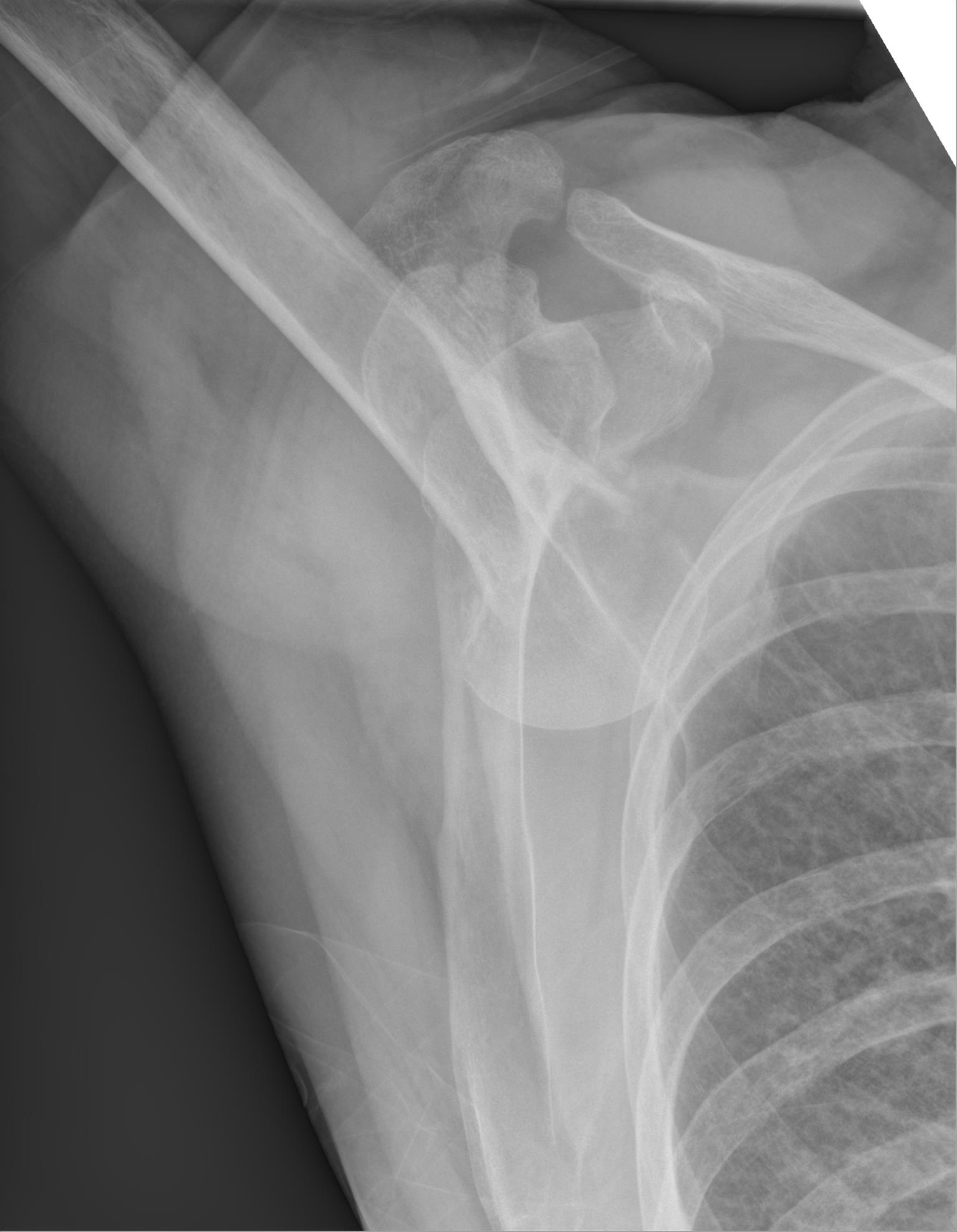
About 95% of shoulder dislocations are anterior: abduction plus external rotation drives the humeral head out the front of the glenoid, and the axillary nerve is the structure to check. Posterior dislocation follows seizure or axial load, shows the light bulb sign, and is the one the boards say is commonly missed. Inferior dislocation, luxatio erecta, locks the arm overhead.
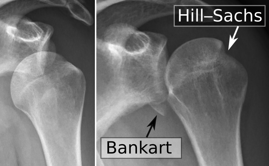
Anterior dislocation is the abduction and external rotation story. A fall on the outstretched arm, a reach-back, or a throwing force lever the humeral head anterior and inferior out of the glenoid. The arm is held slightly abducted and externally rotated, the deltoid contour flattens, and an anterior prominence appears. The axillary nerve wraps the surgical neck of the humerus, so the displaced head stretches it: deltoid weakness plus regimental badge anesthesia (lateral upper arm) is the board combination, and it must be documented before and after reduction. Two lesions ride along and both drive recurrence: the Hill-Sachs compression fracture of the posterolateral humeral head (impact on the anterior glenoid rim, bone side) and the Bankart tear of the anteroinferior labrum (soft-tissue side). The apprehension test recreates the slip: the shoulder placed in abduction and external rotation makes the patient resist, and posteriorly directed pressure on the head (the relocation test) relieves it.
Reduction is Stimson (prone, gravity with the arm hanging), Kocher (lever with the elbow flexed), or traction-countertraction under sedation, followed by post-reduction radiographs and a repeat neurovascular exam. A first-time dislocator gets a sling and early range of motion; the young, active patient with recurrent instability and a Bankart tear is the arthroscopic labral repair candidate. The rotator cuff is the mirror-image trap: a painful arc of 60 to 120 degrees with an internal rotation lag is cuff disease with the joint reduced, not a dislocation.
Flip between the anterior and posterior shoulder.
THE SHOULDER DISLOCATION STAGE
The Shoulder Dislocation Stage: Three Mechanisms, Three Paths
Run each mechanism and watch where the humeral head goes, then commit to the structure at risk before the board readout reveals. The mechanism is the first link of the reflex; the head follows the force.
Three mechanisms, three paths, three structures at risk. Dana, 19, felt her shoulder pop while reaching back for a pass: abduction plus external rotation drove the head anterior and inferior, and the axillary nerve took the stretch. Marcus, 40, seized during an insulin reaction: the internal rotators fired against the adducted arm and drove the head posterior, behind the glenoid rim, the light bulb sign on AP and the one that is routinely missed. Priya, 26, fell with her arm locked overhead: the head went inferior under the glenoid, luxatio erecta, and the axillary artery and nerve were on the line, with a weak radial pulse as the red flag. The luxatio erecta arm cannot come down; reduction is in-line traction in the overhead position with countertraction, then a pulse and nerve check, sling, and rehab.
Tap each site on the stage and read its path.
Tap a site.
THE DASHBOARD AND THE FALL
Hip Dislocations: The Dashboard and the Fall
About 90% of hip dislocations are posterior: the dashboard mechanism drives the femoral head behind the acetabulum, the limb lies shortened, internally rotated, and adducted, and the sciatic nerve is at risk. The anterior pattern is the opposite posture with the femoral nerve and artery on the line.
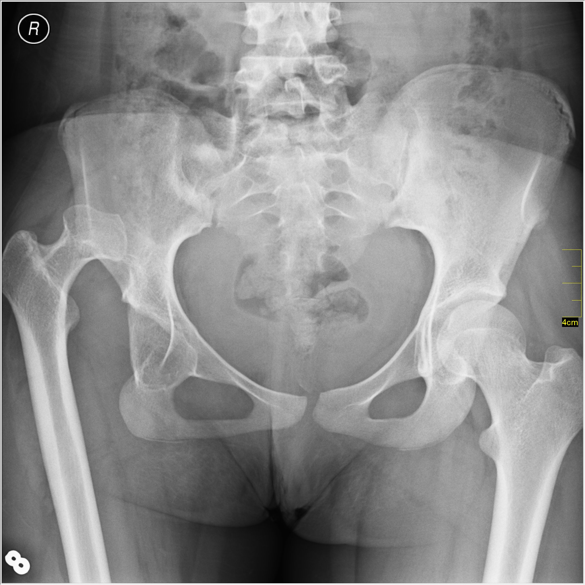
The dashboard story is the classic third-order stem: the knee strikes the dash with the hip flexed and adducted, and the femoral head pops out the back of the acetabulum. The limb lies shortened, internally rotated, and adducted, the sciatic nerve is stretched (especially its common peroneal division, so check dorsiflexion and great toe extension for foot drop), a posterior acetabular wall fracture often rides along, and avascular necrosis of the femoral head is why closed reduction under sedation must happen within about 6 hours: the retinacular circulation enters the femoral neck, the dislocation kinks it, and the head starves while it sits out of the socket.
Anterior dislocation follows forced abduction and external rotation, typically a fall: the limb lies lengthened and externally rotated, and the femoral nerve and artery in the groin are the structures at risk, with the groin pulse and quadriceps strength as the checks. After any hip reduction: post-reduction radiographs, a repeat neurovascular exam, CT of the acetabulum for the wall fracture, and hip precautions. The reflex that separates them never changes: shortened plus internally rotated plus adducted is posterior; lengthened plus externally rotated is anterior.
Commit to the posterior hip reflex.
A 36-year-old man is brought in after a head-on motor vehicle collision in which his knee struck the dashboard. The right leg lies shortened, internally rotated, and adducted, and he cannot dorsiflex the foot. Which of the following best describes the injury and the structure at risk?
A. Posterior hip dislocation with sciatic (peroneal division) injury, reduced within about 6 hours. The dashboard mechanism with the shortened, internally rotated, adducted limb is the posterior hip signature, and the inability to dorsiflex is foot drop from the peroneal division of the sciatic nerve. The 6-hour clock protects the femoral head from avascular necrosis. B. Anterior hip. That is the lengthened, externally rotated limb after forced abduction and external rotation, with the femoral nerve and artery at risk. C. Femoral neck fracture. A fractured neck leaves the leg shortened but externally rotated, not internally rotated and adducted. D. Acetabular fracture without dislocation. The locked posture proves the head is out of the socket. Rule: shortened + internally rotated + adducted is posterior hip, and the clock is 6 hours.
WHICH STRUCTURE TORE
The Knee: Which Structure Tore
The knee stem is a mechanism-and-timing puzzle. A pivot injury with a pop and a hemarthrosis within hours is an ACL tear; a dashboard blow with the tibia sagging backward is the PCL; a valgus blow is the MCL; a twist on a planted foot with locking is a meniscal tear.
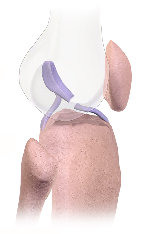
The two clocks split the knee. ACL tears bleed immediately because the ligament is intra-articular and vascular: pop, giving way, and a tense effusion within hours, with Lachman the most sensitive test (knee at 30 degrees, anterior translation), anterior drawer at 90 degrees, and the pivot shift confirming rotational instability. Female athletes tear ACLs at higher rates in cutting sports, and the young, active patient who wants to return to pivoting gets reconstruction. Meniscal tears ooze slowly: a twist on a planted foot with joint line tenderness, a positive McMurray, or a locked knee, with the effusion building over about 24 hours, the reverse of the ACL. A PCL injury follows a dashboard blow to a bent knee, the tibia sags posteriorly at 90 degrees, and the posterior drawer is positive. An MCL tear follows a valgus blow with medial pain and opens to valgus stress, graded I (stretch), II (partial), III (complete); most isolated MCL tears heal nonoperatively. The unhappy triad is the valgus blow that takes the MCL, the ACL, and the medial meniscus together.
The extensor mechanism completes the differential. Patellar dislocation rides lateral in adolescent and young adult women with a high Q angle, often after a valgus blow or twisting injury, and reduces with knee extension and a gentle medial push. Patellar tendon rupture leaves a palpable gap and a high-riding patella; quadriceps tendon rupture hits older patients and drops the patella low (patella baja). Inability to actively extend the knee with a suprapatellar or infrapatellar gap is the rupture story.
Open each structure and hold its signature.
Cutting or pivot injury with a pop, giving way, and a tense effusion within hours. Lachman is the most sensitive test; anterior drawer and pivot shift support it. Female athletes tear ACLs at higher rates. Young and active patients get reconstruction; older or sedentary patients often do well with bracing and quadriceps rehabilitation.
The tibia strikes the dash with the knee bent and the tibia sags posteriorly at 90 degrees; the posterior drawer is the test. Less common than the ACL, and often seen with other ligament injuries in high-energy trauma.
Valgus force opens the medial joint line: grade I stretch, grade II partial tear, grade III complete tear. Most isolated MCL tears are managed nonoperatively with bracing and rehabilitation.
Twist on a planted foot with joint line tenderness, a positive McMurray (click or pain with rotation during flexion and extension), and mechanical locking from a bucket-handle tear. The effusion builds over about 24 hours, the reverse of the ACL's immediate hemarthrosis.
Patellar dislocation rides lateral in young women with a high Q angle and reduces with extension plus a medial push. Patellar tendon rupture: high-riding patella with a palpable gap. Quadriceps tendon rupture in older patients: patella baja. Inability to actively extend the knee is the shared red flag.
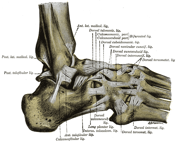
Inversion injury tears the anterior talofibular ligament first, then the calcaneofibular and posterior talofibular ligaments. Grade I is a mild stretch, grade II a partial tear, grade III a complete tear with gross instability; the anterior drawer test checks ATFL integrity. Most sprains heal with rest, ice, compression, elevation, and progressive weight bearing.
INVERSION, THE ATFL, AND THE OTTAWA GATE
The Ankle: Inversion, the ATFL, and the Ottawa Gate
The rolled ankle is the most tested sprain in medicine. The anterior talofibular ligament tears first in the inversion injury, the sprain is graded I to III, and the Ottawa rules decide who actually gets a radiograph.
The anterior talofibular ligament (ATFL) runs from the fibula to the talus and is the first structure to tear when the ankle inverts; the calcaneofibular and posterior talofibular ligaments tear as the injury gets worse. The anterior drawer test checks ATFL integrity, and the sprain is graded by tear size: grade I is a mild stretch, grade II a partial tear, grade III a complete tear with gross instability. Most sprains are managed with rest, ice, compression, elevation, and progressive weight bearing; chronic ATFL laxity shows up as a positive anterior drawer.
The tendon behind the ankle is the Achilles. The classic stem is the middle-aged recreational athlete who felt a sudden snap or pop in the calf during a push-off, "like being kicked," and now cannot push off: the Thompson test is positive (squeezing the calf produces no plantarflexion), the patient cannot perform a single heel raise, and a palpable gap marks the rupture site. Management is early functional rehabilitation versus operative repair, both with good outcomes when the diagnosis is prompt; a missed rupture, often presenting weeks later with a persistent limp and weak push-off, has worse results, and delayed repair carries higher complication rates, so the exam that finds it is the Thompson squeeze, not the radiograph.
The Ottawa ankle rules are the radiograph gate, stated exactly as the boards grade them. Ankle series when there is pain in the malleolar zone plus tenderness at the posterior edge or tip of either malleolus, or inability to bear weight for 4 steps immediately and in the emergency department. Foot series when there is pain in the midfoot zone plus tenderness at the base of the fifth metatarsal or the navicular, or the same inability to bear weight. Bruising, swelling, and the amount of pain never order radiographs; only the two bone-tenderness zones and weight bearing do.
Four ankles walk in. Tick every finding that triggers a radiograph, then read the gate.
Radiograph triggers: 0
POSTURE IS THE DIAGNOSIS
The Limb-Position Discriminator: Posture Is the Diagnosis
Before any radiograph, the limb posture names the dislocation. Four postures, one reflex: mechanism, limb position, nerve at risk, clock. Put the chain in order and the stem answers itself.
The limb position is the discriminator because it is the one finding the boards never hide. Shortened, internally rotated, and adducted is the posterior hip: the sciatic nerve (common peroneal division, foot drop), the posterior acetabular wall fracture, and closed reduction within about 6 hours before the femoral head starves. Lengthened and externally rotated is the anterior hip: the femoral nerve and artery in the groin, urgent reduction, and the groin pulse as the check. Adducted and internally rotated after a seizure is the posterior shoulder: the light bulb sign on AP, commonly missed, reduce and control the seizures. The arm locked overhead is luxatio erecta: inferior dislocation with the axillary artery and nerve stretched, urgent reduction with a pulse check. And abduction plus external rotation with an anterior prominence is the anterior shoulder: the axillary nerve, documented before and after reduction.
The reflex is always the same four links. The mechanism starts the chain, the limb position separates the look-alikes, the nerve follows the direction of the dislocation, and the urgency follows the blood supply: the femoral head starves in about 6 hours, the axillary artery is stretched in luxatio erecta, and the axillary nerve is the structure to document around any shoulder reduction.
Put the dislocation reflex in order.
THE DISLOCATION TRIAGE DECISION TOOL
The Dislocation Triage Decision Tool
Run the whole reflex on one patient: mechanism to limb position to nerve to clock. Every dislocation on boards is one of these doors, and the door decides how fast you move.
Run the doors in order. Dashboard crash with a shortened, internally rotated, adducted leg: posterior hip, sciatic peroneal division, posterior wall fracture, closed reduction within about 6 hours. Fall with a lengthened, externally rotated leg: anterior hip, femoral nerve and artery, urgent reduction with the groin pulse checked. Reach-back or fall on the outstretched arm with an anterior prominence: anterior shoulder, axillary nerve, reduce and document the nerve before and after. Seizure with the arm adducted and internally rotated: posterior shoulder, light bulb sign, confirm with axillary or trans-scapular views and reduce under sedation. Arm locked overhead: luxatio erecta, axillary artery and nerve, urgent in-line traction with a pulse check.
The order matters because each link constrains the next: the mechanism starts the chain, the limb position is the discriminator that separates the look-alikes, the nerve follows the direction of the dislocation, and the urgency follows the blood supply. Answer the four in order and the stem answers itself.
Match each patient to its management card.
Which management card fits which patient?
Tap each letter of the one-line cards.
SIA LE LB
SShortened: the posterior hip limb, with sciatic nerve at risk (peroneal division, foot drop)
IInternally rotated: the posterior hip limb, with the posterior acetabular wall fracture to look for
AAdducted: the posterior hip posture, reduced within about 6 hours to protect the femoral head
LLengthened: the anterior hip limb, externally rotated, with the femoral nerve and artery at risk
EExternally rotated: the anterior hip and the anterior shoulder, the opposite of the internal rotation traps
LLight bulb sign: the AP contour of the posterior shoulder after a seizure, the commonly missed one
BBankart: the anteroinferior labral tear that drives recurrence, repaired arthroscopically in the young and active
Prove it
Walkthrough: run the soft-tissue reflex
Original practice scenarios, one at a time. Choose an answer, then open any option to work its reasoning.
Clinical walkthrough
