⌘ KStart free
0%
Skip to lesson

Neurology

Olfactory Nerve and Cribriform Plate

Trace odor from nasal epithelium to bulb, separate CN I injury from CSF leakage, test smell correctly, and localize skull-base exits.

A fall, a blocked nostril, and a cerebrospinal fluid leak can all change the complaint "I cannot smell." Start by separating odor access, CN I transmission, and the meningeal barrier before naming the lesion.

Trace odor through three gates

The cribriform plate is the perforated horizontal plate of the ethmoid bone forming the roof of the nasal cavity. It lies medial to the fovea ethmoidalis, the lateral part of the ethmoid roof over the ethmoid air cells. [10] [11] The crista galli rises in the midline and anchors the falx cerebri; it is a landmark, not an olfactory receptor. [9]

Bipolar sensory neurons sit high in the olfactory mucosa. Their small axons collect into fila, pass through cribriform foramina, and synapse in the olfactory bulb. The bulb begins central processing rather than serving as the receptor surface. [1] [2]

A conductive failure keeps odorant from reaching intact receptors, as with edema or a polyp. A neural injury damages fila, bulb, or tract, so odor identification can fail even when the nasal passage is open. [1]

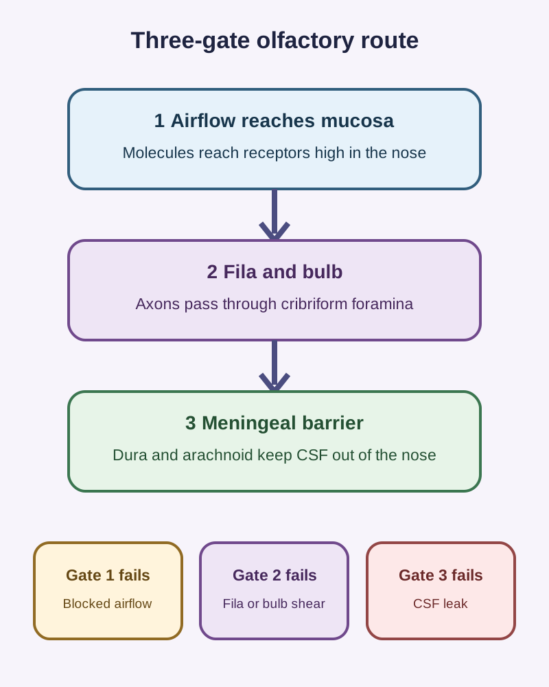
Vertical three-gate route: airflow reaches the mucosa, fila and bulb carry the signal through cribriform foramina, and the meningeal barrier keeps CSF out of the nose, with blocked airflow, fila or bulb shear, and CSF leak as the failure of each gate.
The three-gate route separates blocked odor delivery from fila or bulb injury and from a breach of the meningeal barrier. [1] [2]
The three-gate nasal roof trace
GateQuestionFailure pattern
AirflowCan odor reach the superior nasal mucosa?Side-specific reduction with obstruction
Fila and bulbCan the signal cross the cribriform plate?Loss despite a patent airway
Meningeal barrierIs there a communication with subarachnoid CSF?Clear drainage and infection risk

Use the three-gate trace before choosing a diagnosis. State which gate is abnormal, predict the extra finding that should follow, and compare that prediction with the actual patient pattern.

A visible unilateral polyp supports blocked odor delivery. Abrupt bilateral loss after acceleration with open nasal passages supports fila or bulb injury, even when no fracture line appears on routine imaging. [1]

Try it here · Checkpoint 1 of 3

Make your prediction before reading the choices. A first attempt is just a starting point.

Case 2

A patient has a cribriform plate fracture and cannot identify coffee odor, but has no nasal drainage after repeated observation. Which inference is justified?

Show answer and explanations for case 2
  1. A. A halo sign is needed to establish a CSF leak (Why this does not fit)

    A halo is nonspecific and no fluid is available here. It cannot establish a dural opening.

    Reasoning steps for option A
    1. Why might a halo test seem like the next step after a skull-base fracture?

      The halo sign is widely taught as a bedside screen for CSF in nasal fluid.

    2. Why is the halo irrelevant in this patient?

      No drainage has appeared, and a halo is nonspecific even when fluid exists, so it cannot establish a dural opening.

  2. B. Every cribriform fracture includes a tear of the dura (Why this does not fit)

    The fracture may injure bone or fibers without opening meninges. No leak has been established here.

    Reasoning steps for option B
    1. What makes it tempting to assume the dura is torn?

      The dura is tightly adherent to the cribriform plate, so a plate fracture often raises concern for a tear.

    2. Why is a universal dural tear not justified?

      Bone can break without a meningeal defect, and repeated observation has shown no drainage to suggest one.

  3. C. Olfactory injury can occur without a demonstrated CSF leak (Best answer)

    Sensory fibers can be injured separately from a communicating dural defect. Absence of drainage does not erase anosmia.

    Reasoning steps for option C
    1. Which two findings coexist in this patient?

      Coffee odor cannot be identified, yet no nasal drainage appears despite repeated observation.

    2. Why do these findings support olfactory injury without a proven leak?

      Fila can be torn at the fracture while the meninges stay sealed, so smell loss and a CSF leak are separate states.

  4. D. β2-transferrin must be positive even without collected fluid (Why this does not fit)

    The assay tests collected drainage. Without a sample, neither identity nor a leak can be asserted.

    Reasoning steps for option D
    1. Why might β2-transferrin testing seem decisive here?

      It is the classic laboratory marker for confirming CSF in nasal fluid.

    2. What does the assay need that this patient lacks?

      The test is run on collected drainage, and no fluid has appeared to sample.

  5. E. An intact smell pathway excludes a cribriform fracture (Why this does not fit)

    The patient has smell loss, not preserved smell. Even preserved smell would not by itself exclude bony injury.

    Reasoning steps for option E
    1. What sounds reassuring about an intact smell pathway?

      Preserved smell would suggest the fila crossing the plate survived the injury.

    2. Why does that reasoning fail twice in this case?

      This patient has lost smell, and even normal smell would not exclude a bony fracture.

Takeaway: Bone, fibers, and meninges are separate states.

Case sources: [1] [3]

Separate smell loss from a CSF leak

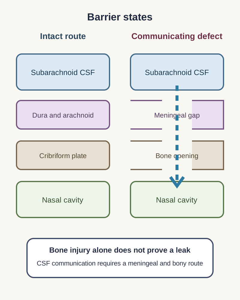
A bony defect does not automatically create a leak. CSF reaches the nose only when a compatible bone opening and a meningeal defect form a communicating route from the subarachnoid space. [3]

Side-by-side intact and communicating skull-base barriers showing that a bone opening plus a meningeal gap creates a CSF route to the nose.
A fracture line alone is not a CSF leak; communication requires both bone and meningeal defects. [3]

Positional clear rhinorrhea after trauma or surgery raises concern for that communication. The open route also raises the risk of meningitis, which is why anosmia, fracture, and leakage must be assessed as related but separate states. [3]

A laboratory assay can test fluid identity. β2-transferrin remains a useful marker, while some laboratories now use β-trace protein; the performing laboratory's method and reference limits govern interpretation. [3] [4]

A halo on absorbent material and a nasal glucose strip are not confirmatory. With intermittent drainage, an unrepresentative negative sample does not end evaluation; targeted imaging answers where a suspected communication travels. [3]

When a breach is suspected, avoid blind nasal instrumentation while arranging prompt assessment. Alertness, intact facial sensation, or preserved smell does not make an uncertain nasal route safe. [3]

Try it here · Checkpoint 2 of 3

Make your prediction before reading the choices. A first attempt is just a starting point.

Case 5

An order set at a laboratory now offers β-trace protein rather than β2-transferrin for suspected CSF rhinorrhea. The patient has intermittent clear drainage after sinus surgery. What is the sound interpretation?

Show answer and explanations for case 5
  1. A. Use the local assay protocol to interpret the specimen result (Best answer)

    Some referral laboratories changed to β-trace protein. The method and its reference limits belong to the performing laboratory.

    Reasoning steps for option A
    1. What changed in the laboratory order set?

      The laboratory now offers β-trace protein in place of β2-transferrin for suspected CSF rhinorrhea.

    2. How should the result be interpreted?

      By the performing laboratory's method and reference limits, since referral laboratories have switched markers.

  2. B. Diagnose a CSF leak from the history of sinus surgery alone (Why this does not fit)

    Surgery raises suspicion. It does not establish that this intermittent drainage contains CSF.

    Reasoning steps for option B
    1. Why does recent sinus surgery raise suspicion?

      Endoscopic sinus surgery is a leading cause of iatrogenic CSF leaks through the skull base.

    2. Why is surgical history alone insufficient?

      Suspicion is not diagnosis; the intermittent drainage still needs a marker test and, if positive, localization.

  3. C. Use a halo sign in place of testing because the assay names differ (Why this does not fit)

    The halo remains nonspecific. Changing laboratory methods does not make a tissue pattern confirmatory.

    Reasoning steps for option C
    1. Why might a halo sign seem like a way around a new assay?

      When a marker name is unfamiliar, a quick bedside sign can seem simpler.

    2. Why can the halo not substitute for the laboratory marker?

      A ring forms whenever blood mixes with any clear fluid, so it cannot confirm CSF; the answer is to learn the new assay, not to abandon testing.

  4. D. Exclude a CSF leak after a negative sample during a dry interval (Why this does not fit)

    An absent or unrepresentative intermittent sample can miss the event. Persistent suspicion calls for further evaluation.

    Reasoning steps for option D
    1. Why could a negative sample during a dry interval feel conclusive?

      A laboratory result seems more objective than an intermittent history.

    2. Why does timing undermine that sample?

      An intermittent leak can be missed when fluid is collected while little is draining, so suspicion should persist.

  5. E. Reject β-trace testing because only β2-transferrin detects CSF (Why this does not fit)

    β-trace protein (prostaglandin D synthase) is a validated CSF marker with diagnostic accuracy comparable to β2-transferrin.

    Reasoning steps for option E
    1. Why might β-trace protein seem less legitimate?

      β2-transferrin has been the familiar teaching marker for decades.

    2. What evidence supports β-trace protein?

      It is a validated CSF marker with accuracy comparable to β2-transferrin, and major referral laboratories now use it.

Takeaway: Know the local assay, rather than treating a historic marker name as universal.

Case sources: [3] [4]

Test smell without testing irritation

Basic taste includes sweet, sour, salty, bitter, and umami. Much of a food's identity comes from retronasal aroma traveling from the pharynx to the olfactory mucosa during eating. [1]

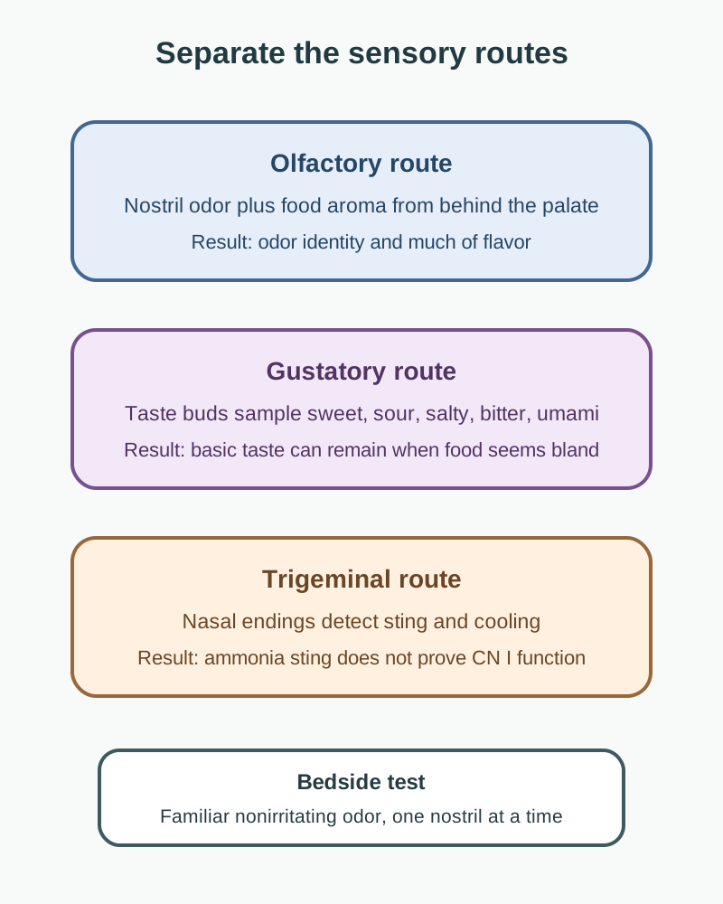
Three stacked sensory routes distinguish olfactory aroma, tongue taste, and trigeminal irritation during bedside assessment.
The sensory comparison explains why food can seem bland while basic taste and ammonia sting remain. [1] [7]

Trigeminal chemesthesis detects sting, cooling, and other irritation. Ammonia can feel sharp despite olfactory loss, so a safe familiar nonirritating odor is the more specific bedside probe. [1]

Test one nostril at a time after checking patency, and ask the patient to identify rather than merely detect the odor. Formal smell testing standardizes stimuli and can separate threshold, discrimination, and identification. [8]

Reduced smell only through a blocked side supports nasal obstruction. Persistent loss after congestion resolves supports postviral dysfunction or another neural cause rather than continued odor-delivery failure. [1]

Lifelong congenital anosmia with normal airflow changes the differential. When it occurs with delayed puberty, evaluate for Kallmann syndrome rather than inventing an acquired skull-base injury. Variants in ANOS1 (X-linked), FGFR1 and other genes disrupt migration of GnRH neurons alongside olfactory axons. [7]

Map each deficit to its own exit

CN I fila cross the cribriform plate, while CN II passes through the optic canal. Smell loss and visual loss therefore name different anterior skull-base routes. [2]

The superior orbital fissure carries III, IV, V1, and VI, so combined ocular motility and forehead sensory deficits belong there. The foramen rotundum carries V2, matching cheek, upper-lip, and maxillary-tooth sensation. [2]

The foramen ovale carries V3, combining mandibular sensation with jaw motor fibers. The internal acoustic meatus groups VII and VIII, so facial weakness with hearing or balance change points to petrous temporal bone. [2]

The jugular foramen carries IX, X, and XI, linking swallowing, voice, and shoulder findings. The hypoglossal canal carries XII, so isolated tongue weakness belongs to a different posterior opening. [2]

The middle meningeal artery enters through foramen spinosum, while the internal carotid artery traverses the carotid canal. Neither vascular opening explains isolated odor identification loss. [2]

For every mixed injury, write the deficit pattern first, then name the shared passage. Do not force cheek numbness, diplopia, hearing loss, hoarseness, or tongue deviation through the cribriform plate merely because trauma involved the skull base.

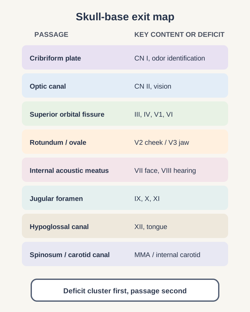
Grouped skull-base passages pair cranial nerve or vascular contents with the deficit pattern each injury produces.
Start with the deficit cluster, then select the matching passage. [2]

Try it here · Checkpoint 3 of 3

Make your prediction before reading the choices. A first attempt is just a starting point.

Case 17

A lesion just behind the orbit limits eye movement in several directions and reduces forehead sensation, while visual acuity is preserved. Which skull-base passage best groups the impaired nerves without requiring optic canal involvement?

Show answer and explanations for case 17
  1. A. Cribriform plate (Why this does not fit)

    CN I carries olfaction there. It does not carry the supplied ocular motor and forehead deficits.

    Reasoning steps for option A
    1. Why might the cribriform plate appear on a list of anterior skull-base passages?

      It is an anterior skull-base opening lying between the orbits, so it can seem relevant to any lesion near the orbit.

    2. Why does the cribriform plate not fit?

      It carries only olfactory fila, not the nerves for eye movement or forehead sensation.

  2. B. Optic canal (Why this does not fit)

    II travels through the optic canal. Isolated canal injury would primarily threaten vision rather than multiple ocular motor nerves and V1.

    Reasoning steps for option B
    1. Why could the optic canal seem likely in a lesion just behind the orbit?

      The optic canal sits beside the superior orbital fissure at the orbital apex.

    2. Which finding argues against optic canal involvement?

      Visual acuity is preserved, and the canal does not carry the ocular motor nerves or V1.

  3. C. Foramen rotundum (Why this does not fit)

    V2 there supplies cheek and upper teeth. It does not carry the ocular motor nerves or forehead V1.

    Reasoning steps for option C
    1. Why might foramen rotundum be considered with facial numbness?

      It carries a trigeminal division near the orbit.

    2. Why is V2 the wrong division?

      V2 supplies the cheek and upper teeth, while this patient has forehead numbness from V1.

  4. D. Jugular foramen (Why this does not fit)

    IX, X, and XI serve swallow, voice, and shoulder functions. Those are not the findings.

    Reasoning steps for option D
    1. Why could the jugular foramen seem relevant to multiple cranial nerve deficits?

      It transmits three cranial nerves together.

    2. Why does the jugular foramen not match?

      IX, X, and XI control swallowing, voice, and shoulder movement, none of which is affected.

  5. E. Superior orbital fissure (Best answer)

    III, IV, V1, and VI pass here. Their combined motor and forehead pattern with preserved vision fits.

    Reasoning steps for option E
    1. Which findings must a single passage explain?

      Limited eye movement in several directions with reduced forehead sensation but normal visual acuity.

    2. Why does the superior orbital fissure group them?

      III, IV, V1, and VI pass through it, while CN II takes the separate optic canal and is spared.

Takeaway: Separate fissure ocular motor and V1 findings from optic-canal vision loss.

Case sources: [2]

Let time course choose the next branch

Abrupt anosmia after a blow can reflect fila or bulb shear. A patent nasal airway and reduced nonirritating odor identification make simple congestion less persuasive, even if routine imaging shows no plate fracture. [1]

Progressive bilateral loss with frontal symptoms suggests a central process. An enlarging lesion in the olfactory groove can compress bulbs or tracts while cheek sensation and basic taste remain intact. [1]

An olfactory groove meningioma can produce anosmia with Foster Kennedy syndrome, which pairs optic atrophy on the side of the tumor with papilledema in the other eye. A unilateral mass high in the nasal cavity near the cribriform plate, with obstruction or epistaxis, suggests esthesioneuroblastoma (olfactory neuroblastoma). [12] [13]

Uncinate seizures arise in mesial temporal structures and can begin with a brief, often unpleasant smell that has no external source. [14] Loss of smell can precede the motor signs of Parkinson disease and the memory decline of Alzheimer disease, so unexplained prodromal hyposmia in an older adult deserves a neurologic history. [8]

After a viral illness, including COVID-19, smell loss that persists once congestion resolves is common. Structured olfactory training, repeated daily sniffing of a small set of strong odors over months, is the recommended first treatment for this postinfectious loss. [8]

Warm freshwater forced into the nose can introduce Naegleria fowleri along the olfactory route without a fracture. Days later, rapidly progressive headache, fever, vomiting, and cognitive or meningeal findings raise concern for primary amebic meningoencephalitis. [2] [5] [6]

Anterior fossa inflammation on imaging does not identify the organism. Confirmation relies on appropriate CSF or tissue methods such as PCR, immunostaining, or specialized microscopy, with urgent expert involvement. Drinking contaminated water alone is not the described route. [5]

Finish with a two-column note: time course on the left and associated deficits on the right. Acute trauma, fluctuating obstruction, gradual compression, and rapidly progressive infection predict different next tests even when each patient says smell is gone.

Practice with clinical cases

Case 1

After a frontal fall, a patient identifies a mild vanilla odor through neither nostril, but feels light touch on the cheeks and sees normally. CT shows a small defect in the midline cribriform plate. Which pathway best accounts for the sensory loss?

Show answer and explanations for case 1
  1. A. Olfactory fila crossing the cribriform plate (Best answer)

    Cribriform plate trauma can disrupt CN I axons. The deficit is odor identification rather than cheek touch or vision.

    Reasoning steps for option A
    1. Which modality is lost after the frontal fall, and which are spared?

      Odor identification fails through both nostrils, while cheek touch and vision are normal.

    2. Why does a midline cribriform defect explain that selective loss?

      CN I fila cross the cribriform plate, so a defect there can tear them without touching trigeminal or optic pathways.

  2. B. Maxillary nerve in foramen rotundum (Why this does not fit)

    V2 carries cheek and upper-teeth sensation. Cheek touch remains and the deficit is smell.

    Reasoning steps for option B
    1. What makes V2 tempting after a facial blow?

      Midface trauma often injures the maxillary nerve, and the fall struck the front of the head.

    2. Which finding rules out the foramen rotundum?

      Light touch on the cheeks is intact, so V2 is working; the lost modality is smell.

  3. C. Ophthalmic nerve in superior orbital fissure (Why this does not fit)

    V1 carries forehead and corneal sensation. It does not carry familiar odor identity.

    Reasoning steps for option C
    1. Why might V1 come to mind with a frontal injury?

      V1 supplies the forehead and passes near the anterior skull base, the region that was struck.

    2. What does V1 fail to explain here?

      V1 carries touch and pain from the forehead and cornea, not odor identity, and no forehead numbness is described.

  4. D. Facial nerve in internal acoustic meatus (Why this does not fit)

    VII can affect facial motor function and anterior tongue taste. Neither explains isolated post-traumatic odor loss at the cribriform plate.

    Reasoning steps for option D
    1. Why could the facial nerve seem relevant to a patient who says food tastes wrong?

      CN VII carries taste from the anterior tongue, and taste and smell are easily confused.

    2. Where does CN VII travel compared with the injury site?

      It enters the internal acoustic meatus in the petrous temporal bone, far from the midline cribriform defect, and no facial weakness is reported.

  5. E. Optic nerve in the optic canal (Why this does not fit)

    CN II damage would disturb visual input. Both eyes see normally while odor identification fails.

    Reasoning steps for option E
    1. Why is the optic canal a nearby candidate?

      The optic canal also lies in the anterior skull base, close to the cribriform region.

    2. What does normal vision in both eyes tell you?

      CN II is functioning, so optic canal injury cannot account for the isolated smell loss.

Takeaway: Match the sensory modality to the cribriform crossing.

Case sources: [1] [2]

Case 3

After anterior skull-base trauma, clear right-sided drainage recurs when a patient leans forward. A paper towel shows a pale ring around blood. Which approach best distinguishes identity from location?

Show answer and explanations for case 3
  1. A. Use smell loss alone to identify the nasal drainage as CSF (Why this does not fit)

    Anosmia may accompany fiber injury without meningeal communication. The actual fluid still needs assessment.

    Reasoning steps for option A
    1. Why might smell loss seem to point toward a CSF leak?

      Both anosmia and CSF rhinorrhea follow anterior skull-base trauma, so they often appear together.

    2. Why can anosmia not identify the fluid?

      Fila can be sheared while the meninges stay intact, so the drainage itself still needs testing.

  2. B. Accept the halo as confirmation without laboratory testing (Why this does not fit)

    Blood mixed with non-CSF fluid can also make a ring. This pattern cannot confirm the fluid.

    Reasoning steps for option B
    1. Why is the pale ring around blood appealing?

      A double ring on a paper towel is the classic bedside picture described for CSF mixed with blood.

    2. Why can the ring not confirm CSF?

      Tears, saline, and other clear fluids mixed with blood can form the same ring.

  3. C. Assay drainage for a CSF marker, then image to locate the defect (Best answer)

    A laboratory marker evaluates fluid identity. Imaging characterizes the bony and meningeal route; the ring is nonspecific.

    Reasoning steps for option C
    1. Which two separate questions does positional clear drainage raise?

      Whether the fluid is CSF, and where the bony and meningeal defect lies.

    2. How do assay and imaging divide that work?

      A CSF marker such as β2-transferrin or β-trace protein establishes identity; targeted imaging then locates the breach.

  4. D. Use a nasal glucose strip to confirm the drainage as CSF (Why this does not fit)

    Nasal glucose has inadequate sensitivity and specificity. It does not establish a CSF leak.

    Reasoning steps for option D
    1. Why does a glucose strip seem convenient?

      CSF contains glucose, and strips are quick and available at the bedside.

    2. Why is a positive strip not confirmation?

      Nasal secretions and blood-tinged fluid can contain glucose, so the strip has poor sensitivity and specificity.

  5. E. Use a normal skull radiograph to exclude all meningeal defects (Why this does not fit)

    A plain skull view cannot establish fluid identity or reliably exclude a small communicating breach.

    Reasoning steps for option E
    1. Why might a plain skull film feel like enough to exclude a defect?

      It is fast and can show large fractures after trauma.

    2. What can a normal radiograph not do?

      It cannot identify the fluid or reliably exclude a small communicating breach in the anterior skull base.

Takeaway: Assay asks what the fluid is; imaging asks where it travels.

Case sources: [3] [4]

Case 4

A lab reports a positive β2-transferrin assay in drainage after combined orbital and nasal trauma. The patient also has a ruptured globe. Which caveat is most relevant?

Show answer and explanations for case 4
  1. A. Any serum sample always contains β2-transferrin (Why this does not fit)

    The CSF-associated isoform is not normally in serum. The particular confound here is damaged ocular vitreous.

    Reasoning steps for option A
    1. Why might a serum source seem to explain a positive marker?

      Transferrin is abundant in blood, and traumatic drainage is often blood-tinged.

    2. Which finding makes serum the wrong explanation?

      The CSF-associated β2 isoform is not normally present in serum; the special confounder here is the ruptured globe.

  2. B. A halo on tissue is more specific than the assay (Why this does not fit)

    Halo patterns are nonspecific. The eye injury calls for interpretation of the assay, not replacement by a ring.

    Reasoning steps for option B
    1. Why could a halo seem more trustworthy than a laboratory result?

      The halo is visible at the bedside and may seem direct when a laboratory result is unexpected.

    2. Why does the halo not settle this sample?

      Blood mixed with tears, saline or vitreous can also separate into a ring, so the halo cannot tell whether ocular fluid explains the positive assay.

  3. C. Vitreous fluid can contain β2-transferrin (Best answer)

    The marker is highly useful for usual nasal fluid. Ocular fluid contamination after globe rupture can produce a positive result without proving CSF.

    Reasoning steps for option C
    1. What unusual injury accompanies the positive assay?

      The globe has ruptured during the same orbital and nasal trauma.

    2. Why does globe rupture weaken the result?

      Vitreous humor can contain β2-transferrin, so ocular fluid in the sample can give a positive result without CSF.

  4. D. A negative nasal glucose strip overrides the assay (Why this does not fit)

    Glucose strips are unreliable. They do not settle the positive marker in this ocular-trauma context.

    Reasoning steps for option D
    1. Why might a negative glucose strip seem to cancel the assay?

      A bedside test result that disagrees with the laboratory can seem like a tiebreaker.

    2. Why should the strip not override the marker?

      Nasal glucose testing is unreliable in both directions and says nothing about ocular contamination.

  5. E. An odor test proves the sample is CSF (Why this does not fit)

    Smell function and fluid identity are different questions. Odor testing cannot identify drainage composition.

    Reasoning steps for option E
    1. Why could smell testing seem connected to the fluid question?

      Olfactory loss often accompanies anterior skull-base injury with drainage.

    2. What does odor testing actually measure?

      It measures olfactory function, not the chemical identity of a fluid sample.

Takeaway: β2-transferrin is specific in context, not literally exclusive to CSF.

Case sources: [3]

Case 6

A patient has positional clear rhinorrhea confirmed as CSF and imaging localizes a small anterior fossa breach. Which anatomical condition explains the increased meningitis risk?

Show answer and explanations for case 6
  1. A. A meningeal breach connecting the nose to the subarachnoid space (Best answer)

    A dural and arachnoid breach lets CSF escape and creates a potential path for organisms into cranial spaces.

    Reasoning steps for option A
    1. What has already been confirmed in this patient?

      The fluid is CSF, and imaging shows a small anterior fossa breach.

    2. Why does that breach raise meningitis risk?

      A dural and arachnoid defect connects the nose to the subarachnoid space, giving nasal organisms a route inward.

  2. B. Severing of the olfactory axons beneath intact meningeal layers (Why this does not fit)

    CN I injury can cause smell loss. By itself it does not make a CSF-to-nose communication.

    Reasoning steps for option B
    1. Why could severed olfactory axons seem important in anterior fossa trauma?

      CN I fila cross the same cribriform region and are often torn.

    2. Why do torn fila alone not raise infection risk?

      If the meninges remain intact, there is no CSF-to-nose channel for leakage or bacterial entry.

  3. C. Compression of the maxillary nerve within foramen rotundum (Why this does not fit)

    V2 injury affects cheek sensation. It does not form a communicating meningeal defect.

    Reasoning steps for option C
    1. Why might V2 compression be considered after facial trauma?

      Maxillary fractures can involve the foramen rotundum.

    2. Why is V2 unrelated to the confirmed leak?

      V2 injury causes cheek numbness and creates no meningeal communication with the nose.

  4. D. Tearing of the middle meningeal artery at foramen spinosum (Why this does not fit)

    An arterial tear can produce bleeding. The confirmed nasal CSF route instead requires a meningeal breach.

    Reasoning steps for option D
    1. Why could the middle meningeal artery seem relevant to a meningeal complication?

      Its name and its dural course make it sound linked to meningeal injury.

    2. What would an arterial tear cause instead?

      Bleeding, typically an epidural hematoma, rather than a CSF route from the nose.

  5. E. Occlusion of the internal carotid canal within temporal bone (Why this does not fit)

    Carotid injury is vascular. It is not the identified anterior fossa CSF route.

    Reasoning steps for option E
    1. Why might carotid canal injury be included in skull-base trauma?

      Basilar fractures can extend through the petrous carotid canal.

    2. Why does carotid injury not explain this patient's risk?

      The carotid canal is a vascular channel in the temporal bone, not the anterior fossa CSF route imaged here.

Takeaway: The leak and infection risk require a barrier defect, not simply anosmia.

Case sources: [2] [3]

Case 7

A patient with a suspected anterior skull-base breach has clear nasal drainage and pneumocephalus after trauma. Which precaution is justified while the injury is evaluated?

Show answer and explanations for case 7
  1. A. Use glucose strips to clear the nasal route (Why this does not fit)

    Glucose strips cannot reliably exclude a leak. A negative strip would not make blind passage safe.

    Reasoning steps for option A
    1. Why might glucose strips seem able to clear the nasal route?

      A negative strip looks like quick evidence that the drainage is not CSF.

    2. Why does a negative strip not make blind passage safe?

      Strips cannot reliably exclude a leak, and pneumocephalus already signals a communication.

  2. B. Treat anosmia alone as proof that the dura is torn (Why this does not fit)

    Smell axons may be injured without a leak. Here drainage and pneumocephalus, not anosmia alone, raise concern.

    Reasoning steps for option B
    1. Why could anosmia seem to prove a dural tear?

      Smell loss and CSF leaks share the same anterior skull-base injury.

    2. What actually raises concern for a breach here?

      Clear drainage and pneumocephalus, not smell loss, which can occur with intact meninges.

  3. C. Avoid blind nasal instrumentation (Best answer)

    A potential cranial-nasal communication makes unguided nasal passage hazardous. Evaluation should account for skull-base injury.

    Reasoning steps for option C
    1. Which findings suggest a skull-base breach?

      Clear nasal drainage and intracranial air after trauma.

    2. Why should blind nasal instrumentation be avoided?

      A tube or probe passed through a breached cribriform region can enter the cranial cavity.

  4. D. Assume all nasal passages are safe because the patient is alert (Why this does not fit)

    Alertness does not restore a breached skull base. The imaging and drainage remain concerning.

    Reasoning steps for option D
    1. Why might an alert patient seem low risk?

      Normal consciousness suggests no major brain injury.

    2. Why does alertness not protect the nasal route?

      Mental status says nothing about the integrity of the skull base, and drainage and air remain concerning.

  5. E. Ignore the nasal finding if facial sensation is intact (Why this does not fit)

    V2 sensation can remain intact despite anterior ethmoid injury. It cannot establish nasal route safety.

    Reasoning steps for option E
    1. Why could intact facial sensation seem reassuring?

      It suggests the trigeminal branches around the nose are uninjured.

    2. Why is normal V2 sensation irrelevant to route safety?

      An anterior ethmoid or cribriform breach can coexist with normal cheek sensation.

Takeaway: Suspicion of skull-base injury is enough to avoid blind nasal passage.

Case sources: [3]

Case 8

A person reports that soup has become bland after a viral illness. Sweet and salty solutions are identified on the tongue, but a mild familiar odor is not recognized through either nostril. Which sensory deficit best explains the perceived flavor loss?

Show answer and explanations for case 8
  1. A. Dysfunction of the optic nerve affecting visual input (Why this does not fit)

    Vision may influence food experience. It cannot explain absent odor identification with preserved basic tastes.

    Reasoning steps for option A
    1. Why might vision be linked to enjoyment of food?

      Appearance does shape appetite and the experience of a meal.

    2. Why can the optic nerve not explain this pattern?

      The deficit is failure to recognize an odor with preserved sweet and salty taste, which vision does not govern.

  2. B. Complete loss of gustatory sensation from facial nerve injury (Why this does not fit)

    VII contributes anterior tongue taste. Preserved sweet and salty perception and lost smell point elsewhere.

    Reasoning steps for option B
    1. Why would a person call soup bland if taste were lost?

      Many people describe flavor loss as losing taste.

    2. Which finding shows gustation is intact?

      Sweet and salty solutions are correctly identified on the tongue.

  3. C. Failure of vagal motor function affecting swallowing or voice (Why this does not fit)

    Vagus dysfunction can alter swallowing or voice. Those deficits are not supplied and do not explain selective odor failure.

    Reasoning steps for option C
    1. Why might the vagus seem connected to eating?

      CN X controls swallowing and carries some taste from the epiglottis.

    2. Why does vagal dysfunction not fit?

      No swallowing or voice change is described, and vagal loss does not remove odor recognition.

  4. D. Olfactory dysfunction impairing retronasal food aroma (Best answer)

    Basic taste survives, while aroma from food normally reaches nasal olfactory receptors from behind. Its loss makes flavors seem flat.

    Reasoning steps for option D
    1. What sensory split appears after the viral illness?

      Basic tastes are preserved, while a familiar odor is not recognized through either nostril.

    2. How does olfactory loss make soup bland?

      Food aroma reaches the olfactory mucosa retronasally from the pharynx, and without it flavor collapses to basic taste.

  5. E. Failure of mandibular nerve motor function affecting chewing (Why this does not fit)

    V3 helps mastication and lower-face sensation. It does not encode food aroma.

    Reasoning steps for option E
    1. Why could V3 matter for eating?

      The mandibular nerve powers chewing and supplies lower-face sensation.

    2. Why does V3 not explain the change in flavor?

      Chewing and oral touch do not encode food aroma, and no jaw weakness is described.

Takeaway: Flavor integrates retronasal smell with intact basic taste.

Case sources: [1]

Case 9

A patient cannot identify a faint floral odor in either nostril but says ammonia stings sharply. Which inference about CN I is best?

Show answer and explanations for case 9
  1. A. The ammonia sting establishes intact olfactory receptor neurons (Why this does not fit)

    Trigeminal chemesthesis can survive CN I impairment. Irritation is not proof of odor identification.

    Reasoning steps for option A
    1. Why might a strong ammonia response seem to show normal smell?

      The patient clearly perceives something when ammonia is held under the nose.

    2. What carries the ammonia sting?

      Trigeminal chemesthetic endings in the nasal mucosa, which can work even when CN I fails.

  2. B. Olfactory loss can coexist with preserved trigeminal irritation (Best answer)

    Ammonia activates irritant sensation. A mild nonirritant odor is the more specific bedside olfactory probe.

    Reasoning steps for option B
    1. What two responses does this patient give?

      A faint floral odor is not identified in either nostril, while ammonia stings sharply.

    2. How do these responses separate two nasal systems?

      The sting shows trigeminal irritation is intact, while the missed floral odor shows olfactory loss.

  3. C. The ammonia and floral findings establish complete taste loss (Why this does not fit)

    Neither the sting nor the floral test measures gustatory qualities on the tongue. Taste was not tested.

    Reasoning steps for option C
    1. Why might a smell deficit be mistaken for taste loss?

      Patients with anosmia often report that food has no taste.

    2. Why can taste not be judged from these tests?

      Neither the floral odor nor the ammonia sting tests sweet, salty, sour, bitter, or umami on the tongue.

  4. D. The absent floral odor identification establishes a cribriform fracture (Why this does not fit)

    Smell loss has many causes including postviral or obstruction. No trauma or imaging establishes a fracture.

    Reasoning steps for option D
    1. Why could smell loss suggest a cribriform fracture?

      Fracture is a well-known cause of anosmia.

    2. What is missing for a fracture diagnosis?

      No trauma history or imaging is given, and postviral or obstructive causes are more common.

  5. E. The ammonia and floral findings establish isolated V1 loss (Why this does not fit)

    A felt sting indicates some nasal irritant sensation remains. It does not demonstrate isolated V1 loss.

    Reasoning steps for option E
    1. Why might trigeminal nerves come to mind?

      The ammonia test probes nasal trigeminal sensation.

    2. Why does the result argue against V1 loss?

      A sharp sting means nasal irritant sensation is working, not that V1 is lost.

Takeaway: Use safe nonirritating odors, one nostril at a time.

Case sources: [1]

Case 10

A person has unilateral nasal obstruction from a visible polyp and reduced odor identification only through that side. There is no injury, headache, or facial numbness. What mechanism most directly accounts for this pattern?

Show answer and explanations for case 10
  1. A. Severing of ipsilateral olfactory fila required by the odor deficit (Why this does not fit)

    Fiber injury can reduce smell. The visible obstruction provides a nearer explanation without requiring axonal severing.

    Reasoning steps for option A
    1. Why might severed fila seem to explain one-sided smell loss?

      Fila injury does reduce smell and can be asymmetric.

    2. What nearer explanation does the examination supply?

      A visible polyp blocks the same side, explaining reduced odor delivery without axonal injury.

  2. B. Compression of the optic nerve within the ipsilateral optic canal (Why this does not fit)

    CN II injury would impair sight. The deficit tracks nasal airflow on one side.

    Reasoning steps for option B
    1. Why could the optic canal be considered for a one-sided problem?

      Unilateral lesions near the anterior skull base can affect CN II.

    2. Why does the optic nerve not fit?

      Vision is not affected, and the deficit tracks nasal airflow on one side.

  3. C. Reduced odor delivery to intact sensory mucosa high in the nose (Best answer)

    The blocked passage prevents molecules from reaching receptors. The side correspondence and polyp support a conductive mechanism.

    Reasoning steps for option C
    1. What links the smell deficit to the polyp?

      Odor identification is reduced only through the obstructed nostril.

    2. Why is this a conductive mechanism?

      The polyp stops odorant molecules from reaching intact receptors high in the nasal cavity.

  4. D. Disease at the jugular foramen affecting the posterior cranial nerves (Why this does not fit)

    IX through XI deficits affect posterior cranial nerve functions. They do not create unilateral nasal blockage.

    Reasoning steps for option D
    1. Why might a skull-base lesion be sought?

      Jugular foramen masses can cause several cranial nerve deficits.

    2. Why does a jugular foramen lesion not fit?

      IX through XI affect swallowing, voice, and shoulder, and cannot block one nostril.

  5. E. Transection of the maxillary division of the trigeminal nerve at rotundum (Why this does not fit)

    V2 carries cheek and upper-tooth sensation. Those symptoms are absent and obstruction explains odor delivery.

    Reasoning steps for option E
    1. Why could V2 be linked to the nose?

      The maxillary nerve supplies sensation to the nasal cavity and cheek.

    2. Which absent finding argues against V2 transection?

      There is no cheek or upper-tooth numbness, and V2 injury would not block odor delivery.

Takeaway: A conductive smell deficit need not be a CN I axon injury.

Case sources: [1] [2]

Case 11

A patient has slowly progressive bilateral smell loss and frontal headaches without congestion. Examination finds intact cheek touch and basic taste. MRI shows an anterior midline extra-axial mass near both olfactory bulbs. Which mechanism best explains the sensory pattern?

Show answer and explanations for case 11
  1. A. Compression limited to the optic chiasm (Why this does not fit)

    A chiasmal lesion produces visual field disturbance. The supplied mass abuts olfactory bulbs and the complaint is smell.

    Reasoning steps for option A
    1. Why might a midline anterior mass suggest the chiasm?

      The optic chiasm is also a midline structure near the anterior skull base.

    2. Why does chiasmal compression not fit?

      It would cause visual field loss, while this patient's deficit is smell and the mass abuts the bulbs.

  2. B. Olfactory bulb or tract compression in the olfactory groove (Best answer)

    A slowly growing anterior mass can disturb bilateral central olfactory input. Intact cheek touch and taste distinguish other modalities.

    Reasoning steps for option B
    1. What does the time course and imaging show?

      Slowly progressive bilateral smell loss with an extra-axial mass beside both olfactory bulbs.

    2. Why does olfactory groove compression explain it?

      A growing mass such as a meningioma presses on both bulbs or tracts while sparing cheek touch and taste.

  3. C. Injury to both facial nerves within the temporal bones (Why this does not fit)

    VII injury may alter facial motion and taste. It does not fit the anterior groove location or selective smell loss.

    Reasoning steps for option C
    1. Why might the facial nerves be considered in a flavor complaint?

      CN VII carries anterior tongue taste.

    2. Why do the facial nerves not fit?

      Taste is intact, there is no facial weakness, and the mass sits in the anterior fossa, not the temporal bones.

  4. D. Compression of both maxillary nerves at the foramina rotunda (Why this does not fit)

    V2 injury impairs cheek and upper-tooth sensation. Those are preserved while smell is lost.

    Reasoning steps for option D
    1. Why might V2 be considered in bilateral facial symptoms?

      The maxillary nerves supply much of the nose and midface.

    2. Which finding excludes V2?

      Cheek touch is intact on examination.

  5. E. Injury to both hypoglossal nerves within the hypoglossal canals (Why this does not fit)

    XII injury affects tongue motion. It does not explain smell loss from a frontal mass.

    Reasoning steps for option E
    1. Why could a skull-base mass suggest lower cranial nerves?

      Large skull-base tumors can involve several nerves.

    2. Why do the hypoglossal canals not fit?

      They lie in the posterior fossa and control tongue movement, which is not affected.

Takeaway: Progressive central smell loss can arise above the plate, not just in the nose.

Case sources: [1] [2]

Case 12

A patient has never identified odors, and adolescence brings absent pubertal progression. Nasal airflow is normal and there is no head injury. Which association warrants evaluation?

Show answer and explanations for case 12
  1. A. Kallmann syndrome with hypogonadotropic hypogonadism (Best answer)

    Congenital anosmia or hyposmia together with delayed puberty points to olfactory and GnRH developmental pathways.

    Reasoning steps for option A
    1. What pair of findings defines this history?

      Lifelong inability to identify odors together with absent pubertal progression.

    2. Which developmental link explains both?

      GnRH neurons migrate from the nasal placode with olfactory axons, so Kallmann syndrome disrupts both smell and puberty.

  2. B. An acute traumatic fracture of the ethmoid cribriform plate (Why this does not fit)

    Fracture follows trauma and has an abrupt course. Neither is present.

    Reasoning steps for option B
    1. Why might a fracture be considered with anosmia?

      Cribriform fracture is a classic cause of smell loss.

    2. Why does the time course rule out fracture?

      Smell has been absent since earliest memory, and there is no head injury.

  3. C. Ocular myasthenia gravis affecting skeletal muscle function (Why this does not fit)

    Myasthenia fluctuates in skeletal muscle function. It does not link congenital smell loss to pubertal delay.

    Reasoning steps for option C
    1. Why might a neuromuscular diagnosis appear here?

      Fatigable weakness can present in adolescence.

    2. Why does myasthenia gravis not fit?

      It fluctuates in skeletal muscle and does not connect congenital anosmia with pubertal delay.

  4. D. Isolated postviral olfactory loss following an infection (Why this does not fit)

    Postviral loss begins after infection, not from earliest memory with delayed puberty.

    Reasoning steps for option D
    1. Why is postviral loss a common suspect?

      Viral infection is one of the most frequent causes of smell loss.

    2. Why does postviral loss not fit this patient?

      It follows a specific illness, whereas this patient has never smelled and also has delayed puberty.

  5. E. Isolated maxillary division neuropathy of the trigeminal nerve (Why this does not fit)

    V2 supplies midface sensation. It does not explain lifelong odor loss and delayed puberty.

    Reasoning steps for option E
    1. Why could V2 seem linked to nasal sensation?

      The maxillary nerve supplies the nasal mucosa.

    2. Why does V2 neuropathy fail to explain the history?

      V2 carries touch and pain, not odor identity, and has no connection to puberty.

Takeaway: Congenital smell loss plus delayed puberty raises a developmental endocrine differential.

Case sources: [1] [7]

Case 13

Five days after warm freshwater entered the nose, a swimmer develops fever, severe headache, vomiting, and rapid confusion. CT shows no cribriform fracture. Which route best explains a suspected Naegleria infection?

Show answer and explanations for case 13
  1. A. Passage through the jugular foramen along the vagus nerve (Why this does not fit)

    Vagus exits posteriorly. It does not link intranasal water exposure to olfactory bulbs.

    Reasoning steps for option A
    1. Why might the vagus nerve seem like a route to the brain?

      CN X is a long cranial nerve that some pathogens travel along.

    2. Why does the jugular foramen not fit this exposure?

      The vagus exits posteriorly and has no link to water forced into the nose.

  2. B. Passage through foramen spinosum along the middle meningeal artery (Why this does not fit)

    The artery enters the skull laterally. It is not the nasal-olfactory path for this exposure.

    Reasoning steps for option B
    1. Why could the middle meningeal artery seem a path to the meninges?

      It supplies the dura, and the patient has meningoencephalitis.

    2. Why is foramen spinosum the wrong route?

      The artery enters the skull laterally from the infratemporal fossa, not from the nasal mucosa.

  3. C. Entry after oral ingestion through an injured optic canal (Why this does not fit)

    Drinking water is not the typical route. The optic canal carries vision, not nasal organism entry.

    Reasoning steps for option C
    1. Why might contaminated water suggest swallowing as the route?

      Many waterborne infections are acquired by drinking.

    2. Why does ingestion not explain Naegleria infection?

      Swallowed water does not cause it, and the optic canal carries vision, not organisms from the gut.

  4. D. Entry from cheek skin along the maxillary nerve at rotundum (Why this does not fit)

    V2 carries facial sensation. The exposure was intranasal and the illness is rapidly meningeal.

    Reasoning steps for option D
    1. Why might skin entry be considered in a swimmer?

      Cheek skin contacts the water and is supplied by V2.

    2. Why does the maxillary nerve not fit?

      The exposure was water forced into the nose, and the rapid meningeal illness follows the olfactory route.

  5. E. Entry from nasal mucosa along olfactory pathways across intact cribriform bone (Best answer)

    The organism can enter from water forced into the nose and follow the olfactory route. A fracture is not required.

    Reasoning steps for option E
    1. Which exposure and imaging findings define this case?

      Warm freshwater entered the nose days before fulminant meningoencephalitis, and CT shows no cribriform fracture.

    2. How does Naegleria reach the brain without a fracture?

      The ameba invades the olfactory mucosa and follows olfactory nerve fibers through the intact cribriform plate to the bulbs.

Takeaway: Intranasal exposure can reach the brain without a bony fracture.

Case sources: [2] [5] [6]

Case 14

A rapidly deteriorating patient has compatible freshwater nasal exposure and meningitis. MRI shows nonspecific anterior fossa inflammation. Which diagnostic evidence most directly confirms the suspected organism?

Show answer and explanations for case 14
  1. A. Naegleria identification in CSF or tissue by PCR or specialized microscopy (Best answer)

    CDC identifies molecular detection and direct organism examination among diagnostic tests. MRI inflammation alone is not species identification.

    Reasoning steps for option A
    1. What makes Naegleria the leading suspicion?

      Recent freshwater nasal exposure with a rapidly deteriorating meningitis.

    2. Which tests confirm the organism?

      PCR or specialized microscopy of CSF or tissue, because imaging inflammation is not specific.

  2. B. A positive glucose strip result from a sample of nasal drainage (Why this does not fit)

    Glucose strips concern suspected CSF drainage and are unreliable even there. They do not identify an ameba.

    Reasoning steps for option B
    1. Why might a glucose strip be tried on nasal fluid?

      It is a quick bedside test associated with CSF.

    2. Why can a glucose strip not answer this question?

      It is unreliable even for CSF identity and cannot identify an ameba.

  3. C. A double-ring halo pattern surrounding a sample of nasal fluid (Why this does not fit)

    Halo patterns are nonspecific for CSF and say nothing about the etiologic organism.

    Reasoning steps for option C
    1. Why might a halo pattern come up in meningitis with nasal findings?

      It is a familiar bedside sign linked to CSF in nasal drainage.

    2. What does a halo fail to show?

      It is nonspecific for CSF and says nothing about which organism is present.

  4. D. An MRI appearance of anterior fossa inflammation without further testing (Why this does not fit)

    Anterior inflammation is not specific for Naegleria. The organism requires laboratory identification.

    Reasoning steps for option D
    1. Why might anterior fossa inflammation seem diagnostic?

      It matches the expected olfactory route of infection.

    2. Why is MRI not confirmation?

      Other infections and inflammatory conditions look similar, so the organism must be identified in a specimen.

  5. E. A cribriform plate fracture demonstrated on a CT scan of the skull base (Why this does not fit)

    The organism can travel through an intact region. Fracture presence would not identify it in CSF.

    Reasoning steps for option E
    1. Why could a cribriform fracture seem to explain entry?

      A fracture would create an obvious path from the nose to the brain.

    2. Why would a fracture not identify the organism?

      Naegleria can cross an intact plate, and bone imaging cannot show which organism is in the CSF.

Takeaway: Suspect from exposure and tempo; identify Naegleria in appropriate specimens.

Case sources: [3] [5] [6]

Case 15

After a lateral sphenoid fracture, a patient loses sensation over the cheek, upper lip, and maxillary teeth but identifies coffee separately through both nostrils. Which exit best matches the injured sensory branch?

Show answer and explanations for case 15
  1. A. Cribriform plate carrying I (Why this does not fit)

    CN I carries odor identity. Coffee recognition remains intact, and the lost territory is V2.

    Reasoning steps for option A
    1. Why might the cribriform plate come to mind after skull trauma?

      It is the skull-base opening most linked to post-traumatic smell loss.

    2. Which finding excludes CN I involvement?

      Coffee is identified separately through both nostrils, so olfaction is intact.

  2. B. Foramen ovale carrying V3 (Why this does not fit)

    V3 supplies lower jaw sensation and mastication. The deficit is upper teeth and cheek.

    Reasoning steps for option B
    1. Why might V3 be confused with V2?

      Both are trigeminal divisions that supply facial sensation and teeth.

    2. What separates V3 from this deficit?

      V3 supplies the lower teeth, chin, and jaw muscles, while this loss involves the cheek, upper lip, and upper teeth.

  3. C. Foramen rotundum carrying V2 (Best answer)

    The maxillary division carries this midface territory. Preserved smell separates it from ethmoid CN I.

    Reasoning steps for option C
    1. Which territory has lost sensation?

      The cheek, upper lip, and maxillary teeth after a lateral sphenoid fracture.

    2. Why does foramen rotundum match?

      V2 exits the middle cranial fossa through foramen rotundum and supplies exactly this midface territory.

  4. D. Superior orbital fissure carrying V1 (Why this does not fit)

    V1 supplies forehead and corneal sensation. The deficit is maxillary territory.

    Reasoning steps for option D
    1. Why could V1 be considered in a sphenoid fracture?

      The superior orbital fissure lies in the sphenoid bone and carries V1.

    2. Why does V1 not fit the sensory map?

      V1 supplies the forehead and cornea, and those areas are not described as numb.

  5. E. Hypoglossal canal carrying XII (Why this does not fit)

    XII supplies tongue motor function. It does not carry midface sensation.

    Reasoning steps for option E
    1. Why might a skull-base canal list include XII?

      The hypoglossal canal is another commonly tested skull-base opening.

    2. Why is XII irrelevant here?

      It is a motor nerve to the tongue and carries no facial sensation.

Takeaway: Cheek and upper teeth implicate V2 rather than the ethmoid sieve.

Case sources: [2]

Case 16

A skull-base lesion produces reduced lower-tooth and chin sensation plus weak jaw clenching. Smell testing and forehead sensation remain normal. Which exit best integrates sensory and motor deficits?

Show answer and explanations for case 16
  1. A. Foramen rotundum carrying V2 (Why this does not fit)

    V2 supplies cheek and upper teeth without jaw motor fibers. The lower-jaw pattern differs.

    Reasoning steps for option A
    1. Why is V2 a natural first thought for dental numbness?

      The maxillary nerve supplies the upper teeth.

    2. What makes V2 the wrong branch?

      The numbness involves lower teeth and chin, and V2 carries no jaw motor fibers.

  2. B. Cribriform plate carrying I (Why this does not fit)

    CN I loss impairs odor identification. Smell is intact and jaw strength is weak.

    Reasoning steps for option B
    1. Why might the cribriform plate appear on a skull-base differential?

      It is one of the main anterior skull-base openings.

    2. Which findings exclude CN I?

      Smell testing is normal, and CN I cannot explain weak jaw clenching.

  3. C. Superior orbital fissure carrying V1 (Why this does not fit)

    V1 supplies forehead and corneal sensation. Forehead sensation is intact and jaw motor loss is unexplained.

    Reasoning steps for option C
    1. Why could V1 be considered with trigeminal symptoms?

      It is the trigeminal division that exits through the superior orbital fissure.

    2. Which finding shows V1 is intact?

      Forehead sensation is normal, and V1 has no motor role in chewing.

  4. D. Foramen ovale carrying V3 (Best answer)

    Mandibular V3 contributes lower-face sensation and mastication. Both deficits match its exit.

    Reasoning steps for option D
    1. Which combination of deficits is present?

      Reduced lower-tooth and chin sensation together with weak jaw clenching.

    2. Why does foramen ovale integrate both?

      V3 is the only trigeminal division with motor fibers to the muscles of mastication and exits through foramen ovale.

  5. E. Internal acoustic meatus carrying VII (Why this does not fit)

    VII supplies facial expression rather than mastication. Chin sensory loss and jaw clench implicate V3.

    Reasoning steps for option E
    1. Why might the facial nerve be considered for facial weakness?

      CN VII supplies the muscles of facial expression.

    2. Why does CN VII not fit?

      Jaw clenching uses the muscles of mastication supplied by V3, and CN VII carries no chin sensation.

Takeaway: V3 is the trigeminal branch with jaw motor and mandibular sensation.

Case sources: [2]

Case 18

A petrous temporal fracture is followed by ipsilateral facial weakness and hearing loss, while smell and forehead sensation are intact. Which passage groups the affected nerves?

Show answer and explanations for case 18
  1. A. Internal acoustic meatus (Best answer)

    VII and VIII travel together into petrous temporal bone. Facial motor and auditory findings align.

    Reasoning steps for option A
    1. Which paired deficits follow the petrous fracture?

      Ipsilateral facial weakness and hearing loss.

    2. Why does the internal acoustic meatus explain both?

      CN VII and CN VIII enter the petrous temporal bone together through this meatus.

  2. B. Jugular foramen (Why this does not fit)

    IX through XI affect swallowing, voice or shoulder. The deficit instead combines face and hearing.

    Reasoning steps for option B
    1. Why might the jugular foramen be considered after a temporal bone fracture?

      It lies next to the petrous temporal bone and transmits several nerves.

    2. Why do the jugular foramen nerves not fit?

      IX, X, and XI would affect swallowing, voice, or shoulder, not facial movement and hearing.

  3. C. Superior orbital fissure (Why this does not fit)

    III, IV, V1, and VI affect ocular motion or forehead sensation. Those functions remain intact.

    Reasoning steps for option C
    1. Why could the superior orbital fissure be considered with facial findings?

      It carries V1, which supplies part of the face.

    2. Why does the superior orbital fissure fail here?

      Eye movement and forehead sensation are intact, and none of its nerves controls facial expression or hearing.

  4. D. Cribriform plate (Why this does not fit)

    CN I fila run through the cribriform plate. Smell is intact and it cannot explain hearing loss.

    Reasoning steps for option D
    1. Why does the cribriform plate appear in skull-base fracture questions?

      It is the classic site of post-traumatic anosmia.

    2. Why is it the wrong site for this patient?

      Smell is intact, and CN I fila through the cribriform plate cannot cause hearing loss.

  5. E. Optic canal (Why this does not fit)

    CN II conveys sight. Facial motion and hearing point to VII and VIII instead.

    Reasoning steps for option E
    1. Why might the optic canal be considered after head trauma?

      Traumatic optic neuropathy is a known complication of skull fractures.

    2. Why does CN II not fit?

      It carries vision, while this patient has facial weakness and hearing loss.

Takeaway: Facial motor plus hearing localizes VII and VIII at petrous temporal entry.

Case sources: [2]

Case 19

After posterior skull-base surgery, a patient is hoarse, aspirates liquids, and has weak ipsilateral shoulder shrug. Smell and ocular motility remain normal. Which exit is most likely affected?

Show answer and explanations for case 19
  1. A. Cribriform plate (Why this does not fit)

    CN I loss changes smell. Smell remains intact and the findings are posterior motor functions.

    Reasoning steps for option A
    1. Why might the cribriform plate be considered after skull-base surgery?

      Skull-base procedures can injure the olfactory region.

    2. Why is CN I excluded?

      Smell remains intact, and the deficits are posterior motor functions.

  2. B. Hypoglossal canal (Why this does not fit)

    XII affects tongue motion. It does not group hoarseness and shoulder weakness.

    Reasoning steps for option B
    1. Why might the hypoglossal canal be tempting after posterior surgery?

      It lies close to the jugular foramen in the posterior fossa.

    2. Why does XII not explain these deficits?

      XII moves the tongue and cannot cause hoarseness, aspiration, or shoulder weakness.

  3. C. Jugular foramen (Best answer)

    X contributes laryngeal function and XI shoulder motion; IX joins them at this posterior exit.

    Reasoning steps for option C
    1. Which deficits appear after posterior skull-base surgery?

      Hoarseness, aspiration of liquids, and weak ipsilateral shoulder shrug.

    2. Why does the jugular foramen group them?

      CN IX and X serve swallowing and the larynx, CN XI supplies the trapezius, and all three exit together here.

  4. D. Foramen ovale (Why this does not fit)

    V3 contributes jaw motor function. It does not account for vagal voice and accessory shoulder findings.

    Reasoning steps for option D
    1. Why could foramen ovale be considered with swallowing trouble?

      V3 moves the jaw, which contributes to eating.

    2. Why does V3 not fit?

      V3 cannot explain hoarseness or trapezius weakness, and chewing is not described as impaired.

  5. E. Internal acoustic meatus (Why this does not fit)

    VII and VIII relate to facial motion and hearing. They do not group voice and shoulder.

    Reasoning steps for option E
    1. Why might the internal acoustic meatus be considered near the posterior fossa?

      It lies close to the surgical field in the petrous bone.

    2. Why do VII and VIII not fit?

      They serve facial movement and hearing, which are not reported, not voice and shoulder.

Takeaway: Voice and shoulder together identify the jugular group.

Case sources: [2]

Case 20

A fracture near the occipital condyle is followed by tongue deviation right on protrusion. Jaw strength, voice, and odor identification are preserved. Which exit most likely contains the injured right motor nerve?

Show answer and explanations for case 20
  1. A. Right hypoglossal canal (Best answer)

    Right XII weakness makes the protruded tongue deviate toward the weak side. Other posterior functions are spared.

    Reasoning steps for option A
    1. Which motor sign follows the condylar fracture?

      The protruded tongue deviates to the right, while jaw, voice, and smell are normal.

    2. Why does the right hypoglossal canal fit?

      The weak right genioglossus lets the tongue deviate toward the injured side, and XII exits through the hypoglossal canal next to the condyle.

  2. B. Right internal acoustic meatus (Why this does not fit)

    VII and VIII injury affects facial expression or hearing. It does not isolate tongue protrusion.

    Reasoning steps for option B
    1. Why might the internal acoustic meatus come up in posterior skull trauma?

      Temporal bone fractures commonly involve it.

    2. Why does the meatus not explain tongue deviation?

      VII and VIII control facial movement and hearing, not tongue protrusion.

  3. C. Right foramen ovale (Why this does not fit)

    V3 injury weakens jaw closure and lower-face sensation. Jaw strength is normal.

    Reasoning steps for option C
    1. Why could foramen ovale be considered for a movement problem of the mouth?

      V3 supplies the muscles of chewing.

    2. Which finding excludes V3?

      Jaw strength is preserved.

  4. D. Right jugular foramen (Why this does not fit)

    IX through XI can affect voice, swallowing and shoulder. Tongue protrusion specifically tests XII.

    Reasoning steps for option D
    1. Why might the jugular foramen be suspected near the occipital condyle?

      It lies right beside the hypoglossal canal.

    2. Why does the jugular foramen not fit?

      Voice is normal, and IX through XI do not control tongue protrusion.

  5. E. Right cribriform plate (Why this does not fit)

    CN I injury impairs right smell. Odor identification remains normal.

    Reasoning steps for option E
    1. Why does the cribriform plate appear in skull-base exit questions?

      It is the opening most often tested with head trauma.

    2. Why is it irrelevant here?

      Odor identification is normal, and the fracture is posterior at the occipital condyle.

Takeaway: Tongue deviation with preserved jaw and voice points to XII canal.

Case sources: [2]

Case 21

After a lateral skull blow, a patient has a temporal epidural collection and fracture at the pterion. There is no nasal drainage or smell loss. Which skull-base opening carries the implicated artery?

Show answer and explanations for case 21
  1. A. Foramen spinosum (Best answer)

    The middle meningeal artery traverses spinosum and is vulnerable near the pterion. This is a vascular lateral injury.

    Reasoning steps for option A
    1. What does a pterion fracture with a temporal epidural collection suggest?

      Tearing of the middle meningeal artery, which runs beneath the pterion.

    2. Why does foramen spinosum fit?

      The middle meningeal artery enters the cranial cavity through foramen spinosum.

  2. B. Jugular foramen (Why this does not fit)

    IX through XI and venous structures occupy the jugular foramen. The pterional arterial source is elsewhere.

    Reasoning steps for option B
    1. Why might the jugular foramen be considered for a vascular injury?

      It transmits the internal jugular vein.

    2. Why is it the wrong passage?

      Its contents are venous and posterior; an epidural bleed at the pterion comes from the middle meningeal artery.

  3. C. Foramen rotundum (Why this does not fit)

    V2 traverses rotundum and supplies cheek sensation. It is not the middle meningeal artery.

    Reasoning steps for option C
    1. Why might foramen rotundum be considered for a lateral skull injury?

      It sits in the middle cranial fossa near the temporal region.

    2. Why does rotundum not fit?

      It carries V2, a sensory nerve, not the middle meningeal artery.

  4. D. Cribriform plate (Why this does not fit)

    CN I fila traverse the cribriform plate. They do not supply the pterional middle meningeal artery.

    Reasoning steps for option D
    1. Why could the cribriform plate be linked to head injury?

      It is a common fracture site with skull trauma.

    2. Why is it excluded here?

      There is no smell loss or nasal drainage, and the cribriform plate carries CN I fila, not the middle meningeal artery.

  5. E. Carotid canal (Why this does not fit)

    The internal carotid artery uses the temporal carotid canal. It is distinct from the middle meningeal artery at spinosum.

    Reasoning steps for option E
    1. Why might the carotid canal be tempting for an arterial injury?

      It carries the major artery entering the skull base.

    2. Why is the carotid canal the wrong passage?

      It transmits the internal carotid artery, while the pterional epidural bleed comes from the middle meningeal artery through spinosum.

Takeaway: Pterion and middle meningeal artery map to spinosum.

Case sources: [2]

Case 22

A petrous temporal lesion surrounds the internal carotid artery as it enters the skull and disrupts its sympathetic plexus. Smell is normal. Which passage is involved?

Show answer and explanations for case 22
  1. A. Optic canal (Why this does not fit)

    CN II and ophthalmic artery use the optic canal. The described petrous carotid segment is earlier and distinct.

    Reasoning steps for option A
    1. Why could the optic canal be tempting for an arterial question?

      The ophthalmic artery, a carotid branch, travels through it.

    2. Why does the optic canal not fit?

      The lesion involves the petrous carotid segment, which lies proximal to the ophthalmic branch.

  2. B. Carotid canal (Best answer)

    The internal carotid travels through petrous temporal carotid canal with sympathetic fibers. This is vascular, not olfactory.

    Reasoning steps for option B
    1. Which structures does the petrous lesion surround?

      The internal carotid artery and its sympathetic plexus as they enter the skull.

    2. Why does the carotid canal fit?

      The internal carotid and its sympathetic fibers traverse the carotid canal in the petrous temporal bone.

  3. C. Foramen spinosum (Why this does not fit)

    Spinosum transmits the middle meningeal artery. It is not the internal carotid entry.

    Reasoning steps for option C
    1. Why might foramen spinosum be confused with the carotid canal?

      Both are arterial passages in the middle cranial fossa region.

    2. Which artery would a spinosum lesion involve instead?

      Foramen spinosum admits the middle meningeal artery from the maxillary artery, an external carotid branch, whereas this lesion surrounds the internal carotid and its sympathetic plexus in the petrous bone.

  4. D. Internal acoustic meatus (Why this does not fit)

    VII and VIII use the meatus. It is not the internal carotid route.

    Reasoning steps for option D
    1. Why could the internal acoustic meatus seem likely in a petrous lesion?

      It also lies within the petrous temporal bone.

    2. Why does the meatus not fit?

      It transmits VII and VIII, not the internal carotid artery or its sympathetic plexus.

  5. E. Cribriform plate (Why this does not fit)

    CN I fila cross the cribriform plate. Normal smell and a petrous carotid finding point elsewhere.

    Reasoning steps for option E
    1. Why might the cribriform plate appear on a skull-base list?

      It is the best-known anterior opening.

    2. Why is the cribriform plate excluded?

      Smell is normal, and the carotid finding is in the petrous bone, far from where CN I fila cross.

Takeaway: Carotid canal and foramen spinosum carry different arteries.

Case sources: [2]

Case 23

A patient has an intact skull on CT after acceleration-deceleration injury yet reports new smell loss. Nonirritating odor testing is reduced bilaterally, and the nasal airway is patent. Which explanation remains plausible?

Show answer and explanations for case 23
  1. A. Nasal obstruction established by reduced bilateral odor identification (Why this does not fit)

    A blocked nose can limit odor access. The supplied airway is patent.

    Reasoning steps for option A
    1. Why might obstruction seem plausible when smell is reduced on both sides?

      Congestion commonly reduces smell bilaterally.

    2. Which finding removes obstruction as the cause?

      The nasal airway is patent.

  2. B. Olfactory dysfunction excluded by normal findings on the CT scan (Why this does not fit)

    Imaging assesses structural injury at its resolution. The functional odor test is abnormal.

    Reasoning steps for option B
    1. Why might a normal CT feel reassuring?

      It shows no skull fracture after the injury.

    2. Why does a normal CT not exclude olfactory dysfunction?

      CT shows bone, not the function of tiny olfactory fibers, and odor testing is already abnormal.

  3. C. Injury of V2 established by loss of odor identification on both sides (Why this does not fit)

    V2 conveys midface sensation rather than odor identity. No cheek deficit is given.

    Reasoning steps for option C
    1. Why could trigeminal injury seem connected to a nasal complaint?

      V2 supplies sensation to the nasal cavity.

    2. Why does V2 not explain the finding?

      V2 carries touch and pain, not odor identity, and no midface numbness is reported.

  4. D. A CSF leak required by the presence of impaired odor identification (Why this does not fit)

    Olfactory injury and meningeal breach are independent possibilities. No drainage establishes a leak.

    Reasoning steps for option D
    1. Why might a CSF leak be suspected after head injury with smell loss?

      Both can follow anterior skull-base trauma.

    2. Why is a leak not required?

      Olfactory injury and meningeal breach are independent, and no drainage is reported.

  5. E. Shear injury of olfactory fila or the bulb despite no visible skull fracture (Best answer)

    Tiny olfactory fibers and bulb connections can be injured by motion. A CT without fracture cannot prove their function intact.

    Reasoning steps for option E
    1. What combination points to a neural mechanism?

      New bilateral loss of nonirritating odor identification after acceleration-deceleration injury, with a patent airway.

    2. How can the fila fail without a fracture?

      Brain movement against the skull can shear fila at the cribriform plate or injure the bulb, which CT does not show.

Takeaway: Anosmia after acceleration can occur without a visible plate fracture.

Case sources: [1] [3]

Case 24

After a cold, a person loses smell while the nose is congested. Weeks later congestion clears, but supervised odor identification remains impaired. Which conclusion best uses the changed airflow finding?

Show answer and explanations for case 24
  1. A. The persistent smell deficit establishes a cribriform plate fracture (Why this does not fit)

    No trauma or bone finding is supplied. Postviral dysfunction can persist without fracture.

    Reasoning steps for option A
    1. Why might persistent anosmia suggest a structural injury?

      Fracture is a classic cause of lasting smell loss.

    2. What history argues against fracture?

      The loss began with a cold, and there is no trauma or bone finding.

  2. B. Nasal glucose testing provides a diagnosis for the persistent odor deficit (Why this does not fit)

    Glucose strips are not olfactory tests. There is no drainage suggesting CSF testing.

    Reasoning steps for option B
    1. Why might a glucose test be considered for nasal symptoms?

      Glucose testing is associated with evaluating nasal fluid.

    2. Why is nasal glucose irrelevant here?

      It is not a smell test, and there is no drainage suggesting CSF.

  3. C. Restored nasal airflow establishes a loss of gustatory sensation (Why this does not fit)

    Airflow and olfactory identity are different. Basic taste was not shown to fail.

    Reasoning steps for option C
    1. Why might restored airflow seem to shift the problem to taste?

      Patients often describe flavor loss as a taste problem.

    2. Why is gustatory loss not established?

      Basic taste was never shown to fail, and airflow does not test the tongue.

  4. D. Persistent postinfectious olfactory dysfunction needs evaluation beyond obstruction (Best answer)

    Initial blockage could prevent odor access. Continued deficit after airflow returns suggests another component may persist.

    Reasoning steps for option D
    1. What changed between the first and second assessments?

      Congestion has cleared, yet supervised odor identification remains impaired.

    2. Why does that point to postinfectious olfactory dysfunction?

      Once the conductive barrier is gone, persistent loss implies injury to the olfactory epithelium or pathway that needs further evaluation.

  5. E. The initial nasal congestion establishes severing of all olfactory receptor neurons (Why this does not fit)

    Obstruction can explain initial loss without severing. Persistent loss requires assessment, not that absolute conclusion.

    Reasoning steps for option E
    1. Why might the initial congestion seem to have destroyed receptors?

      The smell loss started while the nose was blocked.

    2. Why is complete receptor destruction an overstatement?

      Obstruction explains the early loss without cell death, and persistent loss calls for assessment, not an absolute conclusion.

Takeaway: Reassess smell after a conductive barrier resolves.

Case sources: [1]

Case 25

A patient with clear nasal drainage after sinus surgery has a negative β2-transferrin sample collected on a day with barely any leakage. The drainage recurs later and positional headache persists. Which interpretation is safest?

Show answer and explanations for case 25
  1. A. Continue evaluating the suspected intermittent leak and consider imaging (Best answer)

    The negative low-volume sample may not represent recurrent drainage. Clinical suspicion and imaging still matter.

    Reasoning steps for option A
    1. Why is the negative sample weak evidence?

      It was collected on a day with barely any leakage, so it may not represent the recurrent fluid.

    2. What keeps the leak on the differential?

      Drainage recurs and positional headache persists, so evaluation and imaging should continue.

  2. B. Conclude from the negative specimen that the meningeal barrier must be intact (Why this does not fit)

    One negative intermittent sample cannot establish no communication. Symptoms recur.

    Reasoning steps for option B
    1. Why might a negative laboratory test seem to settle the question?

      A laboratory result feels more objective than symptoms.

    2. Why can one sample not prove an intact barrier?

      An intermittent leak can be missed on a low-volume day, and symptoms still recur.

  3. C. Use a nasal glucose result as the definitive test to resolve the suspected leak (Why this does not fit)

    Nasal glucose lacks adequate diagnostic accuracy. It does not settle an intermittent leak.

    Reasoning steps for option C
    1. Why could a nasal glucose result seem like a tiebreaker?

      It is quick and can be repeated whenever fluid appears.

    2. Why should glucose not be the decisive test?

      Nasal glucose has inadequate sensitivity and specificity for CSF.

  4. D. Use a halo sign from the nasal drainage as the final basis for the leak decision (Why this does not fit)

    The halo lacks specificity. Recurrent compatible drainage warrants appropriate evaluation.

    Reasoning steps for option D
    1. Why might a halo sign seem easier than repeating the assay?

      It can be checked at home or at the bedside.

    2. Why is the halo an unsafe basis for the decision?

      It is nonspecific, and recurrent compatible drainage needs a real marker test and imaging.

  5. E. Diagnose shear injury of olfactory nerve fibers on the basis of the negative assay (Why this does not fit)

    The assay asks whether sampled fluid is CSF. It does not measure olfactory axon function.

    Reasoning steps for option E
    1. Why could shear injury seem linked to a skull-base problem?

      Fiber shear and CSF leaks both occur at the cribriform region.

    2. What does the β2-transferrin assay actually measure?

      Whether a fluid sample contains CSF, not whether olfactory axons function.

Takeaway: Sampling limits matter when a leak is intermittent.

Case sources: [3]

Search Bone Wizardry

Quick links