Petrous Temporal Fracture and the Lesser Petrosal Nerve
Prioritize temporal bone trauma, trace parotid parasympathetic fibers, distinguish petrosal routes, and localize deficits without overreading an exit.
What needs attention first after a petrous temporal fracture, and what can a change in parotid secretion tell you? Work through the trauma priorities, trace the secretory pathway, and use the findings to distinguish neighboring nerves and gland disorders.
What must be addressed before localizing a small nerve?
A fractured petrous temporal bone sits beside the middle ear, inner ear, facial canal and major vascular passages. Its presence should widen the injury assessment, not narrow attention to one tiny nerve. The lesser petrosal nerve becomes an important localization question only after urgent trauma problems are addressed.
Consider an adult after a high-speed collision who has blood at an ear, an unreliable examination and persistent hypotension. Airway, breathing and circulation take priority, with cervical protection and assessment for associated injuries. An isolated salivary branch injury cannot explain shock or a declining level of consciousness. Do not delay resuscitation for a detailed salivary examination. [1]
Once immediate threats are controlled, record facial function, hearing symptoms, vertigo, ear or nasal fluid and focal neurological deficits. An early facial examination creates a baseline against which later weakness can be judged. Document inability to examine a sedated patient rather than describing untested function as normal. Continue broader neurological observations. [1][10]
Noncontrast head CT addresses acute cranial trauma when indicated. Modern trauma CT often demonstrates the temporal fracture already. Dedicated thin-section, noncontrast temporal bone CT can clarify a clinically important unresolved bony question, such as the facial canal course; it is not a compulsory repeat study in every patient. Suspected arterial injury requires a vascular study, not simply thinner bone images. [9][10]
Choose the urgent problem before choosing the anatomical detail.
A hypotensive patient has ear bleeding. What takes priority over identifying a petrosal branch?
Stabilization and assessment of the associated trauma. Ear bleeding identifies an injured region but does not explain the circulatory failure.
Transfer: after stabilization, ask what uncertainty remains. A facial canal fracture and a suspected carotid injury need different imaging questions.
How does a CN IX signal reach the parotid through V3?
The parotid gland lies in front of and below the ear. Secretomotor describes a neural signal that promotes gland secretion. A preganglionic parasympathetic neuron sends its axon from the central nervous system to a peripheral autonomic ganglion. A postganglionic neuron sends the next axon from that ganglion to the target. A plexus is a network of fibers; its name does not prove that a parasympathetic synapse occurs there. [2][11]
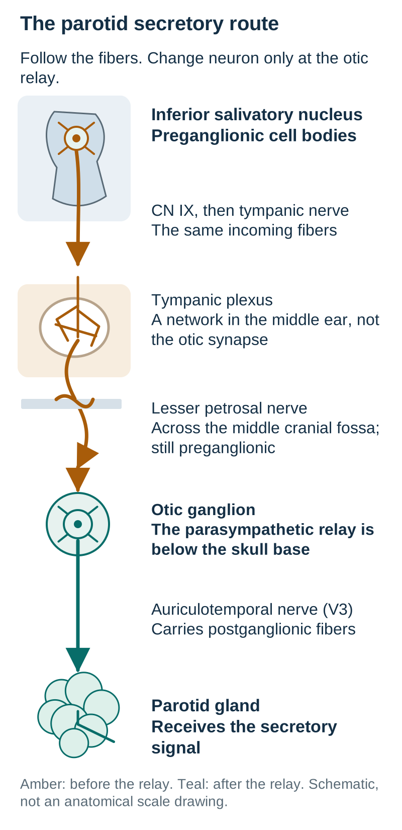
For the standard parotid route, begin at the inferior salivatory nucleus. Its axons travel in CN IX, the glossopharyngeal nerve, then enter the tympanic branch, or Jacobson nerve. That branch reaches the middle ear and contributes to the tympanic plexus on the promontory. The parasympathetic fibers continue in the lesser petrosal nerve toward the otic ganglion. [2][3][6]
The lesser petrosal nerve emerges onto the anterior petrous surface and crosses the middle cranial fossa. Its fibers are still preganglionic. The otic ganglion is the parasympathetic relay, in the infratemporal region near the medial side of V3 below the foramen ovale. Postganglionic fibers then accompany the auriculotemporal nerve, a branch of V3, to the parotid. A route carried by a trigeminal branch is not necessarily a route originating in the trigeminal nerve. [2][4][6]
Use the route diagram as a lesion exercise. Start at the parotid and trace backward to the first relay, then backward to the brainstem. Now imagine an interruption on the middle-fossa segment before the relay. The gland and the downstream neurons can remain anatomically present even though the incoming parasympathetic signal no longer reaches the relay. This is a model of pathway continuity, not a clinical nerve-stimulation protocol.
Trace backward from the gland to the otic relay, then to the brainstem. A carrier nerve and a neuron origin are different facts. [2][3][4][6]The interruption is before the otic ganglion. Which signal is lost first?
The incoming preganglionic signal from the inferior salivatory nucleus is interrupted before it can activate otic neurons.
Which nerve would carry the outgoing fibers if the relay and distal route were intact?
The auriculotemporal nerve carries the postganglionic fibers to the parotid.
Transfer: an injury after the relay can also reduce parotid secretion. The target deficit alone does not tell you which side of the synapse was injured.
Which glands belong to each secretory route?
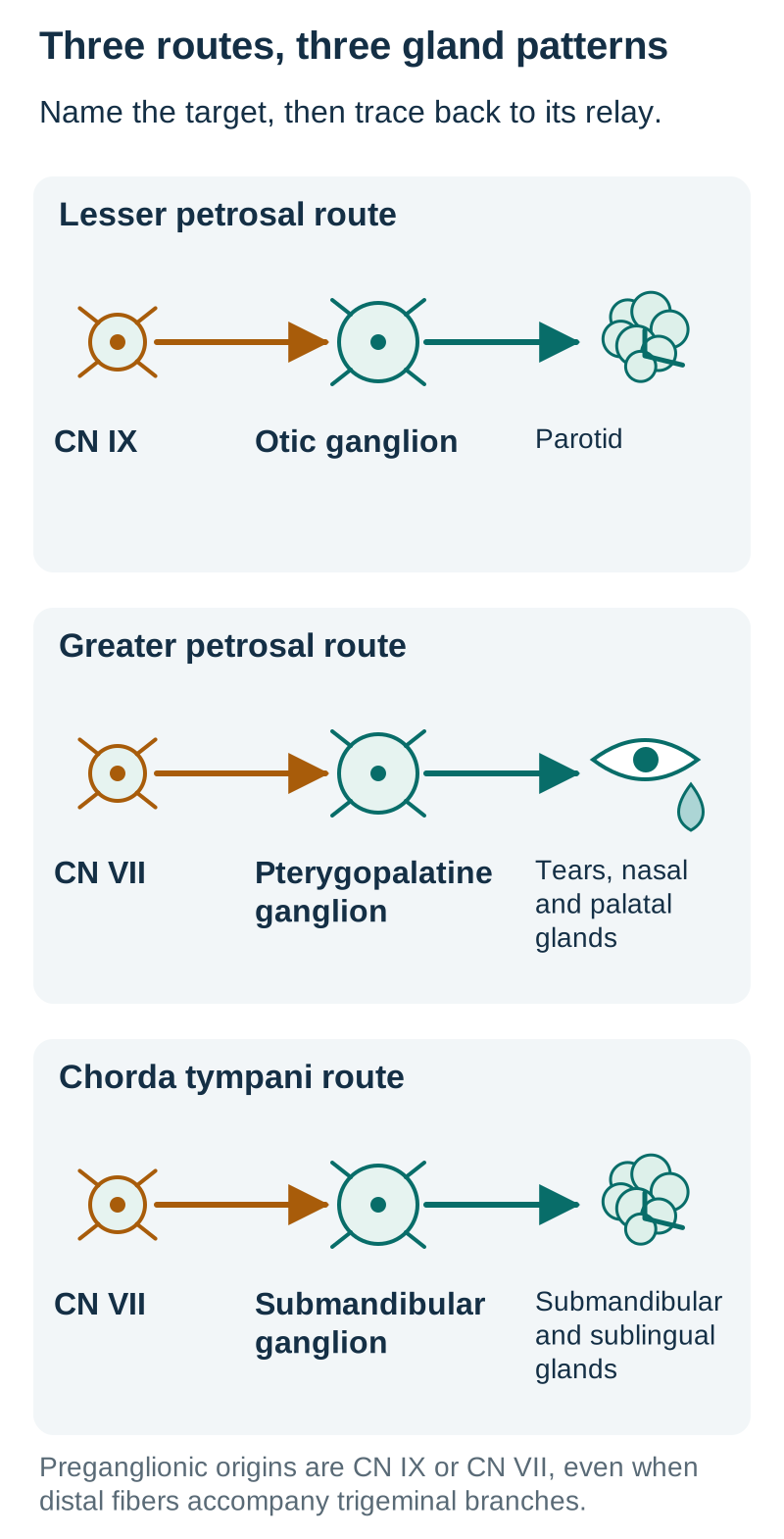
Similar names are a poor substitute for tracing a target. The lesser petrosal route has CN IX input and an otic relay. The greater petrosal route has CN VII input and a pterygopalatine relay. Its postganglionic fibers reach lacrimal, nasal and palatal glands using trigeminal branches. The lacrimal route includes a V2-to-V1 connection. [2]
The greater petrosal nerve arises near the geniculate ganglion and joins the deep petrosal nerve to form the nerve of the pterygoid canal. The greater petrosal component carries preganglionic parasympathetic fibers; the deep petrosal component carries postganglionic sympathetic fibers. The geniculate ganglion is a sensory ganglion, not the parasympathetic relay for these secretory fibers. That relay is the pterygopalatine ganglion. [5]
A third route prevents a common salivation error. CN VII also sends fibers through chorda tympani, which joins the lingual nerve and reaches the submandibular ganglion. This supplies submandibular and sublingual secretion. Chorda tympani also carries taste from the anterior two-thirds of the tongue; the lingual nerve itself carries general sensation there. Thus a middle-ear injury affecting anterior taste and these glands is different from a selective parotid pathway injury. [2][6]
The comparison diagram gives each route a separate starting nerve, relay and target. Point to the target before reading the nerve name. Then trace backward: a tear and nasal secretion deficit leads toward the pterygopalatine route, not toward the otic relay. These are idealized selective patterns; a fracture can affect more than one route.
Compare the target first. The geniculate ganglion is not substituted for a parasympathetic relay. Routes are functional schematics. [2][5][6]Tears and nasal secretion fall, but parotid output is preserved. Which relay belongs to the affected secretory route?
The pterygopalatine ganglion belongs to the greater petrosal route. Preserved parotid secretion argues against using the otic route to explain the entire pattern.
Transfer: loss of anterior tongue taste together with reduced submandibular secretion redirects attention to chorda tympani rather than either petrosal nerve.
Does the exit opening establish the nerve identity?
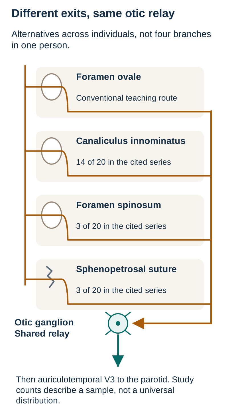
The lesser petrosal nerve is often taught as leaving the middle cranial fossa through foramen ovale. Treat that as a conventional route, not an invariant or a reliable population-frequency estimate. Direct dissections have documented passage through the canaliculus innominatus, foramen spinosum and the sphenopetrosal suture. The exact bony exit can vary without changing the downstream otic relay. [4]
In the 20 middle fossae described by Kakizawa and colleagues, the reported exits were 14 through the canaliculus innominatus, 3 through foramen spinosum and 3 through the sphenopetrosal suture. None in that series used foramen ovale. These observations refute an exclusive foramen-ovale rule; they do not prove that every population has the same distribution. Distinguish a teaching convention, a sample observation and an individual's anatomy. [4]
Two different bony transitions are also easy to confuse. The small petrosal hiatus allows the nerve to reach the middle cranial fossa from the temporal bone. The later exit takes it toward the infratemporal region. Neither should be substituted for the jugular foramen through which the main glossopharyngeal nerve leaves the skull. Track where the fiber is now, rather than remembering a single foramen for the entire route. [4][6]
In the exit diagram, follow each alternative opening to the same destination. A different opening changes exposure to a fracture or surgical corridor; it does not convert the nerve into CN VII or turn its fibers into postganglionic fibers before the otic synapse.
The separate-canal route predominated in the cited sample. Verify the actual individual course; a preserved foramen ovale does not assess every alternative. [4][6]A traced lesser petrosal nerve uses a small separate canal. Does its parasympathetic relay change?
No. The fibers still reach the otic ganglion. Continuity and destination identify the route more reliably than an assumed opening.
Transfer: an intact foramen ovale does not, by itself, establish that every possible lesser petrosal course is intact.
What can a parotid deficit localize?
A selective lesser petrosal injury reduces parasympathetic input to the ipsilateral parotid. It does not directly paralyze facial muscles, interrupt hearing or abolish tears. Those functions belong to different structures. However, low output from one parotid is not a uniquely identifying test for the lesser petrosal nerve. Injury to proximal CN IX, the otic ganglion or the auriculotemporal route can affect the same destination. [2][3][6]
Use additional findings to test those alternatives. Posterior tongue taste or pharyngeal afferent abnormalities suggest a broader CN IX lesion rather than a purely distal secretomotor lesion. A localized auriculotemporal injury can combine reduced parotid output with sensory loss near the anterior ear or temple. Preserved function does not prove a nerve is completely normal, but a pattern involving several distinct CN IX functions requires a broader explanation. [3][6]
The facial nerve traverses the parotid and supplies muscles of facial expression. Passing through the gland does not make it the source of the gland's parasympathetic secretion. A local facial motor branch injury can therefore impair a facial action while leaving the parotid secretory route intact. Conversely, selective parotid denervation need not produce a weak face. [6]
Separate the whole mouth from a single gland. The other parotid, submandibular, sublingual and minor glands can continue contributing saliva. A person may not notice marked dryness after a unilateral deficit. Generalized dryness also has medication, gland and systemic explanations, so it should not be automatically assigned to a temporal bone nerve injury. Painful gland swelling with an obstructed duct offers a local non-neural explanation for poor drainage. [11]
Try a second pathway interruption in the diagram: this time choose the auriculotemporal segment after the otic relay. Compare it with the preganglionic interruption from section 2. Both reduce input reaching the gland, but only the later segment carries fibers whose cell bodies are in the otic ganglion.
Parotid output is reduced but the mouth is still moist. What conclusion is justified?
A unilateral secretory deficit can coexist with saliva from other glands. Overall moisture does not exclude a localized deficit.
What additional evidence would favor a lesser petrosal lesion over another point on the same route?
A demonstrated injury along its middle-fossa course, interpreted with the rest of the examination, is more localizing than the parotid deficit alone.
A target deficit identifies a pathway; additional evidence identifies a site.
Why can similar fracture labels hide different injuries?
The otic capsule is the dense bone surrounding the inner-ear labyrinth. A fracture that violates it raises concern for sensorineural hearing loss and vestibular injury. Facial canal involvement, a route for cerebrospinal fluid leakage and carotid canal involvement are separate structural questions. Ask which structures the fracture crosses instead of relying only on the words longitudinal or transverse. [7][10]
Otic capsule classification helps organize concern, but it is not a deterministic outcome score. A small study by Little and Kesser found stronger associations with major complications than the traditional orientation system. Rafferty and colleagues, also studying a small cohort, did not demonstrate general superiority for predicting all specific complications. Neither result supports declaring a particular patient safe from their category alone. [7][8]
Hearing data add a functional localization. Air-conduction testing includes the external and middle-ear route; bone-conduction testing bypasses those structures. A reproducible air-bone gap with preserved bone thresholds supports a conductive component. Poor air and bone thresholds without a material gap support a sensorineural component. A mixed deficit is possible. These interpretations require appropriate test technique and masking when indicated. [12]
For a concrete comparison, imagine reliable thresholds at the same frequencies of air 50 dB HL and bone 15 dB HL, with a laboratory adult reference of 0 to 20 dB HL. The 35 dB gap and preserved bone thresholds favor a sound-transmission problem. Compare air 65 and bone 60 dB HL with new vertigo and a fracture crossing the cochlea: both thresholds are abnormal and the small gap cannot explain away the inner-ear injury. These are invented teaching values, not a treatment threshold.
Clear ear or nasal drainage after a skull-base injury warrants assessment for a CSF leak. A defect through the middle-ear roof can matter even when the otic capsule is spared. New facial weakness requires timely reassessment, including eye closure. Immediate weakness, delayed weakness and an unexamined face are not interchangeable histories. A fracture near a vascular canal plus a new focal deficit warrants urgent vascular assessment; CTA head and neck is an appropriate study when traumatic arterial injury is suspected. [1][9][10]
Hearing and balance worsen while parotid output is reduced. Can one isolated lesser petrosal lesion explain the entire pattern?
No. The auditory and vestibular findings require assessment of additional structures, rather than expanding the function assigned to one secretomotor branch.
Transfer: a patient can have a real small-nerve deficit and a more urgent coexisting injury. Match each abnormality to its structure, then prioritize the danger. Decisions about facial nerve procedures, CSF repair, hearing rehabilitation or vascular treatment require the full clinical assessment and specialist input; this lesson is not an operative algorithm.
Apply the anatomy
Case 1
Show answer and explanations for case 1
A. Obtain an urgent dedicated temporal bone CT (Why this does not fit)
Thin-section CT can clarify a temporal fracture. The patient first has an unstable airway and circulation. Imaging detail must not delay resuscitation.
Reasoning steps for option A
What can thin-section temporal CT clarify after blood is seen at the ear?
Thin-section CT could delineate a suspected temporal bone fracture associated with right ear bleeding.
Which findings make transport to CT unsafe before resuscitation?
GCS 8, irregular breathing and systolic pressure 82 mm Hg indicate threatened airway, ventilation and perfusion.
Why must temporal fracture mapping wait in this patient?
The patient needs airway and circulatory stabilization before transport for detailed temporal imaging.
B. Transfer directly to a regional neuro-otology unit (Why this does not fit)
A temporal fracture may require specialist care. An unstable patient needs initial resuscitation and stabilization before transfer. Choose a service and transfer plan that can address all active injuries.
Reasoning steps for option B
Why might a temporal fracture eventually require neuro-otology expertise?
Temporal bone fractures can cause facial, hearing or other otologic complications requiring specialist assessment.
What must happen before transfer with systolic pressure of 82 mm Hg and irregular breathing?
Stabilize ventilation and blood pressure with cervical protection before considering interfacility transfer.
Why is a single-specialty transfer not the first trauma intervention?
The unstable patient requires immediate multisystem trauma resuscitation, not direct transfer for an isolated ear injury.
C. Stabilize airway and circulation with cervical protection (Best answer)
Impaired consciousness, irregular breathing and hypotension identify immediate threats. A small secretomotor nerve injury cannot account for this physiological instability. Address life-threatening trauma before detailed localization.
Reasoning steps for option C
Which simultaneous threats do GCS 8, irregular breathing and pressure of 82 mm Hg signal?
GCS 8 and irregular breathing threaten airway and ventilation; systolic pressure 82 mm Hg indicates circulatory shock.
Could an isolated lesser petrosal lesion explain hypotension and disordered ventilation?
No; an isolated lesser petrosal lesion changes parotid secretion, not breathing or blood pressure.
How does possible cervical injury change immediate airway management?
Maintain cervical spine protection while securing ventilation because cervical injury has not been excluded.
D. Complete the facial and salivary examinations (Why this does not fit)
An early cranial nerve baseline can be useful. A detailed examination would delay treatment of the current physiological threats. Record baseline function when feasible without delaying emergency care.
Reasoning steps for option D
What useful baseline could a facial and salivary examination establish?
Facial strength and salivary output could document an early cranial nerve baseline.
Why should that examination not precede airway and circulatory support here?
A detailed cranial nerve exam would delay treatment of compromised ventilation and hypotension.
When can cranial nerve documentation safely follow this rollover injury?
Document facial and salivary function after immediate airway, circulation and cervical spine priorities are addressed.
Takeaway: After high-energy injury, physiological instability takes priority over small-nerve localization.
Hemotympanum is a sign that raises concern for a basal skull fracture. A normal current neurological examination does not exclude associated acute cranial injury. Use acute head-injury imaging rather than starting with a narrow salivary test.
Reasoning steps for option A
What does blood behind an intact tympanic membrane suggest after a fall?
Hemotympanum after trauma raises concern for a basal skull or temporal bone fracture.
Does a normal current neurological examination rule out acute cranial injury?
No; a normal examination now does not rule out associated acute intracranial injury.
Why is noncontrast head CT the initial study rather than gland imaging?
Noncontrast head CT assesses acute cranial trauma raised by hemotympanum; parotid imaging does not.
B. Contrast-enhanced MRI of the internal auditory canals (Why this does not fit)
MRI can investigate selected neural and inner-ear disorders. The immediate question is acute cranial trauma with a basal skull fracture sign. Match the initial modality to the injury phase and question.
Reasoning steps for option B
What sort of neural or inner-ear question could internal auditory canal MRI address?
MRI can investigate selected internal auditory canal neural and inner-ear disorders.
What acute injury concern does hemotympanum raise instead?
Post-traumatic hemotympanum signals possible basal skull fracture and intracranial injury.
Why does an MRI focused on the internal auditory canals not take priority before head CT?
Focused internal auditory canal MRI does not replace initial noncontrast head CT for acute cranial trauma.
C. Ultrasonography of the parotid gland (Why this does not fit)
Ultrasound can assess gland and duct disease. Blood behind the tympanic membrane is not evidence of a parotid duct lesion. Localize the observed abnormality before choosing a gland study.
Reasoning steps for option C
Which abnormalities would parotid ultrasound be suited to investigate?
Parotid ultrasound can assess gland tissue and ducts when local gland disease is suspected.
Does blood behind the tympanic membrane localize disease to the parotid duct?
No; blood behind the intact tympanic membrane is a middle-ear finding, not a parotid duct sign.
Which location should direct imaging selection in this patient?
The middle-ear blood prompts cranial trauma imaging, not a study centered on the parotid gland.
D. Plain radiography of the skull (Why this does not fit)
A radiograph can depict some bony abnormalities. It does not adequately assess the acute intracranial injury concern raised here. Do not substitute a limited skull study for indicated head CT.
Reasoning steps for option D
What can a skull radiograph potentially show?
A skull radiograph may depict some gross bony abnormalities.
Why is a plain skull image insufficient with traumatic hemotympanum?
Plain radiographs cannot adequately evaluate the acute intracranial injury concern associated with hemotympanum.
Which imaging approach addresses possible intracranial injury despite a normal examination?
Noncontrast head CT evaluates that concern even though the current neurological examination is normal.
Takeaway: A basal skull fracture sign warrants acute cranial assessment even when the current neurological examination is normal.
A. Contrast-enhanced MRI of the parotid (Why this does not fit)
MRI can characterize a parotid mass and surrounding soft tissue. The unresolved injury is in the bony facial canal, not a demonstrated gland mass. Choose the field of view from the suspected lesion site.
Reasoning steps for option A
What type of suspected lesion is contrast-enhanced parotid MRI designed to characterize?
Parotid MRI evaluates suspected gland masses and adjacent soft tissue, not the fracture line through bone.
Where is the unanswered fracture relationship in this patient with immediate facial weakness?
The initial head CT has not shown whether the temporal fracture involves the facial canal.
Why should imaging center on the facial canal rather than the parotid?
Immediate facial weakness with an unresolved bony facial canal relationship warrants thin-section temporal CT.
B. CT angiography of the head and neck (Why this does not fit)
CTA assesses suspected traumatic arterial injury. The specified uncertainty concerns bony canal involvement without a vascular indication. A vascular study and a detailed bone study answer different questions.
Reasoning steps for option B
Which trauma complication would justify head and neck CT angiography?
CTA assesses suspected traumatic arterial injury in the head and neck.
Is there an arterial indication in the stated fracture assessment?
No clinical or imaging suspicion of arterial injury is present in the stem.
What study is needed to resolve bony facial canal involvement instead?
Thin-section noncontrast temporal bone CT delineates the fracture against the facial canal.
C. Plain radiographs of the mastoid (Why this does not fit)
Mastoid radiographs depict limited regional bone detail. They do not adequately resolve the fracture relationship to the facial canal. Complex temporal anatomy requires cross-sectional detail.
Reasoning steps for option C
What limited information might mastoid radiographs provide?
Mastoid radiographs can show limited regional bony detail.
Can plain mastoid films define a fracture against the facial canal?
No; overlapping temporal bone anatomy limits visualization of the facial canal relationship.
What anatomical complexity calls for cross-sectional temporal bone imaging?
Cross-sectional thin-section temporal CT resolves the canal and fracture in a patient with immediate facial weakness.
D. Thin-section noncontrast temporal bone CT (Best answer)
Dedicated temporal CT resolves detailed bony anatomy. Initial images leave an important facial-canal question unanswered in a patient with facial weakness. Use additional imaging for a specific unresolved question rather than for every fracture.
Reasoning steps for option D
What can thin-section noncontrast temporal CT show that the initial head CT cannot?
It supplies high-resolution bony detail showing whether the fracture involves the facial canal.
Why does immediate facial weakness make the unresolved facial canal relationship important?
Immediate weakness could reflect injury near the facial nerve; the initial CT leaves canal involvement uncertain.
When is a dedicated bone study justified after an initial trauma CT?
A targeted study is justified because the initial CT cannot answer the specific facial canal question.
Takeaway: Dedicated temporal CT is useful when the initial study leaves an important bony question unresolved.
A. Noncontrast CT limited to the temporal bones (Why this does not fit)
Detailed bone imaging can map the fracture. The fracture is already known, and a new focal deficit raises a vascular question. Do not answer a vessel question using bone anatomy alone.
Reasoning steps for option A
What aspect of a carotid canal fracture could focused temporal CT map?
Focused temporal CT could further delineate the already recognized fracture through the carotid canal.
Why does new left arm weakness raise a question beyond bone mapping?
New contralateral arm weakness without explanatory hemorrhage raises concern for arterial injury affecting cerebral perfusion.
Which structure must be examined rather than merely the canal walls?
The carotid artery traverses the implicated canal, so its lumen requires assessment with CTA.
B. CT angiography of the head and neck (Best answer)
CTA evaluates the cervical and intracranial arterial circulation. Carotid canal involvement plus an unexplained focal deficit raises concern for traumatic arterial injury. Use the new neurological finding to broaden the trauma assessment.
Reasoning steps for option B
What vessels does head and neck CTA assess after skull-base trauma?
CTA assesses cervical and intracranial arteries, including potential traumatic carotid injury.
What do carotid canal extension and unexplained contralateral arm weakness jointly suggest?
A carotid canal fracture plus new left arm weakness raises concern for traumatic carotid injury and cerebral ischemia.
Why does the new focal deficit shift the next study toward arterial imaging?
The new deficit is not explained by head CT hemorrhage, making head and neck arterial imaging the relevant next test.
C. Ultrasonography limited to the parotid (Why this does not fit)
Ultrasound can evaluate superficial salivary structures. A parotid lesion does not explain contralateral arm weakness. A gland-focused explanation cannot account for a cerebral deficit.
Reasoning steps for option C
What superficial structure can parotid ultrasound evaluate?
Parotid ultrasound assesses superficial gland or duct abnormalities.
Can a parotid disorder account for new contralateral arm weakness?
No; parotid disease does not explain a new contralateral focal limb weakness.
What site of injury is suggested by a focal limb deficit instead of a salivary symptom?
A cerebral vascular complication of the carotid canal fracture better matches the new focal deficit.
D. CT venography of the head (Why this does not fit)
CTV assesses suspected cerebral venous injury. The identified at-risk passage contains the carotid artery, directing the immediate vascular question to arteries. Match the vascular study to the structure implicated by the injury.
Reasoning steps for option D
Which suspected injury is head CT venography intended to assess?
The geniculate ganglion contains sensory neuronal cell bodies associated with CN VII. The documented injury interrupts a CN IX secretomotor route rather than a facial sensory route. Do not substitute a nearby sensory ganglion for an autonomic relay.
Reasoning steps for option A
What neuronal cell bodies are found in the geniculate ganglion?
The geniculate ganglion contains sensory neuronal cell bodies associated with the facial nerve.
Does a CN VII sensory ganglion relay the documented CN IX parotid pathway?
No; the injured parotid secretomotor route runs via CN IX and lesser petrosal nerve toward the otic ganglion.
What type of ganglion must supply neurons whose axons leave for the parotid?
The outgoing parotid secretomotor neurons have cell bodies in the otic autonomic ganglion.
B. Trigeminal ganglion (Why this does not fit)
Trigeminal sensory neuronal cell bodies lie in this ganglion. The parotid-directed fibers joining V3 are autonomic fibers from a different ganglion. The nerve carrying an axon does not establish the location of its cell body.
Reasoning steps for option B
What type of neurons reside in the trigeminal ganglion?
The trigeminal ganglion houses trigeminal sensory neuron cell bodies.
Do autonomic fibers carried along a V3 branch originate in the trigeminal ganglion?
No; parotid autonomic fibers merely join a V3 branch after synapsing in the otic ganglion.
Why does the distal carrier of parotid fibers not identify their autonomic relay?
The auriculotemporal carrier is a V3 branch, but its parotid autonomic axons originate in the otic ganglion.
C. Submandibular ganglion (Why this does not fit)
This ganglion relays parasympathetic input for submandibular and sublingual glands. The measured deficit concerns the parotid after a documented lesser petrosal injury. Match the ganglion to the affected gland.
Reasoning steps for option C
Which salivary glands receive fibers relayed in the submandibular ganglion?
The submandibular ganglion supplies postganglionic fibers to submandibular and sublingual glands.
Which gland loses secretion after this lesser petrosal injury?
Parotid secretion is reduced after interruption of the lesser petrosal pathway.
Which ganglion corresponds to the affected gland rather than neighboring glands?
The otic ganglion supplies the affected parotid, rather than the submandibular ganglion.
D. Pterygopalatine ganglion (Why this does not fit)
This ganglion relays greater petrosal parasympathetic input. Its secretory targets include lacrimal and nasal glands, which are not the documented target here. Separate greater and lesser petrosal routes by destination.
Reasoning steps for option D
Which petrosal pathway synapses in the pterygopalatine ganglion?
Greater petrosal preganglionic fibers relay in the pterygopalatine ganglion.
Which secretory targets would that pathway affect rather than the parotid?
Those fibers serve lacrimal and nasal glands, not the selectively affected parotid.
How do preserved tears distinguish this route from the injured parotid pathway?
Preserved tears with reduced parotid secretion favors lesser petrosal to otic rather than greater petrosal to pterygopalatine.
E. Otic ganglion (Best answer)
Otic neurons send postganglionic axons toward the parotid through the auriculotemporal nerve. The recorded injury lies in the preganglionic lesser petrosal segment before this relay. Distinguish the incoming nerve from the cell bodies of the outgoing neurons.
Reasoning steps for option E
Which ganglion supplies the postganglionic parotid neurons?
Postganglionic otic neurons project toward the parotid in the auriculotemporal nerve.
Is the injured lesser petrosal segment before or after that ganglionic relay?
The injured lesser petrosal fibers approach the otic ganglion before the autonomic synapse.
Where are the cell bodies of axons subsequently carried by the auriculotemporal nerve?
The cell bodies of the subsequent parotid-directed axons are in the otic ganglion.
Takeaway: Lesser petrosal fibers approach the otic relay; postganglionic parotid fibers arise from otic neurons.
A. Right lesser petrosal nerve (Why this does not fit)
This nerve carries preganglionic input for the right parotid. An isolated lesion does not explain posterior tongue taste loss or the pharyngeal afferent deficit. A wider set of CN IX functions requires a more proximal explanation.
Reasoning steps for option A
What parotid-directed fibers run in the lesser petrosal nerve?
The lesser petrosal nerve carries preganglionic parasympathetic fibers toward the otic ganglion for parotid secretion.
Can an isolated lesser petrosal lesion abolish posterior tongue taste and pharyngeal sensation?
No; it does not carry posterior tongue taste or the pharyngeal sensory afferent limb of the gag.
What broader CN IX site explains secretory and sensory losses together?
A right CN IX trunk lesion can involve both parotid fibers and posterior tongue and pharyngeal afferents.
B. Right auriculotemporal nerve (Why this does not fit)
This nerve carries postganglionic input toward the right parotid. Its sensory territory is not the posterior tongue and pharynx. Distal carriage of autonomic fibers does not confer the functions of their source nerve.
Reasoning steps for option B
What parotid fibers travel with the auriculotemporal nerve?
The auriculotemporal nerve carries postganglionic otic fibers to the parotid.
Does its sensory distribution include the posterior tongue and pharynx?
No; its sensory territory is anterior ear and temple rather than posterior tongue and pharynx.
Why cannot injury to this distal parotid carrier explain the unilateral gag afferent loss?
A distal auriculotemporal lesion cannot abolish the right pharyngeal gag afferent or posterior tongue taste.
C. Right glossopharyngeal nerve trunk (Best answer)
The main glossopharyngeal nerve carries parotid-directed fibers and posterior tongue and pharyngeal afferents. The side-specific absent afferent response with an intact response from the other side fits this combined lesion. Use the additional sensory deficit to distinguish a main nerve lesion from a distal secretory branch.
Reasoning steps for option C
Which CN IX functions are united in the main glossopharyngeal trunk?
CN IX trunk contains parotid-directed parasympathetic fibers, posterior tongue taste and pharyngeal afferents.
What does absent response to right pharyngeal touch but bilateral response to left touch localize?
Absent gag after right stimulation but bilateral response after left stimulation localizes failure to the right afferent limb.
Why does posterior tongue taste loss favor the trunk over an isolated secretory branch?
Posterior tongue taste and gag afferents travel with CN IX trunk, not the isolated lesser petrosal branch.
D. Right chorda tympani nerve (Why this does not fit)
Chorda tympani carries anterior tongue taste and submandibular secretomotor fibers. The deficits concern posterior tongue taste and the parotid instead. Separate anterior tongue and posterior tongue pathways.
Reasoning steps for option D
Which tongue region and glands are served by chorda tympani?
Chorda tympani carries anterior two-thirds tongue taste and submandibular and sublingual preganglionic fibers.
Do this patient's taste and salivary deficits match those territories?
No; posterior tongue taste and parotid secretion are impaired instead.
Which cranial nerve serves posterior tongue taste rather than anterior tongue taste?
E. Right greater petrosal nerve (Why this does not fit)
This nerve supplies preganglionic input for lacrimal and nasal secretion. It does not account for the parotid and pharyngeal afferent combination. Select a route that explains all of the observed territories.
Reasoning steps for option E
Which glands receive greater petrosal parasympathetic input?
Greater petrosal fibers serve lacrimal and nasal secretion via the pterygopalatine ganglion.
Could this route account for both parotid hyposecretion and absent right gag afference?
No; that route cannot explain parotid loss and an absent right pharyngeal gag afferent.
Which pathway must include pharyngeal sensation as well as parotid fibers?
The right CN IX trunk includes both parotid parasympathetic and pharyngeal sensory fibers.
Takeaway: Parotid dysfunction plus posterior tongue and pharyngeal afferent deficits suggests a broader CN IX lesion.
A. Postganglionic output from the otic ganglion (Why this does not fit)
Postganglionic otic fibers reach the parotid through the auriculotemporal nerve. That distal route is in the infratemporal region rather than on the middle-ear promontory. Use location to distinguish preganglionic from postganglionic injury.
Reasoning steps for option A
How do postganglionic otic fibers reach the parotid?
Otic postganglionic fibers reach parotid via the auriculotemporal nerve.
Is the auriculotemporal route located on the cochlear promontory?
No; auriculotemporal fibers lie in the infratemporal region, not the cochlear promontory.
Why does the site of damage favor input before the otic synapse?
Promontory injury affects tympanic plexus preganglionic input before the otic ganglion synapse.
B. Preganglionic input toward the otic ganglion (Best answer)
The tympanic plexus carries parotid-directed preganglionic fibers onward into the lesser petrosal nerve. Promontory injury with preserved chorda tympani functions fits interruption of this input. A plexus is not necessarily the site of an autonomic synapse.
Reasoning steps for option B
Which preganglionic pathway passes through the tympanic plexus on the promontory?
Preganglionic CN IX parotid fibers run through the promontory tympanic plexus and then lesser petrosal nerve.
How do reduced parotid output and preserved chorda tympani functions identify that pathway?
Parotid loss with intact anterior taste and submandibular secretion indicates tympanic plexus injury rather than chorda tympani injury.
Does the tympanic plexus itself contain the parotid parasympathetic synapse?
No; these fibers synapse at the otic ganglion, not on the middle-ear promontory.
C. Preganglionic input toward the submandibular ganglion (Why this does not fit)
Chorda tympani carries this input through the middle ear. Preserved anterior taste and submandibular secretion argue against that route as the injured one. Neighboring middle-ear nerves have different target patterns.
Reasoning steps for option C
Which nerve brings preganglionic fibers to the submandibular ganglion?
Chorda tympani carries preganglionic fibers toward the submandibular ganglion.
Which preserved functions argue against chorda tympani damage here?
Preserved anterior tongue taste and submandibular secretion argue against chorda tympani injury.
How does the parotid target separate promontory plexus injury from chorda tympani injury?
The injured promontory plexus carries CN IX parotid-directed fibers; chorda tympani serves submandibular and sublingual glands.
D. Postganglionic output from the pterygopalatine ganglion (Why this does not fit)
These fibers serve lacrimal and nasal or palatal glands through trigeminal branches. They do not explain selective parotid dysfunction after promontory injury. Match both the ganglion and its distal targets.
Reasoning steps for option D
What secretory territories receive pterygopalatine postganglionic fibers?
Pterygopalatine postganglionic fibers supply lacrimal, nasal and palatal glands.
Do those targets account for isolated parotid loss after promontory trauma?
No; isolated parotid hyposecretion after promontory trauma does not match those targets.
Which ganglion instead belongs to the parotid secretory pathway?
The otic ganglion, reached via tympanic plexus and lesser petrosal nerve, belongs to the parotid pathway.
E. Motor output from the facial motor nucleus (Why this does not fit)
Facial motor axons supply muscles of facial expression. The described change is parotid secretion, not facial weakness. Motor supply to facial muscles is distinct from gland secretomotor supply.
Reasoning steps for option E
What does facial motor output innervate?
Facial motor axons innervate muscles of facial expression.
What motor deficit would be expected rather than isolated parotid hyposecretion?
Facial motor injury would produce facial weakness, not just reduced parotid output.
Why does gland secretion implicate autonomic fibers rather than facial motor axons?
Parotid hyposecretion after promontory trauma implicates CN IX parasympathetic fibers in the tympanic plexus.
Takeaway: The tympanic plexus carries parotid-directed fibers before their otic ganglion synapse.
Otic neurons provide postganglionic fibers that accompany the auriculotemporal nerve. The combined temporal sensory and parotid findings localize to that distal carrier. Identify the carrier from its sensory territory, then identify the origin of the autonomic fibers it carries.
Reasoning steps for option A
Where do postganglionic parotid secretomotor fibers arise?
Parotid postganglionic secretomotor neurons have cell bodies in the otic ganglion.
What does combined anterior ear and temple numbness with parotid loss identify as the injured carrier?
Anterior ear and temple sensation plus parotid fibers travel together in the auriculotemporal nerve.
Which relay precedes entry of those secretory fibers into that carrier?
Preganglionic lesser petrosal fibers synapse on otic ganglion neurons; those neurons send postganglionic axons into the auriculotemporal nerve.
B. Inferior salivatory nucleus (Why this does not fit)
This nucleus contains the preganglionic neurons of the parotid route. The injured distal carrier contains the fibers after the otic synapse. Do not assign a postganglionic axon to a preganglionic cell body.
Reasoning steps for option B
What class of parotid neurons resides in the inferior salivatory nucleus?
The inferior salivatory nucleus contains preganglionic CN IX parotid neurons.
Are salivary axons in the auriculotemporal nerve before or after the otic synapse?
They are postganglionic axons arising from otic neurons, downstream of the synapse made by incoming lesser petrosal fibers.
Why is a preganglionic nucleus not the cell-body site for the injured distal fibers?
The injured distal axons arise from otic postganglionic neurons, not the inferior salivatory nucleus.
C. Trigeminal ganglion (Why this does not fit)
Sensory fibers of the auriculotemporal nerve have cell bodies in the trigeminal ganglion. The question asks about the salivary fibers, not the neighboring sensory fibers. One peripheral nerve can contain axons with different cell-body locations.
Reasoning steps for option C
Whose cell bodies in the trigeminal ganglion contribute to the auriculotemporal nerve?
Trigeminal ganglion neurons contribute sensory fibers for anterior ear and temple sensation.
Does the question concern its sensory axons or its parotid secretomotor axons?
The question specifies salivary axons accompanying those sensory fibers.
Why can sensory and autonomic fibers in the same peripheral nerve have different origins?
Auriculotemporal sensory axons originate in the trigeminal ganglion, while parotid secretomotor axons originate in the otic ganglion.
D. Pterygopalatine ganglion (Why this does not fit)
This ganglion provides postganglionic fibers for lacrimal and nasal or palatal secretion. Those destinations do not match the parotid deficit and anterior ear sensory territory. A secretory deficit must be assigned to the correct gland pathway.
Reasoning steps for option D
Which glandular territories receive pterygopalatine ganglion output?
Pterygopalatine output serves lacrimal, nasal and palatal glands.
Are lacrimation and parotid secretion impaired together in this patient?
No; parotid output is reduced but lacrimation is preserved.
Which ganglion supplies the affected parotid instead of lacrimal targets?
The otic ganglion supplies postganglionic parotid fibers in the injured auriculotemporal nerve.
E. Geniculate ganglion (Why this does not fit)
This is a sensory ganglion associated with the facial nerve. The combined deficit fits an auriculotemporal route rather than facial sensory fibers. A sensory ganglion is not the parasympathetic source for parotid secretion.
Reasoning steps for option E
What kind of facial nerve cells are found in the geniculate ganglion?
The geniculate ganglion holds sensory cell bodies associated with CN VII.
Does anterior ear numbness with parotid loss implicate a facial sensory route?
No; ear and temple numbness with parotid loss localizes to the auriculotemporal nerve.
Why is a facial sensory ganglion not the source of postganglionic parotid fibers?
The autonomic parotid fibers carried by that nerve originate in the otic ganglion, not a facial sensory ganglion.
Takeaway: Auriculotemporal sensory fibers and its carried parasympathetic fibers have different cell-body locations.
A. Otic injury with preserved facial motor input (Why this does not fit)
Otic injury can impair parotid-directed autonomic signaling. It does not explain a focal facial motor deficit with preserved gland output. Do not make the otic ganglion responsible for muscles of facial expression.
Reasoning steps for option A
Does otic ganglion injury primarily affect parotid secretion or mouth-corner retraction?
Otic ganglion injury impairs parotid secretomotor output, not mouth-corner movement.
Why does unchanged stimulated parotid output argue against otic injury?
Parotid flow is unchanged, so its otic secretomotor pathway appears functional.
Which pathway supplies the impaired mouth-corner movement?
A facial motor branch traversing the parotid supplies that facial-expression movement.
B. Greater petrosal injury with preserved otic input (Why this does not fit)
Greater petrosal injury affects lacrimal and nasal secretory input. It does not explain weakness of a facial muscle supplied beyond the parotid. Separate facial motor branches from facial parasympathetic branches.
Reasoning steps for option B
Which secretions follow the greater petrosal nerve rather than a parotid motor branch?
Greater petrosal fibers supply lacrimal and nasal secretions.
Does reduced tearing explain inability to retract the mouth corner?
No. Lacrimal secretory loss cannot weaken the muscle retracting the mouth corner.
What does preserved parotid secretion say about the otic route?
Preserved parotid secretion is consistent with spared otic secretomotor input.
C. Lesser petrosal injury with preserved facial motor input (Why this does not fit)
Lesser petrosal injury can reduce parasympathetic parotid input. The reported salivary function is preserved while facial muscle function is impaired. An isolated secretomotor lesion does not produce this motor pattern.
Reasoning steps for option C
What parotid function would a lesser petrosal injury be expected to reduce?
Lesser petrosal interruption would reduce parasympathetic parotid input.
Which observed function is preserved despite this proposed injury?
Stimulated parotid output remains unchanged.
Can an isolated lesser petrosal lesion weaken facial expression?
No. The lesser petrosal nerve carries secretomotor rather than facial motor fibers.
D. Facial motor branch injury with preserved otic input (Best answer)
Facial motor branches pass through the parotid and supply facial muscles. They are distinct from the otic and auriculotemporal secretory route that remains functional. A nerve passing through a gland need not supply that gland with parasympathetic input.
Reasoning steps for option D
Which nerve branches traversing the parotid move the corner of the mouth?
Facial motor branches passing through the parotid innervate muscles moving the mouth corner.
Why can parotid output remain normal after one of these motor branches is injured?
Their motor pathway is separate from the otic ganglion and auriculotemporal parotid secretomotor route.
Does passage of facial motor fibers through the parotid make them its secretomotor supply?
No. Facial motor branches traverse the gland without providing its parasympathetic input.
Takeaway: Facial motor branches within the parotid do not provide its principal parasympathetic secretory input.
A. CN IX input; otic ganglion relay (Why this does not fit)
This pair belongs to the parotid secretory route. The affected targets are lacrimal and nasal while parotid output is preserved. Use the target pattern rather than the word petrosal alone.
Reasoning steps for option A
Which cranial nerve and ganglion supply parotid secretomotor input?
CN IX preganglionic fibers relay in the otic ganglion for parotid secretion.
Why does preserved parotid flow weigh against an otic relay lesion?
Normal parotid output weighs against disruption of that route.
Which gland pattern instead points to the greater petrosal route?
Reduced tearing and nasal output instead indicate the greater petrosal CN VII route.
B. CN VII input; submandibular ganglion relay (Why this does not fit)
This pair belongs to the chorda tympani salivary route. It does not account for the combined lacrimal and nasal deficit. CN VII has more than one parasympathetic destination.
Reasoning steps for option B
Where do CN VII chorda tympani preganglionic fibers relay?
Chorda tympani carries CN VII input to the submandibular ganglion.
Would that relay explain reduced tearing plus nasal secretion?
No. This relay serves submandibular and sublingual glands rather than lacrimal and nasal glands.
Which CN VII secretory branch reaches the affected glands instead?
The greater petrosal branch of CN VII supplies the affected glands via the pterygopalatine ganglion.
C. CN VII input; pterygopalatine ganglion relay (Best answer)
Greater petrosal preganglionic input reaches the pterygopalatine ganglion. Its downstream targets include the lacrimal and nasal glands affected here. Facial parasympathetic dysfunction can occur without facial motor weakness.
Reasoning steps for option C
Which CN VII branch carries preganglionic input toward the pterygopalatine ganglion?
The greater petrosal nerve carries CN VII preganglionic fibers to the pterygopalatine ganglion.
Which two reduced secretions are supplied downstream of this ganglion?
Lacrimal and nasal glands both receive secretory output downstream of this ganglion.
Why can facial strength remain normal when this secretory branch is interrupted?
Greater petrosal fibers can be injured separately from facial motor fibers.
D. CN V input; trigeminal ganglion relay (Why this does not fit)
The trigeminal ganglion contains sensory neuronal cell bodies. Trigeminal branches carry secretory fibers but do not use this sensory ganglion as their parasympathetic relay. Distinguish autonomic origin from a distal sensory nerve carrier.
Reasoning steps for option D
What is the trigeminal ganglion function relevant to this proposed relay?
Trigeminal ganglion contains general sensory neuronal cell bodies.
Do trigeminal carriers synapse their lacrimal secretory fibers in that sensory ganglion?
No. Trigeminal branches carry secretory fibers but do not relay them there.
What distinguishes a carrier in CN V from the CN VII autonomic origin?
CN VII supplies preganglionic fibers; CN V branches are distal carriers of postganglionic fibers.
E. CN VII input; geniculate ganglion relay (Why this does not fit)
The greater petrosal nerve arises near the geniculate ganglion. That ganglion is sensory rather than the peripheral parasympathetic synapse for these targets. Passing a sensory ganglion does not make fibers postganglionic.
Reasoning steps for option E
Why is the geniculate ganglion anatomically tempting near greater petrosal origin?
The greater petrosal nerve arises near the geniculate ganglion.
Is the geniculate ganglion the autonomic relay for lacrimal and nasal glands?
No. The geniculate ganglion is sensory and not the parasympathetic synapse.
Where do the preganglionic fibers synapse instead?
These preganglionic fibers synapse in the pterygopalatine ganglion.
Takeaway: Greater petrosal parasympathetic fibers originate with CN VII and relay in the pterygopalatine ganglion.
The otic ganglion relays parotid-directed parasympathetic fibers. Parotid output is preserved while anterior taste and submandibular secretion are affected. Select the relay for the gland actually involved.
Reasoning steps for option A
Which gland receives postganglionic output from the otic ganglion?
Otic ganglion postganglionic neurons supply the parotid gland.
Why does preserved parotid flow exclude it as the relevant relay?
Parotid secretion is unchanged, unlike the affected submandibular output.
Which injured branch combines anterior tongue taste with submandibular secretion?
Chorda tympani carries both anterior tongue taste and submandibular secretomotor fibers.
B. Pterygopalatine ganglion (Why this does not fit)
This ganglion relays lacrimal and nasal or palatal secretory input. The paired anterior tongue taste and submandibular deficit follows a different CN VII branch. Different branches of one cranial nerve can reach different ganglia.
Reasoning steps for option B
Which secretory targets use the pterygopalatine ganglion?
Pterygopalatine neurons supply lacrimal, nasal and palatal glands.
Do lost anterior tongue taste and submandibular flow match that destination?
No. That ganglion is not the relay for submandibular output or anterior tongue taste.
Which CN VII branch supplies the affected modalities?
Chorda tympani carries the affected CN VII modalities.
C. Geniculate ganglion (Why this does not fit)
The geniculate ganglion contains facial taste sensory cell bodies. It is not the autonomic synapse requested in the question. The taste and parasympathetic fibers sharing chorda tympani have different neuronal relationships.
Reasoning steps for option C
What type of neuronal cell bodies lie in the geniculate ganglion?
Geniculate ganglion contains CN VII taste sensory cell bodies.
Does that sensory ganglion synapse the chorda tympani secretomotor fibers?
No. Its taste neurons do not synapse the preganglionic secretomotor fibers.
Where is their downstream parasympathetic synapse instead?
Chorda tympani preganglionic fibers synapse in the submandibular ganglion.
D. Trigeminal ganglion (Why this does not fit)
The trigeminal ganglion contains general sensory cell bodies for the lingual nerve. Light touch is preserved and the question concerns the secretory relay. Do not confuse intact lingual general sensation with intact chorda tympani function.
Reasoning steps for option D
Which modality of anterior tongue sensation uses trigeminal sensory neurons?
General anterior tongue light touch travels through the lingual nerve to trigeminal sensory neurons.
What does preserved light touch imply about the lingual general sensory pathway?
Intact light touch argues that general lingual sensation is preserved.
Can a trigeminal sensory ganglion relay submandibular secretion?
No. The trigeminal ganglion is sensory, whereas secretory fibers relay in the submandibular ganglion.
E. Submandibular ganglion (Best answer)
Chorda tympani carries preganglionic fibers to this relay after joining the lingual nerve. Its combined taste and secretory functions explain the affected modalities despite preserved general sensation. Identify the injured branch from the modality pattern, then trace its autonomic destination.
Reasoning steps for option E
Which CN VII branch carries both lost anterior tongue taste and salivary input?
Chorda tympani carries both anterior tongue taste and submandibular secretomotor fibers.
Which nerve carries its preganglionic fibers onward toward the submandibular ganglion?
After joining the lingual nerve, its preganglionic fibers travel toward the submandibular ganglion.
Why is preserved touch compatible with this branch lesion?
Lingual general sensory fibers can remain intact despite loss of chorda tympani taste and secretomotor fibers.
Takeaway: Chorda tympani can lose taste and submandibular input while lingual general sensation and parotid secretion remain intact.
A. Reduced lacrimal secretion with preserved mastication (Why this does not fit)
Lacrimal secretion depends on postganglionic neurons of the pterygopalatine ganglion. The specified ganglion below foramen ovale is the otic ganglion instead. A ganglion must be identified by its site before assigning its target.
Reasoning steps for option A
Which ganglion supplies lacrimal secretomotor neurons?
Which ganglion actually lies medial to V3 below foramen ovale?
The ganglion medial to V3 below foramen ovale is the otic ganglion.
Would selective destruction there reduce tearing?
No. Isolated otic destruction predicts parotid rather than lacrimal deficit.
B. Reduced parotid secretion with preserved mastication (Best answer)
The otic ganglion supplies postganglionic parotid secretory fibers. The adjacent mandibular motor pathway is explicitly preserved. An autonomic ganglion lesion need not injure nearby somatic motor fibers.
Reasoning steps for option B
Which postganglionic neurons for parotid secretion are destroyed at this location?
The destroyed neurons are otic ganglion postganglionic neurons whose axons provide parotid secretomotor input.
Why should chewing remain intact when V3 motor branches are preserved?
The mandibular motor branches supplying mastication are explicitly spared.
Does adjacency to V3 mean otic neurons innervate masticatory muscle?
No. Otic secretomotor neurons and nearby V3 somatic motor axons have different targets.
C. Reduced mastication with preserved parotid secretion (Why this does not fit)
Mandibular motor fibers supply the muscles of mastication. Those fibers are preserved while the parotid autonomic neurons are destroyed. Physical proximity does not mean two functions share a synapse.
Reasoning steps for option C
Which preserved nerve division supplies muscles of mastication?
Preserved V3 motor fibers supply the masticatory muscles.
What parotid effect follows destruction of otic ganglion neurons?
Why is the proposed preservation of parotid output inconsistent?
Parotid output would not be expected to remain normal after destruction of its otic relay.
D. Reduced facial expression with preserved parotid secretion (Why this does not fit)
Facial expression depends on CN VII motor branches. The lesion is in a V3-adjacent autonomic ganglion, not the facial motor route. Do not infer facial paralysis from a lesion near the parotid secretory pathway.
Reasoning steps for option D
Which cranial nerve supplies facial-expression muscles?
CN VII motor branches supply facial-expression muscles.
Does an isolated otic ganglion lesion interrupt its motor branches?
No. An isolated otic ganglion lesion does not injure those CN VII motor branches.
What secretory deficit, rather than facial paresis, is predicted?
The expected deficit is reduced parotid secretion.
E. Reduced sublingual secretion with preserved mastication (Why this does not fit)
Sublingual secretion receives postganglionic input associated with the submandibular ganglion. That ganglion is not the one described immediately below foramen ovale. Localize the relay and then predict the gland deficit.
Reasoning steps for option E
Which ganglion mediates sublingual secretomotor output?
The submandibular ganglion supplies postganglionic fibers for sublingual secretion.
Is that ganglion medial to V3 below foramen ovale?
No. The ganglion medial to V3 below foramen ovale is otic.
Which gland is instead linked to the ganglion at the described site?
The otic ganglion supplies parotid secretion.
Takeaway: The otic ganglion is an autonomic relay near V3, not a motor relay for mastication.
A. Reduced lacrimal input with intact parotid input (Why this does not fit)
Lacrimal secretomotor input follows the greater petrosal route. The traced nerve reaches the otic rather than the pterygopalatine ganglion. Identify a nerve by continuity and destination even when its exit is unfamiliar.
Reasoning steps for option A
Which petrosal pathway supplies lacrimal rather than parotid secretion?
What does the observed otic ganglion destination rule out?
Its otic destination rules out the pterygopalatine relay used for lacrimal output.
Does a separate bony canal turn lesser petrosal fibers into greater petrosal fibers?
No. The variant exit leaves its otic and parotid destination unchanged.
B. Reduced facial motor input with intact parotid input (Why this does not fit)
Facial motor fibers supply muscles of facial expression. The traced course is toward an autonomic ganglion, and facial motor injury is absent. A secretomotor course should not be assigned a somatic motor target.
Reasoning steps for option B
Which fibers would have to be injured to weaken facial expression?
Facial motor axons to muscles of expression would have to be injured.
Why does a tympanic-to-otic course not identify facial motor fibers?
The observed tympanic-to-otic course identifies lesser petrosal autonomic input instead.
What preserved clinical finding supports intact facial motor input?
The stem explicitly states that no facial motor injury occurs.
C. Reduced submandibular input with intact parotid input (Why this does not fit)
Submandibular secretion depends on the chorda tympani and submandibular route. The directly observed ganglionic destination is the otic ganglion. Different salivary glands use different peripheral relays.
Reasoning steps for option C
Which CN VII branch supplies submandibular secretomotor input?
Chorda tympani carries CN VII input to the submandibular ganglion.
Does that branch terminate at the observed otic ganglion?
No. The traced nerve terminates at the otic ganglion.
What gland receives input via this traced tympanic-to-otic route?
The otic relay serves the parotid gland.
D. Reduced parotid input with intact lacrimal input (Best answer)
A tympanic-to-otic course identifies the standard lesser petrosal secretory route. A separate canal changes the bony exit but not this target assignment. An exit variant does not change the downstream gland.
Reasoning steps for option D
Which nerve travels from the tympanic region toward the otic ganglion?
The lesser petrosal nerve carries tympanic-region preganglionic fibers to the otic ganglion.
What does interruption before its variant canal do to parotid input?
The interruption reduces parasympathetic parotid input before those fibers enter the variant canal.
Why can lacrimal input remain intact despite the alternate bony exit?
Lacrimal fibers travel through the distinct greater petrosal and pterygopalatine route.
E. Reduced nasal secretory input with intact parotid input (Why this does not fit)
Nasal secretory input follows the pterygopalatine route. The traced nerve ends at the otic ganglion instead. Use the observed distal relay rather than a nearby petrosal name.
Reasoning steps for option E
Which ganglion relays nasal secretomotor input?
The pterygopalatine ganglion relays nasal gland secretomotor input.
Does the traced nerve reach that ganglion or the otic ganglion?
The traced nerve reaches the otic ganglion, not the pterygopalatine ganglion.
Which secretion, rather than nasal output, is endangered?
Parotid secretion is endangered by this otic-directed nerve interruption.
Takeaway: A lesser petrosal exit variant still carries preganglionic input toward the otic ganglion and parotid.
A. The reported bone finding leaves the nerve route unresolved (Best answer)
Lesser petrosal exit routes vary and may use a separate canal. The report assesses foramen ovale but not all possible courses or nerve continuity. An intact conventional exit does not establish an intact nerve.
Reasoning steps for option A
Can lesser petrosal fibers leave through a separate canal rather than foramen ovale?
Yes. Lesser petrosal fibers may pass through the canaliculus innominatus or other exits.
Which potential exits and nerve structures were not assessed in the report?
Adjacent small canals and direct lesser petrosal nerve continuity were not described.
Can intact foramen ovale alone exclude a lesser petrosal lesion?
No. The nerve can be injured away from an intact foramen ovale.
B. The reported bone finding establishes normal otic relay function (Why this does not fit)
Otic function contributes to parotid output. An intact bony opening neither measures ganglionic function nor excludes injury elsewhere along the route. Separate structural description from physiological evidence.
Reasoning steps for option B
Does an intact foramen ovale test otic ganglion physiology?
No. Bony patency does not measure ganglionic function.
What missing information prevents confirmation of the relay function?
The report does not examine the otic ganglion or the lesser petrosal nerve.
How does reduced parotid output relate to the proposed normal function?
Reduced parotid output does not support assuming normal otic relay function.
C. The reported bone finding localizes the lesion to CN VII (Why this does not fit)
Some nearby temporal structures contain facial nerve fibers. Preservation of foramen ovale does not select a facial route to explain a parotid deficit. Excluding one assumed opening cannot identify a different cranial nerve.
Reasoning steps for option C
Does sparing foramen ovale localize the parotid deficit to CN VII?
No. Intact foramen ovale is not evidence of a CN VII lesion.
Which unreported lesser petrosal courses remain possible?
Adjacent small canals or another lesser petrosal course were not reported.
What additional evidence would be needed to specify a nerve lesion?
Direct neural or functional evidence would be needed to localize injury to CN VII.
D. The reported bone finding establishes a distal duct obstruction (Why this does not fit)
A duct obstruction can reduce measured parotid drainage. No duct abnormality is demonstrated by the reported skull-base finding. Consider a competing explanation without presenting it as established.
Reasoning steps for option D
Could a parotid duct lesion reduce observed gland output?
Yes. A parotid duct obstruction could impair collected output.
Does this skull-base bone report show a duct obstruction?
No. The report describes skull-base bone, not the parotid duct.
Why is duct obstruction only a possibility, not an established conclusion?
No duct abnormality was established by this foramen ovale report.
Takeaway: Normal appearance of foramen ovale alone does not establish continuity of every lesser petrosal route.
A. Foramen spinosum is the dominant route; plan around that opening (Why this does not fit)
Foramen spinosum was one of the observed exits. It accounted for 3 of 20 specimens rather than the largest group. Read the actual distribution before selecting a dominant sample route.
Reasoning steps for option A
How many of 20 specimens used foramen spinosum?
Three of 20 specimens used foramen spinosum.
Which exit was more frequent than its 3 of 20 observations?
Canaliculus innominatus occurred in 14 of 20.
Can a minority sample route be treated as dominant for this patient?
No. Its 3-of-20 frequency is a sample minority and does not fix this patient’s course.
B. Foramen ovale is the exclusive route; classify the observations as errors (Why this does not fit)
Foramen ovale is a conventional teaching route. The direct observations document other exits rather than supporting exclusivity. Teaching conventions must accommodate observed anatomical variation.
Reasoning steps for option B
How many observed exits traversed foramen ovale?
None of the 20 observed exits used foramen ovale.
Which three alternative routes were actually documented?
Canaliculus innominatus, foramen spinosum and sphenopetrosal suture were documented.
Why does a conventional route not invalidate direct anatomical observations?
Direct observation of all 20 alternative exits contradicts labeling them errors solely from conventional teaching.
C. A separate canal predominated in this sample; verify the individual course (Best answer)
The canaliculus innominatus accounted for 14 of 20 observations. The sample demonstrates variation but does not determine the anatomy of the next patient. Separate the most frequent sample finding from an individual anatomical conclusion.
Reasoning steps for option C
Which exit accounted for 14 of 20 specimens?
Canaliculus innominatus accounted for 14 of 20 exits.
What percentage of this sample does that represent?
Fourteen divided by 20 is 70%.
Why must the next patient’s own lesser petrosal course still be verified?
The sample frequency does not establish which exit this new patient has.
D. The sphenopetrosal route occurred in 70%; use it as the reference course (Why this does not fit)
The sphenopetrosal suture was an observed route. Its 3 observations represent 15%, whereas 70% corresponds to the canaliculus innominatus. Keep the calculated proportion attached to the correct anatomical category.
Reasoning steps for option D
How many specimens used the sphenopetrosal suture?
Three of 20 specimens used the sphenopetrosal suture.
Which exit actually accounted for the 70% figure?
Canaliculus innominatus accounted for 14 of 20, or 70%.
Why is 3 of 20 not a basis for calling the suture the reference course?
Three of 20 is only 15%, not the dominant 70% route.
Takeaway: A cadaveric distribution informs anatomical expectations without fixing the route in an individual patient.
A. Bilateral parotid input failure with compensating submandibular output (Why this does not fit)
Submandibular glands can contribute to overall mouth moisture. The left parotid output did not fall, so bilateral parotid failure is not supported. Use the side-specific measurements rather than the symptom alone.
Reasoning steps for option A
What happened to left parotid flow under matched collection conditions?
Left parotid flow remained 0.7 mL/min.
Why does unchanged left flow contradict bilateral failure?
Its unchanged value contradicts bilateral parotid failure.
Can limited dryness alone establish bilateral gland injury?
No. Other glands maintain output despite the measured unilateral deficit.
B. Right parotid deficit with maintained other-gland output (Best answer)
The measured decline is confined to the right parotid. Unchanged output from the other measured glands can help explain the limited dryness. Whole-mouth symptoms need not mirror a deficit in one gland.
Reasoning steps for option B
How much did right parotid flow fall after surgery?
Right parotid flow fell by 0.5 mL/min, from 0.7 to 0.2 mL/min.
Which measured outputs remained at their preoperative values?
Left parotid remained 0.7 mL/min and combined submandibular remained 1.0 mL/min.
How can preserved output elsewhere coexist with little subjective dryness?
Preserved flow from these other sources can limit whole-mouth dryness.
C. A right submandibular deficit with compensating parotid output (Why this does not fit)
Different major salivary glands can contribute despite loss of one source. The measured submandibular output is unchanged and the right parotid is the declining source. Identify which measured compartment changed before explaining compensation.
Reasoning steps for option C
Did combined submandibular flow change from 1.0 mL/min?
Combined submandibular flow remained 1.0 mL/min.
Which side-specific parotid measurement actually declined?
Right parotid flow fell from 0.7 to 0.2 mL/min.
Can these combined measurements establish a right submandibular deficit?
No. Combined submandibular flow is unchanged and does not identify a right-sided submandibular loss.
D. A bilateral lacrimal deficit with maintained salivary output (Why this does not fit)
Lacrimal dysfunction can occur separately from salivary function. The actual measurements demonstrate a unilateral parotid decline and supply no lacrimal evidence. Do not substitute an unmeasured gland disorder for the observed one.
Reasoning steps for option D
Was lacrimal output measured in this comparison?
No lacrimal secretion was measured.
Which salivary measurement directly contradicts maintained salivary output?
Right parotid output fell from 0.7 to 0.2 mL/min.
Can little dryness override the demonstrated right parotid decline?
No. Subjective dryness cannot negate the matched-condition right parotid decline.
Takeaway: Preserved saliva from other glands can make a unilateral parotid deficit less noticeable.
A. Secretory dysfunction confined to the right lesser petrosal route (Why this does not fit)
This route supplies preganglionic input to the right parotid. It does not supply the left parotid or either submandibular gland. A unilateral branch cannot explain a bilateral multigland pattern.
Reasoning steps for option A
Which gland depends on the right lesser petrosal pathway through the otic ganglion?
The right parotid depends on this pathway: lesser petrosal preganglionic fibers synapse in the right otic ganglion, whose postganglionic axons reach the gland through the auriculotemporal nerve.
Why does reduced left parotid and bilateral submandibular output contradict an isolated right lesser petrosal lesion?
It does not supply the left parotid or either submandibular gland.
What distribution would a solitary right lesser petrosal deficit predict?
Only right parotid output would be expected to fall, not output from left parotid and both submandibular glands.
B. Secretory dysfunction confined to the right otic relay (Why this does not fit)
The right otic ganglion contributes postganglionic input to the right parotid. A lesion there does not account for reduced output from all four measured glands. Map the full distribution before selecting a single ganglion.
Reasoning steps for option B
Which gland receives fibers relayed by the right otic ganglion?
The right otic ganglion contributes postganglionic input to the right parotid.
Can a right otic relay injury reduce secretion from all four measured glands?
A lesion there does not account for reduced output from all four measured glands.
Why is the right otic relay insufficient despite the old right temporal fracture?
The right otic ganglion relays to the right parotid, not the left parotid or either submandibular gland.
C. Secretory dysfunction confined to the bilateral chorda tympani routes (Why this does not fit)
These routes supply submandibular and sublingual parasympathetic input. They do not provide the parotid secretory input that is also reduced. Bilateral involvement of one route still does not explain unrelated gland pathways.
Reasoning steps for option C
Which measured glands receive chorda tympani-mediated parasympathetic input?
Why would bilateral chorda tympani lesions leave the parotid decline unexplained?
They do not provide the parotid secretory input that is also reduced.
Does bilateral involvement of the chorda route cover either parotid?
Neither parotid receives its preganglionic input through chorda tympani; their reduced output requires another pathway or cause.
D. Secretory dysfunction involving more than one gland pathway (Best answer)
Parotid and submandibular secretion depend on distinct cranial parasympathetic routes. The bilateral multigland decline exceeds the territory of the old focal right-sided injury. Review medication, systemic and gland causes without treating timing alone as proof of a particular cause.
Reasoning steps for option D
Which distinct secretomotor pathways serve parotid and submandibular glands?
Parotid input follows CN IX through lesser petrosal and otic pathways; submandibular input follows CN VII through chorda tympani and submandibular ganglia.
Why does a four-gland decline extend beyond the old right-sided injury?
The old right lesser petrosal territory is limited to right parotid input, while secretion now falls from the left parotid and both submandibular glands too.
What further categories merit review after several new medicines and a bilateral multigland decline?
Review the newly started medicines, systemic causes and gland disorders; timing alone does not prove which is responsible.
Takeaway: A bilateral multigland pattern cannot be explained by one isolated right lesser petrosal injury.
A. Impaired ductal outflow despite secretion into the duct (Best answer)
An obstructing ductal stone can prevent saliva from draining normally. The calculus, upstream dilation and meal-related swelling directly support an outflow problem. A visible local mechanism should not be displaced by the history of an old nerve-region injury.
Reasoning steps for option A
What does a calculus within the parotid duct physically impede?
An obstructing ductal stone can prevent saliva from draining normally.
Why do upstream dilation and swelling during meals favor obstruction?
Meal stimulation produces saliva behind the obstructing stone, causing upstream accumulation, duct dilation and painful swelling.
Why should the old petrous fracture not override the duct ultrasound?
The parotid duct calculus with upstream dilation directly demonstrates an obstructed drainage route despite the older nerve-region injury.
B. Loss of preganglionic input with preserved ductal patency (Why this does not fit)
Reduced preganglionic input can lower parotid secretion. The duct is demonstrably obstructed rather than patent. Distinguish reduced production from blocked drainage.
Reasoning steps for option B
How could interrupted preganglionic input affect parotid output?
Reduced preganglionic input can lower parotid secretion.
Which ultrasound finding contradicts preserved ductal patency?
The intraductal calculus with upstream dilation demonstrates obstruction, contradicting the option's premise of a patent duct.
What separates impaired saliva production from blocked exit in this patient?
Ultrasound demonstrates a duct stone and upstream dilation, unlike the patent duct posited by this option.
C. Loss of postganglionic input with preserved ductal patency (Why this does not fit)
Damage after the otic relay can impair parotid-directed signaling. It does not explain the demonstrated stone and upstream dilation as well as obstruction does. Integrate structural gland findings with the neurological differential.
Reasoning steps for option C
Where does postganglionic parotid signaling begin relative to the otic relay?
Postganglionic parotid axons arise from neurons in the otic ganglion and travel to the gland through the auriculotemporal nerve.
Why is postganglionic denervation insufficient to explain upstream duct dilation?
Denervation could reduce secretion, but the visible calculus provides a direct mechanical cause of upstream dilation and impaired drainage.
Which structural finding must be accounted for before attributing low duct output to a nerve?
The calculus and upstream duct dilation must be explained rather than assumed to reflect postganglionic denervation.
D. Reduced systemic fluid supply without focal obstruction (Why this does not fit)
Systemic dehydration can affect salivary output. The focal stone and upstream dilation provide evidence of a local obstruction. Use the most directly demonstrated mechanism for a focal gland presentation.
Reasoning steps for option D
How might reduced fluid supply alter salivary output?
Systemic dehydration can reduce salivary production, but it does not account for this demonstrated focal duct stone.
What demonstrates a focal process rather than dehydration alone?
The focal stone and upstream dilation provide evidence of a local obstruction.
Which meal-linked and imaging findings favor local outflow failure?
Meal-related right parotid swelling, a duct calculus and upstream dilation favor focal outflow obstruction.
Takeaway: A parotid deficit can reflect duct obstruction rather than injury anywhere along the secretory nerve route.
A. Cochlear sensory transduction (Why this does not fit)
A cochlear disorder can impair detection of sounds reaching the inner ear. The preserved bone thresholds argue against it explaining the large air-bone difference. Compare air and bone measurements before labeling hearing loss sensorineural.
Reasoning steps for option A
What would cochlear transduction injury typically do to bone thresholds?
Cochlear injury would impair detection even by bone conduction, raising bone-conduction thresholds.
Why do 15 dB bone thresholds argue against a dominant cochlear deficit?
The preserved bone thresholds argue against it explaining the large air-bone difference.
How does the 35 dB air-bone gap distinguish conduction from sensory loss?
Air thresholds of 50 dB HL minus bone thresholds of 15 dB HL yield a 35 dB conductive gap.
B. Vestibulocochlear nerve signaling (Why this does not fit)
Auditory nerve injury can cause sensorineural hearing loss. It would not explain the marked air-bone gap with preserved bone thresholds as well as a conduction problem. A temporal fracture does not imply that every hearing deficit is neural.
Reasoning steps for option B
How might auditory nerve injury affect hearing thresholds?
Auditory nerve injury can elevate both air- and bone-conduction thresholds through sensorineural loss.
Why does preserved bone conduction disfavor a dominant CN VIII lesion?
It would not explain the marked air-bone gap with preserved bone thresholds as well as a conduction problem.
What audiometric finding prevents assuming the fracture caused neural hearing loss?
Bone thresholds near 15 dB HL are within the laboratory reference despite air thresholds near 50 dB HL.
C. Inferior salivatory nucleus output (Why this does not fit)
This nucleus supplies preganglionic parotid secretomotor input. It is not part of the auditory transmission system measured by the audiogram. Keep gland secretion separate from hearing.
Reasoning steps for option C
What function does the inferior salivatory nucleus control?
This nucleus supplies preganglionic parotid secretomotor input.
Can its parotid secretomotor output generate an air-bone gap?
It is not part of the auditory transmission system measured by the audiogram.
Why is salivary secretion irrelevant to this audiogram?
The inferior salivatory nucleus governs parotid secretion, not middle-ear sound conduction.
D. Otic ganglion secretory transmission (Why this does not fit)
The otic ganglion relays parotid parasympathetic signals. Its anatomical name does not make it the relay for sound. Do not assign auditory function to an autonomic ganglion near the ear.
Reasoning steps for option D
What is relayed in the otic ganglion?
The otic ganglion relays parotid parasympathetic signals.
Does the otic ganglion transmit air-conducted sound?
No. The otic ganglion relays parotid parasympathetic signals rather than transmitting sound through the middle ear.
Why does the name otic not imply an auditory relay?
The otic ganglion relays parotid autonomic fibers, not auditory signals.
E. Middle-ear sound transmission (Best answer)
The middle ear contributes to the air-conduction pathway that bone conduction bypasses. The 35 dB air-bone gap with preserved bone thresholds and middle-ear blood supports a conductive deficit. Use both the physiological pattern and the local structural finding.
Reasoning steps for option E
Which hearing route traverses the blood-filled middle ear?
Air conduction passes through the external and middle ear, including the blood-filled middle ear; bone conduction bypasses that route.
How do 50 dB air and 15 dB bone thresholds localize the deficit?
The 35 dB air-bone gap with preserved bone thresholds and middle-ear blood supports a conductive deficit.
Which local structural finding supports impaired middle-ear sound transmission?
Middle-ear blood supplies a local cause of impaired air conduction with spared bone conduction.
Takeaway: An air-bone gap with preserved bone thresholds supports a conductive component, not an isolated lesser petrosal lesion.
A. Middle-ear ossicular articulations (Why this does not fit)
Ossicular disruption can impair sound conduction. It does not explain the poor bone thresholds and demonstrated cochlear and vestibular fracture. Do not attribute a sensorineural pattern to a conductive structure alone.
Reasoning steps for option A
Which hearing mechanism can ossicular disruption impair?
Ossicular disruption can impair sound conduction.
Why do 60 dB bone thresholds and a cochlear fracture argue against ossicles alone?
Bone conduction bypasses ossicular transmission; its 60 dB HL threshold is markedly abnormal, and CT directly shows cochlear and vestibular injury.
Can an ossicular lesion alone account for vertigo from a vestibular fracture?
The vestibular fracture and acute vertigo require an inner-ear balance mechanism beyond ossicular transmission.
B. Auriculotemporal nerve fibers (Why this does not fit)
This nerve carries temporal-region sensation and parotid autonomic fibers. Those functions do not explain poor bone-conduction hearing and acute vestibular dysfunction. The nerve serving the parotid is distinct from the hearing and balance apparatus.
Reasoning steps for option B
What sensory and autonomic functions does the auriculotemporal nerve carry?
This nerve carries temporal-region sensation and parotid autonomic fibers.
Can auriculotemporal injury cause poor bone thresholds and vertigo?
Those functions do not explain poor bone-conduction hearing and acute vestibular dysfunction.
Which injured structures on CT lie outside the parotid carrier route?
CT shows a fracture through cochlea and vestibule, which mediate hearing and balance.
C. Inner-ear labyrinth (Best answer)
The cochlea and vestibular structures lie in the inner-ear labyrinth. Poor air and bone thresholds with vertigo and a fracture through these structures fit the combined deficit. Integrate physiological localization with the actual fracture trajectory.
Reasoning steps for option C
Which labyrinthine structures are crossed on CT?
The CT fracture crosses the cochlea and vestibule of the inner-ear labyrinth.
Why do poor bone thresholds and vertigo match cochlear and vestibular injury?
Elevated bone thresholds indicate impaired sensorineural hearing, while vertigo implicates balance structures; CT crosses both the cochlea and vestibule.
What combines to favor labyrinthine rather than middle-ear localization?
The 60 dB bone thresholds, vertigo and CT crossing cochlea and vestibule converge on labyrinthine injury.
D. Tympanic membrane tissue (Why this does not fit)
Tympanic injury can create a conductive component. It does not account for the direct inner-ear fracture and the sensorineural measurement pattern. A membrane injury is not a sufficient explanation for all post-traumatic hearing loss.
Reasoning steps for option D
Which hearing deficit can tympanic membrane injury produce?
Tympanic injury can create a conductive component.
Why does a membrane lesion fail to explain 60 dB bone thresholds?
A tympanic membrane lesion chiefly impairs air conduction, whereas bone thresholds of 60 dB HL indicate a deficit beyond that membrane.
Does the tympanic membrane account for a fracture through cochlea and vestibule?
CT directly shows cochlear and vestibular involvement, beyond a membrane lesion.
E. Intraparotid facial branches (Why this does not fit)
These branches supply muscles of facial expression. The demonstrated deficits are auditory and vestibular rather than a facial motor pattern. Separate CN VII motor output from inner-ear function.
Reasoning steps for option E
What do intraparotid facial motor branches innervate?
These branches supply muscles of facial expression.
Why do hearing loss and vertigo not localize to facial motor branches?
Facial motor branches move facial muscles; they do not mediate sound detection or vestibular balance.
Which observed functions instead implicate the fractured labyrinth?
Sensorineural hearing loss implicates the cochlear component, and severe vertigo implicates vestibular function in the fractured labyrinth.
Takeaway: Poor bone thresholds plus vertigo and an otic capsule injury support inner-ear involvement.
A. Repeat salivary measurements before broadening the evaluation (Why this does not fit)
Repeated measurements can characterize a gland deficit. They do not address the new possible skull-base communication and headache. Do not let a previously documented minor deficit delay assessment of a new danger.
Reasoning steps for option A
What could repeat gland-output testing establish?
Repeat salivary testing could quantify the previously documented ipsilateral parotid reduction, but not assess the source of the new ear drainage.
Why should salivary retesting wait after new clear otorrhea and headache?
Persistent clear otorrhea through a fractured roof raises concern for CSF leakage, and worsening headache requires urgent reassessment rather than gland retesting.
Which new complication is not assessed by measuring parotid output?
Persistent clear ear drainage with a roof defect raises concern for a traumatic CSF leak.
B. Urgent assessment for a traumatic CSF leak (Best answer)
A middle-ear roof defect and clear drainage after trauma raise concern for CSF leakage. The new headache also requires reassessment rather than attributing all symptoms to a secretory nerve. Assess the skull-base and intracranial consequences before elective salivary testing.
Reasoning steps for option B
What does clear drainage suggest when the middle-ear roof is fractured?
A middle-ear roof defect and clear drainage after trauma raise concern for CSF leakage.
Why does worsening headache increase the need for urgent reassessment?
Worsening headache accompanying persistent clear drainage warrants reassessment for skull-base and intracranial consequences, rather than attributing symptoms to parotid dysfunction.
Which skull-base problem takes precedence over elective salivary testing?
Possible traumatic CSF leakage through the middle-ear roof takes priority, together with reassessment of the worsening headache.
C. Schedule isolated outpatient audiometry as the next assessment (Why this does not fit)
Audiometry can characterize post-traumatic hearing loss. It does not address the immediate question raised by persistent drainage and worsening headache. A hearing assessment is not a substitute for evaluating possible CSF leakage.
Reasoning steps for option C
What can outpatient audiometry quantify?
Audiometry can characterize post-traumatic hearing loss.
Why is audiometry alone inadequate for clear otorrhea through a roof defect?
Audiometry measures hearing, not the origin of ear fluid or intracranial causes of worsening headache; possible CSF leakage needs urgent assessment.
What must be assessed before routine hearing follow-up?
Assess possible CSF leakage and the worsening headache before isolated outpatient audiometry.
D. Localize the lesser petrosal exit before requesting further review (Why this does not fit)
Exit anatomy can matter during detailed nerve localization. Its variation does not explain the new fluid communication or headache. Separate a small-nerve question from a potentially urgent skull-base complication.
Reasoning steps for option D
When could mapping the lesser petrosal exit help?
Exit mapping may help detailed lesser petrosal localization when investigating the parotid deficit, after urgent skull-base concerns are addressed.
Can exit variation explain clear fluid from a fractured middle-ear roof?
Its variation does not explain the new fluid communication or headache.
Which finding demands attention before fine secretomotor localization?
Persistent clear otorrhea and worsening headache with a roof defect warrant urgent skull-base reassessment.
Takeaway: New clear drainage and worsening symptoms after a skull-base fracture require prompt reassessment beyond salivary localization.
A. It establishes complete motor transection at the time of impact (Why this does not fit)
Complete transection can cause immediate loss of motor function. The documented normal function after impact does not support that interpretation. Do not infer an immediate complete interruption from later weakness.
Reasoning steps for option A
What would complete facial motor transection at impact predict on the initial examination?
Complete motor transection at impact would be expected to cause immediate facial weakness, rather than the documented normal forehead, eye closure and smile.
How does documented early full eye closure contradict immediate complete transection?
The documented normal function after impact does not support that interpretation.
What timing inference cannot be made from weakness appearing two days later?
The later weakness cannot establish complete facial motor interruption at the original impact given documented intact early movement.
B. It excludes a relationship between the fracture and later weakness (Why this does not fit)
An early normal examination establishes the baseline at that time. It does not exclude delayed post-traumatic facial nerve dysfunction. A normal baseline is a comparison point rather than a guarantee about the later course.
Reasoning steps for option B
What does symmetric facial movement immediately after impact document?
It documents intact facial motor function just after injury, allowing the later weakness to be identified as delayed onset.
Can delayed post-traumatic facial weakness still follow a normal early examination?
Yes. Normal early facial movement does not exclude fracture-related dysfunction developing two days later.
Why is the normal early examination a baseline rather than an exclusion?
Early intact facial function does not prevent later post-traumatic facial nerve dysfunction.
C. It establishes isolated lesser petrosal dysfunction as the cause (Why this does not fit)
Lesser petrosal injury affects parotid parasympathetic input. Forehead weakness and incomplete eye closure are facial motor findings. Do not assign facial muscle weakness to an isolated secretomotor branch.
Reasoning steps for option C
What function does the lesser petrosal nerve serve?
Which new signs identify facial motor rather than isolated parotid secretory dysfunction?
Forehead weakness and incomplete eye closure are facial motor findings.
Why cannot a lesser petrosal lesion explain forehead and eye-closure weakness?
The lesser petrosal route controls parotid secretion, not forehead movement or orbicularis oculi function.
D. It supports delayed facial dysfunction requiring reassessment (Best answer)
The patient had demonstrably intact facial function before the later deficit. The new weakness and incomplete eye closure warrant prompt assessment, including ocular protection needs. Document onset and function without using timing alone to prescribe a procedure or guarantee recovery.
Reasoning steps for option D
What does normal forehead movement and eye closure immediately after injury establish?
The patient had demonstrably intact facial function before the later deficit.
Which new deficit requires reassessment and ocular protection consideration?
New forehead and lower facial weakness with incomplete eye closure requires prompt reassessment, including ocular protection needs.
What does the two-day onset establish without dictating procedure or prognosis?
It documents delayed onset after intact early facial movement, without establishing treatment or recovery.
Takeaway: A genuine early normal facial examination distinguishes delayed dysfunction from unrecognized immediate weakness.
A. CSF escape through the roof despite otic capsule sparing (Best answer)
The roof of the middle ear and the inner-ear capsule are different structures. A cranial communication through the roof can coexist with preserved inner-ear bone and hearing. Otic capsule sparing does not exclude every route of CSF leakage.
Reasoning steps for option A
Which bone separates the middle ear from the cranial compartment?
The bony middle-ear roof, or tegmen, separates the middle ear from the cranial compartment.
How can fluid traverse a roof defect while the inner-ear capsule stays intact?
The middle-ear roof and the bone surrounding the inner ear are separate boundaries, so a roof communication can transmit fluid without disrupting the otic capsule.
Why does otic capsule sparing not rule out CSF otorrhea?
A middle-ear roof defect communicates with the cranium independently of the intact bone around the inner ear.
B. An intact otic capsule establishes an intact middle-ear roof (Why this does not fit)
Otic capsule sparing describes the bone enclosing the inner ear. The supplied images independently demonstrate a roof defect. Do not extend a classification label to a structure it does not assess.
Reasoning steps for option B
What bone does the term otic capsule sparing describe?
Otic capsule sparing describes the bone enclosing the inner ear.
Which separate roof finding directly contradicts the claimed intact roof?
The supplied images independently demonstrate a roof defect.
Why cannot an inner-ear classification establish tegmen integrity?
Otic capsule sparing concerns the inner-ear enclosure, while the imaged middle-ear roof is defective.
C. Near-baseline hearing establishes a non-CSF source of the fluid (Why this does not fit)
Hearing testing measures auditory function. Preserved hearing does not negate a demonstrated cranial communication. A functional hearing result cannot identify the biochemical origin of ear fluid.
Reasoning steps for option C
What does near-baseline audiometry establish about hearing?
Near-baseline thresholds show hearing remains close to its prior level; they do not identify the source of the clear drainage.
Why can a cranial communication still leak despite preserved hearing?
Auditory function can remain preserved while fluid traverses a separate bony roof communication with the cranial compartment.
Can hearing thresholds determine whether clear otorrhea is CSF?
No; preserved hearing does not establish the origin of fluid draining through a cranial roof communication.
D. An otic relay injury creates a direct route into the cranial compartment (Why this does not fit)
Otic ganglion injury can affect parotid secretion. It does not create the demonstrated bony roof defect. Autonomic dysfunction and structural skull-base communication are different mechanisms.
Reasoning steps for option D
What does otic ganglion injury affect?
Otic ganglion injury can affect parotid secretion.
Can a secretory ganglion injury itself create the roof defect on imaging?
It does not create the demonstrated bony roof defect.
Which mechanism, rather than autonomic dysfunction, permits cranial fluid to reach the ear?
A structural middle-ear roof defect, not injury to the otic secretory relay, provides that route.
Takeaway: A CSF leakage route can exist through the middle-ear roof while the otic capsule remains intact.
A. The shared longitudinal designation predicts equivalent functional injury (Why this does not fit)
Orientation describes a geometric relationship of the fracture. The patients have different involved structures and different functional findings. Do not substitute a shared orientation label for an assessment of the actual injuries.
Reasoning steps for option A
What anatomical feature does longitudinal describe?
Longitudinal describes fracture orientation relative to the temporal bone, not which functional structures the line actually crosses.
Why do middle-ear-only and cochleovestibular trajectories differ despite that label?
Patient A has middle-ear sound-transmission injury, whereas Patient B has cochlear and vestibular involvement with sensory hearing and balance deficits.
Which structural and functional details should replace orientation alone in assessment?
Assess A’s middle-ear conduction deficit separately from B’s cochleovestibular fracture, poor bone thresholds and vertigo.
B. Patient A has the greater inner-ear concern because a conductive gap is present (Why this does not fit)
A conductive gap identifies impaired sound transmission. Patient B has direct inner-ear involvement with poor bone thresholds and vestibular symptoms. Distinguish a conductive component from evidence of inner-ear injury.
Reasoning steps for option B
What does Patient A’s conductive deficit indicate?
A conductive gap identifies impaired sound transmission.
Why do Patient B’s poor bone thresholds and vertigo raise greater inner-ear concern?
Patient B has direct inner-ear involvement with poor bone thresholds and vestibular symptoms.
Why is an air-bone gap not evidence that A has the worse labyrinthine injury?
A’s conductive deficit localizes to sound transmission, whereas B has direct labyrinth injury on CT.
C. Patient B has inner-ear involvement supported by both CT and function (Best answer)
The cochlea and vestibule belong to the inner-ear labyrinth. The bony involvement agrees with poor bone thresholds and vertigo in Patient B. Combine fracture trajectory with measured function rather than predicting from orientation alone.
Reasoning steps for option C
Which inner-ear structures does Patient B’s fracture cross?
Patient B's line crosses the cochlea and vestibule, both within the inner-ear labyrinth.
How do bone thresholds and vertigo corroborate B’s CT trajectory?
Poor bone thresholds support cochlear injury and marked vertigo supports vestibular injury where Patient B’s fracture crosses both.
Why do trajectory and measured function outrank a shared longitudinal designation?
B’s cochlear and vestibular trajectory plus poor bone thresholds and vertigo establish a different risk from A’s middle-ear-only line.
D. Patient B has a selective parotid-pathway injury because vertigo is present (Why this does not fit)
The parotid pathway includes a nerve crossing the petrous region. Its secretomotor function does not account for vertigo and poor bone thresholds. An anatomical neighborhood does not imply a shared physiological function.
Reasoning steps for option D
What salivary function is associated with the petrosal pathway?
The lesser petrosal pathway supplies preganglionic parasympathetic input for parotid secretion, not hearing or balance.
Why do vertigo and poor bone thresholds not establish selective parotid-pathway injury?
Its secretomotor function does not account for vertigo and poor bone thresholds.
Which physiological structures should be assessed for B’s balance and sensory hearing deficits?
The cochlea and vestibule crossed by B’s fracture explain poor bone thresholds and vertigo, not parotid secretomotor fibers.
Takeaway: The structures crossed and the functional findings matter more than a shared fracture-orientation label.
A. Otic and submandibular ganglia (Why this does not fit)
These ganglia relay salivary parasympathetic input. They do not jointly explain facial motor weakness, sensorineural hearing loss and vertigo. A salivary localization must not absorb unrelated motor and sensory functions.
Reasoning steps for option A
Which outputs are relayed by otic and submandibular ganglia?
Why do those ganglia not explain eye closure failure, sensorineural loss and vertigo?
These autonomic relays do not supply facial-expression muscles or cochlear and vestibular function, so they cannot explain those deficits.
Which non-secretory systems remain to be evaluated after considering salivary ganglia?
Facial motor pathways and inner-ear hearing and balance structures remain unexplained by either salivary relay.
B. Greater petrosal and chorda tympani nerves (Why this does not fit)
These CN VII branches carry secretory fibers, and chorda tympani also carries taste. Neither branch supplies the muscles needed for eye closure or the inner-ear hearing and balance apparatus. Different CN VII components serve different functions.
Reasoning steps for option B
What secretory and taste roles do greater petrosal and chorda tympani fibers have?
Greater petrosal fibers serve lacrimal and nasal secretion; chorda tympani carries anterior tongue taste and submandibular and sublingual secretomotor input.
Why do these branches not account for failed eye closure or vestibular loss?
Neither branch supplies the muscles needed for eye closure or the inner-ear hearing and balance apparatus.
What distinguishes CN VII secretory branches from facial motor and inner-ear structures?
Secretory CN VII branches do not substitute for motor fibers in the facial canal or the auditory-vestibular labyrinth.
C. Auriculotemporal and lingual nerves (Why this does not fit)
These V3 branches carry regional sensation and autonomic fibers from other pathways. They do not explain a facial motor palsy or the sensorineural and vestibular combination. Do not confuse trigeminal carrier nerves with facial motor or inner-ear structures.
Reasoning steps for option C
What do auriculotemporal and lingual V3 branches carry?
Auriculotemporal fibers carry anterior ear and temple sensation plus otic-derived parotid fibers; lingual fibers carry tongue general sensation and joined chorda tympani fibers.
Why cannot these carrier nerves explain facial palsy and sensorineural loss?
Neither V3 carrier supplies facial-expression muscles or the inner-ear structures mediating sensorineural hearing and balance.
Which motor and labyrinthine sites remain outside these V3 branches?
The facial nerve within the facial canal and the cochlear and vestibular structures of the labyrinth require assessment beyond those V3 carrier nerves.
D. Tympanic membrane and middle-ear ossicles (Why this does not fit)
These structures contribute to sound conduction. They do not explain the sensorineural pattern, vertigo and facial motor weakness as a complete pair. A conductive mechanism cannot account for the full deficit pattern.
Reasoning steps for option D
What component of hearing is served by membrane and ossicles?
These structures contribute to sound conduction.
Why do conduction structures fail to account for vertigo and poor bone thresholds?
Membrane and ossicles mediate sound conduction, not vestibular balance or sensorineural hearing; their injury alone cannot explain this combination.
Which additional deficit cannot arise from middle-ear conduction failure?
Failed eye closure indicates facial motor involvement beyond tympanic membrane or ossicular disease.
E. Facial canal and inner-ear labyrinth (Best answer)
The facial canal contains the motor nerve to facial muscles, while the labyrinth supports hearing and balance. These structures address the facial weakness and auditory-vestibular deficits not explained by the lesser petrosal route. A fracture can injure several structures; map each deficit and prioritize the urgent complications.
Reasoning steps for option E
Which two structures respectively contain facial motor fibers and auditory-vestibular receptors?
The facial canal contains facial motor fibers; the cochlea and vestibule of the inner-ear labyrinth support hearing and balance.
How do facial canal and labyrinth cover deficits left by an isolated lesser petrosal lesion?
The facial canal contains the nerve needed for forehead movement and eye closure; the labyrinth accounts for the additional hearing and balance deficits.
Why must the fracture be assessed across facial motor and labyrinthine structures?
The fracture can affect facial motor fibers and cochlear and vestibular structures in addition to parotid secretomotor input.
Takeaway: Multiple cranial functions lost after a fracture require a multi-structure assessment, not an expanded function for one small nerve.