Distinguish brief positional vertigo from continuous syndromes, trace canal debris, interpret eye findings, and match treatment to the demonstrated cause.
A person who feels well until rolling in bed and a person who has been spinning continuously for two days both say that turning the head feels terrible. Only the first history describes a trigger that starts a brief attack. Start there, before naming an ear disorder or choosing a maneuver.
First ask what starts, and what merely worsens
Vertigo is a false sensation of motion. It describes an experience, not a diagnosis or a lesion. Benign paroxysmal positional vertigo (BPPV) is one cause: displaced inner-ear particles generate brief attacks when gravity acts on them in particular head positions. The word benign belongs to an established diagnosis, not to every person who reports spinning. [1]
Trace the baseline before the turn. A trigger starts a new attack; an aggravator makes an existing symptom worse. Original non-scale schematic. [2]Enlarge this diagram
Ask the patient to reconstruct a single episode. Were symptoms already present before turning? How long did the intense spinning last after the head stopped? Is there a symptom-free interval? Rolling in bed, looking upward, or lying back may initiate BPPV. Almost any acute vestibular disorder becomes more unpleasant with head motion. Worsening an ongoing symptom does not make it a triggered episodic syndrome.[2]
Three time patterns organize the next examination. Triggered episodic symptoms suggest positional testing when the history fits. Spontaneous episodes require a differential that includes vestibular migraine, Meniere disease and transient ischemia. An acute continuous vestibular syndrome requires evaluation for vestibular neuritis and posterior circulation stroke. These categories guide testing; they are not verdicts. A continuous syndrome begins before its eventual duration is known, so do not wait a day to assess concerning symptoms. [2]
Presyncope on standing is another useful comparison. A sustained fall of at least 20 mmHg systolic or 10 mmHg diastolic within three minutes of standing supports orthostatic hypotension. Link the measurement to symptoms and assess contributors such as volume depletion or a recent medication change. [4] Upright dimming vision asks a different physiologic question from vertigo on rolling while supine. Describe the trigger precisely. Age, a recent cold, a normal routine strength examination, and a patient's choice of the word dizzy cannot independently rule out stroke.
Predict the test: a patient is comfortable sitting still but has twenty seconds of spinning each time she lies back. A second patient is spinning while motionless and becomes worse on turning. The first needs an appropriately performed positional examination; the second needs assessment of an acute ongoing vestibular syndrome. Transfer this distinction before interpreting any eye finding.
Canal Gravity Lab: the sensor reports the wrong motion
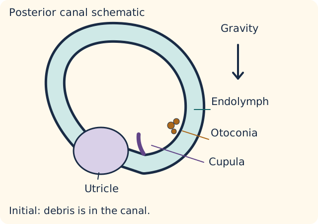
The utricle and saccule are otolith organs. Their sensory surfaces carry calcium carbonate particles called otoconia, which help detect gravity and linear acceleration. The three semicircular canals instead detect rotation. Each fluid-filled canal connects to an expanded ampulla containing sensory hair cells and a gelatinous cupula. Fluid pressure deflects the cupula, altering vestibular nerve activity. [1]
Particles in a fluid-filled canal can deflect its cupula. Settling quiets the signal; correctly directed repositioning changes particle location. Original non-scale schematic. [1]Enlarge this diagram
In canalithiasis, particles detached from the utricle are free within a canal. A change of orientation lets gravity carry them along the canal. Their interaction with endolymph, the canal fluid, deflects the cupula even after the head has stopped. The nervous system interprets the resulting imbalance between vestibular signals as rotation. The posterior canal is most commonly involved; the horizontal canal can also be affected. The affected canal matters because it determines both the eye pattern and the appropriate treatment.
Predict why an attack ends without the particles leaving the ear. When freely mobile particles settle, the fluid disturbance subsides and the false rotation signal diminishes. Settled is not the same as relocated. A later position change may mobilize the particles again. This explains how someone can feel well between attacks yet still have BPPV. A short latency and a transient response support typical posterior canal canalithiasis. They are not properties of every peripheral vestibular disorder.
Particles attached to a cupula can produce more sustained position-dependent deflection. This cupulolithiasis concept helps explain why a universal under-one-minute rule is too simple. The clinical pattern must still be interpreted by a trained examiner; persistent or atypical positional nystagmus can also have a central cause. Do not label a persistent response benign merely by invoking attached particles. [1]
Use the orientations below to predict cupula deflection. Select a discrete orientation or settling state and predict the cupula response before checking it. Clinical repositioning requires selection of the affected canal and side. Repositioning is different from simply waiting: a correctly selected sequence guides debris out of the affected canal toward the utricle, addressing the mechanical source of the signal.
Match the eye response to the tested canal
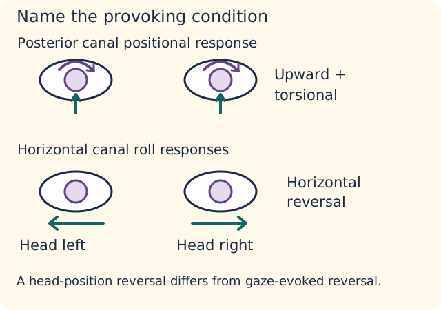
Nystagmus is rhythmic ocular drift with a corrective component; jerk nystagmus is conventionally named for its fast component. Describe its direction, time course and provoking condition. Do not combine a spontaneous vestibular neuritis examination with a positional BPPV response and call the mixture one peripheral pattern. [1][2]
Compare the axis of the response and the provoking condition. This conceptual figure does not determine an affected side. Original non-scale schematic. [1][2]Enlarge this diagram
In an appropriate history, the Dix-Hallpike examination tests for posterior canal BPPV. The examiner supports the patient and positions the head relative to gravity, observing the eyes. A compatible positive response is torsional and upbeating, usually following a brief delay and subsiding. Torsion means rotation of the visible eye landmarks, not simply horizontal motion. The upper poles rotate toward the dependent affected ear. If the initial side is negative, the opposite side is assessed when safe. [1]
If the history is compatible but Dix-Hallpike produces horizontal nystagmus or no nystagmus, assess for horizontal canal disease with a supine roll test or refer to someone who can. Horizontal canal BPPV may produce direction-changing horizontal nystagmus as the head is rolled between sides. Geotropic responses point toward the ground; apogeotropic responses point away. For a clearly asymmetric geotropic pattern, the stronger response identifies the affected ear. For an apogeotropic pattern, the ear opposite the stronger response is favored. These rules require identifying direction relative to the ground first; intensity alone can point to the wrong side. Subtype assessment and technique require expertise. [1]
Now change only the provoking condition. A patient's eyes reverse their fast direction when looking right versus left while the head stays still. That is gaze-evoked direction change, a central concern in an acute vestibular syndrome. It is different from reversal during positional roll testing. Writing merely direction-changing loses the feature that distinguishes these situations.
Before positional testing or treatment, assess neck mobility, relevant cervical or vascular disease, ability to transfer, balance and support needs. An unsafe standard position is a reason for modification by a trained clinician or referral, not forceful extension. A negative or atypical test should prompt reconsideration rather than an automatic Epley maneuver. The diagnosis depends on the history and observed response, not the patient's age or willingness to tolerate the position.
Know where HINTS belongs, and where it does not
HINTS combines head impulse, nystagmus and test of skew. It is intended for a trained examiner evaluating an acute vestibular syndrome with spontaneous nystagmus. It is not a screening checklist for all dizziness, a self-test, or a method for clearing an asymptomatic person between brief positional spells. Without spontaneous nystagmus, assessment of gait severity and the rest of the examination becomes particularly important. [2]
The head impulse tests the vestibulo-ocular reflex: the eyes should remain on a target during a brief head rotation. During a rightward head impulse, normal compensation rotates the eyes leftward relative to the head. A later refixation saccade indicates insufficient compensation for that tested direction. A corrective saccade supports impaired peripheral reflex function, but some strokes can also impair that pathway. A normal head impulse in the appropriate ongoing syndrome raises central concern. Neither result is a stand-alone discharge test. Suppression of nystagmus by visual fixation likewise supports a pattern but does not prove safety.
Central or equivocal findings require further evaluation. Examples include gaze-evoked direction change, skew deviation, or a normal head impulse in the proper syndrome. Do not require all three central findings. New hearing asymmetry, severe inability to sit or walk, diplopia, dysarthria, limb ataxia or other focal findings can alter concern even when one part of the eye examination looks peripheral. [2]
New hearing loss with continuous vertigo is not automatically labyrinthitis. Inflammation involving the labyrinth is one explanation, but ischemia affecting the inner ear or adjacent pathways is another. Assess hearing and the complete clinical picture. The absence of obvious limb weakness is not an exclusion test for posterior stroke.
Routine noncontrast head CT is a poor test for separating peripheral from central dizziness. A normal early scan does not erase a concerning examination. GRACE-3 recommends MRI/MRA for central or equivocal HINTS findings; clinical stroke assessment determines the urgent imaging and vascular pathway. If an appropriately trained examiner is unavailable, uncertainty needs a suitable evaluation, not an improvised normal HINTS label. [2]
Geotropic and apogeotropic describe the direction of a positional response relative to gravity. Neither term alone proves that its cause is peripheral. Persistent apogeotropic responses have been recorded with central cerebellar lesions; associated gaze-holding or other neurologic abnormalities therefore require an interpretation broader than a particle model. [7]
Treat the identified mechanism, then reassess
For confirmed posterior canal BPPV, offer a canalith repositioning procedure such as Epley, or referral to someone who can perform it. The procedure uses a selected sequence to guide particles toward the utricle. Horizontal canal BPPV needs a canal-specific approach; an empiric posterior maneuver for every dizzy patient ignores the diagnosis. Treatment may need repetition, and symptom resolution is not a guarantee against recurrence. [1][2]
Routine vestibular suppressants, including antihistamines or benzodiazepines, are not the treatment of BPPV. They do not relocate debris and may add sedation or fall risk. Observation with follow-up is an option in selected patients, and vestibular rehabilitation can help appropriate patients. Discuss impaired mobility, home support, fall risk and the impact of attacks on daily activities. Routine postprocedure positional restrictions are not recommended after posterior canal repositioning. [1]
Reassess within one month after initial treatment or observation. Persistent symptoms require evaluation for unresolved BPPV and other peripheral or central disorders. A new continuous syndrome, new neurologic findings or new hearing change is a changed clinical problem, not automatically a failed maneuver. Explain recurrence, safety and when to return. Continuing daily sedation without examining the new pattern substitutes symptom suppression for reassessment.
Vestibular neuritis usually presents as an acute continuous syndrome without hearing loss after alternative dangerous causes have been considered. Symptom treatment may be needed briefly, while rehabilitation supports recovery. GRACE-3 frames corticosteroids within the first three days as shared decision-making based on very low-certainty evidence. Do not promise that steroids reliably shorten the illness. [2]
Meniere disease combines recurrent spontaneous attacks with auditory symptoms such as fluctuating sensorineural hearing loss, tinnitus and fullness. Audiometry and the overall pattern matter; endolymphatic hydrops is an associated pathologic concept, not a bedside proof. Lifestyle counseling and maintenance options such as diuretics may be offered, with uncertainty about benefit explained. Vestibular migraine is supported by recurrent episodes and an appropriate migraine history or accompanying migraine features; a normal interval examination does not settle that diagnosis. Sudden transient symptoms can still warrant vascular assessment. [3][2]
Carry forward three questions: What is the time pattern? Which observation actually localizes it? What would change the plan? A positional maneuver treats a demonstrated mechanical problem; a central warning changes the urgency.
Do not transfer the strength of a drug recommendation from one peripheral nerve syndrome to another. Early corticosteroids have a stronger recommendation in appropriately assessed adult Bell palsy than in vestibular neuritis, where benefit evidence is very low certainty and shared decision-making is recommended. Diabetes or a prior severe glucose response adds a patient-specific harm consideration without making the underlying evidence identical. [2][6]
After repositioning, a changed eye-movement pattern needs new localization. A posterior-canal response can resolve while a horizontal response appears on roll testing, compatible with canal conversion. Compare the response plane, its direction relative to gravity and its intensity on each side before choosing the next target; the test pattern does not directly show the path taken by individual particles. [1]
Practice the next decision
Each scenario is an original educational example. Make a prediction before comparing the options. All options have readable explanations, and retrying carries no penalty.
Case 1
Show answer and explanations for case 1
A. Audiometry to evaluate recurrent cochlear dysfunction (Why this does not fit)
Audiometry is useful when recurrent vertigo includes auditory change. These attacks last seconds after rolling, hearing is unchanged, and the immediate unanswered question concerns a positional canal response. Hearing measurements cannot replace the missing horizontal-canal examination.
B. Head impulse, nystagmus and skew testing between attacks (Why this does not fit)
Eye-movement testing can help classify ongoing vestibular dysfunction. This patient has symptom-free intervals and no spontaneous nystagmus, so an interval HINTS examination cannot settle the positional canal question. Match a bedside test to the syndrome in which its interpretation is supported.
C. Supine roll testing of both horizontal canals (Best answer)
The brief attacks begin with head orientation while still supine, supporting a gravity-sensitive vestibular process. A negative posterior-canal test does not examine every canal, so bilateral roll testing addresses the remaining horizontal-canal possibility. Choose the next test from both the triggering event and the canal already assessed.
D. Repeat orthostatic blood pressure measurements as the main evaluation (Why this does not fit)
Getting out of bed can provoke orthostatic symptoms. Here symptoms begin before upright posture and standing pressure is unchanged, whereas the posterior-canal test leaves a different canal unexamined. Separate rolling from standing before attributing bed-related symptoms to blood pressure.
Takeaway: An unrevealing posterior-canal test can leave horizontal-canal disease untested.
A. Right posterior-canal repositioning based on the prior affected side (Why this does not fit)
The old examination identified right peripheral hypofunction and could encourage anchoring to that ear. It did not establish right posterior-canal debris, and the current right positional test is negative while the left is characteristic. A prior vestibular deficit and a new positional canal lesion need not share a side.
B. Left horizontal-canal repositioning for the newly provoked episodes (Why this does not fit)
The new attacks and current left-sided provocation support reassessing for a mechanical positional disorder. Torsional upward rather than horizontal nystagmus identifies the posterior canal, so a left horizontal procedure matches the side but not the plane. Both elements of the present examination must guide the maneuver.
C. Left posterior-canal repositioning for the newly provoked episodes (Best answer)
The new symptom-free intervals and brief lying-back attacks differ from the prior continuous vestibular syndrome. The current left torsional upward response supplies a left posterior-canal target, independent of the previously affected right vestibular system. Reclassify the present time pattern before letting an old side label determine treatment.
D. Right vestibular rehabilitation alone for the previously measured hypofunction (Why this does not fit)
Rehabilitation may remain useful for residual dysfunction from the earlier right-sided illness. The current complaint consists of newly reproducible brief left posterior-canal attacks rather than continuous spinning, so rehabilitation alone does not target the demonstrated debris mechanism. A recovery plan can need an added treatment when a different syndrome appears.
Takeaway: An old peripheral vestibular deficit does not localize a new brief positional syndrome.
A. Proceed with a standard right posterior-canal repositioning procedure (Why this does not fit)
The side and canal match the provoked nystagmus. The standard approach becomes unsuitable when further extension is explicitly unsafe, even though its mechanical target is correct. A correct diagnosis does not remove a positioning limitation.
B. Refer for an adapted right posterior-canal repositioning procedure (Best answer)
The torsional upward response with the right ear dependent localizes the demonstrated disorder to the right posterior canal. The new inability to tolerate extension changes how treatment can be delivered, favoring an adapted procedure by someone able to position her safely. Canal localization identifies the target; physical limitations determine the safe route to that target.
C. Refer for an adapted right horizontal-canal repositioning procedure (Why this does not fit)
A horizontal-canal procedure can also be adapted to physical limitations. The demonstrated response is torsional upward rather than the horizontal pattern that would justify that canal target. Match both the procedural constraint and the eye-movement plane.
D. Refer for an adapted left posterior-canal repositioning procedure (Why this does not fit)
An adapted procedure would respect the painful neck restriction. The left test is negative and the torsional response points toward the right dependent ear, so this plan targets the wrong side. Safety adaptation must preserve the observed localization.
Takeaway: Localize first, then adapt the procedure to what the patient can safely tolerate.
A. Particles returned to the utricle during the pause and remained there during the repeat test (Why this does not fit)
Return to the utricle could account for sustained relief after an effective repositioning procedure. Remaining there does not explain the renewed identical canal-specific response after simple rest, and no procedure or other evidence of removal is supplied. Use the repeat provocation to test what symptom cessation actually established.
B. A fixed cupular load persisted through the pause and explains both responses without changing (Why this does not fit)
A weighted cupula can produce a position-dependent response while gravity continues to load it. An unchanged load alone does not directly explain the observed cessation during a sustained provocative position and a later fresh finite response, making a transient-transit account a better fit. Compare the proposed mechanism with the time course at a fixed position.
C. Particles settled within the canal during the pause and remained available for another transit (Best answer)
The self-limited response during a held position fits a finite fluid disturbance as particles settle rather than an unchanging load that must keep driving the cupula. Reproduction after an untreated quiet interval supports persistence of a position-sensitive canal stimulus rather than demonstrating that the first symptom cessation removed it. A quiet interval is not the same observation as successful relocation.
D. Fixed peripheral vestibular hypofunction improved through compensation during the pause (Why this does not fit)
Compensation can reduce symptoms after a persistent peripheral vestibular loss. It is less directly supported by delayed, brief responses tied to the same specific position with comfortable intervals and reproducible provocation after only fifteen minutes. The timing and stimulus dependence favor a recurring mechanical disturbance rather than compensation for a fixed deficit.
Takeaway: Repeated provocation distinguishes a temporary quiet interval from evidence that the canal stimulus was removed.
The left-ear-down position also provokes symptoms, so it cannot be ignored. Its weaker horizontal response does not demonstrate left posterior-canal disease, which would require the appropriate torsional upward pattern. Bilateral provocation does not by itself mean bilateral posterior-canal disease.
B. Right horizontal canal (Best answer)
The fast phase points toward the ground on either side, defining a geotropic horizontal pattern. In this pattern the stronger response identifies the affected ear, making the right horizontal canal the treatment target. Apply the intensity rule only after identifying the direction relative to gravity.
C. Left horizontal canal (Why this does not fit)
A left horizontal lesion is a plausible explanation for horizontal positional nystagmus. In the supplied geotropic pattern, however, the stronger right-sided response identifies the right ear; choosing the weaker side imports the apogeotropic rule. Direction and relative intensity must be interpreted together.
D. Right posterior canal (Why this does not fit)
The right side produces the more intense attack, making a right-sided process plausible. The response is horizontal rather than torsional upward, so that side information cannot justify a posterior-canal procedure. Determine the canal before choosing its treatment.
Takeaway: In a geotropic roll pattern, the stronger side identifies the affected horizontal canal.
A. Left horizontal-canal apogeotropic pattern (Why this does not fit)
The away-from-ground directions correctly identify the response type. Choosing the left side from its greater intensity applies the geotropic side rule to an apogeotropic pattern. Interpret the direction before assigning the affected ear.
B. Right horizontal-canal geotropic pattern (Why this does not fit)
The right ear is favored by the complete apogeotropic comparison. Calling the pattern geotropic conflicts with fast phases directed away from the dependent ear in both positions. A correct side reached by the wrong pattern would misguide subtype-specific care.
C. Left horizontal-canal geotropic pattern (Why this does not fit)
The stronger left-ear-down response would support the left ear in geotropic disease. Here rightward fast phases with the left ear down point away from the ground, so the response belongs to the other pattern. Intensity without gravity-relative direction can reverse localization.
D. Right horizontal-canal apogeotropic pattern (Best answer)
The fast phase points away from the ground in both positions, identifying an apogeotropic horizontal pattern. For this pattern the affected ear is opposite the side with the stronger response, so the right side is favored. Positional direction reversal must be separated from reversal caused by changing gaze.
Takeaway: Apogeotropic and geotropic roll patterns use different intensity rules.
A. Caloric testing to quantify the suspected left peripheral deficit (Why this does not fit)
Caloric testing could characterize a left vestibular deficit suggested by the impulse. It would not resolve the supplied skew, which changes the immediate question to possible central disease. A test that measures a peripheral component cannot clear a conflicting central component.
B. Dix-Hallpike testing to identify the responsible posterior canal (Why this does not fit)
Position-sensitive symptoms often lead to posterior-canal testing. This patient remains symptomatic continuously and has spontaneous nystagmus with skew, so a brief positional canal response would not explain the complete syndrome. Select testing for the current time pattern and conflicting eye findings.
C. Noncontrast head CT to distinguish peripheral from central vertigo (Why this does not fit)
CT is useful for selected acute intracranial questions, particularly hemorrhage. It is not the recommended discriminator for this central or equivocal vestibular examination and cannot adequately settle posterior ischemia. The imaging method must answer the unresolved clinical question.
D. Brain MRI and indicated vascular imaging for central disease (Best answer)
The impulse error and unidirectional nystagmus can fit a peripheral vestibular deficit. Reproducible skew makes the combined trained examination central or equivocal despite those components, supporting MRI/MRA rather than accepting a purely peripheral interpretation. Interpret HINTS as a combined examination in the correct syndrome.
Takeaway: A peripheral-looking impulse result does not cancel skew in an ongoing vestibular syndrome.
A. Use peripheral follow-up for both because alternate cover testing shows no skew (Why this does not fit)
Absence of skew is one peripheral-looking component shared by these patients. B still has a central-concerning normal impulse in an acute continuous syndrome, so absent skew cannot justify the same peripheral pathway for both. A single reassuring component cannot replace the combined examination.
B. Prioritize central evaluation for both because rightward fast phases are a central pattern (Why this does not fit)
Both patients require a careful examination for dangerous causes. Unidirectional rightward nystagmus alone does not create the central distinction; the different impulse results do. The pattern and test context matter more than the absolute fast-phase direction.
C. Prioritize central evaluation for A; interpret B within the complete peripheral assessment (Why this does not fit)
An abnormal impulse can occur in some strokes, so A still needs clinical integration. It is B whose preserved impulse response is discordant with the usual peripheral loss pattern in this syndrome. Do not equate a visibly abnormal bedside test with the more central HINTS interpretation.
D. Prioritize central evaluation for B; interpret A within the complete peripheral assessment (Best answer)
A normal impulse during this ongoing syndrome raises central concern even when nystagmus is unidirectional and skew is absent. The leftward impulse deficit in A fits a peripheral pattern, although it is not an isolated guarantee of safety. The same negative skew result has different implications when the impulse findings differ.
Takeaway: Identical nystagmus and skew findings can lead to different interpretations when head impulses differ.
A. New left conductive loss; brain MRI with vascular assessment as clinically indicated (Why this does not fit)
The proposed central investigation can address concern about the acute syndrome. The hearing classification is incorrect because the supplied thresholds rise together without the air-bone separation expected from a conductive deficit. Interpret the audiogram independently of choosing the investigation.
B. New left sensorineural loss; repeat noncontrast head CT as the principal discriminator (Why this does not fit)
The parallel thresholds correctly identify sensorineural loss. Repeating the same limited test is not the preferred discriminator of central versus peripheral acute vestibular disease when concern persists after a normal CT, whereas MRI addresses that question more directly. A correct auditory localization does not make an insensitive exclusion strategy adequate.
C. New left sensorineural loss; brain MRI with vascular assessment as clinically indicated (Best answer)
Parallel elevation of left air and bone thresholds without a significant gap identifies sensorineural rather than middle-ear conduction loss. Abrupt combined auditory and vestibular dysfunction warrants evaluation for a vascular cause despite a peripheral-looking impulse and unrevealing noncontrast CT, with MRI and appropriate vascular assessment addressing that remaining concern. A component that looks peripheral does not establish a nonvascular cause.
D. New left conductive loss; temporal-bone CT as the principal discriminator (Why this does not fit)
Temporal-bone imaging can address a structural conduction problem when the examination supports one. Here the absent significant gap and normal otoscopy do not support that localization, and a conduction-focused scan would not resolve the central concern raised by the acute combined syndrome. The measured compartment and the unresolved clinical question should select the investigation.
Takeaway: New sensorineural auditory involvement changes how a peripheral-looking vestibular result and normal CT are interpreted.
A. CTA or MRA of the relevant head and neck vessels (Best answer)
The transient spontaneous episodes with focal speech and coordination changes raise concern for posterior circulation ischemia rather than canal provocation. A CT without hemorrhage does not exclude that mechanism, so vascular imaging addresses the remaining large-vessel question. Symptom resolution does not remove the need to investigate a possible transient ischemic event.
B. Repeat noncontrast head CT as the principal ischemia discriminator (Why this does not fit)
The first scan appropriately reports whether hemorrhage is visible. Repeating the same noncontrast method does not directly evaluate the vascular mechanism suggested by transient focal findings. Choose vascular imaging when the unresolved question is arterial disease in suspected TIA.
C. Audiometry to document a fluctuating cochlear threshold (Why this does not fit)
Audiometry can support an auditory episodic disorder. The supplied episodes instead contain focal speech and limb-coordination abnormalities with a vascular risk factor, making arterial assessment the priority. The most discriminating supplied findings determine which system needs investigation.
D. Video head-impulse testing to document a peripheral vestibular deficit (Why this does not fit)
Instrumented impulse testing can quantify vestibular reflex loss. Here the episodes have ended and included dysarthria with hand incoordination, so a peripheral measurement cannot account for the complete transient syndrome. Do not apply an acute continuous-syndrome test as the primary answer to episodic focal symptoms.
Takeaway: A normal noncontrast CT does not settle transient vertigo accompanied by focal neurologic symptoms.
A. Modified positional testing to identify the involved canal (Best answer)
The witnessed onset during lying back and bed rolling supports a gravity-dependent vestibular trigger despite the initial description of standing symptoms. Cervical restriction prevents standard positioning but does not identify the canal, so a clinician skilled in modified positional testing should establish it. Adapt the examination before choosing a canal-specific treatment.
B. A modified right posterior-canal maneuver without further testing (Why this does not fit)
A right-sided bed trigger makes right posterior-canal disease plausible. It does not establish the canal, and safe modified testing is needed before choosing that treatment. A positioning limitation justifies adaptation, not skipping localization.
C. Video head-impulse testing as the primary positional localizer (Why this does not fit)
Head-impulse testing can quantify vestibular reflex function. The patient has discrete gravity-triggered attacks without a continuous syndrome, so it does not replace positional localization of the responsible canal. Choose a safe version of the relevant test rather than a different test that answers another question.
D. Tilt-table testing to establish a primary orthostatic mechanism (Why this does not fit)
Standing symptoms can make orthostatic testing reasonable. Here pressure is stable and the witnessed attack begins with lying back, directing the main unresolved question to positional vestibular testing. A precise observed trigger outweighs the broad label of dizziness on rising.
Takeaway: Unsafe neck extension changes the examination technique, not the need to establish the canal.
A. Review routine meclizine withdrawal, balance rehabilitation and fall prevention (Best answer)
Absent positional symptoms and negative canal testing do not reproduce the former mechanical treatment target. Dose-linked sleepiness with ongoing imbalance makes routine suppression a relevant modifiable exposure while balance and fall assessment address the remaining functional problem. Treat the current phenotype without claiming that the drug must be its only cause.
B. Review meclizine dosing and repeat the prior right posterior-canal procedure (Why this does not fit)
Medication review fits the drowsiness and falls. Repeating the earlier procedure lacks a current positional or examination target, so it should not replace assessment of the persistent nonpositional imbalance. A historically correct maneuver can be poorly matched to a changed presentation.
C. Maintain scheduled meclizine and add supervised balance rehabilitation (Why this does not fit)
Rehabilitation can address persistent functional imbalance after positional symptoms resolve. Maintaining the scheduled exposure without reviewing its dose-linked sedation leaves a plausible contributor to falls unaddressed, and routine suppression is not recommended treatment for resolved canal disease. Balance care and medication review should respond to different parts of the evidence.
D. Review antihypertensive reduction and increase fluid intake as the primary strategy (Why this does not fit)
A hemodynamic contributor should be considered in an older patient who falls. The stable pressures and dose-linked sedative symptoms do not support prioritizing that mechanism over medication-related impairment and residual balance dysfunction in this case. Match the initial modifiable target to the reproduced pattern.
Takeaway: A resolved canal response and persistent nonpositional imbalance require reassessment, not automatic repetition of the old maneuver.
A. Clinician-assisted left posterior-canal repositioning with transfer support (Why this does not fit)
This delivery plan addresses the transfer risk and supplies appropriate assistance. It targets the negative side, however, while the characteristic response is on the right. A safe procedure can still be mismatched to the observed lesion.
B. Clinician-assisted right posterior-canal repositioning with transfer support (Best answer)
The right torsional upward response identifies a posterior-canal target on that side. Walker dependence and near-falls make clinician-assisted delivery with available transfer support preferable to unsupervised home positioning. A treatment plan must match both the demonstrated canal and the ability to carry out the procedure safely.
C. Clinician-assisted right horizontal-canal repositioning with transfer support (Why this does not fit)
Assisted treatment addresses the documented functional limitations and uses the correct ear. The torsional upward response identifies a posterior rather than horizontal target, so this procedure does not match the observed plane. Side, canal and practical delivery all matter in selecting causal care.
D. Unsupervised right posterior-canal repositioning at home with telephone follow-up (Why this does not fit)
The right posterior target fits the positional response. Near-falls and walker dependence make unsupervised positioning a less suitable delivery plan when hands-on assistance is available. Correct localization does not by itself establish that home performance is safe.
Takeaway: Localize the canal, then choose a delivery plan that the patient can safely complete.
A. Left horizontal canal with an apogeotropic pattern (Why this does not fit)
The direction relative to gravity correctly identifies the apogeotropic subtype. Choosing the stronger left response as the affected ear applies the geotropic side rule to the wrong pattern. A changed canal response requires both direction classification and the matching intensity rule.
B. Left horizontal canal with a geotropic pattern (Why this does not fit)
A stronger left response would favor the left ear in a geotropic roll pattern. Here rightward nystagmus with the left ear down points away from the ground, so that subtype and its side rule do not fit. The most intense response cannot be interpreted without the direction relative to gravity.
C. Right posterior canal with recurrent canalithiasis (Why this does not fit)
The right posterior canal was the original documented target. Its torsional upward response has disappeared, and the new horizontal roll pattern requires a different canal assessment rather than automatic repetition. Preserve the old evidence without allowing it to overwrite a changed present examination.
D. Right horizontal canal with an apogeotropic pattern (Best answer)
The new fast phases point away from the ground on both sides, indicating an apogeotropic horizontal pattern rather than persistence of the original posterior response. In that pattern the affected ear is favored on the side with the weaker response, making the right horizontal canal the new target. Reidentify the response subtype before applying its side rule after symptoms change.
Takeaway: After repositioning, a changed response may require a different canal and subtype assessment.
A. Early benefit is established more clearly for B; evidence for A remains very low certainty (Why this does not fit)
Both syndromes can be described as peripheral nerve dysfunction, but that description does not rank their treatment evidence. The early adult facial-palsy recommendation is more established than the vestibular-neuritis steroid recommendation, so this comparison reverses the guideline distinction. A shared mechanism label is not a shared treatment evidence base.
B. Early benefit is established to a similar degree for A and B (Why this does not fit)
The time window makes an early treatment discussion relevant to both patients. It does not erase the substantially lower certainty behind vestibular-neuritis steroids, especially when a specific adverse-effect risk must be weighed. Separate timing eligibility from the certainty of expected benefit.
C. Early benefit is established more clearly for A; evidence for B remains very low certainty (Best answer)
A has the assessed adult idiopathic facial-palsy pattern within its supported early treatment interval. B has a different peripheral nerve syndrome for which early corticosteroids are a shared-decision option with very low-certainty evidence, and the common glucose risk does not make those evidence bases equivalent. Identify the syndrome before transferring the strength of a treatment recommendation.
D. Early benefit is very low certainty to a similar degree for A and B (Why this does not fit)
Diabetes makes individual risk management important for both patients. It does not change the underlying adult facial-palsy recommendation into the same very-low-certainty category as the vestibular-neuritis evidence. Patient-specific harm and certainty of benefit are distinct dimensions of the decision.
Takeaway: The strength of steroid evidence is syndrome-specific even when timing and metabolic risk are similar.
A. An auditory episodic-disorder plan centered on scheduled daily meclizine maintenance (Why this does not fit)
The proposed disease pattern fits the audiometry and episode duration. Scheduled daily suppression is not the recommended maintenance role of meclizine in this setting; the guideline limits suppressant use to a brief course during attacks. A fitting diagnosis does not establish that an acute symptom drug is a preventive treatment.
B. Auditory episodic-disorder care: lifestyle advice and optional diuretic maintenance (Best answer)
The spontaneous duration and fluctuating sensorineural auditory findings support a Meniere-type phenotype more specifically than a mechanical canal response. For recurrence planning, lifestyle education and an optional maintenance treatment fit the guideline distinction from brief suppression during an attack, while migraine can still be assessed rather than categorically excluded. Match the evidence to both the phenotype and the treatment phase.
C. A migraine-associated-disorder plan centered on migraine preventive medication (Why this does not fit)
Migraine history makes migraine-associated vertigo worth assessing and does not preclude coexistence. These particular attacks repeatedly track unilateral auditory symptoms and sensorineural fluctuation without their own migraine features, giving an auditory-directed plan stronger initial support. Avoid treating a remote history as a complete account of the current attack pattern.
D. A positional canal-disorder plan centered on repeated posterior-canal repositioning (Why this does not fit)
A recurring vestibular complaint can prompt consideration of canal disease. The spontaneous prolonged attacks, auditory fluctuation and negative positional tests do not demonstrate a posterior-canal target, so repeating that procedure would not address the documented recurrence pattern. Select a preventive or mechanical intervention from the mechanism actually supported.
Takeaway: Attack phenotype and treatment phase both matter when distinguishing maintenance options from short-term symptom suppression.
A. Migraine-associated episodic vertigo; current left posterior-canal positional disease (Why this does not fit)
The migraine-associated interpretation fits the older episodes. The current torsional upward response is provoked with the right ear down and beats toward that ear, supporting the right rather than the left posterior canal. Correctly interpreting the longer syndrome does not determine the side of the new mechanical one.
B. Migraine-associated episodic vertigo; current right posterior-canal positional disease (Best answer)
The older spontaneous attacks have a migraine-associated duration and repeated accompanying migraine features without the supplied auditory pattern of Meniere disease. The new brief provoked response independently identifies a right posterior-canal target, so that mechanical component should not be used to explain every older episode. Separate objectively different symptom patterns before assigning their treatment targets.
C. Meniere-type episodic vertigo; current right posterior-canal positional disease (Why this does not fit)
The right posterior-canal assignment fits the newly observed response. The older attacks have migraine features but no reported aural symptoms or audiometric fluctuation, which supports a migraine-associated explanation more strongly than a Meniere-type syndrome in this comparison. A positive canal test does not supply missing auditory evidence for another disorder.
D. Migraine-associated episodic vertigo; current right horizontal-canal positional disease (Why this does not fit)
The older episode pattern supports migraine assessment. The current upward torsional response is characteristic of posterior rather than horizontal-canal involvement, so the proposed mechanical target does not fit the eye movement. Infer the canal from the response plane rather than from the word rolling alone.
Takeaway: An older episodic syndrome and a newly documented canal response can require different, concurrently relevant plans.
A. Urgent central evaluation guided by severe truncal and gait instability (Best answer)
The absence of spontaneous nystagmus means the note is not a valid reassuring HINTS assessment, especially without examiner training. Severe inability to sit or stand independently then becomes a major finding directing central evaluation despite full limb strength. Use the available syndrome-appropriate evidence rather than treating an inapplicable test as negative.
B. Routine medication review based on preserved limb strength (Why this does not fit)
Medication review can contribute to an imbalance evaluation. Full limb strength does not account for abrupt severe truncal and gait dysfunction, so it cannot justify a routine disposition here. Posterior neurologic dysfunction may be prominent in coordination and posture rather than strength.
C. Peripheral vestibular follow-up based on the recorded normal HINTS result (Why this does not fit)
A valid trained peripheral examination may support a peripheral pathway in the appropriate syndrome. This note lacks both the required context and training, and the severe postural instability remains unexplained. A label in a chart is weaker evidence than the conditions under which the examination was performed.
D. Positional-clinic follow-up based on worsening when standing (Why this does not fit)
Standing may worsen symptoms from several vestibular disorders. This patient is persistently dizzy even before standing and cannot maintain posture, rather than having brief discrete gravity-triggered spells. Distinguish a functional consequence from the event that initiates an attack.
Takeaway: When HINTS is inapplicable, severe postural instability remains clinically informative.
A. A current right horizontal geotropic pattern, compatible with conversion from the treated posterior canal (Best answer)
The new roll responses beat toward the ground on both sides and are stronger with the right ear down, favoring the right horizontal canal. Comparing that pattern with the prior posterior response and initially negative roll test makes post-treatment canal conversion a coherent explanation rather than simple persistence of the old target. The observed response localizes treatment while the particle trajectory remains an inference.
B. A current left horizontal geotropic pattern, compatible with conversion from the treated posterior canal (Why this does not fit)
The horizontal ground-beating responses identify a geotropic pattern. For that pattern the stronger right-ear-down response favors the right rather than the left ear, so the proposed side does not follow the measured asymmetry. Apply the side rule to the current response type before interpreting its relation to treatment.
C. A current right posterior pattern, compatible with persistence despite treatment (Why this does not fit)
The prior examination provided a right posterior target. Its torsional upward response has disappeared and the new finding is horizontal on roll testing, so persistence of the original pattern does not explain the changed plane. Reassess the involved canal rather than preserve the prior target by default.
D. A current right horizontal apogeotropic pattern, compatible with conversion after treatment (Why this does not fit)
A horizontal-canal conversion is possible after a posterior-canal procedure. The new fast phases point toward the ground rather than away from it, so calling the response apogeotropic applies the wrong subtype and side framework. Identify the present direction before choosing how to address the changed canal.
Takeaway: After a maneuver, a new response plane can require a different canal target even on the same side.
A. Weak right forehead and right mouth (Best answer)
The conjugate right gaze deficit with ipsilateral coordination findings localizes the new syndrome to right pontine circuitry rather than an isolated canal or eye-muscle nerve. The nearby facial fascicle is the final motor output to the ipsilateral upper and lower face, so its interruption predicts weakness of both right regions. Localize the accompanying systems before applying the facial output pattern.
B. Preserved right forehead with weak right mouth (Why this does not fit)
Relative upper-face preservation is the familiar pattern of a contralateral supranuclear lesion. The supplied conjugate gaze and ipsilateral coordination syndrome instead places the hypothesized fascicular extension in the right pons, after cortical inputs converge. A facial pattern must follow the level identified by the other findings.
C. Weak left forehead and left mouth (Why this does not fit)
Whole-face weakness is compatible with interruption of a facial final-output pathway. A right pontine facial fascicle supplies the right face, however, so this option reverses the predicted side. Brainstem motor output laterality differs from the usual contralateral cortical prediction.
D. Preserved left forehead with weak left mouth (Why this does not fit)
Left lower-face weakness with relative forehead preservation would suggest a right supranuclear input lesion. A right facial fascicle in the focal region suggested here is instead an ipsilateral final pathway to both upper and lower face. Do not substitute a cortical crossing rule for a brainstem fascicular lesion.
Takeaway: A pontine facial fascicle lesion can weaken the ipsilateral whole face despite a central cause.
A. Sleep comfortably without routine upright restrictions (Best answer)
The demonstrated posterior-canal response has resolved after treatment. Routine postprocedure positional restrictions are not recommended, so obligatory chair sleeping adds discomfort without an established procedural requirement. Separate evidence-based follow-up from customary restrictions.
B. Avoid the right side until a month of negative positional tests (Why this does not fit)
Avoiding the previously affected side might seem to prevent recurrence. The current response is negative and routine side restrictions are not recommended after posterior-canal repositioning. Follow-up for recurrence does not require prolonged avoidance of comfortable sleep positions.
C. Use a cervical collar at night to limit positional changes (Why this does not fit)
A collar could restrict head orientation during sleep. There is no separate indication in this patient and no routine postprocedure requirement for immobilization, so it adds burden without an established benefit. Treatment of displaced debris does not imply a need for continuing neck restraint.
D. Sleep upright for a week to preserve the effect of repositioning (Why this does not fit)
Upright sleeping has historically been used after repositioning. Routine restrictions are not supported for this setting, and this patient has a concrete discomfort cost from chair sleeping. A plausible mechanical story is not sufficient evidence for a mandatory restriction.
Takeaway: Routine upright sleeping or side avoidance is not required after posterior-canal repositioning.
A. Geotropic positional response; a central process could account for both abnormalities (Why this does not fit)
The gaze-holding abnormality appropriately leaves a central explanation in consideration. The positional classification is incorrect because the fast phases point away from rather than toward the ground in the two ear-down positions. A reasonable etiologic interpretation does not correct a misread response direction.
B. Apogeotropic positional response; the roll test establishes a separate peripheral cupular disorder (Why this does not fit)
The ground-opposed fast phases correctly identify an apogeotropic response. That pattern can arise centrally as well as peripherally, particularly when additional central ocular-motor findings coexist, so it cannot establish a separate peripheral disease by itself. Compatibility with cupular loading is weaker than proof of that anatomical cause.
C. Geotropic positional response; the roll test establishes a separate peripheral cupular disorder (Why this does not fit)
The fast phases are ground-opposed, which contradicts the proposed geotropic classification. Even a correctly classified positional response would not by itself establish a peripheral cause in the presence of the separate gaze-holding abnormality, because central positional responses are documented. Avoid combining a direction error with unwarranted certainty about origin.
D. Apogeotropic positional response; a central process could account for both abnormalities (Best answer)
On each side the roll-test fast phase beats away from the ground, establishing an apogeotropic response rather than a geotropic one. The separate sustained gaze reversal with the head fixed raises central concern, and a central process can also produce persistent apogeotropic positional nystagmus, so the first test does not prove a peripheral cupular cause. Describe the response before inferring its anatomical origin.
Takeaway: Apogeotropic describes a response direction, not proof of a peripheral cause; associated gaze findings change the interpretation.
A. Compensatory pulse acceleration is blunted; assess volume status and the recent diuretic change (Why this does not fit)
Assessing the new medication exposure and volume status is appropriate. A rise from sixty-four to the low nineties during the pressure fall is not a blunted or absent pulse response, so that physiologic label does not fit the measurements. Preserve the distinction between the observed reflex response and the management target.
B. Compensatory pulse acceleration is present; assess volume status and the recent diuretic change (Best answer)
The sustained pressure fall with symptoms supports orthostatic hypotension, while the substantial pulse rise shows that heart-rate compensation is not absent in this observation. The recent diuretic increase supplies a plausible modifiable volume or medication contributor to assess before attributing the problem primarily to neurogenic failure or adding pressor therapy. Interpret the pressure response and the contributor rather than automatically stopping all blood-pressure treatment.
C. Compensatory pulse acceleration is present; prioritize pressor treatment for primary neurogenic failure (Why this does not fit)
The pulse does rise substantially with the symptomatic pressure fall. That response and the new diuretic exposure support investigating reversible contributors before selecting primary neurogenic failure as the leading mechanism and adding a pressor, although coexistence is not excluded by one observation. A demonstrated compensatory response should inform the initial ranking of causes.
D. Compensatory pulse acceleration is blunted; prioritize pressor treatment for primary neurogenic failure (Why this does not fit)
The pulse measurements do not show the blunted response asserted in this option. The temporal medication change also remains an unassessed contributor, so prioritizing primary neurogenic failure and pressor treatment does not best follow the current evidence. Integrate measured physiology with chronology before choosing a new drug target.
Takeaway: An orthostatic pressure fall, its pulse response and a recent medication change answer different clinical questions.
A. Stop meclizine and use left posterior-canal repositioning (Why this does not fit)
Reviewing the sedating medication addresses one supplied problem. The left positional test is negative while the right response is characteristic, so this maneuver chooses the wrong target. A plan must match both medication effects and current localization.
B. Increase meclizine while retaining observation as the main plan (Why this does not fit)
The drug has reduced the perceived spinning. The objective canal response persists and daytime sleepiness is increasing, so a higher dose intensifies the burden without addressing the demonstrated cause. Subjective relief does not prove mechanical resolution.
C. Retain meclizine and use right horizontal-canal repositioning (Why this does not fit)
A right-sided mechanical disorder is supported. Its torsional upward response identifies the posterior rather than horizontal canal, and retaining routine sedation does not address the increasing sleepiness. Correct side alone is insufficient for a well-matched plan.
D. Right posterior-canal repositioning; review daily meclizine (Best answer)
The persistent side-specific torsional upward response demonstrates a right posterior-canal target despite reduced subjective spinning. Daily sleepiness identifies a cost of suppression, which has not removed the debris and is not routine BPPV treatment. Treat the mechanical source and reassess the symptomatic drug separately.
Takeaway: Less perceived spinning can coexist with persistent canal disease and medication-related impairment.
A. Left horizontal-canal disease identified by positional direction reversal (Why this does not fit)
Horizontal-canal disease can produce direction-changing nystagmus during roll testing. Here the head stays fixed and only gaze changes, so the provoking variable is incompatible with that positional interpretation. Document what changed, not just that the fast phase reversed.
B. A purely left peripheral deficit confirmed by the impulse result (Why this does not fit)
Leftward impulse refixation supports impaired left vestibular reflex function. It cannot account for or neutralize the new gaze-evoked reversal, so the earlier component does not establish a purely peripheral conclusion. Integrate new discordant findings into the current assessment.
C. A central or equivocal combined examination requiring central assessment (Best answer)
The initial impulse deficit could fit peripheral vestibular dysfunction. Reversal with gaze while head orientation is fixed adds a central-concerning pattern, unlike reversal caused by rolling between gravity positions. New examination evidence should revise the combined interpretation rather than be canceled by the earlier impulse.
D. Right posterior-canal disease identified by the initial rightward fast phase (Why this does not fit)
A right-sided positional response can indicate right canal disease when its plane and stimulus fit. Primary-gaze rightward nystagmus during continuous symptoms is not a torsional upward Dix-Hallpike response, and the later gaze reversal adds another mismatch. Absolute fast-phase direction does not select a posterior canal.
Takeaway: Reversal with gaze and reversal with gravity are different observations with different implications.