Cranial Parasympathetic Techniques: Anatomy, Contact and Clinical Judgment
Trace cranial parasympathetic pathways, compare five cranial techniques, and use anatomy, safety findings and clinical evidence to guide decisions.
A dry eye, a stiff suboccipital region and a sudden severe headache can all bring attention to the head. They do not call for the same response. First identify the nerve route, then the tissue a technique actually contacts, then whether treatment is appropriate. By the end, you should be able to distinguish the five main approaches without treating a traditional explanation as a proven clinical effect.
These descriptions support supervised osteopathic education, not self-treatment. Consent, clinical assessment and a comfortable supported position precede contact. Stop for pain, neurological symptoms or withdrawal of consent.
Trace the nerve before choosing a technique
Does a gland receive its parasympathetic supply from the nerve nearest your fingers? Not necessarily. A preganglionic neuron starts in the central nervous system and synapses in a ganglion; a postganglionic neuron reaches the organ. Both use acetylcholine, but ganglionic receptors are nicotinic and target-organ receptors are muscarinic. Cranial nerves III, VII, IX and X carry cranial preganglionic outflow. Trigeminal branches can carry passengers from these routes without being their central origin. [1]
Origin, peripheral relay and destination
Outflow
Route
Destination
OutflowIII
RouteEdinger-Westphal nucleus; ciliary ganglion; short ciliary nerves
DestinationIris sphincter and ciliary muscle: pupil constriction and near accommodation
OutflowVII
RouteSuperior salivatory region; greater petrosal nerve to pterygopalatine ganglion, or chorda tympani to submandibular ganglion
DestinationTear, nasal and palatal glands through the first route; submandibular and sublingual glands through the second
RouteDorsal vagal nucleus and nucleus ambiguus; terminal or intramural ganglia
DestinationThoracic and much abdominal viscera; cardiac parasympathetic control importantly involves nucleus ambiguus
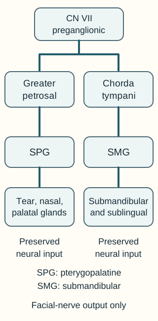
The geniculate ganglion contains sensory cell bodies; it is not the parasympathetic synapse for tear or salivary secretion. Greater petrosal fibers reach the pterygopalatine ganglion, also called the sphenopalatine ganglion or SPG. Its secretomotor fibers subsequently accompany trigeminal branches. Chorda tympani joins the lingual branch of V3 before its fibers synapse in the submandibular ganglion. Facial motor branches passing through the parotid do not provide that gland's secretomotor supply. [3][4][5][11]
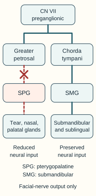
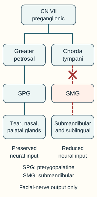
Trace the interrupted branch and identify a gland group that remains supplied. The drawing isolates parasympathetic input, not total gland output. Open full-size diagram
Predict a spared function. Follow one branch from VII to its glands. Before opening either comparison, identify which other gland group should retain its parasympathetic input.
Interrupt the greater petrosal routeTrace the interrupted branch and identify a gland group that remains supplied. The drawing isolates parasympathetic input, not total gland output. Open full-size diagram
The interrupted route no longer delivers its normal preganglionic input to the SPG. Chorda tympani remains connected to the submandibular ganglion.
Interrupt the chorda tympani routeTrace the interrupted branch and identify a gland group that remains supplied. The drawing isolates parasympathetic input, not total gland output. Open full-size diagram
The interrupted route reduces the neural input to the submandibular ganglion. The greater petrosal route remains connected to the SPG.
Worked comparison: an isolated greater petrosal injury can reduce tearing while sparing chorda-mediated salivation and anterior tongue taste. An isolated chorda injury can reduce that salivation and taste while sparing tearing. Close a comparison to return to the baseline, or open both to compare. These drawings isolate neural input; they do not predict a measured secretion volume. [3][4]
Now transfer the route: preserved facial strength does not exclude a secretomotor branch injury. Conversely, weakness after injury distal to the stylomastoid foramen can spare the earlier branches. In the abdomen, vagal supply reaches the midgut, conventionally through the proximal two-thirds of the transverse colon. The distal third and hindgut receive pelvic splanchnic outflow from S2-S4. Constipation therefore cannot be assigned to the vagus alone. [3][6]
Try it here · Checkpoint 1 of 3
Make your prediction before reading the choices. A first attempt is just a starting point.
Case 2
Show answer and explanations for case 2
A. Reduced submandibular secretion; preserved anterior tongue touch (Best answer)
Chorda tympani crosses the middle ear and carries anterior tongue taste plus salivary parasympathetic fibers. The surgical site and taste loss localize this branch before it joins the lingual nerve, so submandibular secretion falls while lingual general sensation remains. Localize the injury before deciding which functions of a shared carrier are affected.
Reasoning steps for option A
What does anterior tongue taste loss after injury to a nerve crossing the tympanic cavity localize?
It localizes chorda tympani before its junction with the lingual nerve, consistent with the middle-ear surgical site.
Why should submandibular secretion fall while anterior tongue touch remains after this injury?
Chorda carries salivary preganglionic fibers as well as taste, but tongue touch remains in the separate lingual nerve at this site.
B. Reduced parotid secretion; preserved anterior tongue touch (Why this does not fit)
Parotid secretion depends on IX, the lesser petrosal nerve and the otic ganglion. That pathway does not account for the new anterior tongue taste loss after injury to a nerve crossing the tympanic cavity. Different salivary glands do not share one secretomotor route.
Reasoning steps for option B
Would loss of the IX-to-otic salivary pathway explain anterior tongue taste loss after middle-ear surgery?
No. That pathway controls parotid secretion, whereas the lost anterior tongue taste localizes the injured branch to chorda tympani.
Which half of the reduced-parotid, preserved-touch prediction fits this surgical site?
Preserved touch fits because lingual sensory fibers are separate in the middle ear. The gland prediction does not: chorda injury reduces submandibular rather than parotid input.
C. Reduced submandibular secretion; reduced anterior tongue touch (Why this does not fit)
A lingual nerve injury after chorda joins it can impair taste, salivary output and general sensation together. The injury is within the middle ear rather than the floor of the mouth, separating chorda from the lingual sensory fibers. The site of convergence determines whether a lesion affects passengers and carrier together.
Reasoning steps for option C
Where would one injury need to occur to reduce anterior taste, tongue touch and submandibular input together?
A lingual nerve injury after chorda tympani joins it could affect all three functions carried together there.
Why does the documented tympanic injury not support reduced anterior tongue touch?
The lesion occurs before chorda joins the lingual nerve, with no floor-of-mouth injury; lingual general-sensory fibers therefore remain separate from the injured branch.
D. Preserved submandibular secretion; reduced anterior tongue touch (Why this does not fit)
An isolated general-sensory lingual injury could reduce tongue touch. It does not explain interruption of chorda-mediated taste at the documented middle-ear site while sparing its salivary fibers. Predict downstream secretion from the branch carrying the lost taste modality.
Reasoning steps for option D
Which isolated nerve function would explain reduced anterior tongue touch without a salivary deficit?
A general-sensory lingual injury could impair touch without interrupting the separate salivary fibers at an upstream site.
Why does preserved submandibular secretion conflict with the documented taste-bearing middle-ear nerve injury?
The injured chorda tympani carries both anterior taste and salivary preganglionic input, so its loss predicts reduced submandibular secretion rather than an isolated touch deficit.
Takeaway: Middle-ear chorda injury reduces anterior taste and submandibular/sublingual output while sparing lingual touch.
Can a surface contact establish that a nerve is trapped in a foramen? It cannot. Use the skull to localize anatomy, then use the neurological examination to assess function. The sphenoid contains the superior orbital fissure for III, IV, V1 and VI, the foramen rotundum for V2, and the foramen ovale for V3. The temporal bone contains the internal acoustic meatus for VII and VIII and the facial canal. [2]
The jugular foramen lies between temporal and occipital bone and transmits IX, X and XI. The hypoglossal canal is in the occipital bone and transmits XII. The foramen magnum transmits the brainstem-spinal cord junction, vertebral arteries and ascending spinal accessory roots. Olfactory fibers traverse the ethmoid cribriform plate. Neither frontal nor parietal surface tenderness proves compression of these deep structures. [2]
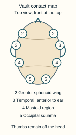
Vault hold: compare paired contacts
Locate each contact on a model before interpreting a palpated pattern. The dots indicate regions, not force or a nerve being pressed. Open full-size diagram
With the patient supine and the head supported, the classic vault hold places index fingers on the greater sphenoid wings, middle fingers on the temporal regions anterior to the ears, ring fingers at the mastoids and little fingers on the occipital squama. Thumbs remain off the head. Use light contact to compare perceived amplitude, symmetry and rhythm rather than squeezing until a pattern appears. [9]
The traditional primary respiratory mechanism links proposed intrinsic nervous-system motion, cerebrospinal-fluid fluctuation, dural tension, cranial articular motion and involuntary sacral motion. The palpated cranial rhythmic impulse, or CRI, is part of that framework, not an established measurement of intracranial pressure, cerebrospinal-fluid production or vagal discharge. Teaching ranges such as 10-14 cycles/minute vary between traditions. A value of 12 alone cannot establish normal neurological function. Published examiner agreement is limited. [10][19]
The sphenobasilar synchondrosis, abbreviated SBS in traditional terminology, supplies a language for perceived relationships. In the flexion-extension description, sphenoid and occiput rotate oppositely around transverse axes. Torsion describes opposite rotation around an anterior-posterior axis and is named for the higher greater sphenoid wing. Right index higher with left little finger higher fits the traditional right-torsion pattern. Sidebending-rotation instead combines vertical-axis sidebending with same-direction rotation around an anterior-posterior axis. Lateral strain instead describes same-direction rotation around vertical axes. A compression label describes limited perceived compliance, not proven brainstem compression. [19]
V-spread: locate the receiving hand
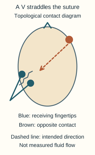
Distinguish the receiving fingers around the target suture from the opposite directing hand. Open full-size diagram
Place the receiving fingertips on opposite sides of the selected suture; the second contact is diagonally across the skull and directed toward the receiving region. For an occipitomastoid target, the V belongs at that target, not at the opposite hand. Traditional descriptions invoke fluid direction and a perceived softening or pulsation. These are not measurements of fluid transport or proof that the suture was mechanically widened. Never pry the skull or increase force to obtain a release. Reassess comfort and the original finding. [19]
Use the drawings. On a skull model, identify the ring-finger contact, then put a paper marker on a different suture and relocate the receiving V. The first remains the mastoid; the second changes with the target. For a new patient with V2 sensory loss, localize the foramen rotundum and investigate the deficit rather than inferring its cause from a tender suture.
Two very different occipital contacts
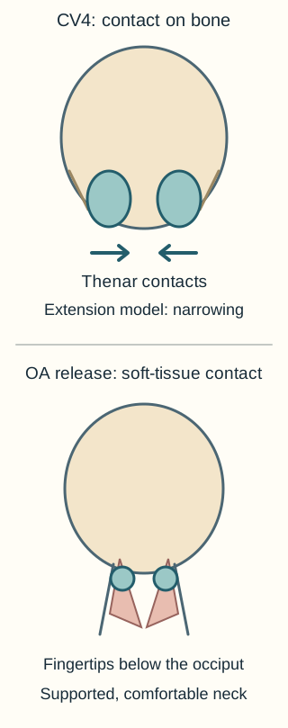
Both CV4 and suboccipital release begin near the back of the head. Identify the contact surface before naming either technique. One uses the broad thenar regions on the occipital squama; the other supports soft tissues below the occiput. Neither reaches the brainstem or vagus directly. [8][12]
Use the surface and tissue being contacted to distinguish CV4 from suboccipital release. Arrows show a teaching model rather than a force prescription. Open full-size diagram
CV4: favor extension, not flexion
For the thenar-contact variant of the traditional compression of the fourth ventricle technique described in the cited written source, support the supine patient's occipital squama on the thenar contacts, medial to the occipitomastoid sutures. Follow perceived cranial extension and gently resist the return toward flexion. In this terminology, extension accompanies relative narrowing and flexion relative widening. This does not mean extending the patient's neck, timing pressure to each breath, or compressing an actual ventricle. [8][19]
A perceived reduction and pause in the rhythm is called a still point. Maintain gentle contact rather than adding force. When the perceived rhythm resumes, release gradually and reassess the original complaint and examination. A still point is not cessation of cerebrospinal-fluid flow. Breathing, pulse and awareness should continue normally; altered consciousness or respiratory distress is not a therapeutic endpoint. The fourth ventricular floor lies on the dorsal pons and medulla, deep to the occipital bone. [7][8]
Use the ventricular floor to localize a neurological pattern, not to describe a superficial hand contact. The facial colliculus lies on its pontine portion, where VII motor fibers pass around the abducens nucleus. Loss of conjugate horizontal gaze to one side together with whole-face weakness on that side can therefore indicate a pontine lesion. An isolated peripheral VI lesion impairs abduction of one eye rather than gaze of both eyes toward that side. These are findings for medical assessment, not a cranial strain diagnosis. [23]
A reported still point also needs a measurement question: do different examiners agree about the same interval, and does the palpated event correspond to an independent physiological measure? Repeated reports by one examiner address repeatability, not proof that cerebrospinal-fluid flow or pressure changed. [10]
OA release: support the suboccipital tissues
The occipitoatlantal joint, or OA, is the occiput-C1 articulation. Its principal motion is flexion-extension, with opposite coupling of sidebending and rotation in the conventional osteopathic description. The atlantoaxial joint, or AA, is C1-C2 and contributes much upper-cervical rotation. Do not substitute one level for the other when interpreting a restriction. [12][19]
After assessing cervical safety, support the head in a comfortable near-neutral position. Place finger pads in the suboccipital muscular region below the occiput, using gentle sustained support or traction until tissue resistance softens. Avoid a thrust, painful end range or forceful rotation. Reassess local tenderness and comfortable range. This can address a coexisting musculoskeletal finding; it does not demonstrate that the jugular foramen enlarged or that a visceral disorder was caused by vagal compression. [12]
Compare and predict. Point to each contact on the drawing, then identify which one belongs to the traditional narrowing phase. CV4 uses the broad bony contact and favors extension; the suboccipital approach uses supported soft-tissue contact. Transfer to a patient whose discomfort increases when the head is unsupported: improve support and reassess rather than applying the CV4 direction to the neck.
Try it here · Checkpoint 2 of 3
Make your prediction before reading the choices. A first attempt is just a starting point.
Case 10
Show answer and explanations for case 10
A. Suboccipital release; follow widening and resist narrowing (Why this does not fit)
Suboccipital release is directed at supported soft tissues beneath the occiput. Broad contacts on the occipital squama identify a different setup, and following widening reverses the extension-directed convention being practiced. Identify the contacted tissue before interpreting a motion instruction.
Reasoning steps for option A
Do broad thenar contacts on the occipital squama match the soft-tissue target of suboccipital release?
No. Suboccipital release supports soft tissues below the occiput with finger pads; the described broad bony contacts identify the CV4 setup.
Would following widening and resisting narrowing correct the trainee's phase error?
No. It repeats the reversed phases. The cited CV4 convention follows relative narrowing and gently resists widening without extending the neck.
B. Suboccipital release; follow narrowing and resist widening (Why this does not fit)
Following narrowing is consistent with the traditional CV4 phase convention. The described bony thenar contacts are not the fingerpad soft-tissue support of suboccipital release. A correct direction does not establish the identity of the technique.
Reasoning steps for option B
Which part of the suboccipital-release and narrowing instruction matches the intended cranial phase?
Following narrowing and resisting widening matches the traditional extension-directed CV4 phase, so the direction is appropriate for that convention.
Why does the technique label remain incorrect despite the right phase instruction?
The trainee uses thenar contacts on the occipital squama, not fingerpad support beneath the occiput; the contact identifies CV4 rather than suboccipital release.
C. CV4; follow narrowing and resist widening (Best answer)
Broad thenar support on the occipital squama matches the cited CV4 procedure. Its traditional extension-directed phase follows relative narrowing and gently resists the return toward widening, so the trainee has reversed the phases. Cranial extension terminology does not require cervical extension.
Reasoning steps for option C
What identifies CV4 before interpreting whether the trainee follows the correct motion?
Broad thenar support on the occipital squama medial to the occipitomastoid sutures distinguishes CV4 from suboccipital soft-tissue support.
How should the widening-following instruction change within the cited CV4 convention?
Follow perceived narrowing and gently resist the return toward widening, favoring traditional cranial extension while keeping the neck comfortably supported.
D. CV4; follow widening and resist narrowing (Why this does not fit)
The contact identifies CV4 rather than the suboccipital soft-tissue approach. The proposed phase instruction repeats the trainee error instead of favoring relative cranial narrowing. Check contact and phase separately rather than accepting the correct technique name alone.
Reasoning steps for option D
Why is the CV4 name correct for these thenar contacts even though the full option is wrong?
The occipital squama contact matches CV4, so the error lies in the phase instruction rather than the name.
What happens to the trainee's mistake if widening is still followed and narrowing resisted?
The original reversal remains: widening corresponds to traditional flexion, while the cited procedure favors extension by following narrowing.
Takeaway: Identify CV4 from its bony contact, then favor traditional cranial narrowing rather than widening.
A deep target does not make every approach equivalent
Does tearing during oral pressure mean a compressed ganglion has been corrected? Tearing is an observation. It does not establish the proposed cause, the structure directly contacted or a lasting treatment effect. The SPG lies in the pterygopalatine fossa behind the maxilla, not on an exposed oral surface. Return to the VII branching diagram: this is the tear and nasal route, not the parotid relay. [11]
SPG-region treatment
A published intraoral osteopathic approach uses a gloved fingertip along the posterior-lateral maxilla toward the lateral pterygoid plate region, followed by gentle superior contact. The clinician contacts overlying tissues, not the ganglion itself. Oral inspection, explicit consent, gag and bite precautions, and stopping for sharp pain are essential. Defer local contact through infected or injured tissue or a recently operated region until appropriately assessed. Do not force deeper contact to produce tears. [11][12]
Manual SPG-region treatment, an extraoral variation and an intranasal local-anesthetic SPG block are different interventions. Their contact sites and active components differ. A result from an anesthetic-block trial cannot be transferred to manual treatment simply because both names contain SPG. Likewise, increased secretion after contact does not by itself establish relief of inflammatory rhinitis, sinus obstruction or sleep apnea. [11][12]
Keep the intervention attached to its outcome
Manual study: a small randomized crossover proof-of-concept study tested awake upper-airway collapsibility after an SPG-region intervention. That physiological endpoint is not a demonstrated reduction in sleep-apnea events.
Sinusitis pilot: participants received multiple osteopathic techniques and reported symptoms afterward. Without a concurrent control or an isolated SPG intervention, the study cannot establish how much change was caused by the SPG component. [11][12]
Change one feature. Compare the same patient receiving manual contact versus local anesthetic. Name what differs before considering efficacy: pressure and tissue contact are not pharmacological conduction block. Now transfer to an oral ulcer at the intended contact: choose a different care plan rather than treating the technique name as permission to proceed.
Related contacts have different targets
Frontal and parietal lifts address their named calvarial regions within the traditional framework. Temporal rocking assesses or encourages a temporal-bone pattern; an ear-pull variation uses a different contact and is not an interchangeable procedure. Occipitomastoid V-spread targets its suture region. Venous sinus drainage describes a sequence of cranial contacts, not established treatment for thrombosis or intracranial hypertension. [18][19]
Galbreath treatment uses gentle mandibular traction in a traditional middle-ear drainage approach; it is not SPG treatment. The ethmoid lies deep at the nasal roof and medial orbital region. Its cribriform plate transmits olfactory fibers, but a frontal, nasal or maxillary contact does not directly palpate that plate. Map the bone and intended tissue before assigning a technique to a symptom. [2][19]
Decide whether contact should happen at all
A gentle technique can still be the wrong decision if it delays assessment. A new thunderclap headache, altered awareness, focal deficit, suspected hemorrhage, acute skull fracture or suspected raised intracranial pressure requires immediate emergency assessment rather than a trial of cranial treatment. Arrange evaluation now; do not wait for a routine appointment or for symptoms to recur. Improvement in pain does not retrospectively establish a benign cause. [14][15]
New or worsening neurological features: stop cranial/cervical contact and arrange immediate emergency assessment. Maintain comfortable head and neck support without provocative testing.
Possible cervical instability: preserve comfortable support and obtain an appropriate evaluation before cranial or cervical treatment.
Stable, evaluated musculoskeletal findings: discuss an adjunctive technique, its uncertainty and a measurable symptom or functional goal.
A negative head CT describes one investigation at one time. Persistent symptoms after trauma need medical reassessment; worsening headache with repeated vomiting or new drowsiness requires emergency assessment now, with urgent repeat neuroimaging as clinically indicated. An unevaluated headache that reached maximum intensity within seconds also requires immediate, same-day emergency assessment, even after the pain resolves and the neurological examination is normal.
For a sudden severe nontraumatic headache, decisions about further testing after CT depend on timing, scan quality, examination and residual risk; do not teach an automatic CT-then-lumbar-puncture schedule for every patient. Neither an earlier negative scan nor four symptom-free days independently clears a new presentation. [14][15]
Rheumatoid arthritis, Down syndrome and congenital cervical abnormalities such as Klippel-Feil syndrome warrant attention to stability; the diagnosis alone is not a radiographic finding. Known instability, new neck pain, weakness, gait change, hyperreflexia or bowel/bladder change takes precedence over choosing a technique. CV4 is not an automatic safe substitute for OA treatment while instability remains unresolved.
The AAP does not recommend routine cervical screening radiographs in asymptomatic children with Down syndrome; old normal films do not exclude a later problem. New gait deterioration, impaired hand function and bladder changes raise concern for cervical myelopathy. For that combined progressive presentation, defer cranial/cervical contact and arrange emergency assessment now, with comfortable neck support and clinician-directed imaging.
Do not perform provocative neck testing or arrange routine follow-up in place of urgent evaluation. [13]
Recent cranial or cervical surgery, infection, a bleeding disorder, anticoagulation, a seizure history or pregnancy needs individualized assessment of the proposed contact and clinical circumstances. An exclusion from a small experiment is not a universally proven contraindication, and inclusion in a study is not proof of safety for every patient. [8][15]
A newborn who becomes less alert and takes substantially less milk, especially with coughing during feeds, needs immediate emergency pediatric assessment. Defer cranial treatment; do not infer hemorrhage, infection, aspiration or a cranial-nerve lesion from this pattern alone. NICE parent guidance identifies listlessness and new feeding difficulty as reasons to seek urgent medical help. IX exits through the jugular foramen, whereas XII uses the separate hypoglossal canal in the occipital bone; a proposed shared exit does not explain the deterioration. [2][22]
An otherwise well infant whose head tilts left and rotates right may have left sternocleidomastoid shortening; that posture alone does not prove accessory-nerve injury. Its principal motor supply is the spinal accessory nerve, XI. [20] Examine for ocular, neurological and vertebral causes when the pattern is atypical. [16]
Compare two timelines. One patient has a stable, previously evaluated tension-type headache with local muscle tenderness; another has new vomiting and a worsening headache after trauma despite an earlier normal scan. Identify the finding that changes the plan: the second patient's deterioration requires emergency assessment now, with urgent repeat neuroimaging as clinically indicated. For a third patient with known AA instability, do not substitute a cranial contact until an appropriate plan is established.
Try it here · Checkpoint 3 of 3
Make your prediction before reading the choices. A first attempt is just a starting point.
Case 22
Show answer and explanations for case 22
A. Defer the session; observe at home while awaiting an outpatient repeat CT tomorrow (Why this does not fit)
An earlier normal CT does not assess a later change in condition. New drowsiness with worsening headache and repeated vomiting requires emergency evaluation now rather than waiting at home for tomorrow's scan. Timing follows current deterioration, not the date of the prior test.
Reasoning steps for option A
What does the normal CT from the day of the collision fail to assess four days later?
It does not assess the later worsening headache, repeated vomiting and new drowsiness that now change the risk assessment.
Why is home observation until tomorrow's repeat scan not an adequate response to those changes?
The patient is already deteriorating and needs emergency assessment now, with urgent repeat neuroimaging as determined by the evaluating team.
B. Defer the session; provide symptomatic treatment and arrange concussion-clinic review this week (Why this does not fit)
Concussion follow-up can be appropriate for stable symptoms after clinical assessment. The worsening trajectory and altered alertness require an immediate emergency pathway with urgent reassessment and neuroimaging decisions first. Do not classify a new red-flag pattern as routine postconcussion follow-up.
Reasoning steps for option B
Which features distinguish this patient from a stable concussion-clinic follow-up?
Substantially worsening headache, repeated vomiting and new drowsiness indicate deterioration rather than a stable postconcussion symptom course.
Why cannot symptomatic treatment and review later this week address the immediate concern?
They delay emergency reassessment of the new neurological warning signs and the urgent neuroimaging decisions that must come before routine follow-up.
C. Defer the session; observe in the office before deciding whether further imaging is needed (Why this does not fit)
Serial examination can contribute to monitored medical care. An outpatient cranial-treatment setting should not delay transfer for new drowsiness and repeated vomiting after head injury. Observation is not a substitute for emergency evaluation when deterioration is already present.
Reasoning steps for option C
Has the threshold for emergency reassessment already been met before beginning office observation?
Yes. New drowsiness with repeated vomiting and worsening headache after trauma already warrants emergency evaluation.
Why should a cranial-treatment office not wait to see whether further imaging becomes necessary?
Waiting there can delay transfer despite established deterioration. The emergency team should promptly reassess and determine urgent repeat neuroimaging needs.
D. Defer the session; arrange emergency assessment now to guide urgent repeat neuroimaging (Best answer)
An earlier normal scan does not exclude evolving or delayed complications. Worsening headache, repeated vomiting and new drowsiness require immediate emergency assessment, with urgent repeat neuroimaging determined by the evaluating team. Current neurological deterioration overrides the earlier reassuring snapshot.
Reasoning steps for option D
Why does today's symptom trajectory override the earlier normal trauma CT?
The scan preceded new drowsiness, repeated vomiting and worsening headache, so it cannot exclude a later or evolving complication.
How should the current deterioration determine the timing of assessment and repeat imaging?
Defer the session and arrange emergency assessment now; the evaluating team should determine urgent repeat neuroimaging as clinically indicated, without an outpatient delay.
Takeaway: Post-traumatic worsening headache, repeated vomiting and drowsiness require emergency assessment now despite an earlier normal CT.
Does an autonomic change establish a useful treatment? First ask which organ response is expected, then which clinical outcome was measured. Parasympathetic signaling increases gland secretion but can constrict bronchial smooth muscle. It is not a universal relaxation command. Acute wheeze or respiratory distress calls for appropriate respiratory assessment and treatment, not an attempt to adjust vagal tone by palpation. [1][21]
Rhinitis, sinus discomfort, middle-ear symptoms, headache, sleep difficulty and stress have been proposed settings for cranial adjuncts. The diagnosis and standard care still matter. Persistent fever is not an indication to suppress symptoms with CV4 without investigating the cause. New vertigo or tinnitus requires its own vestibular, auditory and neurological assessment rather than an automatic temporal-rotation diagnosis. Constipation involves enteric, autonomic, medication, metabolic and mechanical contributors; reflux cannot be reduced to a single lower-esophageal-sphincter response to the vagus. [1][6][12][19]
What the cited studies can and cannot establish
Study
Measured finding
Boundary
StudyCV4 randomized trial, 2015
Measured findingCompared healthy adults receiving CV4 with nontherapeutic contact; no clear intervention-specific effect on the selected autonomic measures
BoundaryDoes not establish treatment of insomnia, hypertension or autonomic disease
StudyModified V-spread, online 2024; issue 2025
Measured findingPre/post HRV measurements in 30 healthy participants; RMSSD rose from 50.5 to 55.0 ms (P = .013), but unnormalized high-frequency power was not significantly changed (P = .103)
BoundaryNo concurrent randomized sham group; does not establish disease benefit or anatomical decompression
StudyPostconcussion pilot, 2018
Measured findingNine participants received one vault-hold session; no immediate self-reported adverse events
BoundarySmall observational sample cannot establish efficacy or exclude uncommon or delayed harms
Keep those designs distinct. A change within one group can reflect quiet rest, breathing, expectation or time. A nonsignificant comparison does not prove exact equivalence, either. Report the observed result and its uncertainty instead of calling a technique the strongest parasympathetic treatment. HRV is an indirect cardiac measure, not a direct assay of secretion, bowel transit or vagal compression. [8][9][17]
Interpret a worked example. In a demonstration, pulse falls 4/min after manual contact and 4/min after quiet rest. Subtract the changes: the observed difference is zero. This does not prove the procedures are identical, but it gives no observed extra pulse effect from the contact. For a new study of constipation, require a relevant bowel outcome and a suitable comparison rather than transferring an HRV result.
After an appropriate adjunctive session, reassess the complaint and an agreed functional goal, document adverse symptoms, and continue cause-directed care. A softer tissue finding can be recorded as a finding. It should not be rewritten as proof that a nerve, ventricle or disease mechanism was corrected.
Apply the lesson
Case 1
Show answer and explanations for case 1
A. Chorda tympani; submandibular ganglion (Why this does not fit)
Its injury would impair the salivary route and often anterior tongue taste, the functions preserved here. Localize a branch using both the lost and spared functions.
Reasoning steps for option A
What would chorda tympani injury predict for the taste and salivation preserved after this operation?
It would reduce anterior tongue taste and submandibular secretomotor input, whereas both remain intact here.
Why does the submandibular relay fail to account for the isolated tear deficit?
It serves the salivary branch of VII; lacrimal input instead follows greater petrosal fibers to the pterygopalatine ganglion.
B. Lesser petrosal nerve; otic ganglion (Why this does not fit)
The lesser petrosal route belongs to IX and supplies the parotid rather than the lacrimal gland. Similar branch names do not imply the same cranial-nerve origin.
Reasoning steps for option B
Which gland receives the lesser petrosal route through the otic ganglion?
The parotid receives this IX secretomotor route, not the lacrimal gland that has reduced secretion.
Does surgery near the petrous temporal bone make the two petrosal nerves functionally interchangeable?
No. The isolated tear deficit requires the greater petrosal branch of VII, while lesser petrosal fibers belong to the separate IX-to-parotid pathway.
C. Greater petrosal nerve; pterygopalatine ganglion (Best answer)
Preserved chorda tympani functions separate the tear pathway from the other secretomotor branch of VII. The greater petrosal route carries preganglionic input to the pterygopalatine ganglion, with subsequent fibers reaching the lacrimal gland.
Reasoning steps for option C
How do preserved anterior tongue taste and submandibular flow narrow this postoperative VII branch injury?
They spare the chorda tympani route, separating the lost lacrimal input from VII's other secretomotor branch.
Which branch and relay then explain reduced right tearing despite intact facial strength?
Greater petrosal fibers carry VII preganglionic input to the pterygopalatine ganglion; this separate route can be injured while facial motor output remains intact.
D. Oculomotor nerve; ciliary ganglion (Why this does not fit)
It controls pupil constriction and accommodation rather than tear secretion. An orbital symptom must be assigned to its specific effector.
Reasoning steps for option D
Which ocular responses would a ciliary-ganglion pathway lesion impair instead of tearing?
It would impair pupil constriction and near accommodation, the intrinsic ocular functions supplied by III.
Why does an orbital parasympathetic relay not explain this patient's lacrimal deficit?
The affected effector is a tear gland rather than an iris or ciliary muscle; its secretomotor route is VII through the pterygopalatine ganglion.
Takeaway: Loss of tearing with preserved chorda functions localizes beyond the facial-nerve division into separate secretomotor routes.
A. Reduced lacrimal secretion; reduced parotid secretion (Why this does not fit)
Proximal VII carries lacrimal fibers, while IX supplies the parotid through the otic relay. An extracranial VII injury after the stylomastoid exit is distal to the tear branch and does not interrupt parotid secretomotor fibers. Passing through a gland does not establish secretomotor innervation of that gland.
Reasoning steps for option A
Has the lacrimal branch already left VII at the documented stylomastoid-exit injury?
Yes. Greater petrosal fibers separate within the temporal bone, so this distal motor injury does not interrupt lacrimal secretomotor input.
Why does passage of the injured facial trunk through the parotid not predict loss of that gland's secretion?
Parotid parasympathetic input comes from IX through the otic ganglion and auriculotemporal nerve, not the facial motor trunk; neither proposed secretion loss follows.
B. Reduced lacrimal secretion; preserved parotid secretion (Why this does not fit)
A more proximal VII lesion could reduce tears without interrupting the IX parotid route. The documented lesion lies after the greater petrosal branch has separated, so lacrimal secretomotor input should remain. Use the branch level rather than facial weakness alone to predict tearing.
Reasoning steps for option B
Which part of reduced tearing with preserved parotid secretion is inconsistent with the distal VII lesion?
Reduced tearing is inconsistent because the greater petrosal branch has already separated before VII reaches the stylomastoid foramen.
Why could a more proximal VII injury fit this option better than the recorded surgical injury?
A proximal lesion could involve greater petrosal fibers while still sparing the separate IX parotid route. The operative report instead places the injury distal to the tear branch.
C. Preserved lacrimal secretion; reduced parotid secretion (Why this does not fit)
Lacrimal input has already separated from distal VII. Parotid parasympathetic input travels from IX through the otic ganglion and auriculotemporal nerve, not through the injured facial motor trunk. Separate a nerve traversing the parotid from the nerve regulating its secretion.
Reasoning steps for option C
Why is preserved lacrimal secretion plausible despite weakness of the entire right face?
The tear branch leaves VII before the extracranial motor trunk injured here, separating lacrimal input from the weak facial muscles.
Which intact pathway contradicts the predicted reduction in parotid secretion?
IX supplies the parotid through the otic ganglion and auriculotemporal nerve. Injury confined to distal VII with intact gland tissue does not interrupt that route.
D. Preserved lacrimal secretion; preserved parotid secretion (Best answer)
The greater petrosal branch departs within the temporal bone, before the stylomastoid exit. The lacrimal route is therefore spared, and parotid secretion is also spared because its parasympathetic supply is from IX. Distal facial motor weakness need not imply either tear-gland or parotid denervation.
Reasoning steps for option D
What does the intact temporal bone imply about lacrimal fibers after this extracranial VII injury?
Their greater petrosal branch departed within the temporal bone before the injured segment, so lacrimal secretomotor input is spared.
How can parotid secretion also remain when facial motor weakness follows parotid-region surgery?
The operation injured VII after the stylomastoid exit, not the separate IX-to-otic-to-auriculotemporal secretomotor pathway; both glandular inputs can remain intact.
Takeaway: A distal VII motor injury spares earlier tear fibers and the separate IX-to-parotid secretomotor route.
A. Muscarinic acetylcholine receptors on the gland (Why this does not fit)
A gland with failed muscarinic signaling would not retain the specified response to postganglionic stimulation. Use stimulation above and below a relay to localize a transmission defect.
Reasoning steps for option A
What does retained secretion during postganglionic stimulation show about the gland's muscarinic response?
It shows that downstream gland signaling still works, arguing against muscarinic receptor failure at the effector.
Why does the failed preganglionic stimulus move localization upstream of the gland?
Stimulation below the ganglion produces secretion, but stimulation above it fails to activate the ganglion cell; the interrupted transmission lies at the neuronal relay.
B. Nicotinic acetylcholine receptors on the ganglion cell (Best answer)
The defect lies at the synapse between preganglionic and postganglionic neurons. Preserved secretion when the downstream axon is stimulated demonstrates functioning target-organ signaling; ganglionic transmission uses nicotinic acetylcholine receptors.
Reasoning steps for option B
Which junction is bypassed when direct postganglionic stimulation restores gland secretion?
The preganglionic-to-postganglionic synapse is bypassed, localizing the functional defect to the ganglionic relay rather than the gland.
Why do ganglion-cell nicotinic receptors fit the failure of the preganglionic stimulus?
Preganglionic parasympathetic axons release acetylcholine onto nicotinic receptors there. Failure at those receptors prevents ganglion-cell activation while leaving the downstream muscarinic response intact.
C. Alpha-1 adrenergic receptors on the ganglion cell (Why this does not fit)
The preganglionic parasympathetic synapse is cholinergic and nicotinic, not an alpha-1 synapse. Ganglionic and end-organ receptor classes must be distinguished.
Reasoning steps for option C
Does locating the failure on a ganglion cell make an alpha-1 receptor the appropriate transmitter target?
No. This is a preganglionic parasympathetic synapse, where acetylcholine acts on nicotinic receptors rather than alpha-1 adrenergic receptors.
Which paired stimulation results identify the correct relay but reject the proposed adrenergic mechanism?
Preganglionic stimulation fails and postganglionic stimulation still causes secretion. That places the defect at the cholinergic ganglionic relay, not at an alpha-1 synapse.
D. Beta-2 adrenergic receptors on the gland (Why this does not fit)
That does not explain failure specifically between two parasympathetic neurons with an intact downstream response. Identify the failed junction before selecting an autonomic receptor.
Reasoning steps for option D
Would a beta-2 defect on the gland explain why the postganglionic axon still produces secretion?
No. The observed downstream response is retained, while the failure occurs when input must cross from the preganglionic axon to the ganglion cell.
Which receptor location should replace the proposed glandular beta-2 localization?
The ganglion-cell nicotinic acetylcholine receptor fits the failed neuronal transmission; choosing another end-organ receptor does not account for the bypass experiment.
Takeaway: Preganglionic-to-ganglion transmission is nicotinic; parasympathetic target-organ signaling is muscarinic.
A. Hypoglossal canal within the occipital bone (Why this does not fit)
It contains XII and would not by itself account for the IX, X and XI pattern with preserved tongue function. Adjacent skull-base openings transmit different nerve groups.
Reasoning steps for option A
Which examination finding argues against a lesion confined to the hypoglossal canal?
The tongue protrudes in the midline, indicating preserved XII function despite swallowing, voice and shoulder deficits.
Why can an isolated XII canal lesion not unify the hoarseness and trapezius weakness?
Those findings require lower cranial nerves beyond XII, including X and XI; the combined IX/X/XI pattern instead points to their shared jugular exit.
B. Jugular foramen at the temporal-occipital junction (Best answer)
The combination of IX/X-related swallowing and phonation findings with XI-related sternocleidomastoid and trapezius weakness localizes to the jugular foramen. Preserved tongue function makes an isolated XII lesion inadequate.
Reasoning steps for option B
How do head-turning and trapezius weakness extend the localization beyond the swallowing complaint?
They implicate XI in addition to the IX/X-related swallowing and phonation findings, requiring a location shared by those nerves.
Which opening explains that combination while allowing tongue function to remain intact?
The jugular foramen at the temporal-occipital junction carries IX, X and XI; XII exits separately through the hypoglossal canal.
C. Internal acoustic meatus within the temporal bone (Why this does not fit)
Its VII/VIII contents do not explain this lower-cranial-nerve and shoulder pattern. Match the full deficit pattern rather than one nearby bone.
Reasoning steps for option C
Which nerve pair would an internal acoustic meatus lesion affect rather than the bulbar and shoulder group?
It would affect VII and VIII, producing facial motor or auditory/vestibular findings rather than the documented IX/X/XI pattern.
Why is choosing a temporal-bone passage alone insufficient for this skull-base mass?
The lesion must explain dysphagia, hoarseness, weak head turning and weak trapezius together. Their shared exit is the jugular foramen, not the VII/VIII meatus.
D. Foramen ovale within the sphenoid bone (Why this does not fit)
V3 controls mastication rather than the documented swallowing, voice and shoulder functions. A motor finding must be assigned to the appropriate muscle group.
Reasoning steps for option D
Which motor function belongs to V3 at the foramen ovale rather than XI?
V3 supplies mastication, whereas the weak sternocleidomastoid and trapezius are supplied principally by XI.
Why does a mandibular-division lesion fail to unify the voice, swallowing and shoulder abnormalities?
Those abnormalities involve the IX/X/XI group, not a single V3 motor territory; preserved facial sensation further weakens this alternative.
Takeaway: Combined IX, X and XI deficits suggest the jugular foramen; XII exits separately.
A. Left jugular foramen between temporal and occipital bones (Why this does not fit)
It carries IX, X and XI rather than XII, and those functions are preserved in this examination. Use spared lower-cranial-nerve functions to refine localization.
Reasoning steps for option A
What do normal voice, palate elevation and shoulder strength indicate about the neighboring jugular-foramen nerves?
They indicate preserved functions of the IX/X/XI region despite the isolated tongue deficit.
Why does left tongue deviation after a condyle fracture favor a different occipital opening?
It localizes ipsilateral XII motor weakness, whose nerve traverses the hypoglossal canal rather than the jugular foramen.
B. Left internal acoustic meatus in the temporal bone (Why this does not fit)
VII and VIII dysfunction would cause a different pattern, not isolated ipsilateral tongue weakness. A trauma history does not replace neurological localization.
Reasoning steps for option B
Would VII or VIII injury at the internal acoustic meatus produce isolated left tongue weakness?
No. Their facial motor and auditory/vestibular functions do not account for the tongue motor deficit attributed to XII.
How does the location of the fracture reinforce rejection of the temporal meatus?
The injury is at the occipital condyle, near the occipital hypoglossal canal; the examination also localizes to its XII contents rather than temporal VII/VIII pathways.
C. Left foramen ovale in the sphenoid bone (Why this does not fit)
Those fibers supply mastication rather than tongue protrusion, and the specified fracture lies at the occipital condyle. Separate tongue motor control from jaw motor control.
Reasoning steps for option C
Does the motor component passing through foramen ovale control the weak tongue or the jaw?
V3 motor fibers control mastication rather than tongue protrusion, so they do not explain the leftward tongue deviation.
Which combination of bone and nerve replaces the sphenoid-ovale proposal here?
An occipital condyle fracture with ipsilateral tongue weakness points toward XII in the occipital hypoglossal canal, not V3 in the sphenoid.
D. Left hypoglossal canal in the occipital bone (Best answer)
Ipsilateral tongue weakness points to a lower motor neuron XII injury. The preserved palate, voice and shoulder findings argue against a combined jugular-foramen lesion; XII traverses the occipital hypoglossal canal.
Reasoning steps for option D
Which nerve and side are identified by a weak tongue that deviates left on protrusion?
A left lower motor neuron XII injury fits ipsilateral tongue weakness and deviation toward that side.
How do the fracture and spared neighboring functions select the hypoglossal canal?
The canal lies in the occipital bone near the injured condyle, while preserved palate, voice and shoulder function argue against the adjacent jugular-foramen group.
Takeaway: The hypoglossal canal belongs to the occipital bone, not the temporal bone.
The sensory territory is V2, while V1 and V3 functions remain intact. V2 passes through the foramen rotundum in the sphenoid.
Reasoning steps for option A
Which trigeminal division joins the numb cheek, upper lip and upper teeth into one territory?
V2 supplies that maxillary sensory territory, while the spared forehead, lower lip and jaw strength indicate preserved V1 and V3 functions.
Which sphenoid opening carries the division isolated by that examination?
Foramen rotundum transmits V2 and therefore matches a lesion confined to the affected maxillary sensory route.
B. Superior orbital fissure (Why this does not fit)
V1 supplies the forehead and corneal region, not the isolated cheek and upper-tooth territory. Map the sensory division before selecting a skull opening.
Reasoning steps for option B
Which preserved sensory territory contradicts selecting the superior orbital fissure for this cheek deficit?
The forehead is supplied by V1, which passes through the superior orbital fissure, and its sensation is normal.
Why does upper-tooth numbness redirect the localization away from V1's fissure?
Upper teeth and the upper lip belong to V2. Their shared maxillary deficit points to foramen rotundum instead.
C. Foramen ovale (Why this does not fit)
Preserved lower-lip sensation and jaw strength argue against a V3 lesion. The ovale route belongs to the mandibular division.
Reasoning steps for option C
Which two preserved V3 functions weigh against the foramen ovale choice?
Lower-lip sensation and jaw strength are preserved, arguing against the mandibular division transmitted by foramen ovale.
Why should the lesion follow the upper rather than lower dental-facial territory?
The affected cheek, upper lip and upper teeth identify V2, whose sphenoid opening is foramen rotundum rather than the V3 ovale route.
D. Optic canal (Why this does not fit)
A lesion there concerns optic-nerve function rather than the documented maxillary sensory loss. A sphenoid lesion must be localized by function, not bone name alone.
Reasoning steps for option D
What functional deficit would make the optic canal relevant instead of cheek and upper-tooth numbness?
An optic-nerve visual deficit would fit that canal; the described loss is maxillary somatic sensation rather than vision.
Why does the lesion being in the sphenoid not distinguish the optic canal from the correct opening?
Several openings share that bone. The V2 sensory distribution, not the bone name alone, selects foramen rotundum.
Takeaway: Isolated maxillary sensory loss localizes V2 and then the foramen rotundum.
A. Pterygopalatine ganglion; lacrimal secretion (Why this does not fit)
The retained tearing and III motor pattern do not identify the VII-SPG pathway. Separate ocular glandular responses from pupillary and accommodative responses.
Reasoning steps for option A
Which preserved response directly weakens a pterygopalatine-lacrimal explanation of the ocular findings?
Tear production remains intact, indicating that the affected pattern is not loss of the VII-SPG lacrimal route.
What motor and pupil combination instead links the additional deficit to III?
Ptosis, a down-and-out eye and a dilated pupil identify III involvement, whose ciliary-ganglion pathway also supports near accommodation.
B. Otic ganglion; parotid secretion (Why this does not fit)
It receives IX-related fibers for the parotid and does not explain ptosis, eye position or pupil dilation. A cranial ganglion must be linked to its parent outflow and effector.
Reasoning steps for option B
Which parent outflow and effector belong to the otic relay?
IX supplies the parotid through the otic ganglion, an anatomically separate salivary route from the injured ocular motor nerve.
Why cannot the otic-parotid pair predict an additional deficit from this nerve injury?
The examination localizes III through ptosis, eye position and pupil dilation. Its parasympathetic relay is ciliary, not otic.
C. Ciliary ganglion; near accommodation (Best answer)
The motor pattern and dilated pupil indicate III involvement. Its parasympathetic fibers relay in the ciliary ganglion and supply both the iris sphincter and ciliary muscle; the latter is required for near accommodation.
Reasoning steps for option C
How do the pupil and eye position identify the injured outflow before choosing a ganglion?
A dilated pupil with ptosis and a down-and-out eye combines the parasympathetic and motor findings of III involvement.
Why does interruption of III input to the ciliary ganglion also impair near accommodation?
Postganglionic fibers supply the ciliary muscle as well as the iris sphincter; loss of ciliary-muscle signaling impairs focusing for near vision.
D. Submandibular ganglion; sublingual secretion (Why this does not fit)
The affected functions localize to III rather than the facial secretomotor route. Localize the nerve using the examination before predicting an autonomic deficit.
Reasoning steps for option D
Which cranial outflow serves the proposed submandibular-sublingual pathway?
VII supplies that salivary route through chorda tympani and the submandibular ganglion, not through the ocular motor nerve involved here.
Why does the down-and-out eye reject a salivary prediction despite both routes being parasympathetic?
The motor pattern identifies III, so the additional deficit must follow its ciliary relay to accommodation rather than VII input to sublingual secretion.
Takeaway: CN III parasympathetic injury can impair both pupil constriction and accommodation.
A. S2-S4 spinal cord; nicotinic acetylcholine receptor (Best answer)
Pelvic splanchnic parasympathetic outflow to the hindgut and bladder originates at S2-S4. Preganglionic fibers activate nicotinic receptors on postganglionic neurons; the preserved muscarinic response concerns the organ rather than this relay. An organ pattern localizes the outflow, and the site of transmission determines the receptor.
Reasoning steps for option A
Which outflow territory fits bladder and rectal dysfunction with preserved stomach and small intestine?
The affected pelvic organs fit pelvic splanchnic supply from S2-S4, while preserved upper-gut function separates the pattern from vagal outflow.
Why does a preserved muscarinic organ response still point to nicotinic receptors at the requested neuronal relay?
The question asks about the receptor on the postganglionic neuron. S2-S4 preganglionic input uses nicotinic acetylcholine receptors there, whereas muscarinic receptors mediate the downstream smooth-muscle response.
B. S2-S4 spinal cord; muscarinic acetylcholine receptor (Why this does not fit)
The pelvic-organ pattern does fit S2-S4 outflow. Muscarinic receptors mediate the described smooth-muscle response, not the usual synapse onto the postganglionic neuron. Do not assign an effector receptor to the peripheral ganglionic relay.
Reasoning steps for option B
Which part of the S2-S4 and muscarinic pairing correctly localizes the postoperative organ deficits?
S2-S4 correctly identifies pelvic splanchnic outflow to the bladder and hindgut, the affected territory after pelvic surgery.
Why does the smooth-muscle agonist result not make the ganglionic receptor muscarinic?
The drug tests the effector's response, not the synapse onto the postganglionic neuron; that neuronal relay normally uses nicotinic receptors.
C. Dorsal vagal nucleus; nicotinic acetylcholine receptor (Why this does not fit)
Vagal preganglionic fibers also use nicotinic receptors at peripheral ganglia. The affected bladder and rectum instead implicate pelvic splanchnic outflow, while preserved upper-gut function weighs against the proposed vagal localization. A correct receptor does not rescue a mismatched organ territory.
Reasoning steps for option C
Is nicotinic transmission compatible with a dorsal vagal preganglionic neuron in isolation?
Yes. Vagal preganglionic fibers also activate nicotinic receptors at peripheral ganglia, so the receptor half of this option is plausible.
Which organ pattern makes that vagal origin wrong for this postoperative deficit?
The bladder and rectum have reduced input while the stomach and small intestine function normally, localizing the affected supply to pelvic S2-S4 outflow rather than the dorsal vagal nucleus.
D. Dorsal vagal nucleus; muscarinic acetylcholine receptor (Why this does not fit)
Vagal output and muscarinic effectors both participate in gastrointestinal physiology. This combination mismatches the pelvic distribution and places an organ receptor at the postganglionic-neuron synapse. Identify both the outflow territory and the junction being tested.
Reasoning steps for option D
Why does the preserved upper-gut function challenge assigning the rectal deficit to the dorsal vagal nucleus?
Vagal supply concerns the upper gut, while the combined rectal and bladder deficits fit the pelvic splanchnic territory.
What second mismatch remains even apart from this option's vagal origin?
Muscarinic receptors describe the intact smooth-muscle response; the requested preganglionic synapse onto the postganglionic neuron is nicotinic.
Takeaway: Pelvic-organ deficits point toward S2-S4 outflow; its ganglionic relay is nicotinic, not muscarinic.
A. Keep the neck extended while reducing the pressure applied to the occiput (Why this does not fit)
The discomfort tracks the cervical position, which is not required for CV4 even when hand pressure is light. Correct the provoking position rather than assuming every symptom comes from contact pressure.
Reasoning steps for option A
Does reducing occipital pressure remove the feature that repeatedly brings on this neck ache?
No. The ache appears when the chin is tipped up and stops in neutral, linking it to cervical position rather than establishing pressure as the cause.
Why should the extended neck not be retained just because the CV4 instruction says extension?
The instruction concerns perceived cranial extension, not cervical extension. Comfortable head support and reassessment must replace the provoking position.
B. Restore comfortable head support; distinguish cranial from neck extension (Best answer)
The symptom appears with an unnecessary cervical position and stops when support is restored. The procedural phase refers to perceived cranial behavior, not tipping the neck into extension; comfort and reassessment precede further contact.
Reasoning steps for option B
What does immediate relief on returning the head to neutral reveal about the trainee's error?
It identifies the unnecessary extended neck position as the provocation in this setup, despite an unchanged neurological examination.
How can comfortable support be restored without abandoning the intended CV4 phase?
Keep cranial and cervical extension distinct: the traditional phase describes perceived cranial motion and does not require tipping the neck into extension.
C. Keep the neck extended until the perceived cranial rhythm reaches a pause (Why this does not fit)
A hoped-for endpoint does not justify continuing a position that provokes a new symptom. Patient comfort and new symptoms take priority over a palpatory endpoint.
Reasoning steps for option C
Why is waiting for a palpatory pause inappropriate while this positional neck ache continues?
A hoped-for still point does not justify retaining a position that has produced a new symptom and is unnecessary for the technique.
Which observed response should guide the correction instead of the anticipated pause?
The ache stops with neutral support. Restore that comfortable position and reassess before considering any further contact.
D. Keep the neck extended while moving the contact to the suboccipital muscles (Why this does not fit)
Changing the contact does not correct the position that produces the ache. Selecting a different technique does not make a painful setup appropriate.
Reasoning steps for option D
Would changing from bony contact to suboccipital muscle contact correct the chin-up positioning error?
No. The head would remain in the same cervical extension that produced the ache, even though the contacted tissue changes.
What must change before a suboccipital approach could be reconsidered for this patient?
The provoking neck position must be corrected with comfortable support and reassessment; a different technique cannot make that painful setup appropriate.
Takeaway: The word extension in CV4 is not permission to extend a painful neck.
A. Within-examiner repeatability; compare palpation with an independent flow measurement (Why this does not fit)
Within-examiner repeatability concerns repeated assessments by the same examiner. The disagreement shown here is between examiners, although comparison with an independent flow measurement would address the proposed physiologic interpretation. First classify the observed measurement problem, then choose the validation comparison.
Reasoning steps for option A
Does one examiner recording a pause while another records rhythm demonstrate within-examiner repeatability?
No. The conflicting assessments come from different masked examiners during the same interval, so the demonstrated problem is between-examiner agreement.
Which half of the independent-flow option would still help test the claimed still-point physiology?
An independent flow measurement would test whether palpation corresponds to cerebrospinal-fluid flow, but it does not correct the option's misclassification of the observer disagreement.
B. Between-examiner agreement; compare each examiner with their own later palpation (Why this does not fit)
Different reports during the same interval indicate a between-examiner agreement problem. Repeating palpation with each examiner may assess repeatability but would still not test the claimed relationship to cerebrospinal-fluid flow. Repeated subjective observations are not an independent physiologic criterion.
Reasoning steps for option B
Why is between-examiner agreement the right concern when only one observer records a 20-second still point?
Both observe the same interval but report different findings, so their agreement rather than one observer's repeatability is in question.
Why would comparing each observer with their own later palpation leave the fluid-flow claim untested?
Later palpation remains a subjective observation. It can assess within-examiner repeatability but provides no independent measurement of cerebrospinal-fluid flow.
C. Within-examiner repeatability; compare each examiner with their own later palpation (Why this does not fit)
Repeat assessments by the same examiner can estimate within-examiner repeatability. That design neither classifies the reported disagreement correctly nor independently tests a fluid-flow explanation of the still point. A repeatability study and a physiologic validation study answer different questions.
Reasoning steps for option C
What would comparing an examiner's present and later palpation actually measure?
It would measure within-examiner repeatability, rather than explain disagreement between the two observers in the current interval.
Which two questions would remain unanswered by that repeat-palpation design?
It would leave the observed between-examiner disagreement unresolved and would not independently test whether a reported still point corresponds to altered fluid flow.
D. Between-examiner agreement; compare palpation with an independent flow measurement (Best answer)
Different masked examiners reported different findings for the same interval, indicating a between-examiner agreement problem. Testing the fluid-flow interpretation also requires an independent flow measurement rather than another palpatory label. A still point is a reported palpatory event, not a measurement of stopped fluid flow.
Reasoning steps for option D
How should simultaneous reports of a pause and continuing rhythm be classified?
They demonstrate a between-examiner agreement problem because different masked observers assess the same interval.
Why must testing the fluid-flow interpretation add an independent measure rather than rely on regular pulse and breathing?
Pulse and breathing document those continuing functions, not cerebrospinal-fluid flow. An independent flow measure is needed to test whether it corresponds to the palpatory event.
Takeaway: A palpatory still point needs both examiner agreement and independent physiologic validation before a fluid-flow interpretation.
A. Medullary fourth-ventricular floor near the hypoglossal trigone (Why this does not fit)
The medullary floor includes landmarks related to lower cranial-nerve nuclei. A hypoglossal-region lesion would suggest tongue motor findings rather than the paired horizontal-gaze and facial-motor deficits described. Use the affected functions to select the level of the ventricular floor.
Reasoning steps for option A
Which motor deficit would make the hypoglossal-trigone region of the medullary floor a better fit?
Tongue weakness would fit a XII-related medullary region, whereas the case combines horizontal-gaze and whole-face weakness.
Why do failure of both eyes to look left and ipsilateral facial weakness select a level above the medulla?
They implicate pontine horizontal-gaze circuitry and neighboring VII motor fibers, the relationship found at the facial colliculus rather than the hypoglossal trigone.
B. Cerebellar fourth-ventricular roof near the inferior vermis (Why this does not fit)
The cerebellum contributes to the posterior boundary of the fourth ventricle. A vermian lesion more directly suggests truncal coordination problems, not the adjacent abducens-nuclear and facial-fiber pattern. A structure bordering the ventricle is not automatically its brainstem floor.
Reasoning steps for option B
What type of finding would be more directly associated with an inferior vermian lesion than the stated VI/VII pattern?
A vermian lesion would more directly suggest truncal coordination problems, not the combined conjugate-gaze and facial-motor deficits.
Why does bordering the fourth ventricle not make the cerebellar roof the correct brainstem surface?
The implicated VII fibers loop around the abducens nucleus at the pontine floor. A cerebellar roof location does not contain that adjacent motor relationship.
C. Pontine fourth-ventricular floor near the facial colliculus (Best answer)
Loss of conjugate gaze to the left implicates left pontine horizontal-gaze circuitry rather than an isolated lateral rectus muscle. Nearby VII motor fibers loop around the abducens nucleus at the facial colliculus, explaining the associated ipsilateral facial weakness at the pontine floor. The fourth-ventricular floor is neural tissue on the dorsal pons and medulla, not the occipital bone contacted in CV4.
Reasoning steps for option C
Why does inability of either eye to look left indicate more than an isolated peripheral left VI lesion?
A peripheral VI lesion impairs abduction of one eye. Loss of conjugate left gaze implicates left pontine horizontal-gaze circuitry.
Which nearby fibers explain whole left-face weakness and locate the lesion on the ventricular boundary?
VII motor fibers loop around the abducens nucleus at the facial colliculus, placing the combined pattern at the pontine fourth-ventricular floor.
D. Midbrain cerebral-aqueduct wall near the oculomotor complex (Why this does not fit)
The oculomotor complex lies near the cerebral aqueduct and participates in eye control. Preserved vertical gaze and pupil responses with a conjugate horizontal-gaze deficit plus whole-face weakness favor the pontine VI/VII relationship instead. Combine the eye and facial findings before choosing a brainstem level.
Reasoning steps for option D
Which preserved ocular functions weaken an oculomotor-complex explanation of this eye-movement deficit?
Vertical gaze and pupil responses are preserved, while the deficit is specifically conjugate left gaze.
How does associated whole-face weakness redirect the lesion away from the midbrain aqueduct wall?
It links the horizontal-gaze deficit to neighboring VII fibers in the pontine facial-colliculus region rather than the midbrain oculomotor complex.
Takeaway: Conjugate horizontal-gaze palsy plus ipsilateral whole-face weakness can localize to the facial-colliculus region of the pontine fourth-ventricular floor.
A. Broad thenar support on the occipital squama with an extension-favoring hold (Why this does not fit)
Its bony contact and traditional cranial phase are not the specified soft-tissue target. Nearby techniques are distinguished by what their hands contact.
Reasoning steps for option A
Which tissue would broad thenar support on the occipital squama contact in this headache patient?
It would contact the occipital bone in a CV4-type setup rather than directly support the tender suboccipital muscles.
Why does an extension-favoring bony hold not best match the agreed soft-tissue goal?
The examination and consent identify tenderness below the occiput as the treatment target; fingerpad support of those tissues matches that goal more directly.
B. Comfortable head support with fingertip contact below the occiput (Best answer)
The stated goal is the suboccipital soft-tissue finding, with a nodding complaint involving the OA region. A supported fingertip approach addresses that region more directly than a bony occipital contact, suture contact or intraoral procedure.
Reasoning steps for option B
How do tenderness below the occiput and discomfort with nodding narrow the intended target?
They identify suboccipital soft tissues near the OA region rather than a selected suture or intraoral site; relatively preserved rotation supports keeping those targets distinct.
Which contact directly serves that goal after the stable assessment and consent?
Comfortable head support with fingertips below the occiput addresses the examined soft tissues rather than applying a bony cranial-phase hold.
C. Receiving fingertips on opposite sides of a cranial suture with an opposing hand (Why this does not fit)
The stem identifies muscular tenderness and a comfortable soft-tissue goal, not a selected suture. Match the procedure to the actual examination finding.
Reasoning steps for option C
What examination target would normally justify receiving fingers straddling a suture?
A selected cranial suture would fit a V-spread contact, but the stated finding is suboccipital muscle tenderness.
Why does the patient's headache alone not justify substituting the suture contact?
The agreed goal is treatment of the tender soft tissues below the occiput. Sharing a headache context does not make suture and muscular contacts equivalent.
D. A gloved fingertip along the posterior-lateral maxillary region (Why this does not fit)
That contact is not at the suboccipital muscles identified in this patient. A shared cranial category does not make techniques interchangeable.
Reasoning steps for option D
Where is the posterior-lateral maxillary contact relative to the tender tissues in this case?
It is in a different cranial region from the suboccipital muscles identified beneath the occiput.
Why does consent to a gentle suboccipital technique not make an intraoral setup the best match?
The accepted target is the examined muscular tenderness near the OA region, which is addressed more directly by supported fingertips below the occiput.
Takeaway: Select the contact for the examined tissue, not merely for a symptom shared by several techniques.
A. Receiving V at left lambdoid suture; other contact diagonally opposite (Why this does not fit)
The lambdoid suture joins parietal and occipital bones. An opposite directing contact corrects the hand relationship but leaves the receiving fingers at the wrong bony junction. Both target anatomy and hand roles must match the intended comparison.
Reasoning steps for option A
Does the lambdoid suture join the two bones marked by the instructor?
No. It joins parietal and occipital bones, while the mark identifies the temporal mastoid-occipital border.
What remains wrong after moving the second contact diagonally opposite this lambdoid receiving V?
The hand relationship is corrected, but the receiving V still straddles the wrong suture; it belongs at the marked left occipitomastoid target.
B. Receiving V at left occipitomastoid suture; other contact alongside it (Why this does not fit)
The temporal mastoid-occipital border is the occipitomastoid suture. The target is identified correctly, but the described traditional receiving and directing contacts are separated across the head rather than placed alongside each other. Correct target selection does not resolve a reversed contact relationship.
Reasoning steps for option B
Why does the left occipitomastoid label correctly identify the marked bony border?
The mastoid portion of the temporal bone meets the occipital bone there, rather than at the parietal-occipital lambdoid junction.
Why is placing the other contact alongside that correctly located V still inconsistent with the convention?
The receiving fingers straddle the target, but the directing contact belongs diagonally across the skull, not next to them on the same side.
C. Receiving V at left lambdoid suture; other contact alongside it (Why this does not fit)
A parietal-occipital junction can be a separate sutural target. It is not the marked temporal-occipital border, and adjacent same-side contacts retain the second setup error. Read the participating bones before assigning the receiving hand.
Reasoning steps for option C
Which anatomical mistake is retained by putting the V at the parietal-occipital junction?
That selects the lambdoid suture rather than the instructor's temporal mastoid-occipital target.
Which additional setup mistake remains when the other contact stays alongside the receiving fingers?
The traditional directing contact should be diagonally opposite. This choice retains both the incorrect suture and the adjacent same-side hand relationship.
D. Receiving V at left occipitomastoid suture; other contact diagonally opposite (Best answer)
The marked mastoid-occipital border identifies the occipitomastoid suture rather than the lambdoid suture. The receiving fingers straddle that target and the other contact is diagonally opposite in the traditional convention. This is a contact-map exercise, not evidence that a fluid wave is transported or a suture is forced open.
Reasoning steps for option D
How does the marked temporal mastoid-occipital border identify the receiving-V location?
Those two bones meet at the left occipitomastoid suture, which the receiving fingertips should straddle.
Where must the second contact move to correct the trainee's same-side arrangement?
It belongs diagonally opposite the receiving V in the stated convention; this maps contact positions without demonstrating fluid transport or forced sutural opening.
Takeaway: Identify the two bones at the target, then place the receiving V there and the other contact opposite.
A. Temporal bone; ring-finger pads at the mastoid region (Best answer)
The combination of facial motor and auditory/vestibular deficits implicates VII and VIII at their shared internal acoustic meatus. That opening lies in the temporal bone; the mastoid is its palpable posterior projection, contacted by the ring fingers in the cited classic vault hold. This is anatomical localization on a model, not a recommendation to manipulate a fracture.
Reasoning steps for option A
Which shared nerve passage is suggested by whole-face weakness with hearing loss and vertigo?
The combined VII motor and VIII auditory/vestibular deficits fit the internal acoustic meatus, which lies in the temporal bone.
Which classic vault contact maps to that bone's projection behind the ear on the model?
The ring-finger pads contact the mastoid region, the temporal bone's posterior projection; the exercise does not authorize manipulation of the fracture.
B. Temporal bone; little-finger pads at the mastoid region (Why this does not fit)
The VII/VIII entry does identify the temporal bone and its mastoid projection. In the cited classic vault convention, the little fingers contact the occiput, while ring fingers belong at the mastoid regions. After localizing a bone, verify the specific contact rather than changing the landmark to fit a finger.
Reasoning steps for option B
Why does temporal bone correctly localize the shared VII/VIII entry in this chart?
The internal acoustic meatus carries the affected facial and vestibulocochlear nerves and lies within the temporal bone.
Which contact error remains in assigning the little fingers to its mastoid projection?
In the cited classic vault hold, ring fingers contact mastoids and little fingers contact the occiput; the right bone does not make the proposed finger correct.
C. Occipital bone; ring-finger pads at the mastoid region (Why this does not fit)
Ring-finger contact at the mastoid regions is part of the cited classic vault hold. The mastoid and the VII/VIII internal acoustic meatus belong to the temporal bone, not the occipital bone; spared tongue function also does not support a hypoglossal-canal localization. A correctly named finger cannot rescue the wrong bony localization.
Reasoning steps for option C
Which part of the occipital-bone and ring-finger pairing matches the classic mastoid contact?
The ring-finger contact is correct for the mastoid region in the cited vault convention.
Why is assigning that mastoid and the affected shared nerve entry to occipital bone incorrect?
Both the mastoid projection and the VII/VIII internal acoustic meatus belong to temporal bone; preserved tongue function also does not favor an occipital hypoglossal-canal lesion.
D. Occipital bone; little-finger pads at the mastoid region (Why this does not fit)
Little-finger contacts belong to the occipital component of the classic vault hold. The affected VII/VIII entry and the projection behind the ear are temporal, and the relevant mastoid contact uses the ring fingers. Localize the neurological pattern before applying the surface-contact map.
Reasoning steps for option D
Does choosing an occipital little-finger contact explain the facial and auditory deficits at a shared opening?
No. Those VII/VIII findings localize to the temporal internal acoustic meatus, not an occipital nerve passage.
What separate contact-map error remains at the stated projection behind the ear?
The mastoid region requires ring-finger rather than little-finger pads in the cited hold; little fingers map to occiput, so both bone and mastoid finger are mismatched.
Takeaway: VII/VIII findings can localize to the temporal internal acoustic meatus; the classic vault ring contact maps to the same bone at its mastoid region.
Traditional torsion is named for the higher greater sphenoid wing. The higher index contact is on the right greater wing; naming it from the higher left occipital contact reverses the convention. Translate each finger to its bone before assigning the side.
Reasoning steps for option A
Which higher contact might tempt a learner to call this left torsion?
The higher left little finger corresponds to the left occiput, but the torsion naming landmark is the greater sphenoid wing rather than the occipital contact.
Which verified finger-to-bone relationship contradicts the left-torsion name?
The higher right index finger lies at the right greater sphenoid wing, so the opposed sphenoid-occipital pattern is named right torsion in the traditional convention.
B. Right sidebending-rotation (Why this does not fit)
Sidebending-rotation includes a different combined-axis relationship from torsion. The index and little-finger height pattern places opposite sides of the sphenoid and occiput higher, fitting their opposite rotation about an anteroposterior axis. The side alone is insufficient without identifying the relative pattern.
Reasoning steps for option B
Does a right-sided label alone distinguish torsion from sidebending-rotation in this vault pattern?
No. The relationship between the sphenoid and occipital contacts must also fit the type of strain being named.
Why do the higher right index and higher left little finger favor torsion instead of right sidebending-rotation?
They place opposite sides of sphenoid and occiput higher, fitting opposite rotation about an anteroposterior axis rather than the combined-axis sidebending-rotation pattern.
C. Left sidebending-rotation (Why this does not fit)
A left-sided label could be chosen incorrectly from the higher left occipital contact. That both misnames the sphenoid side and fails to recognize the opposed sphenoid-occipital height relationship of torsion. Determine the pattern first and then apply its naming landmark.
Reasoning steps for option C
Why is using the higher left occipital contact to select a left-sided strain misleading here?
The left little finger marks the occiput, while the higher greater sphenoid wing is on the right and supplies the torsion naming side.
What feature also contradicts the sidebending-rotation part of this option?
The right sphenoid and left occiput are higher on opposite sides, indicating the opposed rotational relationship of traditional torsion rather than sidebending-rotation.
D. Right torsion (Best answer)
Index contacts correspond to the greater sphenoid wings and little-finger contacts to the occiput. The right sphenoid wing and left occiput are higher, fitting traditional torsion, which is named for the higher right sphenoid wing. This describes a palpatory convention, not an imaging diagnosis or evidence of brainstem compression.
Reasoning steps for option D
What does translating the two elevated fingers to their bony landmarks reveal?
The right index marks a higher right greater sphenoid wing and the left little finger a higher left occiput, fitting the opposed relationship of traditional torsion.
Why is that torsion named right rather than left?
Torsion is named for the higher greater sphenoid wing, which is on the right here. The label describes a palpatory convention, not an imaging diagnosis.
Takeaway: Opposite higher sides at sphenoid and occiput fit traditional torsion; the higher sphenoid wing names the side.
A. Pterygopalatine ganglion; zygomatic V2 then lacrimal V1 branches (Best answer)
The combined tear and nasal secretory deficit with preserved chorda functions identifies the pterygopalatine relay of VII. Postganglionic fibers reach the lacrimal route through a zygomatic V2 connection to the lacrimal branch of V1. A V branch can carry parasympathetic fibers without being their central origin.
Reasoning steps for option A
Which relay fits loss of tearing and nasal secretion with preserved chorda-mediated functions behind the maxilla?
The pterygopalatine ganglion receives the VII greater petrosal pathway to tear and nasal glands, separate from the spared chorda salivary route.
How do its postganglionic fibers reach the lacrimal gland without making trigeminal nerve their central origin?
They accompany the zygomatic V2 route and communicate with the lacrimal V1 branch; these trigeminal branches carry fibers whose preganglionic origin is VII.
B. Submandibular ganglion; lingual V3 branches to salivary glands (Why this does not fit)
The submandibular relay receives VII input through chorda tympani and the lingual nerve. That route supplies submandibular and sublingual secretion, which is preserved, rather than the affected tear and nasal pathway. Compare the spared salivary branch with the affected secretory territory.
Reasoning steps for option B
Which preserved glandular function argues against selecting the submandibular ganglion?
Submandibular secretion remains intact, while the affected secretion is tear and nasal output rather than the chorda-mediated salivary route.
Why does a lingual V3 carrier not complete a route to the affected lacrimal gland?
That carrier belongs to the chorda-submandibular salivary pathway; the tear route instead uses the pterygopalatine relay with zygomatic V2 and lacrimal V1 branches.
C. Otic ganglion; auriculotemporal V3 branches to the parotid (Why this does not fit)
The otic relay and auriculotemporal carrier are a valid pathway for IX secretomotor input. Parotid secretion is preserved, and the lesion behind the maxilla with tear and nasal deficits fits the pterygopalatine region instead. Choose among anatomically valid routes using the affected and spared glands.
Reasoning steps for option C
Which intact secretion would be threatened by an otic-to-auriculotemporal lesion?
Parotid secretion would be affected because that is the IX secretomotor pathway, but parotid output is preserved here.
How do the posterior-maxillary location and combined tear-nasal deficit distinguish the alternative relay?
They fit the pterygopalatine ganglion and its VII input, not the otic relay to the parotid, even though both use trigeminal carriers.
D. Ciliary ganglion; short ciliary branches to the intrinsic eye (Why this does not fit)
The ciliary ganglion relays III parasympathetic input to pupil constriction and accommodation. It does not explain the tear and nasal secretory deficit, even though one affected organ is near the orbit. Distinguish orbital gland secretion from intrinsic ocular-muscle function.
Reasoning steps for option D
Do short ciliary branches supply the tear and nasal glands that are affected in this case?
No. The ciliary ganglion relays III input to intrinsic ocular muscles for pupil constriction and accommodation.
Why does a tear deficit near the orbit not select the ciliary relay by proximity alone?
The deficit is glandular and accompanies reduced nasal secretion. Together those findings identify the VII-pterygopalatine route rather than the III-ciliary muscle pathway.
Takeaway: Tear and nasal deficits with chorda functions spared identify the pterygopalatine pathway; lacrimal fibers travel onward with V2 and V1 branches.
A. Extraoral manual contact versus credible sham; measure changes in tear production (Why this does not fit)
This comparison studies the proposed extraoral intervention rather than the anesthetic block. Tear production is a physiologic response, not the headache-day benefit the patient is asking about. Match both the intervention and the patient-relevant endpoint.
Reasoning steps for option A
What part of an extraoral-versus-sham tear-production study matches the patient's proposed substitute?
It tests extraoral manual contact against a credible sham, rather than transferring an intranasal anesthetic result to a different intervention.
Why would increased tearing still not answer her question about recurrent headaches?
She seeks fewer monthly headache days. Tear production is a physiologic response and does not establish that clinical benefit.
B. Intranasal anesthetic versus placebo; measure changes in monthly headache days (Why this does not fit)
Headache days match the patient's desired outcome and a placebo comparison can estimate a drug-block effect. Repeating the intranasal intervention still does not test the proposed extraoral manual substitute. Evidence remains specific to the intervention actually studied.
Reasoning steps for option B
Which part of the intranasal-anesthetic comparison does match the patient's desired benefit?
Monthly headache days are the relevant clinical outcome, and placebo comparison can estimate an effect of the anesthetic intervention.
Why would another positive anesthetic trial leave the requested extraoral manual benefit unresolved?
It would still test a pharmacological intranasal block, not the extraoral contact whose benefit she is asking about.
C. Extraoral manual contact versus credible sham; measure changes in monthly headache days (Best answer)
The existing trial does not establish a manual effect because it used an intranasal anesthetic. A sham-controlled extraoral comparison with headache days addresses both the proposed intervention and the desired clinical outcome. The local lesion and refusal still preclude intraoral contact; a different contact requires its own consent and suitability assessment.
Reasoning steps for option C
What must change in the study intervention to evaluate the requested substitute rather than repeat the supplied trial?
The active intervention must be extraoral manual contact, compared with a credible sham, because the supplied trial tested intranasal anesthetic.
Which outcome and consent boundary must accompany that extraoral comparison?
Measure monthly headache days rather than secretion. The painful ulcer and refusal still preclude intraoral contact, and any alternative requires separate consent and suitability assessment.
D. Intraoral manual contact versus credible sham; measure changes in monthly headache days (Why this does not fit)
A controlled manual study with headache days could answer a question about its own intervention. It would test the declined intraoral approach rather than the requested extraoral substitute, so it leaves the relevant comparison unresolved. Changing the contact changes the intervention and does not bypass consent.
Reasoning steps for option D
Why does measuring headache days not make an intraoral trial evidence for the proposed extraoral substitute?
The outcome matches, but the contact differs. An intraoral intervention does not directly test the extraoral manual approach under consideration.
Which two patient-specific facts also prevent using the intraoral study as permission for contact today?
She has a painful oral ulcer and has declined intraoral contact. Evidence about that procedure does not override either the local concern or her refusal.
Takeaway: After respecting the local lesion and refusal, evaluate a proposed substitute using its own controlled clinical-outcome evidence.
A. Use supported CV4 without deliberate rotation; reassess constipation after treatment (Why this does not fit)
Avoiding deliberate rotation reduces one component of cervical stress. It does not establish safe head support and positioning in a patient with documented instability and no current plan. Changing cranial technique is not a substitute for resolving a known stability problem.
Reasoning steps for option A
Which element of the pending stability decision remains unresolved even when supported CV4 avoids rotation?
Safe head and neck positioning remains unresolved in documented atlantoaxial instability; avoiding one movement does not supply the missing specialist plan.
Why should checking bowel symptoms after CV4 not replace the two assessments needed now?
Cranial/cervical contact should wait for the stability plan, while constipation needs its own evaluation rather than being treated as a purely vagal problem.
B. Defer cranial/cervical contact pending the stability plan; assess bowel causes separately (Best answer)
Known instability requires an appropriate assessment and positioning plan before cranial or cervical contact. Constipation also has multiple causes, with pelvic splanchnic rather than exclusively vagal supply to the hindgut. Resolve the safety question and the organ-specific clinical question separately.
Reasoning steps for option B
Why does documented atlantoaxial instability take priority over choosing CV4 instead of OA contact?
Either contact still requires suitable head support and positioning, and the specialist plan is pending. Changing techniques does not establish safety.
Why must the bowel complaint be assessed separately while cranial/cervical contact is deferred?
Constipation has multiple causes, and the hindgut receives pelvic splanchnic rather than exclusively vagal input; the proposed cranial mechanism does not explain it by itself.
C. Use neutral OA soft-tissue support without rotation; reassess constipation after treatment (Why this does not fit)
A neutral soft-tissue approach differs from a rotational cervical technique. Neutral positioning alone does not resolve the documented instability or justify bypassing the pending specialist plan. A gentler contact still requires an appropriate safety assessment.
Reasoning steps for option C
Does neutral OA soft-tissue support resolve the documented instability because it avoids rotation?
No. Neutral positioning describes the proposed setup but does not establish its suitability before the pending specialist assessment and positioning plan.
Why is post-session bowel reassessment insufficient to justify that OA contact now?
A later symptom check cannot clear the current cervical risk, and the constipation requires cause-directed assessment independent of choosing a gentler contact.
D. Wait for neck discomfort to settle before using supported CV4 to address the bowel symptoms (Why this does not fit)
Symptoms can inform reassessment but do not measure stability by themselves. Improved comfort would not replace a plan for documented atlantoaxial instability, and constipation should not be assigned a single vagal mechanism. Do not use symptom relief as clearance for an established structural risk.
Reasoning steps for option D
Would improvement in neck discomfort demonstrate that the known atlantoaxial instability is cleared?
No. Comfort does not measure structural stability or replace a specialist positioning plan, even in the absence of new neurological symptoms.
Why does waiting for pain relief still leave the proposed CV4-for-constipation plan incomplete?
The cervical safety plan remains necessary and constipation has neural and nonneural causes, including a hindgut territory not supplied solely by the vagus.
Takeaway: Known cervical instability is not cleared by changing cranial contact; assess constipation independently of a purely vagal explanation.
A. Defer cranial contact and arrange a routine pediatric review with the old cervical films (Why this does not fit)
Earlier films document the cervical appearance at an earlier time. New gait, hand-function and bladder changes suggest possible myelopathy and make routine follow-up too slow; this child needs immediate emergency assessment. Old normal imaging cannot clear a new neurological presentation.
Reasoning steps for option A
What makes a routine pediatric appointment too slow for this child's new presentation?
Progressive gait and hand-function deterioration with new bladder dysfunction raises concern for cervical cord compromise and requires emergency assessment now.
Why cannot the normal cervical films from two years ago justify that routine disposition?
They describe an earlier cervical assessment and do not exclude a later problem producing the current neurological changes.
B. Defer cranial contact and request outpatient cervical imaging for later this week (Why this does not fit)
Repeat imaging may be part of the medical evaluation. Scheduling it later does not address progressive neurological dysfunction with new bladder involvement; assessment and imaging decisions belong in an immediate emergency pathway. Do not let an outpatient test queue delay evaluation of possible cord compromise.
Reasoning steps for option B
Why is repeating cervical imaging potentially relevant but scheduling it later this week inadequate?
Imaging may help evaluate the suspected cord problem, but the progressive neurological deficits and new incontinence require an immediate emergency pathway.
Which current finding prevents using an outpatient imaging queue as the next step?
New bladder dysfunction joins worsening gait and hand function, increasing concern for myelopathy; emergency assessment now should direct the imaging rather than await it.
C. Defer cranial contact and arrange emergency assessment now with the neck comfortably supported (Best answer)
Combined gait, fine-motor and bladder deterioration raises concern for cervical cord compromise in this context. Arrange immediate, same-day emergency evaluation and clinician-directed imaging; support the neck comfortably and avoid cranial/cervical treatment or provocative testing. The AAP distinction between asymptomatic screening and symptomatic assessment is crucial: old normal films do not reassure against this new pattern.
Reasoning steps for option C
How do the gait, buttoning and bladder changes combine into a cervical safety concern?
They suggest possible cervical myelopathy rather than a stable baseline finding, despite the previously normal radiographs.
What disposition follows from that changed neurological pattern before any cranial session?
Defer cranial/cervical contact, keep the neck comfortably supported and arrange emergency assessment now with clinician-directed imaging rather than provocative neck testing.
D. Defer cranial contact and obtain clinic flexion-extension films before arranging specialist referral (Why this does not fit)
Dynamic studies may have a selected role when directed in collaboration with a specialist. They are not the first office step in this child with possible myelopathy; emergency assessment now should guide imaging without provoking neck motion. A testing maneuver must not precede the safety decision it is meant to inform.
Reasoning steps for option D
Why should clinic flexion-extension films not precede referral in this child with possible myelopathy?
The combined progressive neurological findings require emergency assessment now; provoking neck motion in the clinic should not come before that safety decision.
When should the role of additional cervical imaging be determined for this changed presentation?
It should be directed through the immediate evaluating team and relevant specialists, not used as an outpatient prerequisite that delays referral.
Takeaway: New gait, hand-function and bladder deterioration requires emergency assessment now, not clearance from old normal films.
A. Defer cranial treatment and start an outpatient migraine evaluation with a headache diary (Why this does not fit)
A headache diary can help characterize recurrent primary headaches. It is not the first response to an unevaluated thunderclap onset, even when the pain has resolved; emergency assessment is needed now. The onset pattern can outweigh the present pain level.
Reasoning steps for option A
Which part of yesterday's headache history makes a migraine diary an inadequate first response?
The headache reached maximum intensity within seconds during exertion and has never been medically investigated, a thunderclap history requiring emergency assessment.
Why does absence of pain today not make routine outpatient migraine evaluation sufficient?
Resolution and a normal current examination do not exclude a dangerous secondary cause of the original event; assessment is needed now.
B. Defer cranial treatment and arrange emergency assessment now for a secondary cause (Best answer)
Sudden maximal-intensity onset raises concern for a dangerous secondary headache. Resolution and a normal current examination do not remove that concern, so arrange immediate, same-day emergency evaluation. Subsequent testing depends on timing, scan quality, examination and residual risk, not an automatic CT-then-lumbar-puncture sequence.
Reasoning steps for option B
Which historical feature preserves concern for a secondary cause after the headache has resolved?
Sudden maximal intensity within seconds during exertion remains a concerning thunderclap onset even with a normal examination today.
Why should the cranial session be deferred for emergency assessment rather than a fixed outpatient testing sequence?
The event is unevaluated and requires immediate, same-day emergency assessment. Further testing depends on timing, examination, scan quality and residual risk rather than an automatic sequence.
C. Defer cranial treatment and schedule an outpatient brain MRI later in the week (Why this does not fit)
Imaging can be part of investigating a secondary headache. A delayed outpatient study is not an adequate disposition for a previously unevaluated thunderclap event; emergency evaluation should determine the appropriate tests now. Choose the urgency of evaluation before selecting a convenient test.
Reasoning steps for option C
Why is choosing an outpatient brain MRI not enough to address this resolved exertional headache?
The onset was thunderclap and has not been investigated, so the missing step is emergency assessment now rather than merely choosing an imaging modality.
What risk is introduced by waiting until later in the week despite a normal examination today?
A dangerous secondary cause of the earlier sudden-onset event could remain unassessed. Current improvement does not remove the need for immediate evaluation.
D. Defer cranial treatment and reassess after rest, with emergency care for recurrent pain (Why this does not fit)
Recurrence would add concern but is not required to justify evaluation. The initial thunderclap history already warrants emergency assessment now; waiting for another episode risks delaying care. A resolved red-flag event remains relevant to today's disposition.
Reasoning steps for option D
Must the pain recur before the original seconds-to-maximum headache justifies emergency care?
No. The unevaluated thunderclap onset itself already warrants emergency assessment, regardless of whether another episode occurs.
Why would rest and reassessment for neck tension leave the central concern unresolved?
They do not investigate a secondary cause of yesterday's sudden severe headache and could delay the emergency evaluation required by that history.
Takeaway: An unevaluated thunderclap headache requires emergency assessment now even after pain resolves and the examination normalizes.
A. Physiologic validity; compare counts with independent clinically indicated pressure measurements (Best answer)
Using one examiner may reduce variation caused by switching observers. It does not establish a relationship between palpated counts and intracranial pressure; that requires an independent pressure criterion in an appropriate clinical study. Repeatability and physiologic validity are different properties of a measurement.
Reasoning steps for option A
What uncertainty remains after one examiner is assigned to all CRI follow-up visits?
Observer switching is reduced, but it remains untested whether changes in that examiner's palpated counts correspond to changes in intracranial pressure.
Why are independent clinically indicated pressure measurements the relevant comparison?
They provide a separate physiological criterion against which CRI can be compared in an appropriate clinical study; repeating palpation alone cannot validate the pressure interpretation.
B. Within-examiner repeatability; compare repeated counts by that examiner at one visit (Why this does not fit)
Repeating one examiner's measurements evaluates within-examiner repeatability. The study already found that property to be good, and repeating it would not test the proposed pressure interpretation. Identify the remaining inference rather than repeating a question already answered.
Reasoning steps for option B
Which result in the study already addresses repeated counts by one examiner?
The study found close within-examiner repeatability, so repeating that comparison would revisit a property already demonstrated in the scenario.
What would remain unknown even if the examiner's additional repeated counts matched perfectly?
Whether CRI tracks intracranial pressure would remain unknown without an independent pressure criterion; repeatability does not establish physiological validity.
C. Between-examiner agreement; compare counts from several examiners during one visit (Why this does not fit)
Comparing different examiners assesses their agreement. The proposed one-examiner follow-up limits that source of variation but still lacks a pressure criterion, so this comparison does not answer the stated physiologic question. An operational response to observer disagreement does not establish measurement validity.
Reasoning steps for option C
What would comparing several examiners during one visit measure after the clinic's staffing change?
It would assess between-examiner agreement, the variation the clinic has tried to limit by keeping one examiner for each patient.
Why does even improved examiner agreement not validate the proposed pressure inference?
The observers would still only be compared with one another. The claim that CRI changes track pressure requires an independent pressure measurement.
D. Sampling precision; average more consecutive counts from the same examiner per visit (Why this does not fit)
Averaging measurements may reduce some random sampling variation. Greater precision in a palpatory count does not show that the count tracks intracranial pressure. A precisely repeated observation can still lack the proposed physiologic interpretation.
Reasoning steps for option D
What kind of improvement might averaging more counts from the same examiner provide?
It may reduce random sampling variation in the palpated count and improve precision.
Why can a more precise CRI average still be unsuitable for inferring intracranial pressure?
Precision does not show what the count measures. Its relationship to pressure remains untested until compared with an independent clinical pressure criterion.
Takeaway: Keeping the examiner constant addresses an observer issue, not whether CRI measures intracranial pressure.
A. Vagal acetylcholine and muscarinic antagonism both activate airway beta-2 receptors directly (Why this does not fit)
Neither the cholinergic vagal signal nor muscarinic blockade is direct beta-2 agonism. Different bronchodilator mechanisms should not be treated as the same receptor action.
Reasoning steps for option A
Does either vagal acetylcholine or the stated muscarinic antagonist directly act as a beta-2 agonist?
No. Vagal acetylcholine signals through cholinergic receptors, while the drug blocks muscarinic action; neither mechanism is direct beta-2 activation.
Why does a shared discussion of bronchodilation not make the two interventions equivalent?
Muscarinic blockade opposes cholinergic bronchial contraction, whereas greater vagal signaling can promote it. A receptor's direction of action must be distinguished from a different bronchodilator pathway.
B. Vagal acetylcholine contracts bronchial smooth muscle; muscarinic blockade opposes that action (Best answer)
The medication blocks a parasympathetic end-organ effect rather than mimicking increased vagal signaling. Cranial palpation also does not establish a controllable dose or direction of airway autonomic output and should not delay respiratory care.
Reasoning steps for option B
What airway action follows parasympathetic acetylcholine signaling at bronchial muscarinic receptors?
It can promote bronchial smooth-muscle contraction rather than a universal relaxation response.
Why does the inhaled antagonist oppose rather than reproduce greater vagal drive?
It blocks that muscarinic contribution to contraction. Cranial palpation does not establish controlled airway autonomic output or replace the respiratory treatment already underway.
C. Both relax bronchial muscle because parasympathetic activity relaxes smooth muscle throughout the body (Why this does not fit)
The direction of an organ response depends on the target; bronchial muscarinic signaling can cause contraction. Learn the effector response rather than applying one universal autonomic slogan.
Reasoning steps for option C
Which organ-specific response contradicts the assertion that parasympathetic activity relaxes all smooth muscle?
Bronchial muscarinic signaling can produce contraction, so a general resting label cannot predict airway relaxation.
How do that contraction and muscarinic blockade make the proposed two-way relaxation claim incorrect?
Greater vagal acetylcholine signaling promotes the muscarinic contractile contribution, while the antagonist reduces it; the two are not the same airway intervention.
D. Muscarinic antagonism increases nicotinic transmission at autonomic ganglia, reproducing greater vagal drive (Why this does not fit)
The specified drug blocks muscarinic signaling at the target rather than increasing nicotinic ganglionic activation. Distinguish the peripheral relay from the organ receptor.
Reasoning steps for option D
Which cholinergic receptor site does the inhaled antagonist block rather than stimulate?
It blocks muscarinic action at the bronchial target rather than increasing nicotinic transmission in autonomic ganglia.
Why would greater nicotinic ganglionic transmission not describe the stated medication's mechanism?
The drug opposes a downstream parasympathetic effect instead of amplifying the relay that conveys vagal input; confusing the ganglion with the effector reverses its action.
Takeaway: Parasympathetic effects are organ-specific; muscarinic airway blockade does not mimic greater vagal signaling.
A. Next-day pediatric feeding review; IX exits the jugular foramen and XII the hypoglossal canal (Why this does not fit)
IX and XII do leave through separate openings, as this option states. Reduced alertness with a substantial new feeding decline requires emergency pediatric assessment now rather than waiting for next-day feeding review. Correct anatomy does not reduce the urgency of a deteriorating infant.
Reasoning steps for option A
Which part of the next-day review and separate-exits option correctly addresses the caregiver's anatomical claim?
IX exits through the jugular foramen, whereas XII uses the separate hypoglossal canal, so a shared exit is not supported.
Why does that correct anatomy not justify waiting for next-day feeding review?
The infant is newly less alert with a marked feeding decline and coughing during feeds, requiring emergency pediatric assessment now rather than delayed review.
B. Emergency pediatric assessment now; both IX and XII leave the skull through the jugular foramen (Why this does not fit)
Immediate emergency pediatric assessment is appropriate for the current deterioration. The anatomical statement is incorrect: IX uses the jugular foramen, whereas XII uses the separate hypoglossal canal in the occipital bone. Urgent referral and accurate localization are separate requirements.
Reasoning steps for option B
Why is emergency pediatric assessment now the correct disposition despite uncertainty about the cause?
Reduced alertness and substantial new feeding difficulty indicate deterioration that needs immediate assessment without waiting to establish a diagnosis through cranial treatment.
Which anatomical fact makes the shared-jugular-foramen part of this option wrong?
IX passes through the jugular foramen, but XII leaves through the hypoglossal canal in the occipital bone; the two do not share the proposed opening.
C. Emergency pediatric assessment now; IX exits the jugular foramen and XII the hypoglossal canal (Best answer)
The change in alertness and feeding warrants immediate emergency pediatric evaluation, with no cranial treatment while the infant is assessed. IX exits through the jugular foramen and XII through the separate occipital hypoglossal canal; their anatomy does not support the proposed single-opening explanation. These findings do not by themselves diagnose infection, intracranial bleeding, aspiration or a cranial-nerve lesion.
Reasoning steps for option C
What makes this more urgent than a stable feeding issue after delivery?
The infant has become less alert and takes substantially less milk, with coughing during feeds, requiring emergency pediatric assessment now while cranial treatment remains deferred.
Why does the anatomy not support diagnosing one restricted opening as the cause of deterioration?
IX and XII exit separately through the jugular foramen and occipital hypoglossal canal. The symptoms alone also do not establish infection, bleeding, aspiration or a cranial-nerve lesion.
D. Next-day pediatric feeding review; both IX and XII leave the skull through the jugular foramen (Why this does not fit)
A feeding assessment may eventually contribute to care, but the current change is more than an isolated stable feeding problem. Emergency pediatric assessment is needed now, and XII does not share the jugular foramen with IX. Do not combine delayed disposition with an unsupported common-exit diagnosis.
Reasoning steps for option D
Why does a next-day feeding appointment not match the infant's changed alertness and intake?
This is a new deterioration rather than an isolated stable feeding complaint, so emergency pediatric assessment is needed now.
What second error remains in attributing IX and XII dysfunction to one jugular exit?
XII uses the separate occipital hypoglossal canal. A shared-jugular explanation is anatomically incorrect and cannot account for the deterioration by itself.
Takeaway: A newborn with reduced alertness and new feeding decline needs emergency pediatric assessment now; IX and XII use different exits.
A. Left sternocleidomastoid; spinal accessory nerve (Best answer)
A unilateral sternocleidomastoid tilts the head toward its own side and turns the chin away. The left tilt with rightward chin and opposite-direction restriction therefore fit left SCM shortening; its principal motor supply is XI. A muscular posture does not by itself diagnose spinal accessory neuropathy.
Reasoning steps for option A
Which SCM side fits a left head tilt, rightward chin and restriction of the opposite movements?
Left SCM shortening fits ipsilateral left tilt and contralateral right rotation, with resistance to stretching toward right sidebending and left rotation.
Why is spinal accessory nerve the appropriate motor pairing without diagnosing neuropathy from this posture?
XI provides the SCM's principal motor supply, but identifying its action and innervation does not establish injury to that nerve in an otherwise well infant.
B. Right sternocleidomastoid; spinal accessory nerve (Why this does not fit)
XI is the principal motor supply of sternocleidomastoid on either side. Right SCM shortening would favor right tilt and leftward chin, the mirror of the observed posture. Derive the side from both actions before assigning the nerve.
Reasoning steps for option B
What posture would right SCM shortening predict instead of the observed left tilt and rightward chin?
It would favor a right tilt with leftward chin rotation, the mirror of this infant's posture.
Does correctly naming spinal accessory nerve compensate for choosing the wrong muscle side?
No. XI supplies SCM on either side, but the observed actions and opposite-direction restrictions identify the left muscle rather than the right.
C. Left sternocleidomastoid; upper cervical plexus (Why this does not fit)
The posture does fit the left SCM, which also has upper cervical sensory and proprioceptive connections. Those cervical contributions do not replace XI as the principal motor supply asked for here. Distinguish the principal motor nerve from other connections to the same muscle.
Reasoning steps for option C
Which part of the left-SCM and upper-cervical-plexus pairing is supported by the infant's posture?
Left SCM is supported because that muscle tilts the head left and turns the chin right.
Why do upper cervical sensory and proprioceptive connections not answer the requested principal motor supply?
Those connections do not replace spinal accessory nerve XI as the SCM's principal motor input; this option identifies the side but not the motor nerve.
D. Right sternocleidomastoid; upper cervical plexus (Why this does not fit)
Upper cervical connections can be confused with the primary motor supply of SCM. This choice also reverses the muscle side: the observed left tilt and rightward chin fit left SCM shortening, principally supplied by XI. Resolve muscle action and motor innervation as separate anatomical questions.
Reasoning steps for option D
Why does right SCM not fit the combined resting posture and passive restrictions?
Right SCM would produce right tilt and leftward chin, while the observed left tilt, rightward chin and opposite-motion restriction fit left SCM shortening.
What motor-innervation error remains even after correcting the muscle side?
The principal motor nerve is XI, not the upper cervical plexus; cervical sensory and proprioceptive contributions do not change that pairing.
Takeaway: Left tilt with rightward chin fits left SCM shortening; XI supplies its principal motor input, but the posture alone does not prove a nerve lesion.
A. 4/min additional reduction; quantify uncertainty for the between-group contrast (Why this does not fit)
The CV4 group has a raw reduction of 4/min. The sham group has the same reduction, so subtracting the two changes yields 0/min additional reduction, not 4/min. The correct analysis compares groups rather than using the active group's change alone.
Reasoning steps for option A
What does the 4/min value represent before subtracting the sham group's pulse change?
It is the raw drop from 76/min to 72/min in the CV4 group, not the additional reduction beyond sham.
Why is a between-group uncertainty analysis appropriate even though this option's point estimate is wrong?
The sham group also falls 4/min, giving 0/min additional reduction. Uncertainty should be quantified for that between-group contrast rather than treating the raw CV4 drop as extra benefit.
B. 0/min additional reduction; use the treatment group's within-person P value (Why this does not fit)
The difference between the two observed reductions is 0/min. A within-treatment P value asks whether that group changed, not whether its change differs from sham. A correct point estimate still needs uncertainty for the appropriate between-group contrast.
Reasoning steps for option B
Why does 0/min correctly describe the observed additional pulse reduction?
Both groups fall from 76/min to 72/min, so subtracting the sham reduction from the CV4 reduction gives 4 minus 4, or 0/min.
Why does the treatment group's within-person P value not supply the needed uncertainty for that zero contrast?
It tests change within the CV4 group, not the difference between CV4 and sham changes. The relevant uncertainty belongs to the between-group contrast.
C. 4/min additional reduction; use the treatment group's within-person P value (Why this does not fit)
A pre/post comparison can detect a change within the treated group. It neither subtracts the identical sham change nor estimates uncertainty in the technique-specific contrast. Within-group significance is not evidence of superiority over the comparator.
Reasoning steps for option C
Which comparator change is omitted by calling the observed CV4 drop a 4/min additional reduction?
The identical 4/min sham reduction is omitted; including it makes the observed extra reduction 0/min.
Why does the significant within-treatment P value not rescue the claim of technique-specific benefit?
It does not compare the CV4 change with sham or estimate uncertainty in their difference, so within-group significance cannot establish superiority.
D. 0/min additional reduction; quantify uncertainty for the between-group contrast (Best answer)
Each group has a reduction of 4/min, so the observed additional reduction is 4 minus 4, or 0/min. Assess the between-group contrast and its confidence interval rather than relying on the treatment group's pre/post P value. A zero observed contrast without an appropriate uncertainty analysis does not establish clinical equivalence.
Reasoning steps for option D
How is the technique-specific pulse contrast calculated from the two 76-to-72 changes?
CV4 and sham each reduce pulse by 4/min; the observed additional reduction is 4 minus 4, or 0/min.
Which analysis is still needed before interpreting the zero point estimate as equivalence?
Estimate the between-group change contrast and its confidence interval. A zero observed difference without that uncertainty analysis does not demonstrate clinical equivalence.
Takeaway: Subtract the sham change and evaluate the between-group contrast; a within-group P value does not establish a specific treatment effect.
A. Randomized sham-controlled trial in symptomatic patients, measuring orthostatic symptoms and HRV (Best answer)
The original pre/post design cannot separate a technique effect from time, rest and other accompanying factors. A randomized sham comparison in symptomatic patients with clinical symptom outcomes addresses both causal attribution and applicability beyond healthy participants. A change in an HRV surrogate is not itself demonstrated improvement in dysautonomia symptoms.
Reasoning steps for option A
Which causal limitation of the 30-person pre/post study would randomized sham control address?
Without a concurrent comparator, the HRV change cannot be separated from rest, time, expectation or other accompanying factors; a randomized sham comparison addresses that attribution gap.
Why must the new trial also recruit symptomatic patients and measure orthostatic symptoms rather than HRV alone?
The clinic asks about dysautonomia symptom benefit, not healthy-volunteer physiology. Matching the population and clinical endpoint tests that question rather than transferring a surrogate result.
B. Randomized sham-controlled trial in healthy volunteers, measuring resting pulse and HRV (Why this does not fit)
A randomized sham comparison would strengthen inference about physiological effects in healthy volunteers. It would still not study the clinic's symptomatic population or establish improvement in their orthostatic symptoms. A better control does not automatically resolve a population and endpoint mismatch.
Reasoning steps for option B
What would a randomized sham trial in healthy volunteers improve over the original V-spread design?
It would add a causal comparator for resting pulse and HRV, helping distinguish intervention-specific physiological change from accompanying effects.
Why would that stronger design still not directly answer the dysautonomia clinic's symptom question?
Healthy volunteers and their resting autonomic measures do not establish whether patients with dysautonomia have fewer orthostatic symptoms.
C. Uncontrolled repeated-treatment series in symptomatic patients, measuring orthostatic symptoms and HRV (Why this does not fit)
Studying symptomatic patients and their symptoms improves applicability to the clinical question. Without a comparator, time effects, concurrent care and expectation remain alternative explanations for observed improvement. Population relevance does not replace a causal comparison.
Reasoning steps for option C
Which applicability problem is improved by studying orthostatic symptoms in symptomatic patients?
The population and symptom outcome now match the clinic's desired dysautonomia benefit rather than healthy-volunteer HRV alone.
Why does an uncontrolled repeated-treatment series still not attribute improvement to V-spread?
Without a comparator, time, expectation and concurrent care remain alternative explanations for symptom changes despite the more relevant population.
D. Uncontrolled repeated-treatment series in healthy volunteers, measuring resting pulse and HRV (Why this does not fit)
Replication can assess whether a similar pre/post physiological pattern appears again. This design preserves both the missing comparator and the gap between healthy-volunteer physiology and dysautonomia symptom benefit. Replicating the same design can leave the same inferential limits unresolved.
Reasoning steps for option D
What could repeating an uncontrolled series in healthy volunteers establish about the original HRV observation?
It could show whether a similar pre/post physiological pattern is observed again, but replication alone does not isolate the technique's effect.
Which two gaps would still separate that replicated series from evidence of orthostatic symptom relief?
It still lacks a concurrent comparator and still studies healthy-volunteer pulse and HRV rather than symptoms in patients with dysautonomia.
Takeaway: To test a dysautonomia benefit, add a controlled comparison in symptomatic patients and measure the symptoms that matter.
A. Compare awake closing pressure after active and sham sessions (Why this does not fit)
This retains a concurrent treatment comparison for awake airway physiology. It repeats the original type of endpoint rather than measuring obstructive breathing events during sleep. A controlled surrogate comparison does not substitute for the desired clinical outcome.
Reasoning steps for option A
Which useful design feature is retained by comparing awake closing pressure after active and sham sessions?
The sham comparison is retained, permitting comparison of the interventions for an awake airway physiology outcome.
Why does that endpoint still fail to answer whether obstructive events fall during sleep?
Awake closing pressure repeats the pilot's surrogate state rather than measuring sleep breathing-event burden, so controlled awake results do not establish the requested sleep benefit.
B. Compare sleep apnea-hypopnea index after active and sham sessions (Best answer)
The apnea-hypopnea index measures obstructive breathing-event burden during sleep rather than awake collapsibility alone. An active-versus-sham comparison with other care held constant tests whether the manual intervention changes that sleep outcome. Match the clinical state and retain the causal comparison.
Reasoning steps for option B
Which endpoint replaces awake collapsibility with the breathing-event burden asked about during sleep?
The sleep apnea-hypopnea index addresses the sleep breathing-event outcome rather than airway physiology measured while awake.
Why compare that sleep endpoint after active and sham sessions while other care is unchanged?
The comparison helps attribute a difference to the manual intervention rather than accompanying care or variation between observations, directly testing the requested sleep outcome.
C. Compare sleep apnea-hypopnea index before and after active sessions (Why this does not fit)
Measuring apnea-hypopnea index addresses the desired sleep endpoint. An uncontrolled before/after comparison remains vulnerable to night-to-night variation and other changes, unlike the requested active-versus-sham comparison. A relevant endpoint still needs an appropriate comparator.
Reasoning steps for option C
What is improved by measuring sleep apnea-hypopnea index rather than awake closing pressure?
It assesses the relevant sleep breathing-event endpoint instead of transferring an awake physiological surrogate to a sleep claim.
Why is measuring that index only before and after active treatment still insufficient for the intended comparison?
Without sham comparison, night-to-night variation and other changes can explain the difference; the endpoint is relevant but the causal comparator is missing.
D. Compare awake closing pressure before and after active sessions (Why this does not fit)
Before/after awake pressure measurements can describe short-term physiology. They neither measure sleep breathing-event burden nor retain the sham comparison needed for a technique-specific inference. Address the endpoint gap and the comparator together.
Reasoning steps for option D
What can a before/after awake closing-pressure measurement show after the manual session?
It can describe a short-term change in awake airway physiology, not directly establish a change in obstructive breathing events during sleep.
What else is missing besides the sleep endpoint when only active sessions are measured?
A sham comparator is also missing, so the design neither tests the desired sleep outcome nor separates an intervention-specific change from other influences.
Takeaway: An awake airway result does not establish sleep-apnea benefit; test sleep events in an active-versus-sham comparison.
A. Randomized sham-controlled trial; measure immediate tolerance and post-session comfort (Why this does not fit)
Randomization and a sham comparison can improve causal inference for the outcomes measured. Restricting assessment to immediate tolerance and comfort still leaves later symptom benefit and delayed adverse effects unmeasured. A stronger comparator does not compensate for an insufficient follow-up window.
Reasoning steps for option A
Which limitation of the nine-person vault-hold pilot would a randomized sham comparison improve?
It would strengthen causal inference for the measured outcomes compared with the original uncontrolled observation.
Why do immediate tolerance and post-session comfort still leave both follow-up questions incomplete?
They do not track later postconcussion symptom benefit or adverse effects emerging after the visit; a better comparator does not extend the observation window.
B. Larger uncontrolled cohort; follow symptoms and monitor adverse effects after the visit (Why this does not fit)
Longer follow-up can capture outcomes and adverse effects missed at the initial visit. Without a comparator, symptom improvement remains difficult to separate from natural recovery and concurrent care. Follow-up duration and causal design solve different limitations.
Reasoning steps for option B
Which missed outcomes would longer symptom follow-up and delayed adverse-event monitoring help capture?
They could detect symptom trajectories and adverse effects that were not observed during the pilot's immediate post-session assessment.
Why does a larger uncontrolled cohort still limit claims of vault-hold symptom efficacy?
Without a comparator, improvement cannot readily be separated from natural recovery, concurrent care or contextual effects, even when follow-up is longer.
C. Larger uncontrolled cohort; measure immediate tolerance and post-session comfort (Why this does not fit)
A larger cohort can provide more observations of immediate tolerance. It preserves both the lack of a causal comparator and the absence of delayed monitoring needed for the two research questions. A larger sample does not by itself repair endpoint and design limitations.
Reasoning steps for option C
What is gained by increasing the cohort size while measuring only immediate tolerance and comfort?
It adds observations of immediate tolerance, but still measures the same narrow post-session period.
Which two central limitations remain despite having more participants than the original nine?
There is still no causal comparator for symptom efficacy and no later observation period to detect delayed adverse effects.
D. Randomized sham-controlled trial; follow symptoms and monitor delayed adverse effects (Best answer)
The uncontrolled nine-person observation cannot establish symptom efficacy or exclude delayed adverse effects. A randomized sham comparison with follow-up for symptoms and adverse events addresses those separate gaps, within an appropriately screened clinical study. No immediate reported events in a small selected sample does not demonstrate safety for all concussion presentations.
Reasoning steps for option D
Why is controlled symptom follow-up needed beyond the original report of no immediate adverse events?
Absence of immediate reported harm in nine participants does not establish symptom improvement beyond natural recovery or concurrent care; a randomized sham comparison addresses that benefit question.
What separate purpose does monitoring adverse effects after the visit serve?
It extends assessment beyond immediate tolerance to possible delayed effects. Even that study must be appropriately screened and cannot infer universal safety from the original small pilot.
Takeaway: A small immediate-tolerance pilot needs controlled symptom follow-up and delayed safety monitoring before broader conclusions.
A. Proximal to greater petrosal origin, before the geniculate region (Why this does not fit)
A proximal intratemporal lesion can affect facial motor, stapedius and chorda fibers. It would also threaten the greater petrosal tear pathway, which is preserved in this pattern. Use a spared early branch to set the proximal limit of localization.
Reasoning steps for option A
Which preserved branch function conflicts with locating the intratemporal injury before the greater petrosal origin?
Normal right tear production indicates spared greater petrosal input, which a lesion proximal to that branch would also threaten.
Why do facial weakness, sound sensitivity and taste loss not overcome that proximal-boundary mismatch?
A proximal lesion could affect those downstream functions, but it fails to explain why the earlier lacrimal route remains intact; the lesion must be beyond greater petrosal branching.
B. Distal to stapedius origin but proximal to chorda tympani (Why this does not fit)
This interval can interrupt taste and distal facial motor fibers while sparing lacrimal input. It would spare the stapedius branch and therefore does not explain the new sound sensitivity attributed to stapedius weakness. A lost intermediate branch function sets a second boundary.
Reasoning steps for option B
Which observed functions could a lesion after stapedius but before chorda tympani explain?
It could impair anterior taste and distal facial motor output while sparing the earlier lacrimal branch.
Which additional symptom excludes that post-stapedius interval in this patient?
New sound sensitivity implicates stapedius weakness, but its branch would already be spared by a lesion distal to its origin.
C. Distal to greater petrosal origin but proximal to stapedius (Best answer)
Preserved tearing places the lesion beyond the greater petrosal origin. Sound sensitivity and anterior taste loss place it before the stapedius and chorda branches, respectively, matching this interval. Combine preserved and lost branch functions to bracket the lesion.
Reasoning steps for option C
How does preserved tearing set the proximal boundary of the facial-nerve lesion?
It places the injury distal to the greater petrosal origin, allowing lacrimal parasympathetic input to remain intact.
How do sound sensitivity and anterior taste loss set the other branch relationship?
Sound sensitivity places the injury proximal to stapedius branching; the later chorda tympani route is also downstream and can be affected, explaining taste loss along with facial weakness.
D. Distal to chorda tympani origin but proximal to stylomastoid exit (Why this does not fit)
This distal intratemporal interval can impair facial motor output. It lies beyond both stapedius and chorda branches, so it does not explain their associated sound and taste findings. Motor weakness alone does not identify the correct intratemporal level.
Reasoning steps for option D
Which finding is compatible with a lesion after chorda tympani but before the stylomastoid exit?
Facial motor weakness fits because motor fibers still pass through that distal intratemporal segment.
Why do the accompanying sound and taste deficits rule out that distal interval as the full explanation?
Both stapedius and chorda tympani have already branched before that segment, so their functions would be spared rather than producing the observed sound sensitivity and taste loss.
Takeaway: Preserved tears with stapedius and chorda deficits brackets VII injury after greater petrosal but before stapedius branching.