Lymphatics: Fluid, Anatomy, and Safe Technique Selection
Trace lymphatic drainage, explain edema, compare regional techniques, and use clinical findings to choose safe assessment and care without relying on shortcuts.
A swollen limb is a problem to explain before it is a reason to choose a pump. Follow the lymphatic pathway, compare fluid entry with drainage, then decide whether the patient needs urgent investigation, established edema care or a carefully selected adjunct.
Does the side of the swelling identify the blocked vessel?
Not by itself. A drainage map predicts where lymph normally travels; examination and appropriate imaging establish what is actually wrong. Lymphatic capillaries collect filtered interstitial fluid and proteins. Collecting vessels, valves and regional nodes convey that lymph toward the venous circulation. [1]
Trace a right hand and then a right foot. Body side alone does not choose the final duct. [1][2]
The right lymphatic duct serves the right upper territory: right head and neck, right upper limb and right thorax. The thoracic duct receives the remainder, including both lower limbs, pelvis, abdomen and left upper territory. Terminal trunks can vary or drain separately. Do not infer that all pulmonary or cardiac lymph has one exclusive right-sided outlet. The usual venous angles are where the internal jugular and subclavian veins meet, not openings directly into the right atrium. [1]
Follow the abdominal route: lumbar and intestinal trunks often join a cisterna chyli near L1-L2, usually to the right of the aorta. Its form, level and presence vary. The thoracic duct enters the thorax through the aortic hiatus at about T12, ascends in the posterior mediastinum and eventually reaches the left venous angle. The familiar diaphragm levels are IVC T8, esophagus T10, and aorta with thoracic duct T12; they are orientation landmarks, not proof of an individual's anatomy. [1][2]
Try another route: a right forearm tracer reaches a right-neck leak, but tracer from both feet reaches the left neck normally. Predict another territory that might share the leak. The right head and neck fits the usual right terminal pathway; the right foot does not. A traced pathway is stronger evidence than swelling distribution alone.
Within a lymph node, afferent vessels deliver lymph to sinuses and an efferent vessel carries it away at the hilum. Cortical follicles contain B cells, with germinal centers supporting antigen-driven proliferation and selection; the paracortex is T-cell rich. Initial B-cell development occurs in marrow, not simply wherever a germinal center appears. [1]
Try it here · Checkpoint 1 of 3
Make your prediction before reading the choices. A first attempt is just a starting point.
Case 1
Show answer and explanations for case 1
A. Right lymphatic duct (Why this does not fit)
A right-sided effusion can tempt a right-sided duct label. The right lymphatic duct is near the root of the neck and does not normally carry intestinal chyle. Effusion side alone does not identify the injured duct.
Reasoning steps for option A
Does a right pleural effusion place the injury in the right lymphatic duct?
No. A right pleural effusion does not establish right lymphatic duct injury; the chylomicrons and T9 operative site must also fit.
Does that cervical duct normally carry chylomicron-rich intestinal lymph past T9?
The right lymphatic duct is near the root of the neck and does not normally carry intestinal chyle.
Why is effusion side insufficient to name the injured duct?
The right lymphatic duct drains the right upper territory near the neck; chylomicrons after T9 surgery instead implicate the thoracic duct despite a right effusion.
B. Azygos vein (Why this does not fit)
The operative site is next to this vein. Venous bleeding would not account for chylomicrons in the fluid. Combine the fluid identity with the regional anatomy.
Reasoning steps for option B
Why might the azygos vein be suspected at this operative site?
The azygos vein lies beside the posterior esophageal dissection between it and the aorta at T9.
Would azygos bleeding explain pleural chylomicrons?
Venous bleeding would not account for chylomicrons in the fluid.
Which combination of T9 anatomy and fluid composition argues against venous injury?
At T9 the azygos vein is nearby, but chylomicron-rich pleural fluid points to injury of the adjacent thoracic duct rather than venous bleeding.
C. Thoracic duct (Best answer)
Chylomicrons identify intestinal lymph in the pleural space. The duct passes through this lower posterior mediastinal region before crossing toward the left superiorly. The documented surgical level and fluid composition support duct injury, even though its usual venous termination is on the left.
Reasoning steps for option C
What does the pleural chylomicron result identify after esophageal surgery?
Chylomicrons identify intestinal lymph in the pleural space.
Which chyle-bearing vessel crosses the posterior mediastinum between aorta and azygos at T9?
The thoracic duct ascends in the lower posterior mediastinum between the aorta and azygos vein at this T9 operative site.
Why does a right effusion not contradict thoracic duct injury?
The thoracic duct carries intestinal chyle past T9 before its usual left-sided venous termination, so the right pleural collection is compatible with its injury.
D. Esophageal lymphatic capillaries (Why this does not fit)
These capillaries drain the tissue being dissected. A local esophageal capillary does not normally convey the main intestinal chyle stream. The larger central conduit best links abdominal lymph with this thoracic injury.
Reasoning steps for option D
Why consider esophageal lymphatic capillaries during dissection behind the esophagus?
Esophageal lymphatic capillaries drain the dissected esophageal tissue, making local injury superficially plausible.
Do local esophageal capillaries convey the main intestinal chyle stream detected here?
A local esophageal capillary does not normally convey the main intestinal chyle stream.
Which central vessel better explains chyle in this postoperative pleural effusion?
The thoracic duct conveys intestinal chyle through this region; local esophageal lymphatic capillaries do not normally carry that main stream.
E. Cisterna chyli (Why this does not fit)
The cisterna receives intestinal lymph. It is usually below this thoracic operative level in the retrocrural region. Use the surgical level to distinguish the abdominal reservoir from the ascending duct.
Reasoning steps for option E
Why might the cisterna chyli explain a chylomicron-rich effusion?
The cisterna receives intestinal lymph.
Where is the cisterna relative to dissection at T9?
It is usually below this thoracic operative level in the retrocrural region.
How does the operative level favor ascending thoracic duct over abdominal reservoir?
The cisterna chyli is usually a retrocrural abdominal reservoir near L1-L2, whereas the T9 dissection is beside the ascending thoracic duct.
Takeaway: Localize its central transport vessel to the lower posterior mediastinum.
Yes. Early lymphedema can pit. Longstanding transport failure can produce inflammation, fibrosis and fat deposition, so later swelling may become firmer and less pitting. Retained protein is part of the process, but protein concentration alone does not dictate whether a finger leaves an indentation. [4][5]
Think of two rates: fluid entering tissue and lymph actually leaving it. Increased capillary hydrostatic pressure favors filtration, as with venous congestion. Reduced plasma oncotic pressure reduces opposition to filtration, as with major protein loss. Inflammation can increase permeability. A damaged lymphatic bed can fail even when the incoming load is ordinary. Modern microvascular physiology also considers the endothelial glycocalyx; avoid a universal rule that the venous end reabsorbs a fixed large percentage of all filtered fluid. [1][18]
Count the fluid units, not the diagnostic labels. These arbitrary teaching units demonstrate conservation, not normal human flow rates or treatment doses. [1][18]
Change one part of the fluid budget
Begin at two units entering and two leaving per interval: no additional accumulation. Predict the change before opening a worked comparison. The essential rule remains visible: change in stored fluid = inflow minus actual outflow. Existing swelling may still be present when the change is zero.
Test increased inflow: four enter, two leave
Two additional units accumulate per interval. The drain is still functioning, but it cannot match this stated inflow. This models excess filtration, not proof of a physically blocked duct.
In: ● ● ● ● Out: ● ● Retained: ● ●
Test reduced drainage: two enter, one leaves
One additional unit accumulates per interval. No rise in incoming filtration was required. This models reduced transport capacity after regional damage.
In: ● ● Out: ● Retained: ●
Test a new balance: one enters, one leaves
No additional unit accumulates. This does not mean earlier accumulated fluid has disappeared. A period with outflow exceeding inflow would be needed to reduce that stored amount.
In: ● Out: ● Additional retained: 0
Transfer: both nephrotic edema and postoperative lymphedema can swell. The first may increase filtration demand through low albumin; the second may reduce transport capacity. A dent cannot distinguish them without the rest of the clinical picture.
Valves limit backward flow. Skeletal muscle activity, breathing-related pressure changes and intrinsic lymphatic contractions assist transport. During spontaneous inspiration, thoracic pressure falls while abdominal pressure rises, favoring abdominal-to-thoracic transport. A high venous outlet pressure can oppose drainage; actual flow also depends on valves, resistance and active contraction. [1][19]
Human thoracic ducts have predominantly adrenergic innervation, with cholinergic components identified in experimental tissue. Adrenergic contraction is not synonymous with squeezing a vessel completely shut. Likewise, a rib-raising response does not demonstrate that sympathetic activity has been normalized or that a terminal duct has opened. [3]
Try it here · Checkpoint 2 of 3
Make your prediction before reading the choices. A first attempt is just a starting point.
Case 5
Show answer and explanations for case 5
A. Systemic loss of plasma oncotic pressure (Why this does not fit)
Low albumin can produce pitting edema. Albumin is normal and the swelling is confined to the operated drainage territory. Pitting does not make a systemic protein disorder the default diagnosis.
Reasoning steps for option A
Why might indentation of the swollen hand suggest low oncotic pressure?
Low albumin can produce pitting edema.
What do normal albumin and unilateral post-axillary distribution show?
Albumin is normal and the swelling is confined to the operated drainage territory.
Does pitting require systemic protein loss in this patient?
Pitting alone does not establish low oncotic pressure: albumin is normal and swelling follows the operated arm drainage territory.
B. Chronic systemic venous hypertension (Why this does not fit)
Venous hypertension can increase filtration and swelling. No systemic congestion is supplied, and the onset follows unilateral axillary disruption. Use distribution and chronology rather than pitting alone.
Reasoning steps for option B
How might venous hypertension produce pitting arm edema?
Venous hypertension can increase filtration and swelling.
Does isolated postoperative ipsilateral arm swelling indicate systemic congestion?
The stem provides no systemic congestion; swelling is unilateral in the arm after ipsilateral axillary node dissection.
Why do onset and territory favor axillary lymphatic disruption?
The unilateral arm onset after axillary dissection is more consistent with regional lymphatic disruption than chronic systemic venous hypertension.
C. Acute bacterial inflammation of the arm (Why this does not fit)
Skin infection can impair lymphatic drainage. There is no erythema, pain or febrile illness in this progressive postoperative presentation. The case supports transport loss rather than an acute inflammatory episode.
Reasoning steps for option C
Why should infection be considered in a swollen post-dissection arm?
A skin infection can produce swollen tissue and compromise lymph drainage, particularly in a vulnerable postoperative limb.
Are redness, pain or febrile features present?
There is no erythema, pain or febrile illness in this progressive postoperative presentation.
Why is acute bacterial inflammation less supported than progressive transport failure?
The progressive arm swelling without redness, pain or fever fits postoperative lymphatic transport loss better than acute bacterial inflammation.
D. Early loss of regional lymphatic transport capacity (Best answer)
Axillary surgery can reduce drainage from the ipsilateral arm. Early lymphedema may pit and a negative Stemmer examination does not exclude it. The surgical territory and exclusion of more likely alternatives support early secondary lymphedema.
Reasoning steps for option D
What did axillary node dissection remove from the affected arm drainage route?
The dissection removed axillary nodes and disrupted regional drainage pathways serving the ipsilateral hand and forearm.
Do pitting and a pinchable second-finger skin fold rule out early lymphedema?
Early lymphedema may pit and a negative Stemmer examination does not exclude it.
How do negative duplex and the surgical territory support early secondary lymphedema?
Ipsilateral axillary disruption explains early secondary lymphedema despite pitting and a negative finger Stemmer test; venous duplex does not assess lymph transport.
E. New obstruction of the brachial veins (Why this does not fit)
A venous thrombus can produce unilateral swelling. The venous study is negative and there are no further findings suggesting missed thrombosis. A negative venous study does not test or exclude lymphatic dysfunction.
Reasoning steps for option E
Why consider brachial venous obstruction in unilateral arm swelling?
A venous thrombus can produce unilateral swelling.
What did the venous duplex show?
The venous study is negative and there are no further findings suggesting missed thrombosis.
Why does a negative venous study leave lymphatic injury possible?
Negative venous duplex argues against the proposed new brachial vein obstruction but does not exclude axillary lymphatic transport loss.
Takeaway: Recognize that early pitting and a negative Stemmer examination do not exclude lymphatic disease.
New painful unilateral swelling after surgery requires assessment for thrombosis. Hypoxemia, hypotension, cool extremities and pulmonary congestion require urgent cardiopulmonary assessment. New abdominal or back pain with a suspected aneurysm needs urgent vascular evaluation. None becomes an elective pumping problem because swelling is present. [17][23][24]
Make the decision: compare a stable patient with chronic dependent swelling against a postoperative patient with a tender calf 4 cm larger than the other side. The second presentation changes the task from selecting a drainage adjunct to evaluating possible DVT. A negative venous study, when appropriate, answers a venous question; it does not measure lymphatic transport.
For a suitable stable patient, a common osteopathic teaching sequence is central outflow assessment and thoracic inlet soft-tissue treatment, attention to respiratory diaphragm and rib motion, then selected regional or pumping techniques. Some curricula place Chapman reflex treatment after broader structural and pelvic assessment. This is an organizational convention, not a proven universal sequence for every cause of edema. The pneumonia trial used a different multitechnique order and did not establish that any single ordering is mandatory. [6]
Sibson's fascia is the suprapleural membrane over the lung apex. Its proximity to the terminal drainage region does not establish that a palpated restriction obstructs a duct or that a technique has restored patency. A first-rib motion finding does not prove the cause of postoperative arm lymphedema. Treating rib mobility cannot reconstruct excised axillary pathways. [1][2][4]
Screen the contact as well as the patient: acute fracture, dislocation, osteonecrosis or suspected instability at the loaded region argues against force there. Painful fragile ribs, recent incisions, local infection, unexplained masses and suspected organ injury require appropriate assessment. Osteoporosis is a risk modifier, not permission for a fixed force or frequency. A full stomach or reflux can make abdominal pressure poorly tolerated; defer or adapt rather than prescribe a universal dose. [6]
Pregnancy alone is not a blanket prohibition on every gentle technique. It also does not establish safety: gestational stage, symptoms, obstetric complications, position and proposed contact require assessment. Do not apply abdominal pressure across an unexplained mass, painful operative site or suspected vascular abnormality. A trial of a specified third-trimester protocol cannot establish safety for every technique or pregnancy; it also reported longer labor in the treatment group. These are supervised technique-selection principles, not self-treatment instructions. [29]
A history of cancer is not a universal reason to withhold lymphedema care. Cancer-related lymphedema is commonly managed with coordinated skin care, activity, appropriate compression and specialist decongestive therapy. Active tumor, bone fragility, recurrence concern and healing tissues change the plan. The claim that all manual lymphatic care seeds tumor cells is not established; conversely, that does not make every osteopathic pump suitable for every cancer patient. [4][6]
Transfer: a patient with pneumonia and painful lytic rib lesions has a contact-specific reason to avoid anterior chest compression. The reason is fragile tissue beneath the force, not a rule that every person with a cancer history must be denied edema care.
Try it here · Checkpoint 3 of 3
Make your prediction before reading the choices. A first attempt is just a starting point.
Case 20
Show answer and explanations for case 20
A. Lymphoscintigraphy as the initial urgent study (Why this does not fit)
Lymphatic imaging can clarify persistent unexplained lymphatic dysfunction. It is not the first test for a likely acute venous thrombus. Assess the dangerous competing cause before characterizing chronic drainage capacity.
Reasoning steps for option A
When can lymphoscintigraphy clarify persistent limb swelling?
Lymphatic imaging can clarify persistent unexplained lymphatic dysfunction.
Does it urgently test the suspected clot in this painful calf nine days after surgery?
It is not the first test for a likely acute venous thrombus.
What dangerous vascular cause must be assessed first?
Evaluate suspected acute leg DVT before investigating chronic lymphatic transport.
B. D-dimer alone followed by discharge if negative (Why this does not fit)
D-dimer is useful in selected lower-probability diagnostic pathways. The postoperative findings support a likely-DVT assessment rather than a low-probability presentation. Do not use an isolated low-probability strategy in the stated high-probability setting.
Reasoning steps for option B
In which probability setting can D-dimer contribute to DVT exclusion?
D-dimer is useful in selected lower-probability diagnostic pathways.
How do recent surgery, a 4 cm calf difference and deep-vein tenderness affect DVT probability?
The postoperative findings support a likely-DVT assessment rather than a low-probability presentation.
Is a negative D-dimer alone a safe discharge strategy here?
No. Recent pelvic surgery, a calf 4 cm larger and venous tenderness warrant a likely-DVT imaging pathway, not D-dimer-only discharge.
C. Thoracic duct imaging as the initial urgent study (Why this does not fit)
Central lymphatic imaging has a role in selected lymphatic disorders. It does not directly evaluate the acute unilateral painful venous presentation. The first study should target the suspected clot.
Reasoning steps for option C
When is central thoracic duct imaging relevant?
Thoracic duct imaging may assess selected central lymphatic disorders, but it does not target the likely acute venous clot in this painful swollen calf.
Would it investigate unilateral calf tenderness and new postoperative pitting as directly as venous imaging?
It does not directly evaluate the acute unilateral painful venous presentation.
Which suspected vascular lesion should the first study target?
Prompt leg-vein ultrasound should investigate the suspected deep venous clot, not the thoracic duct.
D. Prompt proximal leg-vein ultrasonography (Best answer)
Recent surgery and the examination produce a high-probability thrombotic presentation. A likely-DVT assessment directs prompt venous ultrasound under a time-sensitive diagnostic pathway. Defer manual pumping while the possible clot is evaluated.
Reasoning steps for option D
Which postoperative calf findings make acute DVT likely?
Recent pelvic surgery, a 4 cm calf-size difference, unilateral pitting and deep venous tenderness together support a likely-DVT assessment.
What study follows a likely-DVT assessment in this time-sensitive presentation?
A likely-DVT assessment directs prompt venous ultrasound under a time-sensitive diagnostic pathway.
What should happen to proposed manual pumping while clot is evaluated?
Defer pedal or other manual pumping until suspected postoperative DVT is evaluated with venous imaging.
E. Repeat calf measurement after pedal pumping (Why this does not fit)
Serial measurements can document a stable edema course. Pumping before evaluating likely thrombosis is not a diagnostic safety strategy. A change in circumference cannot exclude DVT.
Reasoning steps for option E
What can serial calf circumference document in stable swelling?
Serial measurements can document a stable edema course.
Can pedal pumping before imaging safely distinguish postoperative edema from DVT?
Pumping before evaluating likely thrombosis is not a diagnostic safety strategy.
Would any change in circumference exclude a venous clot?
No. A smaller or larger calf after pumping cannot rule out DVT.
Takeaway: Prioritize the venous diagnostic pathway before manual treatment.
Where is the contact, and what is it meant to accomplish?
Identify the contact before memorizing the name. These descriptions help distinguish techniques in supervised training; they do not establish clinical efficacy, prescribe force or replace assessment. Respiratory illness still needs cause-directed medical care. [6][16]
Three different contacts, not three interchangeable ways to treat any swelling. The arrows distinguish intended force directions only; they are not a dosing or positioning guide. [6]
Thorax and upper limb
Miller thoracic pump: anterior chest contact with rhythmic compression and release. It is traditionally considered a thoracic respiratory-circulatory adjunct, not an established stand-alone treatment for pneumonia. Do not compress painful fractured or structurally fragile ribs. Avoid memorizing one frequency as a universally validated dose.
Rib raising: posterior rib-angle contact with anterior lifting pressure, aiming to improve rib and adjacent tissue mobility. Proposed autonomic effects are distinct from demonstrated changes in clinical outcomes. Fracture, instability and painful vulnerable tissue remain important exclusions. [3][6]
Pectoral traction: contact at the anterior axillary folds with lateral traction coordinated with breathing. The anterior fold includes pectoralis major; do not describe the contact as isolated direct stretching of pectoralis minor. A fresh axillary or mastectomy wound makes this local force inappropriate until the care team establishes healing-related precautions.
Feet, abdomen and jaw
Dalrymple pedal pump: foot or ankle contact produces gentle rhythmic oscillation through the legs and trunk. This remote contact is not a treatment for every cause of edema. Suspected acute thrombosis, acute limb injury and unstable cardiopulmonary status take priority over elective pumping. [6][17][23]
Diaphragm doming and abdominal pumping: these use different respiratory-related contacts at the lower rib margin or abdominal wall. Doming aims at diaphragmatic and associated tissue mobility; abdominal pumping applies rhythmic abdominal pressure. Neither directly palpates and squeezes a clearly isolated cisterna chyli through the body wall. Avoid force over suspected aneurysm, traumatic liver or spleen injury, painful healing tissue or an unexplained abdominal process. Reflux and a recent large meal affect tolerance. [2][24]
Galbreath technique: gentle mandibular traction is described as a regional adjunct for middle-ear or Eustachian function. Evidence from a technical case report is not proof that it prevents recurrent otitis or replaces appropriate otologic care. Recent mandibular trauma, malocclusion, painful temporomandibular dysfunction or limited opening needs assessment before traction. [16]
Cervical soft-tissue or drainage contacts: do not apply force over an unexplained neck mass, intensely tender inflamed tissue or a vulnerable carotid region. Known carotid disease or bradyarrhythmia calls for particular caution about carotid sinus stimulation. Do not label every reactive node a target for compression.
Choose by anatomy: posterior rib-angle lifting identifies rib raising, not a Miller pump. Now add a fresh anterior axillary incision: that specifically changes the suitability of pectoral traction. Reordering other techniques does not protect the wound from local traction.
The randomized pneumonia study tested a package of adjunctive techniques. Its intention-to-treat analysis did not demonstrate significant group differences in the main outcomes; favorable per-protocol comparisons did not establish superiority over light touch or identify an effective individual pump. Explain that uncertainty rather than promise shorter admission, infection clearance or restored immunity. [6]
What does the skin show, and what still needs testing?
Compare distribution, time course, warmth, pain, tissue texture and the history. A positive Stemmer examination means the skin fold at the base of a digit, classically the second toe, cannot be pinched normally. It supports lymphedema but is not independently definitive; false positives occur, and a negative result can occur in early disease. Do not describe a toe finding as if it had been examined on a swollen arm. [4][5]
Look for the distribution and deep skin folds. The uploader identifies this as stage-3 lymphedema after treatment. A photograph cannot establish pitting, a Stemmer result, the cause of swelling or treatment response. Original. Image: AmyKenneth, dedicated to the public domain; no alteration. Image provenance. [4][5].
Separate observation from inference: the folds are visible; the response to finger pressure is not. Now consider an early postoperative swollen hand that still pits. That difference in texture does not exclude a lymphatic cause. Obtain the missing examination and clinical history rather than treating one advanced photograph as the only appearance of the disease.
Venous insufficiency often produces dependent pitting, ankle pigmentation and medial ankle ulceration. Foot and toe involvement and persistent tissue thickening support considering lymphatic dysfunction, but mixed venous-lymphatic disease occurs. Neither improvement with leg support nor a skin-color pattern creates an absolute diagnostic boundary. [1][4]
Primary disease: congenital familial lower-limb lymphedema suggests Milroy disease, associated with FLT4/VEGFR3 dysfunction. Turner syndrome can present with fetal neck lymphatic abnormalities, later neck webbing and neonatal hand or foot edema; confirm the suspected syndrome appropriately and assess associated cardiac disease. These features are not a karyotype result. [14][15][26]
Secondary disease: node dissection, radiation, tumor-related obstruction and filarial infection can impair transport. Wuchereria bancrofti and other lymphatic filarial parasites are mosquito-borne; chronic disease may include marked limb swelling and hydrocele. Established tissue change is not simply a vessel filled with circulating microfilariae. [4][9]
New persistent purple papules or nodules in a chronically lymphedematous limb warrant prompt assessment and often biopsy. Angiosarcoma arising in chronic lymphedema is associated with Stewart-Treves syndrome, but color alone cannot distinguish it from other lesions. Do not simply increase compression or apply a local pump to an unexplained growing lesion. [21]
Read a node in its regional and clinical context
A persistent firm or fixed adult supraclavicular mass is concerning, especially with weight loss. A left supraclavicular, or Virchow, node can reflect intra-abdominal or other malignancy, but does not identify the primary site by itself. The initial adult neck-mass pathway commonly includes contrast imaging and fine-needle aspiration, rather than automatic initial open biopsy. A shrinking reactive node can merit planned reassessment; persistence, growth or concerning systemic features require escalation. [11]
An epitrochlear node lies near the medial elbow and prompts inspection of its ulnar hand and forearm drainage region; individual pathways vary. [28] Inguinal nodes are often reactive to lower-limb or genital skin processes, but growth, fixation and unexplained persistence still matter. Testicular tissue usually drains to retroperitoneal nodes, unlike scrotal skin; prior surgery can alter pathways. Ovarian drainage also includes retroperitoneal pathways and should not be reduced to a skin-based inguinal rule. [1][20]
A Sister Mary Joseph nodule is a suspicious umbilical tumor deposit, not a normal named lymph node. [27] Carcinomas commonly spread through lymphatics and many sarcomas through blood, but these are tendencies rather than exclusive routes. A sentinel node is a first draining node identified in the relevant mapping context, not necessarily the largest or nearest palpable node; there can be more than one. [12]
Transfer: tracer reaches a small internal mammary node before a palpable axillary node. The first mapped node has sentinel status because of drainage order. That designation does not prove that it contains cancer or dictate every detail of the sampling plan.
Does a red streak identify a bacterium, or milky fluid identify chyle?
Neither appearance is sufficient by itself. Acute tender erythema tracking proximally from a wound, often with a regional tender node, suggests lymphangitis accompanying a skin infection. Streptococci are common causes in nonpurulent disease, but staphylococci and exposure-specific organisms can also be involved. The claim that streptococci must form lines while staphylococci form circles is false. Assess severity, purulence and exposures to guide treatment. [7]
Compare the time course: a painful streak developing within a day differs from separate nodules appearing over weeks after a thorn puncture. The latter pattern raises sporotrichosis among other nodular inoculation infections. Fungal culture can establish the cause; appropriate cutaneous sporotrichosis is commonly treated with itraconazole. Plant work is a useful exposure, not proof of an organism. Fever with rapid progression or systemic illness changes the urgency. [7][8]
Lacteals in intestinal villi accept chylomicrons carrying absorbed dietary lipid. Lipid-rich intestinal lymph is called chyle. Many shorter fatty acids reach portal blood without requiring chylomicron transport through lacteals. Ezetimibe is a separate intestinal concept: it inhibits NPC1L1-mediated cholesterol uptake at enterocytes, reducing cholesterol delivery to the liver and increasing hepatic LDL-receptor activity. It does not plug lacteals or broadly abolish fat absorption. [1][13]
Predict the diagnosis from composition before using color. These are samples, not patient specimens. Original comparison diagram. [10][25]
Chylothorax is chyle in the pleural space. Thoracic surgery, trauma or obstruction, including lymphoma, can cause it. Pleural triglycerides above 110 mg/dL strongly support the diagnosis in the appropriate setting; values around 50-110 call for chylomicron analysis when suspicion remains. Fasting or malnutrition can produce lower triglycerides, including occasional values below 50. Chylomicrons establish the chylous nature of the fluid. It is not always milky or always exudative. [10]
Pseudochylothorax is a cholesterol-rich effusion, often associated with longstanding pleural inflammation. High cholesterol, cholesterol crystals and absent chylomicrons favor it over a true chyle leak. Cloudy fluid also has nonlipid causes, including infection; fluid appearance does not replace laboratory analysis. [25]
Test the exception: a fasting postoperative patient has pale fluid, triglycerides of 74 mg/dL and detected chylomicrons. The correct interpretation is chylothorax despite the appearance and intermediate triglyceride result. In the second sample, cholesterol-rich crystals without chylomicrons support pseudochylothorax. The two samples can look similar while representing different processes.
An abdominal mass with a chylous effusion suggests investigating lymphatic obstruction, not automatically naming a lymphoma subtype. Burkitt lymphoma is assessed through integrated morphology, mature B-cell markers, brisk proliferation and characteristic MYC-associated genetics. A starry-sky appearance or a location left of the umbilicus is not independently diagnostic. [22]
Carry forward: first identify the fluid or tissue process, then localize the pathway or select the next test. This order prevents a familiar photograph, eponym or technique name from replacing the clinical decision.
Apply the findings to a new patient
Case 2
Show answer and explanations for case 2
A. Left side of the head and neck (Why this does not fit)
The two sides of the neck are anatomically close. The left head and neck normally join left-sided terminal drainage. Physical proximity does not establish a shared lymphatic endpoint.
Reasoning steps for option A
Why might the nearby left neck seem to share a right cervical leak?
The left and right cervical outlets are physically close, but the observed leak is on the right.
Where does left head and neck lymph normally terminate in the mapped anatomy?
The left head and neck normally join left-sided terminal drainage.
Does proximity across the neck establish a shared right terminal pathway?
The left head and neck usually drain toward the left venous angle, not the mapped right-sided leak.
B. Left upper limb (Why this does not fit)
Both arms have regional axillary drainage. The left forearm tracer is explicitly reaching the unaffected left endpoint. Upper-limb symmetry does not mean a common final duct.
Reasoning steps for option B
Why might the left arm seem analogous to the leaking right arm?
The left and right arms each drain through regional axillary pathways, but their terminal destinations differ.
Where did the actual left forearm tracer drain?
The left forearm tracer is explicitly reaching the unaffected left endpoint.
Why does the observed left-sided endpoint exclude the left upper limb?
The left forearm tracer reaches the normal left neck outlet, so left arm symmetry does not imply a shared right terminal duct.
C. Right lower limb (Why this does not fit)
The right leg is on the same side as the neck leak. Both feet are documented to drain through the unaffected left terminal pathway. Lower-body lymph normally enters the thoracic duct regardless of side.
Reasoning steps for option C
Why might the right leg seem to feed a right-neck leak?
The right leg is on the same side as the neck leak.
Where did tracer from both feet reach instead?
Both feet are documented to drain through the unaffected left terminal pathway.
What does foot drainage show about using body side to assign a terminal duct?
Both feet drain to the left neck through the thoracic duct despite the right foot being ipsilateral to the leak.
D. Small-intestinal wall (Why this does not fit)
Intestinal lymph can contribute to a postoperative leak. In the mapped anatomy, intestinal lymph joins the thoracic duct rather than the right terminal pathway. A right cervical leak is not automatically a chyle leak.
Reasoning steps for option D
Why consider intestinal lymph as a source of a cervical lymph leak?
Postoperative cervical lymph leaks can involve intestinal chyle if the thoracic duct is damaged, but this mapped leak follows right forearm tracer.
Which terminal route receives intestinal lymph under the confirmed usual anatomy?
In the mapped anatomy, intestinal lymph joins the thoracic duct rather than the right terminal pathway.
Does this right cervical leak necessarily involve intestinal chyle?
Intestinal lymph normally enters the thoracic duct and left terminal pathway, not this mapped right upper-body leak.
E. Right side of the head and neck (Best answer)
Right forearm tracer identifies the right upper-body terminal pathway. The right head and neck normally share that terminal drainage territory. The traced distribution, rather than the appearance of swelling alone, supports this localization.
Reasoning steps for option E
Which tracer injection actually entered the right jugulosubclavian leak?
Right forearm tracer identifies the right upper-body terminal pathway.
What other territory ordinarily shares terminal drainage with the right forearm?
The right head and neck normally share that terminal drainage territory.
Why does tracer mapping favor right head and neck over the other proposed territories?
The right head and neck share the usual right upper-body terminal drainage identified by the leaking right forearm tracer.
Takeaway: Predict another region using that same endpoint.
A. Jugular and subclavian trunks (Why this does not fit)
These trunks are part of central lymphatic return. They carry head, neck and upper-limb lymph near the venous outlets, not into an L1-L2 reservoir. Match tributaries to the correct body compartment.
Reasoning steps for option A
Why are jugular and subclavian trunks plausible lymphatic tributaries generally?
Jugular and subclavian trunks convey lymph centrally from head, neck and upper limb, but at cervical outlets.
Would head-neck and arm trunks enter a right retrocrural reservoir at L1-L2?
They carry head, neck and upper-limb lymph near the venous outlets, not into an L1-L2 reservoir.
What anatomic mismatch rules out these upper-body trunks?
Jugular and subclavian trunks collect head, neck and arm lymph near cervical venous outlets, not lower-body lymph entering an L1-L2 cisterna.
B. Lumbar and intestinal trunks (Best answer)
A fluid-filled retrocrural structure can be the cisterna chyli. Continuity through the aortic hiatus and inflow from lumbar and intestinal trunks identify its drainage role. Location and continuity together are more informative than labeling every retroperitoneal structure a node.
Reasoning steps for option B
What structure fits a fluid-filled right retrocrural finding near L1-L2?
A fluid-filled retrocrural structure can be the cisterna chyli.
Which incoming trunks and superior continuation establish cisterna chyli anatomy?
Continuity through the aortic hiatus and inflow from lumbar and intestinal trunks identify its drainage role.
Why does aortic-hiatus continuity favor a lymphatic reservoir over an enlarged solid node?
A thin-walled fluid structure at L1-L2 that continues into a vessel through the aortic hiatus fits the cisterna chyli. Lumbar and intestinal inflow would further support that identification rather than an enlarged solid node.
C. Bronchomediastinal and jugular trunks (Why this does not fit)
Both drain regions near the central venous system. Their upper-body territories do not explain this abdominal reservoir. An abdominal structure receiving lower-body lymph needs abdominal tributaries.
Reasoning steps for option C
What regions do bronchomediastinal and jugular trunks drain?
Bronchomediastinal trunks drain thoracic structures, and jugular trunks drain head and neck near the upper venous outlets.
Would those territories feed the fluid structure below the diaphragm?
Bronchomediastinal and jugular trunks drain upper-body territories rather than the L1-L2 abdominal reservoir.
Which lower-body inflow is missing from this proposed pair?
An L1-L2 reservoir receiving abdominal and lower-body lymph requires lumbar and intestinal inflow, not bronchomediastinal and jugular trunks.
D. Subclavian and bronchomediastinal trunks (Why this does not fit)
These may join terminal ducts or drain independently. Their variable cervical endpoints are distant from the imaged retrocrural reservoir. Terminal variants do not relocate upper-body inflow to the cisterna.
Reasoning steps for option D
Where could subclavian and bronchomediastinal trunks join lymphatic return?
Subclavian and bronchomediastinal trunks may enter terminal lymphatic ducts or drain independently near the neck.
Does a cervical terminal drainage variant explain an L1-L2 reservoir?
Their cervical terminal drainage lies far above the retrocrural L1-L2 fluid structure.
Why does terminal variability not make these cisterna tributaries?
Cervical subclavian and bronchomediastinal trunks may vary at terminal outlets, but they do not become tributaries of the L1-L2 cisterna.
E. Jugular and intestinal trunks (Why this does not fit)
The intestinal component fits chyle transport. The jugular component drains the head and neck, not the usual cisterna inflow. Both proposed tributaries must fit the localization.
Reasoning steps for option E
Which half of the jugular-and-intestinal pair fits the suspected cisterna?
The intestinal trunk carries abdominal lymph and therefore fits the proposed cisterna chyli.
Why does a jugular trunk fail the L1-L2 localization?
The jugular component drains the head and neck, not the usual cisterna inflow.
Why must both proposed incoming trunks match the abdominal reservoir?
The intestinal trunk fits the cisterna, but a jugular trunk drains the head and neck, so the pair cannot describe its usual inflow.
Takeaway: Predict lumbar and intestinal inflow into a cisterna chyli.
A. Reduced plasma oncotic opposition to filtration (Best answer)
Heavy urinary protein loss accounts for the low albumin concentration. Lower plasma oncotic pressure favors net filtration without requiring an initial lymphatic obstruction. The resulting fluid load can exceed drainage capacity and produce systemic edema.
Reasoning steps for option A
How does 6.2 g/day proteinuria explain albumin of 1.8 g/dL?
Heavy urinary protein loss accounts for the low albumin concentration.
What does low plasma oncotic pressure do to capillary filtration?
Lower plasma oncotic pressure favors net filtration without requiring an initial lymphatic obstruction.
How can intact lymphatics become overwhelmed without a primary obstruction?
Urinary protein loss lowers albumin and oncotic opposition to filtration, increasing tissue inflow beyond otherwise functioning lymphatic capacity.
B. Reduced regional lymphatic transport capacity (Why this does not fit)
A damaged regional drainage bed can cause swelling. This bilateral and periorbital pattern accompanies substantial protein loss rather than local surgery or a mapped drainage defect. Do not substitute a local obstruction for the demonstrated systemic filtration abnormality.
Reasoning steps for option B
When can reduced regional lymphatic transport produce edema?
Regional transport loss causes edema when a damaged drainage bed cannot clear its tissue fluid load; this patient instead has bilateral ankle and eyelid swelling with marked proteinuria.
Does bilateral ankle and eyelid edema with heavy proteinuria indicate a focal disrupted drainage bed?
This bilateral and periorbital pattern accompanies substantial protein loss rather than local surgery or a mapped drainage defect.
Why is systemic filtration load more supported than regional transport loss?
Bilateral ankle and eyelid edema with severe urinary protein loss points to systemic excess filtration, not a demonstrated regional lymphatic obstruction.
C. Increased systemic venous outlet pressure (Why this does not fit)
High venous pressure can impair lymph return and increase filtration. Normal jugular venous pressure and cardiac imaging do not support that explanation here. The urinary and albumin findings identify the stronger mechanism.
Reasoning steps for option C
How could elevated systemic venous pressure increase swelling?
High systemic venous pressure raises capillary filtration and can hinder lymph return at venous outlets.
What do normal jugular venous pressure and cardiac imaging imply here?
Normal jugular venous pressure and cardiac imaging do not support that explanation here.
Which measured urinary and plasma abnormalities offer the stronger explanation?
Normal jugular venous pressure and cardiac imaging weaken systemic venous hypertension; 6.2 g/day proteinuria with albumin 1.8 g/dL favors oncotic loss.
D. Increased local endothelial permeability (Why this does not fit)
Inflammatory permeability can increase tissue fluid and protein. There is no regional inflammatory lesion, while urinary protein loss directly explains the biochemical abnormality. Do not label all protein-related edema inflammatory.
Reasoning steps for option D
How can inflammatory endothelial permeability cause edema?
Increased endothelial permeability allows more fluid and protein to enter tissues, raising the drainage load; no regional inflammatory lesion is supplied here.
Is a local inflammatory lesion present to explain the protein findings?
There is no regional inflammatory lesion, while urinary protein loss directly explains the biochemical abnormality.
Why does renal protein loss not establish inflammatory permeability?
Renal protein loss and low albumin explain the swelling without an identified local inflammatory lesion or increased endothelial permeability.
Takeaway: Predict increased filtration demand on lymphatic return.
A. Isolated venous hypertension with preserved lymphatic drainage (Why this does not fit)
The ulcer location and reflux support venous disease. Preserved drainage contradicts the measured delay in lymphatic transport. A correct venous diagnosis does not erase a second documented abnormality.
Reasoning steps for option A
Which duplex and skin findings support a venous contribution?
Superficial venous reflux and a healed medial ankle ulcer with brown discoloration support chronic venous hypertension.
Does delayed foot tracer transport fit preserved lymphatic drainage?
Preserved drainage contradicts the measured delay in lymphatic transport.
Why cannot isolated venous disease account for both tests?
Venous reflux explains ankle pigmentation and ulcer history, but delayed foot tracer transport contradicts preserved lymphatic drainage.
B. Isolated congenital lymphatic hypoplasia without venous disease (Why this does not fit)
Congenital lymphatic abnormalities can affect the forefoot. The established reflux and venous skin history are not explained by an isolated congenital process. The observed combination is better explained by mixed disease.
Reasoning steps for option B
Why can forefoot involvement suggest a lymphatic problem?
Persistent forefoot involvement can accompany impaired lymphatic transport, making congenital lymphatic hypoplasia superficially plausible.
What reflux and medial ankle skin history contradict absence of venous disease?
The established reflux and venous skin history are not explained by an isolated congenital process.
Why is an exclusively congenital lymphatic diagnosis incomplete?
Congenital hypoplasia alone cannot account for documented superficial venous reflux and a healed medial ankle ulcer.
C. Venous hypertension with secondary lymphatic insufficiency (Best answer)
Venous reflux explains the ankle skin changes and prolonged filtration load. Delayed lymphatic tracer transport independently demonstrates impaired clearance. Both abnormalities can coexist rather than requiring an exclusively venous or exclusively lymphatic diagnosis.
Reasoning steps for option C
How does venous reflux increase ankle filtration over time?
Venous reflux sustains elevated venous pressure and increases the filtration load at the ankle, fitting the brown discoloration and healed medial ulcer.
What does delayed lymphatic tracer movement from the forefoot independently show?
Delayed lymphatic tracer transport independently demonstrates impaired clearance.
Why does combined venous and lymphatic insufficiency fit better than either alone?
Venous hypertension can increase filtration while delayed tracer transport shows secondary lymphatic insufficiency; both contribute to the persistent foot edema.
D. Reduced plasma oncotic pressure with normal drainage (Why this does not fit)
Low oncotic pressure may cause persistent edema. Normal albumin and objectively abnormal local venous and lymphatic tests oppose this explanation. The measured local abnormalities are more informative than edema alone.
Reasoning steps for option D
Why might low oncotic pressure cause persistent swelling?
Reduced plasma oncotic pressure would favor capillary filtration and persistent edema, but this patient has normal albumin.
How do normal albumin and the two abnormal local tests challenge this?
Albumin is normal while duplex documents reflux and lymphatic imaging documents delayed transport from the foot.
Why would normal drainage conflict with foot tracer delay?
Normal albumin and delayed foot tracer transport contradict both low oncotic pressure and the claim of normal drainage.
E. Acute bacterial lymphangitis superimposed on normal veins (Why this does not fit)
An infection can cause regional swelling. There is no acute painful erythema, and the veins are demonstrably abnormal. The chronic vascular pattern is not an acute infectious presentation.
Reasoning steps for option E
What acute process could cause lymphangitic regional swelling?
An infection can cause regional swelling.
Is acute painful erythema described, and what did venous duplex show?
There is no acute painful erythema; duplex instead demonstrates abnormal superficial venous reflux.
Why does longstanding ulcer-related discoloration favor chronic mixed disease?
The chronic reflux and ankle skin changes, without acute painful erythema, contradict acute lymphangitis on normal veins.
Takeaway: Add impaired lymphatic clearance shown independently by tracer imaging.
A. Increase garment pressure and review in three months (Why this does not fit)
Poorly fitting garments can contribute to local skin injury. The lesions continue to enlarge after refitting and no trauma is described. Do not delay assessment of a new persistent nodular lesion.
Reasoning steps for option A
Could a poorly fitting compression garment irritate skin?
Poorly fitting garments can contribute to local skin injury.
Did purple nodules stop enlarging after garment refitting?
The lesions continue to enlarge after refitting and no trauma is described.
Why is a three-month delay unsafe for persistent growing lesions?
Eight weeks of enlarging purple nodules despite garment refitting warrant prompt lesion assessment, not greater pressure and a three-month wait.
B. Begin an oral antibiotic course and review in three months (Why this does not fit)
Cellulitis is an important complication of lymphedema. This slowly enlarging, afebrile nodular presentation lacks the supplied features of acute cellulitis. Empiric infection treatment should not defer tissue evaluation here.
Reasoning steps for option B
Why does chronic lymphedema raise concern for cellulitis?
Cellulitis can complicate a chronically lymphedematous limb, but these slowly enlarging purple nodules lack the supplied pattern of acute cellulitis.
Do afebrile purple nodules enlarging for eight weeks resemble acute cellulitis?
This slowly enlarging, afebrile nodular presentation lacks the supplied features of acute cellulitis.
Why would antibiotics and delayed review miss the needed assessment?
Afebrile, slowly growing purple nodules need tissue assessment rather than empiric antibiotics and delayed review for presumed cellulitis.
C. Apply local manual drainage and track skin color (Why this does not fit)
Manual drainage may be part of stable lymphedema care. It does not establish the cause of new growing nodules. Separate ongoing edema management from assessment of a possible malignancy.
Reasoning steps for option C
What role might manual drainage have in stable arm lymphedema?
Manual drainage may help manage established stable arm lymphedema, but this patient has a separate new skin lesion.
Can manual drainage identify the histology of new purple nodules?
Manual drainage cannot determine whether these enlarging purple nodules are angiosarcoma or another skin process.
Why should lesion assessment take priority over tracking color during drainage?
Local drainage cannot diagnose the new enlarging nodules or exclude angiosarcoma; assess the lesions separately from stable edema management.
D. Order repeat venous duplex as the sole investigation (Why this does not fit)
Venous obstruction can worsen arm swelling. It does not account for the new localized skin nodules or determine their histology. The new lesion, not just the background swelling, needs direct evaluation.
Reasoning steps for option D
Can venous obstruction contribute to worsening arm edema?
Venous obstruction can worsen arm swelling.
Would repeat venous duplex characterize these enlarging skin nodules?
Repeat duplex could investigate venous obstruction but cannot identify the histology of purple nodules.
Why is duplex alone insufficient to evaluate the new lesion?
A venous duplex could assess venous flow but not determine the histology of new purple skin nodules; direct skin evaluation is needed.
E. Arrange prompt dermatologic assessment and skin biopsy (Best answer)
Persistent enlarging lesions require a tissue diagnosis. New purple nodules in a chronically lymphedematous limb raise concern for angiosarcoma, among other possibilities. Prompt assessment is preferable to assuming that the lesions are another expression of edema.
Reasoning steps for option E
What warrants tissue assessment beyond routine edema management?
The eight-week growth of new purple nodules in a chronically lymphedematous arm warrants tissue assessment.
What serious diagnosis is raised, but not established, by purple nodules in chronic lymphedema?
New purple nodules in a chronically lymphedematous limb raise concern for angiosarcoma, among other possibilities.
Why should prompt dermatologic assessment and biopsy precede assuming edema-related skin change?
Prompt dermatologic assessment and biopsy address possible angiosarcoma or another lesion without treating purple color as a definitive diagnosis.
Takeaway: Investigate possible angiosarcoma with tissue rather than diagnose it by color.
A. Loss of plasma proteins through renal glomeruli (Why this does not fit)
Renal protein loss can cause infantile edema. Normal albumin and renal studies conflict with that mechanism, whereas lymphatic imaging is abnormal. Select the mechanism supported by the functional test and family pattern.
Reasoning steps for option A
How can renal glomerular protein loss create edema in an infant?
Glomerular protein loss can lower plasma albumin and oncotic opposition to filtration, increasing tissue fluid accumulation; normal albumin and renal studies argue against it here.
What do normal renal studies and serum albumin show in this child?
Normal albumin and renal studies conflict with that mechanism, whereas lymphatic imaging is abnormal.
Why does familial birth-onset foot swelling with abnormal tracer uptake favor a lymphatic cause?
Normal albumin and renal studies oppose glomerular protein loss; familial birth-onset foot edema and poor lymphatic uptake instead support primary lymphatic dysfunction.
B. Abnormal development of peripheral lymphatic vessels (Best answer)
Birth-onset familial foot edema with abnormal lymphatic transport suggests a primary developmental disorder. Milroy disease is associated with FLT4/VEGFR3 signaling abnormalities rather than a new systemic filtration disturbance. The phenotype supports investigating inherited lymphatic development, without claiming that examination alone proves a genotype.
Reasoning steps for option B
What do affected father and birth-onset bilateral foot swelling suggest?
Birth-onset familial foot edema with abnormal lymphatic transport suggests a primary developmental disorder.
How does poor foot tracer transport relate to developmental FLT4/VEGFR3-associated disease?
Milroy disease can involve FLT4/VEGFR3 signaling and impaired peripheral lymphatic development, fitting poor foot tracer transport.
Why is a developmental mechanism supported without asserting a proven genotype?
Birth-onset familial foot edema with impaired transport supports abnormal peripheral lymphatic development, potentially FLT4/VEGFR3-related, but does not prove genotype.
C. Increased venous pressure from ventricular dysfunction (Why this does not fit)
Cardiac dysfunction can raise the filtration burden. The cardiac assessment is normal and the transport defect is regional and present from birth in two generations. Systemic venous congestion is not the demonstrated abnormality.
Reasoning steps for option C
How would ventricular dysfunction raise edema-producing filtration?
Ventricular dysfunction can raise venous pressure and capillary filtration, but the normal cardiac assessment does not support that mechanism in this infant.
Does normal cardiac assessment account for two generations of localized foot transport failure?
The cardiac assessment is normal and the transport defect is regional and present from birth in two generations.
Why is systemic venous congestion not the demonstrated mechanism?
Normal cardiac assessment and localized foot transport failure since infancy in two generations argue against ventricular dysfunction causing systemic venous congestion.
D. Increased permeability from acquired skin inflammation (Why this does not fit)
Skin inflammation can increase local fluid entry. The longstanding familial pattern and lack of an inflammatory presentation argue against an acquired inflammatory episode. The onset and inheritance pattern favor a developmental drainage disorder.
Reasoning steps for option D
Could acquired skin inflammation raise local fluid entry?
Skin inflammation can increase local fluid entry.
Does an inflammatory episode explain swelling since birth in both child and father?
Swelling since birth in both child and father, without an inflammatory episode, conflicts with acquired skin inflammation.
What chronology distinguishes inherited drainage dysfunction from acquired permeability?
Foot edema from birth in child and father without an inflammatory presentation supports inherited lymphatic development rather than acquired skin permeability.
E. Loss of tissue lymphatics through regional fibrosis (Why this does not fit)
Acquired scarring can impair lymphatic transport. Neither infant nor parent has a preceding regional injury or operation, and both were affected in infancy. Separate an inherited developmental defect from acquired destruction.
Reasoning steps for option E
How might regional fibrosis damage lymphatics?
Acquired regional scarring can destroy or impair lymphatic pathways and reduce transport; no preceding regional injury explains this familial infantile swelling.
Is there prior surgery or injury in either affected family member?
Neither infant nor father has antecedent regional surgery or injury, and each had swelling since infancy.
Why does infancy-onset swelling in two generations favor developmental rather than acquired vessel loss?
Neither affected family member has preceding injury or surgery, and both had infantile foot edema, favoring congenital lymphatic dysfunction over acquired fibrosis.
Takeaway: Distinguish impaired lymphatic development from systemic filtration or acquired tissue injury.
The prenatal and neonatal lymphatic findings raise suspicion for Turner syndrome. Coarctation is an important associated cardiovascular lesion, especially in the setting of neck webbing. The phenotype prompts genetic and cardiac evaluation rather than proving a diagnosis by itself.
Reasoning steps for option A
How does prenatal posterior neck fluid plus hand and foot edema frame coarctation risk?
Together they suggest Turner syndrome, which carries congenital aortic risk.
Why does neck webbing make aortic narrowing a priority?
Coarctation is an important Turner-associated lesion in an infant with neck webbing.
Does this phenotype establish coarctation without evaluation?
No. Chromosome testing and cardiac assessment are warranted; the phenotype alone proves neither diagnosis.
B. Tetralogy of Fallot (Why this does not fit)
A syndromic presentation can include congenital heart disease. The lymphatic phenotype here more specifically directs attention to Turner-associated aortic abnormalities. The common association in this presentation is not a conotruncal defect.
Reasoning steps for option B
Why might tetralogy enter a newborn cardiac differential?
Congenital heart disease can accompany syndromes, so a structural defect merits consideration.
Which syndrome does the neck webbing and distal edema suggest instead?
Prenatal neck fluid, webbing and distal edema favor evaluation for Turner syndrome.
Why is a conotruncal defect less targeted than coarctation?
Coarctation is the targeted aortic association; tetralogy is not the usual association of this phenotype.
C. Ebstein anomaly of the tricuspid valve (Why this does not fit)
Congenital valve disease can produce abnormal circulation. This right-sided valve lesion is not the characteristic association of the suspected lymphatic syndrome. Identify the syndrome before choosing its cardiovascular association.
Reasoning steps for option C
Could Ebstein anomaly explain a congenital cardiac finding?
It is a congenital valve defect, but a possible cardiac defect is not automatically the syndrome-associated lesion.
Is a tricuspid valve anomaly the characteristic Turner association?
No. Ebstein anomaly affects the right-sided tricuspid valve rather than the characteristic Turner-associated aorta.
Which cardiac territory deserves priority in this suspected syndrome?
Assess the aorta for coarctation in this infant rather than selecting a tricuspid anomaly.
D. Total anomalous pulmonary venous return (Why this does not fit)
A venous return defect can cause neonatal cardiopulmonary illness. No cyanotic respiratory presentation is supplied, and the lymphatic phenotype points elsewhere. Neonatal edema does not automatically indicate anomalous pulmonary veins.
Reasoning steps for option D
Why consider abnormal pulmonary venous return in a neonate?
Anomalous pulmonary venous return can cause serious illness in a newborn.
What is missing from this infant compared with a cyanotic pulmonary venous presentation?
The vignette supplies lymphatic swelling and neck webbing, not a cyanotic respiratory syndrome.
Does distal edema with neck webbing implicate pulmonary venous anatomy?
No. These findings point to Turner-associated aortic evaluation, not pulmonary venous return.
E. Transposition of the great arteries (Why this does not fit)
A major cardiac lesion can present in a newborn. The described neck and limb findings do not establish the usual cyanotic presentation of transposition. Turner-associated aortic assessment is the stronger targeted prediction.
Reasoning steps for option E
Why might transposition be considered in any newborn cardiac evaluation?
Transposition can cause critical congenital disease in neonates.
Does the stem describe cyanosis typical of transposition?
No cyanotic presentation is described; neck webbing and distal edema instead suggest Turner syndrome.
What cardiovascular target better fits the suspected Turner phenotype?
Coarctation of the aorta is the stronger Turner-associated target.
Takeaway: Predict an associated aortic lesion requiring assessment.
A. Microfilarial occlusion of the systemic veins (Why this does not fit)
Microfilariae can circulate in blood. The demonstrated abnormality is in lymphatic vessels, and chronic filarial edema is not explained by mechanical venous plugging by microfilariae. Match the parasite life stage with the affected compartment.
Reasoning steps for option A
Why does filarial infection make circulating microfilariae plausible?
Microfilariae circulate in blood, making a bloodstream mechanism superficially plausible.
Where are the abnormal vessels actually documented in this patient?
Imaging demonstrates dilated lymphatic vessels, not obstructed systemic veins.
Why does venous microfilarial plugging fail to explain the hydrocele and leg edema?
Adult-worm-associated lymphatic dysfunction better explains the regional edema and hydrocele than venous microfilarial plugs.
B. Loss of albumin through glomerular capillaries (Why this does not fit)
Protein loss can cause swelling in multiple regions. There is no supplied renal protein loss, while infection and lymphatic injury are demonstrated. Use the positive disease-specific findings rather than edema alone.
Reasoning steps for option B
How might urinary albumin loss cause edema in general?
Hypoalbuminemia from renal loss can promote more generalized edema.
What renal evidence is absent versus the positive Wuchereria antigen?
No proteinuria or renal disease is supplied; Wuchereria antigen and lymphatic abnormalities are documented.
Why favor lymphatic transport failure over glomerular protein loss?
The demonstrated filarial lymphatic injury supplies a more direct mechanism than hypothetical glomerular loss.
C. Increased venous pressure from isolated left ventricular failure (Why this does not fit)
Cardiac failure can cause edema. The regional lymphatic findings and hydrocele in a confirmed filarial infection provide a different direct explanation. Do not replace demonstrated lymphatic disease with an unsupported cardiac diagnosis.
Reasoning steps for option C
Why can heart failure cause lower-limb swelling?
Raised venous pressures in cardiac failure can produce dependent edema.
How do the hydrocele and dilated lymphatics redirect localization?
The hydrocele and abnormal lymphatic vessels occur alongside confirmed filarial infection.
What evidence is lacking for isolated left ventricular failure?
No cardiac failure findings are supplied to replace this documented lymphatic cause.
D. Adult parasite-associated injury to lymphatic vessels (Best answer)
The exposure and positive antigen establish a relevant filarial infection. Adult worms inhabit lymphatics and contribute to chronic vessel dysfunction and tissue change. Persistent edema is a consequence of impaired transport, not simply parasites circulating in blood.
Reasoning steps for option D
What do residence in a filarial region and positive Wuchereria antigen establish?
They support lymphatic filariasis rather than a nonspecific edema diagnosis.
Which parasite stage injures the dilated lymphatic vessels?
Adult worms inhabit lymphatic vessels and contribute to chronic dysfunction and tissue change.
How does chronic lymphatic injury produce persistent leg swelling and hydrocele?
Impaired lymphatic transport causes persistent fluid accumulation in the leg and scrotal region.
E. Failure of chylomicron assembly in enterocytes (Why this does not fit)
Abnormal lipid transport can involve the lymphatic system. This would affect intestinal fat absorption rather than explain the infected peripheral vessels and hydrocele. Different lymphatic functions produce different clinical patterns.
Reasoning steps for option E
Why might chylomicron biology appear in a lymphatic differential?
Intestinal fat normally enters lymphatic transport as chylomicrons.
Would defective enterocyte assembly account for dilated infected peripheral lymphatics?
No. Enterocyte assembly failure would implicate fat absorption, not infected dilated leg lymphatics and a hydrocele.
What function is impaired in this patient instead of intestinal fat assembly?
Peripheral lymphatic transport is impaired by adult parasite-associated vessel injury.
Takeaway: Relate chronic adult-worm-associated vessel injury to impaired transport.
A nodular lymphatic infection after environmental inoculation can require antifungal treatment. This is a rapidly developing tender flat streak, not a weeks-long series of nodules. The time course and lesion form favor acute bacterial disease.
Reasoning steps for option A
What kind of lymphatic skin infection might merit itraconazole?
A chronic nodular inoculation infection such as sporotrichosis can require antifungal therapy.
How does a one-day flat red streak differ from sporotrichoid nodules?
This tender erythematous streak developed in one day rather than as ascending nodules over weeks.
What infection pattern better fits this abrasion?
The hand abrasion and acute nonpurulent streak favor bacterial cellulitis with lymphangitis.
B. Trimethoprim-sulfamethoxazole alone (Why this does not fit)
This drug is useful in selected purulent infections where MRSA is a concern. There is no purulence or abscess, and reliable streptococcal coverage is the priority here. Do not choose an MRSA-focused regimen merely because a skin infection is present.
Reasoning steps for option B
When would trimethoprim-sulfamethoxazole alone be attractive?
It can be useful for selected purulent skin infections with MRSA concern.
What does the absence of purulence or abscess imply for coverage?
Without abscess or purulence, reliable streptococcal coverage is the initial priority.
Why is MRSA-directed monotherapy a weaker initial choice?
Trimethoprim-sulfamethoxazole alone is not the preferred streptococcal-focused initial regimen here.
C. Cephalexin (Best answer)
Acute nonpurulent cellulitis with lymphangitis commonly requires streptococcal coverage. The case supplies no abscess or special exposure that would make the alternative regimens preferable. A suitable oral beta-lactam is a reasonable initial choice in this stable presentation.
Reasoning steps for option C
What organism group should acute nonpurulent cellulitis with lymphangitis cover?
Streptococci are important targets in acute nonpurulent cellulitis with lymphangitis.
Which absent complications or exposures support ordinary oral coverage?
No abscess, bite, aquatic exposure or systemic instability calls for an alternative syndrome-specific regimen.
Why is cephalexin reasonable for this stable patient without allergies?
An oral beta-lactam such as cephalexin provides reasonable initial coverage in this stable patient.
D. Azithromycin for cat-scratch disease (Why this does not fit)
Bartonella infection may produce regional lymphadenopathy. There is no cat exposure and the dominant process is acute spreading nonpurulent erythema. A tender node can accompany ordinary bacterial lymphangitis.
Reasoning steps for option D
Why could a tender regional node raise Bartonella as a possibility?
Regional lymphadenopathy can occur with Bartonella infection.
Which missing exposure and acute lesion pattern argue against cat-scratch disease?
There is no cat exposure, and the dominant presentation is a rapidly spreading nonpurulent streak.
How can ordinary lymphangitis explain this node?
The mildly tender node can accompany ordinary bacterial lymphangitis without cat-scratch disease.
E. Doxycycline for an aquatic infection (Why this does not fit)
Some exposure-associated soft-tissue infections call for doxycycline-containing regimens. There is no aquatic exposure and no supporting exposure-specific presentation. Choose coverage based on the clinical pattern, not a streak as a species identifier.
Reasoning steps for option E
When would doxycycline for an aquatic pathogen make sense?
A relevant water-associated inoculation could warrant such a regimen.
Which aquatic exposure is absent from this hand abrasion history?
The history specifically lacks water exposure or another aquatic infection clue.
Can a red lymphatic streak alone identify an aquatic organism?
No. A streak identifies lymphatic involvement, not a particular waterborne organism.
Takeaway: Choose initial streptococcal coverage rather than a treatment for a different syndrome.
A. Routine bacterial blood cultures (Why this does not fit)
Bacteremia can accompany severe acute bacterial infection. The patient is well and has a localized chronic nodular process rather than a septic presentation. Sample the lesion for the suspected pathogen instead of relying on blood cultures.
Reasoning steps for option A
Why consider blood cultures in a patient with an infection?
Severe acute bacterial infection with bacteremia could justify blood cultures.
Does five weeks of localized nodules without fever suggest bacteremia?
The patient is afebrile and well with five weeks of localized nodules, not sepsis.
Which specimen would better identify the thorn-related cause?
Culture lesion material for a suspected inoculated organism instead of using routine blood cultures.
B. Bartonella serology as the sole study (Why this does not fit)
Bartonella may follow animal exposure and cause regional nodes. The reported exposure and multiple ascending cutaneous nodules favor a different inoculation infection. A disease-specific test should fit the complete presentation.
Reasoning steps for option B
What presentation would make Bartonella serology more relevant?
Animal exposure and regional adenopathy could prompt Bartonella testing.
How does thorn inoculation with ascending skin nodules differ from cat-associated adenopathy?
A thorn puncture led to multiple ascending skin nodules rather than a reported animal exposure and dominant adenopathy.
Why is Bartonella serology alone poorly matched to these lesions?
Bartonella serology alone does not establish the suspected plant-associated nodular infection.
C. Venous duplex ultrasonography (Why this does not fit)
Venous obstruction can cause forearm swelling. It does not identify the cause of successive discrete skin nodules after a puncture. Do not substitute vascular imaging for lesion microbiology.
Reasoning steps for option C
Why might duplex imaging be ordered for a swollen forearm?
A venous disorder might produce arm swelling.
Can a venous study identify the cause of discrete ascending nodules?
No. Duplex assesses veins, not the organism behind successive cutaneous nodules.
What investigation addresses the actual skin lesions?
Microbiologic sampling of a nodule addresses this chronic inoculation pattern.
D. Filarial antigen testing (Why this does not fit)
Filarial disease can affect lymphatic vessels. A local thorn inoculation followed by nodules is not the usual mosquito-associated chronic filarial phenotype. The route of exposure helps select the diagnostic test.
Reasoning steps for option D
What lymphatic disorder could filarial antigen testing detect?
It can document infection in a compatible mosquito-exposed patient.
Does a thorn puncture followed by five weeks of nodules fit mosquito-borne filariasis?
No. This illness began at a thorn puncture and formed ascending discrete nodules.
Which inoculation route should guide the test here?
Plant inoculation favors testing lesion material for a fungal cause.
E. Fungal culture of a skin lesion (Best answer)
Slowly progressive nodules after plant inoculation suggest cutaneous sporotrichosis. Culture of lesion material can identify Sporothrix rather than inferring the organism from occupation alone. Confirmation supports appropriate targeted antifungal treatment.
Reasoning steps for option E
Which exposure and time course point toward sporotrichosis?
The thorn puncture, five-week course and ascending nodules suggest cutaneous sporotrichosis.
What can culture of an ascending forearm lesion establish?
Fungal culture of lesion material can establish Sporothrix rather than relying on occupation alone.
Why confirm Sporothrix before prolonged targeted antifungal treatment?
Confirming the organism supports an appropriate prolonged antifungal course.
Takeaway: Select fungal culture rather than assume all lymphatic inflammation is bacterial.
A. Empiric antibiotics and review in three months (Why this does not fit)
Reactive infectious nodes are common. No bacterial syndrome is present, and the size, fixation and persistence are concerning. Antibiotics should not delay the diagnostic evaluation of this mass.
Reasoning steps for option A
When could empiric antibiotics be justified for a neck node?
A convincing acute bacterial lymphadenitis syndrome could support antibiotics.
Which features of this fixed 2.3 cm mass argue against presumed bacterial adenitis?
This mass is fixed, 2.3 cm and persistent for four weeks with weight loss and no infection.
Why would a three-month delay be inappropriate?
Empiric antibiotics and a three-month review could postpone needed malignancy assessment.
B. Contrast neck imaging and fine-needle aspiration (Best answer)
Persistent fixed adult neck masses with systemic symptoms require malignancy evaluation. Contrast imaging defines the anatomy and fine-needle aspiration obtains tissue without an initial open procedure. A left supraclavicular location increases concern but does not identify the primary cancer by itself.
Reasoning steps for option B
Which features of this adult supraclavicular mass warrant malignancy evaluation?
Age, persistence, fixation, supraclavicular location and weight loss raise malignancy concern.
What distinct roles do contrast neck imaging and fine-needle aspiration serve?
Contrast imaging defines the neck anatomy; fine-needle aspiration provides initial tissue sampling without open surgery.
Can left supraclavicular location alone name a primary tumor?
No. Location increases concern but cannot establish which primary malignancy, if any, is present.
C. Open excisional biopsy before diagnostic imaging (Why this does not fit)
Excision can provide tissue in selected circumstances. The initial adult neck mass evaluation favors imaging and fine-needle aspiration before an open biopsy. Choose the appropriate first tissue approach rather than equating urgency with immediate excision.
Reasoning steps for option C
Why could excision eventually be useful for a neck mass?
Excision provides more tissue in selected later diagnostic situations.
What first imaging and sampling approach is preferred here?
Contrast neck imaging and fine-needle aspiration are appropriate initial steps for this suspicious adult mass.
Why not begin with open excision solely because the mass is concerning?
An immediate open procedure bypasses the preferred initial anatomic assessment and needle sample.
D. Observation until the mass becomes painful (Why this does not fit)
Pain can accompany inflammation. Malignant nodes may be painless; waiting for pain ignores the existing risk features. The current findings already justify investigation.
Reasoning steps for option D
Could pain help identify an inflamed node?
Tenderness can accompany an inflammatory node.
Can a fixed malignant node remain painless?
Yes. Malignant masses need not become painful despite being fixed and persistent.
Which existing findings already warrant prompt investigation?
Fixation, size, duration and weight loss already justify evaluation without waiting for pain.
E. Manual cervical drainage followed by a size check (Why this does not fit)
Manual techniques may be considered for selected stable musculoskeletal or drainage problems. They cannot diagnose a persistent fixed mass or safely substitute for its investigation. Evaluate the unexplained mass before considering local treatment.
Reasoning steps for option E
When might manual cervical drainage be contemplated?
It can be considered in selected stable drainage or musculoskeletal problems.
Would drainage reveal the cause of this persistent fixed mass?
No. It neither identifies a malignancy nor diagnoses another cause of the fixed mass.
What must precede local manual treatment in this patient?
Investigate this unexplained supraclavicular mass before local manual therapy.
Takeaway: Arrange appropriate imaging and needle sampling rather than delay or initial open biopsy.
The location identifies an epitrochlear node. This nodal group receives drainage from the ulnar hand and adjacent forearm. A regional skin or soft-tissue source should be sought before treating the node as an isolated elbow disorder.
Reasoning steps for option A
What nodal group lies just above the medial epicondyle?
A node just proximal to the medial epicondyle is epitrochlear.
Which distal skin territory drains to this epitrochlear node?
Epitrochlear nodes receive lymph from the ulnar hand and medial forearm.
Where should an isolated regional source be sought first?
Inspect that distal territory for a skin or soft-tissue source before treating the elbow as an isolated tendon problem.
B. Lateral shoulder and proximal upper arm (Why this does not fit)
Upper-limb lymph can eventually reach axillary nodes. That does not make the lateral shoulder the primary territory of this medial elbow node. Use the regional nodal group rather than the final shared drainage basin.
Reasoning steps for option B
Why does the axilla make the lateral shoulder superficially plausible?
Upper-limb lymph eventually reaches the axilla, creating a superficial anatomical link.
Does the medial elbow node directly sample the proximal lateral shoulder?
No. The lateral proximal shoulder is not the primary drainage territory of an epitrochlear node.
Which nodal location should control the first examination site?
The medial elbow node points first to the ulnar hand and medial forearm.
C. Anterior chest wall near the clavicle (Why this does not fit)
Chest-wall disease can produce regional lymphadenopathy. Its usual regional pathways do not make it the first site to inspect for an isolated epitrochlear node. The node location narrows the local examination.
Reasoning steps for option C
Could a chest-wall lesion enlarge regional nodes?
Chest-wall disease can enlarge its regional nodes.
Is the clavicular chest wall the usual source of an epitrochlear node?
No. Clavicular chest-wall drainage does not usually reach an epitrochlear node first.
What does the medial epicondyle location narrow down?
The medial elbow location prioritizes distal ulnar hand and forearm examination.
D. Occipital scalp and posterior neck (Why this does not fit)
Scalp lesions can cause palpable nodes. Their usual drainage is to head and neck nodal groups, not the medial elbow. A palpable node should be interpreted in its own drainage territory.
Reasoning steps for option D
Can scalp disease cause palpable lymphadenopathy?
Scalp infection can cause palpable regional nodes.
Which nodal basin normally receives occipital scalp lymph?
Occipital scalp lymph goes to head and neck nodal groups rather than medial elbow nodes.
Why is the scalp not the first target for this elbow node?
The isolated epitrochlear enlargement directs examination to the ipsilateral distal upper limb.
E. Lower abdominal wall below the umbilicus (Why this does not fit)
Superficial abdominal lesions can produce nodal enlargement. This territory is not the usual source for an isolated epitrochlear node. Choose the anatomically connected region rather than a generic skin survey alone.
Reasoning steps for option E
Can lower abdominal skin lesions enlarge lymph nodes?
Superficial abdominal disease can cause regional lymphadenopathy.
Do they normally drain into a medial elbow node?
No. Lower abdominal wall lymph does not ordinarily drain to an epitrochlear node.
Which connected upper-limb territory should be examined instead?
Examine the ulnar hand and medial forearm rather than an unrelated abdominal territory.
Takeaway: Examine its ulnar hand and forearm drainage territory.
A. Superficial inguinal nodes (Why this does not fit)
Scrotal skin drains toward superficial inguinal nodes. The lesion is intratesticular and the scrotal skin is normal. Do not assign skin drainage to the organ underneath it.
Reasoning steps for option A
Why are superficial inguinal nodes tempting for a scrotal-area mass?
The overlying scrotal skin drains to superficial inguinal nodes.
Does the ultrasound place this lesion in skin or testicular tissue?
Ultrasound shows the lesion inside the testis, with normal scrotal skin.
Why does normal scrotal skin make superficial inguinal staging less apt?
Skin drainage is not the usual first route for this intratesticular mass.
B. Deep inguinal nodes (Why this does not fit)
Deep inguinal pathways receive selected deep limb and genital drainage. They are not the expected first regional basin of an isolated testicular lesion with usual anatomy. The gonadal vascular pathway directs attention to the retroperitoneum.
Reasoning steps for option B
Which structures can reach deep inguinal nodes?
Selected deep lower-limb and genital lymphatic pathways can enter deep inguinal nodes.
Is an isolated intratesticular tumor expected to drain there first?
No. Usual testicular lymphatics do not first reach deep inguinal nodes.
Where does the gonadal vessel route lead instead?
They follow gonadal vessels toward retroperitoneal para-aortic nodes.
C. External iliac nodes (Why this does not fit)
External iliac nodes participate in pelvic and lower-limb drainage. The testis developed in the abdomen and retains its characteristic gonadal lymphatic pathway. Current surface position does not establish a pelvic nodal origin.
Reasoning steps for option C
What territories ordinarily reach external iliac nodes?
They participate in pelvic and lower-limb drainage.
How does the testis embryologic origin affect its lymphatic course?
The testis arose in the abdomen and retains a lymphatic pathway alongside gonadal vessels.
Why does present-day scrotal position not determine pelvic nodal staging?
Its scrotal position does not redirect usual testicular lymph to external iliac nodes.
D. Retroperitoneal para-aortic nodes (Best answer)
The lesion arises in the testis rather than the scrotal skin. Testicular lymphatics follow the gonadal vessels toward the retroperitoneum; prior surgery that might alter pathways is absent. Organ origin determines the expected regional basin.
Reasoning steps for option D
Which tissue of origin does ultrasound identify?
This is an intratesticular lesion, not a scrotal skin mass.
Along which vessels do testicular lymphatics reach their regional basin?
Testicular lymphatics accompany gonadal vessels to retroperitoneal para-aortic nodes.
How does absence of prior inguinal or scrotal surgery affect that prediction?
Without prior surgery altering drainage, that usual basin remains the initial regional staging target.
E. Internal iliac nodes (Why this does not fit)
Internal iliac nodes drain several pelvic and perineal structures. An isolated testicular mass without altered postoperative pathways does not normally use this as its initial regional basin. Distinguish a gonadal organ from neighboring pelvic tissues.
A. It is the largest node in the drainage region (Why this does not fit)
Enlarged nodes may deserve assessment. The first mapped node is small, and size does not define sentinel status. Do not replace functional mapping with a size criterion.
Reasoning steps for option A
Why might a palpable enlarged node draw attention?
An enlarged axillary node may merit separate clinical assessment.
What is the size of the first tracer-positive internal mammary node?
The tracer first enters a small internal mammary node, despite a palpable axillary node.
Why does size not confer sentinel status?
Sentinel status follows first drainage, not which node is largest.
B. It is the node closest to the tumor on examination (Why this does not fit)
Proximity can suggest a plausible regional pathway. Lymphatic drainage need not reach the nearest palpable node first. The demonstrated tracer pathway is more informative than surface distance.
Reasoning steps for option B
Why might proximity to a medial breast tumor seem relevant?
Nearby nodes might seem likely to receive tumor-region lymph.
Does the palpable axillary node receive tracer before the internal mammary node?
No. The tracer reaches the internal mammary node before the later axillary node.
What evidence outranks examination distance for sentinel designation?
Observed lymphatic mapping outranks palpability or apparent proximity.
C. It is among the first nodes receiving tumor-region drainage (Best answer)
Sentinel status is a functional drainage designation. Tracer reaches this node first regardless of its size or whether it can be palpated. Mapping identifies the drainage order; the clinical sampling plan is a separate decision.
Reasoning steps for option C
What functional property defines a sentinel node?
It is among the first nodes to receive lymph from the tumor region.
Which node receives tracer first from this medial breast lesion?
The small internal mammary node receives tracer before the axillary node.
Does the mapping order itself dictate histology or a sampling plan?
Order establishes sentinel status; neither tumor positivity nor a specific sampling plan follows automatically.
D. It is the node most likely to contain a macrometastasis (Why this does not fit)
A sentinel node is sampled to assess regional spread in appropriate settings. It can be free of tumor, and its designation does not depend on a preexisting metastasis. Sentinel status describes drainage, not a positive histology result.
Reasoning steps for option D
Why might clinicians assess sentinel nodes for metastasis?
In appropriate settings, sentinel sampling can assess regional tumor spread.
Must the first internal mammary node contain a macrometastasis?
No. The first-draining node may be tumor-free, and no histology is given.
What does tracer arrival establish without pathology?
Tracer order establishes drainage status, not a macrometastasis.
E. It is the last node before the thoracic duct (Why this does not fit)
Central ducts eventually return lymph to veins. Sentinel mapping concerns the first regional nodes, not the final central endpoint. Distinguish entry into a nodal basin from exit into the venous system.
Reasoning steps for option E
Where do central lymphatic ducts eventually deliver lymph?
Lymph ultimately returns through central ducts to the venous circulation.
Does sentinel designation refer to the last node before the thoracic duct?
No. The sentinel node is first in regional drainage, not the last node near a central duct.
Which end of the mapped regional pathway matters here?
The internal mammary node first reached by tracer is relevant, not a hypothetical terminal node.
Takeaway: Identify sentinel status without assuming a positive tumor result or mandatory sampling approach.
A. First-rib restriction establishes the primary cause of the edema (Why this does not fit)
Thoracic inlet mechanics may be relevant to an osteopathic assessment. Palpated restriction alone does not establish the cause of a documented postoperative lymphatic disorder. Do not infer a confirmed central obstruction from a somatic finding.
Reasoning steps for option A
Can first-rib restriction matter to an osteopathic examination?
Thoracic inlet mechanics may be relevant to an osteopathic assessment.
Does limited ipsilateral first-rib motion establish why the arm swelled after axillary dissection?
Palpated restriction alone does not establish the cause of a documented postoperative lymphatic disorder.
What central obstruction should not be diagnosed from palpation alone?
A limited first rib does not demonstrate terminal duct obstruction or supersede axillary dissection as the established transport injury.
B. Normal venous imaging establishes normal lymphatic transport (Why this does not fit)
Venous imaging is useful when excluding a venous cause. The study does not measure the damaged lymphatic system. A negative venous result cannot cancel the history of axillary disruption.
Reasoning steps for option B
What cause can normal venous imaging help exclude in this swollen arm?
Normal venous imaging helps assess and exclude venous thrombosis as the explanation for this persistently swollen postoperative arm.
Does a negative venous study measure lymphatic transport after axillary surgery?
The study does not measure the damaged lymphatic system.
Why does the negative clot evaluation not erase the axillary injury?
No venous thrombus was found, but the venous study does not measure the lymphatic pathways disrupted by axillary dissection.
C. Negative recurrence imaging excludes a need for edema care (Why this does not fit)
Excluding recurrent disease is clinically important. It does not restore transport capacity or resolve persistent swelling. Continue symptom and tissue management after dangerous alternatives are assessed.
Reasoning steps for option C
Why was excluding recurrent mass important in this arm?
A recurrent mass could contribute to impaired arm drainage; excluding recurrence leaves the known axillary transport injury relevant to ongoing edema care.
Does absence of recurrent tumor restore lymph drainage after dissection?
It does not restore transport capacity or resolve persistent swelling.
What ongoing care remains indicated for persistent postoperative swelling?
Persistent postoperative arm edema still merits symptom, skin and lymphedema management after recurrence is excluded.
D. Persistent edema establishes that compression is contraindicated (Why this does not fit)
An ineffective or poorly fitted garment may need reassessment. Persistence alone does not contraindicate appropriately prescribed compression or comprehensive care. Adjust the care plan rather than assume all compression is harmful.
Reasoning steps for option D
When might a compression garment need reconsideration in persistent arm edema?
An ineffective or poorly fitted garment may need reassessment.
Does persistent swelling alone make prescribed compression unsafe?
Persistence alone does not contraindicate appropriately prescribed compression or comprehensive care.
Should the garment and comprehensive plan be adjusted or abandoned?
Reassess fit and the comprehensive lymphedema plan rather than declaring all compression harmful because swelling persists.
E. Regional transport loss remains important despite the rib finding (Best answer)
Axillary dissection can permanently reduce regional lymphatic transport capacity. A first-rib motion restriction does not prove that the terminal duct is occluded or that correcting the rib replaces missing nodal pathways. Address the edema with established lymphedema care while assessing any musculoskeletal finding on its own merits.
Reasoning steps for option E
How does axillary dissection affect regional transport two years later?
Axillary dissection can permanently reduce regional lymphatic transport capacity.
Can first-rib mobility restore excised axillary drainage pathways or prove duct blockage?
A first-rib motion restriction does not prove that the terminal duct is occluded or that correcting the rib replaces missing nodal pathways.
What care should address the arm edema alongside separate assessment of rib motion?
Use established lymphedema care for transport loss after axillary dissection; assess the rib finding separately.
Takeaway: Avoid inferring duct occlusion or cure from a rib-motion finding.
A. A productive cough during antibiotic treatment (Why this does not fit)
Productive cough is part of the respiratory presentation. It does not by itself account for the local bone-injury risk from anterior compression. Safety depends on the affected tissue as well as the intended regional purpose.
Reasoning steps for option A
Why is cough relevant to the patient treated for pneumonia?
Productive cough is part of the respiratory presentation.
Does productive cough explain the danger of pressing on anterior lytic ribs?
It does not by itself account for the local bone-injury risk from anterior compression.
Should the choice of chest contact depend only on respiratory symptoms?
No. Painful lytic ribs beneath the anterior chest contact, not cough alone, determine compression safety.
B. Fragile painful ribs at the compression site (Best answer)
The proposed thoracic pump applies force directly to the anterior chest wall. Lytic rib lesions and local pain create a substantial tissue-injury concern at that contact. Avoid that technique without asserting that every form of lymphedema care spreads cancer.
Reasoning steps for option B
Where does the proposed rhythmic anterior chest compression load tissue?
The proposed thoracic pump applies force directly to the anterior chest wall.
What do CT-proven lytic rib metastases and pain on light palpation imply for that load?
Lytic rib lesions and local pain create a substantial tissue-injury concern at that contact.
Which contact should be avoided without banning all cancer-related edema care?
Avoid rhythmic anterior thoracic compression over the painful metastatic ribs; this is not a ban on all cancer-associated lymphedema care.
C. An infection located in a lower pulmonary lobe (Why this does not fit)
A lower-lobe infection may lead someone to consider a respiratory adjunct. Lobar location does not protect fragile ribs from the applied contact. Use structural safety findings before choosing an adjunct by region.
Reasoning steps for option C
Why might a clinician consider respiratory adjuncts for lower-lobe pneumonia?
A lower-lobe infection may lead someone to consider a respiratory adjunct.
Does lower-lobe location shield metastatic anterior ribs from compression?
Lobar location does not protect fragile ribs from the applied contact.
Which finding should govern safety before selecting a regional adjunct?
The CT-demonstrated lytic ribs and pain on light anterior palpation govern safety, not the lower-lobe site of pneumonia.
D. A history of malignancy without regard to the contact site (Why this does not fit)
Cancer history appropriately prompts additional assessment. The demonstrated painful lytic lesions provide a specific risk, unlike a universal claim about all manual care after cancer. Describe the actual contraindicating tissue rather than an unsupported blanket prohibition.
Reasoning steps for option D
Why does malignancy history warrant assessment before hands-on care?
Cancer history warrants checking tissue integrity and the planned contact; this patient has painful lytic rib lesions exactly where compression is proposed.
Is cancer history itself the injury mechanism, or are painful lytic ribs at the contact the concern?
The demonstrated painful lytic lesions provide a specific risk, unlike a universal claim about all manual care after cancer.
What specific contraindication should replace a blanket tumor-spread claim?
Painful lytic rib lesions exactly beneath the proposed compressive contact pose bone-injury risk; cancer history alone is not a universal prohibition.
E. A requirement to release the diaphragm before the inlet (Why this does not fit)
Traditional sequencing can inform selected stable-patient protocols. Changing the order does not make compression of painful diseased ribs acceptable. A sequence cannot override a local safety exclusion.
Reasoning steps for option E
Why might a trainee consider sequencing diaphragm and inlet treatment?
Traditional sequencing can inform selected stable-patient protocols.
Would reordering the steps remove fracture risk from anterior compression of lytic ribs?
Changing the order does not make compression of painful diseased ribs acceptable.
Can technique order override fragile painful bone at the contact site?
No. Changing diaphragm and inlet order cannot make anterior pressure on fragile lytic ribs safe.
Takeaway: Withhold the technique because of bone fragility at that site.
A. Urgent monitored stabilization and heart-failure assessment (Best answer)
Hypoxemia, poor perfusion and congestion indicate acute decompensation. This is not stable dependent edema suitable for an elective whole-body pump. Prioritize respiratory and circulatory support with individualized medical management.
Reasoning steps for option A
What do 85% room-air saturation, 88/56 pressure, cool limbs and crackles indicate?
Hypoxemia, poor perfusion and congestion indicate acute decompensation.
Is this rapidly worsening heart-failure presentation stable dependent edema?
This is not stable dependent edema suitable for an elective whole-body pump.
Which monitored respiratory and circulatory response takes priority?
Urgent monitored respiratory and circulatory stabilization with individualized heart-failure evaluation takes priority over edema procedures.
B. Pedal pumping followed by routine outpatient follow-up (Why this does not fit)
Pedal pumping may be proposed for benign stable swelling. Severe hypoxemia and hypotension indicate a different risk category. Edema does not justify manual augmentation in an unstable patient.
Reasoning steps for option B
In what sort of leg swelling might pedal pumping be considered?
Pedal pumping may be considered for selected stable swelling, unlike this hypotensive, severely hypoxemic heart-failure presentation.
How do severe hypoxemia and hypotension change the safety of pumping here?
Blood pressure of 88/56 mmHg and oxygen saturation of 85% indicate instability requiring urgent monitored care, not elective pumping.
Does leg edema justify outpatient pumping despite unstable perfusion?
No. Saturation of 85% and blood pressure of 88/56 make pedal pumping and routine outpatient follow-up inappropriate.
C. Thoracic inlet release followed by a pedal pump (Why this does not fit)
An inlet-first approach appears in traditional lymphatic teaching. Preparatory treatment does not resolve shock or make an elective pump appropriate. Clinical stabilization precedes optional manual treatment.
Reasoning steps for option C
Why might an inlet-first sequence be suggested for lymphatic techniques?
An inlet-first approach appears in traditional lymphatic teaching.
Can inlet release reverse this patient's hypotension and pulmonary congestion?
Preparatory treatment does not resolve shock or make an elective pump appropriate.
What must precede an optional pedal pump in acute decompensation?
Stabilize hypoxemia, hypotension and congestion before considering any optional inlet or pedal technique.
D. Compression-garment fitting as the principal immediate treatment (Why this does not fit)
Compression is useful in selected chronic edema disorders. It does not address acute hypoxemia and poor systemic perfusion. The immediate problem is cardiopulmonary instability rather than garment selection.
Reasoning steps for option D
When can compression garments help manage leg edema?
Compression is useful in selected chronic edema disorders.
Will garment fitting address saturation of 85% and cool poorly perfused limbs?
It does not address acute hypoxemia and poor systemic perfusion.
Which urgent problem outranks garment selection in this patient?
Acute hypoxemia and poor perfusion with pulmonary congestion outrank compression-garment fitting.
E. Abdominal pumping followed by reassessment of ankle size (Why this does not fit)
Abdominal techniques are sometimes described as respiratory-circulatory adjuncts. Ankle size is not the priority outcome in a patient with severe oxygenation and perfusion abnormalities. Do not substitute an edema-focused procedure for acute medical assessment.
Reasoning steps for option E
Why might abdominal pumping be proposed as a circulatory adjunct?
Abdominal techniques are sometimes described as respiratory-circulatory adjuncts.
Is ankle circumference the immediate endpoint in hypoxemic hypotensive heart failure?
Ankle size is not the priority outcome in a patient with severe oxygenation and perfusion abnormalities.
What assessment must replace an edema-focused procedure now?
Urgent cardiopulmonary assessment must replace abdominal pumping and ankle-size reassessment.
Takeaway: Prioritize urgent stabilization instead of elective lymphatic pumping.
A. Continue gentle doming and reassess the pain afterward (Why this does not fit)
A gentle technique may seem less likely to provoke discomfort. The unresolved symptomatic aneurysm requires evaluation before elective manipulation. Lower force does not turn this into a routine treatment encounter.
Reasoning steps for option A
Why might gentle doming appear preferable to strong abdominal pressure?
A gentle technique may seem less likely to provoke discomfort.
Can lower force make a newly painful ultrasound-confirmed aneurysm safe for elective manipulation?
The unresolved symptomatic aneurysm requires evaluation before elective manipulation.
What evaluation must precede any abdominal manual treatment?
Urgent vascular evaluation of the newly painful imaged aneurysm must precede even gentle abdominal doming.
B. Schedule surveillance ultrasonography in several months (Why this does not fit)
Surveillance is appropriate for selected asymptomatic aneurysms. This patient has new pain and cannot be managed as an asymptomatic screening finding. Symptoms alter the urgency even without hypotension.
Reasoning steps for option B
When is delayed ultrasound surveillance reasonable for an aneurysm?
Surveillance is appropriate for selected asymptomatic aneurysms.
How does new persistent back and abdominal pain change this normotensive patient's classification?
This patient has new pain and cannot be managed as an asymptomatic screening finding.
Does preserved pressure justify months of delay?
No. New abdominal and back pain makes the aneurysm symptomatic and urgent despite preserved blood pressure.
C. Stop elective treatment and obtain urgent vascular assessment (Best answer)
New pain with a demonstrated aneurysm raises concern for symptomatic aortic disease. A normal blood pressure does not exclude an urgent aneurysm complication. Arrange urgent assessment rather than apply abdominal force or wait for hypotension.
Reasoning steps for option C
What do new back and abdominal pain plus a pulsatile imaged aneurysm suggest?
New pain with a demonstrated aneurysm raises concern for symptomatic aortic disease.
Does normotension rule out a dangerous symptomatic aortic process?
A normal blood pressure does not exclude an urgent aneurysm complication.
What urgent action should replace elective abdominal force?
Stop elective abdominal manipulation and arrange urgent vascular assessment of the symptomatic aneurysm.
D. Choose a pedal pump instead of abdominal contact (Why this does not fit)
A remote contact avoids direct pressure over the abdominal mass. It still delays assessment of a possible aortic emergency. Changing the technique does not address the urgent underlying problem.
Reasoning steps for option D
What local contact risk might a pedal pump avoid?
A remote contact avoids direct pressure over the abdominal mass.
Would a foot-based pump resolve or assess the newly symptomatic aneurysm?
It still delays assessment of a possible aortic emergency.
Why is switching contact insufficient before vascular assessment?
A pedal pump avoids direct abdominal force but delays evaluation of a potentially dangerous symptomatic aneurysm.
E. Treat lumbar restriction before pursuing aortic assessment (Why this does not fit)
Musculoskeletal pain can coexist with an aneurysm. New pain in a patient with an imaged aneurysm cannot first be assigned to a somatic restriction. Investigate the high-risk competing diagnosis before elective treatment.
Reasoning steps for option E
Can lumbar restriction coexist with aortic disease?
Musculoskeletal pain can coexist with an aneurysm.
Why can new pain not be attributed first to somatic dysfunction with a confirmed aneurysm?
New pain in a patient with an imaged aneurysm cannot first be assigned to a somatic restriction.
Which high-risk cause requires assessment before lumbar treatment?
The ultrasound-confirmed aneurysm with new abdominal and back pain needs urgent vascular assessment before attributing pain to lumbar restriction.
Takeaway: Prioritize urgent vascular assessment over elective manual treatment.
A. The lymph eventually returns through the thoracic duct (Why this does not fit)
The thoracic duct carries much of the abdominal lymph. An anatomically relevant destination does not establish that force across a fresh incision is safe. Knowing the pathway is not sufficient to choose the contact.
Reasoning steps for option A
Where does much abdominal lymph eventually drain?
The thoracic duct carries much of the abdominal lymph.
Does thoracic duct anatomy establish safety of pressure over a four-day-old painful incision?
An anatomically relevant destination does not establish that force across a fresh incision is safe.
What local issue must guide abdominal pump selection instead?
The painful fresh upper abdominal incision at the proposed pressure site matters more than the downstream thoracic duct route.
B. The patient is breathing spontaneously rather than on a ventilator (Why this does not fit)
Respiratory status is relevant to overall assessment. Spontaneous breathing does not protect the painful healing abdominal wall from mechanical stress. Local tissue safety still governs the proposed technique.
Reasoning steps for option B
Why does spontaneous breathing matter to overall postoperative assessment?
Spontaneous breathing informs respiratory assessment, but this patient still breathes shallowly because the fresh upper abdominal incision is painful.
Does absence of a ventilator protect the incised abdominal wall from pump pressure?
Spontaneous breathing does not protect the painful healing abdominal wall from mechanical stress.
Which tissue risk remains despite independent breathing?
The healing painful abdominal wall remains vulnerable to pump pressure even while the patient breathes spontaneously.
C. The incision lies below the usual venous outlet (Why this does not fit)
The abdominal contact is distant from the terminal neck drainage sites. Distance from the outlet does not reduce stress at the incision itself. Evaluate the tissue actually receiving pressure.
Reasoning steps for option C
How is the incision positioned relative to the terminal venous outlets?
The abdominal contact is distant from the terminal neck drainage sites.
Does distance from the neck prevent an abdominal pump from loading the incision?
Distance from the outlet does not reduce stress at the incision itself.
Where should the clinician assess pressure-related harm?
Assess the four-day-old upper abdominal incision where pump pressure would actually be applied, not merely the distant neck outlet.
D. The patient has no history of lymphatic malignancy (Why this does not fit)
A cancer history is only one part of assessment. The absence of malignancy does not negate the current postoperative contact risk. Do not treat a short contraindication list as proof of safety.
Reasoning steps for option D
Why might malignancy history enter an assessment of lymphatic care?
Malignancy history is one part of lymphatic-care assessment, but its absence cannot negate the current risk of pressure across a fresh painful incision.
Does no lymphatic cancer history remove the fresh-incision hazard?
The absence of malignancy does not negate the current postoperative contact risk.
Can absence of one contraindication establish abdominal-pump safety?
No. Lack of lymphatic malignancy does not make pressure across a painful postoperative incision safe.
E. The proposed pressure overlaps painful healing tissue (Best answer)
Abdominal pumping applies pressure through the abdominal wall. The planned contact overlaps a fresh painful incision and the patient also has nausea. Defer that contact and coordinate postoperative pain and respiratory care rather than treat the incision as an obstacle to push through.
Reasoning steps for option E
Which tissue receives force during the proposed abdominal pump?
Abdominal pumping applies pressure through the abdominal wall.
How do a painful fresh incision and post-meal nausea affect that contact?
The planned contact overlaps a fresh painful incision and the patient also has nausea.
What should replace direct abdominal pumping while postoperative care is coordinated?
Defer abdominal pumping across the incision and coordinate postoperative pain, nausea and respiratory care.
Takeaway: Defer force across painful healing tissue while the postoperative care plan is assessed.
A. Proceed because the tympanic membrane is the intended target (Why this does not fit)
The regional purpose concerns middle-ear or Eustachian function. The applied force still passes through the injured jaw rather than bypassing it. Select a technique according to its actual contact, not just its intended effect.
Reasoning steps for option A
What ear-related rationale might lead to a mandibular adjunct?
The regional purpose concerns middle-ear or Eustachian function.
Does an intended middle-ear benefit bypass the jaw injured in yesterday's fall?
The applied force still passes through the injured jaw rather than bypassing it.
Should suitability depend on the actual jaw contact or only the tympanic membrane?
Suitability depends on traction through the newly injured mandible, not only the intended middle-ear benefit.
B. Assess the jaw injury before mandibular treatment (Best answer)
The described contact is the mandibular approach associated with Galbreath technique. An altered bite and restricted painful opening after trauma require assessment for injury before traction. An ear-related treatment goal does not override a new jaw safety concern.
Reasoning steps for option B
What technique uses the proposed gentle mandibular traction?
The described contact is the mandibular approach associated with Galbreath technique.
What do new malocclusion and limited painful opening imply before Galbreath traction?
An altered bite and restricted painful opening after trauma require assessment for injury before traction.
How should jaw assessment relate to continued ear care?
Assess the traumatic altered bite and painful limited jaw opening before Galbreath traction while continuing appropriate otologic follow-up.
C. Increase traction to overcome the limited opening (Why this does not fit)
Restricted opening can reflect a painful structural injury. Greater force would act on tissue that has not been evaluated after trauma. Do not interpret a possible fracture or dislocation as resistance to overcome.
Reasoning steps for option C
What structural problem could explain restricted opening after a fall?
After a fall, altered bite and painful restricted opening raise concern for a structural jaw injury such as fracture or dislocation, requiring assessment before traction.
What would stronger traction do to a potentially fractured or dislocated jaw?
Greater force would act on tissue that has not been evaluated after trauma.
Is painful restriction something to force through before evaluation?
No. Limited painful opening and altered bite after a fall may signify fracture or dislocation; stronger traction risks injury.
D. Substitute a thoracic pump as definitive ear treatment (Why this does not fit)
A thoracic technique acts at a different region. It neither evaluates the new jaw injury nor replaces the otologic care plan. Changing the manual technique does not resolve the new diagnostic concern.
Reasoning steps for option D
Why might a thoracic pump seem to avoid direct jaw pressure?
A thoracic technique acts at a different region.
Would it diagnose the new jaw injury or definitively manage persistent effusion?
It neither evaluates the new jaw injury nor replaces the otologic care plan.
What remains necessary despite choosing a remote technique?
The new jaw injury still needs assessment, and a thoracic pump neither diagnoses it nor replaces the established ear-care plan.
E. Stop the otologic follow-up and monitor the jaw alone (Why this does not fit)
The jaw symptoms need attention. They do not eliminate the established need to follow the ear condition. Address the new injury while preserving appropriate ongoing care.
Reasoning steps for option E
Why must the child's new jaw pain receive attention?
New jaw pain, altered bite and limited opening after a fall require evaluation for traumatic injury before mandibular treatment.
Does jaw evaluation eliminate the established follow-up for middle-ear effusion?
They do not eliminate the established need to follow the ear condition.
How can both injury assessment and otologic care be maintained?
Assess the jaw injury and maintain the planned follow-up for persistent middle-ear effusion.
Takeaway: Evaluate the traumatic abnormality before an elective mandibular adjunct.
A. Rib raising; improve rib and adjacent soft-tissue mobility (Best answer)
The posterior rib-angle contact and anterior lifting direction identify rib raising. An immediate mobility objective fits the examination better than a claim of proven terminal duct opening. Keep proposed autonomic effects separate from directly assessed rib motion.
Reasoning steps for option A
Which technique places finger pads at posterior rib angles and lifts anteriorly?
The posterior rib-angle contact and anterior lifting direction identify rib raising.
What immediate goal matches restricted rib excursion in a stable healed chest wall?
Improving restricted rib and adjacent soft-tissue mobility is the immediate objective; the examination does not demonstrate terminal duct opening.
Can measured mobility be distinguished from unproven duct-opening or autonomic claims?
Yes. Posterior rib-angle lifting can target restricted rib mobility without claiming demonstrated duct opening or autonomic normalization.
B. Miller pump; produce rhythmic anterior chest compression (Why this does not fit)
The Miller approach applies rhythmic force to the anterior thorax. The described contact is posterior at the rib angles rather than over the anterior chest. Distinguish the location and direction of force before naming the technique.
Reasoning steps for option B
Where does a Miller thoracic pump apply rhythmic force?
The Miller approach applies rhythmic force to the anterior thorax.
Is the demonstrated lifting contact anterior over the chest or posterior beneath rib angles?
The described contact is posterior at the rib angles rather than over the anterior chest.
Which contact distinction rules out Miller pumping here?
Miller pumping compresses the anterior chest rhythmically; these hands lift at the posterior rib angles instead.
C. Pedal pump; produce oscillation through the lower limbs (Why this does not fit)
A pedal pump creates oscillation from contact at the feet. No foot contact is described. A shared respiratory-circulatory rationale does not make the contacts interchangeable.
Reasoning steps for option C
At which body site is a pedal pump applied?
A pedal pump creates oscillation from contact at the feet.
Are the feet contacted during this posterior-rib demonstration?
No foot contact is described.
Can shared circulatory intent override the observed contact difference?
No. A pedal pump starts at the feet, whereas this maneuver directly lifts the posterior rib angles.
D. Pectoral traction; stretch the anterior axillary folds (Why this does not fit)
Pectoral traction uses contact at the anterior axillary folds. The demonstration instead places the hands beneath the posterior ribs. Name the technique from the actual anatomical contact.
Reasoning steps for option D
Where are hands placed for pectoral traction?
Pectoral traction uses contact at the anterior axillary folds.
Are the anterior axillary folds or posterior rib angles contacted in this demonstration?
The demonstration instead places the hands beneath the posterior ribs.
What anatomy determines the proper technique name?
Posterior rib-angle contact with anterior lifting identifies rib raising, not traction at the anterior axillary folds.
E. Galbreath technique; apply traction through the mandible (Why this does not fit)
Galbreath describes a mandibular approach. The head and jaw are not the treated region in this demonstration. Regional purpose cannot substitute for observed contact anatomy.
Reasoning steps for option E
What structure receives traction in Galbreath technique?
Galbreath describes a mandibular approach.
Does the demonstrated maneuver contact the mandible or posterior ribs?
The head and jaw are not the treated region in this demonstration.
Why can a regional treatment purpose not substitute for contact identification?
Galbreath uses mandibular traction, whereas this demonstration contacts the posterior ribs and lifts them anteriorly.
Takeaway: Select a mobility objective rather than assert proven autonomic or duct effects.
A. Apply stronger traction because the scar is recent (Why this does not fit)
A scar may eventually require individualized rehabilitation. A three-day-old wound is not a mature restriction to stretch forcefully. The timing of healing changes the safety assessment.
Reasoning steps for option A
Why might recent scar care tempt stronger axillary-fold traction?
Scar mobility may eventually become a rehabilitation goal, but a wound revised only three days ago is still healing rather than a mature scar to stretch.
What does revision only three days ago imply for forceful scar loading?
A three-day-old wound is not a mature restriction to stretch forcefully.
Why reject stronger traction at this stage?
A wound revised three days ago is not a mature scar; stronger traction could stress healing tissue.
B. Use the same contact after a thoracic inlet release (Why this does not fit)
An inlet release may appear in a broader treatment sequence. It does not protect the fresh axillary wound from local mechanical stress. Preparatory sequencing cannot neutralize a contact-site risk.
Reasoning steps for option B
Why might an inlet release precede pectoral traction?
An inlet release may be included before peripheral techniques in a broader treatment sequence, but it cannot protect this fresh axillary wound from traction.
Does inlet preparation shield the revised axillary wound from lateral traction?
It does not protect the fresh axillary wound from local mechanical stress.
What local hazard persists despite that sequence?
An inlet release does not prevent lateral axillary-fold traction from stressing the fresh revision.
C. Treat the contralateral fold and declare the local problem resolved (Why this does not fit)
A contralateral technique avoids the fresh wound. It does not demonstrate restoration of transport in the operated arm or resolve the wound-related decision. Do not equate a different contact with proof of local therapeutic success.
Reasoning steps for option C
How does treating the opposite axillary fold avoid direct wound contact?
A contralateral technique avoids the fresh wound.
Does opposite-side contact prove drainage has recovered in the operated arm?
It does not demonstrate restoration of transport in the operated arm or resolve the wound-related decision.
Why is declaring the local swelling resolved unjustified?
Avoiding the wound on the other side does not demonstrate improved lymph transport in the operated arm.
D. Defer traction across the healing axillary region (Best answer)
The planned contact lies directly beside the recently revised wound. Lateral traction would load healing tissue rather than bypass it. Coordinate wound clearance and other elements of edema care before reconsidering that contact.
Reasoning steps for option D
Where would the proposed anterior-fold hand contact fall relative to the revised wound?
The planned contact lies directly beside the recently revised wound.
What would lateral traction do to tissue only three days into healing?
Lateral traction would load healing tissue rather than bypass it.
What should happen before pectoral contact is reconsidered?
Defer local traction, coordinate wound clearance and continue appropriate edema care before reconsidering it.
E. Stop prescribed edema care because traction is deferred (Why this does not fit)
The unsafe contact is only one proposed adjunct. Wound-appropriate compression, skin care and other prescribed measures require individualized coordination rather than blanket withdrawal. Separate one deferred technique from the full care plan.
Reasoning steps for option E
Is pectoral traction the whole chronic arm-edema regimen?
The unsafe contact is only one proposed adjunct.
Which prescribed measures may still need wound-specific coordination?
Wound-appropriate compression, skin care and other prescribed measures require individualized coordination rather than blanket withdrawal.
Why does deferring traction not require stopping all edema care?
Only the risky local traction is deferred; wound-appropriate compression, skin care and other prescribed care need individualized coordination.
Takeaway: Defer that contact without abandoning coordinated edema care.
A. The thoracic pump alone accounts for the reported difference (Why this does not fit)
A thoracic pump was included in the treatment package. No isolated thoracic-pump comparison was performed. Presence in a multicomponent protocol does not identify the active component.
Reasoning steps for option A
Why is a thoracic pump a tempting explanation for the pneumonia trial signal?
A thoracic pump was included in the treatment package.
Was the pump tested separately from the other protocol techniques?
No isolated thoracic-pump comparison was performed.
Can a package-level per-protocol signal establish pump-specific efficacy?
No. A multitechnique package and an adherence-only signal cannot isolate the thoracic pump as the cause.
B. The per-protocol result overrides the randomized comparison (Why this does not fit)
Adherence-based analyses can provide additional information. Excluding participants can compromise the balance created by randomization and does not erase the intention-to-treat finding. Interpret the secondary analysis alongside, not in place of, the main comparison.
Reasoning steps for option B
What information can the adherent-participant analysis add?
The per-protocol analysis describes outcomes among adherent participants; its favorable comparison with usual care, but not light touch, does not replace the randomized intention-to-treat result.
What randomized balance may be lost when nonadherent participants are excluded?
Excluding participants can compromise the balance created by randomization and does not erase the intention-to-treat finding.
How should its signal be weighed against the nonsignificant intention-to-treat comparison?
The secondary per-protocol signal does not supersede the nonsignificant randomized intention-to-treat analysis.
C. The findings do not establish benefit from a specific pump (Best answer)
The trial assessed a package of techniques and different analyses gave different results. An individual pump was not isolated, and the primary randomized comparison did not establish the asserted benefit. The study cannot justify claiming that one pump reliably shortens pneumonia admissions.
Reasoning steps for option C
What exactly was randomized in the pneumonia study?
The randomized comparison evaluated a multitechnique osteopathic protocol added to usual pneumonia care, not a thoracic pump in isolation.
Which missing isolation and primary-result finding limit a specific-pump claim?
An individual pump was not isolated, and the primary randomized comparison did not establish the asserted benefit.
What claim about shortening pneumonia admissions remains unsupported?
This trial does not establish that a specific pump reliably shortens pneumonia admissions.
D. The results prove manual adjuncts are ineffective in every patient (Why this does not fit)
A negative trial result limits claims of demonstrated benefit. It does not establish universal absence of any possible effect in every setting or patient. Avoid converting uncertainty into an equally unsupported absolute negative claim.
Reasoning steps for option D
What does the nonsignificant main analysis constrain?
The nonsignificant intention-to-treat main outcomes prevent claiming established benefit from the protocol on those outcomes.
Does it show that no patient in any setting could benefit from manual adjuncts?
It does not establish universal absence of any possible effect in every setting or patient.
Why is universal ineffectiveness too strong an inference?
Nonsignificant main outcomes limit demonstrated benefit but do not prove every manual adjunct ineffective for every patient.
E. The study establishes one mandatory sequence for all edema (Why this does not fit)
The investigators used a defined protocol for a pneumonia trial. They did not compare every sequence or study all causes of edema. A protocol is not proof of a universal ordering rule.
Reasoning steps for option E
Why could the pneumonia protocol look like a prescribed order?
The investigators used a defined protocol for a pneumonia trial.
Did investigators test all sequences or all forms of edema?
They did not compare every sequence or study all causes of edema.
Can its chosen protocol establish mandatory ordering for every edema patient?
The pneumonia protocol neither compared all technique sequences nor studied all edema causes, so it establishes no universal order.
Takeaway: Avoid attributing an uncertain package effect to one pump or universal sequence.
A. Pseudochylothorax because the triglycerides are below 110 mg/dL (Why this does not fit)
Low triglycerides can occur in cholesterol-rich pseudochylothorax. The detected chylomicrons directly support true chyle, and fasting explains a lower triglyceride concentration. Do not use a threshold without considering the confirmatory test and nutritional context.
Reasoning steps for option A
Why could 74 mg/dL triglycerides suggest pseudochylothorax?
Low triglycerides can occur in cholesterol-rich pseudochylothorax.
How do fasting and detected chylomicrons challenge that threshold-based label?
The detected chylomicrons directly support true chyle, and fasting explains a lower triglyceride concentration.
Which test takes priority over the low triglyceride cutoff here?
The positive chylomicron assay supports true chyle despite 74 mg/dL triglycerides after limited food intake.
B. Hydrostatic effusion because the fluid is pale yellow (Why this does not fit)
Hydrostatic effusions can appear clear or pale. Appearance cannot explain away the presence of chylomicrons. Fluid color is not a reliable substitute for biochemical identification.
Reasoning steps for option B
Why might pale yellow fluid look hydrostatic?
Hydrostatic effusions can appear clear or pale.
Can a hydrostatic explanation dismiss detected pleural chylomicrons?
Appearance cannot explain away the presence of chylomicrons.
Why is color insufficient to classify this effusion?
Pale yellow appearance cannot outweigh chylomicrons identifying intestinal lymph in pleural fluid.
C. Empyema because the effusion followed surgery (Why this does not fit)
Postoperative infection is an important competing consideration. No purulence or infectious profile is given, while chylomicrons identify a lymphatic component. Use the demonstrated composition rather than timing alone.
Reasoning steps for option C
Why should postoperative empyema be considered?
Postoperative infection is an important competing consideration.
Which missing infection evidence and positive lipid test weigh against it?
No purulence or infectious profile is given, while chylomicrons identify a lymphatic component.
What does the actual fluid composition favor over timing alone?
Detected chylomicrons favor chylothorax; postoperative timing without purulence or other infectious findings does not establish empyema.
D. Hemothorax because a vessel was injured during surgery (Why this does not fit)
Surgery can cause bleeding into the pleural space. A hemothorax is established with an appropriate fluid hematocrit, not by chylomicrons. Match the diagnostic fluid component to the proposed injury.
Reasoning steps for option D
Why can thoracic surgery raise concern for hemothorax?
Surgery can cause bleeding into the pleural space.
Which measurement would establish hemothorax instead of chylomicrons?
A hemothorax is established with an appropriate fluid hematocrit, not by chylomicrons.
Why does this pleural assay favor a lymphatic rather than blood diagnosis?
Without a diagnostic pleural hematocrit, surgery alone does not establish blood as the source; chylomicrons support chyle.
E. Chylothorax despite nonmilky fluid and borderline triglycerides (Best answer)
Chylomicrons demonstrate intestinal lymph in the pleural space. Fasting can reduce triglyceride content and chylous fluid is not invariably milky. The positive chylomicron result outweighs an unreliable appearance-based exclusion.
Reasoning steps for option E
What does chylomicron detection establish in this pleural sample?
Chylomicrons demonstrate intestinal lymph in the pleural space.
Why can fasting yield 74 mg/dL triglycerides and nonmilky chyle?
Fasting can reduce triglyceride content and chylous fluid is not invariably milky.
What diagnosis remains supported despite the misleading appearance?
The chylomicron-positive postoperative effusion is chylothorax even though it is pale and triglycerides are only 74 mg/dL.
Takeaway: Use chylomicrons to identify a chylous effusion.
A. Thoracic duct leak with chylothorax (Why this does not fit)
Chyle may produce a cloudy or milky effusion. This sample contains cholesterol crystals but no chylomicrons and has a cholesterol-dominant profile. The chemistry favors pseudochylothorax rather than the central duct leak implied by the appearance.
Reasoning steps for option A
Why could cloudy pleural fluid suggest a thoracic duct leak?
Chyle may produce a cloudy or milky effusion.
How do cholesterol crystals and absent chylomicrons oppose true chyle?
This sample contains cholesterol crystals but no chylomicrons and has a cholesterol-dominant profile.
What diagnosis better explains the cholesterol-dominant sample?
The cholesterol-rich, crystal-positive, chylomicron-negative chronic effusion favors pseudochylothorax over a thoracic duct leak.
B. Pseudochylothorax (Best answer)
Chronic pleural inflammation can produce a cholesterol-rich effusion. The high cholesterol, crystals, low triglycerides and absent chylomicrons support pseudochylothorax rather than a chyle leak. A cloudy appearance alone cannot distinguish the two.
Reasoning steps for option B
What chronic pleural process fits the cholesterol-rich effusion?
A longstanding inflammatory pleural effusion can become cholesterol-rich, fitting the crystals and cholesterol of 312 mg/dL in this sample.
Which lipid measurements and particles distinguish this from chylothorax?
The high cholesterol, crystals, low triglycerides and absent chylomicrons support pseudochylothorax rather than a chyle leak.
Why can cloudiness alone not decide between the two?
Cloudiness occurs with more than one effusion type; the cholesterol crystals and absent chylomicrons favor pseudochylothorax.
C. Empyema from acute bacterial infection (Why this does not fit)
Pus can make pleural fluid cloudy. Cloudiness does not account for the supplied cholesterol-rich profile and crystals in a longstanding inflammatory effusion. Do not equate every cloudy fluid with acute pleural infection.
Reasoning steps for option C
Why might acute pleural pus look like this cloudy sample?
Pus can make pleural fluid cloudy.
What findings instead identify longstanding cholesterol accumulation?
The longstanding inflammatory effusion has cholesterol of 312 mg/dL and cholesterol crystals, with low triglycerides and no chylomicrons.
Why is appearance not enough to call this empyema?
The chronic cholesterol-rich crystal-containing sample is not identified as acute infection merely because it is cloudy.
D. Hemothorax from recent trauma (Why this does not fit)
Blood can alter the appearance of pleural fluid. Neither recent trauma nor a diagnostic pleural hematocrit is supplied. The demonstrated lipid pattern is the relevant discriminator.
Reasoning steps for option D
How could blood change pleural-fluid appearance?
Blood can alter pleural fluid appearance, but a diagnosis of hemothorax requires an appropriate pleural hematocrit rather than cloudiness alone.
What trauma history or pleural hematocrit is missing here?
Neither recent trauma nor a diagnostic pleural hematocrit is supplied.
Which measured lipid features better explain this fluid?
High cholesterol and crystals with absent chylomicrons are demonstrated, whereas a traumatic blood effusion is not.
E. Uncomplicated hydrostatic transudate (Why this does not fit)
Hydrostatic imbalance can cause a persistent effusion. It does not explain the striking cholesterol-rich composition and crystals. Interpret the specific sample findings rather than the duration alone.
Reasoning steps for option E
Can hydrostatic imbalance cause persistent pleural fluid?
Hydrostatic imbalance can cause a persistent effusion.
Why do cholesterol of 312 mg/dL and crystals oppose a simple transudate?
It does not explain the striking cholesterol-rich composition and crystals.
Which specific sample findings should govern classification?
The measured cholesterol of 312 mg/dL, crystals and low triglycerides outweigh duration alone and support pseudochylothorax.
Takeaway: Distinguish pseudochylothorax from true chylothorax despite similar appearance.
The mature B-cell phenotype, brisk proliferation and MYC rearrangement support Burkitt lymphoma in this morphologic setting. The effusion is compatible with lymphatic obstruction but does not establish the tumor type by itself. Use the integrated tissue profile rather than the abdominal location or macrophage pattern alone.
Reasoning steps for option A
Which biopsy features point toward Burkitt lymphoma?
The mature B-cell phenotype, brisk proliferation and MYC rearrangement support Burkitt lymphoma in this morphologic setting.
Does chylomicron-positive pleural fluid independently type the abdominal tumor?
The effusion is compatible with lymphatic obstruction but does not establish the tumor type by itself.
Why must the MYC finding be integrated with morphology and B-cell phenotype?
The uniform medium-sized CD20/CD10-positive cells, near-100% proliferation and IGH::MYC together favor Burkitt lymphoma.
B. B-lymphoblastic lymphoma (Why this does not fit)
A rapidly growing lymphoid mass can arise from immature B cells. The supplied mature B-cell phenotype and characteristic integrated MYC-associated pattern favor Burkitt rather than a precursor-cell neoplasm. Age and growth rate alone cannot classify a lymphoma.
Reasoning steps for option B
Why could a fast-growing mass in an adolescent suggest a lymphoblastic neoplasm?
A rapidly growing lymphoid mass can arise from immature B cells.
What mature B-cell and IGH::MYC pattern instead supports Burkitt lymphoma?
The supplied mature B-cell phenotype and characteristic integrated MYC-associated pattern favor Burkitt rather than a precursor-cell neoplasm.
Why cannot age and rapid growth alone establish precursor B-cell disease?
The mature B-cell phenotype plus MYC-associated morphology, not adolescence or rapid growth alone, favors Burkitt.
C. Classic Hodgkin lymphoma (Why this does not fit)
Hodgkin lymphoma can obstruct lymphatic drainage. The monomorphic mature B-cell population and specified genetic profile do not describe classic Hodgkin morphology. A chylous effusion identifies a drainage problem, not a Hodgkin diagnosis.
Reasoning steps for option C
How might Hodgkin lymphoma produce a chylous effusion?
Hodgkin lymphoma can obstruct lymphatic drainage.
Does this monomorphic CD20/CD10-positive MYC-rearranged biopsy resemble classic Hodgkin morphology?
The monomorphic mature B-cell population and specified genetic profile do not describe classic Hodgkin morphology.
Why does a chylous effusion not type the underlying lymphoma?
Chylomicrons indicate intestinal lymph in the effusion, whereas the biopsy identifies a Burkitt-type B-cell tumor rather than classic Hodgkin lymphoma.
D. Follicular lymphoma (Why this does not fit)
Follicular lymphoma can express germinal-center markers such as CD10. The uniform medium-sized cells, near-total proliferation and MYC-associated pattern differ from the usual follicular presentation. Interpret immunophenotype together with morphology and proliferation.
Follicular lymphoma can express germinal-center markers such as CD10.
How do uniform medium-sized cells, near-100% proliferation and IGH::MYC argue otherwise?
The uniform medium-sized cells, near-total proliferation and MYC-associated pattern differ from the usual follicular presentation.
Which findings must accompany CD10 in classifying this biopsy?
CD10 must be read alongside near-total proliferation, monomorphic medium-sized cells and IGH::MYC, which favor Burkitt rather than usual follicular lymphoma.
E. Anaplastic large-cell lymphoma (Why this does not fit)
This can be an aggressive lymphoma in younger patients. The provided mature B-cell markers and IGH-associated MYC rearrangement do not fit the usual large-cell CD30-defined pattern. Do not classify the tumor by patient age alone.
Reasoning steps for option E
Why might an aggressive lymphoma in an adolescent suggest anaplastic large-cell lymphoma?
This can be an aggressive lymphoma in younger patients.
How do CD20/CD10 and IGH::MYC differ from a usual CD30-defined large-cell pattern?
The provided mature B-cell markers and IGH-associated MYC rearrangement do not fit the usual large-cell CD30-defined pattern.
Why does age alone not support anaplastic large-cell lymphoma?
The mature B-cell markers and IGH::MYC profile outweigh age and do not fit the usual CD30-defined anaplastic large-cell pattern.
Takeaway: Integrate the genetic and morphologic profile to favor Burkitt lymphoma.
A. Reduced lacteal flow followed by retention of chylomicrons in the gut wall (Why this does not fit)
Dietary lipid transport involves lacteals. Ezetimibe targets an enterocyte cholesterol transporter rather than lymphatic flow. Do not confuse intestinal cholesterol uptake with downstream lymphatic transport.
Reasoning steps for option A
Why are intestinal lacteals relevant to dietary lipid transport?
Lacteals carry intestinal chylomicrons, so they participate downstream of enterocyte lipid handling; ezetimibe does not target this lymphatic flow.
Does ezetimibe obstruct lacteals or inhibit enterocyte NPC1L1?
Ezetimibe targets an enterocyte cholesterol transporter rather than lymphatic flow.
Why is retained chylomicron flow not the mechanism of the LDL reduction?
Ezetimibe blocks enterocyte NPC1L1 cholesterol uptake, not lacteal transport of intestinal chylomicrons.
B. Reduced hepatic cholesterol synthesis followed by reduced mevalonate production (Why this does not fit)
Statins inhibit HMG-CoA reductase in the liver. The newly added drug is ezetimibe, whose main target is intestinal NPC1L1. Distinguish an intestinal uptake inhibitor from a synthesis inhibitor.
Reasoning steps for option B
Which drug class blocks hepatic mevalonate production rather than intestinal cholesterol uptake?
Statins inhibit HMG-CoA reductase in the liver.
What intestinal target identifies the newly added ezetimibe instead?
The newly added drug is ezetimibe, whose main target is intestinal NPC1L1.
Why should the new LDL response not be attributed to synthesis blockade?
Statins inhibit hepatic HMG-CoA reductase; the added ezetimibe lowers cholesterol uptake through intestinal NPC1L1 instead.
C. Reduced bile acid reabsorption followed by increased fecal bile loss (Why this does not fit)
Bile acid sequestration can lower LDL. Ezetimibe does not act primarily by binding bile acids in the lumen. Similar lipid outcomes can arise from different intestinal targets.
Reasoning steps for option C
Which intestinal process is altered by bile acid sequestration rather than ezetimibe?
Bile acid sequestration reduces reabsorption and increases fecal bile acid loss; this is a different LDL-lowering mechanism from ezetimibe inhibition of NPC1L1.
Does ezetimibe primarily bind luminal bile acids?
Ezetimibe does not act primarily by binding bile acids in the lumen.
What distinct intestinal target accounts for this LDL reduction?
Ezetimibe acts on NPC1L1 rather than binding bile acids, despite both approaches potentially lowering LDL.
D. Reduced enterocyte cholesterol uptake followed by increased hepatic LDL receptors (Best answer)
Ezetimibe inhibits intestinal NPC1L1-mediated cholesterol uptake. Lower delivery of cholesterol to the liver promotes increased hepatic LDL-receptor activity. This is not mechanical obstruction of lacteals or global failure of fat transport.
Reasoning steps for option D
Which enterocyte transporter does ezetimibe inhibit?
How does lower intestinal cholesterol delivery affect hepatic LDL-receptor activity?
Lower delivery of cholesterol to the liver promotes increased hepatic LDL-receptor activity.
Why does the absence of new steatorrhea fit targeted uptake inhibition rather than lacteal obstruction?
Targeted NPC1L1 inhibition reduces cholesterol delivery and increases hepatic LDL-receptor activity without implying global fat malabsorption.
E. Reduced triglyceride hydrolysis followed by reduced fatty-acid absorption (Why this does not fit)
Lipase inhibition can reduce dietary fat absorption and cause steatorrhea. The targeted cholesterol effect without a global fat-malabsorption phenotype fits NPC1L1 inhibition instead. A cholesterol transport target is not the same as pancreatic lipase inhibition.
Reasoning steps for option E
What would pancreatic lipase inhibition do to fat absorption?
Lipase inhibition can reduce dietary fat absorption and cause steatorrhea.
How do lower LDL without new steatorrhea and the named drug favor NPC1L1 inhibition?
The targeted cholesterol effect without a global fat-malabsorption phenotype fits NPC1L1 inhibition instead.
Why is triglyceride hydrolysis not the intended ezetimibe target?
The LDL response without new steatorrhea fits intestinal cholesterol-uptake inhibition rather than impaired triglyceride hydrolysis.
Takeaway: Predict increased hepatic LDL-receptor activity after reduced cholesterol delivery.
A. Initial B-cell maturation from hematopoietic precursors (Why this does not fit)
B cells undergo early developmental maturation before participating in peripheral responses. A vaccine-responsive germinal center is not the primary marrow developmental compartment. Do not call every B-cell-rich structure a site of initial B-cell development.
Reasoning steps for option A
Where do B cells initially mature before reaching a draining lymph node?
Initial B-cell developmental maturation occurs in bone marrow, not in the vaccine-responsive germinal centers of a draining lymph node.
Is a postvaccination cortical germinal center that developmental site?
A vaccine-responsive germinal center is not the primary marrow developmental compartment.
What activity rather than initial marrow maturation explains follicular enlargement?
The enlarged postvaccination follicles reflect antigen-driven B-cell proliferation and selection, not initial B-cell maturation in marrow.
B. Predominant T-cell expansion within the paracortex (Why this does not fit)
T cells are concentrated in the paracortex. The described expansion is in cortical follicles. Regional node architecture helps identify the principal cellular response.
Reasoning steps for option B
Which nodal region normally contains concentrated T cells?
T cells are concentrated in the paracortex.
Is that paracortex or the reported enlarged cortical follicles dominant here?
The described expansion is in cortical follicles.
Why is predominant paracortical T-cell expansion not the explanation?
The biopsy localizes expansion to cortical follicles rather than the T-cell-rich paracortex.
C. Antigen-driven B-cell proliferation and selection (Best answer)
Germinal centers are sites of activated B-cell responses. The expansion is cortical and follicular rather than predominantly paracortical. Distinguish peripheral antigen-driven selection from initial B-cell development in marrow.
Reasoning steps for option C
Which immune cells respond within vaccine-associated germinal centers?
Germinal centers are sites of activated B-cell responses.
Which cortical rather than paracortical compartment is enlarged?
The expansion is cortical and follicular rather than predominantly paracortical.
What antigen-dependent B-cell process accounts for that enlargement?
Vaccine antigen drives B-cell proliferation and selection within the enlarged cortical germinal centers.
D. Filtration of lymph through medullary sinuses (Why this does not fit)
Lymph traverses sinuses as it passes through the node. The supplied change is a cellular follicular expansion rather than a sinus-flow measurement. Separate the node structural route from the active immune-cell response.
Reasoning steps for option D
Why are medullary sinuses relevant to lymph-node function?
Lymph traverses sinuses as it passes through the node.
Does lymph filtration through sinuses explain expanded cortical germinal centers?
The supplied change is a cellular follicular expansion rather than a sinus-flow measurement.
Which cellular response better explains the biopsy architecture?
The observed follicular cellular expansion reflects a B-cell response rather than medullary sinus filtration.
Takeaway: Identify antigen-driven B-cell activity rather than initial marrow maturation.
A. The forward pressure difference rises from 7 to 10 mmHg (Why this does not fit)
Outlet pressure is an important variable in lymphatic drainage. An increased downstream pressure subtracts more from the fixed upstream pressure rather than adding to it. Keep the direction of the pressure difference consistent.
Reasoning steps for option A
How is forward pressure difference defined with a 12 mmHg upstream pressure?
The forward driving difference is 12 mmHg minus the venous outlet pressure, not the outlet pressure itself.
What happens when outlet pressure increases from 5 to 10 mmHg?
With upstream pressure fixed at 12 mmHg, raising the outlet from 5 to 10 mmHg reduces the difference from 7 to 2 mmHg.
Why cannot the pressure difference rise to 10 mmHg?
The gradient is 12 minus 5 = 7 mmHg initially and 12 minus 10 = 2 mmHg afterward; it decreases.
B. The forward pressure difference falls from 17 to 12 mmHg (Why this does not fit)
Adding the two pressures can produce these numbers. The driving difference is not the sum of upstream and downstream pressures. Subtract the outlet pressure when evaluating forward drainage.
Reasoning steps for option B
What arithmetic error could produce the proposed initial 17 mmHg?
Adding 12 and 5 gives 17 mmHg, but that sum is not the forward pressure difference; the initial difference is 12 minus 5, or 7 mmHg.
Should downstream pressure be added to or subtracted from 12 mmHg?
Subtract downstream pressure: 12 minus 10 gives 2 mmHg after the outlet rises, not the proposed final 12 mmHg.
What initial and final differences replace the claimed 17-to-12 change?
Subtracting outlet from upstream gives 7 then 2 mmHg, not 17 then 12 mmHg.
C. The forward pressure difference remains fixed at 12 mmHg (Why this does not fit)
The upstream pressure remains fixed in the demonstration. The outlet pressure also contributes to the driving difference and has changed. A single measured pressure cannot describe the full gradient.
Reasoning steps for option C
Which pressure stays at 12 mmHg in the demonstration?
The upstream pressure remains fixed in the demonstration.
Does a fixed upstream value keep the gradient fixed when the outlet rises?
The outlet pressure also contributes to the driving difference and has changed.
Why is 12 mmHg not the forward difference?
The upstream pressure is 12 mmHg, but the forward difference falls from 7 to 2 mmHg as outlet pressure rises.
D. The forward pressure difference reverses to negative 2 mmHg (Why this does not fit)
A sufficiently high outlet pressure could oppose the usual direction. The stated upstream pressure remains greater than the outlet pressure. The gradient becomes smaller but does not reverse in this example.
Reasoning steps for option D
When could an elevated outlet pressure reverse forward drive?
Reversal of the driving difference would require venous outlet pressure to exceed the fixed 12 mmHg upstream pressure; the new outlet is only 10 mmHg.
Is the new 10 mmHg outlet greater than the 12 mmHg upstream pressure?
The stated upstream pressure remains greater than the outlet pressure.
What positive gradient remains rather than negative 2 mmHg?
At an outlet of 10 mmHg the upstream 12 mmHg still exceeds it by 2 mmHg, so the gradient has not reversed.
E. The forward pressure difference falls from 7 to 2 mmHg (Best answer)
Forward pressure difference is upstream pressure minus outlet pressure. The stated change reduces that difference from 12 minus 5 to 12 minus 10. The smaller driving difference opposes passive forward flow, but does not by itself quantify actual lymph flow.
Reasoning steps for option E
What calculation gives the initial 12-minus-5 difference?
The initial difference is upstream minus outlet pressure: 12 minus 5 equals 7 mmHg.
What difference results after the outlet rises to 10 mmHg?
The final difference is 12 minus 10 equals 2 mmHg, a reduction from the initial 7 mmHg.
What can this smaller gradient predict without quantifying actual lymph flow?
The gradient falls from 7 to 2 mmHg; that opposes passive forward flow but cannot alone specify the actual flow rate.
Takeaway: Predict a smaller forward driving difference without inventing a proportional clinical flow change.

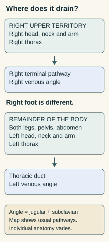
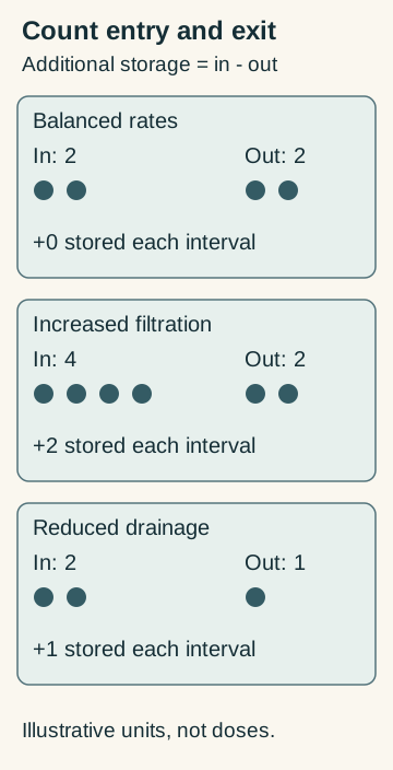
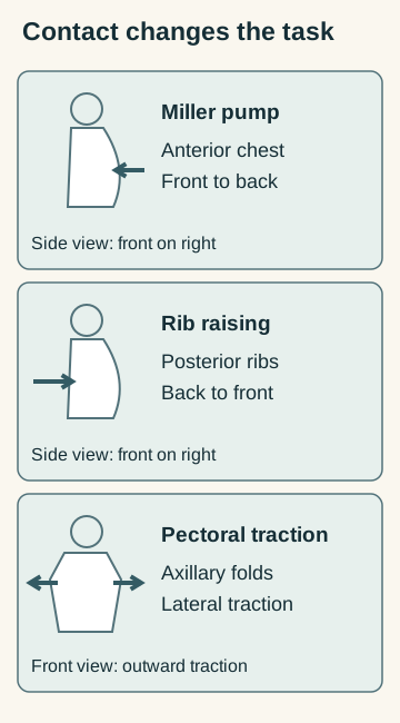
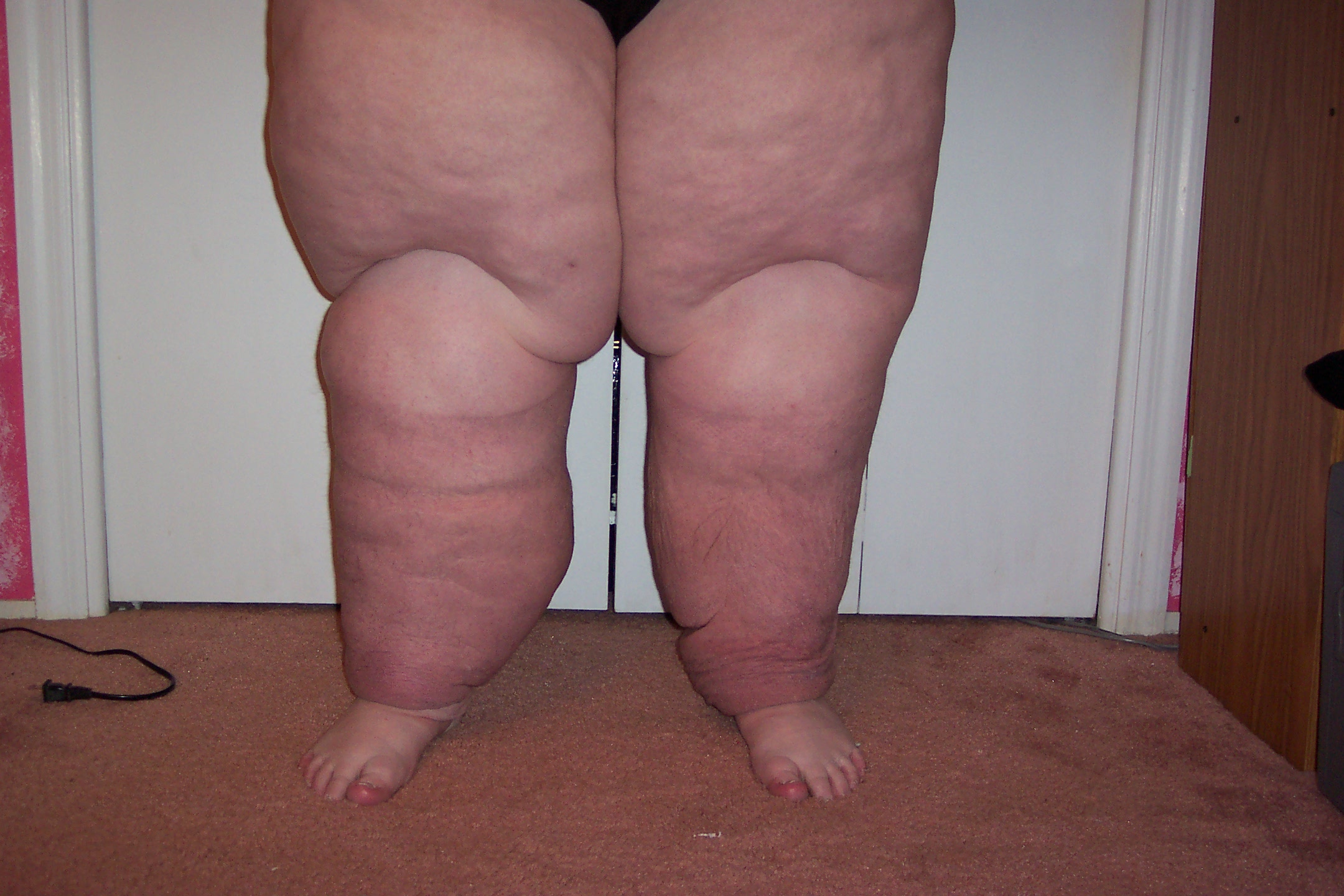
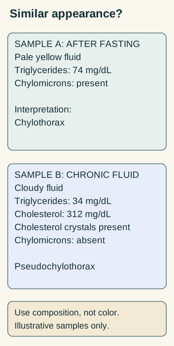
.JPG)