Level 1 learners classify cranial strain patterns from axes, relative sphenoid-occiput motion, vault contacts, and the traditional primary respiratory mechanism.
Level 1 starts with the traditional mechanism, axes, paired-bone behavior, sacral linkage, and the landmark that names each strain. Level 2 and Level 3 start with safety, diagnosis, workup order, and follow-up before a manual label influences care. Beyond the boards, use consent, referral, outcome tracking, and evidence limits so a descriptive cranial finding is never mistaken for a proven structural lesion or the cause of a patient symptom.
Find the bones before naming a pattern
Level map: Level 1 asks which axes,
relative rotations, vault contacts, and traditional craniosacral relationships define the
pattern. Level 2 asks whether the presentation needs another diagnosis or workup before
osteopathic treatment. Level 3 asks how to reassess symptoms and function over time.
Beyond the boards, document consent, response, adverse symptoms, and referral decisions
without presenting a palpatory model as imaging or a validated causal diagnosis.
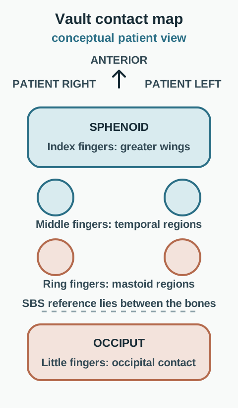
What does a high finger actually represent? In the vault hold, an index finger monitors a greater wing of the sphenoid. A little finger monitors the occiput. Confusing these contacts reverses a torsion name even when the asymmetry was noticed correctly. The middle fingers lie near the temporal region anterior to the ears; the ring fingers lie near the mastoid regions. Use the paired anterior and posterior contacts to compare the two bones, not to diagnose from one isolated point.
Use patient right and left. The map is conceptual and not an anatomical surface rendering. [1][5][14]
The sphenobasilar synchondrosis, or SBS, is the relationship between the basisphenoid and basiocciput. The sphenoid body is anterior; the basilar occiput is posterior, in front of the foramen magnum. The developmental cartilage junction is also called the spheno-occipital synchondrosis. It normally fuses as skeletal maturation proceeds. In an adult, a cranial strain description must not be presented as proof that an open cartilage joint has slipped. [1][5]
Orient by patient right and left. The apparent separation at the junction is exaggerated for teaching.
Trace from a greater wing to the sphenoid body, then posteriorly across the junction to the occiput. Next locate the foramen magnum. This separates a skull-base relationship from the coronal suture in the vault and from the occipitoatlantal joint below the skull. A focal finding at one of those locations does not automatically establish a finding at the others.
Now orient three axis families. Transverse axes run left to right, one for each bone. Vertical axes run superior to inferior, one through the sphenoid and one through the occipital region. A shared anteroposterior axis runs front to back. Always compare rotations from the same viewing direction. Clockwise changes when the observer changes sides; patient right does not. [1][2]
An axis gives the family. The relative rotations distinguish neighboring patterns. A specified landmark supplies the name.
Apply that order to an unfamiliar observation. If the left little finger is high, you know something about the occiput. You still need the greater-wing finding before assigning a torsion side. A photograph of the whole head tilted toward a shoulder also cannot supply the missing relative bone motion.
Build the traditional mechanism before naming a strain
William Garner Sutherland is traditionally credited with the cranial concept. Traditional primary respiratory mechanism (PRM) teaching assembles five proposals: spontaneous activity within brain and cord tissue; cyclic variation of cerebrospinal fluid (CSF); shifting tension within cranial and spinal membranes; small articular behavior among skull bones; and passive sacral response between the innominates. This is a historical osteopathic model, not proof that every proposal can be measured by vault palpation in an individual. [11][15]
The craniosacral linkage in that model follows dural attachments between the
skull and sacrum. The respiratory axis is the superior transverse axis near the second sacral
segment. During cranial flexion, the sacral base is described as traveling posteriorly, which
structural terminology calls counternutation; during cranial extension, the base travels
anteriorly. Some cranial texts call the first phase sacral flexion, so translate the geometry
rather than relying on the word alone. [15]
The vault hold maps the index fingers to the greater wings of the sphenoid,
middle fingers to the temporal regions anterior to the ears, ring fingers to the mastoid
regions, and little fingers to the occiput. Patient right and left always describe the side
being contacted. The hands compare relative motion; one high finger by itself is not a complete
strain diagnosis. [14]
Flexion and extension apply to midline bones in the model. Paired bones are
described as externally rotating during cranial flexion and internally rotating during cranial
extension. In the vault hold, the full pattern matters more than a memorized direction from one
contact. [2][4]
The sphenobasilar synchondrosis remains the reference relationship for naming
these patterns even though the developmental cartilage normally becomes bone. A reduced
perceived cranial rhythmic impulse after trauma may be framed as dysfunction in traditional
teaching, but the count does not establish an open adult joint, intracranial pressure, venous
obstruction, or the cause of symptoms. [5][6][7]
Try it here · Checkpoint 1 of 3
Make your prediction before reading the choices. A first attempt is just a starting point.
Case 27
Show answer and explanations for case 27
A. List B is the traditional five-component model because it uses measurable cardiopulmonary drivers (Why this does not fit)
Cardiac and respiratory influences can affect physiology, but they do not replace the named articular and sacral components in the traditional osteopathic list.
Reasoning steps for option A
For the traditional five-component primary respiratory mechanism, what must be tested first for option 1, "List B is the traditional five-component model because it uses measurable cardiopulmonary drivers"?
Test option 1 against which elements belong to the historical list. Keep the supplied observation separate from the proposed label.
What second comparison decides whether option 1, "List B is the traditional five-component model because it uses measurable cardiopulmonary drivers", remains viable in case csp-27?
Then compare option 1 with whether list membership validates a patient-level mechanism test. Any mismatch makes that alternative unsupported.
B. Both lists are equivalent descriptions of the same validated physiologic measurement (Why this does not fit)
The lists are not equivalent, and vault palpation has not validated all proposed components as one objective physiologic measurement.
Reasoning steps for option B
For the traditional five-component primary respiratory mechanism, what must be tested first for option 2, "Both lists are equivalent descriptions of the same validated physiologic measurement"?
Test option 2 against which elements belong to the historical list. Keep the supplied observation separate from the proposed label.
What second comparison decides whether option 2, "Both lists are equivalent descriptions of the same validated physiologic measurement", remains viable in case csp-27?
Then compare option 2 with whether list membership validates a patient-level mechanism test. Any mismatch makes that alternative unsupported.
C. List A is incomplete unless voluntary skeletal muscle contraction is added (Why this does not fit)
Voluntary skeletal muscle contraction is not one of the five traditionally taught components.
Reasoning steps for option C
For the traditional five-component primary respiratory mechanism, what must be tested first for option 3, "List A is incomplete unless voluntary skeletal muscle contraction is added"?
Test option 3 against which elements belong to the historical list. Keep the supplied observation separate from the proposed label.
What second comparison decides whether option 3, "List A is incomplete unless voluntary skeletal muscle contraction is added", remains viable in case csp-27?
Then compare option 3 with whether list membership validates a patient-level mechanism test. Any mismatch makes that alternative unsupported.
D. List B is preferred because the adult spheno-occipital junction remains cartilaginous (Why this does not fit)
The developmental junction normally fuses, and its fusion does not convert the PRM list into a cardiopulmonary list.
Reasoning steps for option D
For the traditional five-component primary respiratory mechanism, what must be tested first for option 4, "List B is preferred because the adult spheno-occipital junction remains cartilaginous"?
Test option 4 against which elements belong to the historical list. Keep the supplied observation separate from the proposed label.
What second comparison decides whether option 4, "List B is preferred because the adult spheno-occipital junction remains cartilaginous", remains viable in case csp-27?
Then compare option 4 with whether list membership validates a patient-level mechanism test. Any mismatch makes that alternative unsupported.
E. List A matches the traditional Sutherland model, but the model is not a validated patient-level mechanism test (Best answer)
List A contains the five traditionally taught components. Identifying the historical model does not establish that each component is objectively measured by an individual examination.
Reasoning steps for option E
For the traditional five-component primary respiratory mechanism, what must be tested first for option 5, "List A matches the traditional Sutherland model, but the model is not a validated patient-level mechanism test"?
Test option 5 against which elements belong to the historical list. Keep the supplied observation separate from the proposed label.
What second comparison decides whether option 5, "List A matches the traditional Sutherland model, but the model is not a validated patient-level mechanism test", remains viable in case csp-27?
Then compare option 5 with whether list membership validates a patient-level mechanism test. Any mismatch makes that alternative unsupported.
Takeaway: Know the five traditional components, then keep the historical model separate from measurement validity.
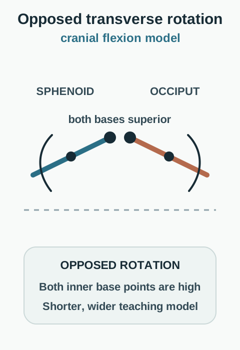
Both flexion-extension and vertical strain use two transverse axes. What separates them is the relationship between rotations. In the traditional flexion model, rotation is opposed and both basilar portions are superior, increasing the superior convexity of the junction. In extension, rotation is also opposed, but both bases are inferior. The traditional shape comparison is shorter and wider in flexion, longer and narrower in extension. Paired cranial bones are described as externally rotating with flexion and internally rotating with extension. These are descriptions of the teaching model, not measurements that establish a headache cause. [2][4]
Opposed rotation with both bases superior is the cranial flexion model. [1][2][4]A side-view comparison. Track the black inner tips, not the outer ends of the levers.
A source wording issue matters here: AACOM term 398 describes extension with both bases inferior. Term 399 is headed Flexion and describes opposite transverse rotations with both bases superior, but its final clause says "in SBS extension". That last word is an apparent editorial error; it does not reverse the flexion heading or the stated geometry. [2]
Use the black inner tips to track the bases. They are at a similar superior level in the starting picture. Predict what happens if only one bone reverses its rotation while both axes stay fixed. You can open either alternative, compare both, and close it again to return to the starting view. No answer is required to read the explanation.
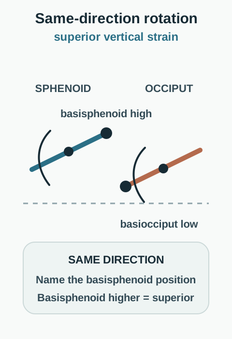
Reverse only the occiputA side-view comparison. Track the black inner tips, not the outer ends of the levers.
The two rotations now agree. The sphenoid base remains higher while the occipital base becomes lower. This is superior vertical strain, named for the basisphenoid relative to the basiocciput. Calling it flexion because the sphenoid is high misses the changed relationship.
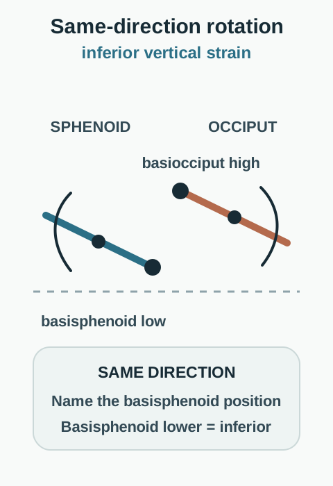
Reverse only the sphenoidA side-view comparison. Track the black inner tips, not the outer ends of the levers.
The two rotations again agree, but the sphenoid base is now lower than the occipital base. This is inferior vertical strain. Naming the higher occiput instead would reverse the correct designation.
Vertical strain means same-direction rotation around two transverse axes. A superior basisphenoid gives a superior strain; an inferior basisphenoid gives an inferior strain. By contrast, flexion and extension have opposed rotations and describe the shared superior or inferior basilar configuration. The exercise exaggerates angles and spacing to reveal that distinction. It is not a skull deformation calculator or a treatment simulation. [1][2]
Same-direction rotation plus a higher basisphenoid gives superior vertical strain. [1][2][4]
Same-direction rotation plus a lower basisphenoid gives inferior vertical strain. [1][2][4]
For transfer, imagine both basilar portions inferior with opposed rotations. That is extension, not inferior vertical strain. An index finger is also not the basisphenoid itself. Do not substitute an anterior greater-wing contact for the base that supplies the vertical-strain name.
A high wing and a convex side name different things
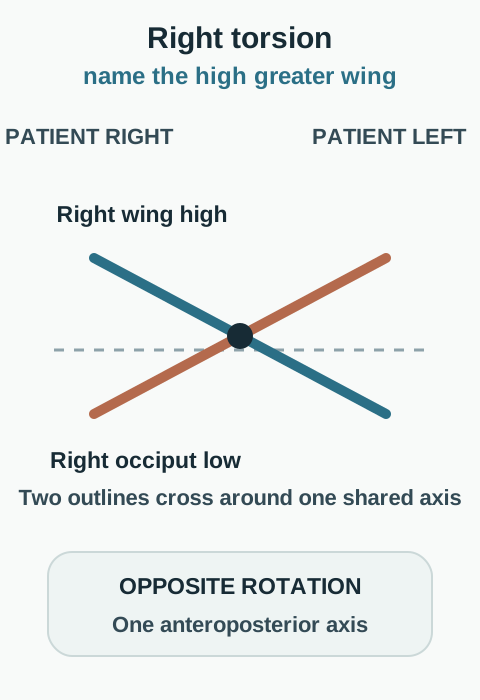
Why can a high right greater wing appear in more than one description? A side name is meaningful only after the family is established. In torsion, the sphenoid and occiput rotate oppositely around one shared anteroposterior axis. Name the high greater wing. A high right wing with the right occipital side inferior is right torsion; reversing both gives left torsion. [1]
Name the high greater wing after confirming opposite rotation around one shared axis. [1][4]The two outlines cross because the rotations oppose. Name the high sphenoid wing, not the occiput.
Point to the high wing, then to the occipital contact on that same patient side. Their superior-inferior relationship is opposite. If both contacts on a side were inferior together, the torsion relationship would no longer be established. This check prevents a high-wing shortcut from being applied to every cranial asymmetry.
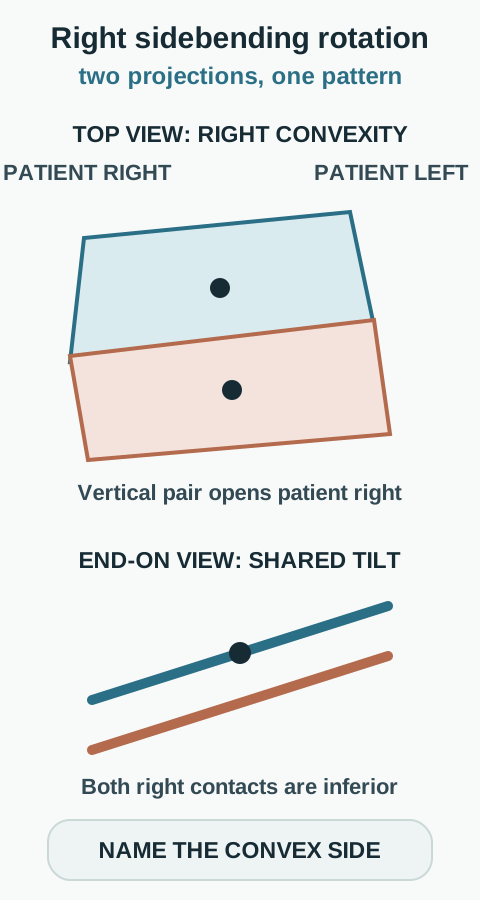
Sidebending rotation has two components. The bones rotate oppositely about their two vertical axes, producing a convex side, and in the same direction about the shared anteroposterior axis. The convex side is inferior in this combined model. Name the convexity, not the high wing. In right sidebending rotation, the right side is fuller or wider between the vault contacts and both right contacts tip inferiorly. [1][4][12]
The top view explains convexity; the end-on view explains the shared inferior side. [1][4][12]Different projections explain different axes. The separated lower traces show shared tilt, not two distinct anteroposterior axes.
Compare the two projections before naming the pattern. The top view explains why one side spreads. The end-on traces explain why both contacts on that side are inferior. The lower traces are separated for visibility, not to create two anteroposterior axes. Saying that all rotations are in the same direction loses the defining vertical-axis component.
Now apply a less familiar combination. If the left side is convex and both left contacts are inferior, the designation is left sidebending rotation even though the right greater wing is higher. An isolated high right wing would not distinguish that combination from right torsion; the occipital relationship resolves the ambiguity.
Torsion names the high wing. Sidebending rotation names the convex, inferior side.
Separate lateral shear from global restriction
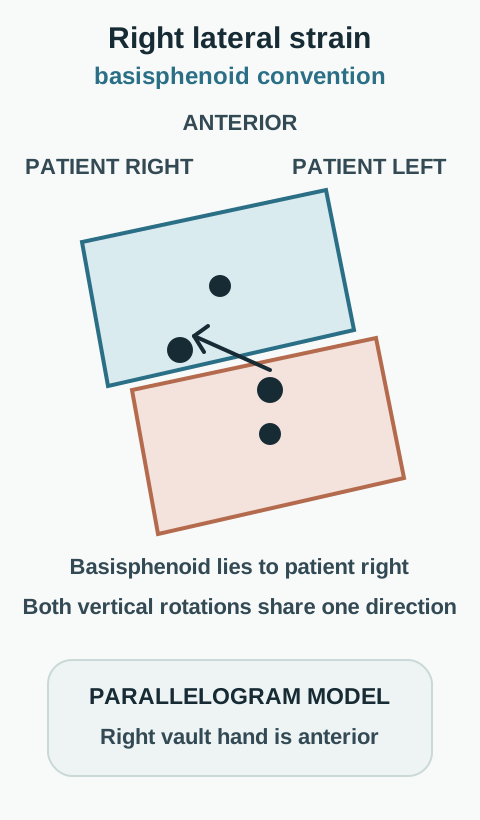
What does a parallelogram tell you that a forehead impact does not? Lateral strain uses same-direction rotation around two vertical axes. The relative shape resembles a parallelogram. This lesson uses the basisphenoid convention explicitly discussed by Capobianco and Shermon because lateral-strain naming has varied across teaching sources. A basisphenoid right of the basiocciput is a right lateral strain. Under that convention the right greater wing is more prominent anteriorly, the right vault hand is anterior, and the left hand is posterior. Reverse the relationship for a left lateral strain. [1][3]
Patient right is page left. Naming follows the declared basisphenoid convention. [1][3]The two center dots are separate vertical axes. The small inner points represent the two bases, not their palpation sites.
Throughout this lesson, a right or left vault hand means the hand contacting that side of the patient, not the examiner's own anatomical side. In the superior-view diagram, patient right is at page left. Mirroring a drawing changes its page coordinates, not the patient-relative name. [3]
Trace the small inner base points, not the side edges. Patient right is on the left of this top-view drawing. Now mentally reverse both rotations. Their same-direction relationship remains, so the family stays lateral strain, but the relative base position and anterior hand reverse. The hand pattern corroborates a stated convention; it should not replace the axis and base description.
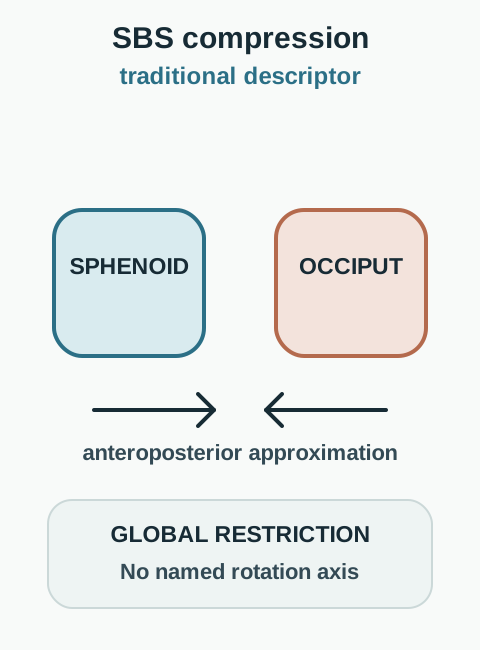
SBS compression is different. The traditional descriptor is approximation of the basisphenoid and basiocciput with markedly limited overall excursion. There is no named rotational axis for compression. A low perceived rhythm or a tender point alone does not demonstrate this global relationship. [2][6]
The arrows illustrate a traditional descriptor, not a measured adult joint gap or headache cause. [2][5][6]The inward arrows illustrate the traditional descriptor. They do not establish an injury mechanism or headache cause.
Compare organized directional shear with generalized restriction. A model with one base displaced rightward has a directional relationship. A report of generalized restriction without a reproducible shear describes something different. The diagrams explain those terms; they do not show an actual adult joint opening or closing.
Traditional teaching groups flexion-extension, torsion, and sidebending rotation as physiologic patterns; vertical strain, lateral strain, and compression are commonly grouped as nonphysiologic. Those categories do not mean that the first group always represents health or that the second proves trauma. Birth molding, forehead or vertex impacts, and lateral blows may appear in clinical histories. None of them alone selects a strain, proves a birth injury, or identifies the source of persistent symptoms. Patterns have also been reported in healthy subjects. [1][4]
Try the competing example. A forehead-impact history followed by same-direction vertical rotation and a rightward basisphenoid supports a right lateral description, not an automatic compression label. The injury still matters for the separate safety assessment.
Turn the model into a safe clinical sequence
Start with the patient, not the strain name. Review the symptom trajectory,
recent trauma, anticoagulation, neurological symptoms, age-specific risks, and the reason for
the visit. Obtain consent, explain that the examination is gentle and descriptive, and use a
neutral, supported position. New focal deficit, repeated vomiting, altered alertness, seizure,
or progressive post-injury symptoms changes the plan from manual assessment to urgent medical
evaluation. [8]
Classify in two passes. First identify the axis family and whether the
sphenoid and occiput rotate together or oppositely. Then use the correct landmark: the high
greater wing for torsion, the convex inferior side for sidebending rotation, the basisphenoid
position for vertical or lateral strain, and global approximation for compression. Recheck
patient-side orientation before naming the pattern. [1][2][3]
Balanced ligamentous tension (BLT) and related balanced-tension descriptions
use a position of relative balance and the patient's inherent physiologic motion as the
traditional corrective influence. Operator effort, voluntary muscle contraction, and a rapid
thrust are not interchangeable with that model. The technique name does not prove that a fused
adult junction opened or that venous flow changed. [11][15]
Infant head shape requires a different differential. Positional
plagiocephaly typically produces occipital flattening with ipsilateral frontal bossing and
anterior displacement of the ear, creating a parallelogram from above. Lambdoid
craniosynostosis is uncommon and more often pulls the affected ear posteriorly and inferiorly,
with mastoid prominence. A cranial strain label cannot replace pediatric assessment of suture
fusion, torticollis, progression, development, and head growth. [16][17]
First-line care follows the actual diagnosis. A stable musculoskeletal
complaint may be managed with an agreed trial of conservative care and a defined functional
outcome. Positional head-shape counseling preserves supine sleep, adds supervised awake prone
time and varied positioning, and addresses torticollis when present. Suspected craniosynostosis,
progressive asymmetry, abnormal neurological findings, or failure to improve warrants pediatric
or craniofacial referral rather than repeated cranial sessions. [17][18]
Try it here · Checkpoint 2 of 3
Make your prediction before reading the choices. A first attempt is just a starting point.
Case 29
Show answer and explanations for case 29
A. Positional plagiocephaly (Best answer)
Ipsilateral occipital flattening, frontal bossing, and anterior ear displacement form the typical positional parallelogram. Continued pediatric follow-up still assesses progression and associated torticollis.
Reasoning steps for option A
For infant occipital flattening with frontal bossing and anterior ear displacement, what must be tested first for option 1, "Positional plagiocephaly"?
Test option 1 against the full top-view head-shape pattern. Keep the supplied observation separate from the proposed label.
What second comparison decides whether option 1, "Positional plagiocephaly", remains viable in case csp-29?
Then compare option 1 with the ear direction that distinguishes suspected lambdoid synostosis. Any mismatch makes that alternative unsupported.
B. Unilateral lambdoid craniosynostosis with posterior and inferior ear displacement (Why this does not fit)
Lambdoid synostosis more often shifts the affected ear posteriorly and inferiorly and can produce mastoid prominence. The supplied ear direction is the opposite.
Reasoning steps for option B
For infant occipital flattening with frontal bossing and anterior ear displacement, what must be tested first for option 2, "Unilateral lambdoid craniosynostosis with posterior and inferior ear displacement"?
Test option 2 against the full top-view head-shape pattern. Keep the supplied observation separate from the proposed label.
What second comparison decides whether option 2, "Unilateral lambdoid craniosynostosis with posterior and inferior ear displacement", remains viable in case csp-29?
Then compare option 2 with the ear direction that distinguishes suspected lambdoid synostosis. Any mismatch makes that alternative unsupported.
C. Unilateral coronal craniosynostosis (Why this does not fit)
Coronal synostosis primarily alters the forehead and orbital region rather than producing this posterior positional parallelogram.
Reasoning steps for option C
For infant occipital flattening with frontal bossing and anterior ear displacement, what must be tested first for option 3, "Unilateral coronal craniosynostosis"?
Test option 3 against the full top-view head-shape pattern. Keep the supplied observation separate from the proposed label.
What second comparison decides whether option 3, "Unilateral coronal craniosynostosis", remains viable in case csp-29?
Then compare option 3 with the ear direction that distinguishes suspected lambdoid synostosis. Any mismatch makes that alternative unsupported.
D. SBS compression caused by birth trauma (Why this does not fit)
A manual SBS label cannot establish birth trauma or replace the pediatric head-shape differential.
Reasoning steps for option D
For infant occipital flattening with frontal bossing and anterior ear displacement, what must be tested first for option 4, "SBS compression caused by birth trauma"?
Test option 4 against the full top-view head-shape pattern. Keep the supplied observation separate from the proposed label.
What second comparison decides whether option 4, "SBS compression caused by birth trauma", remains viable in case csp-29?
Then compare option 4 with the ear direction that distinguishes suspected lambdoid synostosis. Any mismatch makes that alternative unsupported.
E. Right torsion based only on the side of occipital flattening (Why this does not fit)
Torsion requires relative sphenoid and occipital findings around an anteroposterior axis. Head-shape asymmetry alone does not supply that examination.
Reasoning steps for option E
For infant occipital flattening with frontal bossing and anterior ear displacement, what must be tested first for option 5, "Right torsion based only on the side of occipital flattening"?
Test option 5 against the full top-view head-shape pattern. Keep the supplied observation separate from the proposed label.
What second comparison decides whether option 5, "Right torsion based only on the side of occipital flattening", remains viable in case csp-29?
Then compare option 5 with the ear direction that distinguishes suspected lambdoid synostosis. Any mismatch makes that alternative unsupported.
Takeaway: Anterior displacement of the ear on the flattened side favors positional plagiocephaly; suspected synostosis needs pediatric or craniofacial assessment.
A person has a progressively worsening headache several weeks after striking the forehead. Does an apparently normal neurological examination or an earlier negative CT make a manual explanation sufficient? No. A changed or persistent post-injury complaint needs appropriate reassessment. Worsening headache, repeated vomiting, altered alertness, a seizure, or a new focal deficit increases urgency; anticoagulation adds important bleeding risk. A previous scan describes a previous time point. Do not delay reassessment to try decompression, and do not wait for limb weakness before responding to a concerning trajectory. [8]
Consider a patient with worsening headache and new imbalance while taking an anticoagulant. Choose the next action, then compare its consequence.
Choose a manual treatment trial first
This delays assessment of an evolving post-injury problem. A report of cranial restriction does not exclude an intracranial cause, and symptom response would not safely rule one out. Arrange urgent emergency reassessment instead.
Choose urgent medical reassessment
This prioritizes the changed symptoms and bleeding risk. The acute team can reassess the neurological state and decide whether repeat imaging or other evaluation is needed. The cranial description does not determine that decision.
After the safety assessment, compare positive localizing findings. Familiar headache reproduced during restricted upper-cervical motion supports investigating a cervical contribution with a fuller joint and muscle examination. An occipitoatlantal flexion preference, restricted extension and concordant suboccipital pain can be documented; a cranial rhythm count cannot establish or exclude that contribution. Normal cervical radiographs also do not directly assess functional motion or muscle performance. [2][6][10]
The controlled-block study by Getsoian and colleagues evaluated a combination of cervical motion, joint and muscle findings, not isolated pain provocation. Its block targets were C2/C3 and C3/C4, so it does not establish an exclusive occipitoatlantal headache source. Keep the wider headache differential open, and compare headache outcomes separately from changes in neck function. [13]
A focal tender coronal region is not the same examination as an SBS relationship. Temple pain modified by chewing or clenching, with familiar pain reproduced by temporalis palpation in a person with a painful temporomandibular disorder, supports a jaw-related assessment. Primary headache disorders and other causes can coexist, so localization does not end the differential. [9]
Apply this to a stable patient whose familiar pain occurs with upper-neck extension, not jaw function. The positive cervical examination guides the next assessment. Change the history to new drowsiness and an arm drift after injury, and the priority changes to emergency care regardless of the prior strain name.
Longitudinal care uses separate outcomes.
Record headache frequency, intensity, medication use, sleep, work or school function, neck
motion, and adverse symptoms at baseline. Improvement in neck motion without improvement in the
headache diary does not prove failure of the examination, but it does argue against claiming
that the cervical or cranial finding was the main headache mechanism. Escalate evaluation when
the trajectory changes, function worsens, or new red flags appear. [8][13]
Keep a useful model separate from a proven mechanism
What can a rhythm count establish? The cranial rhythmic impulse, or CRI, is a perceived rhythm within the traditional primary respiratory mechanism model. Teaching ranges such as 10 to 14 cycles per minute are not validated diagnostic cutoffs for compression, intracranial pressure, or a headache cause. It is not a substitute for measuring the pulse, breathing, or the neurological state. Primary studies have found important limitations in agreement between examiners. Better repeatability by one examiner does not solve diagnostic validity. [6][7]
Imagine an examiner records similar counts on two occasions while a second examiner records substantially different values. Decide which question was tested. This concerns measurement agreement, not whether a disease is present, whether a proposed mechanism is true, or whether treatment improves outcomes. Each requires different evidence. Headache, low mood, concentration problems, and a low counted rhythm do not form a validated SBS-compression triad. Persistent post-injury symptoms still deserve clinical follow-up. [6][7][8]
The reciprocal tension membrane concept uses dural relationships, including the falx and tentorium, to describe linked cranial behavior. Anatomical connections to venous sinuses do not establish that a manually named strain obstructs venous drainage or causes the patient's headache. Likewise, describing coupled cranial and sacral rhythms does not demonstrate the proposed coupling in an individual. Keep those traditional explanatory models labeled as such. [2][6][7]
Gentle cranial approaches may be described as decompression or balanced membranous or ligamentous tension. CV4 is a traditional technique name referring to compression of the fourth ventricle; it does not mean that a practitioner directly squeezes the ventricle through the skull. Naming a technique is not evidence that it reopens a fused SBS, restores measured venous flow, or is appropriate before an injury has been assessed. Selection belongs to trained clinical care after assessment, contraindication review, and consent, not to an unsupervised forceful maneuver. [5][8][11]
For transfer, suppose a patient reports less pain after one session. Document the response without claiming that it proves the proposed structural or vascular mechanism. A favorable experience, a reliable measurement, an accurate diagnosis, and an effective treatment are distinct findings.
Use a measurement ladder. Repeatability asks
whether one examiner obtains a similar result. Interexaminer agreement asks whether two
examiners agree. Diagnostic accuracy needs an independently justified reference. Treatment
effectiveness needs a comparison group and patient-important outcomes. A result at one rung
cannot be promoted to the next without new evidence. [6][7]
Prevent avoidable complications by screening for urgent illness, using
comfortable positioning, avoiding forceful contact, explaining uncertainty, and stopping when
symptoms worsen or the patient withdraws consent. In infants, persistent asymmetry and
suspected synostosis require age-appropriate pediatric pathways. In adults, a named strain does
not lower the threshold for evaluating a dangerous headache or neurological change.
[8][17]
For the examination, use axes, relative rotation, and the naming landmark. For the patient, use the clinical assessment and the limits of the evidence.
Beyond the boards: counseling, referral, and real-world limits
Use transparent counseling. Describe a cranial strain as an osteopathic
palpatory pattern, state the naming convention, and explain that the label is not an imaging
diagnosis or proof of symptom causation. Discuss reasonable alternatives, the option of no
manual treatment, expected comfort, and the plan for stopping or reassessment.
Referral follows the clinical problem. Emergency services evaluate acute
neurological deterioration or concerning head injury. Neurology or a headache clinician may
help with persistent or atypical headache. Dental or temporomandibular expertise may help when
familiar temple pain is modified by jaw function. Pediatric and craniofacial teams evaluate
suspected craniosynostosis or progressive infant asymmetry. [8][9][17]
No medication dose treats a named cranial strain. Medication, imaging, and
laboratory choices belong to the actual diagnosis. When a separate condition requires a drug,
verify typical adult dosing against current labeling and account for age, pregnancy, kidney and
liver function, interactions, and local protocols. Do not attach an analgesic, anticoagulation,
or imaging schedule to the palpatory label alone.
A common real-world pitfall is repeating the same intervention because a
local finding remains palpable while the patient-important outcome is unchanged. Predefine the
outcome and review interval, document any adverse response, and change course when benefit is
absent or the presentation changes. A symptom response can be worth documenting without
treating it as proof of a proposed structural or vascular mechanism.
Read research by the question it answers. Reliability studies address
agreement. Observational prevalence studies describe selected populations. A sham-controlled
trial would be needed to estimate a specific treatment effect, and a mechanism claim would need
a direct mechanism measure. Keep those questions separate when counseling patients or planning
future study. [4][6][7]
Try it here · Checkpoint 3 of 3
Make your prediction before reading the choices. A first attempt is just a starting point.
Case 24
Show answer and explanations for case 24
A. Within-examiner agreement increased; diagnostic accuracy remains unmeasured (Why this does not fit)
The absence of a diagnostic reference leaves accuracy unmeasured. The changed coefficient is the between-examiner estimate, not the within-examiner value that remains 0.65.
Reasoning steps for option A
For stable within-examiner ICC with improved between-examiner ICC, what must be tested first for option 1, "Within-examiner agreement increased; diagnostic accuracy remains unmeasured"?
Test option 1 against the estimate that changed. Keep the supplied observation separate from the proposed label.
What second comparison decides whether option 1, "Within-examiner agreement increased; diagnostic accuracy remains unmeasured", remains viable in case csp-24?
Then compare option 1 with the missing reference standard and diagnostic-accuracy evidence. Any mismatch makes that alternative unsupported.
B. Between-examiner agreement increased; diagnostic accuracy remains unmeasured (Best answer)
The reported between-examiner estimate rises from 0.10 to 0.60 while the within-examiner estimate is unchanged. Improved agreement in this illustrative exercise still does not supply a disease reference or a sensitivity estimate.
Reasoning steps for option B
For stable within-examiner ICC with improved between-examiner ICC, what must be tested first for option 2, "Between-examiner agreement increased; diagnostic accuracy remains unmeasured"?
Test option 2 against the estimate that changed. Keep the supplied observation separate from the proposed label.
What second comparison decides whether option 2, "Between-examiner agreement increased; diagnostic accuracy remains unmeasured", remains viable in case csp-24?
Then compare option 2 with the missing reference standard and diagnostic-accuracy evidence. Any mismatch makes that alternative unsupported.
C. Within-examiner agreement increased; diagnostic sensitivity increased (Why this does not fit)
The within-examiner coefficient does not increase in the supplied data. Sensitivity also cannot be inferred from either agreement coefficient without an appropriate diagnostic comparison.
Reasoning steps for option C
For stable within-examiner ICC with improved between-examiner ICC, what must be tested first for option 3, "Within-examiner agreement increased; diagnostic sensitivity increased"?
Test option 3 against the estimate that changed. Keep the supplied observation separate from the proposed label.
What second comparison decides whether option 3, "Within-examiner agreement increased; diagnostic sensitivity increased", remains viable in case csp-24?
Then compare option 3 with the missing reference standard and diagnostic-accuracy evidence. Any mismatch makes that alternative unsupported.
D. Between-examiner agreement increased; diagnostic sensitivity increased (Why this does not fit)
The between-examiner estimate does increase. That is an agreement result, not evidence that sensitivity increased; the exercise never compared findings against an independent diagnostic reference.
Reasoning steps for option D
For stable within-examiner ICC with improved between-examiner ICC, what must be tested first for option 4, "Between-examiner agreement increased; diagnostic sensitivity increased"?
Test option 4 against the estimate that changed. Keep the supplied observation separate from the proposed label.
What second comparison decides whether option 4, "Between-examiner agreement increased; diagnostic sensitivity increased", remains viable in case csp-24?
Then compare option 4 with the missing reference standard and diagnostic-accuracy evidence. Any mismatch makes that alternative unsupported.
Takeaway: Improving agreement between examiners is not the same as demonstrating diagnostic accuracy.
Use the supplied findings to decide what is supported and what remains unknown. Read the options only after forming your answer. The explanations compare every alternative, including the finding that would have made it a better fit.
Case 1
Show answer and explanations for case 1
A. Initial cranial flexion; final superior vertical strain (Why this does not fit)
This reverses the order of the two configurations. The initial rotations agree, and the isolated occipital reversal produces opposed rotations with both bases superior.
Reasoning steps for option A
For same-direction transverse rotations with opposite base heights followed by an occipital reversal, what must be tested first for option 1, "Initial cranial flexion; final superior vertical strain"?
Test option 1 against the transverse-axis relationship before reversal. Keep the supplied observation separate from the proposed label.
What second comparison decides whether option 1, "Initial cranial flexion; final superior vertical strain", remains viable in case csp-01?
Then compare option 1 with the final base heights and whether rotations oppose. Any mismatch makes that alternative unsupported.
B. Initial superior vertical strain; final cranial flexion (Best answer)
Same-direction transverse rotation with a relatively high basisphenoid identifies the initial superior vertical strain. Reversing only the occiput raises its base while the sphenoid stays superior, producing the opposed-rotation flexion configuration.
Reasoning steps for option B
For same-direction transverse rotations with opposite base heights followed by an occipital reversal, what must be tested first for option 2, "Initial superior vertical strain; final cranial flexion"?
Test option 2 against the transverse-axis relationship before reversal. Keep the supplied observation separate from the proposed label.
What second comparison decides whether option 2, "Initial superior vertical strain; final cranial flexion", remains viable in case csp-01?
Then compare option 2 with the final base heights and whether rotations oppose. Any mismatch makes that alternative unsupported.
C. Initial inferior vertical strain; final cranial flexion (Why this does not fit)
A low basisphenoid would support an initial inferior vertical strain. Here the sphenoid base starts high; the predicted final flexion does not correct the initial naming error.
Reasoning steps for option C
For same-direction transverse rotations with opposite base heights followed by an occipital reversal, what must be tested first for option 3, "Initial inferior vertical strain; final cranial flexion"?
Test option 3 against the transverse-axis relationship before reversal. Keep the supplied observation separate from the proposed label.
What second comparison decides whether option 3, "Initial inferior vertical strain; final cranial flexion", remains viable in case csp-01?
Then compare option 3 with the final base heights and whether rotations oppose. Any mismatch makes that alternative unsupported.
D. Initial superior vertical strain; final cranial extension (Why this does not fit)
The initial superior vertical label fits the supplied relationship. Extension would require both bases inferior, but the unchanged sphenoid base remains superior after the occipital reversal.
Reasoning steps for option D
For same-direction transverse rotations with opposite base heights followed by an occipital reversal, what must be tested first for option 4, "Initial superior vertical strain; final cranial extension"?
Test option 4 against the transverse-axis relationship before reversal. Keep the supplied observation separate from the proposed label.
What second comparison decides whether option 4, "Initial superior vertical strain; final cranial extension", remains viable in case csp-01?
Then compare option 4 with the final base heights and whether rotations oppose. Any mismatch makes that alternative unsupported.
E. Initial cranial extension; final inferior vertical strain (Why this does not fit)
An initial extension configuration would require opposed rotations and two inferior bases. Neither is present, and the isolated reversal produces matched superior bases rather than inferior vertical shear.
Reasoning steps for option E
For same-direction transverse rotations with opposite base heights followed by an occipital reversal, what must be tested first for option 5, "Initial cranial extension; final inferior vertical strain"?
Test option 5 against the transverse-axis relationship before reversal. Keep the supplied observation separate from the proposed label.
What second comparison decides whether option 5, "Initial cranial extension; final inferior vertical strain", remains viable in case csp-01?
Then compare option 5 with the final base heights and whether rotations oppose. Any mismatch makes that alternative unsupported.
Takeaway: Track the changed bone separately from the unchanged bone, then classify their new relationship.
A. Initial cranial extension; final inferior vertical strain (Why this does not fit)
Extension correctly describes the starting state. Reversing the sphenoid raises its base above the unchanged inferior occiput, giving superior rather than inferior vertical strain.
Reasoning steps for option A
For opposed inferior bases followed by a sphenoid reversal, what must be tested first for option 1, "Initial cranial extension; final inferior vertical strain"?
Test option 1 against opposed transverse rotations with both bases inferior. Keep the supplied observation separate from the proposed label.
What second comparison decides whether option 1, "Initial cranial extension; final inferior vertical strain", remains viable in case csp-02?
Then compare option 1 with the final same-direction rotations and relative basisphenoid height. Any mismatch makes that alternative unsupported.
B. Initial cranial flexion; final superior vertical strain (Why this does not fit)
The final superior vertical designation fits the predicted relative bases. The starting bases are both inferior, which identifies extension rather than flexion.
Reasoning steps for option B
For opposed inferior bases followed by a sphenoid reversal, what must be tested first for option 2, "Initial cranial flexion; final superior vertical strain"?
Test option 2 against opposed transverse rotations with both bases inferior. Keep the supplied observation separate from the proposed label.
What second comparison decides whether option 2, "Initial cranial flexion; final superior vertical strain", remains viable in case csp-02?
Then compare option 2 with the final same-direction rotations and relative basisphenoid height. Any mismatch makes that alternative unsupported.
C. Initial cranial flexion; final inferior vertical strain (Why this does not fit)
Flexion would begin with both bases superior. The supplied inferior starting bases and the subsequent rise of only the sphenoid disagree with both labels in this option.
Reasoning steps for option C
For opposed inferior bases followed by a sphenoid reversal, what must be tested first for option 3, "Initial cranial flexion; final inferior vertical strain"?
Test option 3 against opposed transverse rotations with both bases inferior. Keep the supplied observation separate from the proposed label.
What second comparison decides whether option 3, "Initial cranial flexion; final inferior vertical strain", remains viable in case csp-02?
Then compare option 3 with the final same-direction rotations and relative basisphenoid height. Any mismatch makes that alternative unsupported.
D. Initial cranial extension; final superior vertical strain (Best answer)
Opposed transverse rotations with both bases inferior identify extension. Reversing only the sphenoid makes the rotations agree and places its base above the occipital base, producing superior vertical strain.
Reasoning steps for option D
For opposed inferior bases followed by a sphenoid reversal, what must be tested first for option 4, "Initial cranial extension; final superior vertical strain"?
Test option 4 against opposed transverse rotations with both bases inferior. Keep the supplied observation separate from the proposed label.
What second comparison decides whether option 4, "Initial cranial extension; final superior vertical strain", remains viable in case csp-02?
Then compare option 4 with the final same-direction rotations and relative basisphenoid height. Any mismatch makes that alternative unsupported.
Takeaway: A shared inferior starting position is extension; the direction of the single-bone change determines the later shear.
A. Right torsion, named by the superior right greater wing (Best answer)
Torsion uses opposite rotation around a shared anteroposterior axis. The index finger follows the greater wing; the high right greater wing names a right torsion.
Reasoning steps for option A
For a high right greater wing with a high left occipital contact, what must be tested first for option 1, "Right torsion, named by the superior right greater wing"?
Test option 1 against opposite rotation about one anteroposterior axis. Keep the supplied observation separate from the proposed label.
What second comparison decides whether option 1, "Right torsion, named by the superior right greater wing", remains viable in case csp-03?
Then compare option 1 with the greater wing that supplies the torsion name. Any mismatch makes that alternative unsupported.
B. Left torsion (Why this does not fit)
A left torsion has the left greater wing superior. The left high contact here is on the occiput, not the sphenoid.
Reasoning steps for option B
For a high right greater wing with a high left occipital contact, what must be tested first for option 2, "Left torsion"?
Test option 2 against opposite rotation about one anteroposterior axis. Keep the supplied observation separate from the proposed label.
What second comparison decides whether option 2, "Left torsion", remains viable in case csp-03?
Then compare option 2 with the greater wing that supplies the torsion name. Any mismatch makes that alternative unsupported.
C. Right sidebending rotation (Why this does not fit)
Right sidebending rotation has right convexity and a same-direction anteroposterior component. Neither right convexity nor same-direction rotation is supplied.
Reasoning steps for option C
For a high right greater wing with a high left occipital contact, what must be tested first for option 3, "Right sidebending rotation"?
Test option 3 against opposite rotation about one anteroposterior axis. Keep the supplied observation separate from the proposed label.
What second comparison decides whether option 3, "Right sidebending rotation", remains viable in case csp-03?
Then compare option 3 with the greater wing that supplies the torsion name. Any mismatch makes that alternative unsupported.
D. Left sidebending rotation (Why this does not fit)
Left sidebending rotation is named for left convexity. The paired index and little finger findings indicate opposite, not shared, anteroposterior rotation.
Reasoning steps for option D
For a high right greater wing with a high left occipital contact, what must be tested first for option 4, "Left sidebending rotation"?
Test option 4 against opposite rotation about one anteroposterior axis. Keep the supplied observation separate from the proposed label.
What second comparison decides whether option 4, "Left sidebending rotation", remains viable in case csp-03?
Then compare option 4 with the greater wing that supplies the torsion name. Any mismatch makes that alternative unsupported.
E. Right lateral strain (Why this does not fit)
Right lateral strain is a relative rightward basisphenoid pattern around vertical axes. The examination instead describes superior and inferior contacts around one front-to-back axis.
Reasoning steps for option E
For a high right greater wing with a high left occipital contact, what must be tested first for option 5, "Right lateral strain"?
Test option 5 against opposite rotation about one anteroposterior axis. Keep the supplied observation separate from the proposed label.
What second comparison decides whether option 5, "Right lateral strain", remains viable in case csp-03?
Then compare option 5 with the greater wing that supplies the torsion name. Any mismatch makes that alternative unsupported.
Takeaway: For torsion, name the high sphenoid greater wing, not the high occipital contact.
A. Right index superior and left little finger superior; right torsion (Why this does not fit)
This would follow reversal of the sphenoid rather than the occiput from the stated starting tilt. The unchanged right sphenoid contact stays inferior, so the right index cannot become superior.
Reasoning steps for option A
For right sidebending rotation followed by an isolated occipital reversal, what must be tested first for option 1, "Right index superior and left little finger superior; right torsion"?
Test option 1 against whether both contacts remain inferior on one side. Keep the supplied observation separate from the proposed label.
What second comparison decides whether option 1, "Right index superior and left little finger superior; right torsion", remains viable in case csp-04?
Then compare option 1 with whether opposed vertical components are still present. Any mismatch makes that alternative unsupported.
B. Both left contacts inferior with left-sided widening; left sidebending rotation (Why this does not fit)
Left sidebending rotation would require left convexity and a shared inferior left tilt. Neutralizing the vertical components removes the widening, and reversing only the occiput produces opposite anteroposterior rotations.
Reasoning steps for option B
For right sidebending rotation followed by an isolated occipital reversal, what must be tested first for option 2, "Both left contacts inferior with left-sided widening; left sidebending rotation"?
Test option 2 against whether both contacts remain inferior on one side. Keep the supplied observation separate from the proposed label.
What second comparison decides whether option 2, "Both left contacts inferior with left-sided widening; left sidebending rotation", remains viable in case csp-04?
Then compare option 2 with whether opposed vertical components are still present. Any mismatch makes that alternative unsupported.
C. Left index superior and right little finger superior; left torsion (Best answer)
The right sphenoid contact stays inferior while the right occipital contact becomes superior. Their rotations now oppose about the shared anteroposterior axis; the left greater wing is high, so the pattern is left torsion.
Reasoning steps for option C
For right sidebending rotation followed by an isolated occipital reversal, what must be tested first for option 3, "Left index superior and right little finger superior; left torsion"?
Test option 3 against whether both contacts remain inferior on one side. Keep the supplied observation separate from the proposed label.
What second comparison decides whether option 3, "Left index superior and right little finger superior; left torsion", remains viable in case csp-04?
Then compare option 3 with whether opposed vertical components are still present. Any mismatch makes that alternative unsupported.
D. Both right contacts inferior with right-sided widening; right sidebending rotation (Why this does not fit)
This describes the starting sidebending configuration. The specified changes remove its vertical-axis component and reverse its occipital tilt, so the original contact pairing cannot persist.
Reasoning steps for option D
For right sidebending rotation followed by an isolated occipital reversal, what must be tested first for option 4, "Both right contacts inferior with right-sided widening; right sidebending rotation"?
Test option 4 against whether both contacts remain inferior on one side. Keep the supplied observation separate from the proposed label.
What second comparison decides whether option 4, "Both right contacts inferior with right-sided widening; right sidebending rotation", remains viable in case csp-04?
Then compare option 4 with whether opposed vertical components are still present. Any mismatch makes that alternative unsupported.
Takeaway: Neutralize one component before interpreting a change in another projection.
Right torsion is named for a high right greater wing. Torsion would require the occiput to rotate oppositely, not for both left-sided contacts to lie inferior together.
Reasoning steps for option A
For left convexity with both left vault contacts inferior, what must be tested first for option 1, "Right torsion"?
Test option 1 against the convex inferior naming side. Keep the supplied observation separate from the proposed label.
What second comparison decides whether option 1, "Right torsion", remains viable in case csp-05?
Then compare option 1 with the distinction from the torsion high-wing rule. Any mismatch makes that alternative unsupported.
B. Left torsion (Why this does not fit)
Left torsion has a high left greater wing. The left wing is inferior and the left side is convex.
Reasoning steps for option B
For left convexity with both left vault contacts inferior, what must be tested first for option 2, "Left torsion"?
Test option 2 against the convex inferior naming side. Keep the supplied observation separate from the proposed label.
What second comparison decides whether option 2, "Left torsion", remains viable in case csp-05?
Then compare option 2 with the distinction from the torsion high-wing rule. Any mismatch makes that alternative unsupported.
C. Right sidebending rotation (Why this does not fit)
Right sidebending rotation is named for right convexity. The observed widening is on the left.
Reasoning steps for option C
For left convexity with both left vault contacts inferior, what must be tested first for option 3, "Right sidebending rotation"?
Test option 3 against the convex inferior naming side. Keep the supplied observation separate from the proposed label.
What second comparison decides whether option 3, "Right sidebending rotation", remains viable in case csp-05?
Then compare option 3 with the distinction from the torsion high-wing rule. Any mismatch makes that alternative unsupported.
D. Left lateral strain (Why this does not fit)
Left lateral strain follows a leftward basisphenoid relationship around vertical axes. The stated shared inferior tilt and left convexity identify the combined pattern instead.
Reasoning steps for option D
For left convexity with both left vault contacts inferior, what must be tested first for option 4, "Left lateral strain"?
Test option 4 against the convex inferior naming side. Keep the supplied observation separate from the proposed label.
What second comparison decides whether option 4, "Left lateral strain", remains viable in case csp-05?
Then compare option 4 with the distinction from the torsion high-wing rule. Any mismatch makes that alternative unsupported.
E. Left sidebending rotation, named by the convex inferior left side (Best answer)
Sidebending rotation is named for its convex side. The left widening and both left contacts inferior fit left sidebending rotation; the high-wing naming rule belongs to torsion.
Reasoning steps for option E
For left convexity with both left vault contacts inferior, what must be tested first for option 5, "Left sidebending rotation, named by the convex inferior left side"?
Test option 5 against the convex inferior naming side. Keep the supplied observation separate from the proposed label.
What second comparison decides whether option 5, "Left sidebending rotation, named by the convex inferior left side", remains viable in case csp-05?
Then compare option 5 with the distinction from the torsion high-wing rule. Any mismatch makes that alternative unsupported.
Takeaway: First identify the pattern family; only then select its side-naming rule.
A. Initial basisphenoid left; final widening on patient left (Why this does not fit)
Left-sided hand prominence would support an initial leftward basisphenoid under the declared convention. The initial prominent wing and anterior hand are on patient right, although the predicted left widening is correct.
Reasoning steps for option A
For a right lateral pattern followed by a sphenoid reversal, what must be tested first for option 1, "Initial basisphenoid left; final widening on patient left"?
Test option 1 against the initial basisphenoid side under the declared convention. Keep the supplied observation separate from the proposed label.
What second comparison decides whether option 1, "Initial basisphenoid left; final widening on patient left", remains viable in case csp-06?
Then compare option 1 with the side that widens when rotations become opposed. Any mismatch makes that alternative unsupported.
B. Initial basisphenoid right; final widening on patient right (Best answer)
The right prominent wing and anterior patient-right hand correspond to an initial rightward basisphenoid. Reversing only the sphenoid converts parallel vertical rotations to opposed rotations and separates the patient-left anterior and posterior contacts in this symmetric model.
Reasoning steps for option B
For a right lateral pattern followed by a sphenoid reversal, what must be tested first for option 2, "Initial basisphenoid right; final widening on patient right"?
Test option 2 against the initial basisphenoid side under the declared convention. Keep the supplied observation separate from the proposed label.
What second comparison decides whether option 2, "Initial basisphenoid right; final widening on patient right", remains viable in case csp-06?
Then compare option 2 with the side that widens when rotations become opposed. Any mismatch makes that alternative unsupported.
C. Initial basisphenoid right; final widening on patient left (Why this does not fit)
This pair fits a different starting lateral configuration and a different widening direction. The stated right-sided hand pattern identifies right lateral strain, and sphenoid-only reversal widens patient left.
Reasoning steps for option C
For a right lateral pattern followed by a sphenoid reversal, what must be tested first for option 3, "Initial basisphenoid right; final widening on patient left"?
Test option 3 against the initial basisphenoid side under the declared convention. Keep the supplied observation separate from the proposed label.
What second comparison decides whether option 3, "Initial basisphenoid right; final widening on patient left", remains viable in case csp-06?
Then compare option 3 with the side that widens when rotations become opposed. Any mismatch makes that alternative unsupported.
D. Initial basisphenoid left; final widening on patient right (Why this does not fit)
The initial rightward basisphenoid is correctly identified. Reversing only the occiput would widen patient right; the specified sphenoid reversal instead widens patient left.
Reasoning steps for option D
For a right lateral pattern followed by a sphenoid reversal, what must be tested first for option 4, "Initial basisphenoid left; final widening on patient right"?
Test option 4 against the initial basisphenoid side under the declared convention. Keep the supplied observation separate from the proposed label.
What second comparison decides whether option 4, "Initial basisphenoid left; final widening on patient right", remains viable in case csp-06?
Then compare option 4 with the side that widens when rotations become opposed. Any mismatch makes that alternative unsupported.
Takeaway: Use patient-side labels for lateral naming, then track the specific bone changed in the model.
A. Left hand anterior with left greater wing more prominent (Best answer)
Reversing both vertical rotations reverses the lateral-strain side while preserving its family. The new leftward basisphenoid relationship corresponds to an anterior left vault hand and more prominent left greater wing.
Reasoning steps for option A
For same-direction vertical rotations reversed together, what must be tested first for option 1, "Left hand anterior with left greater wing more prominent"?
Test option 1 against whether the family remains lateral strain. Keep the supplied observation separate from the proposed label.
What second comparison decides whether option 1, "Left hand anterior with left greater wing more prominent", remains viable in case csp-07?
Then compare option 1 with the new basisphenoid side and vault-hand relationship. Any mismatch makes that alternative unsupported.
B. Right hand anterior with right greater wing more prominent (Why this does not fit)
Those findings correspond to the original right lateral strain. Both modeled rotations have reversed, so the side cannot remain the same.
Reasoning steps for option B
For same-direction vertical rotations reversed together, what must be tested first for option 2, "Right hand anterior with right greater wing more prominent"?
Test option 2 against whether the family remains lateral strain. Keep the supplied observation separate from the proposed label.
What second comparison decides whether option 2, "Right hand anterior with right greater wing more prominent", remains viable in case csp-07?
Then compare option 2 with the new basisphenoid side and vault-hand relationship. Any mismatch makes that alternative unsupported.
C. Both left contacts inferior with left convexity (Why this does not fit)
Shared inferior tilt and convexity characterize left sidebending rotation. Reversing both vertical rotations does not add the anteroposterior component or change them to opposed vertical rotation.
Reasoning steps for option C
For same-direction vertical rotations reversed together, what must be tested first for option 3, "Both left contacts inferior with left convexity"?
Test option 3 against whether the family remains lateral strain. Keep the supplied observation separate from the proposed label.
What second comparison decides whether option 3, "Both left contacts inferior with left convexity", remains viable in case csp-07?
Then compare option 3 with the new basisphenoid side and vault-hand relationship. Any mismatch makes that alternative unsupported.
D. Left greater wing superior with right occipital contact superior (Why this does not fit)
That crossed superior-contact pattern is compatible with left torsion. The reported change remains around vertical axes, not a shared front-to-back axis.
Reasoning steps for option D
For same-direction vertical rotations reversed together, what must be tested first for option 4, "Left greater wing superior with right occipital contact superior"?
Test option 4 against whether the family remains lateral strain. Keep the supplied observation separate from the proposed label.
What second comparison decides whether option 4, "Left greater wing superior with right occipital contact superior", remains viable in case csp-07?
Then compare option 4 with the new basisphenoid side and vault-hand relationship. Any mismatch makes that alternative unsupported.
Takeaway: Reversing both rotations can reverse the side without changing the strain family.
A. First superior vertical strain; then cranial flexion (Why this does not fit)
Superior vertical strain would require the sphenoid base to remain above the occipital base. The first reversal lowers the sphenoid, and the second lowers the occiput rather than restoring two superior bases.
Reasoning steps for option A
For opposed superior bases changed in two stages, what must be tested first for option 1, "First superior vertical strain; then cranial flexion"?
Test option 1 against the relationship after sphenoid reversal. Keep the supplied observation separate from the proposed label.
What second comparison decides whether option 1, "First superior vertical strain; then cranial flexion", remains viable in case csp-08?
Then compare option 1 with the relationship after occipital reversal. Any mismatch makes that alternative unsupported.
B. First inferior vertical strain; then cranial extension (Why this does not fit)
The first inferior vertical designation fits the lowered sphenoid and unchanged high occiput. The second change lowers the occiput too, producing extension rather than flexion.
Reasoning steps for option B
For opposed superior bases changed in two stages, what must be tested first for option 2, "First inferior vertical strain; then cranial extension"?
Test option 2 against the relationship after sphenoid reversal. Keep the supplied observation separate from the proposed label.
What second comparison decides whether option 2, "First inferior vertical strain; then cranial extension", remains viable in case csp-08?
Then compare option 2 with the relationship after occipital reversal. Any mismatch makes that alternative unsupported.
C. First cranial flexion; then superior vertical strain (Why this does not fit)
The final extension label fits the two inferior bases after both changes. The first isolated sphenoid reversal, however, produces an inferior rather than superior vertical strain.
Reasoning steps for option C
For opposed superior bases changed in two stages, what must be tested first for option 3, "First cranial flexion; then superior vertical strain"?
Test option 3 against the relationship after sphenoid reversal. Keep the supplied observation separate from the proposed label.
What second comparison decides whether option 3, "First cranial flexion; then superior vertical strain", remains viable in case csp-08?
Then compare option 3 with the relationship after occipital reversal. Any mismatch makes that alternative unsupported.
D. First cranial extension; then inferior vertical strain (Best answer)
The first change lowers only the sphenoid base, yielding inferior vertical strain with same-direction rotations. The second lowers the occipital base as well and restores opposed rotations, now with both bases inferior in extension.
Reasoning steps for option D
For opposed superior bases changed in two stages, what must be tested first for option 4, "First cranial extension; then inferior vertical strain"?
Test option 4 against the relationship after sphenoid reversal. Keep the supplied observation separate from the proposed label.
What second comparison decides whether option 4, "First cranial extension; then inferior vertical strain", remains viable in case csp-08?
Then compare option 4 with the relationship after occipital reversal. Any mismatch makes that alternative unsupported.
Takeaway: Update both base positions after each change; do not carry the original shared height into the second state.
A. Lateral strain to right torsion (Why this does not fit)
Right torsion involves opposite rotation around a shared anteroposterior axis. The second configuration instead has same-direction anteroposterior rotation with an additional opposed vertical component.
Reasoning steps for option A
For a lateral pattern changed to opposed vertical rotation plus shared tilt, what must be tested first for option 1, "Lateral strain to right torsion"?
Test option 1 against the number and direction of vertical axes. Keep the supplied observation separate from the proposed label.
What second comparison decides whether option 1, "Lateral strain to right torsion", remains viable in case csp-09?
Then compare option 1 with the common anteroposterior component and convex side. Any mismatch makes that alternative unsupported.
B. Vertical strain to right sidebending rotation (Why this does not fit)
Vertical strain begins with same-direction transverse rotation. The starting axes are vertical, so the first family is lateral rather than vertical strain.
Reasoning steps for option B
For a lateral pattern changed to opposed vertical rotation plus shared tilt, what must be tested first for option 2, "Vertical strain to right sidebending rotation"?
Test option 2 against the number and direction of vertical axes. Keep the supplied observation separate from the proposed label.
What second comparison decides whether option 2, "Vertical strain to right sidebending rotation", remains viable in case csp-09?
Then compare option 2 with the common anteroposterior component and convex side. Any mismatch makes that alternative unsupported.
C. Lateral strain to right sidebending rotation (Best answer)
The first configuration is a lateral strain because the vertical rotations agree. Opposed vertical rotations with right convexity plus a shared right inferior anteroposterior tilt create right sidebending rotation in the second configuration.
Reasoning steps for option C
For a lateral pattern changed to opposed vertical rotation plus shared tilt, what must be tested first for option 3, "Lateral strain to right sidebending rotation"?
Test option 3 against the number and direction of vertical axes. Keep the supplied observation separate from the proposed label.
What second comparison decides whether option 3, "Lateral strain to right sidebending rotation", remains viable in case csp-09?
Then compare option 3 with the common anteroposterior component and convex side. Any mismatch makes that alternative unsupported.
D. Right torsion to inferior vertical strain (Why this does not fit)
Torsion uses a shared front-to-back axis, while vertical strain uses transverse axes. Neither describes the given first and second axis combinations.
Reasoning steps for option D
For a lateral pattern changed to opposed vertical rotation plus shared tilt, what must be tested first for option 4, "Right torsion to inferior vertical strain"?
Test option 4 against the number and direction of vertical axes. Keep the supplied observation separate from the proposed label.
What second comparison decides whether option 4, "Right torsion to inferior vertical strain", remains viable in case csp-09?
Then compare option 4 with the common anteroposterior component and convex side. Any mismatch makes that alternative unsupported.
Takeaway: Changing the direction of one bone can change the family; consider a second axis component separately.
A. Right occiput superior initially; both right contacts inferior after reversal (Why this does not fit)
A superior right occiput alongside a superior right wing favors a shared tilt, not the required opposite rotation. Reversing only the occiput also cannot lower the unchanged high right sphenoid contact.
Reasoning steps for option A
For a high right wing and proposed occipital reversal, what must be tested first for option 1, "Right occiput superior initially; both right contacts inferior after reversal"?
Test option 1 against the starting occipital side in torsion. Keep the supplied observation separate from the proposed label.
What second comparison decides whether option 1, "Right occiput superior initially; both right contacts inferior after reversal", remains viable in case csp-10?
Then compare option 1 with whether same-side contacts become jointly inferior. Any mismatch makes that alternative unsupported.
B. Right occiput inferior initially; both right contacts superior after reversal (Best answer)
An inferior right occiput opposes the high right sphenoid wing and supports right torsion. Reversing only that occipital tilt makes the right occipital side superior too; both right contacts then become high, without establishing a sidebending pattern unless its vertical component is also known.
Reasoning steps for option B
For a high right wing and proposed occipital reversal, what must be tested first for option 2, "Right occiput inferior initially; both right contacts superior after reversal"?
Test option 2 against the starting occipital side in torsion. Keep the supplied observation separate from the proposed label.
What second comparison decides whether option 2, "Right occiput inferior initially; both right contacts superior after reversal", remains viable in case csp-10?
Then compare option 2 with whether same-side contacts become jointly inferior. Any mismatch makes that alternative unsupported.
C. Left occiput superior initially; both left contacts superior after reversal (Why this does not fit)
A superior left occiput is consistent with the initial right torsion. After occipital reversal, however, the left occiput becomes inferior alongside the already-inferior left wing, not superior.
Reasoning steps for option C
For a high right wing and proposed occipital reversal, what must be tested first for option 3, "Left occiput superior initially; both left contacts superior after reversal"?
Test option 3 against the starting occipital side in torsion. Keep the supplied observation separate from the proposed label.
What second comparison decides whether option 3, "Left occiput superior initially; both left contacts superior after reversal", remains viable in case csp-10?
Then compare option 3 with whether same-side contacts become jointly inferior. Any mismatch makes that alternative unsupported.
D. Left occiput inferior initially; both left contacts inferior after reversal (Why this does not fit)
An inferior left occiput implies a superior right occiput and a shared initial tilt with the sphenoid. It does not support the proposed right torsion, and its reversal would raise rather than lower the left occipital contact.
Reasoning steps for option D
For a high right wing and proposed occipital reversal, what must be tested first for option 4, "Left occiput inferior initially; both left contacts inferior after reversal"?
Test option 4 against the starting occipital side in torsion. Keep the supplied observation separate from the proposed label.
What second comparison decides whether option 4, "Left occiput inferior initially; both left contacts inferior after reversal", remains viable in case csp-10?
Then compare option 4 with whether same-side contacts become jointly inferior. Any mismatch makes that alternative unsupported.
Takeaway: Use the posterior contact to distinguish the initial pattern, then predict the effect of changing that bone alone.
A. SBS compression initially; superior vertical strain in the comparison (Why this does not fit)
The original approximation and global restriction fit compression terminology. In the changed configuration the sphenoid base is relatively lower, not higher, so the vertical strain is inferior.
Reasoning steps for option A
For reported compression compared with a freed two-axis model, what must be tested first for option 1, "SBS compression initially; superior vertical strain in the comparison"?
Test option 1 against global approximation and restricted excursion. Keep the supplied observation separate from the proposed label.
What second comparison decides whether option 1, "SBS compression initially; superior vertical strain in the comparison", remains viable in case csp-11?
Then compare option 1 with the final transverse direction and basisphenoid height. Any mismatch makes that alternative unsupported.
B. Cranial extension initially; inferior vertical strain in the comparison (Why this does not fit)
The new inferior vertical designation fits the supplied comparison. The original report lacks the opposed transverse rotations and two inferior bases required for extension.
Reasoning steps for option B
For reported compression compared with a freed two-axis model, what must be tested first for option 2, "Cranial extension initially; inferior vertical strain in the comparison"?
Test option 2 against global approximation and restricted excursion. Keep the supplied observation separate from the proposed label.
What second comparison decides whether option 2, "Cranial extension initially; inferior vertical strain in the comparison", remains viable in case csp-11?
Then compare option 2 with the final transverse direction and basisphenoid height. Any mismatch makes that alternative unsupported.
C. Inferior vertical strain initially; SBS compression in the comparison (Why this does not fit)
The original report contains global restriction rather than a directional transverse shear. The second state supplies an organized inferior vertical relationship, not the generalized approximation of compression.
Reasoning steps for option C
For reported compression compared with a freed two-axis model, what must be tested first for option 3, "Inferior vertical strain initially; SBS compression in the comparison"?
Test option 3 against global approximation and restricted excursion. Keep the supplied observation separate from the proposed label.
What second comparison decides whether option 3, "Inferior vertical strain initially; SBS compression in the comparison", remains viable in case csp-11?
Then compare option 3 with the final transverse direction and basisphenoid height. Any mismatch makes that alternative unsupported.
D. SBS compression initially; inferior vertical strain in the comparison (Best answer)
The original approximation and globally limited excursion describe a traditional compression pattern. In the separate model, reversing only the sphenoid lowers its base while the occiput stays superior, giving same-direction transverse rotation and an inferior vertical strain.
Reasoning steps for option D
For reported compression compared with a freed two-axis model, what must be tested first for option 4, "SBS compression initially; inferior vertical strain in the comparison"?
Test option 4 against global approximation and restricted excursion. Keep the supplied observation separate from the proposed label.
What second comparison decides whether option 4, "SBS compression initially; inferior vertical strain in the comparison", remains viable in case csp-11?
Then compare option 4 with the final transverse direction and basisphenoid height. Any mismatch makes that alternative unsupported.
Takeaway: Generalized restriction and a directional shear are different observations; compare each configuration on its own findings.
A. Right lateral strain; sphenoid base appears right of the occipital base (Best answer)
The patient-right anterior hand and prominent wing identify right lateral strain under the declared convention. Mirroring the picture changes page coordinates, not the patient-relative designation; the rightward sphenoid base now appears to the right on the page.
Reasoning steps for option A
For a patient-right lateral finding in a mirrored drawing, what must be tested first for option 1, "Right lateral strain; sphenoid base appears right of the occipital base"?
Test option 1 against patient-relative naming rather than page side. Keep the supplied observation separate from the proposed label.
What second comparison decides whether option 1, "Right lateral strain; sphenoid base appears right of the occipital base", remains viable in case csp-12?
Then compare option 1 with the displayed basisphenoid position after mirroring. Any mismatch makes that alternative unsupported.
B. Left lateral strain; sphenoid base appears left of the occipital base (Why this does not fit)
This treats a mirrored display as a reversal of the patient findings. The examination still identifies a patient-right basisphenoid, and the stated mirrored display puts that side at page right.
Reasoning steps for option B
For a patient-right lateral finding in a mirrored drawing, what must be tested first for option 2, "Left lateral strain; sphenoid base appears left of the occipital base"?
Test option 2 against patient-relative naming rather than page side. Keep the supplied observation separate from the proposed label.
What second comparison decides whether option 2, "Left lateral strain; sphenoid base appears left of the occipital base", remains viable in case csp-12?
Then compare option 2 with the displayed basisphenoid position after mirroring. Any mismatch makes that alternative unsupported.
C. Right lateral strain; sphenoid base appears left of the occipital base (Why this does not fit)
The patient-relative right lateral designation is preserved correctly. Its display position is not: page left was patient right before mirroring, whereas page right is patient right afterward.
Reasoning steps for option C
For a patient-right lateral finding in a mirrored drawing, what must be tested first for option 3, "Right lateral strain; sphenoid base appears left of the occipital base"?
Test option 3 against patient-relative naming rather than page side. Keep the supplied observation separate from the proposed label.
What second comparison decides whether option 3, "Right lateral strain; sphenoid base appears left of the occipital base", remains viable in case csp-12?
Then compare option 3 with the displayed basisphenoid position after mirroring. Any mismatch makes that alternative unsupported.
D. Left lateral strain; sphenoid base appears right of the occipital base (Why this does not fit)
The displayed page-right relationship is correct for the mirrored image. The patient-relative label remains right lateral strain rather than changing with the drawing.
Reasoning steps for option D
For a patient-right lateral finding in a mirrored drawing, what must be tested first for option 4, "Left lateral strain; sphenoid base appears right of the occipital base"?
Test option 4 against patient-relative naming rather than page side. Keep the supplied observation separate from the proposed label.
What second comparison decides whether option 4, "Left lateral strain; sphenoid base appears right of the occipital base", remains viable in case csp-12?
Then compare option 4 with the displayed basisphenoid position after mirroring. Any mismatch makes that alternative unsupported.
E. SBS compression pattern; sphenoid base remains centered on the occipital base (Why this does not fit)
Forehead impact can prompt safety assessment, but it does not determine compression. The supplied directional vertical-axis and hand relationships support lateral strain, and mirroring does not eliminate the relative base offset.
Reasoning steps for option E
For a patient-right lateral finding in a mirrored drawing, what must be tested first for option 5, "SBS compression pattern; sphenoid base remains centered on the occipital base"?
Test option 5 against patient-relative naming rather than page side. Keep the supplied observation separate from the proposed label.
What second comparison decides whether option 5, "SBS compression pattern; sphenoid base remains centered on the occipital base", remains viable in case csp-12?
Then compare option 5 with the displayed basisphenoid position after mirroring. Any mismatch makes that alternative unsupported.
Takeaway: A mirrored picture changes display coordinates, not the patient-side strain name.
A left lateral strain requires a leftward basisphenoid relationship around vertical axes. A left-sided impact does not replace that missing relationship.
Reasoning steps for option A
For post-impact left wing elevation with opposite anteroposterior rotation, what must be tested first for option 1, "Left lateral strain"?
Test option 1 against the finding-based torsion rule. Keep the supplied observation separate from the proposed label.
What second comparison decides whether option 1, "Left lateral strain", remains viable in case csp-13?
Then compare option 1 with the absence of lateral base displacement. Any mismatch makes that alternative unsupported.
B. Right torsion (Why this does not fit)
Right torsion has the right greater wing superior. The superior right-sided contact in this case belongs to the occiput.
Reasoning steps for option B
For post-impact left wing elevation with opposite anteroposterior rotation, what must be tested first for option 2, "Right torsion"?
Test option 2 against the finding-based torsion rule. Keep the supplied observation separate from the proposed label.
What second comparison decides whether option 2, "Right torsion", remains viable in case csp-13?
Then compare option 2 with the absence of lateral base displacement. Any mismatch makes that alternative unsupported.
C. Left sidebending rotation (Why this does not fit)
Left sidebending rotation has left convexity with shared anteroposterior rotation. The bones instead rotate oppositely about that axis.
Reasoning steps for option C
For post-impact left wing elevation with opposite anteroposterior rotation, what must be tested first for option 3, "Left sidebending rotation"?
Test option 3 against the finding-based torsion rule. Keep the supplied observation separate from the proposed label.
What second comparison decides whether option 3, "Left sidebending rotation", remains viable in case csp-13?
Then compare option 3 with the absence of lateral base displacement. Any mismatch makes that alternative unsupported.
D. SBS compression (Why this does not fit)
Compression describes generalized restriction with approximation. The examination supplies a reproducible asymmetric rotational pattern instead.
Reasoning steps for option D
For post-impact left wing elevation with opposite anteroposterior rotation, what must be tested first for option 4, "SBS compression"?
Test option 4 against the finding-based torsion rule. Keep the supplied observation separate from the proposed label.
What second comparison decides whether option 4, "SBS compression", remains viable in case csp-13?
Then compare option 4 with the absence of lateral base displacement. Any mismatch makes that alternative unsupported.
E. Left torsion (Best answer)
Torsion requires opposite anteroposterior rotation. The high left greater wing supplies the left designation, regardless of the side of the impact.
Reasoning steps for option E
For post-impact left wing elevation with opposite anteroposterior rotation, what must be tested first for option 5, "Left torsion"?
Test option 5 against the finding-based torsion rule. Keep the supplied observation separate from the proposed label.
What second comparison decides whether option 5, "Left torsion", remains viable in case csp-13?
Then compare option 5 with the absence of lateral base displacement. Any mismatch makes that alternative unsupported.
Takeaway: Name the sphenoid side after establishing the torsion relationship; an injury side is not the naming landmark.
A. One-half; the positive label increases the observed injury frequency (Why this does not fit)
One-half is the proportion labeled positive within each injury-history group. The requested denominator is all 60 label-positive infants, of whom 20 had documented injury.
Reasoning steps for option A
For fictional injury prevalence among label-positive infants, what must be tested first for option 1, "One-half; the positive label increases the observed injury frequency"?
Test option 1 against the conditional numerator and denominator. Keep the supplied observation separate from the proposed label.
What second comparison decides whether option 1, "One-half; the positive label increases the observed injury frequency", remains viable in case csp-14?
Then compare option 1 with the sample baseline injury frequency. Any mismatch makes that alternative unsupported.
B. One-quarter; the positive label decreases the observed injury frequency (Why this does not fit)
One-quarter can result from dividing the 20 positive injury cases by the 80 infants without documented injury. That is not the label-positive denominator, and the correct conditional frequency equals baseline.
Reasoning steps for option B
For fictional injury prevalence among label-positive infants, what must be tested first for option 2, "One-quarter; the positive label decreases the observed injury frequency"?
Test option 2 against the conditional numerator and denominator. Keep the supplied observation separate from the proposed label.
What second comparison decides whether option 2, "One-quarter; the positive label decreases the observed injury frequency", remains viable in case csp-14?
Then compare option 2 with the sample baseline injury frequency. Any mismatch makes that alternative unsupported.
C. One-third; the positive label leaves the injury frequency unchanged (Best answer)
There are 60 label-positive infants, and 20 had documented injury: one-third. Baseline is also 40 of 120, or one-third, so the label supplies no injury discrimination in this fictional sample; these invented proportions are not an estimate for this infant.
Reasoning steps for option C
For fictional injury prevalence among label-positive infants, what must be tested first for option 3, "One-third; the positive label leaves the injury frequency unchanged"?
Test option 3 against the conditional numerator and denominator. Keep the supplied observation separate from the proposed label.
What second comparison decides whether option 3, "One-third; the positive label leaves the injury frequency unchanged", remains viable in case csp-14?
Then compare option 3 with the sample baseline injury frequency. Any mismatch makes that alternative unsupported.
D. Two-thirds; the positive label increases the observed injury frequency (Why this does not fit)
Two-thirds is the proportion of label-positive infants without documented injury, 40 of 60. Reversing the outcome being counted incorrectly makes the label appear to support traumatic causation.
Reasoning steps for option D
For fictional injury prevalence among label-positive infants, what must be tested first for option 4, "Two-thirds; the positive label increases the observed injury frequency"?
Test option 4 against the conditional numerator and denominator. Keep the supplied observation separate from the proposed label.
What second comparison decides whether option 4, "Two-thirds; the positive label increases the observed injury frequency", remains viable in case csp-14?
Then compare option 4 with the sample baseline injury frequency. Any mismatch makes that alternative unsupported.
Takeaway: Compare a label-positive group with the baseline before treating a model-based category as evidence of injury.
A. Open-cartilage-joint explanation untested; increased venous-outflow explanation contradicted (Why this does not fit)
Venous outflow would require an appropriate physiological measurement, which was not obtained. The scans do address the open-joint premise because they show osseous continuity at that very junction.
Reasoning steps for option A
For a fused junction with symptom response but no venous measurement, what must be tested first for option 1, "Open-cartilage-joint explanation untested; increased venous-outflow explanation contradicted"?
Test option 1 against what computed tomography can contradict. Keep the supplied observation separate from the proposed label.
What second comparison decides whether option 1, "Open-cartilage-joint explanation untested; increased venous-outflow explanation contradicted", remains viable in case csp-15?
Then compare option 1 with what symptom change cannot establish without flow data. Any mismatch makes that alternative unsupported.
B. Open-cartilage-joint explanation contradicted; increased venous-outflow explanation untested (Best answer)
Continuous bone at the spheno-occipital junction conflicts with the premise of a persistently open cartilage joint being repositioned. The symptom change does not measure venous outflow, so that separate mechanism remains untested rather than demonstrated or directly refuted.
Reasoning steps for option B
For a fused junction with symptom response but no venous measurement, what must be tested first for option 2, "Open-cartilage-joint explanation contradicted; increased venous-outflow explanation untested"?
Test option 2 against what computed tomography can contradict. Keep the supplied observation separate from the proposed label.
What second comparison decides whether option 2, "Open-cartilage-joint explanation contradicted; increased venous-outflow explanation untested", remains viable in case csp-15?
Then compare option 2 with what symptom change cannot establish without flow data. Any mismatch makes that alternative unsupported.
C. Open-cartilage-joint explanation contradicted; increased venous-outflow explanation contradicted (Why this does not fit)
The imaging conflicts with the literal open-cartilage-joint account. It does not measure cerebral venous outflow, so it cannot directly contradict that separate physiological claim.
Reasoning steps for option C
For a fused junction with symptom response but no venous measurement, what must be tested first for option 3, "Open-cartilage-joint explanation contradicted; increased venous-outflow explanation contradicted"?
Test option 3 against what computed tomography can contradict. Keep the supplied observation separate from the proposed label.
What second comparison decides whether option 3, "Open-cartilage-joint explanation contradicted; increased venous-outflow explanation contradicted", remains viable in case csp-15?
Then compare option 3 with what symptom change cannot establish without flow data. Any mismatch makes that alternative unsupported.
D. Open-cartilage-joint explanation untested; increased venous-outflow explanation untested (Why this does not fit)
An unmeasured physiological mechanism remains untested. The anatomical premise is different: the scans specifically document fusion at the relevant junction, so the open-joint explanation is contradicted rather than simply unexamined.
Reasoning steps for option D
For a fused junction with symptom response but no venous measurement, what must be tested first for option 4, "Open-cartilage-joint explanation untested; increased venous-outflow explanation untested"?
Test option 4 against what computed tomography can contradict. Keep the supplied observation separate from the proposed label.
What second comparison decides whether option 4, "Open-cartilage-joint explanation untested; increased venous-outflow explanation untested", remains viable in case csp-15?
Then compare option 4 with what symptom change cannot establish without flow data. Any mismatch makes that alternative unsupported.
Takeaway: Use structural findings for structural claims and physiological measurements for physiological claims.
A. Spheno-occipital synchondrosis; developmental fusion of the junction (Best answer)
The participating bones and position anterior to the foramen magnum localize the spheno-occipital synchondrosis. Cartilage followed by osseous continuity across maturation, without a fracture history, supports developmental fusion rather than injury healing.
Reasoning steps for option A
For an age-12 cartilage junction and age-24 osseous continuity, what must be tested first for option 1, "Spheno-occipital synchondrosis; developmental fusion of the junction"?
Test option 1 against the location anterior to the foramen magnum. Keep the supplied observation separate from the proposed label.
What second comparison decides whether option 1, "Spheno-occipital synchondrosis; developmental fusion of the junction", remains viable in case csp-16?
Then compare option 1 with developmental fusion rather than injury. Any mismatch makes that alternative unsupported.
B. Spheno-occipital synchondrosis; healing of a traumatic fracture (Why this does not fit)
The location correctly identifies the spheno-occipital synchondrosis. The smooth developmental junction and absence of an intervening fracture make ordinary maturation a better explanation than traumatic healing.
Reasoning steps for option B
For an age-12 cartilage junction and age-24 osseous continuity, what must be tested first for option 2, "Spheno-occipital synchondrosis; healing of a traumatic fracture"?
Test option 2 against the location anterior to the foramen magnum. Keep the supplied observation separate from the proposed label.
What second comparison decides whether option 2, "Spheno-occipital synchondrosis; healing of a traumatic fracture", remains viable in case csp-16?
Then compare option 2 with developmental fusion rather than injury. Any mismatch makes that alternative unsupported.
C. Lambdoid suture; developmental fusion of the junction (Why this does not fit)
Developmental fusion explains the interval change, but the location is wrong. The lambdoid suture joins the posterior parietal region with the occiput, not the sphenoid body with the basilar occiput.
Reasoning steps for option C
For an age-12 cartilage junction and age-24 osseous continuity, what must be tested first for option 3, "Lambdoid suture; developmental fusion of the junction"?
Test option 3 against the location anterior to the foramen magnum. Keep the supplied observation separate from the proposed label.
What second comparison decides whether option 3, "Lambdoid suture; developmental fusion of the junction", remains viable in case csp-16?
Then compare option 3 with developmental fusion rather than injury. Any mismatch makes that alternative unsupported.
D. Lambdoid suture; healing of a traumatic fracture (Why this does not fit)
A healed posterior-vault fracture could involve the lambdoid region in a different history. The supplied scans instead localize a developmental skull-base cartilage junction and provide no fracture evidence.
Reasoning steps for option D
For an age-12 cartilage junction and age-24 osseous continuity, what must be tested first for option 4, "Lambdoid suture; healing of a traumatic fracture"?
Test option 4 against the location anterior to the foramen magnum. Keep the supplied observation separate from the proposed label.
What second comparison decides whether option 4, "Lambdoid suture; healing of a traumatic fracture", remains viable in case csp-16?
Then compare option 4 with developmental fusion rather than injury. Any mismatch makes that alternative unsupported.
Takeaway: Localize the junction first, then interpret interval change in its developmental context without assigning a universal fusion age.
A. The rhythm count lies within a traditionally taught range (Why this does not fit)
A traditional range may help describe a manual observation. A value in that range does not rule out other findings or establish the cause of pain.
Reasoning steps for option A
For familiar headache reproduced by restricted upper-cervical extension, what must be tested first for option 1, "The rhythm count lies within a traditionally taught range"?
Test option 1 against direct reproduction of familiar pain. Keep the supplied observation separate from the proposed label.
What second comparison decides whether option 1, "The rhythm count lies within a traditionally taught range", remains viable in case csp-17?
Then compare option 1 with why a rhythm count does not identify compression. Any mismatch makes that alternative unsupported.
B. The patient has headaches lasting longer than three months (Why this does not fit)
Duration helps characterize a headache presentation. It does not localize the source to the neck rather than cranial or other structures.
Reasoning steps for option B
For familiar headache reproduced by restricted upper-cervical extension, what must be tested first for option 2, "The patient has headaches lasting longer than three months"?
Test option 2 against direct reproduction of familiar pain. Keep the supplied observation separate from the proposed label.
What second comparison decides whether option 2, "The patient has headaches lasting longer than three months", remains viable in case csp-17?
Then compare option 2 with why a rhythm count does not identify compression. Any mismatch makes that alternative unsupported.
C. The patient has no urgent secondary cause on the previous assessment (Why this does not fit)
Previous assessment informs the safety context. It does not by itself identify which musculoskeletal structure reproduces the complaint.
Reasoning steps for option C
For familiar headache reproduced by restricted upper-cervical extension, what must be tested first for option 3, "The patient has no urgent secondary cause on the previous assessment"?
Test option 3 against direct reproduction of familiar pain. Keep the supplied observation separate from the proposed label.
What second comparison decides whether option 3, "The patient has no urgent secondary cause on the previous assessment", remains viable in case csp-17?
Then compare option 3 with why a rhythm count does not identify compression. Any mismatch makes that alternative unsupported.
D. Restricted upper-cervical motion reproduces the familiar headache (Best answer)
Familiar headache reproduced with concordant cervical restriction supports a more complete cervical assessment. This is positive localizing information rather than a rhythm-based exclusion rule, but an isolated extension finding does not prove an OA source; the cited controlled-block study evaluated a multicomponent examination and did not validate an isolated OA diagnosis.
Reasoning steps for option D
For familiar headache reproduced by restricted upper-cervical extension, what must be tested first for option 4, "Restricted upper-cervical motion reproduces the familiar headache"?
Test option 4 against direct reproduction of familiar pain. Keep the supplied observation separate from the proposed label.
What second comparison decides whether option 4, "Restricted upper-cervical motion reproduces the familiar headache", remains viable in case csp-17?
Then compare option 4 with why a rhythm count does not identify compression. Any mismatch makes that alternative unsupported.
Takeaway: Favor concordant pain reproduction and local examination over a rhythm-based exclusion rule.
A. Cervical functional impairment unsupported; the diary strengthens cervical headache attribution (Why this does not fit)
Normal structural films do not erase the concordant functional examination findings. The unchanged headache burden also fails to add the proposed evidence linking improved neck function to improved headaches.
Reasoning steps for option A
For cervical impairments improve while the headache diary does not, what must be tested first for option 1, "Cervical functional impairment unsupported; the diary strengthens cervical headache attribution"?
Test option 1 against initial evidence for a cervical contribution. Keep the supplied observation separate from the proposed label.
What second comparison decides whether option 1, "Cervical functional impairment unsupported; the diary strengthens cervical headache attribution", remains viable in case csp-18?
Then compare option 1 with the separation between neck function and headache outcome. Any mismatch makes that alternative unsupported.
B. Cervical functional impairment supported; the diary strengthens cervical headache attribution (Why this does not fit)
The initial functional cervical findings are supported despite normal films. Improvement in neck function without improvement in headache burden does not strengthen attribution of those headaches to that impairment.
Reasoning steps for option B
For cervical impairments improve while the headache diary does not, what must be tested first for option 2, "Cervical functional impairment supported; the diary strengthens cervical headache attribution"?
Test option 2 against initial evidence for a cervical contribution. Keep the supplied observation separate from the proposed label.
What second comparison decides whether option 2, "Cervical functional impairment supported; the diary strengthens cervical headache attribution", remains viable in case csp-18?
Then compare option 2 with the separation between neck function and headache outcome. Any mismatch makes that alternative unsupported.
C. Cervical functional impairment supported; the diary does not strengthen cervical headache attribution (Best answer)
The initial restriction, concordant pain and muscle impairment support a functional cervical problem even with normal films. The later dissociation between better neck function and unchanged headache burden does not strengthen a cervical explanation for the headaches; reassess the wider differential rather than asserting an exclusive OA cause.
Reasoning steps for option C
For cervical impairments improve while the headache diary does not, what must be tested first for option 3, "Cervical functional impairment supported; the diary does not strengthen cervical headache attribution"?
Test option 3 against initial evidence for a cervical contribution. Keep the supplied observation separate from the proposed label.
What second comparison decides whether option 3, "Cervical functional impairment supported; the diary does not strengthen cervical headache attribution", remains viable in case csp-18?
Then compare option 3 with the separation between neck function and headache outcome. Any mismatch makes that alternative unsupported.
D. Cervical functional impairment unsupported; the diary does not strengthen cervical headache attribution (Why this does not fit)
The diary correctly fails to add evidence of a headache response. Calling the cervical impairment unsupported nevertheless discards the initial concordant examination and the independently improved neck function.
Reasoning steps for option D
For cervical impairments improve while the headache diary does not, what must be tested first for option 4, "Cervical functional impairment unsupported; the diary does not strengthen cervical headache attribution"?
Test option 4 against initial evidence for a cervical contribution. Keep the supplied observation separate from the proposed label.
What second comparison decides whether option 4, "Cervical functional impairment unsupported; the diary does not strengthen cervical headache attribution", remains viable in case csp-18?
Then compare option 4 with the separation between neck function and headache outcome. Any mismatch makes that alternative unsupported.
Takeaway: A functional impairment can be present without explaining every symptom; compare the measured outcomes separately.
A. Local right coronal tenderness with a right torsion pattern (Why this does not fit)
The frontal-parietal vault finding is correctly localized to the coronal region. The high left index monitors the left greater wing, so the separate opposite-rotation pattern is left rather than right torsion.
Reasoning steps for option A
For local coronal tenderness plus a separate left-torsion relationship, what must be tested first for option 1, "Local right coronal tenderness with a right torsion pattern"?
Test option 1 against the local tender region versus the SBS pattern. Keep the supplied observation separate from the proposed label.
What second comparison decides whether option 1, "Local right coronal tenderness with a right torsion pattern", remains viable in case csp-19?
Then compare option 1 with the high wing and opposite occiput naming rule. Any mismatch makes that alternative unsupported.
B. Local right coronal tenderness with a left torsion pattern (Best answer)
The frontal-parietal junction is the coronal region, and its focal tenderness is not generalized SBS restriction. In the separate directional assessment, the high left greater wing with opposite occipital rotation identifies left torsion; neither observation alone establishes the headache cause.
Reasoning steps for option B
For local coronal tenderness plus a separate left-torsion relationship, what must be tested first for option 2, "Local right coronal tenderness with a left torsion pattern"?
Test option 2 against the local tender region versus the SBS pattern. Keep the supplied observation separate from the proposed label.
What second comparison decides whether option 2, "Local right coronal tenderness with a left torsion pattern", remains viable in case csp-19?
Then compare option 2 with the high wing and opposite occiput naming rule. Any mismatch makes that alternative unsupported.
C. Generalized SBS compression with a right torsion pattern (Why this does not fit)
Generalized compression would require approximation and global restriction rather than preserved excursion with one tender vault site. The high left greater wing also contradicts the proposed right torsion.
Reasoning steps for option C
For local coronal tenderness plus a separate left-torsion relationship, what must be tested first for option 3, "Generalized SBS compression with a right torsion pattern"?
Test option 3 against the local tender region versus the SBS pattern. Keep the supplied observation separate from the proposed label.
What second comparison decides whether option 3, "Generalized SBS compression with a right torsion pattern", remains viable in case csp-19?
Then compare option 3 with the high wing and opposite occiput naming rule. Any mismatch makes that alternative unsupported.
D. Generalized SBS compression with a left torsion pattern (Why this does not fit)
The left torsion designation correctly follows the separate paired-contact findings. Focal coronal tenderness with preserved excursion does not supply the global restriction needed to add SBS compression.
Reasoning steps for option D
For local coronal tenderness plus a separate left-torsion relationship, what must be tested first for option 4, "Generalized SBS compression with a left torsion pattern"?
Test option 4 against the local tender region versus the SBS pattern. Keep the supplied observation separate from the proposed label.
What second comparison decides whether option 4, "Generalized SBS compression with a left torsion pattern", remains viable in case csp-19?
Then compare option 4 with the high wing and opposite occiput naming rule. Any mismatch makes that alternative unsupported.
Takeaway: Localize a tender site and classify a separate bone relationship independently; neither substitutes for the other.
A. Migraine with incidental tenderness of the masticatory muscles (Why this does not fit)
Migraine can coexist with jaw symptoms and remains part of a complete history. The supplied painful jaw dysfunction, modification by jaw activity and reproduction of familiar temple headache directly support investigating a temporomandibular contribution instead of treating the tenderness as incidental.
Reasoning steps for option A
For temple headache modified by chewing and reproduced at the temporalis, what must be tested first for option 1, "Migraine with incidental tenderness of the masticatory muscles"?
Test option 1 against jaw-function modification of familiar pain. Keep the supplied observation separate from the proposed label.
What second comparison decides whether option 1, "Migraine with incidental tenderness of the masticatory muscles", remains viable in case csp-20?
Then compare option 1 with the absent cervical reproduction and need for temporomandibular assessment. Any mismatch makes that alternative unsupported.
B. Cervical headache with referred pain in the temporal region (Why this does not fit)
A cervical contribution would be supported by concordant cervical examination findings. Here neck rotation does not reproduce the complaint, whereas chewing, clenching and temporalis palpation do.
Reasoning steps for option B
For temple headache modified by chewing and reproduced at the temporalis, what must be tested first for option 2, "Cervical headache with referred pain in the temporal region"?
Test option 2 against jaw-function modification of familiar pain. Keep the supplied observation separate from the proposed label.
What second comparison decides whether option 2, "Cervical headache with referred pain in the temporal region", remains viable in case csp-20?
Then compare option 2 with the absent cervical reproduction and need for temporomandibular assessment. Any mismatch makes that alternative unsupported.
C. Tension-type headache with tenderness of the pericranial tissues (Why this does not fit)
Pericranial tenderness can accompany tension-type headache. The more specific combination of a painful jaw disorder, jaw-dependent symptoms and reproduction of the familiar temple headache favors a temporomandibular assessment.
Reasoning steps for option C
For temple headache modified by chewing and reproduced at the temporalis, what must be tested first for option 3, "Tension-type headache with tenderness of the pericranial tissues"?
Test option 3 against jaw-function modification of familiar pain. Keep the supplied observation separate from the proposed label.
What second comparison decides whether option 3, "Tension-type headache with tenderness of the pericranial tissues", remains viable in case csp-20?
Then compare option 3 with the absent cervical reproduction and need for temporomandibular assessment. Any mismatch makes that alternative unsupported.
D. Dental pain referred from an inflamed maxillary molar (Why this does not fit)
Dental disease can refer pain to the temporal region and belongs in an appropriate oral examination. No tooth-specific abnormality is supplied, while painful masticatory dysfunction and familiar headache reproduction are documented.
Reasoning steps for option D
For temple headache modified by chewing and reproduced at the temporalis, what must be tested first for option 4, "Dental pain referred from an inflamed maxillary molar"?
Test option 4 against jaw-function modification of familiar pain. Keep the supplied observation separate from the proposed label.
What second comparison decides whether option 4, "Dental pain referred from an inflamed maxillary molar", remains viable in case csp-20?
Then compare option 4 with the absent cervical reproduction and need for temporomandibular assessment. Any mismatch makes that alternative unsupported.
E. Headache attributed to a painful temporomandibular disorder (Best answer)
Jaw-function modification identifies a plausible masticatory contribution rather than nonspecific temple tenderness. Reproduction of the familiar headache in the setting of painful temporomandibular dysfunction further supports that assessment, without excluding coexisting primary headaches.
Reasoning steps for option E
For temple headache modified by chewing and reproduced at the temporalis, what must be tested first for option 5, "Headache attributed to a painful temporomandibular disorder"?
Test option 5 against jaw-function modification of familiar pain. Keep the supplied observation separate from the proposed label.
What second comparison decides whether option 5, "Headache attributed to a painful temporomandibular disorder", remains viable in case csp-20?
Then compare option 5 with the absent cervical reproduction and need for temporomandibular assessment. Any mismatch makes that alternative unsupported.
Takeaway: Use the activity that changes the familiar pain to guide localization, while retaining the wider headache differential.
A. Arrange same-day outpatient head CT with subsequent telephone review (Why this does not fit)
Outpatient imaging can be appropriate for selected stable presentations. This anticoagulated patient has worsening post-injury symptoms and new imbalance, requiring an acute team able to reassess and respond promptly rather than separating imaging from clinical reassessment.
Reasoning steps for option A
For worsening post-injury headache and imbalance during apixaban use, what must be tested first for option 1, "Arrange same-day outpatient head CT with subsequent telephone review"?
Test option 1 against the changed trajectory and bleeding risk. Keep the supplied observation separate from the proposed label.
What second comparison decides whether option 1, "Arrange same-day outpatient head CT with subsequent telephone review", remains viable in case csp-21?
Then compare option 1 with why an earlier negative scan does not close reassessment. Any mismatch makes that alternative unsupported.
B. Arrange expedited neurology review with an outpatient MRI request (Why this does not fit)
Expedited specialist review can address a stable chronic headache. The changed post-injury trajectory and new balance difficulty raise a more immediate concern despite the earlier negative CT.
Reasoning steps for option B
For worsening post-injury headache and imbalance during apixaban use, what must be tested first for option 2, "Arrange expedited neurology review with an outpatient MRI request"?
Test option 2 against the changed trajectory and bleeding risk. Keep the supplied observation separate from the proposed label.
What second comparison decides whether option 2, "Arrange expedited neurology review with an outpatient MRI request", remains viable in case csp-21?
Then compare option 2 with why an earlier negative scan does not close reassessment. Any mismatch makes that alternative unsupported.
C. Arrange immediate emergency reassessment with a repeat-imaging decision (Best answer)
The initial negative scan addressed the earlier presentation, not the current deterioration. Worsening headache, new imbalance and anticoagulation justify immediate emergency reassessment, including a decision about repeat imaging, before manual treatment.
Reasoning steps for option C
For worsening post-injury headache and imbalance during apixaban use, what must be tested first for option 3, "Arrange immediate emergency reassessment with a repeat-imaging decision"?
Test option 3 against the changed trajectory and bleeding risk. Keep the supplied observation separate from the proposed label.
What second comparison decides whether option 3, "Arrange immediate emergency reassessment with a repeat-imaging decision", remains viable in case csp-21?
Then compare option 3 with why an earlier negative scan does not close reassessment. Any mismatch makes that alternative unsupported.
D. Arrange same-day office observation with repeated neurological examinations (Why this does not fit)
Serial neurological examinations are useful within an appropriate acute-care setting. Office observation alone does not provide the urgent injury reassessment and imaging access warranted by this evolving presentation.
Reasoning steps for option D
For worsening post-injury headache and imbalance during apixaban use, what must be tested first for option 4, "Arrange same-day office observation with repeated neurological examinations"?
Test option 4 against the changed trajectory and bleeding risk. Keep the supplied observation separate from the proposed label.
What second comparison decides whether option 4, "Arrange same-day office observation with repeated neurological examinations", remains viable in case csp-21?
Then compare option 4 with why an earlier negative scan does not close reassessment. Any mismatch makes that alternative unsupported.
E. Arrange overnight home observation with head CT the following morning (Why this does not fit)
Home observation with return precautions can suit an appropriately assessed low-risk course. New imbalance and worsening headache during anticoagulation make deferral until the next morning unsuitable.
Reasoning steps for option E
For worsening post-injury headache and imbalance during apixaban use, what must be tested first for option 5, "Arrange overnight home observation with head CT the following morning"?
Test option 5 against the changed trajectory and bleeding risk. Keep the supplied observation separate from the proposed label.
What second comparison decides whether option 5, "Arrange overnight home observation with head CT the following morning", remains viable in case csp-21?
Then compare option 5 with why an earlier negative scan does not close reassessment. Any mismatch makes that alternative unsupported.
Takeaway: An earlier negative scan does not close the assessment when post-injury symptoms worsen.
A. Glasgow Coma Scale 13; emergency transfer for acute head CT without office observation (Best answer)
The component scores sum to 13, not a normal 15. Repeated vomiting, new focal dysfunction and declining alertness after injury require emergency transfer and acute assessment with urgent head CT; the prior strain label does not alter that priority.
Reasoning steps for option A
For Glasgow Coma Scale components with vomiting, drowsiness, and arm drift, what must be tested first for option 1, "Glasgow Coma Scale 13; emergency transfer for acute head CT without office observation"?
Test option 1 against the sum of eye, verbal, and motor scores. Keep the supplied observation separate from the proposed label.
What second comparison decides whether option 1, "Glasgow Coma Scale 13; emergency transfer for acute head CT without office observation", remains viable in case csp-22?
Then compare option 1 with the acute neurological deterioration and transfer priority. Any mismatch makes that alternative unsupported.
B. Glasgow Coma Scale 14; urgent transfer with immediate neuroimaging (Why this does not fit)
Urgent acute assessment and CT are appropriate, but the score is incorrect. Eye 3 plus verbal 4 plus motor 6 totals 13.
Reasoning steps for option B
For Glasgow Coma Scale components with vomiting, drowsiness, and arm drift, what must be tested first for option 2, "Glasgow Coma Scale 14; urgent transfer with immediate neuroimaging"?
Test option 2 against the sum of eye, verbal, and motor scores. Keep the supplied observation separate from the proposed label.
What second comparison decides whether option 2, "Glasgow Coma Scale 14; urgent transfer with immediate neuroimaging", remains viable in case csp-22?
Then compare option 2 with the acute neurological deterioration and transfer priority. Any mismatch makes that alternative unsupported.
C. Glasgow Coma Scale 13; office observation before imaging (Why this does not fit)
The total score of 13 is correct. Observation without the indicated urgent imaging decision is insufficient when new focal dysfunction and repeated vomiting already meet adult head-injury escalation criteria.
Reasoning steps for option C
For Glasgow Coma Scale components with vomiting, drowsiness, and arm drift, what must be tested first for option 3, "Glasgow Coma Scale 13; office observation before imaging"?
Test option 3 against the sum of eye, verbal, and motor scores. Keep the supplied observation separate from the proposed label.
What second comparison decides whether option 3, "Glasgow Coma Scale 13; office observation before imaging", remains viable in case csp-22?
Then compare option 3 with the acute neurological deterioration and transfer priority. Any mismatch makes that alternative unsupported.
D. Glasgow Coma Scale 14; monitored observation before head CT (Why this does not fit)
This option both overcounts the GCS and defers indicated imaging. The recorded total is 13, and the new focal deficit with repeated vomiting warrants urgent CT as part of emergency assessment.
Reasoning steps for option D
For Glasgow Coma Scale components with vomiting, drowsiness, and arm drift, what must be tested first for option 4, "Glasgow Coma Scale 14; monitored observation before head CT"?
Test option 4 against the sum of eye, verbal, and motor scores. Keep the supplied observation separate from the proposed label.
What second comparison decides whether option 4, "Glasgow Coma Scale 14; monitored observation before head CT", remains viable in case csp-22?
Then compare option 4 with the acute neurological deterioration and transfer priority. Any mismatch makes that alternative unsupported.
E. Glasgow Coma Scale 15; outpatient follow-up with later imaging (Why this does not fit)
Obeying commands gives the best motor component a score of 6, not a total GCS of 15. The eye and verbal components are abnormal, so the correct total is 13 even though the urgent-care destination is appropriate.
Reasoning steps for option E
For Glasgow Coma Scale components with vomiting, drowsiness, and arm drift, what must be tested first for option 5, "Glasgow Coma Scale 15; outpatient follow-up with later imaging"?
Test option 5 against the sum of eye, verbal, and motor scores. Keep the supplied observation separate from the proposed label.
What second comparison decides whether option 5, "Glasgow Coma Scale 15; outpatient follow-up with later imaging", remains viable in case csp-22?
Then compare option 5 with the acute neurological deterioration and transfer priority. Any mismatch makes that alternative unsupported.
Takeaway: Calculate the complete neurological score and apply the injury findings; a prior cranial label does not delay emergency assessment.
A. Visit-related variation dominates; compare repeated classifications from blinded examiners at the same visit (Why this does not fit)
A same-visit examiner comparison would investigate reliability. The larger observed difference is already between examiners, six cycles per minute, rather than the one-cycle change between visits, and reliability is not the requested diagnostic-accuracy endpoint.
Reasoning steps for option A
For close within-examiner counts but large between-examiner differences, what must be tested first for option 1, "Visit-related variation dominates; compare repeated classifications from blinded examiners at the same visit"?
Test option 1 against within-examiner versus between-examiner agreement. Keep the supplied observation separate from the proposed label.
What second comparison decides whether option 1, "Visit-related variation dominates; compare repeated classifications from blinded examiners at the same visit", remains viable in case csp-23?
Then compare option 1 with the independent reference needed to test accuracy. Any mismatch makes that alternative unsupported.
B. Examiner-related variation dominates; compare headache outcomes after randomized active treatment and sham sessions (Why this does not fit)
The counts do differ more by examiner than by visit. A randomized symptom-outcome comparison addresses treatment effect, however, not whether the proposed compression classification agrees with an independently justified disease reference.
Reasoning steps for option B
For close within-examiner counts but large between-examiner differences, what must be tested first for option 2, "Examiner-related variation dominates; compare headache outcomes after randomized active treatment and sham sessions"?
Test option 2 against within-examiner versus between-examiner agreement. Keep the supplied observation separate from the proposed label.
What second comparison decides whether option 2, "Examiner-related variation dominates; compare headache outcomes after randomized active treatment and sham sessions", remains viable in case csp-23?
Then compare option 2 with the independent reference needed to test accuracy. Any mismatch makes that alternative unsupported.
C. Visit-related variation dominates; compare classifications with an independently justified reference for the diagnosis (Why this does not fit)
An independently justified diagnostic reference is the appropriate requirement for an accuracy comparison. The first half misreads the data: each examiner changes by one cycle while the between-examiner gap is six.
Reasoning steps for option C
For close within-examiner counts but large between-examiner differences, what must be tested first for option 3, "Visit-related variation dominates; compare classifications with an independently justified reference for the diagnosis"?
Test option 3 against within-examiner versus between-examiner agreement. Keep the supplied observation separate from the proposed label.
What second comparison decides whether option 3, "Visit-related variation dominates; compare classifications with an independently justified reference for the diagnosis", remains viable in case csp-23?
Then compare option 3 with the independent reference needed to test accuracy. Any mismatch makes that alternative unsupported.
D. Examiner-related variation dominates; compare classifications with an independently justified reference for the diagnosis (Best answer)
The six-cycle examiner difference exceeds the one-cycle visit difference, so examiner-related variation dominates this fictional example. Testing diagnostic accuracy would first require a defensible independent reference and comparison of classifications against it; neither a symptom cluster nor more rhythm counts supplies that reference.
Reasoning steps for option D
For close within-examiner counts but large between-examiner differences, what must be tested first for option 4, "Examiner-related variation dominates; compare classifications with an independently justified reference for the diagnosis"?
Test option 4 against within-examiner versus between-examiner agreement. Keep the supplied observation separate from the proposed label.
What second comparison decides whether option 4, "Examiner-related variation dominates; compare classifications with an independently justified reference for the diagnosis", remains viable in case csp-23?
Then compare option 4 with the independent reference needed to test accuracy. Any mismatch makes that alternative unsupported.
Takeaway: Separate observer differences from a clinical trajectory, and do not replace persistent-symptom follow-up with an unvalidated rhythm diagnosis.
A. Three points; serial quantitative measurements of cerebral venous outflow (Why this does not fit)
Three points is the active group improvement before accounting for the comparison group. The comparator improves by two points, leaving a one-point difference in mean change; venous outflow would be the relevant physiological measurement.
Reasoning steps for option A
For active and comparator pain reductions without venous measures, what must be tested first for option 1, "Three points; serial quantitative measurements of cerebral venous outflow"?
Test option 1 against the comparator-adjusted symptom change. Keep the supplied observation separate from the proposed label.
What second comparison decides whether option 1, "Three points; serial quantitative measurements of cerebral venous outflow", remains viable in case csp-25?
Then compare option 1 with the direct measure needed for the venous proposal. Any mismatch makes that alternative unsupported.
B. One point; serial palpatory measurements of cranial rhythm frequency (Why this does not fit)
The one-point difference in mean change is correctly calculated. A palpated rhythm count does not directly measure cerebral venous outflow and would not test the proposed flow change.
Reasoning steps for option B
For active and comparator pain reductions without venous measures, what must be tested first for option 2, "One point; serial palpatory measurements of cranial rhythm frequency"?
Test option 2 against the comparator-adjusted symptom change. Keep the supplied observation separate from the proposed label.
What second comparison decides whether option 2, "One point; serial palpatory measurements of cranial rhythm frequency", remains viable in case csp-25?
Then compare option 2 with the direct measure needed for the venous proposal. Any mismatch makes that alternative unsupported.
C. Three points; serial structural images of the spheno-occipital junction (Why this does not fit)
Structural imaging could address a structural claim, not directly quantify the proposed venous change. This option also uses the unadjusted three-point active improvement rather than subtracting the two-point comparison improvement.
Reasoning steps for option C
For active and comparator pain reductions without venous measures, what must be tested first for option 3, "Three points; serial structural images of the spheno-occipital junction"?
Test option 3 against the comparator-adjusted symptom change. Keep the supplied observation separate from the proposed label.
What second comparison decides whether option 3, "Three points; serial structural images of the spheno-occipital junction", remains viable in case csp-25?
Then compare option 3 with the direct measure needed for the venous proposal. Any mismatch makes that alternative unsupported.
D. Two points; serial quantitative measurements of cerebral venous outflow (Why this does not fit)
Two points is the comparison group improvement. Subtracting that from the active group improvement of three points gives a one-point difference, although the proposed venous measurement is appropriate.
Reasoning steps for option D
For active and comparator pain reductions without venous measures, what must be tested first for option 4, "Two points; serial quantitative measurements of cerebral venous outflow"?
Test option 4 against the comparator-adjusted symptom change. Keep the supplied observation separate from the proposed label.
What second comparison decides whether option 4, "Two points; serial quantitative measurements of cerebral venous outflow", remains viable in case csp-25?
Then compare option 4 with the direct measure needed for the venous proposal. Any mismatch makes that alternative unsupported.
E. One point; serial quantitative measurements of cerebral venous outflow (Best answer)
The active mean reduction is three points and the comparison reduction is two, giving a one-point difference in change. Quantitative venous-outflow measurements would address the separate flow hypothesis; the fictional pain means alone establish neither its mechanism nor structural reopening, and provide no uncertainty estimate for efficacy.
Reasoning steps for option E
For active and comparator pain reductions without venous measures, what must be tested first for option 5, "One point; serial quantitative measurements of cerebral venous outflow"?
Test option 5 against the comparator-adjusted symptom change. Keep the supplied observation separate from the proposed label.
What second comparison decides whether option 5, "One point; serial quantitative measurements of cerebral venous outflow", remains viable in case csp-25?
Then compare option 5 with the direct measure needed for the venous proposal. Any mismatch makes that alternative unsupported.
Takeaway: Account for the comparator before interpreting symptom change, and measure a proposed mechanism separately.
A. Initial photograph unclassified; right torsion on the relative examination (Why this does not fit)
The initial photograph correctly remains unclassified because it supplies whole-head position rather than relative geometry. The later high left greater wing names left rather than right torsion.
Reasoning steps for option A
For room-relative head tilt followed by neutral patient-relative findings, what must be tested first for option 1, "Initial photograph unclassified; right torsion on the relative examination"?
Test option 1 against why room orientation cannot define relative cranial rotation. Keep the supplied observation separate from the proposed label.
What second comparison decides whether option 1, "Initial photograph unclassified; right torsion on the relative examination", remains viable in case csp-26?
Then compare option 1 with the high wing and opposite occiput under neutral examination. Any mismatch makes that alternative unsupported.
B. Initial photograph left sidebending rotation; right torsion on the relative examination (Why this does not fit)
Whole-head tilt without convexity or relative rotation does not establish a sidebending pattern. The later examination also supplies a high left wing with opposite occipital rotation, which identifies left torsion.
Reasoning steps for option B
For room-relative head tilt followed by neutral patient-relative findings, what must be tested first for option 2, "Initial photograph left sidebending rotation; right torsion on the relative examination"?
Test option 2 against why room orientation cannot define relative cranial rotation. Keep the supplied observation separate from the proposed label.
What second comparison decides whether option 2, "Initial photograph left sidebending rotation; right torsion on the relative examination", remains viable in case csp-26?
Then compare option 2 with the high wing and opposite occiput under neutral examination. Any mismatch makes that alternative unsupported.
C. Initial photograph unclassified; left torsion on the relative examination (Best answer)
The initial photograph lacks the relative relationships needed to classify an SBS pattern. The later examination supplies opposite shared-axis rotations and a high left greater wing, identifying left torsion in patient coordinates.
Reasoning steps for option C
For room-relative head tilt followed by neutral patient-relative findings, what must be tested first for option 3, "Initial photograph unclassified; left torsion on the relative examination"?
Test option 3 against why room orientation cannot define relative cranial rotation. Keep the supplied observation separate from the proposed label.
What second comparison decides whether option 3, "Initial photograph unclassified; left torsion on the relative examination", remains viable in case csp-26?
Then compare option 3 with the high wing and opposite occiput under neutral examination. Any mismatch makes that alternative unsupported.
D. Initial photograph left sidebending rotation; left torsion on the relative examination (Why this does not fit)
The later left torsion designation fits the supplied relative examination. The initial photograph alone still does not establish left sidebending rotation, because its two low landmarks can reflect the common head tilt without a documented vertical-axis component.
Reasoning steps for option D
For room-relative head tilt followed by neutral patient-relative findings, what must be tested first for option 4, "Initial photograph left sidebending rotation; left torsion on the relative examination"?
Test option 4 against why room orientation cannot define relative cranial rotation. Keep the supplied observation separate from the proposed label.
What second comparison decides whether option 4, "Initial photograph left sidebending rotation; left torsion on the relative examination", remains viable in case csp-26?
Then compare option 4 with the high wing and opposite occiput under neutral examination. Any mismatch makes that alternative unsupported.
Takeaway: Do not classify a room-relative photograph as a strain; use the later relative bone findings when they are actually supplied.
A. Posterior sacral base around an oblique axis used for gait (Why this does not fit)
The direction is compatible with the flexion phase, but an oblique axis belongs to gait mechanics rather than the craniosacral respiratory model.
Reasoning steps for option A
For cranial flexion paired with a traditional sacral response, what must be tested first for option 1, "Posterior sacral base around an oblique axis used for gait"?
Test option 1 against the respiratory axis near S2. Keep the supplied observation separate from the proposed label.
What second comparison decides whether option 1, "Posterior sacral base around an oblique axis used for gait", remains viable in case csp-28?
Then compare option 1 with the direction of sacral-base travel and its terminology. Any mismatch makes that alternative unsupported.
B. Anterior sacral base around the superior transverse axis near S2 (Why this does not fit)
The axis fits the traditional model, but anterior base travel is paired with cranial extension rather than flexion.
Reasoning steps for option B
For cranial flexion paired with a traditional sacral response, what must be tested first for option 2, "Anterior sacral base around the superior transverse axis near S2"?
Test option 2 against the respiratory axis near S2. Keep the supplied observation separate from the proposed label.
What second comparison decides whether option 2, "Anterior sacral base around the superior transverse axis near S2", remains viable in case csp-28?
Then compare option 2 with the direction of sacral-base travel and its terminology. Any mismatch makes that alternative unsupported.
C. Posterior sacral base around the middle transverse postural axis (Why this does not fit)
A posterior base is the expected direction, but the respiratory axis is the superior transverse axis rather than the middle transverse postural axis.
Reasoning steps for option C
For cranial flexion paired with a traditional sacral response, what must be tested first for option 3, "Posterior sacral base around the middle transverse postural axis"?
Test option 3 against the respiratory axis near S2. Keep the supplied observation separate from the proposed label.
What second comparison decides whether option 3, "Posterior sacral base around the middle transverse postural axis", remains viable in case csp-28?
Then compare option 3 with the direction of sacral-base travel and its terminology. Any mismatch makes that alternative unsupported.
D. Posterior sacral base around the superior transverse axis near S2 (Best answer)
Traditional cranial flexion is paired with posterior sacral-base travel around the superior transverse axis near S2. Structural language calls this counternutation.
Reasoning steps for option D
For cranial flexion paired with a traditional sacral response, what must be tested first for option 4, "Posterior sacral base around the superior transverse axis near S2"?
Test option 4 against the respiratory axis near S2. Keep the supplied observation separate from the proposed label.
What second comparison decides whether option 4, "Posterior sacral base around the superior transverse axis near S2", remains viable in case csp-28?
Then compare option 4 with the direction of sacral-base travel and its terminology. Any mismatch makes that alternative unsupported.
E. Anterior sacral base around an inferior transverse axis because the pelvis externally rotates (Why this does not fit)
The inferior transverse axis is not the respiratory axis, and the predicted base direction is reversed for cranial flexion.
Reasoning steps for option E
For cranial flexion paired with a traditional sacral response, what must be tested first for option 5, "Anterior sacral base around an inferior transverse axis because the pelvis externally rotates"?
Test option 5 against the respiratory axis near S2. Keep the supplied observation separate from the proposed label.
What second comparison decides whether option 5, "Anterior sacral base around an inferior transverse axis because the pelvis externally rotates", remains viable in case csp-28?
Then compare option 5 with the direction of sacral-base travel and its terminology. Any mismatch makes that alternative unsupported.
Takeaway: Cranial flexion pairs with posterior sacral-base travel around the superior transverse axis near S2.
A. A rapid high-velocity thrust supplies the corrective force (Why this does not fit)
A rapid thrust is a different treatment model and is not the corrective influence described in balanced ligamentous tension.
Reasoning steps for option A
For a balanced ligamentous tension demonstration at relative balance, what must be tested first for option 1, "A rapid high-velocity thrust supplies the corrective force"?
Test option 1 against whether operator force or voluntary contraction is required. Keep the supplied observation separate from the proposed label.
What second comparison decides whether option 1, "A rapid high-velocity thrust supplies the corrective force", remains viable in case csp-30?
Then compare option 1 with the traditional role of inherent patient motion. Any mismatch makes that alternative unsupported.
B. A sustained voluntary neck contraction supplies the corrective force (Why this does not fit)
Voluntary muscle contraction characterizes muscle energy approaches, not the traditional balanced-tension explanation.
Reasoning steps for option B
For a balanced ligamentous tension demonstration at relative balance, what must be tested first for option 2, "A sustained voluntary neck contraction supplies the corrective force"?
Test option 2 against whether operator force or voluntary contraction is required. Keep the supplied observation separate from the proposed label.
What second comparison decides whether option 2, "A sustained voluntary neck contraction supplies the corrective force", remains viable in case csp-30?
Then compare option 2 with the traditional role of inherent patient motion. Any mismatch makes that alternative unsupported.
C. The operator progressively increases force until the fused junction separates (Why this does not fit)
Balanced tension minimizes imposed force, and no technique demonstration proves separation of a fused adult junction.
Reasoning steps for option C
For a balanced ligamentous tension demonstration at relative balance, what must be tested first for option 3, "The operator progressively increases force until the fused junction separates"?
Test option 3 against whether operator force or voluntary contraction is required. Keep the supplied observation separate from the proposed label.
What second comparison decides whether option 3, "The operator progressively increases force until the fused junction separates", remains viable in case csp-30?
Then compare option 3 with the traditional role of inherent patient motion. Any mismatch makes that alternative unsupported.
D. Cardiac pulsation alone is treated as proof that the strain diagnosis is valid (Why this does not fit)
A physiologic rhythm does not validate the diagnosis or prove a structural mechanism.
Reasoning steps for option D
For a balanced ligamentous tension demonstration at relative balance, what must be tested first for option 4, "Cardiac pulsation alone is treated as proof that the strain diagnosis is valid"?
Test option 4 against whether operator force or voluntary contraction is required. Keep the supplied observation separate from the proposed label.
What second comparison decides whether option 4, "Cardiac pulsation alone is treated as proof that the strain diagnosis is valid", remains viable in case csp-30?
Then compare option 4 with the traditional role of inherent patient motion. Any mismatch makes that alternative unsupported.
E. The patient's inherent physiologic motion is the traditional corrective influence (Best answer)
Balanced ligamentous tension is traditionally described as finding relative balance and allowing inherent physiologic motion, rather than operator force or voluntary contraction, to supply the corrective influence.
Reasoning steps for option E
For a balanced ligamentous tension demonstration at relative balance, what must be tested first for option 5, "The patient's inherent physiologic motion is the traditional corrective influence"?
Test option 5 against whether operator force or voluntary contraction is required. Keep the supplied observation separate from the proposed label.
What second comparison decides whether option 5, "The patient's inherent physiologic motion is the traditional corrective influence", remains viable in case csp-30?
Then compare option 5 with the traditional role of inherent patient motion. Any mismatch makes that alternative unsupported.
Takeaway: In the traditional BLT model, the corrective influence is inherent patient motion, not operator force or muscle contraction.