Special tests: load the anatomy, then choose the action
Use special tests to load anatomy, combine findings for diagnosis and management, and guide referral, follow-up and safer OMT beyond the boards.
Central question: Does a positive special test identify a damaged structure, or show what happens when a region is loaded? Start with the second question. Match the patient's familiar symptom to the anatomy, compare a second finding, and decide whether the result changes the next clinical decision.
Safety boundary: These descriptions support supervised clinical learning, not self-testing or a manual-treatment prescription. Obtain consent, compare sides when appropriate, and stop when pain or neurologic or vascular symptoms make further provocation unsafe. After trauma, assess stability before loading a neck or limb.
Begin with load, not the eponym
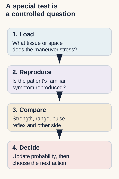
Mechanism first: a special test changes force, space or tension. Spurling narrows a cervical foramen, valgus stress loads the medial knee, and passive shoulder rotation asks whether the joint itself is stiff. The named maneuver is only shorthand for that controlled load.
Diagnosis second: call a test positive only when it reproduces the familiar complaint or reveals a reproducible deficit. Then compare an independent channel such as motor pattern, reflex, passive range, endpoint quality, pulse, swelling or the opposite side. One painful position rarely establishes one injured structure.
Management last: a result is useful when it changes an action. New cranial symptoms, progressive weakness, an ischemic limb, traumatic instability, a locked knee or an unsafe cervical spine ends provocative testing and redirects care. In a stable presentation, the same finding may support observation, targeted imaging, rehabilitation or specialty referral.
Use every maneuver in the same order: know the load, require a familiar response, compare independent findings, then decide whether to observe, investigate, refer or treat. [2][6][17]
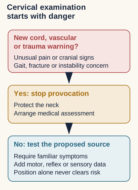
The first decision is not which provocative test to perform. It is whether the presentation is safe to provoke at all. [1][18][21]
Beyond the boards: record what happened
Reproducibility matters: document the starting position, side, load, familiar symptom, comparison finding and what changed after the maneuver. Avoid the unsupported statement that a test was simply positive. This makes follow-up meaningful when pain, strength or range changes over time.
1. Separate a nerve-root pattern from a safety problem
Predict first: a person reports thumb-side arm pain, but another reports sudden double vision with a new severe occipital headache. Should both receive a neck compression test? No. The second presentation requires urgent assessment for a potentially serious neurologic or vascular cause before manual examination. A normal pulse, normal resting blood pressure, or brief symptom resolution does not provide clearance. [1][18]
Spurling: in an appropriate, stable patient, the trained examiner uses cervical extension, ipsilateral lateral flexion and cautious axial loading. Reproduction of the familiar radiating arm symptom supports a cervical radicular source; local neck discomfort alone is a different finding. Cervical distraction may relieve familiar arm symptoms by reducing mechanical loading around the root. Neither response proves a particular disc lesion, and a negative result does not exclude radiculopathy. Use the neurologic examination and clinical context. [2]
Root localization needs agreement across findings
Predominant level
Useful comparison
Important limit
Predominant levelC5
Useful comparisonLateral upper arm; deltoid and biceps weakness
Important limitThese muscles have different peripheral nerves and overlapping C5-C6 supply.
Predominant levelC6
Useful comparisonLateral forearm/thumb; wrist extension; brachioradialis or biceps reflex
Important limitNo single reflex or sensory patch establishes a level.
Important limitConfirm strength, sensation and the symptom pattern together.
Predominant levelC8-T1
Useful comparisonFinger flexion, hand intrinsic function and medial hand/forearm symptoms
Important limitDistinguish root or lower-plexus disease from an isolated ulnar lesion.
A weak deltoid alone could reflect an axillary neuropathy. Deltoid plus biceps abnormalities involve two peripheral nerves; a concordant neck-provoked pattern makes a proximal source more persuasive. Normal biceps reflexes do not cancel partial root dysfunction. Hoffmann sign is not a diminished C8 tendon reflex: interpret it with other possible upper motor neuron findings, including brisk reflexes, gait change and hand dysfunction. [21]
Retire the historical clearance test. Sustained extension-rotation was traditionally called a vertebral artery or pre-manipulation positional test. IFOMPT does not recommend using positional vascular testing to exclude arterial pathology. A negative test cannot establish that cervical manipulation is safe. Do not deliberately repeat a provocative position when concerning symptoms appear, and do not substitute muscle energy, traction or another manual technique for necessary medical assessment. [1]
The historical 5 D's and 3 N's list includes dizziness, drop attacks, diplopia, dysarthria, dysphagia, nausea, nystagmus and facial numbness. It is a reminder, not a validated clearance checklist. Isolated nausea or dizziness has many causes; new focal deficits or a suspicious acute head/neck-pain presentation demand particular concern. Atlantoaxial instability is a separate structural safety issue, not something that this vascular positional test excludes. [1]
Wallenberg syndrome is a syndrome, not a clearance maneuver. Lateral medullary injury can combine ipsilateral facial and contralateral body pain-temperature loss with hoarseness, dysphagia, vertigo or ataxia. Vertebral/PICA circulation is relevant, but the exact vascular lesion requires investigation. A presentation after neck treatment does not, by itself, establish which artery dissected or prove causation. [18]
Transfer: familiar arm pain relieved by gentle distraction supports a root-loading interpretation. New dysarthria and imbalance change the task from localization by provocation to emergency assessment. Do not wait for the full historical mnemonic to appear.
Choose between two priorities: A: stable, familiar thumb-side radiation without warning findings. B: new double vision with severe new occipital pain. Which presentation changes the examination priority?
Compare your explanation
Safety hierarchy: B requires urgent medical assessment rather than more provocation. A permits a contextual root-focused examination only after safety assessment.
Try it here · Checkpoint 1 of 3
Make your prediction before reading the choices. A first attempt is just a starting point.
Case 6
Show answer and explanations for case 6
A. Repeat the positional test on both sides (Why this does not fit)
The earlier test is part of the recorded examination. Reprovocation does not safely exclude the acute vascular concern. A negative positional response cannot clear cervical arterial disease.
Reasoning steps for option A
In case 6, why can option A, “Repeat the positional test on both sides,” seem plausible before every finding is integrated?
Case 6, option A is a plausible competing interpretation; begin by matching its proposed tissue, level, force or action to the presenting region rather than accepting the name alone.
In case 6, which independent channel must be reconciled before accepting option A?
Case 6, option A must also account for the supplied strength, passive range, reflex, endpoint, perfusion, pain location or side-to-side comparison.
In case 6, how would option A change the immediate clinical decision if it were correct?
Case 6, option A should change urgency, testing, treatment, follow-up or counseling only after safety findings and the second examination channel agree.
B. Stop manual care and arrange emergency assessment (Best answer)
New head pain with diplopia and dysarthria is concerning. The prior negative test and usual blood pressure do not resolve that concern. Treat an acute focal neurologic presentation as a medical priority.
Reasoning steps for option B
In case 6, why can option B, “Stop manual care and arrange emergency assessment,” seem plausible before every finding is integrated?
Case 6, option B is the keyed synthesis; begin by matching its proposed tissue, level, force or action to the presenting region rather than accepting the name alone.
In case 6, which independent channel must be reconciled before accepting option B?
Case 6, option B must also account for the supplied strength, passive range, reflex, endpoint, perfusion, pain location or side-to-side comparison.
In case 6, how would option B change the immediate clinical decision if it were correct?
Case 6, option B should change urgency, testing, treatment, follow-up or counseling only after safety findings and the second examination channel agree.
C. Replace the planned treatment with muscle energy (Why this does not fit)
A non-thrust approach differs mechanically from a thrust. Changing manual technique does not address the acute neurologic presentation. Referral priority precedes choosing a manual method.
Reasoning steps for option C
In case 6, why can option C, “Replace the planned treatment with muscle energy,” seem plausible before every finding is integrated?
Case 6, option C is a plausible competing interpretation; begin by matching its proposed tissue, level, force or action to the presenting region rather than accepting the name alone.
In case 6, which independent channel must be reconciled before accepting option C?
Case 6, option C must also account for the supplied strength, passive range, reflex, endpoint, perfusion, pain location or side-to-side comparison.
In case 6, how would option C change the immediate clinical decision if it were correct?
Case 6, option C should change urgency, testing, treatment, follow-up or counseling only after safety findings and the second examination channel agree.
D. Observe until the symptoms fully resolve before referral (Why this does not fit)
Transient symptoms can improve spontaneously. Resolution would not exclude a serious vascular event. Do not require persistent deficits before urgent assessment.
Reasoning steps for option D
In case 6, why can option D, “Observe until the symptoms fully resolve before referral,” seem plausible before every finding is integrated?
Case 6, option D is a plausible competing interpretation; begin by matching its proposed tissue, level, force or action to the presenting region rather than accepting the name alone.
In case 6, which independent channel must be reconciled before accepting option D?
Case 6, option D must also account for the supplied strength, passive range, reflex, endpoint, perfusion, pain location or side-to-side comparison.
In case 6, how would option D change the immediate clinical decision if it were correct?
Case 6, option D should change urgency, testing, treatment, follow-up or counseling only after safety findings and the second examination channel agree.
Takeaway: A negative cervical positional test is not vascular clearance; new focal neurologic symptoms require urgent assessment.
Compare the two observations: a pulse fades during an overhead position in an otherwise asymptomatic arm; a different arm becomes acutely cold and painful and remains pale after being lowered. The first observation alone does not diagnose thoracic outlet syndrome. The persistent second pattern raises an urgent arterial concern. Repeated provocation is not the next useful step. [3][4]
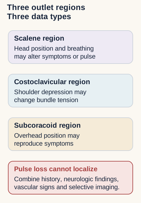
Outlet anatomy: The scalene triangle lies between the anterior and middle scalenes above the first rib. It contains the subclavian artery and brachial plexus; the subclavian vein passes anterior to the anterior scalene. The costoclavicular region lies between the clavicle and first rib. More distally, the bundle passes beneath pectoralis minor near its coracoid attachment. This anatomical map explains why position can alter symptoms, not why each provocative test would uniquely locate a lesion. [4]
Traditional outlet maneuvers: recognize the action without overcalling the result
Test
Traditional description
Interpretation boundary
TestAdson
Traditional descriptionHead toward the tested side, inspiration and an arm position that tensions the outlet; observe symptoms and pulse.
Interpretation boundaryTraditionally associated with the scalene region. Neither pulse loss nor symptom reproduction independently confirms TOS.
TestWright
Traditional descriptionAbduction of the arm overhead while observing the response.
Interpretation boundaryTraditionally associated with the subcoracoid region; does not uniquely prove pectoralis minor compression.
TestMilitary posture / costoclavicular
Traditional descriptionShoulders drawn back and down.
Interpretation boundaryChanges costoclavicular relationships, with nonspecific provocative responses.
TestRoos / EAST
Traditional descriptionShoulders and elbows near 90 degrees; repeated hand opening and closing within tolerance.
Interpretation boundaryInterpret familiar symptoms, not fatigue alone. False-positive responses occur.
Provocation limit: For the anatomical comparison, the anterior scalene typically connects the C3-C6 transverse processes to the first rib, whereas pectoralis minor connects ribs 3-5 to the coracoid. Their different attachments explain the traditional regional associations, not the diagnostic specificity of a maneuver.
A cervical rib is an anatomical risk factor, not proof of disease. Lower-plexus symptoms may include medial hand/forearm sensory change and weakness extending beyond a single distal nerve. Compare cervical radiculopathy, ulnar neuropathy and other explanations. Allen testing concerns radial-ulnar hand collateral circulation, not localization of a thoracic outlet lesion. Clinical assessment and selected electrodiagnostic or vascular studies answer different questions. [3][4]
State the subtype concern: neurologic pain/paresthesia and weakness suggest neural involvement; ischemic coldness or pallor suggests arterial compromise; a swollen cyanotic arm, particularly after effort, raises venous obstruction. New persistent ischemic findings or sudden marked swelling need prompt vascular assessment, not a sequence of repeated outlet tests. [4]
Transfer: a negative Adson result plus a symptomatic Wright position does not rule out a scalene contribution or establish a single subcoracoid lesion. Record the actual symptom, starting circulation, recovery and competing diagnoses.
Trace two vascular paths: Point to the scalene triangle in the diagram. Does the subclavian vein follow the artery through that triangle?
Compare your explanation
Vascular priority: No. The vein passes anterior to the anterior scalene. This is why a regional label must not erase the different anatomical pathways.
Read neural, arterial and venous patterns separately
Neural pattern: neurogenic thoracic outlet presentations are judged from symptom distribution, weakness, atrophy and exclusion of root or distal nerve disease. A cervical rib can increase anatomical risk, but it does not make a provocative pulse response diagnostic [4][20].
Arterial pattern: persistent pallor, coolness, ischemic pain, embolic change or loss of perfusion is not a reason to repeat Adson or Wright testing. It is a reason for urgent vascular assessment. A transient pulse change in an otherwise well limb is common enough that it cannot localize a compression site by itself [3][20].
Venous pattern: unilateral swelling, cyanosis, heaviness and venous distention with preserved arterial pulses suggests axillosubclavian venous obstruction. The clinical priority is thrombotic or obstructive evaluation rather than proving which posture narrows which region [20].
Provocative positions can organize the examination, but the diagnosis depends on the whole neural, arterial or venous pattern. [3][4][20]Recall the three classic positions without overclaiming
Adson, Wright and military posture emphasize the scalene, subcoracoid and costoclavicular regions, respectively. They are regional provocation maneuvers. They do not independently establish thoracic outlet syndrome or the exact tissue responsible.
3. Compare motion, strength and sensation
Predict: two patients cannot reach behind the back. One has nearly full passive range; the other remains restricted in external rotation even with examiner assistance. The same failed reach can have different explanations.
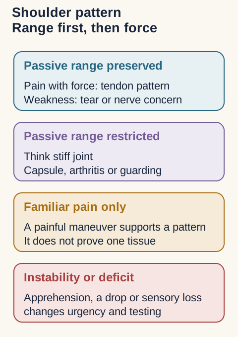
Apley scratch screens combined shoulder function. Reaching behind the head requires abduction and external rotation; reaching behind the back requires adduction and internal rotation. Scapular contribution and pain affect the result. Compare active and passive range. Restriction of both supports a stiff glenohumeral process, such as adhesive capsulitis or arthritis. Weak active function with better passive range suggests a contractile or neurologic problem. Coexisting disease can blur this distinction. [5][6]
Drop arm assesses control during lowering after assisted shoulder abduction. An uncontrolled drop supports substantial cuff dysfunction in context, but does not establish tear size. A negative result does not exclude a tear. Compare abduction, external rotation and internal rotation strength. Pain inhibition and nerve dysfunction can also cause weakness. [5][6]
Shoulder comparison: Supraspinatus contributes to abduction beyond initiation alone; infraspinatus and teres minor contribute to external rotation, and subscapularis to internal rotation. These are overlapping functions, not isolated diagnostic tests. The long head of biceps attaches at the superior labrum/supraglenoid region before entering the groove. [5][8]
Speed versus Yergason: Speed uses resisted shoulder flexion with an extended elbow and supinated forearm. Yergason uses resisted supination with the elbow flexed near 90 degrees. Record whether familiar pain localizes to the bicipital groove. Both load the biceps system, but neither reliably separates groove tendinopathy, labral disease and associated shoulder pathology. A negative Neer response does not establish isolated biceps disease. [7]
Hand localization: Deep pain or clicking during overhead activity can justify evaluating the superior labrum and biceps attachment. Speed and O'Brien findings alone do not establish a SLAP tear. Demonstrable biceps displacement suggests instability of its stabilizing apparatus, not necessarily an isolated transverse humeral ligament tear. The rotator-interval sling and adjacent cuff anatomy matter. [7][8]
Locate radial wrist pain before interpreting a stretch. De Quervain disease involves abductor pollicis longus (APL) and extensor pollicis brevis (EPB) in the first dorsal compartment near the radial styloid. Repetitive infant lifting or occupational thumb loading supplies context, not a diagnostic requirement. Maximum tenderness several centimeters more proximally with crepitus during repetitive wrist use instead suggests intersection-region irritation, where APL/EPB cross the second-compartment ECRL/ECRB tendons.
Finkelstein is not Eichhoff. Thumb inside a closed fist followed by ulnar wrist deviation describes the commonly mislabeled Eichhoff maneuver. Finkelstein uses examiner-controlled thumb traction and ulnar deviation without trapping the thumb inside the fist. Reproduced styloid pain is supportive, not conclusive. The comparative study in asymptomatic people addresses false-positive discomfort, not diagnostic sensitivity in patients with de Quervain disease. [9]
Force quality: After trauma, snuffbox tenderness requires a fracture-focused assessment rather than an immediate tendon label. Dorsoradial numbness suggests a superficial radial sensory nerve problem; palmar thumb catching suggests an A1 pulley problem; ulnar-sided pain redirects attention toward structures such as the TFCC. A mildly painful thumb stretch does not erase these localization differences.
Median nerve provocation at the wrist
Test
Action
Relevant response
TestTinel
ActionGentle tapping over the median nerve at the volar wrist.
Relevant responseFamiliar tingling into the fingers, not local tenderness alone.
TestPhalen
ActionWrist flexion, commonly with the hand dorsums together, for up to a minute as tolerated.
Relevant responseMedian-distribution paresthesia.
TestReverse Phalen
ActionPalms together with the wrists extended.
Relevant responseThe same sensory distribution with a different wrist position.
Clinical follow-up: Median digital symptoms usually concern the thumb, index, middle and radial half of the ring finger. Thenar eminence skin sensation may be spared because its palmar cutaneous branch leaves proximal to the tunnel. A negative Tinel or Phalen does not exclude carpal tunnel syndrome, and an isolated positive result is insufficient for diagnosis. [10]
Transfer: select a location and functional deficit before a test name. Groove pain with resisted supination, styloid pain with thumb loading, and index-finger paresthesia with wrist flexion are different outputs.
Compare the two dashed arm positions: Both solid arms show a limited active attempt. Which diagram redirects attention toward a stiff joint rather than active weakness alone?
Compare your explanation
Shoulder comparison: The lower diagram limits both active and examiner-assisted range. The upper diagram preserves passive range and requires a strength, pain and neurologic comparison.
Shoulder tests answer different questions
Range comes first: the Apley scratch pattern is a quick screen, not a diagnosis. Compare active and passive motion. Preserved passive motion with painful or weak active elevation redirects attention toward the cuff, pain inhibition or neural function. Restriction of both active and passive motion suggests a stiff glenohumeral joint, arthritis, fracture, guarding or adhesive capsulitis [5][26].
Range separates a stiff joint from active weakness. The quality of the response then separates pain provocation from loss of force, instability or nerve injury. [5][6][23][26]
Best interpretationPain with preserved force supports a painful cuff pattern; reproducible weakness raises concern for tear or nerve disease.
ManeuverDrop arm
Primary loadEccentric control during lowering
Best interpretationAn uncontrolled drop supports substantial cuff dysfunction, but a controlled descent does not exclude a tear.
ManeuverNeer or Hawkins-Kennedy
Primary loadSubacromial compression during elevation or internal rotation
Best interpretationFamiliar pain supports a subacromial pain pattern, not a unique tendon diagnosis.
ManeuverSpeed or Yergason
Primary loadLong-head biceps and adjacent stabilizing structures
Best interpretationAnterior pain may implicate the biceps-labral region, but neither maneuver proves an isolated tendon lesion.
ManeuverCross-body adduction
Primary loadAcromioclavicular joint compression
Best interpretationFocal superior joint pain supports an acromioclavicular source; diffuse pain needs broader assessment.
ManeuverO'Brien active compression
Primary loadAcromioclavicular and superior labral loading
Best interpretationPain location and change with forearm position matter more than the word positive.
ManeuverApprehension with relocation
Primary loadAnterior instability in abduction and external rotation
Best interpretationFear or guarding, relieved by posterior support, supports instability more than pain alone.
Pain versus weakness: this is the practical cuff distinction. Pain during resisted elevation with full force fits tendinopathy or pain inhibition better than a large tear. Definite weakness, a traumatic onset, inability to lower the arm, atrophy or a neurologic deficit deserves earlier imaging or specialty assessment. The current AAOS guideline addresses adult rotator-cuff management, but no single maneuver determines that management [6][23].
After dislocation: anterior shoulder dislocation can injure the axillary nerve. Test deltoid strength and sensation over the lateral shoulder once reduction and fracture safety are addressed. Medial scapular winging instead points toward long thoracic nerve dysfunction and serratus anterior weakness, not a cuff tear.
Adhesive capsulitis is a longitudinal diagnosis
Capsular pattern: progressive pain and loss of both active and passive glenohumeral motion, usually with prominent external-rotation restriction, is more informative than a painful impingement maneuver. Diabetes, thyroid disease and dyslipidemia increase risk. Ultrasound or MRI can exclude competing disease but should support, not replace, the clinical diagnosis [26].
Follow the trajectory: symptoms may persist beyond two years in some patients. Education, graded motion and rehabilitation are central; intra-articular corticosteroid injection can reduce pain and improve function, and physiotherapy or hydrodilatation with steroid may help selected adults. A positive shoulder test does not supply a medication dose. Verify any drug or injection plan against current labeling, comorbidities and local protocol [26].
Spencer sequence: the seven-stage articulatory sequence can be used as a controlled shoulder-mobility treatment after fracture, instability, infection and other contraindications are excluded. Circumduction with compression is passive and does not require a muscle-energy contraction. Current synthesis suggests improvement in pain, disability and range in adhesive capsulitis, without clear superiority over other manual therapies [29].
Transfer the shoulder rule to a new patient
Preserved passive range plus pain with full strength favors a painful tendon pattern. Loss of passive range redirects toward the joint capsule or arthritis. True focal weakness, instability or a nerve deficit changes the urgency and the workup.
Hand and elbow traps
Thumb-side wrist pain: Finkelstein uses examiner-controlled thumb traction and ulnar deviation without trapping the thumb inside a closed fist; the patient-made fist version is Eichhoff. Both load the first dorsal compartment, so maximal radial-styloid tenderness and the broader examination matter more than the label [9].
Median nerve pattern: Phalen and Tinel can support carpal tunnel syndrome, but thenar-eminence skin sensation is supplied before the tunnel. Thenar skin numbness or pronation weakness therefore argues for a more proximal median lesion despite positive wrist provocation [10].
Epicondylar pattern: pain at the medial epicondyle with resisted wrist flexion or pronation supports common-flexor loading, while lateral pain with resisted wrist extension supports common-extensor loading. Strength loss, locking, trauma or a broad neurologic pattern should not be reduced to golfer or tennis elbow.
4. Keep the pelvis in the reference frame
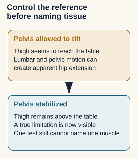
Predict: if a thigh appears to extend only when the pelvis tilts forward, has true hip extension improved? No. Some of the excursion came from the pelvis and lumbar region. Stabilization changes what the observation means. [11]
Crossed straight-leg raise: raising the less symptomatic leg with its knee extended reproduces familiar radicular pain in the opposite, symptomatic leg. Distinguish this from stretching discomfort in the raised hamstring. A concordant crossed response supports lumbar disc-related root irritation, but does not establish disc size, location or root level without other findings. New bladder dysfunction, saddle sensory change or progressive bilateral deficits requires urgent assessment. [17]
Thomas test: flex one hip sufficiently to control pelvic tilt while assessing the opposite thigh's extension position. Persistent opposite-thigh flexion supports an extension limitation, not necessarily an isolated psoas contracture. Knee position can alter rectus femoris tension, but several tissues and pain affect the result. [11]
Psoas tension assessment: the traditional prone assessment combines knee flexion near 90 degrees with controlled hip extension and observation of early pelvic/ASIS compensation. It is a length/loading observation, not an isolated psoas measurement. Rectus femoris, other anterior tissues, joint restriction and discomfort can affect it. This assessment is also distinct from the abdominal psoas sign. [13]
Ober test: side-lying hip positioning followed by controlled adduction assesses adduction limitation with a stabilized pelvis. It does not prove isolated iliotibial-band tightness or ITB syndrome. In a cadaveric study, ITB transection did not produce the expected change in the test; gluteal and capsular restraints contributed. This challenges tissue-specific interpretation, not the observed restriction. [12]
Patrick / FABERE (FABER): flexion, abduction and external rotation form a figure-four position; stabilize the opposite pelvis and assess tolerated range and pain location. Deep anterior groin pain favors a hip-region concern; posterior SI-region pain warrants assessment of that region. Neither location alone proves a diagnosis. Compare other hip findings or concordant SI pain-provocation findings. [15]
Transfer: first name the output: contralateral radicular pain, extension limitation with pelvic control, limited adduction, or localized pain. These are not interchangeable single-muscle diagnoses.
Predict after stabilizing the reference: The thigh appears to reach the table only while the pelvis tilts forward. Predict the finding after pelvic substitution is controlled.
Compare your explanation
Reference control: A hip extension limitation may become apparent. The diagram explains the reference error; it does not establish which single tissue caused the limitation.
Use provocation to localize, not to overname
Straight-leg raise: familiar below-knee radicular pain during ipsilateral elevation supports lumbosacral nerve-root irritation. Symptoms on the affected side when the opposite leg is raised are less sensitive but more specific for a disc-related process. Hip or hamstring discomfort without the neurologic pattern is not equivalent to radiculopathy [17].
Single-leg extension: the Stork or one-leg hyperextension maneuver is classically associated with pars stress, but pain can arise from several posterior structures and diagnostic accuracy is limited. Persistent focal extension pain in a young athlete is assessed with history, activity modification and appropriate imaging rather than declared spondylolysis from one maneuver [31].
The result depends on what was held still. Stabilization improves the measurement but does not turn a multistructure test into an isolated psoas diagnosis. [11][12][13]
FABER location: anterior groin pain redirects toward the hip joint, whereas posterior pain may implicate the sacroiliac region only when it agrees with other pain-provocation findings. A positive position never names a sacral torsion by itself [15][22].
Beyond the boards: decide when not to continue
Escalation findings: new urinary retention, saddle sensory loss, bilateral progressive weakness, fever, major trauma or cancer risk requires urgent medical assessment. Persistent radicular pain without progression can be followed over time, with imaging chosen when the result is likely to change management [17].
Osteopathic considerations
Keep descriptive tests descriptive
Compare two questions: does the pelvis permit a particular motion, and does that motion reproduce the person's familiar pain? The answers are not equivalent. Traditional motion labels describe an examination pattern; they do not independently identify a pain generator, torn tissue or radiographic lesion. [14][15]
Hip drop: in the traditional standing screen, bending one knee while keeping the heel supported allows that side of the pelvis to descend; observe the compensatory lumbar side-bending response. A right-sided pelvic drop assesses the left lumbar side-bending response. Describe asymmetry rather than applying an unsupported universal angle cutoff. Balance, hip mobility, leg-length differences and pain can affect the observation. This is not the same as Trendelenburg testing of stance-side abductor function.
Pelvic side shift: stabilize the upper trunk and compare lateral translation of the pelvis. Record the actual easier and restricted directions. Some teaching conventions name a positive result by the freer side; the name alone therefore invites confusion. An easier left translation does not, by itself, establish a right psoas lesion or a sacral torsion. Compare symptoms, hip examination and repeated findings. [13]
Spring test: in traditional OMM assessment, reduced anterior springing near the lumbosacral junction/sacral base is described as a positive spring and associated with a posterior-base/extension pattern. Guarding, pain and examiner technique affect the finding. It does not diagnose a fracture, establish a pain source, or identify a torsional axis. Structural injury concerns take priority over springing.
Sphinx / backward-bending test: supported prone lumbar extension is followed by reassessment of sacral asymmetry. Improvement traditionally fits an anterior-base/flexion or forward-torsion pattern; persistent or increased asymmetry fits a posterior-base/extension or backward-torsion pattern. Traditional forward labels include left-on-left and right-on-right; backward labels include left-on-right and right-on-left. Sphinx alone cannot select the axis or a unique named torsion. These are descriptive conventions, not independently validated tissue diagnoses. [14]
When findings disagree: a painful positive spring with an improving sphinx result is not a reason to force a diagnostic label. Recheck positioning, patient tolerance, landmarks and the broader examination. SI pain-provocation findings address a different question from positional palpation. [15]
Transfer: document “left translation easier; right restricted” before proposing an explanation. For a sacral label, require a coherent set of observations; for the pain source, require a separate concordant assessment.
Reconcile discordant observations: A painful spring assessment appears restricted, but supported extension reduces the observed asymmetry. Should this be forced into one named torsion?
Compare your explanation
OMT principle: No. Recheck tolerance, positioning and the other observations. Report the discordance rather than inventing an axis or assuming a pain source.
Barrier language predicts the technique
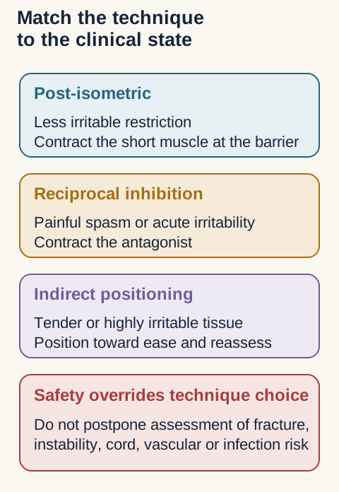
Restrictive barrier: this is the first limit to physiologic motion in the direction of restriction. A direct technique engages that barrier. An indirect technique carries the region away from the barrier toward ease. A pathologic barrier reflects disease or injury and is not a target to push through.
Muscle energy: muscle energy is active and usually direct. Post-isometric relaxation gently contracts the shortened muscle at the barrier, then takes up the new range. Reciprocal inhibition contracts the antagonist and is often chosen when the short muscle is acutely painful or in spasm. The oculocephalogyric approach uses voluntary eye movement to recruit small cervical motor activity in the intended direction [27].
Technique names become useful only after the clinician has established safety, irritability and the direction of the restrictive barrier. [27][28]Choose the muscle-energy strategy
Less irritable chronic restriction favors a gentle contraction of the shortened muscle followed by a new barrier. Acute painful spasm favors antagonist contraction away from the barrier. Neither strategy overrides consent, trauma assessment or neurologic safety.
SCM, torticollis and cervical treatment recognition
SCM action: one sternocleidomastoid side-bends the head toward itself and rotates the face away. Congenital shortening therefore produces ipsilateral head tilt with contralateral facial rotation [30].
Anterior C7 counterstrain: the classic tender point lies on the superior clavicle at the clavicular attachment of the SCM. The position of ease uses marked flexion to C7, side-bending toward the tender point and rotation away. Reassess tenderness rather than forcing a memorized angle, and do not apply cervical treatment when instability, fracture, vascular or cord risk is unresolved [28].
Contralateral traction: in this soft-tissue setup, the clinician stabilizes the shoulder while gently lengthening the cervical tissues away from it. Treat it as a low-force soft-tissue method, not as a clearance test for whiplash or vascular disease.
Whiplash sequence: a rear impact can produce rapid cervical extension followed by flexion. The board-level sequence helps predict which tissues are loaded, but severe midline pain, neurologic deficit, altered mental status or high-risk trauma requires formal cervical-spine assessment before any OMT.
Shoulder-girdle OMT belongs after structural safety
Clavicle and scapula: Still technique combines a position of ease, a compressive or traction force and a smooth path through the barrier. Indirect scapular myofascial release follows tissue ease before reassessment. Neither technique substitutes for imaging of a suspected fracture or management of an unstable acromioclavicular or sternoclavicular injury.
Upper crossed pattern: overactivity of upper trapezius, levator scapulae, pectoral muscles and cervical extensors with relative weakness of deep neck flexors and lower scapular stabilizers is a useful postural pattern, not a diagnosis for every neck or shoulder complaint. Link it to the patient's symptoms and function before treating.
Sacral screens: hip-drop, side-shift, spring and sphinx findings can describe motion preferences within traditional OMM terminology. They do not independently validate a tissue lesion, pain generator or named torsional axis. Discordant findings should be documented and rechecked rather than forced into one label.
Beyond the boards: consent, response and follow-up
Treatment record: document indication, consent, region, technique, pre-treatment finding, immediate response and any adverse symptom. Stop when pain becomes unfamiliar, neurologic findings appear or tolerance worsens. Reassessment is part of the technique, not an optional final step.
Try it here · Checkpoint 2 of 3
Make your prediction before reading the choices. A first attempt is just a starting point.
Case 41
Show answer and explanations for case 41
A. Contract the antagonist using reciprocal inhibition (Best answer)
Reciprocal inhibition asks the antagonist to contract so the acutely painful agonist can reduce tone without being directly recruited against a painful barrier.
Reasoning steps for option A
In case 41, why can option A, “Contract the antagonist using reciprocal inhibition,” seem plausible before every finding is integrated?
Case 41, option A is the keyed synthesis; begin by matching its proposed tissue, level, force or action to the presenting region rather than accepting the name alone.
In case 41, which independent channel must be reconciled before accepting option A?
Case 41, option A must also account for the supplied strength, passive range, reflex, endpoint, perfusion, pain location or side-to-side comparison.
In case 41, how would option A change the immediate clinical decision if it were correct?
Case 41, option A should change urgency, testing, treatment, follow-up or counseling only after safety findings and the second examination channel agree.
B. Contract the painful muscle using post-isometric relaxation (Why this does not fit)
Post-isometric relaxation recruits the shortened muscle at the barrier. It is often useful for less irritable chronic restriction, but direct recruitment is poorly tolerated here.
Reasoning steps for option B
In case 41, why can option B, “Contract the painful muscle using post-isometric relaxation,” seem plausible before every finding is integrated?
Case 41, option B is a plausible competing interpretation; begin by matching its proposed tissue, level, force or action to the presenting region rather than accepting the name alone.
In case 41, which independent channel must be reconciled before accepting option B?
Case 41, option B must also account for the supplied strength, passive range, reflex, endpoint, perfusion, pain location or side-to-side comparison.
In case 41, how would option B change the immediate clinical decision if it were correct?
Case 41, option B should change urgency, testing, treatment, follow-up or counseling only after safety findings and the second examination channel agree.
C. Apply cervical high-velocity low-amplitude thrust (Why this does not fit)
A thrust is passive and direct, not muscle energy. Acute pain and incomplete safety assessment also make an immediate cervical thrust inappropriate.
Reasoning steps for option C
In case 41, why can option C, “Apply cervical high-velocity low-amplitude thrust,” seem plausible before every finding is integrated?
Case 41, option C is a plausible competing interpretation; begin by matching its proposed tissue, level, force or action to the presenting region rather than accepting the name alone.
In case 41, which independent channel must be reconciled before accepting option C?
Case 41, option C must also account for the supplied strength, passive range, reflex, endpoint, perfusion, pain location or side-to-side comparison.
In case 41, how would option C change the immediate clinical decision if it were correct?
Case 41, option C should change urgency, testing, treatment, follow-up or counseling only after safety findings and the second examination channel agree.
D. Hold the tissue at the restrictive barrier without contraction (Why this does not fit)
Holding the barrier without patient effort is not an active muscle-energy setup. The question specifically asks for antagonist recruitment to reduce the painful muscle's activity.
Reasoning steps for option D
In case 41, why can option D, “Hold the tissue at the restrictive barrier without contraction,” seem plausible before every finding is integrated?
Case 41, option D is a plausible competing interpretation; begin by matching its proposed tissue, level, force or action to the presenting region rather than accepting the name alone.
In case 41, which independent channel must be reconciled before accepting option D?
Case 41, option D must also account for the supplied strength, passive range, reflex, endpoint, perfusion, pain location or side-to-side comparison.
In case 41, how would option D change the immediate clinical decision if it were correct?
Case 41, option D should change urgency, testing, treatment, follow-up or counseling only after safety findings and the second examination channel agree.
Takeaway: Reciprocal inhibition contracts the antagonist and is useful when directly recruiting an acutely painful spastic muscle is poorly tolerated.
6. Establish the starting position before judging laxity
Predict: two tibias travel the same distance during an anterior pull. One starts posteriorly sagged and reaches neutral; the other starts neutral and travels beyond it. Are both ACL-deficient? No. The starting reference and final endpoint matter, not excursion alone.
Restore the posteriorly sagged tibia to neutral and compare
Knee reference: The illustrated posteriorly sagged tibia has returned to the expected starting relation. Its apparent anterior travel was restoration, not excess anterior translation. A further anterior pull from the corrected reference, compared with the other knee and the endpoint, asks a different question. The distances are conceptual, not diagnostic thresholds.
Force direction: The comparison is available without JavaScript: open the control to display the corrected anatomical reference, then close it to compare with the resting position. Both explanations remain readable. Posterior sag suggests PCL dysfunction; an apparent positive anterior drawer can be misleading when the initial position is ignored.
Loading direction determines the question
Test
Action and useful finding
Interpretation limit
TestLachman
Action and useful findingKnee near 20-30 degrees; assess anterior tibial translation relative to a stabilized femur and its endpoint.
Interpretation limitAsymmetry with a soft endpoint supports ACL dysfunction. Guarding and technique affect interpretation.
TestAnterior drawer
Action and useful findingKnee near 90 degrees; assess anterior translation after checking the resting tibial position.
Interpretation limitA posteriorly sagged starting point can create apparent anterior laxity.
TestPosterior drawer
Action and useful findingKnee near 90 degrees; assess posterior translation and compare the resting step-off with the other side.
Interpretation limitPosterior laxity/sag supports PCL dysfunction, especially after a posterior force to the flexed tibia.
TestApley compression
Action and useful findingProne, knee near 90 degrees; axial tibial compression with rotation assesses familiar joint-line pain.
Interpretation limitSupports meniscal pathology in context; not a definitive meniscal diagnosis.
TestApley distraction
Action and useful findingTraction with rotation assesses capsular/ligamentous pain rather than compression loading.
Interpretation limitDoes not by itself identify an individual collateral ligament.
TestMcMurray
Action and useful findingRotation during extension from knee flexion; external tibial rotation emphasizes medial assessment and internal rotation lateral assessment.
Interpretation limitA concordant painful joint-line click is more meaningful than painless noise. A negative result does not exclude a tear.
Endpoint quality: Combine the examination with the event: pivoting with early swelling and anterior laxity supports ACL injury; a dashboard-type force with posterior sag supports PCL injury; twisting with delayed swelling, joint-line pain and catching supports a meniscal concern. Valgus or varus stress assesses collateral stability and adds information that an Apley distraction response cannot supply. Major trauma can injure several structures, so one positive test should not end the examination. [16]
Transfer: record the baseline tibial position, direction of the applied load, familiar symptom, endpoint and comparison side. Then decide whether the finding supports instability, compression-sensitive joint-line pathology, or an examination limited by pain.
Translate force direction into the injured restraint
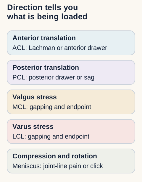
Cruciate direction: the ACL resists anterior tibial translation and rotational instability; the PCL resists posterior translation. A posteriorly sagged tibia can make the anterior drawer appear excessive, so restore the starting reference before judging the endpoint [19].
Collateral direction: valgus stress loads the MCL and varus stress loads the LCL. Compare pain, gapping and endpoint at the tested knee angle. Joint-line tenderness or swelling can coexist and should not be relabeled as isolated ligament injury.
Start with force direction and tibial position. Then add mechanism, effusion, joint-line findings and endpoint quality. [16][19][24][25]
Meniscal loading: McMurray combines tibial rotation with flexion-extension while the examiner follows the joint line. Apley compression loads the meniscus, while distraction shifts stress toward the collateral ligaments. A painful click supports a meniscal pattern only when mechanism, location and mechanical symptoms agree.
LIME setup mnemonic: lateral meniscus with internal tibial rotation, medial meniscus with external tibial rotation. Use LIME to remember the intended rotational bias during McMurray testing, then require concordant joint-line symptoms, an appropriate mechanism and the rest of the examination. The mnemonic does not convert a painless click or isolated maneuver into a diagnosis. [16]
The unhappy triad is a mnemonic, not a complete injury report
Classic triad: a lateral blow to a planted knee creates valgus and rotational load, classically linking ACL, MCL and medial-meniscus injury. The medial meniscus is less mobile because of its capsular and MCL attachments. That framing remains board-relevant [32].
Modern correction: acute ACL-MCL injuries frequently involve the lateral meniscus, and combined patterns vary. Use the classic triad to remember the force and restraints, then report the actual MRI, examination and operative findings rather than assuming the meniscus side [32].
Effusion, alignment and surface landmarks refine the differential
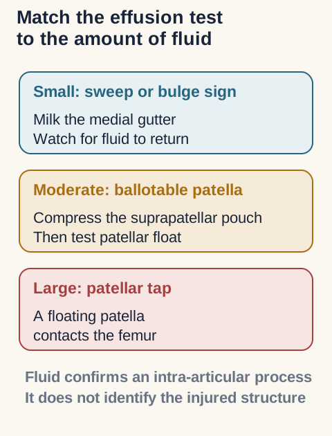
Choose the fluid test by expected volume, then return to mechanism, instability and joint-line findings to identify the lesion. [16][24][25]
Fluid volume: a small effusion is best sought with a sweep or bulge sign; a moderate collection can ballot the patella; a large collection may create a patellar tap. Fluid confirms an intra-articular process but does not identify ACL, meniscus, fracture or inflammation by itself.
Bursa location: prepatellar bursitis produces anterior swelling over the patella, often after kneeling. Pes anserine pain is inferomedial to the joint line at the sartorius, gracilis and semitendinosus insertion. Neither should create true cruciate laxity.
Alignment link: varus alignment increases medial-compartment load and often accompanies medial knee osteoarthritis. Pes planus can increase tibial internal rotation and alter medial knee loading, but foot posture alone does not diagnose a meniscal tear.
Beyond the boards: the result must change care
Urgent patterns: a dislocated or grossly unstable knee requires immediate neurovascular assessment, even if it has reduced spontaneously. A locked knee, displaced tear, fracture concern or rapidly expanding effusion warrants expedited imaging or orthopedic evaluation.
ACL trajectory: treatment depends on instability, associated injury, activity goals, age and shared decision-making. Nonsurgical care can succeed in selected patients, but recurrent giving-way can lead to additional meniscal or cartilage injury. When reconstruction is chosen, the rehabilitation plan and return criteria matter as much as the examination name [24].
Meniscus scope: the 2024 AAOS guideline addresses acute isolated meniscal pathology and explicitly excludes chronic, degenerative, root, repeat and combined ACL-associated tears. Apply its recommendations only inside that boundary [25].
Reconcile a negative McMurray test with recurrent locking
History still matters: one quiet examination does not erase recurrent mechanical episodes. Reassess the joint line, effusion, range and ligament stability, then choose imaging or referral according to persistence, locking and functional loss.
Try it here · Checkpoint 3 of 3
Make your prediction before reading the choices. A first attempt is just a starting point.
Case 45
Show answer and explanations for case 45
A. Classic ACL-MCL-medial meniscus triad (Best answer)
Anterior laxity implicates the ACL, valgus gapping implicates the MCL, and a medial joint-line click supports medial meniscal injury. Together they form the classic triad.
Reasoning steps for option A
In case 45, why can option A, “Classic ACL-MCL-medial meniscus triad,” seem plausible before every finding is integrated?
Case 45, option A is the keyed synthesis; begin by matching its proposed tissue, level, force or action to the presenting region rather than accepting the name alone.
In case 45, which independent channel must be reconciled before accepting option A?
Case 45, option A must also account for the supplied strength, passive range, reflex, endpoint, perfusion, pain location or side-to-side comparison.
In case 45, how would option A change the immediate clinical decision if it were correct?
Case 45, option A should change urgency, testing, treatment, follow-up or counseling only after safety findings and the second examination channel agree.
B. PCL-LCL-lateral meniscus triad (Why this does not fit)
The described forces and examination are opposite: posterior translation would implicate the PCL, varus gapping the LCL, and lateral joint-line findings the lateral meniscus.
Reasoning steps for option B
In case 45, why can option B, “PCL-LCL-lateral meniscus triad,” seem plausible before every finding is integrated?
Case 45, option B is a plausible competing interpretation; begin by matching its proposed tissue, level, force or action to the presenting region rather than accepting the name alone.
In case 45, which independent channel must be reconciled before accepting option B?
Case 45, option B must also account for the supplied strength, passive range, reflex, endpoint, perfusion, pain location or side-to-side comparison.
In case 45, how would option B change the immediate clinical decision if it were correct?
Case 45, option B should change urgency, testing, treatment, follow-up or counseling only after safety findings and the second examination channel agree.
C. Isolated medial collateral ligament sprain (Why this does not fit)
Valgus gapping supports MCL injury, but anterior laxity and a medial meniscal finding demonstrate additional intra-articular structures rather than an isolated sprain.
Reasoning steps for option C
In case 45, why can option C, “Isolated medial collateral ligament sprain,” seem plausible before every finding is integrated?
Case 45, option C is a plausible competing interpretation; begin by matching its proposed tissue, level, force or action to the presenting region rather than accepting the name alone.
In case 45, which independent channel must be reconciled before accepting option C?
Case 45, option C must also account for the supplied strength, passive range, reflex, endpoint, perfusion, pain location or side-to-side comparison.
In case 45, how would option C change the immediate clinical decision if it were correct?
Case 45, option C should change urgency, testing, treatment, follow-up or counseling only after safety findings and the second examination channel agree.
D. Isolated anterior cruciate ligament tear (Why this does not fit)
The ACL explains anterior laxity, but it does not account for valgus gapping or the concordant medial joint-line click. This is a combined injury pattern.
Reasoning steps for option D
In case 45, why can option D, “Isolated anterior cruciate ligament tear,” seem plausible before every finding is integrated?
Case 45, option D is a plausible competing interpretation; begin by matching its proposed tissue, level, force or action to the presenting region rather than accepting the name alone.
In case 45, which independent channel must be reconciled before accepting option D?
Case 45, option D must also account for the supplied strength, passive range, reflex, endpoint, perfusion, pain location or side-to-side comparison.
In case 45, how would option D change the immediate clinical decision if it were correct?
Case 45, option D should change urgency, testing, treatment, follow-up or counseling only after safety findings and the second examination channel agree.
Takeaway: The classic unhappy triad is ACL, MCL, and medial meniscus, but actual combined injuries must be reported from the full examination and imaging.
Choose a level: Level 1 emphasizes mechanism and localization, Level 2 adds diagnostic comparison, Level 3 adds urgency, follow-up and management scope, and Beyond adds current evidence limits, referral thresholds and counseling.
Apply the comparisons
Retrieval practice: Use the clinical findings together. Select one best answer, then compare the explanation for every option with the feature that would change your conclusion.
Case 1
Show answer and explanations for case 1
A. Prioritize cervical protection and trauma assessment (Best answer)
The fall, midline pain and neurologic symptoms raise structural injury concern. Stability must be considered before compressive or traction maneuvers. Triage the risk before pursuing a special-test result.
Reasoning steps for option A
In case 1, why can option A, “Prioritize cervical protection and trauma assessment,” seem plausible before every finding is integrated?
Case 1, option A is the keyed synthesis; begin by matching its proposed tissue, level, force or action to the presenting region rather than accepting the name alone.
In case 1, which independent channel must be reconciled before accepting option A?
Case 1, option A must also account for the supplied strength, passive range, reflex, endpoint, perfusion, pain location or side-to-side comparison.
In case 1, how would option A change the immediate clinical decision if it were correct?
Case 1, option A should change urgency, testing, treatment, follow-up or counseling only after safety findings and the second examination channel agree.
B. Perform Spurling testing before ordering imaging (Why this does not fit)
Thumb symptoms could reflect a cervical root. Recent trauma and midline pain make provocative loading premature. A plausible localization does not replace a safety assessment.
Reasoning steps for option B
In case 1, why can option B, “Perform Spurling testing before ordering imaging,” seem plausible before every finding is integrated?
Case 1, option B is a plausible competing interpretation; begin by matching its proposed tissue, level, force or action to the presenting region rather than accepting the name alone.
In case 1, which independent channel must be reconciled before accepting option B?
Case 1, option B must also account for the supplied strength, passive range, reflex, endpoint, perfusion, pain location or side-to-side comparison.
In case 1, how would option B change the immediate clinical decision if it were correct?
Case 1, option B should change urgency, testing, treatment, follow-up or counseling only after safety findings and the second examination channel agree.
C. Perform cervical distraction before checking stability (Why this does not fit)
Traction can sometimes relieve radicular symptoms. Symptom relief would not establish stability after this fall. A relieving maneuver is not a trauma clearance test.
Reasoning steps for option C
In case 1, why can option C, “Perform cervical distraction before checking stability,” seem plausible before every finding is integrated?
Case 1, option C is a plausible competing interpretation; begin by matching its proposed tissue, level, force or action to the presenting region rather than accepting the name alone.
In case 1, which independent channel must be reconciled before accepting option C?
Case 1, option C must also account for the supplied strength, passive range, reflex, endpoint, perfusion, pain location or side-to-side comparison.
In case 1, how would option C change the immediate clinical decision if it were correct?
Case 1, option C should change urgency, testing, treatment, follow-up or counseling only after safety findings and the second examination channel agree.
D. Use shoulder testing to exclude a cervical injury (Why this does not fit)
A shoulder impact can injure local tissues. A shoulder examination cannot exclude cervical injury with these findings. Adjacent-region findings do not clear the spine.
Reasoning steps for option D
In case 1, why can option D, “Use shoulder testing to exclude a cervical injury,” seem plausible before every finding is integrated?
Case 1, option D is a plausible competing interpretation; begin by matching its proposed tissue, level, force or action to the presenting region rather than accepting the name alone.
In case 1, which independent channel must be reconciled before accepting option D?
Case 1, option D must also account for the supplied strength, passive range, reflex, endpoint, perfusion, pain location or side-to-side comparison.
In case 1, how would option D change the immediate clinical decision if it were correct?
Case 1, option D should change urgency, testing, treatment, follow-up or counseling only after safety findings and the second examination channel agree.
Takeaway: After trauma, establish safety before provoking a suspected nerve-root pattern.
A. Median nerve at the carpal tunnel (Why this does not fit)
Thumb paresthesia can occur in carpal tunnel syndrome. A wrist-level median lesion does not explain wrist-extensor weakness and a reduced brachioradialis reflex. Compare functions supplied outside the suspected peripheral nerve.
Reasoning steps for option A
In case 2, why can option A, “Median nerve at the carpal tunnel,” seem plausible before every finding is integrated?
Case 2, option A is a plausible competing interpretation; begin by matching its proposed tissue, level, force or action to the presenting region rather than accepting the name alone.
In case 2, which independent channel must be reconciled before accepting option A?
Case 2, option A must also account for the supplied strength, passive range, reflex, endpoint, perfusion, pain location or side-to-side comparison.
In case 2, how would option A change the immediate clinical decision if it were correct?
Case 2, option A should change urgency, testing, treatment, follow-up or counseling only after safety findings and the second examination channel agree.
B. C6-predominant cervical nerve root (Best answer)
Lateral forearm/thumb symptoms align with the motor and reflex pattern. Concordant cervical loading responses further support a proximal source. Several agreeing findings are stronger than a sensory patch alone.
Reasoning steps for option B
In case 2, why can option B, “C6-predominant cervical nerve root,” seem plausible before every finding is integrated?
Case 2, option B is the keyed synthesis; begin by matching its proposed tissue, level, force or action to the presenting region rather than accepting the name alone.
In case 2, which independent channel must be reconciled before accepting option B?
Case 2, option B must also account for the supplied strength, passive range, reflex, endpoint, perfusion, pain location or side-to-side comparison.
In case 2, how would option B change the immediate clinical decision if it were correct?
Case 2, option B should change urgency, testing, treatment, follow-up or counseling only after safety findings and the second examination channel agree.
C. C7-predominant cervical nerve root (Why this does not fit)
Cervical disease can produce arm pain and weakness. A middle-finger and triceps-predominant pattern would support C7 more strongly. Root territories overlap, so use the dominant combined pattern.
Reasoning steps for option C
In case 2, why can option C, “C7-predominant cervical nerve root,” seem plausible before every finding is integrated?
Case 2, option C is a plausible competing interpretation; begin by matching its proposed tissue, level, force or action to the presenting region rather than accepting the name alone.
In case 2, which independent channel must be reconciled before accepting option C?
Case 2, option C must also account for the supplied strength, passive range, reflex, endpoint, perfusion, pain location or side-to-side comparison.
In case 2, how would option C change the immediate clinical decision if it were correct?
Case 2, option C should change urgency, testing, treatment, follow-up or counseling only after safety findings and the second examination channel agree.
D. Ulnar nerve at the cubital tunnel (Why this does not fit)
Entrapment can cause hand sensory symptoms. The thumb-side distribution and wrist-extensor/reflex findings do not fit an isolated ulnar lesion. Medial digital symptoms require a different comparison.
Reasoning steps for option D
In case 2, why can option D, “Ulnar nerve at the cubital tunnel,” seem plausible before every finding is integrated?
Case 2, option D is a plausible competing interpretation; begin by matching its proposed tissue, level, force or action to the presenting region rather than accepting the name alone.
In case 2, which independent channel must be reconciled before accepting option D?
Case 2, option D must also account for the supplied strength, passive range, reflex, endpoint, perfusion, pain location or side-to-side comparison.
In case 2, how would option D change the immediate clinical decision if it were correct?
Case 2, option D should change urgency, testing, treatment, follow-up or counseling only after safety findings and the second examination channel agree.
Takeaway: Spurling and distraction support a root-loading interpretation; the accompanying neurologic pattern supplies localization.
A. Axillary nerve near the humeral neck (Why this does not fit)
An axillary lesion can weaken the deltoid. It does not explain biceps and cervical paraspinal abnormalities. Test a second muscle outside the proposed nerve.
Reasoning steps for option A
In case 3, why can option A, “Axillary nerve near the humeral neck,” seem plausible before every finding is integrated?
Case 3, option A is a plausible competing interpretation; begin by matching its proposed tissue, level, force or action to the presenting region rather than accepting the name alone.
In case 3, which independent channel must be reconciled before accepting option A?
Case 3, option A must also account for the supplied strength, passive range, reflex, endpoint, perfusion, pain location or side-to-side comparison.
In case 3, how would option A change the immediate clinical decision if it were correct?
Case 3, option A should change urgency, testing, treatment, follow-up or counseling only after safety findings and the second examination channel agree.
B. Musculocutaneous nerve in the arm (Why this does not fit)
This nerve supplies the biceps. It does not explain deltoid and cervical paraspinal abnormalities. One weak muscle should not define the whole localization.
Reasoning steps for option B
In case 3, why can option B, “Musculocutaneous nerve in the arm,” seem plausible before every finding is integrated?
Case 3, option B is a plausible competing interpretation; begin by matching its proposed tissue, level, force or action to the presenting region rather than accepting the name alone.
In case 3, which independent channel must be reconciled before accepting option B?
Case 3, option B must also account for the supplied strength, passive range, reflex, endpoint, perfusion, pain location or side-to-side comparison.
In case 3, how would option B change the immediate clinical decision if it were correct?
Case 3, option B should change urgency, testing, treatment, follow-up or counseling only after safety findings and the second examination channel agree.
C. C5-predominant cervical nerve root (Best answer)
The deltoid and biceps share proximal root contributions. Paraspinal involvement and concordant neck symptoms support a root lesion across peripheral nerves. A preserved reflex does not exclude a partial root disorder.
Reasoning steps for option C
In case 3, why can option C, “C5-predominant cervical nerve root,” seem plausible before every finding is integrated?
Case 3, option C is the keyed synthesis; begin by matching its proposed tissue, level, force or action to the presenting region rather than accepting the name alone.
In case 3, which independent channel must be reconciled before accepting option C?
Case 3, option C must also account for the supplied strength, passive range, reflex, endpoint, perfusion, pain location or side-to-side comparison.
In case 3, how would option C change the immediate clinical decision if it were correct?
Case 3, option C should change urgency, testing, treatment, follow-up or counseling only after safety findings and the second examination channel agree.
D. Suprascapular nerve near the scapula (Why this does not fit)
A proximal shoulder neuropathy can impair shoulder function. Its principal cuff muscles do not match the supplied deltoid-biceps pattern. Use the actual affected muscles rather than the region alone.
Reasoning steps for option D
In case 3, why can option D, “Suprascapular nerve near the scapula,” seem plausible before every finding is integrated?
Case 3, option D is a plausible competing interpretation; begin by matching its proposed tissue, level, force or action to the presenting region rather than accepting the name alone.
In case 3, which independent channel must be reconciled before accepting option D?
Case 3, option D must also account for the supplied strength, passive range, reflex, endpoint, perfusion, pain location or side-to-side comparison.
In case 3, how would option D change the immediate clinical decision if it were correct?
Case 3, option D should change urgency, testing, treatment, follow-up or counseling only after safety findings and the second examination channel agree.
Takeaway: Weakness across different peripheral nerves with compatible paraspinal findings supports a root-level localization.
A. The sensory territory establishes an ulnar elbow lesion (Why this does not fit)
Medial digital symptoms commonly occur with ulnar neuropathy. Index distal flexion is not an ulnar function. Sensory overlap cannot outweigh a broader motor distribution.
Reasoning steps for option A
In case 4, why can option A, “The sensory territory establishes an ulnar elbow lesion,” seem plausible before every finding is integrated?
Case 4, option A is a plausible competing interpretation; begin by matching its proposed tissue, level, force or action to the presenting region rather than accepting the name alone.
In case 4, which independent channel must be reconciled before accepting option A?
Case 4, option A must also account for the supplied strength, passive range, reflex, endpoint, perfusion, pain location or side-to-side comparison.
In case 4, how would option A change the immediate clinical decision if it were correct?
Case 4, option A should change urgency, testing, treatment, follow-up or counseling only after safety findings and the second examination channel agree.
B. The index weakness establishes a distal median lesion (Why this does not fit)
The median/anterior interosseous pathway supplies index distal flexion. A distal median lesion does not explain ulnar intrinsic weakness with cervical provocation. A second affected nerve changes the localization.
Reasoning steps for option B
In case 4, why can option B, “The index weakness establishes a distal median lesion,” seem plausible before every finding is integrated?
Case 4, option B is a plausible competing interpretation; begin by matching its proposed tissue, level, force or action to the presenting region rather than accepting the name alone.
In case 4, which independent channel must be reconciled before accepting option B?
Case 4, option B must also account for the supplied strength, passive range, reflex, endpoint, perfusion, pain location or side-to-side comparison.
In case 4, how would option B change the immediate clinical decision if it were correct?
Case 4, option B should change urgency, testing, treatment, follow-up or counseling only after safety findings and the second examination channel agree.
C. The hand weakness establishes cervical myelopathy (Why this does not fit)
Cord disease can impair hand function. The supplied focal lower-motor pattern lacks corroborating cord findings. Hand weakness alone is not an upper motor neuron diagnosis.
Reasoning steps for option C
In case 4, why can option C, “The hand weakness establishes cervical myelopathy,” seem plausible before every finding is integrated?
Case 4, option C is a plausible competing interpretation; begin by matching its proposed tissue, level, force or action to the presenting region rather than accepting the name alone.
In case 4, which independent channel must be reconciled before accepting option C?
Case 4, option C must also account for the supplied strength, passive range, reflex, endpoint, perfusion, pain location or side-to-side comparison.
In case 4, how would option C change the immediate clinical decision if it were correct?
Case 4, option C should change urgency, testing, treatment, follow-up or counseling only after safety findings and the second examination channel agree.
D. The motor distribution favors a proximal root process (Best answer)
The weak muscles receive supply through more than one peripheral nerve. The cervical response and C8-predominant motor pattern support a proximal explanation. Compare root, plexus and distal nerve findings before assigning certainty.
Reasoning steps for option D
In case 4, why can option D, “The motor distribution favors a proximal root process,” seem plausible before every finding is integrated?
Case 4, option D is the keyed synthesis; begin by matching its proposed tissue, level, force or action to the presenting region rather than accepting the name alone.
In case 4, which independent channel must be reconciled before accepting option D?
Case 4, option D must also account for the supplied strength, passive range, reflex, endpoint, perfusion, pain location or side-to-side comparison.
In case 4, how would option D change the immediate clinical decision if it were correct?
Case 4, option D should change urgency, testing, treatment, follow-up or counseling only after safety findings and the second examination channel agree.
Takeaway: Medial-hand symptoms do not automatically mean ulnar neuropathy; examine muscles outside the ulnar nerve.
A. Possible cervical cord dysfunction warrants medical assessment (Best answer)
The hand symptoms accompany gait and long-tract abnormalities. Hoffmann responses in this context support concern rather than a diagnosis of C8 reflex loss. Interpret a sign with the entire neurologic examination.
Reasoning steps for option A
In case 5, why can option A, “Possible cervical cord dysfunction warrants medical assessment,” seem plausible before every finding is integrated?
Case 5, option A is the keyed synthesis; begin by matching its proposed tissue, level, force or action to the presenting region rather than accepting the name alone.
In case 5, which independent channel must be reconciled before accepting option A?
Case 5, option A must also account for the supplied strength, passive range, reflex, endpoint, perfusion, pain location or side-to-side comparison.
In case 5, how would option A change the immediate clinical decision if it were correct?
Case 5, option A should change urgency, testing, treatment, follow-up or counseling only after safety findings and the second examination channel agree.
B. An isolated C8 root disorder explains the entire pattern (Why this does not fit)
C8-related dysfunction can affect hand function. A focal root lesion does not explain brisk leg reflexes and gait impairment. Findings below the arm can reveal a cord-level problem.
Reasoning steps for option B
In case 5, why can option B, “An isolated C8 root disorder explains the entire pattern,” seem plausible before every finding is integrated?
Case 5, option B is a plausible competing interpretation; begin by matching its proposed tissue, level, force or action to the presenting region rather than accepting the name alone.
In case 5, which independent channel must be reconciled before accepting option B?
Case 5, option B must also account for the supplied strength, passive range, reflex, endpoint, perfusion, pain location or side-to-side comparison.
In case 5, how would option B change the immediate clinical decision if it were correct?
Case 5, option B should change urgency, testing, treatment, follow-up or counseling only after safety findings and the second examination channel agree.
C. Bilateral carpal tunnel syndrome explains the entire pattern (Why this does not fit)
Median neuropathy can impair hand dexterity. Wrist entrapment does not explain the leg reflex and gait findings. Distal nerve pathology cannot account for central signs.
Reasoning steps for option C
In case 5, why can option C, “Bilateral carpal tunnel syndrome explains the entire pattern,” seem plausible before every finding is integrated?
Case 5, option C is a plausible competing interpretation; begin by matching its proposed tissue, level, force or action to the presenting region rather than accepting the name alone.
In case 5, which independent channel must be reconciled before accepting option C?
Case 5, option C must also account for the supplied strength, passive range, reflex, endpoint, perfusion, pain location or side-to-side comparison.
In case 5, how would option C change the immediate clinical decision if it were correct?
Case 5, option C should change urgency, testing, treatment, follow-up or counseling only after safety findings and the second examination channel agree.
D. Normal age-related reflex variation explains the entire pattern (Why this does not fit)
An isolated brisk response can occur without cord disease. Progressive bilateral dysfunction and gait abnormalities make that reassurance inadequate. A benign isolated sign differs from a progressive syndrome.
Reasoning steps for option D
In case 5, why can option D, “Normal age-related reflex variation explains the entire pattern,” seem plausible before every finding is integrated?
Case 5, option D is a plausible competing interpretation; begin by matching its proposed tissue, level, force or action to the presenting region rather than accepting the name alone.
In case 5, which independent channel must be reconciled before accepting option D?
Case 5, option D must also account for the supplied strength, passive range, reflex, endpoint, perfusion, pain location or side-to-side comparison.
In case 5, how would option D change the immediate clinical decision if it were correct?
Case 5, option D should change urgency, testing, treatment, follow-up or counseling only after safety findings and the second examination channel agree.
Takeaway: Hoffmann sign is not a diminished C8 reflex; associated gait and long-tract findings change the clinical priority.
A. Right posterior inferior cerebellar artery (Why this does not fit)
PICA is relevant to lateral medullary blood supply. It does not ascend through the cervical transverse foramina. An infarct territory and the proximal diseased artery are different questions.
Reasoning steps for option A
In case 7, why can option A, “Right posterior inferior cerebellar artery,” seem plausible before every finding is integrated?
Case 7, option A is a plausible competing interpretation; begin by matching its proposed tissue, level, force or action to the presenting region rather than accepting the name alone.
In case 7, which independent channel must be reconciled before accepting option A?
Case 7, option A must also account for the supplied strength, passive range, reflex, endpoint, perfusion, pain location or side-to-side comparison.
In case 7, how would option A change the immediate clinical decision if it were correct?
Case 7, option A should change urgency, testing, treatment, follow-up or counseling only after safety findings and the second examination channel agree.
B. Right internal carotid artery (Why this does not fit)
Carotid dissection can cause neurologic deficits. Its course and typical circulation do not match the supplied anatomy. Combine territorial localization with the actual vascular finding.
Reasoning steps for option B
In case 7, why can option B, “Right internal carotid artery,” seem plausible before every finding is integrated?
Case 7, option B is a plausible competing interpretation; begin by matching its proposed tissue, level, force or action to the presenting region rather than accepting the name alone.
In case 7, which independent channel must be reconciled before accepting option B?
Case 7, option B must also account for the supplied strength, passive range, reflex, endpoint, perfusion, pain location or side-to-side comparison.
In case 7, how would option B change the immediate clinical decision if it were correct?
Case 7, option B should change urgency, testing, treatment, follow-up or counseling only after safety findings and the second examination channel agree.
C. Right vertebral artery (Best answer)
The vertebral circulation can supply the lateral medullary territory. The cervical transverse-foramen course identifies the imaged vessel. Use vascular evidence to identify the lesion, not timing alone.
Reasoning steps for option C
In case 7, why can option C, “Right vertebral artery,” seem plausible before every finding is integrated?
Case 7, option C is the keyed synthesis; begin by matching its proposed tissue, level, force or action to the presenting region rather than accepting the name alone.
In case 7, which independent channel must be reconciled before accepting option C?
Case 7, option C must also account for the supplied strength, passive range, reflex, endpoint, perfusion, pain location or side-to-side comparison.
In case 7, how would option C change the immediate clinical decision if it were correct?
Case 7, option C should change urgency, testing, treatment, follow-up or counseling only after safety findings and the second examination channel agree.
D. Right anterior spinal artery (Why this does not fit)
This vessel is relevant to medullary and spinal ischemia. Its course does not match the artery described in the cervical transverse foramina. A posterior-circulation label alone is not sufficient localization.
Reasoning steps for option D
In case 7, why can option D, “Right anterior spinal artery,” seem plausible before every finding is integrated?
Case 7, option D is a plausible competing interpretation; begin by matching its proposed tissue, level, force or action to the presenting region rather than accepting the name alone.
In case 7, which independent channel must be reconciled before accepting option D?
Case 7, option D must also account for the supplied strength, passive range, reflex, endpoint, perfusion, pain location or side-to-side comparison.
In case 7, how would option D change the immediate clinical decision if it were correct?
Case 7, option D should change urgency, testing, treatment, follow-up or counseling only after safety findings and the second examination channel agree.
Takeaway: Lateral medullary signs identify a territory; the supplied vascular study identifies the diseased vessel.
A. Both arms have established arterial thoracic outlet syndrome (Why this does not fit)
Bilateral pulse responses can appear concerning. The asymptomatic response does not establish bilateral disease. Do not diagnose vascular TOS from a positional pulse change alone.
Reasoning steps for option A
In case 8, why can option A, “Both arms have established arterial thoracic outlet syndrome,” seem plausible before every finding is integrated?
Case 8, option A is a plausible competing interpretation; begin by matching its proposed tissue, level, force or action to the presenting region rather than accepting the name alone.
In case 8, which independent channel must be reconciled before accepting option A?
Case 8, option A must also account for the supplied strength, passive range, reflex, endpoint, perfusion, pain location or side-to-side comparison.
In case 8, how would option A change the immediate clinical decision if it were correct?
Case 8, option A should change urgency, testing, treatment, follow-up or counseling only after safety findings and the second examination channel agree.
B. The right scalene region has been excluded as a contributor (Why this does not fit)
A negative Adson response may seem reassuring. The test does not reliably exclude a scalene-region contribution. A negative provocative test is not an anatomical exclusion.
Reasoning steps for option B
In case 8, why can option B, “The right scalene region has been excluded as a contributor,” seem plausible before every finding is integrated?
Case 8, option B is a plausible competing interpretation; begin by matching its proposed tissue, level, force or action to the presenting region rather than accepting the name alone.
In case 8, which independent channel must be reconciled before accepting option B?
Case 8, option B must also account for the supplied strength, passive range, reflex, endpoint, perfusion, pain location or side-to-side comparison.
In case 8, how would option B change the immediate clinical decision if it were correct?
Case 8, option B should change urgency, testing, treatment, follow-up or counseling only after safety findings and the second examination channel agree.
C. The right pectoralis minor is the proven compression site (Why this does not fit)
Overhead positioning is traditionally associated with the subcoracoid region. Symptoms and pulse changes do not uniquely identify that structure. An anatomical association is not definitive localization.
Reasoning steps for option C
In case 8, why can option C, “The right pectoralis minor is the proven compression site,” seem plausible before every finding is integrated?
Case 8, option C is a plausible competing interpretation; begin by matching its proposed tissue, level, force or action to the presenting region rather than accepting the name alone.
In case 8, which independent channel must be reconciled before accepting option C?
Case 8, option C must also account for the supplied strength, passive range, reflex, endpoint, perfusion, pain location or side-to-side comparison.
In case 8, how would option C change the immediate clinical decision if it were correct?
Case 8, option C should change urgency, testing, treatment, follow-up or counseling only after safety findings and the second examination channel agree.
D. The right symptoms need assessment beyond positional pulse findings (Best answer)
Only the right arm reproduces the familiar complaint. That increases clinical relevance but still requires assessment of competing explanations. Match history and examination without treating provocation as proof.
Reasoning steps for option D
In case 8, why can option D, “The right symptoms need assessment beyond positional pulse findings,” seem plausible before every finding is integrated?
Case 8, option D is the keyed synthesis; begin by matching its proposed tissue, level, force or action to the presenting region rather than accepting the name alone.
In case 8, which independent channel must be reconciled before accepting option D?
Case 8, option D must also account for the supplied strength, passive range, reflex, endpoint, perfusion, pain location or side-to-side comparison.
In case 8, how would option D change the immediate clinical decision if it were correct?
Case 8, option D should change urgency, testing, treatment, follow-up or counseling only after safety findings and the second examination channel agree.
Takeaway: Familiar symptoms matter, but neither a negative Adson test nor a positive Wright response provides definitive anatomical localization.
Lower-plexus dysfunction can involve several distal nerves and medial forearm sensation. The supplied sensory response supports a postganglionic location. A cervical rib supplies context but does not independently prove TOS.
Reasoning steps for option A
In case 9, why can option A, “Lower brachial plexus,” seem plausible before every finding is integrated?
Case 9, option A is the keyed synthesis; begin by matching its proposed tissue, level, force or action to the presenting region rather than accepting the name alone.
In case 9, which independent channel must be reconciled before accepting option A?
Case 9, option A must also account for the supplied strength, passive range, reflex, endpoint, perfusion, pain location or side-to-side comparison.
In case 9, how would option A change the immediate clinical decision if it were correct?
Case 9, option A should change urgency, testing, treatment, follow-up or counseling only after safety findings and the second examination channel agree.
B. Ulnar nerve at the elbow (Why this does not fit)
Medial digital symptoms and interosseous weakness fit ulnar involvement. The medial forearm sensory pathway and median thenar weakness extend beyond that nerve. Look for findings outside the proposed mononeuropathy.
Reasoning steps for option B
In case 9, why can option B, “Ulnar nerve at the elbow,” seem plausible before every finding is integrated?
Case 9, option B is a plausible competing interpretation; begin by matching its proposed tissue, level, force or action to the presenting region rather than accepting the name alone.
In case 9, which independent channel must be reconciled before accepting option B?
Case 9, option B must also account for the supplied strength, passive range, reflex, endpoint, perfusion, pain location or side-to-side comparison.
In case 9, how would option B change the immediate clinical decision if it were correct?
Case 9, option B should change urgency, testing, treatment, follow-up or counseling only after safety findings and the second examination channel agree.
C. Median nerve at the wrist (Why this does not fit)
Median thenar weakness can occur at the carpal tunnel. It cannot account for ulnar intrinsic and medial forearm findings. A distal median lesion has a narrower distribution.
Reasoning steps for option C
In case 9, why can option C, “Median nerve at the wrist,” seem plausible before every finding is integrated?
Case 9, option C is a plausible competing interpretation; begin by matching its proposed tissue, level, force or action to the presenting region rather than accepting the name alone.
In case 9, which independent channel must be reconciled before accepting option C?
Case 9, option C must also account for the supplied strength, passive range, reflex, endpoint, perfusion, pain location or side-to-side comparison.
In case 9, how would option C change the immediate clinical decision if it were correct?
Case 9, option C should change urgency, testing, treatment, follow-up or counseling only after safety findings and the second examination channel agree.
D. C8-predominant nerve root (Why this does not fit)
A root process can produce a broad C8-pattern deficit across distal nerves. An abnormal distal sensory response favors a postganglionic process in this comparison. Use electrodiagnostic context rather than a cervical rib alone.
Reasoning steps for option D
In case 9, why can option D, “C8-predominant nerve root,” seem plausible before every finding is integrated?
Case 9, option D is a plausible competing interpretation; begin by matching its proposed tissue, level, force or action to the presenting region rather than accepting the name alone.
In case 9, which independent channel must be reconciled before accepting option D?
Case 9, option D must also account for the supplied strength, passive range, reflex, endpoint, perfusion, pain location or side-to-side comparison.
In case 9, how would option D change the immediate clinical decision if it were correct?
Case 9, option D should change urgency, testing, treatment, follow-up or counseling only after safety findings and the second examination channel agree.
Takeaway: A lower-plexus pattern requires evidence beyond medial-finger symptoms; a cervical rib is not a diagnosis by itself.
A. Repeat Wright testing to determine the compression level (Why this does not fit)
Overhead activity supplied the initial context. Persistent ischemic findings make additional provocative localization inappropriate. Resting perfusion abnormalities change the priority.
Reasoning steps for option A
In case 10, why can option A, “Repeat Wright testing to determine the compression level,” seem plausible before every finding is integrated?
Case 10, option A is a plausible competing interpretation; begin by matching its proposed tissue, level, force or action to the presenting region rather than accepting the name alone.
In case 10, which independent channel must be reconciled before accepting option A?
Case 10, option A must also account for the supplied strength, passive range, reflex, endpoint, perfusion, pain location or side-to-side comparison.
In case 10, how would option A change the immediate clinical decision if it were correct?
Case 10, option A should change urgency, testing, treatment, follow-up or counseling only after safety findings and the second examination channel agree.
B. Arrange emergency vascular assessment of the affected limb (Best answer)
Persistent pallor, coldness, pulse loss and weakness are concerning. A prior negative Adson result does not exclude acute arterial disease. Triage the threatened limb before assigning a compression site.
Reasoning steps for option B
In case 10, why can option B, “Arrange emergency vascular assessment of the affected limb,” seem plausible before every finding is integrated?
Case 10, option B is the keyed synthesis; begin by matching its proposed tissue, level, force or action to the presenting region rather than accepting the name alone.
In case 10, which independent channel must be reconciled before accepting option B?
Case 10, option B must also account for the supplied strength, passive range, reflex, endpoint, perfusion, pain location or side-to-side comparison.
In case 10, how would option B change the immediate clinical decision if it were correct?
Case 10, option B should change urgency, testing, treatment, follow-up or counseling only after safety findings and the second examination channel agree.
C. Arrange routine therapy for presumed pectoralis minor tightness (Why this does not fit)
Muscle-related compression is one possible explanation. A presumed cause does not address the immediate perfusion concern. Do not start routine treatment before evaluating acute ischemia.
Reasoning steps for option C
In case 10, why can option C, “Arrange routine therapy for presumed pectoralis minor tightness,” seem plausible before every finding is integrated?
Case 10, option C is a plausible competing interpretation; begin by matching its proposed tissue, level, force or action to the presenting region rather than accepting the name alone.
In case 10, which independent channel must be reconciled before accepting option C?
Case 10, option C must also account for the supplied strength, passive range, reflex, endpoint, perfusion, pain location or side-to-side comparison.
In case 10, how would option C change the immediate clinical decision if it were correct?
Case 10, option C should change urgency, testing, treatment, follow-up or counseling only after safety findings and the second examination channel agree.
D. Begin an outpatient workup for cervical radiculopathy (Why this does not fit)
Neurologic disease can reduce hand strength. It does not adequately explain persistent pallor, coldness and absent pulse. Circulatory findings require their own urgent assessment.
Reasoning steps for option D
In case 10, why can option D, “Begin an outpatient workup for cervical radiculopathy,” seem plausible before every finding is integrated?
Case 10, option D is a plausible competing interpretation; begin by matching its proposed tissue, level, force or action to the presenting region rather than accepting the name alone.
In case 10, which independent channel must be reconciled before accepting option D?
Case 10, option D must also account for the supplied strength, passive range, reflex, endpoint, perfusion, pain location or side-to-side comparison.
In case 10, how would option D change the immediate clinical decision if it were correct?
Case 10, option D should change urgency, testing, treatment, follow-up or counseling only after safety findings and the second examination channel agree.
Takeaway: Persistent ischemic findings are not simply a positive thoracic outlet test.
Pain and a restricted Apley reach can occur with capsulitis. Nearly full passive range is not the characteristic capsular pattern. A failed active reach alone does not diagnose a stiff capsule.
Reasoning steps for option A
In case 11, why can option A, “Adhesive capsulitis,” seem plausible before every finding is integrated?
Case 11, option A is a plausible competing interpretation; begin by matching its proposed tissue, level, force or action to the presenting region rather than accepting the name alone.
In case 11, which independent channel must be reconciled before accepting option A?
Case 11, option A must also account for the supplied strength, passive range, reflex, endpoint, perfusion, pain location or side-to-side comparison.
In case 11, how would option A change the immediate clinical decision if it were correct?
Case 11, option A should change urgency, testing, treatment, follow-up or counseling only after safety findings and the second examination channel agree.
B. Glenohumeral osteoarthritis (Why this does not fit)
Joint disease can restrict shoulder function. The abrupt traumatic weakness and preserved passive range are a poorer fit. Joint restriction and contractile weakness need separate assessment.
Reasoning steps for option B
In case 11, why can option B, “Glenohumeral osteoarthritis,” seem plausible before every finding is integrated?
Case 11, option B is a plausible competing interpretation; begin by matching its proposed tissue, level, force or action to the presenting region rather than accepting the name alone.
In case 11, which independent channel must be reconciled before accepting option B?
Case 11, option B must also account for the supplied strength, passive range, reflex, endpoint, perfusion, pain location or side-to-side comparison.
In case 11, how would option B change the immediate clinical decision if it were correct?
Case 11, option B should change urgency, testing, treatment, follow-up or counseling only after safety findings and the second examination channel agree.
C. Rotator cuff tear (Best answer)
Traumatic weakness and impaired controlled lowering support cuff dysfunction. Better passive than active range argues against primary global capsular restriction. These findings support a working diagnosis, not a specific tear size.
Reasoning steps for option C
In case 11, why can option C, “Rotator cuff tear,” seem plausible before every finding is integrated?
Case 11, option C is the keyed synthesis; begin by matching its proposed tissue, level, force or action to the presenting region rather than accepting the name alone.
In case 11, which independent channel must be reconciled before accepting option C?
Case 11, option C must also account for the supplied strength, passive range, reflex, endpoint, perfusion, pain location or side-to-side comparison.
In case 11, how would option C change the immediate clinical decision if it were correct?
Case 11, option C should change urgency, testing, treatment, follow-up or counseling only after safety findings and the second examination channel agree.
D. Isolated biceps tendinopathy (Why this does not fit)
Loading the arm can provoke biceps-associated pain. The dominant deficit is abduction/lowering control rather than focal groove pain. Use the impaired function as well as the pain location.
Reasoning steps for option D
In case 11, why can option D, “Isolated biceps tendinopathy,” seem plausible before every finding is integrated?
Case 11, option D is a plausible competing interpretation; begin by matching its proposed tissue, level, force or action to the presenting region rather than accepting the name alone.
In case 11, which independent channel must be reconciled before accepting option D?
Case 11, option D must also account for the supplied strength, passive range, reflex, endpoint, perfusion, pain location or side-to-side comparison.
In case 11, how would option D change the immediate clinical decision if it were correct?
Case 11, option D should change urgency, testing, treatment, follow-up or counseling only after safety findings and the second examination channel agree.
Takeaway: An Apley reach limitation requires active/passive comparison before a capsular diagnosis.
A. Isolated supraspinatus tear (Why this does not fit)
Cuff disease can cause pain and impaired active reach. It does not best explain this global passive restriction. Contractile failure and capsular stiffness are different findings.
Reasoning steps for option A
In case 12, why can option A, “Isolated supraspinatus tear,” seem plausible before every finding is integrated?
Case 12, option A is a plausible competing interpretation; begin by matching its proposed tissue, level, force or action to the presenting region rather than accepting the name alone.
In case 12, which independent channel must be reconciled before accepting option A?
Case 12, option A must also account for the supplied strength, passive range, reflex, endpoint, perfusion, pain location or side-to-side comparison.
In case 12, how would option A change the immediate clinical decision if it were correct?
Case 12, option A should change urgency, testing, treatment, follow-up or counseling only after safety findings and the second examination channel agree.
B. C6-predominant radiculopathy (Why this does not fit)
A root disorder may present with shoulder-region pain. The supplied examination shows joint restriction rather than a concordant neurologic pattern. Local passive restriction requires a joint-focused explanation.
Reasoning steps for option B
In case 12, why can option B, “C6-predominant radiculopathy,” seem plausible before every finding is integrated?
Case 12, option B is a plausible competing interpretation; begin by matching its proposed tissue, level, force or action to the presenting region rather than accepting the name alone.
In case 12, which independent channel must be reconciled before accepting option B?
Case 12, option B must also account for the supplied strength, passive range, reflex, endpoint, perfusion, pain location or side-to-side comparison.
In case 12, how would option B change the immediate clinical decision if it were correct?
Case 12, option B should change urgency, testing, treatment, follow-up or counseling only after safety findings and the second examination channel agree.
C. Advanced glenohumeral osteoarthritis (Why this does not fit)
Arthritis can reduce active and passive range. Preserved joint space and the supplied course favor a different stiff-shoulder process. Interpret imaging and range together rather than stiffness alone.
Reasoning steps for option C
In case 12, why can option C, “Advanced glenohumeral osteoarthritis,” seem plausible before every finding is integrated?
Case 12, option C is a plausible competing interpretation; begin by matching its proposed tissue, level, force or action to the presenting region rather than accepting the name alone.
In case 12, which independent channel must be reconciled before accepting option C?
Case 12, option C must also account for the supplied strength, passive range, reflex, endpoint, perfusion, pain location or side-to-side comparison.
In case 12, how would option C change the immediate clinical decision if it were correct?
Case 12, option C should change urgency, testing, treatment, follow-up or counseling only after safety findings and the second examination channel agree.
D. Adhesive capsulitis (Best answer)
The gradual course and loss of active and passive external rotation fit capsular restriction. Preserved joint space makes advanced osteoarthritis less persuasive. Diabetes adds context; it does not establish the diagnosis alone.
Reasoning steps for option D
In case 12, why can option D, “Adhesive capsulitis,” seem plausible before every finding is integrated?
Case 12, option D is the keyed synthesis; begin by matching its proposed tissue, level, force or action to the presenting region rather than accepting the name alone.
In case 12, which independent channel must be reconciled before accepting option D?
Case 12, option D must also account for the supplied strength, passive range, reflex, endpoint, perfusion, pain location or side-to-side comparison.
In case 12, how would option D change the immediate clinical decision if it were correct?
Case 12, option D should change urgency, testing, treatment, follow-up or counseling only after safety findings and the second examination channel agree.
Takeaway: Both active and passive restriction suggest stiffness; the rest of the examination distinguishes its likely cause.
A. The findings support biceps-related pain without proving its cause (Best answer)
Both loads reproduce concordant groove pain. The test combination does not reliably separate tendon, labral and associated pathology. Use the pattern to guide assessment rather than declare certainty.
Reasoning steps for option A
In case 13, why can option A, “The findings support biceps-related pain without proving its cause,” seem plausible before every finding is integrated?
Case 13, option A is the keyed synthesis; begin by matching its proposed tissue, level, force or action to the presenting region rather than accepting the name alone.
In case 13, which independent channel must be reconciled before accepting option A?
Case 13, option A must also account for the supplied strength, passive range, reflex, endpoint, perfusion, pain location or side-to-side comparison.
In case 13, how would option A change the immediate clinical decision if it were correct?
Case 13, option A should change urgency, testing, treatment, follow-up or counseling only after safety findings and the second examination channel agree.
B. The findings establish an isolated long-head biceps tear (Why this does not fit)
Both described maneuvers can load the biceps system. Pain responses do not establish structural tearing or isolation. A positive provocation is not a tissue diagnosis.
Reasoning steps for option B
In case 13, why can option B, “The findings establish an isolated long-head biceps tear,” seem plausible before every finding is integrated?
Case 13, option B is a plausible competing interpretation; begin by matching its proposed tissue, level, force or action to the presenting region rather than accepting the name alone.
In case 13, which independent channel must be reconciled before accepting option B?
Case 13, option B must also account for the supplied strength, passive range, reflex, endpoint, perfusion, pain location or side-to-side comparison.
In case 13, how would option B change the immediate clinical decision if it were correct?
Case 13, option B should change urgency, testing, treatment, follow-up or counseling only after safety findings and the second examination channel agree.
C. The negative overhead response excludes rotator cuff disease (Why this does not fit)
A subacromial provocation may contribute to cuff assessment. A negative isolated test does not exclude cuff pathology. Negative findings have limits that depend on the test and setting.
Reasoning steps for option C
In case 13, why can option C, “The negative overhead response excludes rotator cuff disease,” seem plausible before every finding is integrated?
Case 13, option C is a plausible competing interpretation; begin by matching its proposed tissue, level, force or action to the presenting region rather than accepting the name alone.
In case 13, which independent channel must be reconciled before accepting option C?
Case 13, option C must also account for the supplied strength, passive range, reflex, endpoint, perfusion, pain location or side-to-side comparison.
In case 13, how would option C change the immediate clinical decision if it were correct?
Case 13, option C should change urgency, testing, treatment, follow-up or counseling only after safety findings and the second examination channel agree.
D. The findings establish a superior labral detachment (Why this does not fit)
The biceps attachment connects to the superior labral region. These pain responses do not demonstrate labral detachment. An anatomical connection is not proof of a specific lesion.
Reasoning steps for option D
In case 13, why can option D, “The findings establish a superior labral detachment,” seem plausible before every finding is integrated?
Case 13, option D is a plausible competing interpretation; begin by matching its proposed tissue, level, force or action to the presenting region rather than accepting the name alone.
In case 13, which independent channel must be reconciled before accepting option D?
Case 13, option D must also account for the supplied strength, passive range, reflex, endpoint, perfusion, pain location or side-to-side comparison.
In case 13, how would option D change the immediate clinical decision if it were correct?
Case 13, option D should change urgency, testing, treatment, follow-up or counseling only after safety findings and the second examination channel agree.
Takeaway: Speed and Yergason can support biceps-related pain, but they do not reliably prove an isolated tendon or labral lesion.
A. Isolated groove biceps tendinopathy (Why this does not fit)
Biceps-related pain can produce a positive Speed response. The demonstrated lesion is at the superior attachment, not the intact groove segment. Use the structural finding rather than the provocative test alone.
Reasoning steps for option A
In case 14, why can option A, “Isolated groove biceps tendinopathy,” seem plausible before every finding is integrated?
Case 14, option A is a plausible competing interpretation; begin by matching its proposed tissue, level, force or action to the presenting region rather than accepting the name alone.
In case 14, which independent channel must be reconciled before accepting option A?
Case 14, option A must also account for the supplied strength, passive range, reflex, endpoint, perfusion, pain location or side-to-side comparison.
In case 14, how would option A change the immediate clinical decision if it were correct?
Case 14, option A should change urgency, testing, treatment, follow-up or counseling only after safety findings and the second examination channel agree.
B. Superior labral anterior-posterior lesion (Best answer)
The separation involves the superior labrum and biceps attachment. That structural finding supplies the specificity the clinical tests lack. Imaging must still be interpreted with a concordant clinical presentation.
Reasoning steps for option B
In case 14, why can option B, “Superior labral anterior-posterior lesion,” seem plausible before every finding is integrated?
Case 14, option B is the keyed synthesis; begin by matching its proposed tissue, level, force or action to the presenting region rather than accepting the name alone.
In case 14, which independent channel must be reconciled before accepting option B?
Case 14, option B must also account for the supplied strength, passive range, reflex, endpoint, perfusion, pain location or side-to-side comparison.
In case 14, how would option B change the immediate clinical decision if it were correct?
Case 14, option B should change urgency, testing, treatment, follow-up or counseling only after safety findings and the second examination channel agree.
C. Anterior inferior labral detachment (Why this does not fit)
A labral injury can cause mechanical symptoms. The reported location is superior at the biceps attachment, not anterior inferior. Different labral regions support different diagnoses.
Reasoning steps for option C
In case 14, why can option C, “Anterior inferior labral detachment,” seem plausible before every finding is integrated?
Case 14, option C is a plausible competing interpretation; begin by matching its proposed tissue, level, force or action to the presenting region rather than accepting the name alone.
In case 14, which independent channel must be reconciled before accepting option C?
Case 14, option C must also account for the supplied strength, passive range, reflex, endpoint, perfusion, pain location or side-to-side comparison.
In case 14, how would option C change the immediate clinical decision if it were correct?
Case 14, option C should change urgency, testing, treatment, follow-up or counseling only after safety findings and the second examination channel agree.
D. Isolated supraspinatus tendon tear (Why this does not fit)
Overhead pain can occur with cuff pathology. The supplied study identifies a labral attachment lesion instead. Do not substitute a common diagnosis for the observed structure.
Reasoning steps for option D
In case 14, why can option D, “Isolated supraspinatus tendon tear,” seem plausible before every finding is integrated?
Case 14, option D is a plausible competing interpretation; begin by matching its proposed tissue, level, force or action to the presenting region rather than accepting the name alone.
In case 14, which independent channel must be reconciled before accepting option D?
Case 14, option D must also account for the supplied strength, passive range, reflex, endpoint, perfusion, pain location or side-to-side comparison.
In case 14, how would option D change the immediate clinical decision if it were correct?
Case 14, option D should change urgency, testing, treatment, follow-up or counseling only after safety findings and the second examination channel agree.
Takeaway: The biceps attachment finding, not Speed plus clicking alone, supports the superior labral diagnosis.
A. Isolated transverse humeral ligament failure (Why this does not fit)
The tendon occupies the bicipital groove, so its roof is a tempting explanation. That isolated model does not adequately incorporate the known stabilizing sling and adjacent cuff injury. Do not equate every biceps displacement with a single ligament tear.
Reasoning steps for option A
In case 15, why can option A, “Isolated transverse humeral ligament failure,” seem plausible before every finding is integrated?
Case 15, option A is a plausible competing interpretation; begin by matching its proposed tissue, level, force or action to the presenting region rather than accepting the name alone.
In case 15, which independent channel must be reconciled before accepting option A?
Case 15, option A must also account for the supplied strength, passive range, reflex, endpoint, perfusion, pain location or side-to-side comparison.
In case 15, how would option A change the immediate clinical decision if it were correct?
Case 15, option A should change urgency, testing, treatment, follow-up or counseling only after safety findings and the second examination channel agree.
B. Isolated inferior glenohumeral ligament failure (Why this does not fit)
Capsular ligament injury can cause shoulder instability after trauma. It does not best explain the specifically documented biceps displacement and upper cuff injury. Distinguish tendon instability from whole-joint instability.
Reasoning steps for option B
In case 15, why can option B, “Isolated inferior glenohumeral ligament failure,” seem plausible before every finding is integrated?
Case 15, option B is a plausible competing interpretation; begin by matching its proposed tissue, level, force or action to the presenting region rather than accepting the name alone.
In case 15, which independent channel must be reconciled before accepting option B?
Case 15, option B must also account for the supplied strength, passive range, reflex, endpoint, perfusion, pain location or side-to-side comparison.
In case 15, how would option B change the immediate clinical decision if it were correct?
Case 15, option B should change urgency, testing, treatment, follow-up or counseling only after safety findings and the second examination channel agree.
C. Disruption of the biceps stabilizing sling (Best answer)
The biceps is supported by rotator-interval structures and adjacent cuff anatomy. Dynamic displacement with subscapularis injury supports assessing that apparatus together. A positive Yergason response alone would not supply this structural evidence.
Reasoning steps for option C
In case 15, why can option C, “Disruption of the biceps stabilizing sling,” seem plausible before every finding is integrated?
Case 15, option C is the keyed synthesis; begin by matching its proposed tissue, level, force or action to the presenting region rather than accepting the name alone.
In case 15, which independent channel must be reconciled before accepting option C?
Case 15, option C must also account for the supplied strength, passive range, reflex, endpoint, perfusion, pain location or side-to-side comparison.
In case 15, how would option C change the immediate clinical decision if it were correct?
Case 15, option C should change urgency, testing, treatment, follow-up or counseling only after safety findings and the second examination channel agree.
D. Isolated supraspinatus insertional tendon failure (Why this does not fit)
Cuff pathology can coexist with painful shoulder function. The observed tendon displacement and supplied upper subscapularis finding favor another stabilizing region. Use the actual structural relationships rather than the most common cuff lesion.
Reasoning steps for option D
In case 15, why can option D, “Isolated supraspinatus insertional tendon failure,” seem plausible before every finding is integrated?
Case 15, option D is a plausible competing interpretation; begin by matching its proposed tissue, level, force or action to the presenting region rather than accepting the name alone.
In case 15, which independent channel must be reconciled before accepting option D?
Case 15, option D must also account for the supplied strength, passive range, reflex, endpoint, perfusion, pain location or side-to-side comparison.
In case 15, how would option D change the immediate clinical decision if it were correct?
Case 15, option D should change urgency, testing, treatment, follow-up or counseling only after safety findings and the second examination channel agree.
Takeaway: Biceps instability involves a stabilizing apparatus; snapping is not proof of an isolated transverse humeral ligament tear.
A. ECRL and ECRB in compartment two (Why this does not fit)
These tendons can participate in dorsoradial forearm pain. The maximal styloid tenderness without proximal crepitus favors the first compartment. Location is more informative than an imprecisely named stretch.
Reasoning steps for option A
In case 16, why can option A, “ECRL and ECRB in compartment two,” seem plausible before every finding is integrated?
Case 16, option A is a plausible competing interpretation; begin by matching its proposed tissue, level, force or action to the presenting region rather than accepting the name alone.
In case 16, which independent channel must be reconciled before accepting option A?
Case 16, option A must also account for the supplied strength, passive range, reflex, endpoint, perfusion, pain location or side-to-side comparison.
In case 16, how would option A change the immediate clinical decision if it were correct?
Case 16, option A should change urgency, testing, treatment, follow-up or counseling only after safety findings and the second examination channel agree.
B. EPL within the third dorsal compartment (Why this does not fit)
This thumb extensor is a plausible tendon-region competitor. The supplied first-compartment pattern does not primarily implicate the third compartment. Thumb function spans several tendons with different anatomical courses.
Reasoning steps for option B
In case 16, why can option B, “EPL within the third dorsal compartment,” seem plausible before every finding is integrated?
Case 16, option B is a plausible competing interpretation; begin by matching its proposed tissue, level, force or action to the presenting region rather than accepting the name alone.
In case 16, which independent channel must be reconciled before accepting option B?
Case 16, option B must also account for the supplied strength, passive range, reflex, endpoint, perfusion, pain location or side-to-side comparison.
In case 16, how would option B change the immediate clinical decision if it were correct?
Case 16, option B should change urgency, testing, treatment, follow-up or counseling only after safety findings and the second examination channel agree.
C. ECU within the sixth dorsal compartment (Why this does not fit)
ECU disease can cause activity-related wrist pain. Its ulnar-sided location is inconsistent with the supplied maximal tenderness. Confirm the side of the wrist before naming a tendon.
Reasoning steps for option C
In case 16, why can option C, “ECU within the sixth dorsal compartment,” seem plausible before every finding is integrated?
Case 16, option C is a plausible competing interpretation; begin by matching its proposed tissue, level, force or action to the presenting region rather than accepting the name alone.
In case 16, which independent channel must be reconciled before accepting option C?
Case 16, option C must also account for the supplied strength, passive range, reflex, endpoint, perfusion, pain location or side-to-side comparison.
In case 16, how would option C change the immediate clinical decision if it were correct?
Case 16, option C should change urgency, testing, treatment, follow-up or counseling only after safety findings and the second examination channel agree.
D. APL and EPB in compartment one (Best answer)
These tendons cross the radial styloid in the first dorsal compartment. The symptom location and thumb-loading context support this group despite the naming error. Thumb inside the fist describes Eichhoff, not the classic Finkelstein technique.
Reasoning steps for option D
In case 16, why can option D, “APL and EPB in compartment one,” seem plausible before every finding is integrated?
Case 16, option D is the keyed synthesis; begin by matching its proposed tissue, level, force or action to the presenting region rather than accepting the name alone.
In case 16, which independent channel must be reconciled before accepting option D?
Case 16, option D must also account for the supplied strength, passive range, reflex, endpoint, perfusion, pain location or side-to-side comparison.
In case 16, how would option D change the immediate clinical decision if it were correct?
Case 16, option D should change urgency, testing, treatment, follow-up or counseling only after safety findings and the second examination channel agree.
Takeaway: A correct anatomical localization can coexist with an incorrect test label; thumb-in-fist describes Eichhoff.
Repetitive wrist loading can irritate the first/second compartment crossing region. Proximal maximal tenderness and crepitus favor this location. A nonspecific painful stretch should not outweigh the dominant anatomical pattern.
Reasoning steps for option A
In case 17, why can option A, “Intersection syndrome,” seem plausible before every finding is integrated?
Case 17, option A is the keyed synthesis; begin by matching its proposed tissue, level, force or action to the presenting region rather than accepting the name alone.
In case 17, which independent channel must be reconciled before accepting option A?
Case 17, option A must also account for the supplied strength, passive range, reflex, endpoint, perfusion, pain location or side-to-side comparison.
In case 17, how would option A change the immediate clinical decision if it were correct?
Case 17, option A should change urgency, testing, treatment, follow-up or counseling only after safety findings and the second examination channel agree.
B. De Quervain disease (Why this does not fit)
The same first-compartment tendon group contributes to the nearby anatomy. The maximal tenderness is proximal rather than at the radial styloid. Different sites along related tendons can produce different syndromes.
Reasoning steps for option B
In case 17, why can option B, “De Quervain disease,” seem plausible before every finding is integrated?
Case 17, option B is a plausible competing interpretation; begin by matching its proposed tissue, level, force or action to the presenting region rather than accepting the name alone.
In case 17, which independent channel must be reconciled before accepting option B?
Case 17, option B must also account for the supplied strength, passive range, reflex, endpoint, perfusion, pain location or side-to-side comparison.
In case 17, how would option B change the immediate clinical decision if it were correct?
Case 17, option B should change urgency, testing, treatment, follow-up or counseling only after safety findings and the second examination channel agree.
C. Superficial radial neuropathy (Why this does not fit)
This nerve can produce dorsoradial hand symptoms. Intact sensation and palpable tendon-region crepitus are a poorer fit. Distinguish sensory symptoms from loading-related tendon findings.
Reasoning steps for option C
In case 17, why can option C, “Superficial radial neuropathy,” seem plausible before every finding is integrated?
Case 17, option C is a plausible competing interpretation; begin by matching its proposed tissue, level, force or action to the presenting region rather than accepting the name alone.
In case 17, which independent channel must be reconciled before accepting option C?
Case 17, option C must also account for the supplied strength, passive range, reflex, endpoint, perfusion, pain location or side-to-side comparison.
In case 17, how would option C change the immediate clinical decision if it were correct?
Case 17, option C should change urgency, testing, treatment, follow-up or counseling only after safety findings and the second examination channel agree.
D. Extensor carpi ulnaris tendinopathy (Why this does not fit)
Repetitive wrist use can provoke ECU pain. The supplied site is radial and proximal, not ulnar-sided. Use symptom location before attributing pain to a common overuse diagnosis.
Reasoning steps for option D
In case 17, why can option D, “Extensor carpi ulnaris tendinopathy,” seem plausible before every finding is integrated?
Case 17, option D is a plausible competing interpretation; begin by matching its proposed tissue, level, force or action to the presenting region rather than accepting the name alone.
In case 17, which independent channel must be reconciled before accepting option D?
Case 17, option D must also account for the supplied strength, passive range, reflex, endpoint, perfusion, pain location or side-to-side comparison.
In case 17, how would option D change the immediate clinical decision if it were correct?
Case 17, option D should change urgency, testing, treatment, follow-up or counseling only after safety findings and the second examination channel agree.
Takeaway: Proximal maximal tenderness and crepitus can outweigh a mildly painful thumb stretch.
A. Ulnar nerve at the elbow (Why this does not fit)
Entrapment can cause nocturnal hand paresthesia. The supplied fingers are median-predominant rather than ulnar. The involved and spared digits help separate distal nerves.
Reasoning steps for option A
In case 18, why can option A, “Ulnar nerve at the elbow,” seem plausible before every finding is integrated?
Case 18, option A is a plausible competing interpretation; begin by matching its proposed tissue, level, force or action to the presenting region rather than accepting the name alone.
In case 18, which independent channel must be reconciled before accepting option A?
Case 18, option A must also account for the supplied strength, passive range, reflex, endpoint, perfusion, pain location or side-to-side comparison.
In case 18, how would option A change the immediate clinical decision if it were correct?
Case 18, option A should change urgency, testing, treatment, follow-up or counseling only after safety findings and the second examination channel agree.
B. Median nerve within the carpal tunnel (Best answer)
The finger distribution and wrist-flexion response are concordant. Thenar skin sparing fits a palmar cutaneous branch that bypasses the tunnel. A negative Tinel response does not exclude the diagnosis.
Reasoning steps for option B
In case 18, why can option B, “Median nerve within the carpal tunnel,” seem plausible before every finding is integrated?
Case 18, option B is the keyed synthesis; begin by matching its proposed tissue, level, force or action to the presenting region rather than accepting the name alone.
In case 18, which independent channel must be reconciled before accepting option B?
Case 18, option B must also account for the supplied strength, passive range, reflex, endpoint, perfusion, pain location or side-to-side comparison.
In case 18, how would option B change the immediate clinical decision if it were correct?
Case 18, option B should change urgency, testing, treatment, follow-up or counseling only after safety findings and the second examination channel agree.
C. Superficial radial nerve in the forearm (Why this does not fit)
Radial sensory disease can affect the thumb-side hand. The supplied nocturnal median digital pattern and wrist-flexion response are a poorer fit. Dorsoradial sensory symptoms differ from classic median digital symptoms.
Reasoning steps for option C
In case 18, why can option C, “Superficial radial nerve in the forearm,” seem plausible before every finding is integrated?
Case 18, option C is a plausible competing interpretation; begin by matching its proposed tissue, level, force or action to the presenting region rather than accepting the name alone.
In case 18, which independent channel must be reconciled before accepting option C?
Case 18, option C must also account for the supplied strength, passive range, reflex, endpoint, perfusion, pain location or side-to-side comparison.
In case 18, how would option C change the immediate clinical decision if it were correct?
Case 18, option C should change urgency, testing, treatment, follow-up or counseling only after safety findings and the second examination channel agree.
D. Median nerve proximal to its palmar cutaneous branch (Why this does not fit)
A proximal median lesion can affect the same fingers. Thenar skin sparing and normal forearm function favor a more distal site here. Compare a branch that leaves proximal to the tunnel.
Reasoning steps for option D
In case 18, why can option D, “Median nerve proximal to its palmar cutaneous branch,” seem plausible before every finding is integrated?
Case 18, option D is a plausible competing interpretation; begin by matching its proposed tissue, level, force or action to the presenting region rather than accepting the name alone.
In case 18, which independent channel must be reconciled before accepting option D?
Case 18, option D must also account for the supplied strength, passive range, reflex, endpoint, perfusion, pain location or side-to-side comparison.
In case 18, how would option D change the immediate clinical decision if it were correct?
Case 18, option D should change urgency, testing, treatment, follow-up or counseling only after safety findings and the second examination channel agree.
Takeaway: A negative Tinel result cannot erase a concordant median sensory pattern and wrist-provoked symptoms.
A. The presence of symptoms in the index and middle fingers (Why this does not fit)
These fingers are compatible with median sensory involvement. Their involvement does not distinguish wrist from proximal median pathology. A shared symptom cannot localize the level by itself.
Reasoning steps for option A
In case 19, why can option A, “The presence of symptoms in the index and middle fingers,” seem plausible before every finding is integrated?
Case 19, option A is a plausible competing interpretation; begin by matching its proposed tissue, level, force or action to the presenting region rather than accepting the name alone.
In case 19, which independent channel must be reconciled before accepting option A?
Case 19, option A must also account for the supplied strength, passive range, reflex, endpoint, perfusion, pain location or side-to-side comparison.
In case 19, how would option A change the immediate clinical decision if it were correct?
Case 19, option A should change urgency, testing, treatment, follow-up or counseling only after safety findings and the second examination channel agree.
B. The reproduction of symptoms during wrist flexion (Why this does not fit)
Phalen positioning can reproduce symptoms in median neuropathy. This does not explain away the proximal motor and palmar sensory findings. Provocation contributes to assessment but is not definitive localization.
Reasoning steps for option B
In case 19, why can option B, “The reproduction of symptoms during wrist flexion,” seem plausible before every finding is integrated?
Case 19, option B is a plausible competing interpretation; begin by matching its proposed tissue, level, force or action to the presenting region rather than accepting the name alone.
In case 19, which independent channel must be reconciled before accepting option B?
Case 19, option B must also account for the supplied strength, passive range, reflex, endpoint, perfusion, pain location or side-to-side comparison.
In case 19, how would option B change the immediate clinical decision if it were correct?
Case 19, option B should change urgency, testing, treatment, follow-up or counseling only after safety findings and the second examination channel agree.
C. The combined thenar skin and pronation abnormalities (Best answer)
Both involve functions supplied proximal to or outside the carpal tunnel. Their combination is not explained by an isolated tunnel lesion. A positive provocation cannot override an incompatible distribution.
Reasoning steps for option C
In case 19, why can option C, “The combined thenar skin and pronation abnormalities,” seem plausible before every finding is integrated?
Case 19, option C is the keyed synthesis; begin by matching its proposed tissue, level, force or action to the presenting region rather than accepting the name alone.
In case 19, which independent channel must be reconciled before accepting option C?
Case 19, option C must also account for the supplied strength, passive range, reflex, endpoint, perfusion, pain location or side-to-side comparison.
In case 19, how would option C change the immediate clinical decision if it were correct?
Case 19, option C should change urgency, testing, treatment, follow-up or counseling only after safety findings and the second examination channel agree.
D. The reproduction of symptoms during wrist extension (Why this does not fit)
Reverse Phalen can provoke median-distribution symptoms. Changing wrist position does not establish that all deficits originate in the tunnel. Account for every supplied abnormal function.
Reasoning steps for option D
In case 19, why can option D, “The reproduction of symptoms during wrist extension,” seem plausible before every finding is integrated?
Case 19, option D is a plausible competing interpretation; begin by matching its proposed tissue, level, force or action to the presenting region rather than accepting the name alone.
In case 19, which independent channel must be reconciled before accepting option D?
Case 19, option D must also account for the supplied strength, passive range, reflex, endpoint, perfusion, pain location or side-to-side comparison.
In case 19, how would option D change the immediate clinical decision if it were correct?
Case 19, option D should change urgency, testing, treatment, follow-up or counseling only after safety findings and the second examination channel agree.
Takeaway: Median symptoms with proximal motor or palmar cutaneous involvement require a broader localization than isolated carpal tunnel syndrome.
A. Left S1-predominant nerve root (Why this does not fit)
The examiner raised the left leg. The reproduced familiar pain and neurologic deficits are on the right. In a crossed response, distinguish the raised limb from the symptomatic limb.
Reasoning steps for option A
In case 20, why can option A, “Left S1-predominant nerve root,” seem plausible before every finding is integrated?
Case 20, option A is a plausible competing interpretation; begin by matching its proposed tissue, level, force or action to the presenting region rather than accepting the name alone.
In case 20, which independent channel must be reconciled before accepting option A?
Case 20, option A must also account for the supplied strength, passive range, reflex, endpoint, perfusion, pain location or side-to-side comparison.
In case 20, how would option A change the immediate clinical decision if it were correct?
Case 20, option A should change urgency, testing, treatment, follow-up or counseling only after safety findings and the second examination channel agree.
B. Right L5-predominant nerve root (Why this does not fit)
L5 disease can cause radiating leg pain. The supplied plantar-flexion and Achilles pattern favors S1 over a dorsiflexion-dominant pattern. Use motor and reflex findings rather than radiation alone.
Reasoning steps for option B
In case 20, why can option B, “Right L5-predominant nerve root,” seem plausible before every finding is integrated?
Case 20, option B is a plausible competing interpretation; begin by matching its proposed tissue, level, force or action to the presenting region rather than accepting the name alone.
In case 20, which independent channel must be reconciled before accepting option B?
Case 20, option B must also account for the supplied strength, passive range, reflex, endpoint, perfusion, pain location or side-to-side comparison.
In case 20, how would option B change the immediate clinical decision if it were correct?
Case 20, option B should change urgency, testing, treatment, follow-up or counseling only after safety findings and the second examination channel agree.
C. Right L4-predominant nerve root (Why this does not fit)
L4 disease can produce leg pain and reflex change. Preserved knee extension and patellar reflex are less consistent with that localization. Match the altered function to the proposed root.
Reasoning steps for option C
In case 20, why can option C, “Right L4-predominant nerve root,” seem plausible before every finding is integrated?
Case 20, option C is a plausible competing interpretation; begin by matching its proposed tissue, level, force or action to the presenting region rather than accepting the name alone.
In case 20, which independent channel must be reconciled before accepting option C?
Case 20, option C must also account for the supplied strength, passive range, reflex, endpoint, perfusion, pain location or side-to-side comparison.
In case 20, how would option C change the immediate clinical decision if it were correct?
Case 20, option C should change urgency, testing, treatment, follow-up or counseling only after safety findings and the second examination channel agree.
D. Right S1-predominant nerve root (Best answer)
The painful side is right, with plantar-flexion and Achilles abnormalities. The crossed response adds support for a disc-related radicular process. The examination still does not specify disc size or morphology.
Reasoning steps for option D
In case 20, why can option D, “Right S1-predominant nerve root,” seem plausible before every finding is integrated?
Case 20, option D is the keyed synthesis; begin by matching its proposed tissue, level, force or action to the presenting region rather than accepting the name alone.
In case 20, which independent channel must be reconciled before accepting option D?
Case 20, option D must also account for the supplied strength, passive range, reflex, endpoint, perfusion, pain location or side-to-side comparison.
In case 20, how would option D change the immediate clinical decision if it were correct?
Case 20, option D should change urgency, testing, treatment, follow-up or counseling only after safety findings and the second examination channel agree.
Takeaway: Crossed straight-leg raise identifies a clinically relevant response; the side, strength and reflex findings supply localization.
A. Arrange emergency assessment for possible cauda equina compression (Best answer)
New bladder and saddle findings with bilateral symptoms are concerning. They require urgent investigation beyond a special-test examination. Do not await a complete classic syndrome before escalating concern.
Reasoning steps for option A
In case 21, why can option A, “Arrange emergency assessment for possible cauda equina compression,” seem plausible before every finding is integrated?
Case 21, option A is the keyed synthesis; begin by matching its proposed tissue, level, force or action to the presenting region rather than accepting the name alone.
In case 21, which independent channel must be reconciled before accepting option A?
Case 21, option A must also account for the supplied strength, passive range, reflex, endpoint, perfusion, pain location or side-to-side comparison.
In case 21, how would option A change the immediate clinical decision if it were correct?
Case 21, option A should change urgency, testing, treatment, follow-up or counseling only after safety findings and the second examination channel agree.
B. Repeat crossed straight-leg raise to identify disc size (Why this does not fit)
A crossed response can support a radicular interpretation. It cannot establish disc size or resolve the new red-flag symptoms. Urgency is determined by the new neurologic findings.
Reasoning steps for option B
In case 21, why can option B, “Repeat crossed straight-leg raise to identify disc size,” seem plausible before every finding is integrated?
Case 21, option B is a plausible competing interpretation; begin by matching its proposed tissue, level, force or action to the presenting region rather than accepting the name alone.
In case 21, which independent channel must be reconciled before accepting option B?
Case 21, option B must also account for the supplied strength, passive range, reflex, endpoint, perfusion, pain location or side-to-side comparison.
In case 21, how would option B change the immediate clinical decision if it were correct?
Case 21, option B should change urgency, testing, treatment, follow-up or counseling only after safety findings and the second examination channel agree.
C. Begin hip-flexor testing to distinguish psoas shortening (Why this does not fit)
Hip-related problems can coexist with back pain. They do not adequately explain the new autonomic and saddle findings. A musculoskeletal alternative must account for the dangerous findings.
Reasoning steps for option C
In case 21, why can option C, “Begin hip-flexor testing to distinguish psoas shortening,” seem plausible before every finding is integrated?
Case 21, option C is a plausible competing interpretation; begin by matching its proposed tissue, level, force or action to the presenting region rather than accepting the name alone.
In case 21, which independent channel must be reconciled before accepting option C?
Case 21, option C must also account for the supplied strength, passive range, reflex, endpoint, perfusion, pain location or side-to-side comparison.
In case 21, how would option C change the immediate clinical decision if it were correct?
Case 21, option C should change urgency, testing, treatment, follow-up or counseling only after safety findings and the second examination channel agree.
D. Reassess after a routine course of conservative therapy (Why this does not fit)
Many uncomplicated radicular episodes are managed conservatively. This episode has developed findings that change that pathway. Reconsider the plan when the clinical state changes.
Reasoning steps for option D
In case 21, why can option D, “Reassess after a routine course of conservative therapy,” seem plausible before every finding is integrated?
Case 21, option D is a plausible competing interpretation; begin by matching its proposed tissue, level, force or action to the presenting region rather than accepting the name alone.
In case 21, which independent channel must be reconciled before accepting option D?
Case 21, option D must also account for the supplied strength, passive range, reflex, endpoint, perfusion, pain location or side-to-side comparison.
In case 21, how would option D change the immediate clinical decision if it were correct?
Case 21, option D should change urgency, testing, treatment, follow-up or counseling only after safety findings and the second examination channel agree.
Takeaway: New bladder or saddle symptoms change the priority from routine localization to emergency assessment.
A. The initial position proves normal right hip extension (Why this does not fit)
The thigh visually reached the table. Pelvic substitution changed the reference and masked the limitation. Apparent excursion is not necessarily motion at the measured joint.
Reasoning steps for option A
In case 22, why can option A, “The initial position proves normal right hip extension,” seem plausible before every finding is integrated?
Case 22, option A is a plausible competing interpretation; begin by matching its proposed tissue, level, force or action to the presenting region rather than accepting the name alone.
In case 22, which independent channel must be reconciled before accepting option A?
Case 22, option A must also account for the supplied strength, passive range, reflex, endpoint, perfusion, pain location or side-to-side comparison.
In case 22, how would option A change the immediate clinical decision if it were correct?
Case 22, option A should change urgency, testing, treatment, follow-up or counseling only after safety findings and the second examination channel agree.
B. Pelvic substitution masked a right hip extension limitation (Best answer)
The apparent extension depended on anterior pelvic tilt. The controlled reference exposes the limitation without establishing its unique tissue cause. Stabilize the reference before interpreting a length test.
Reasoning steps for option B
In case 22, why can option B, “Pelvic substitution masked a right hip extension limitation,” seem plausible before every finding is integrated?
Case 22, option B is the keyed synthesis; begin by matching its proposed tissue, level, force or action to the presenting region rather than accepting the name alone.
In case 22, which independent channel must be reconciled before accepting option B?
Case 22, option B must also account for the supplied strength, passive range, reflex, endpoint, perfusion, pain location or side-to-side comparison.
In case 22, how would option B change the immediate clinical decision if it were correct?
Case 22, option B should change urgency, testing, treatment, follow-up or counseling only after safety findings and the second examination channel agree.
C. The stabilized position proves an isolated right psoas contracture (Why this does not fit)
The result indicates reduced extension under controlled conditions. The test does not isolate psoas from other tissues. A restricted angle is not a single-muscle diagnosis.
Reasoning steps for option C
In case 22, why can option C, “The stabilized position proves an isolated right psoas contracture,” seem plausible before every finding is integrated?
Case 22, option C is a plausible competing interpretation; begin by matching its proposed tissue, level, force or action to the presenting region rather than accepting the name alone.
In case 22, which independent channel must be reconciled before accepting option C?
Case 22, option C must also account for the supplied strength, passive range, reflex, endpoint, perfusion, pain location or side-to-side comparison.
In case 22, how would option C change the immediate clinical decision if it were correct?
Case 22, option C should change urgency, testing, treatment, follow-up or counseling only after safety findings and the second examination channel agree.
D. The change proves a fixed sacral torsion on the right (Why this does not fit)
Pelvic position affected the observation. That does not establish a sacral positional diagnosis. Keep a measurement artifact separate from a structural label.
Reasoning steps for option D
In case 22, why can option D, “The change proves a fixed sacral torsion on the right,” seem plausible before every finding is integrated?
Case 22, option D is a plausible competing interpretation; begin by matching its proposed tissue, level, force or action to the presenting region rather than accepting the name alone.
In case 22, which independent channel must be reconciled before accepting option D?
Case 22, option D must also account for the supplied strength, passive range, reflex, endpoint, perfusion, pain location or side-to-side comparison.
In case 22, how would option D change the immediate clinical decision if it were correct?
Case 22, option D should change urgency, testing, treatment, follow-up or counseling only after safety findings and the second examination channel agree.
Takeaway: Pelvic control changes what a Thomas assessment measures; the result still does not isolate a single muscle.
A. Psoas is directly lengthened by knee flexion (Why this does not fit)
Psoas is relevant to hip flexion and extension assessment. It does not cross the knee, so knee flexion does not directly lengthen it. Use the joints crossed by a muscle to predict positional effects.
Reasoning steps for option A
In case 23, why can option A, “Psoas is directly lengthened by knee flexion,” seem plausible before every finding is integrated?
Case 23, option A is a plausible competing interpretation; begin by matching its proposed tissue, level, force or action to the presenting region rather than accepting the name alone.
In case 23, which independent channel must be reconciled before accepting option A?
Case 23, option A must also account for the supplied strength, passive range, reflex, endpoint, perfusion, pain location or side-to-side comparison.
In case 23, how would option A change the immediate clinical decision if it were correct?
Case 23, option A should change urgency, testing, treatment, follow-up or counseling only after safety findings and the second examination channel agree.
B. A fixed sacral torsion changes with knee flexion (Why this does not fit)
Pelvic compensation is part of the observation. The described positional effect does not establish a sacral torsion. Do not replace a biomechanical comparison with an unsupported label.
Reasoning steps for option B
In case 23, why can option B, “A fixed sacral torsion changes with knee flexion,” seem plausible before every finding is integrated?
Case 23, option B is a plausible competing interpretation; begin by matching its proposed tissue, level, force or action to the presenting region rather than accepting the name alone.
In case 23, which independent channel must be reconciled before accepting option B?
Case 23, option B must also account for the supplied strength, passive range, reflex, endpoint, perfusion, pain location or side-to-side comparison.
In case 23, how would option B change the immediate clinical decision if it were correct?
Case 23, option B should change urgency, testing, treatment, follow-up or counseling only after safety findings and the second examination channel agree.
C. Knee-dependent rectus femoris tension can contribute (Best answer)
Rectus femoris crosses both the hip and knee. Changing knee position can alter its restraint during hip extension. This observation limits an isolated-psoas interpretation.
Reasoning steps for option C
In case 23, why can option C, “Knee-dependent rectus femoris tension can contribute,” seem plausible before every finding is integrated?
Case 23, option C is the keyed synthesis; begin by matching its proposed tissue, level, force or action to the presenting region rather than accepting the name alone.
In case 23, which independent channel must be reconciled before accepting option C?
Case 23, option C must also account for the supplied strength, passive range, reflex, endpoint, perfusion, pain location or side-to-side comparison.
In case 23, how would option C change the immediate clinical decision if it were correct?
Case 23, option C should change urgency, testing, treatment, follow-up or counseling only after safety findings and the second examination channel agree.
D. Hamstring length explains the hip extension restriction (Why this does not fit)
Hamstrings cross the hip and knee. They shorten with hip extension and knee flexion rather than explain an anterior length restraint in this pattern. Consider the direction of length change at both joints.
Reasoning steps for option D
In case 23, why can option D, “Hamstring length explains the hip extension restriction,” seem plausible before every finding is integrated?
Case 23, option D is a plausible competing interpretation; begin by matching its proposed tissue, level, force or action to the presenting region rather than accepting the name alone.
In case 23, which independent channel must be reconciled before accepting option D?
Case 23, option D must also account for the supplied strength, passive range, reflex, endpoint, perfusion, pain location or side-to-side comparison.
In case 23, how would option D change the immediate clinical decision if it were correct?
Case 23, option D should change urgency, testing, treatment, follow-up or counseling only after safety findings and the second examination channel agree.
Takeaway: A knee-dependent change during hip extension argues against calling the finding an isolated psoas measurement.
A. An isolated iliotibial-band contracture has been demonstrated (Why this does not fit)
Ober testing is traditionally associated with the ITB. Other restraints contribute, so the result does not isolate that tissue. A traditional association is not tissue-specific validation.
Reasoning steps for option A
In case 24, why can option A, “An isolated iliotibial-band contracture has been demonstrated,” seem plausible before every finding is integrated?
Case 24, option A is a plausible competing interpretation; begin by matching its proposed tissue, level, force or action to the presenting region rather than accepting the name alone.
In case 24, which independent channel must be reconciled before accepting option A?
Case 24, option A must also account for the supplied strength, passive range, reflex, endpoint, perfusion, pain location or side-to-side comparison.
In case 24, how would option A change the immediate clinical decision if it were correct?
Case 24, option A should change urgency, testing, treatment, follow-up or counseling only after safety findings and the second examination channel agree.
B. Symptomatic iliotibial-band syndrome has been established (Why this does not fit)
Runners can develop activity-related lateral knee pain. The supplied complaint and examination lack that concordant symptom pattern. An asymptomatic restriction is not the same as a pain syndrome.
Reasoning steps for option B
In case 24, why can option B, “Symptomatic iliotibial-band syndrome has been established,” seem plausible before every finding is integrated?
Case 24, option B is a plausible competing interpretation; begin by matching its proposed tissue, level, force or action to the presenting region rather than accepting the name alone.
In case 24, which independent channel must be reconciled before accepting option B?
Case 24, option B must also account for the supplied strength, passive range, reflex, endpoint, perfusion, pain location or side-to-side comparison.
In case 24, how would option B change the immediate clinical decision if it were correct?
Case 24, option B should change urgency, testing, treatment, follow-up or counseling only after safety findings and the second examination channel agree.
C. A hip abductor tendon tear has been demonstrated (Why this does not fit)
Gluteal structures can affect lateral hip function. Restricted adduction alone does not establish tendon failure. Strength, pain and structural evidence answer different questions.
Reasoning steps for option C
In case 24, why can option C, “A hip abductor tendon tear has been demonstrated,” seem plausible before every finding is integrated?
Case 24, option C is a plausible competing interpretation; begin by matching its proposed tissue, level, force or action to the presenting region rather than accepting the name alone.
In case 24, which independent channel must be reconciled before accepting option C?
Case 24, option C must also account for the supplied strength, passive range, reflex, endpoint, perfusion, pain location or side-to-side comparison.
In case 24, how would option C change the immediate clinical decision if it were correct?
Case 24, option C should change urgency, testing, treatment, follow-up or counseling only after safety findings and the second examination channel agree.
D. Hip adduction is restricted without a unique tissue diagnosis (Best answer)
The observed limitation is real under the controlled conditions. Its cause cannot be assigned to the ITB alone from this test. Describe the finding before naming a tissue or syndrome.
Reasoning steps for option D
In case 24, why can option D, “Hip adduction is restricted without a unique tissue diagnosis,” seem plausible before every finding is integrated?
Case 24, option D is the keyed synthesis; begin by matching its proposed tissue, level, force or action to the presenting region rather than accepting the name alone.
In case 24, which independent channel must be reconciled before accepting option D?
Case 24, option D must also account for the supplied strength, passive range, reflex, endpoint, perfusion, pain location or side-to-side comparison.
In case 24, how would option D change the immediate clinical decision if it were correct?
Case 24, option D should change urgency, testing, treatment, follow-up or counseling only after safety findings and the second examination channel agree.
Takeaway: A positive Ober assessment describes restricted adduction; it does not independently establish isolated ITB tightness or ITB syndrome.
A. Prioritize hip-joint evaluation using the concordant findings (Best answer)
Anterior groin pain and painful hip rotation agree anatomically. Together they support a hip-region source more than an isolated SI interpretation. Further assessment distinguishes the specific hip pathology.
Reasoning steps for option A
In case 25, why can option A, “Prioritize hip-joint evaluation using the concordant findings,” seem plausible before every finding is integrated?
Case 25, option A is the keyed synthesis; begin by matching its proposed tissue, level, force or action to the presenting region rather than accepting the name alone.
In case 25, which independent channel must be reconciled before accepting option A?
Case 25, option A must also account for the supplied strength, passive range, reflex, endpoint, perfusion, pain location or side-to-side comparison.
In case 25, how would option A change the immediate clinical decision if it were correct?
Case 25, option A should change urgency, testing, treatment, follow-up or counseling only after safety findings and the second examination channel agree.
B. Assign a specific sacral torsion from the FABER response (Why this does not fit)
FABER can provoke pain in the pelvic region. The actual pain is anterior, and the second finding is at the hip. A pain-provocation result does not supply a sacral torsional axis.
Reasoning steps for option B
In case 25, why can option B, “Assign a specific sacral torsion from the FABER response,” seem plausible before every finding is integrated?
Case 25, option B is a plausible competing interpretation; begin by matching its proposed tissue, level, force or action to the presenting region rather than accepting the name alone.
In case 25, which independent channel must be reconciled before accepting option B?
Case 25, option B must also account for the supplied strength, passive range, reflex, endpoint, perfusion, pain location or side-to-side comparison.
In case 25, how would option B change the immediate clinical decision if it were correct?
Case 25, option B should change urgency, testing, treatment, follow-up or counseling only after safety findings and the second examination channel agree.
C. Establish an isolated iliopsoas tear from the groin pain (Why this does not fit)
Iliopsoas-related pathology can produce anterior pain. The supplied findings do not demonstrate a tear or isolate that muscle. Regional localization is not a unique tissue diagnosis.
Reasoning steps for option C
In case 25, why can option C, “Establish an isolated iliopsoas tear from the groin pain,” seem plausible before every finding is integrated?
Case 25, option C is a plausible competing interpretation; begin by matching its proposed tissue, level, force or action to the presenting region rather than accepting the name alone.
In case 25, which independent channel must be reconciled before accepting option C?
Case 25, option C must also account for the supplied strength, passive range, reflex, endpoint, perfusion, pain location or side-to-side comparison.
In case 25, how would option C change the immediate clinical decision if it were correct?
Case 25, option C should change urgency, testing, treatment, follow-up or counseling only after safety findings and the second examination channel agree.
D. Prioritize lumbar root testing as the demonstrated pain source (Why this does not fit)
Lumbar disease can refer symptoms toward the hip region. The supplied joint findings are more concordant than an absent radicular pattern. Choose the assessment supported by the actual examination.
Reasoning steps for option D
In case 25, why can option D, “Prioritize lumbar root testing as the demonstrated pain source,” seem plausible before every finding is integrated?
Case 25, option D is a plausible competing interpretation; begin by matching its proposed tissue, level, force or action to the presenting region rather than accepting the name alone.
In case 25, which independent channel must be reconciled before accepting option D?
Case 25, option D must also account for the supplied strength, passive range, reflex, endpoint, perfusion, pain location or side-to-side comparison.
In case 25, how would option D change the immediate clinical decision if it were correct?
Case 25, option D should change urgency, testing, treatment, follow-up or counseling only after safety findings and the second examination channel agree.
Takeaway: FABER pain location plus a second concordant hip finding can guide assessment without proving a specific lesion.
A. Name a right-sided sacral torsion from the FABER result (Why this does not fit)
Traditional OMM assessment includes sacral positional labels. FABER pain alone does not identify a torsional axis or unique position. Position and pain source are separate questions.
Reasoning steps for option A
In case 26, why can option A, “Name a right-sided sacral torsion from the FABER result,” seem plausible before every finding is integrated?
Case 26, option A is a plausible competing interpretation; begin by matching its proposed tissue, level, force or action to the presenting region rather than accepting the name alone.
In case 26, which independent channel must be reconciled before accepting option A?
Case 26, option A must also account for the supplied strength, passive range, reflex, endpoint, perfusion, pain location or side-to-side comparison.
In case 26, how would option A change the immediate clinical decision if it were correct?
Case 26, option A should change urgency, testing, treatment, follow-up or counseling only after safety findings and the second examination channel agree.
B. Assess agreement across SI pain-provocation findings (Best answer)
Several concordant pain responses address the proposed symptom source. They are more relevant to this question than a single positional label. Even a composite requires clinical context and appropriate differential assessment.
Reasoning steps for option B
In case 26, why can option B, “Assess agreement across SI pain-provocation findings,” seem plausible before every finding is integrated?
Case 26, option B is the keyed synthesis; begin by matching its proposed tissue, level, force or action to the presenting region rather than accepting the name alone.
In case 26, which independent channel must be reconciled before accepting option B?
Case 26, option B must also account for the supplied strength, passive range, reflex, endpoint, perfusion, pain location or side-to-side comparison.
In case 26, how would option B change the immediate clinical decision if it were correct?
Case 26, option B should change urgency, testing, treatment, follow-up or counseling only after safety findings and the second examination channel agree.
C. Use an isolated Ober result to confirm the SI joint (Why this does not fit)
Ober assesses a hip adduction response. That result does not directly test concordant SI-region pain. Select an assessment that addresses the actual symptom hypothesis.
Reasoning steps for option C
In case 26, why can option C, “Use an isolated Ober result to confirm the SI joint,” seem plausible before every finding is integrated?
Case 26, option C is a plausible competing interpretation; begin by matching its proposed tissue, level, force or action to the presenting region rather than accepting the name alone.
In case 26, which independent channel must be reconciled before accepting option C?
Case 26, option C must also account for the supplied strength, passive range, reflex, endpoint, perfusion, pain location or side-to-side comparison.
In case 26, how would option C change the immediate clinical decision if it were correct?
Case 26, option C should change urgency, testing, treatment, follow-up or counseling only after safety findings and the second examination channel agree.
D. Use a normal hip examination to prove SI disease (Why this does not fit)
The hip findings make one competing explanation less persuasive. Weakening one alternative does not establish the remaining diagnosis. Exclusion and positive evidence are not interchangeable.
Reasoning steps for option D
In case 26, why can option D, “Use a normal hip examination to prove SI disease,” seem plausible before every finding is integrated?
Case 26, option D is a plausible competing interpretation; begin by matching its proposed tissue, level, force or action to the presenting region rather than accepting the name alone.
In case 26, which independent channel must be reconciled before accepting option D?
Case 26, option D must also account for the supplied strength, passive range, reflex, endpoint, perfusion, pain location or side-to-side comparison.
In case 26, how would option D change the immediate clinical decision if it were correct?
Case 26, option D should change urgency, testing, treatment, follow-up or counseling only after safety findings and the second examination channel agree.
Takeaway: SI pain-provocation assessment and sacral positional description answer different clinical questions.
A. Reduced right lumbar response with demonstrated right abductor failure (Why this does not fit)
Right knee bending identifies the descending side of the first task. It is not the named left lumbar response, and the stance result does not demonstrate failure. Distinguish the dropped side from the assessed response.
Reasoning steps for option A
In case 27, why can option A, “Reduced right lumbar response with demonstrated right abductor failure,” seem plausible before every finding is integrated?
Case 27, option A is a plausible competing interpretation; begin by matching its proposed tissue, level, force or action to the presenting region rather than accepting the name alone.
In case 27, which independent channel must be reconciled before accepting option A?
Case 27, option A must also account for the supplied strength, passive range, reflex, endpoint, perfusion, pain location or side-to-side comparison.
In case 27, how would option A change the immediate clinical decision if it were correct?
Case 27, option A should change urgency, testing, treatment, follow-up or counseling only after safety findings and the second examination channel agree.
B. Normal lumbar response with demonstrated left abductor failure (Why this does not fit)
A stance observation can assess abductor-related pelvic control. The supplied limitation was lumbar, while the stance observation was normal. Do not exchange the outputs of two different tests.
Reasoning steps for option B
In case 27, why can option B, “Normal lumbar response with demonstrated left abductor failure,” seem plausible before every finding is integrated?
Case 27, option B is a plausible competing interpretation; begin by matching its proposed tissue, level, force or action to the presenting region rather than accepting the name alone.
In case 27, which independent channel must be reconciled before accepting option B?
Case 27, option B must also account for the supplied strength, passive range, reflex, endpoint, perfusion, pain location or side-to-side comparison.
In case 27, how would option B change the immediate clinical decision if it were correct?
Case 27, option B should change urgency, testing, treatment, follow-up or counseling only after safety findings and the second examination channel agree.
C. Reduced left lumbar response without demonstrated stance-side abductor failure (Best answer)
The first task observes compensatory lumbar response during a hip drop. The separate stance task does not show the pelvic drop expected with abductor insufficiency. Two pelvic observations can test different functions.
Reasoning steps for option C
In case 27, why can option C, “Reduced left lumbar response without demonstrated stance-side abductor failure,” seem plausible before every finding is integrated?
Case 27, option C is the keyed synthesis; begin by matching its proposed tissue, level, force or action to the presenting region rather than accepting the name alone.
In case 27, which independent channel must be reconciled before accepting option C?
Case 27, option C must also account for the supplied strength, passive range, reflex, endpoint, perfusion, pain location or side-to-side comparison.
In case 27, how would option C change the immediate clinical decision if it were correct?
Case 27, option C should change urgency, testing, treatment, follow-up or counseling only after safety findings and the second examination channel agree.
D. Bilateral lumbar restriction with bilateral abductor failure (Why this does not fit)
Multiple pelvic tasks can reveal more than one abnormality. Neither bilateral restriction nor bilateral stance failure was supplied. Document the actual side and task rather than expanding the findings.
Reasoning steps for option D
In case 27, why can option D, “Bilateral lumbar restriction with bilateral abductor failure,” seem plausible before every finding is integrated?
Case 27, option D is a plausible competing interpretation; begin by matching its proposed tissue, level, force or action to the presenting region rather than accepting the name alone.
In case 27, which independent channel must be reconciled before accepting option D?
Case 27, option D must also account for the supplied strength, passive range, reflex, endpoint, perfusion, pain location or side-to-side comparison.
In case 27, how would option D change the immediate clinical decision if it were correct?
Case 27, option D should change urgency, testing, treatment, follow-up or counseling only after safety findings and the second examination channel agree.
Takeaway: Traditional hip drop and Trendelenburg-style stance assessment are not interchangeable tests.
A. Left translation restricted; right translation easier (Why this does not fit)
This wording translates a positive label as restriction. It reverses the actual easier and restricted directions supplied. Confirm the naming convention before translating a test label.
Reasoning steps for option A
In case 28, why can option A, “Left translation restricted; right translation easier,” seem plausible before every finding is integrated?
Case 28, option A is a plausible competing interpretation; begin by matching its proposed tissue, level, force or action to the presenting region rather than accepting the name alone.
In case 28, which independent channel must be reconciled before accepting option A?
Case 28, option A must also account for the supplied strength, passive range, reflex, endpoint, perfusion, pain location or side-to-side comparison.
In case 28, how would option A change the immediate clinical decision if it were correct?
Case 28, option A should change urgency, testing, treatment, follow-up or counseling only after safety findings and the second examination channel agree.
B. Right psoas shortening demonstrated by leftward translation (Why this does not fit)
Traditional teaching may associate psoas patterns with side-shift asymmetry. The single observed comparison does not demonstrate an isolated psoas cause. Association should not replace the actual examination finding.
Reasoning steps for option B
In case 28, why can option B, “Right psoas shortening demonstrated by leftward translation,” seem plausible before every finding is integrated?
Case 28, option B is a plausible competing interpretation; begin by matching its proposed tissue, level, force or action to the presenting region rather than accepting the name alone.
In case 28, which independent channel must be reconciled before accepting option B?
Case 28, option B must also account for the supplied strength, passive range, reflex, endpoint, perfusion, pain location or side-to-side comparison.
In case 28, how would option B change the immediate clinical decision if it were correct?
Case 28, option B should change urgency, testing, treatment, follow-up or counseling only after safety findings and the second examination channel agree.
C. Left sacral torsion demonstrated by rightward restriction (Why this does not fit)
Pelvic asymmetry may prompt further sacral assessment. No landmarks or axis-defining findings were supplied. One translation comparison cannot establish a named torsion.
Reasoning steps for option C
In case 28, why can option C, “Left sacral torsion demonstrated by rightward restriction,” seem plausible before every finding is integrated?
Case 28, option C is a plausible competing interpretation; begin by matching its proposed tissue, level, force or action to the presenting region rather than accepting the name alone.
In case 28, which independent channel must be reconciled before accepting option C?
Case 28, option C must also account for the supplied strength, passive range, reflex, endpoint, perfusion, pain location or side-to-side comparison.
In case 28, how would option C change the immediate clinical decision if it were correct?
Case 28, option C should change urgency, testing, treatment, follow-up or counseling only after safety findings and the second examination channel agree.
D. Left translation easier; right translation restricted (Best answer)
This records both observed directions without relying on a positive-side convention. It preserves the comparison while leaving the tissue explanation open. Reproducible observations precede a diagnostic label.
Reasoning steps for option D
In case 28, why can option D, “Left translation easier; right translation restricted,” seem plausible before every finding is integrated?
Case 28, option D is the keyed synthesis; begin by matching its proposed tissue, level, force or action to the presenting region rather than accepting the name alone.
In case 28, which independent channel must be reconciled before accepting option D?
Case 28, option D must also account for the supplied strength, passive range, reflex, endpoint, perfusion, pain location or side-to-side comparison.
In case 28, how would option D change the immediate clinical decision if it were correct?
Case 28, option D should change urgency, testing, treatment, follow-up or counseling only after safety findings and the second examination channel agree.
Takeaway: Document the direction actually tested and observed before assigning a cause or interpreting a positive-side label.
A. A posterior-base pattern without a specified torsional axis (Best answer)
Reduced spring and worsening asymmetry traditionally align with an extension/backward pattern. The findings do not select one named torsion or prove its role in pain. Descriptive agreement has narrower meaning than a complete diagnosis.
Reasoning steps for option A
In case 29, why can option A, “A posterior-base pattern without a specified torsional axis,” seem plausible before every finding is integrated?
Case 29, option A is the keyed synthesis; begin by matching its proposed tissue, level, force or action to the presenting region rather than accepting the name alone.
In case 29, which independent channel must be reconciled before accepting option A?
Case 29, option A must also account for the supplied strength, passive range, reflex, endpoint, perfusion, pain location or side-to-side comparison.
In case 29, how would option A change the immediate clinical decision if it were correct?
Case 29, option A should change urgency, testing, treatment, follow-up or counseling only after safety findings and the second examination channel agree.
B. An anterior-base pattern with a specified left-on-left axis (Why this does not fit)
Improvement during extension would traditionally favor a forward pattern. The supplied asymmetry worsens, and no axis-defining findings were given. Do not reverse the response or invent missing landmarks.
Reasoning steps for option B
In case 29, why can option B, “An anterior-base pattern with a specified left-on-left axis,” seem plausible before every finding is integrated?
Case 29, option B is a plausible competing interpretation; begin by matching its proposed tissue, level, force or action to the presenting region rather than accepting the name alone.
In case 29, which independent channel must be reconciled before accepting option B?
Case 29, option B must also account for the supplied strength, passive range, reflex, endpoint, perfusion, pain location or side-to-side comparison.
In case 29, how would option B change the immediate clinical decision if it were correct?
Case 29, option B should change urgency, testing, treatment, follow-up or counseling only after safety findings and the second examination channel agree.
C. A posterior-base pattern with a specified right-on-left axis (Why this does not fit)
A backward torsion can fit the broad traditional response. The supplied information does not establish that particular axis and rotation. Broad pattern classification is not exact torsional localization.
Reasoning steps for option C
In case 29, why can option C, “A posterior-base pattern with a specified right-on-left axis,” seem plausible before every finding is integrated?
Case 29, option C is a plausible competing interpretation; begin by matching its proposed tissue, level, force or action to the presenting region rather than accepting the name alone.
In case 29, which independent channel must be reconciled before accepting option C?
Case 29, option C must also account for the supplied strength, passive range, reflex, endpoint, perfusion, pain location or side-to-side comparison.
In case 29, how would option C change the immediate clinical decision if it were correct?
Case 29, option C should change urgency, testing, treatment, follow-up or counseling only after safety findings and the second examination channel agree.
D. A normal sacral pattern with a specified SI pain source (Why this does not fit)
Some motion findings vary with examination conditions. The observations are not described as normal and do not uniquely identify the pain source. Motion assessment and pain-provocation assessment are different tasks.
Reasoning steps for option D
In case 29, why can option D, “A normal sacral pattern with a specified SI pain source,” seem plausible before every finding is integrated?
Case 29, option D is a plausible competing interpretation; begin by matching its proposed tissue, level, force or action to the presenting region rather than accepting the name alone.
In case 29, which independent channel must be reconciled before accepting option D?
Case 29, option D must also account for the supplied strength, passive range, reflex, endpoint, perfusion, pain location or side-to-side comparison.
In case 29, how would option D change the immediate clinical decision if it were correct?
Case 29, option D should change urgency, testing, treatment, follow-up or counseling only after safety findings and the second examination channel agree.
Takeaway: Spring and sphinx can support a traditional broad pattern, but cannot independently specify an axis or establish the pain source.
A. Posterior cruciate ligament (Why this does not fit)
Major knee trauma can injure the PCL. The supplied abnormality is anterior laxity without posterior sag or posterior laxity. Use the direction of instability rather than swelling alone.
Reasoning steps for option A
In case 30, why can option A, “Posterior cruciate ligament,” seem plausible before every finding is integrated?
Case 30, option A is a plausible competing interpretation; begin by matching its proposed tissue, level, force or action to the presenting region rather than accepting the name alone.
In case 30, which independent channel must be reconciled before accepting option A?
Case 30, option A must also account for the supplied strength, passive range, reflex, endpoint, perfusion, pain location or side-to-side comparison.
In case 30, how would option A change the immediate clinical decision if it were correct?
Case 30, option A should change urgency, testing, treatment, follow-up or counseling only after safety findings and the second examination channel agree.
B. Anterior cruciate ligament (Best answer)
The ACL restrains excessive anterior tibial translation. A soft asymmetric endpoint from a normal starting relation fits its dysfunction. Associated injuries still require assessment after a major pivot injury.
Reasoning steps for option B
In case 30, why can option B, “Anterior cruciate ligament,” seem plausible before every finding is integrated?
Case 30, option B is the keyed synthesis; begin by matching its proposed tissue, level, force or action to the presenting region rather than accepting the name alone.
In case 30, which independent channel must be reconciled before accepting option B?
Case 30, option B must also account for the supplied strength, passive range, reflex, endpoint, perfusion, pain location or side-to-side comparison.
In case 30, how would option B change the immediate clinical decision if it were correct?
Case 30, option B should change urgency, testing, treatment, follow-up or counseling only after safety findings and the second examination channel agree.
C. Medial collateral ligament (Why this does not fit)
Sports-related contact or valgus loading can injure the MCL. The defining abnormality here is sagittal anterior translation, not valgus opening. A collateral stress examination asks a different stability question.
Reasoning steps for option C
In case 30, why can option C, “Medial collateral ligament,” seem plausible before every finding is integrated?
Case 30, option C is a plausible competing interpretation; begin by matching its proposed tissue, level, force or action to the presenting region rather than accepting the name alone.
In case 30, which independent channel must be reconciled before accepting option C?
Case 30, option C must also account for the supplied strength, passive range, reflex, endpoint, perfusion, pain location or side-to-side comparison.
In case 30, how would option C change the immediate clinical decision if it were correct?
Case 30, option C should change urgency, testing, treatment, follow-up or counseling only after safety findings and the second examination channel agree.
D. Medial meniscus (Why this does not fit)
A pivot can injure a meniscus and produce swelling. The soft anterior endpoint is stronger evidence of ACL dysfunction than isolated meniscal pathology. Mechanism alone does not outweigh the specific laxity finding.
Reasoning steps for option D
In case 30, why can option D, “Medial meniscus,” seem plausible before every finding is integrated?
Case 30, option D is a plausible competing interpretation; begin by matching its proposed tissue, level, force or action to the presenting region rather than accepting the name alone.
In case 30, which independent channel must be reconciled before accepting option D?
Case 30, option D must also account for the supplied strength, passive range, reflex, endpoint, perfusion, pain location or side-to-side comparison.
In case 30, how would option D change the immediate clinical decision if it were correct?
Case 30, option D should change urgency, testing, treatment, follow-up or counseling only after safety findings and the second examination channel agree.
Takeaway: Anterior laxity from a normal reference with a soft endpoint supports ACL dysfunction in a concordant injury pattern.
A. An isolated ACL tear created the posterior resting position (Why this does not fit)
ACL injury can increase anterior translation. It does not explain the supplied posterior sag and normal subsequent anterior endpoint. Differentiate a starting-position error from true anterior instability.
Reasoning steps for option A
In case 31, why can option A, “An isolated ACL tear created the posterior resting position,” seem plausible before every finding is integrated?
Case 31, option A is a plausible competing interpretation; begin by matching its proposed tissue, level, force or action to the presenting region rather than accepting the name alone.
In case 31, which independent channel must be reconciled before accepting option A?
Case 31, option A must also account for the supplied strength, passive range, reflex, endpoint, perfusion, pain location or side-to-side comparison.
In case 31, how would option A change the immediate clinical decision if it were correct?
Case 31, option A should change urgency, testing, treatment, follow-up or counseling only after safety findings and the second examination channel agree.
B. A meniscal tear produced the normal anterior endpoint (Why this does not fit)
Meniscal pathology can accompany knee trauma. It does not best explain restoration from a posteriorly sagged baseline. Joint-line symptoms and sagittal laxity are different outputs.
Reasoning steps for option B
In case 31, why can option B, “A meniscal tear produced the normal anterior endpoint,” seem plausible before every finding is integrated?
Case 31, option B is a plausible competing interpretation; begin by matching its proposed tissue, level, force or action to the presenting region rather than accepting the name alone.
In case 31, which independent channel must be reconciled before accepting option B?
Case 31, option B must also account for the supplied strength, passive range, reflex, endpoint, perfusion, pain location or side-to-side comparison.
In case 31, how would option B change the immediate clinical decision if it were correct?
Case 31, option B should change urgency, testing, treatment, follow-up or counseling only after safety findings and the second examination channel agree.
C. Posterior sag was mistaken for excess anterior laxity (Best answer)
The tibia began behind the normal reference after a PCL-type injury. The initial anterior travel restored that reference rather than exceeded it. Establish the resting position before judging excursion.
Reasoning steps for option C
In case 31, why can option C, “Posterior sag was mistaken for excess anterior laxity,” seem plausible before every finding is integrated?
Case 31, option C is the keyed synthesis; begin by matching its proposed tissue, level, force or action to the presenting region rather than accepting the name alone.
In case 31, which independent channel must be reconciled before accepting option C?
Case 31, option C must also account for the supplied strength, passive range, reflex, endpoint, perfusion, pain location or side-to-side comparison.
In case 31, how would option C change the immediate clinical decision if it were correct?
Case 31, option C should change urgency, testing, treatment, follow-up or counseling only after safety findings and the second examination channel agree.
D. A collateral tear reversed the direction of the drawer test (Why this does not fit)
Collateral injury can coexist after significant trauma. The observed discrepancy is in the sagittal reference rather than coronal opening. Record the direction and endpoint actually observed.
Reasoning steps for option D
In case 31, why can option D, “A collateral tear reversed the direction of the drawer test,” seem plausible before every finding is integrated?
Case 31, option D is a plausible competing interpretation; begin by matching its proposed tissue, level, force or action to the presenting region rather than accepting the name alone.
In case 31, which independent channel must be reconciled before accepting option D?
Case 31, option D must also account for the supplied strength, passive range, reflex, endpoint, perfusion, pain location or side-to-side comparison.
In case 31, how would option D change the immediate clinical decision if it were correct?
Case 31, option D should change urgency, testing, treatment, follow-up or counseling only after safety findings and the second examination channel agree.
Takeaway: A posteriorly sagged tibia can simulate an anterior drawer abnormality when restoration to neutral is counted as anterior laxity.
A twisting event and catching can involve either meniscus. The supplied tenderness and painful click are medial. Use the location of concordant symptoms to refine the hypothesis.
Reasoning steps for option A
In case 32, why can option A, “Lateral meniscus,” seem plausible before every finding is integrated?
Case 32, option A is a plausible competing interpretation; begin by matching its proposed tissue, level, force or action to the presenting region rather than accepting the name alone.
In case 32, which independent channel must be reconciled before accepting option A?
Case 32, option A must also account for the supplied strength, passive range, reflex, endpoint, perfusion, pain location or side-to-side comparison.
In case 32, how would option A change the immediate clinical decision if it were correct?
Case 32, option A should change urgency, testing, treatment, follow-up or counseling only after safety findings and the second examination channel agree.
B. Medial collateral ligament (Why this does not fit)
Medial pain can occur with an MCL injury. Preserved collateral stability and compression-sensitive catching favor a meniscal source. Pain location alone is less discriminating than the loading pattern.
Reasoning steps for option B
In case 32, why can option B, “Medial collateral ligament,” seem plausible before every finding is integrated?
Case 32, option B is a plausible competing interpretation; begin by matching its proposed tissue, level, force or action to the presenting region rather than accepting the name alone.
In case 32, which independent channel must be reconciled before accepting option B?
Case 32, option B must also account for the supplied strength, passive range, reflex, endpoint, perfusion, pain location or side-to-side comparison.
In case 32, how would option B change the immediate clinical decision if it were correct?
Case 32, option B should change urgency, testing, treatment, follow-up or counseling only after safety findings and the second examination channel agree.
C. Anterior cruciate ligament (Why this does not fit)
A twisting event can injure the ACL. Normal anterior endpoints and the dominant joint-line mechanical findings favor another structure. Event type alone does not determine the injured tissue.
Reasoning steps for option C
In case 32, why can option C, “Anterior cruciate ligament,” seem plausible before every finding is integrated?
Case 32, option C is a plausible competing interpretation; begin by matching its proposed tissue, level, force or action to the presenting region rather than accepting the name alone.
In case 32, which independent channel must be reconciled before accepting option C?
Case 32, option C must also account for the supplied strength, passive range, reflex, endpoint, perfusion, pain location or side-to-side comparison.
In case 32, how would option C change the immediate clinical decision if it were correct?
Case 32, option C should change urgency, testing, treatment, follow-up or counseling only after safety findings and the second examination channel agree.
D. Medial meniscus (Best answer)
Twisting, catching and medial compression-sensitive symptoms are concordant. The painful medial rotation response adds support without being individually definitive. A meniscal working diagnosis should integrate several findings.
Reasoning steps for option D
In case 32, why can option D, “Medial meniscus,” seem plausible before every finding is integrated?
Case 32, option D is the keyed synthesis; begin by matching its proposed tissue, level, force or action to the presenting region rather than accepting the name alone.
In case 32, which independent channel must be reconciled before accepting option D?
Case 32, option D must also account for the supplied strength, passive range, reflex, endpoint, perfusion, pain location or side-to-side comparison.
In case 32, how would option D change the immediate clinical decision if it were correct?
Case 32, option D should change urgency, testing, treatment, follow-up or counseling only after safety findings and the second examination channel agree.
Takeaway: Meniscal assessment is stronger when event, joint-line location and compression/rotation symptoms agree.
A lateral force can stress the medial stabilizers. Concordant valgus opening supplies specificity beyond Apley distraction pain. Distraction alone would not identify this ligament.
Reasoning steps for option A
In case 33, why can option A, “Medial collateral ligament,” seem plausible before every finding is integrated?
Case 33, option A is the keyed synthesis; begin by matching its proposed tissue, level, force or action to the presenting region rather than accepting the name alone.
In case 33, which independent channel must be reconciled before accepting option A?
Case 33, option A must also account for the supplied strength, passive range, reflex, endpoint, perfusion, pain location or side-to-side comparison.
In case 33, how would option A change the immediate clinical decision if it were correct?
Case 33, option A should change urgency, testing, treatment, follow-up or counseling only after safety findings and the second examination channel agree.
B. Medial meniscus (Why this does not fit)
Medial knee pain can arise from a meniscus. The defining additional finding is valgus laxity rather than compression-sensitive catching. A second directional stress finding changes the tissue hypothesis.
Reasoning steps for option B
In case 33, why can option B, “Medial meniscus,” seem plausible before every finding is integrated?
Case 33, option B is a plausible competing interpretation; begin by matching its proposed tissue, level, force or action to the presenting region rather than accepting the name alone.
In case 33, which independent channel must be reconciled before accepting option B?
Case 33, option B must also account for the supplied strength, passive range, reflex, endpoint, perfusion, pain location or side-to-side comparison.
In case 33, how would option B change the immediate clinical decision if it were correct?
Case 33, option B should change urgency, testing, treatment, follow-up or counseling only after safety findings and the second examination channel agree.
C. Lateral collateral ligament (Why this does not fit)
Traction-related discomfort can suggest ligamentous involvement. The supplied opening and pain are medial, not varus/lateral. Use the direction of instability rather than traction pain alone.
Reasoning steps for option C
In case 33, why can option C, “Lateral collateral ligament,” seem plausible before every finding is integrated?
Case 33, option C is a plausible competing interpretation; begin by matching its proposed tissue, level, force or action to the presenting region rather than accepting the name alone.
In case 33, which independent channel must be reconciled before accepting option C?
Case 33, option C must also account for the supplied strength, passive range, reflex, endpoint, perfusion, pain location or side-to-side comparison.
In case 33, how would option C change the immediate clinical decision if it were correct?
Case 33, option C should change urgency, testing, treatment, follow-up or counseling only after safety findings and the second examination channel agree.
D. Posterior cruciate ligament (Why this does not fit)
Contact injury can involve a cruciate ligament. The posterior endpoint is symmetric while valgus opening is abnormal. Sagittal and coronal instability assess different restraints.
Reasoning steps for option D
In case 33, why can option D, “Posterior cruciate ligament,” seem plausible before every finding is integrated?
Case 33, option D is a plausible competing interpretation; begin by matching its proposed tissue, level, force or action to the presenting region rather than accepting the name alone.
In case 33, which independent channel must be reconciled before accepting option D?
Case 33, option D must also account for the supplied strength, passive range, reflex, endpoint, perfusion, pain location or side-to-side comparison.
In case 33, how would option D change the immediate clinical decision if it were correct?
Case 33, option D should change urgency, testing, treatment, follow-up or counseling only after safety findings and the second examination channel agree.
Takeaway: Apley distraction pain is not an individual-ligament diagnosis; the concordant valgus finding supplies additional localization.
A. The negative test establishes a normal meniscus (Why this does not fit)
A negative McMurray result is a useful observation. One negative maneuver does not exclude a lesion in a concordant clinical pattern. Interpret a negative result using the test's limitations.
Reasoning steps for option A
In case 34, why can option A, “The negative test establishes a normal meniscus,” seem plausible before every finding is integrated?
Case 34, option A is a plausible competing interpretation; begin by matching its proposed tissue, level, force or action to the presenting region rather than accepting the name alone.
In case 34, which independent channel must be reconciled before accepting option A?
Case 34, option A must also account for the supplied strength, passive range, reflex, endpoint, perfusion, pain location or side-to-side comparison.
In case 34, how would option A change the immediate clinical decision if it were correct?
Case 34, option A should change urgency, testing, treatment, follow-up or counseling only after safety findings and the second examination channel agree.
B. The history still warrants meniscal-focused clinical assessment (Best answer)
Persistent catching and focal joint-line symptoms remain relevant. A single negative maneuver does not erase the broader pattern. Choose further assessment according to symptoms and clinical context, not one test.
Reasoning steps for option B
In case 34, why can option B, “The history still warrants meniscal-focused clinical assessment,” seem plausible before every finding is integrated?
Case 34, option B is the keyed synthesis; begin by matching its proposed tissue, level, force or action to the presenting region rather than accepting the name alone.
In case 34, which independent channel must be reconciled before accepting option B?
Case 34, option B must also account for the supplied strength, passive range, reflex, endpoint, perfusion, pain location or side-to-side comparison.
In case 34, how would option B change the immediate clinical decision if it were correct?
Case 34, option B should change urgency, testing, treatment, follow-up or counseling only after safety findings and the second examination channel agree.
C. The stable ligaments establish patellofemoral pain (Why this does not fit)
Stability makes major ligament laxity less persuasive. It does not establish a patellofemoral explanation for medial catching. Reducing one hypothesis does not prove another.
Reasoning steps for option C
In case 34, why can option C, “The stable ligaments establish patellofemoral pain,” seem plausible before every finding is integrated?
Case 34, option C is a plausible competing interpretation; begin by matching its proposed tissue, level, force or action to the presenting region rather than accepting the name alone.
In case 34, which independent channel must be reconciled before accepting option C?
Case 34, option C must also account for the supplied strength, passive range, reflex, endpoint, perfusion, pain location or side-to-side comparison.
In case 34, how would option C change the immediate clinical decision if it were correct?
Case 34, option C should change urgency, testing, treatment, follow-up or counseling only after safety findings and the second examination channel agree.
D. The catching establishes an ACL tear despite the examination (Why this does not fit)
Mechanical symptoms can accompany several knee disorders. The supplied stability findings do not support an ACL-specific conclusion. A nonspecific symptom cannot override the rest of the examination.
Reasoning steps for option D
In case 34, why can option D, “The catching establishes an ACL tear despite the examination,” seem plausible before every finding is integrated?
Case 34, option D is a plausible competing interpretation; begin by matching its proposed tissue, level, force or action to the presenting region rather than accepting the name alone.
In case 34, which independent channel must be reconciled before accepting option D?
Case 34, option D must also account for the supplied strength, passive range, reflex, endpoint, perfusion, pain location or side-to-side comparison.
In case 34, how would option D change the immediate clinical decision if it were correct?
Case 34, option D should change urgency, testing, treatment, follow-up or counseling only after safety findings and the second examination channel agree.
Takeaway: A negative McMurray response does not independently exclude meniscal pathology in a concordant clinical presentation.
A. Isolated median nerve entrapment (Why this does not fit)
Repetitive activity can provoke median symptoms. A distal mononeuropathy does not explain whole-arm swelling and cyanosis. Match the physical change to the affected physiological system.
Reasoning steps for option A
In case 35, why can option A, “Isolated median nerve entrapment,” seem plausible before every finding is integrated?
Case 35, option A is a plausible competing interpretation; begin by matching its proposed tissue, level, force or action to the presenting region rather than accepting the name alone.
In case 35, which independent channel must be reconciled before accepting option A?
Case 35, option A must also account for the supplied strength, passive range, reflex, endpoint, perfusion, pain location or side-to-side comparison.
In case 35, how would option A change the immediate clinical decision if it were correct?
Case 35, option A should change urgency, testing, treatment, follow-up or counseling only after safety findings and the second examination channel agree.
B. Acute arterial inflow occlusion (Why this does not fit)
Arterial disease can cause an acutely abnormal limb. The supplied warm swollen cyanotic limb with preserved pulses favors venous obstruction in this comparison. Arterial and venous patterns require different interpretation.
Reasoning steps for option B
In case 35, why can option B, “Acute arterial inflow occlusion,” seem plausible before every finding is integrated?
Case 35, option B is a plausible competing interpretation; begin by matching its proposed tissue, level, force or action to the presenting region rather than accepting the name alone.
In case 35, which independent channel must be reconciled before accepting option B?
Case 35, option B must also account for the supplied strength, passive range, reflex, endpoint, perfusion, pain location or side-to-side comparison.
In case 35, how would option B change the immediate clinical decision if it were correct?
Case 35, option B should change urgency, testing, treatment, follow-up or counseling only after safety findings and the second examination channel agree.
C. Axillosubclavian venous obstruction (Best answer)
Effort-related persistent swelling and cyanosis suggest impaired venous drainage. Preserved arterial pulses do not exclude a serious venous problem. A vascular symptom pattern requires assessment beyond nerve provocation tests.
Reasoning steps for option C
In case 35, why can option C, “Axillosubclavian venous obstruction,” seem plausible before every finding is integrated?
Case 35, option C is the keyed synthesis; begin by matching its proposed tissue, level, force or action to the presenting region rather than accepting the name alone.
In case 35, which independent channel must be reconciled before accepting option C?
Case 35, option C must also account for the supplied strength, passive range, reflex, endpoint, perfusion, pain location or side-to-side comparison.
In case 35, how would option C change the immediate clinical decision if it were correct?
Case 35, option C should change urgency, testing, treatment, follow-up or counseling only after safety findings and the second examination channel agree.
D. Isolated cervical root compression (Why this does not fit)
Cervical disease can produce arm symptoms. A root lesion does not adequately explain persistent whole-arm swelling and cyanosis. Do not force a vascular presentation into a neurologic test framework.
Reasoning steps for option D
In case 35, why can option D, “Isolated cervical root compression,” seem plausible before every finding is integrated?
Case 35, option D is a plausible competing interpretation; begin by matching its proposed tissue, level, force or action to the presenting region rather than accepting the name alone.
In case 35, which independent channel must be reconciled before accepting option D?
Case 35, option D must also account for the supplied strength, passive range, reflex, endpoint, perfusion, pain location or side-to-side comparison.
In case 35, how would option D change the immediate clinical decision if it were correct?
Case 35, option D should change urgency, testing, treatment, follow-up or counseling only after safety findings and the second examination channel agree.
Takeaway: Effort-related persistent arm swelling and cyanosis raise venous obstruction concern even when arterial pulses are present.
This nerve supplies muscles important to shoulder function. Deltoid is spared, while both affected muscles use another nerve. Use the actual muscle distribution rather than the region alone.
Reasoning steps for option A
In case 36, why can option A, “Axillary nerve,” seem plausible before every finding is integrated?
Case 36, option A is a plausible competing interpretation; begin by matching its proposed tissue, level, force or action to the presenting region rather than accepting the name alone.
In case 36, which independent channel must be reconciled before accepting option A?
Case 36, option A must also account for the supplied strength, passive range, reflex, endpoint, perfusion, pain location or side-to-side comparison.
In case 36, how would option A change the immediate clinical decision if it were correct?
Case 36, option A should change urgency, testing, treatment, follow-up or counseling only after safety findings and the second examination channel agree.
B. C5-predominant nerve root (Why this does not fit)
Both affected muscles receive C5-C6 contributions. Sparing other muscles from those roots and the supplied paraspinals favors the shared peripheral nerve in this comparison. A normal sampled muscle is supportive context, not an absolute exclusion.
Reasoning steps for option B
In case 36, why can option B, “C5-predominant nerve root,” seem plausible before every finding is integrated?
Case 36, option B is a plausible competing interpretation; begin by matching its proposed tissue, level, force or action to the presenting region rather than accepting the name alone.
In case 36, which independent channel must be reconciled before accepting option B?
Case 36, option B must also account for the supplied strength, passive range, reflex, endpoint, perfusion, pain location or side-to-side comparison.
In case 36, how would option B change the immediate clinical decision if it were correct?
Case 36, option B should change urgency, testing, treatment, follow-up or counseling only after safety findings and the second examination channel agree.
C. Musculocutaneous nerve (Why this does not fit)
This proximal nerve can affect upper-limb strength. Biceps is normal, and it does not supply the two abnormal cuff muscles. Link each muscle to its peripheral nerve before choosing a site.
Reasoning steps for option C
In case 36, why can option C, “Musculocutaneous nerve,” seem plausible before every finding is integrated?
Case 36, option C is a plausible competing interpretation; begin by matching its proposed tissue, level, force or action to the presenting region rather than accepting the name alone.
In case 36, which independent channel must be reconciled before accepting option C?
Case 36, option C must also account for the supplied strength, passive range, reflex, endpoint, perfusion, pain location or side-to-side comparison.
In case 36, how would option C change the immediate clinical decision if it were correct?
Case 36, option C should change urgency, testing, treatment, follow-up or counseling only after safety findings and the second examination channel agree.
D. Suprascapular nerve (Best answer)
It supplies both supraspinatus and infraspinatus. The selective denervation pattern agrees with this distribution more than a broad root disorder. Weak shoulder function is not always a tendon-only problem.
Reasoning steps for option D
In case 36, why can option D, “Suprascapular nerve,” seem plausible before every finding is integrated?
Case 36, option D is the keyed synthesis; begin by matching its proposed tissue, level, force or action to the presenting region rather than accepting the name alone.
In case 36, which independent channel must be reconciled before accepting option D?
Case 36, option D must also account for the supplied strength, passive range, reflex, endpoint, perfusion, pain location or side-to-side comparison.
In case 36, how would option D change the immediate clinical decision if it were correct?
Case 36, option D should change urgency, testing, treatment, follow-up or counseling only after safety findings and the second examination channel agree.
Takeaway: A negative drop-arm result does not settle the cause of weakness; the complete muscle and electrodiagnostic pattern can identify a neurologic contribution.
A. Painful rotator-cuff tendinopathy pattern (Best answer)
Familiar pain during supraspinatus-biased loading with preserved passive range, full force, and controlled lowering fits a painful cuff pattern better than a large structural failure.
Reasoning steps for option A
In case 37, why can option A, “Painful rotator-cuff tendinopathy pattern,” seem plausible before every finding is integrated?
Case 37, option A is the keyed synthesis; begin by matching its proposed tissue, level, force or action to the presenting region rather than accepting the name alone.
In case 37, which independent channel must be reconciled before accepting option A?
Case 37, option A must also account for the supplied strength, passive range, reflex, endpoint, perfusion, pain location or side-to-side comparison.
In case 37, how would option A change the immediate clinical decision if it were correct?
Case 37, option A should change urgency, testing, treatment, follow-up or counseling only after safety findings and the second examination channel agree.
B. Large full-thickness supraspinatus tear (Why this does not fit)
A large tear can cause weakness and an uncontrolled drop. This patient has full force and eccentric control, findings that substantially weaken that interpretation despite pain.
Reasoning steps for option B
In case 37, why can option B, “Large full-thickness supraspinatus tear,” seem plausible before every finding is integrated?
Case 37, option B is a plausible competing interpretation; begin by matching its proposed tissue, level, force or action to the presenting region rather than accepting the name alone.
In case 37, which independent channel must be reconciled before accepting option B?
Case 37, option B must also account for the supplied strength, passive range, reflex, endpoint, perfusion, pain location or side-to-side comparison.
In case 37, how would option B change the immediate clinical decision if it were correct?
Case 37, option B should change urgency, testing, treatment, follow-up or counseling only after safety findings and the second examination channel agree.
C. Adhesive capsulitis (Why this does not fit)
Adhesive capsulitis restricts both active and passive glenohumeral motion, especially external rotation. Her passive range is full, so a capsular stiffness disorder is unlikely.
Reasoning steps for option C
In case 37, why can option C, “Adhesive capsulitis,” seem plausible before every finding is integrated?
Case 37, option C is a plausible competing interpretation; begin by matching its proposed tissue, level, force or action to the presenting region rather than accepting the name alone.
In case 37, which independent channel must be reconciled before accepting option C?
Case 37, option C must also account for the supplied strength, passive range, reflex, endpoint, perfusion, pain location or side-to-side comparison.
In case 37, how would option C change the immediate clinical decision if it were correct?
Case 37, option C should change urgency, testing, treatment, follow-up or counseling only after safety findings and the second examination channel agree.
D. C5-predominant cervical radiculopathy (Why this does not fit)
C5 disease can weaken abduction, but it should prompt a cervical, reflex, sensory, and multi-nerve pattern. The deficit here is pain without objective neurologic loss.
Reasoning steps for option D
In case 37, why can option D, “C5-predominant cervical radiculopathy,” seem plausible before every finding is integrated?
Case 37, option D is a plausible competing interpretation; begin by matching its proposed tissue, level, force or action to the presenting region rather than accepting the name alone.
In case 37, which independent channel must be reconciled before accepting option D?
Case 37, option D must also account for the supplied strength, passive range, reflex, endpoint, perfusion, pain location or side-to-side comparison.
In case 37, how would option D change the immediate clinical decision if it were correct?
Case 37, option D should change urgency, testing, treatment, follow-up or counseling only after safety findings and the second examination channel agree.
Takeaway: Pain with preserved strength supports a painful cuff pattern; reproducible weakness or loss of control raises greater concern for tear or nerve injury.
A. Musculocutaneous nerve injury (Why this does not fit)
Musculocutaneous injury would weaken elbow flexion and alter lateral forearm sensation. Those functions are preserved, while deltoid function and lateral-shoulder sensation are impaired.
Reasoning steps for option A
In case 38, why can option A, “Musculocutaneous nerve injury,” seem plausible before every finding is integrated?
Case 38, option A is a plausible competing interpretation; begin by matching its proposed tissue, level, force or action to the presenting region rather than accepting the name alone.
In case 38, which independent channel must be reconciled before accepting option A?
Case 38, option A must also account for the supplied strength, passive range, reflex, endpoint, perfusion, pain location or side-to-side comparison.
In case 38, how would option A change the immediate clinical decision if it were correct?
Case 38, option A should change urgency, testing, treatment, follow-up or counseling only after safety findings and the second examination channel agree.
B. Axillary nerve injury (Best answer)
The axillary nerve supplies the deltoid and sensation over the lateral shoulder. Its course around the humeral neck makes it vulnerable during anterior dislocation.
Reasoning steps for option B
In case 38, why can option B, “Axillary nerve injury,” seem plausible before every finding is integrated?
Case 38, option B is the keyed synthesis; begin by matching its proposed tissue, level, force or action to the presenting region rather than accepting the name alone.
In case 38, which independent channel must be reconciled before accepting option B?
Case 38, option B must also account for the supplied strength, passive range, reflex, endpoint, perfusion, pain location or side-to-side comparison.
In case 38, how would option B change the immediate clinical decision if it were correct?
Case 38, option B should change urgency, testing, treatment, follow-up or counseling only after safety findings and the second examination channel agree.
C. Long thoracic nerve injury (Why this does not fit)
Long thoracic injury weakens serratus anterior and produces medial scapular winging. It does not explain lateral-shoulder sensory loss after a glenohumeral dislocation.
Reasoning steps for option C
In case 38, why can option C, “Long thoracic nerve injury,” seem plausible before every finding is integrated?
Case 38, option C is a plausible competing interpretation; begin by matching its proposed tissue, level, force or action to the presenting region rather than accepting the name alone.
In case 38, which independent channel must be reconciled before accepting option C?
Case 38, option C must also account for the supplied strength, passive range, reflex, endpoint, perfusion, pain location or side-to-side comparison.
In case 38, how would option C change the immediate clinical decision if it were correct?
Case 38, option C should change urgency, testing, treatment, follow-up or counseling only after safety findings and the second examination channel agree.
D. C7-predominant cervical root injury (Why this does not fit)
C7 disease centers on elbow extension, wrist flexion, the triceps reflex, and middle-finger sensation. This pattern is focal to the axillary nerve territory.
Reasoning steps for option D
In case 38, why can option D, “C7-predominant cervical root injury,” seem plausible before every finding is integrated?
Case 38, option D is a plausible competing interpretation; begin by matching its proposed tissue, level, force or action to the presenting region rather than accepting the name alone.
In case 38, which independent channel must be reconciled before accepting option D?
Case 38, option D must also account for the supplied strength, passive range, reflex, endpoint, perfusion, pain location or side-to-side comparison.
In case 38, how would option D change the immediate clinical decision if it were correct?
Case 38, option D should change urgency, testing, treatment, follow-up or counseling only after safety findings and the second examination channel agree.
Takeaway: After anterior shoulder dislocation, assess axillary-nerve motor and sensory function in addition to reduction and fracture safety.
O'Brien testing can load the superior labrum, but deep joint pain or clicking would be more persuasive. Focal pain on top of the joint favors the acromioclavicular region.
Reasoning steps for option A
In case 39, why can option A, “Superior labral tear,” seem plausible before every finding is integrated?
Case 39, option A is a plausible competing interpretation; begin by matching its proposed tissue, level, force or action to the presenting region rather than accepting the name alone.
In case 39, which independent channel must be reconciled before accepting option A?
Case 39, option A must also account for the supplied strength, passive range, reflex, endpoint, perfusion, pain location or side-to-side comparison.
In case 39, how would option A change the immediate clinical decision if it were correct?
Case 39, option A should change urgency, testing, treatment, follow-up or counseling only after safety findings and the second examination channel agree.
B. Full-thickness supraspinatus tear (Why this does not fit)
A cuff tear is assessed through force, active control, passive range, and tendon-biased loading. The vignette instead provides two concordant acromioclavicular pain responses.
Reasoning steps for option B
In case 39, why can option B, “Full-thickness supraspinatus tear,” seem plausible before every finding is integrated?
Case 39, option B is a plausible competing interpretation; begin by matching its proposed tissue, level, force or action to the presenting region rather than accepting the name alone.
In case 39, which independent channel must be reconciled before accepting option B?
Case 39, option B must also account for the supplied strength, passive range, reflex, endpoint, perfusion, pain location or side-to-side comparison.
In case 39, how would option B change the immediate clinical decision if it were correct?
Case 39, option B should change urgency, testing, treatment, follow-up or counseling only after safety findings and the second examination channel agree.
C. Acromioclavicular joint pain (Best answer)
Cross-body adduction compresses the acromioclavicular joint, and the O'Brien response localizes to the same superior point. Agreement in pain location supports this source.
Reasoning steps for option C
In case 39, why can option C, “Acromioclavicular joint pain,” seem plausible before every finding is integrated?
Case 39, option C is the keyed synthesis; begin by matching its proposed tissue, level, force or action to the presenting region rather than accepting the name alone.
In case 39, which independent channel must be reconciled before accepting option C?
Case 39, option C must also account for the supplied strength, passive range, reflex, endpoint, perfusion, pain location or side-to-side comparison.
In case 39, how would option C change the immediate clinical decision if it were correct?
Case 39, option C should change urgency, testing, treatment, follow-up or counseling only after safety findings and the second examination channel agree.
D. Anterior glenohumeral instability (Why this does not fit)
Instability produces apprehension in abduction and external rotation, often relieved by relocation. Focal superior pain with cross-body compression is a different mechanical pattern.
Reasoning steps for option D
In case 39, why can option D, “Anterior glenohumeral instability,” seem plausible before every finding is integrated?
Case 39, option D is a plausible competing interpretation; begin by matching its proposed tissue, level, force or action to the presenting region rather than accepting the name alone.
In case 39, which independent channel must be reconciled before accepting option D?
Case 39, option D must also account for the supplied strength, passive range, reflex, endpoint, perfusion, pain location or side-to-side comparison.
In case 39, how would option D change the immediate clinical decision if it were correct?
Case 39, option D should change urgency, testing, treatment, follow-up or counseling only after safety findings and the second examination channel agree.
Takeaway: Pain location determines how to interpret cross-body adduction and O'Brien testing; focal superior pain supports an acromioclavicular source.
A. Left sternocleidomastoid shortening (Why this does not fit)
A shortened left sternocleidomastoid would tilt the head left and rotate the face right. The observed posture is the mirror image of that pattern.
Reasoning steps for option A
In case 40, why can option A, “Left sternocleidomastoid shortening,” seem plausible before every finding is integrated?
Case 40, option A is a plausible competing interpretation; begin by matching its proposed tissue, level, force or action to the presenting region rather than accepting the name alone.
In case 40, which independent channel must be reconciled before accepting option A?
Case 40, option A must also account for the supplied strength, passive range, reflex, endpoint, perfusion, pain location or side-to-side comparison.
In case 40, how would option A change the immediate clinical decision if it were correct?
Case 40, option A should change urgency, testing, treatment, follow-up or counseling only after safety findings and the second examination channel agree.
B. Right upper trapezius weakness (Why this does not fit)
Upper trapezius weakness can alter shoulder elevation and scapular mechanics, but it does not produce this fixed combination of ipsilateral tilt and contralateral facial rotation.
Reasoning steps for option B
In case 40, why can option B, “Right upper trapezius weakness,” seem plausible before every finding is integrated?
Case 40, option B is a plausible competing interpretation; begin by matching its proposed tissue, level, force or action to the presenting region rather than accepting the name alone.
In case 40, which independent channel must be reconciled before accepting option B?
Case 40, option B must also account for the supplied strength, passive range, reflex, endpoint, perfusion, pain location or side-to-side comparison.
In case 40, how would option B change the immediate clinical decision if it were correct?
Case 40, option B should change urgency, testing, treatment, follow-up or counseling only after safety findings and the second examination channel agree.
C. Left cervical radiculopathy (Why this does not fit)
An infant with a shortened palpable neck muscle and characteristic posture has a muscular torticollis pattern, not an adult-style cervical root syndrome.
Reasoning steps for option C
In case 40, why can option C, “Left cervical radiculopathy,” seem plausible before every finding is integrated?
Case 40, option C is a plausible competing interpretation; begin by matching its proposed tissue, level, force or action to the presenting region rather than accepting the name alone.
In case 40, which independent channel must be reconciled before accepting option C?
Case 40, option C must also account for the supplied strength, passive range, reflex, endpoint, perfusion, pain location or side-to-side comparison.
In case 40, how would option C change the immediate clinical decision if it were correct?
Case 40, option C should change urgency, testing, treatment, follow-up or counseling only after safety findings and the second examination channel agree.
D. Right sternocleidomastoid shortening (Best answer)
A shortened right sternocleidomastoid produces right lateral flexion and left facial rotation. The palpable right-sided muscle abnormality completes the congenital muscular torticollis pattern.
Reasoning steps for option D
In case 40, why can option D, “Right sternocleidomastoid shortening,” seem plausible before every finding is integrated?
Case 40, option D is the keyed synthesis; begin by matching its proposed tissue, level, force or action to the presenting region rather than accepting the name alone.
In case 40, which independent channel must be reconciled before accepting option D?
Case 40, option D must also account for the supplied strength, passive range, reflex, endpoint, perfusion, pain location or side-to-side comparison.
In case 40, how would option D change the immediate clinical decision if it were correct?
Case 40, option D should change urgency, testing, treatment, follow-up or counseling only after safety findings and the second examination channel agree.
Takeaway: Unilateral sternocleidomastoid shortening tilts toward the involved side and rotates the face away.
A. Post-isometric relaxation of the shortened muscle (Why this does not fit)
Post-isometric relaxation requires a voluntary contraction of the shortened muscle against resistance at the barrier, not eye movement as the primary recruitment cue.
Reasoning steps for option A
In case 42, why can option A, “Post-isometric relaxation of the shortened muscle,” seem plausible before every finding is integrated?
Case 42, option A is a plausible competing interpretation; begin by matching its proposed tissue, level, force or action to the presenting region rather than accepting the name alone.
In case 42, which independent channel must be reconciled before accepting option A?
Case 42, option A must also account for the supplied strength, passive range, reflex, endpoint, perfusion, pain location or side-to-side comparison.
In case 42, how would option A change the immediate clinical decision if it were correct?
Case 42, option A should change urgency, testing, treatment, follow-up or counseling only after safety findings and the second examination channel agree.
B. Oculocephalogyric muscle-energy recruitment (Best answer)
Voluntary eye movement can recruit small cervical motor activity in the intended direction while the clinician supplies resistance, making this an oculocephalogyric muscle-energy approach.
Reasoning steps for option B
In case 42, why can option B, “Oculocephalogyric muscle-energy recruitment,” seem plausible before every finding is integrated?
Case 42, option B is the keyed synthesis; begin by matching its proposed tissue, level, force or action to the presenting region rather than accepting the name alone.
In case 42, which independent channel must be reconciled before accepting option B?
Case 42, option B must also account for the supplied strength, passive range, reflex, endpoint, perfusion, pain location or side-to-side comparison.
In case 42, how would option B change the immediate clinical decision if it were correct?
Case 42, option B should change urgency, testing, treatment, follow-up or counseling only after safety findings and the second examination channel agree.
C. Counterstrain at a position of maximal ease (Why this does not fit)
Counterstrain is passive and indirect. The patient does not actively contract or use eye direction to recruit cervical motion during the hold.
Reasoning steps for option C
In case 42, why can option C, “Counterstrain at a position of maximal ease,” seem plausible before every finding is integrated?
Case 42, option C is a plausible competing interpretation; begin by matching its proposed tissue, level, force or action to the presenting region rather than accepting the name alone.
In case 42, which independent channel must be reconciled before accepting option C?
Case 42, option C must also account for the supplied strength, passive range, reflex, endpoint, perfusion, pain location or side-to-side comparison.
In case 42, how would option C change the immediate clinical decision if it were correct?
Case 42, option C should change urgency, testing, treatment, follow-up or counseling only after safety findings and the second examination channel agree.
D. Balanced ligamentous tension through a neutral point (Why this does not fit)
Balanced ligamentous tension is passive and seeks a point of balanced tension. The active eye-directed contraction in this case identifies muscle energy instead.
Reasoning steps for option D
In case 42, why can option D, “Balanced ligamentous tension through a neutral point,” seem plausible before every finding is integrated?
Case 42, option D is a plausible competing interpretation; begin by matching its proposed tissue, level, force or action to the presenting region rather than accepting the name alone.
In case 42, which independent channel must be reconciled before accepting option D?
Case 42, option D must also account for the supplied strength, passive range, reflex, endpoint, perfusion, pain location or side-to-side comparison.
In case 42, how would option D change the immediate clinical decision if it were correct?
Case 42, option D should change urgency, testing, treatment, follow-up or counseling only after safety findings and the second examination channel agree.
Takeaway: The oculocephalogyric approach uses voluntary eye direction to assist a small, resisted cervical muscle-energy contraction.
A. Right anterior C8 counterstrain tender point (Why this does not fit)
The anterior C8 point lies more medially at the sternal sternocleidomastoid attachment and uses side-bending away rather than the anterior C7 exception.
Reasoning steps for option A
In case 43, why can option A, “Right anterior C8 counterstrain tender point,” seem plausible before every finding is integrated?
Case 43, option A is a plausible competing interpretation; begin by matching its proposed tissue, level, force or action to the presenting region rather than accepting the name alone.
In case 43, which independent channel must be reconciled before accepting option A?
Case 43, option A must also account for the supplied strength, passive range, reflex, endpoint, perfusion, pain location or side-to-side comparison.
In case 43, how would option A change the immediate clinical decision if it were correct?
Case 43, option A should change urgency, testing, treatment, follow-up or counseling only after safety findings and the second examination channel agree.
B. Right posterior C7 counterstrain tender point (Why this does not fit)
Posterior cervical points are palpated posteriorly near spinous or paraspinal landmarks. A point on the superior clavicle is an anterior lower-cervical location.
Reasoning steps for option B
In case 43, why can option B, “Right posterior C7 counterstrain tender point,” seem plausible before every finding is integrated?
Case 43, option B is a plausible competing interpretation; begin by matching its proposed tissue, level, force or action to the presenting region rather than accepting the name alone.
In case 43, which independent channel must be reconciled before accepting option B?
Case 43, option B must also account for the supplied strength, passive range, reflex, endpoint, perfusion, pain location or side-to-side comparison.
In case 43, how would option B change the immediate clinical decision if it were correct?
Case 43, option B should change urgency, testing, treatment, follow-up or counseling only after safety findings and the second examination channel agree.
C. Right anterior C7 counterstrain tender point (Best answer)
The anterior C7 point lies at the clavicular sternocleidomastoid attachment. Its classic position of ease uses flexion, side-bending toward, and rotation away.
Reasoning steps for option C
In case 43, why can option C, “Right anterior C7 counterstrain tender point,” seem plausible before every finding is integrated?
Case 43, option C is the keyed synthesis; begin by matching its proposed tissue, level, force or action to the presenting region rather than accepting the name alone.
In case 43, which independent channel must be reconciled before accepting option C?
Case 43, option C must also account for the supplied strength, passive range, reflex, endpoint, perfusion, pain location or side-to-side comparison.
In case 43, how would option C change the immediate clinical decision if it were correct?
Case 43, option C should change urgency, testing, treatment, follow-up or counseling only after safety findings and the second examination channel agree.
D. Left anterior C7 counterstrain tender point (Why this does not fit)
The tender point and side-bending are on the right, with rotation away to the left. Those findings identify the right-sided point.
Reasoning steps for option D
In case 43, why can option D, “Left anterior C7 counterstrain tender point,” seem plausible before every finding is integrated?
Case 43, option D is a plausible competing interpretation; begin by matching its proposed tissue, level, force or action to the presenting region rather than accepting the name alone.
In case 43, which independent channel must be reconciled before accepting option D?
Case 43, option D must also account for the supplied strength, passive range, reflex, endpoint, perfusion, pain location or side-to-side comparison.
In case 43, how would option D change the immediate clinical decision if it were correct?
Case 43, option D should change urgency, testing, treatment, follow-up or counseling only after safety findings and the second examination channel agree.
Takeaway: Anterior C7 lies at the clavicular sternocleidomastoid attachment and is treated in flexion, side-bending toward, and rotation away.
A. Diagnose a right pars fracture from the maneuver alone (Why this does not fit)
Pain during one-leg extension is not specific for a pars defect. Declaring a fracture without imaging or a broader assessment overstates the test.
Reasoning steps for option A
In case 44, why can option A, “Diagnose a right pars fracture from the maneuver alone,” seem plausible before every finding is integrated?
Case 44, option A is a plausible competing interpretation; begin by matching its proposed tissue, level, force or action to the presenting region rather than accepting the name alone.
In case 44, which independent channel must be reconciled before accepting option A?
Case 44, option A must also account for the supplied strength, passive range, reflex, endpoint, perfusion, pain location or side-to-side comparison.
In case 44, how would option A change the immediate clinical decision if it were correct?
Case 44, option A should change urgency, testing, treatment, follow-up or counseling only after safety findings and the second examination channel agree.
B. Perform lumbar high-velocity low-amplitude treatment immediately (Why this does not fit)
A possible stress injury should be assessed before forceful direct treatment. Provocation during extension is a reason to reduce load, not intensify it.
Reasoning steps for option B
In case 44, why can option B, “Perform lumbar high-velocity low-amplitude treatment immediately,” seem plausible before every finding is integrated?
Case 44, option B is a plausible competing interpretation; begin by matching its proposed tissue, level, force or action to the presenting region rather than accepting the name alone.
In case 44, which independent channel must be reconciled before accepting option B?
Case 44, option B must also account for the supplied strength, passive range, reflex, endpoint, perfusion, pain location or side-to-side comparison.
In case 44, how would option B change the immediate clinical decision if it were correct?
Case 44, option B should change urgency, testing, treatment, follow-up or counseling only after safety findings and the second examination channel agree.
C. Exclude spondylolysis because the neurologic examination is normal (Why this does not fit)
Pars stress injuries often have a normal neurologic examination. Normal strength, reflexes, and sensation do not resolve the structural question.
Reasoning steps for option C
In case 44, why can option C, “Exclude spondylolysis because the neurologic examination is normal,” seem plausible before every finding is integrated?
Case 44, option C is a plausible competing interpretation; begin by matching its proposed tissue, level, force or action to the presenting region rather than accepting the name alone.
In case 44, which independent channel must be reconciled before accepting option C?
Case 44, option C must also account for the supplied strength, passive range, reflex, endpoint, perfusion, pain location or side-to-side comparison.
In case 44, how would option C change the immediate clinical decision if it were correct?
Case 44, option C should change urgency, testing, treatment, follow-up or counseling only after safety findings and the second examination channel agree.
D. Treat the maneuver as suggestive and evaluate persistent symptoms appropriately (Best answer)
The maneuver can reproduce posterior-element pain but cannot establish a pars lesion alone. Activity history, symptom persistence, examination, and selective imaging should guide the next step.
Reasoning steps for option D
In case 44, why can option D, “Treat the maneuver as suggestive and evaluate persistent symptoms appropriately,” seem plausible before every finding is integrated?
Case 44, option D is the keyed synthesis; begin by matching its proposed tissue, level, force or action to the presenting region rather than accepting the name alone.
In case 44, which independent channel must be reconciled before accepting option D?
Case 44, option D must also account for the supplied strength, passive range, reflex, endpoint, perfusion, pain location or side-to-side comparison.
In case 44, how would option D change the immediate clinical decision if it were correct?
Case 44, option D should change urgency, testing, treatment, follow-up or counseling only after safety findings and the second examination channel agree.
Takeaway: A painful Stork test is nonspecific; suspected spondylolysis requires clinical context and selective imaging rather than a one-test diagnosis.
A. A positive patellar tap (Why this does not fit)
Patellar tap is easier to elicit with a large effusion that floats the patella away from the femur. The suspected volume here is small.
Reasoning steps for option A
In case 46, why can option A, “A positive patellar tap,” seem plausible before every finding is integrated?
Case 46, option A is a plausible competing interpretation; begin by matching its proposed tissue, level, force or action to the presenting region rather than accepting the name alone.
In case 46, which independent channel must be reconciled before accepting option A?
Case 46, option A must also account for the supplied strength, passive range, reflex, endpoint, perfusion, pain location or side-to-side comparison.
In case 46, how would option A change the immediate clinical decision if it were correct?
Case 46, option A should change urgency, testing, treatment, follow-up or counseling only after safety findings and the second examination channel agree.
B. A positive sweep or bulge sign (Best answer)
Milking fluid from the medial gutter and watching it return is suited to a small effusion that may be insufficient to ballot or tap the patella.
Reasoning steps for option B
In case 46, why can option B, “A positive sweep or bulge sign,” seem plausible before every finding is integrated?
Case 46, option B is the keyed synthesis; begin by matching its proposed tissue, level, force or action to the presenting region rather than accepting the name alone.
In case 46, which independent channel must be reconciled before accepting option B?
Case 46, option B must also account for the supplied strength, passive range, reflex, endpoint, perfusion, pain location or side-to-side comparison.
In case 46, how would option B change the immediate clinical decision if it were correct?
Case 46, option B should change urgency, testing, treatment, follow-up or counseling only after safety findings and the second examination channel agree.
C. A positive posterior drawer test (Why this does not fit)
Posterior drawer evaluates PCL restraint, not joint-fluid volume. It can coexist with an effusion but does not measure the effusion itself.
Reasoning steps for option C
In case 46, why can option C, “A positive posterior drawer test,” seem plausible before every finding is integrated?
Case 46, option C is a plausible competing interpretation; begin by matching its proposed tissue, level, force or action to the presenting region rather than accepting the name alone.
In case 46, which independent channel must be reconciled before accepting option C?
Case 46, option C must also account for the supplied strength, passive range, reflex, endpoint, perfusion, pain location or side-to-side comparison.
In case 46, how would option C change the immediate clinical decision if it were correct?
Case 46, option C should change urgency, testing, treatment, follow-up or counseling only after safety findings and the second examination channel agree.
D. A positive Apley distraction test (Why this does not fit)
Apley distraction shifts stress toward collateral structures. It is not an effusion maneuver and would not be selected for a low fluid volume.
Reasoning steps for option D
In case 46, why can option D, “A positive Apley distraction test,” seem plausible before every finding is integrated?
Case 46, option D is a plausible competing interpretation; begin by matching its proposed tissue, level, force or action to the presenting region rather than accepting the name alone.
In case 46, which independent channel must be reconciled before accepting option D?
Case 46, option D must also account for the supplied strength, passive range, reflex, endpoint, perfusion, pain location or side-to-side comparison.
In case 46, how would option D change the immediate clinical decision if it were correct?
Case 46, option D should change urgency, testing, treatment, follow-up or counseling only after safety findings and the second examination channel agree.
Takeaway: Use the sweep or bulge sign for a small effusion, ballotability for a moderate collection, and a patellar tap for a large collection.
A meniscal tear produces joint-line tenderness and may cause catching, locking, or effusion. This tenderness lies well below the joint line at the pes insertion.
Reasoning steps for option A
In case 47, why can option A, “Medial meniscus tear,” seem plausible before every finding is integrated?
Case 47, option A is a plausible competing interpretation; begin by matching its proposed tissue, level, force or action to the presenting region rather than accepting the name alone.
In case 47, which independent channel must be reconciled before accepting option A?
Case 47, option A must also account for the supplied strength, passive range, reflex, endpoint, perfusion, pain location or side-to-side comparison.
In case 47, how would option A change the immediate clinical decision if it were correct?
Case 47, option A should change urgency, testing, treatment, follow-up or counseling only after safety findings and the second examination channel agree.
B. Prepatellar bursitis (Why this does not fit)
Prepatellar bursitis produces swelling and tenderness directly over the patella, often after kneeling. The described site is inferomedial rather than anterior.
Reasoning steps for option B
In case 47, why can option B, “Prepatellar bursitis,” seem plausible before every finding is integrated?
Case 47, option B is a plausible competing interpretation; begin by matching its proposed tissue, level, force or action to the presenting region rather than accepting the name alone.
In case 47, which independent channel must be reconciled before accepting option B?
Case 47, option B must also account for the supplied strength, passive range, reflex, endpoint, perfusion, pain location or side-to-side comparison.
In case 47, how would option B change the immediate clinical decision if it were correct?
Case 47, option B should change urgency, testing, treatment, follow-up or counseling only after safety findings and the second examination channel agree.
C. Pes anserine bursitis (Best answer)
Inferomedial tenderness below the joint line at the pes tendons with stable ligaments and no effusion fits pes anserine bursal irritation.
Reasoning steps for option C
In case 47, why can option C, “Pes anserine bursitis,” seem plausible before every finding is integrated?
Case 47, option C is the keyed synthesis; begin by matching its proposed tissue, level, force or action to the presenting region rather than accepting the name alone.
In case 47, which independent channel must be reconciled before accepting option C?
Case 47, option C must also account for the supplied strength, passive range, reflex, endpoint, perfusion, pain location or side-to-side comparison.
In case 47, how would option C change the immediate clinical decision if it were correct?
Case 47, option C should change urgency, testing, treatment, follow-up or counseling only after safety findings and the second examination channel agree.
D. Medial collateral ligament rupture (Why this does not fit)
An MCL rupture should produce pain along the ligament and abnormal valgus gapping or endpoint. The ligament examination is firm and symmetric.
Reasoning steps for option D
In case 47, why can option D, “Medial collateral ligament rupture,” seem plausible before every finding is integrated?
Case 47, option D is a plausible competing interpretation; begin by matching its proposed tissue, level, force or action to the presenting region rather than accepting the name alone.
In case 47, which independent channel must be reconciled before accepting option D?
Case 47, option D must also account for the supplied strength, passive range, reflex, endpoint, perfusion, pain location or side-to-side comparison.
In case 47, how would option D change the immediate clinical decision if it were correct?
Case 47, option D should change urgency, testing, treatment, follow-up or counseling only after safety findings and the second examination channel agree.
Takeaway: Pes anserine pain localizes below the medial joint line; prepatellar bursitis localizes over the patella, and neither creates cruciate laxity.
A. Indirect passive counterstrain technique (Why this does not fit)
Counterstrain places the patient passively toward ease and holds the position. It does not require contraction at the restrictive barrier or repeated barrier engagement.
Reasoning steps for option A
In case 48, why can option A, “Indirect passive counterstrain technique,” seem plausible before every finding is integrated?
Case 48, option A is a plausible competing interpretation; begin by matching its proposed tissue, level, force or action to the presenting region rather than accepting the name alone.
In case 48, which independent channel must be reconciled before accepting option A?
Case 48, option A must also account for the supplied strength, passive range, reflex, endpoint, perfusion, pain location or side-to-side comparison.
In case 48, how would option A change the immediate clinical decision if it were correct?
Case 48, option A should change urgency, testing, treatment, follow-up or counseling only after safety findings and the second examination channel agree.
B. Direct passive high-velocity low-amplitude technique (Why this does not fit)
High-velocity low-amplitude treatment is direct but passive and uses a rapid thrust. This case uses a gentle patient contraction and no thrust.
Reasoning steps for option B
In case 48, why can option B, “Direct passive high-velocity low-amplitude technique,” seem plausible before every finding is integrated?
Case 48, option B is a plausible competing interpretation; begin by matching its proposed tissue, level, force or action to the presenting region rather than accepting the name alone.
In case 48, which independent channel must be reconciled before accepting option B?
Case 48, option B must also account for the supplied strength, passive range, reflex, endpoint, perfusion, pain location or side-to-side comparison.
In case 48, how would option B change the immediate clinical decision if it were correct?
Case 48, option B should change urgency, testing, treatment, follow-up or counseling only after safety findings and the second examination channel agree.
C. Indirect active reciprocal-inhibition technique (Why this does not fit)
Reciprocal inhibition recruits an antagonist, often away from an acutely painful barrier. The shortened muscle is contracted here at the barrier, which identifies post-isometric relaxation.
Reasoning steps for option C
In case 48, why can option C, “Indirect active reciprocal-inhibition technique,” seem plausible before every finding is integrated?
Case 48, option C is a plausible competing interpretation; begin by matching its proposed tissue, level, force or action to the presenting region rather than accepting the name alone.
In case 48, which independent channel must be reconciled before accepting option C?
Case 48, option C must also account for the supplied strength, passive range, reflex, endpoint, perfusion, pain location or side-to-side comparison.
In case 48, how would option C change the immediate clinical decision if it were correct?
Case 48, option C should change urgency, testing, treatment, follow-up or counseling only after safety findings and the second examination channel agree.
D. Direct active muscle-energy technique using post-isometric relaxation (Best answer)
The patient actively contracts while the clinician engages the restrictive barrier. Relaxation followed by a new barrier is the classic post-isometric sequence.
Reasoning steps for option D
In case 48, why can option D, “Direct active muscle-energy technique using post-isometric relaxation,” seem plausible before every finding is integrated?
Case 48, option D is the keyed synthesis; begin by matching its proposed tissue, level, force or action to the presenting region rather than accepting the name alone.
In case 48, which independent channel must be reconciled before accepting option D?
Case 48, option D must also account for the supplied strength, passive range, reflex, endpoint, perfusion, pain location or side-to-side comparison.
In case 48, how would option D change the immediate clinical decision if it were correct?
Case 48, option D should change urgency, testing, treatment, follow-up or counseling only after safety findings and the second examination channel agree.
Takeaway: Muscle energy is active; post-isometric relaxation is direct because the shortened muscle contracts at the restrictive barrier before a new barrier is engaged.