Breast and axilla masses: trace the tissue and choose the test
Trace breast tissue and axillary drainage, compare masses and discharge patterns, and select diagnostic imaging without overlooking discordant findings.
An axillary fullness may be breast tissue, a lymph node, or another lesion. Trace where it begins, compare the tissue pattern, then choose a test that can resolve uncertainty. By the end, you should be able to explain why a reassuring feature never overrides a discordant examination.
Where can breast tissue appear?
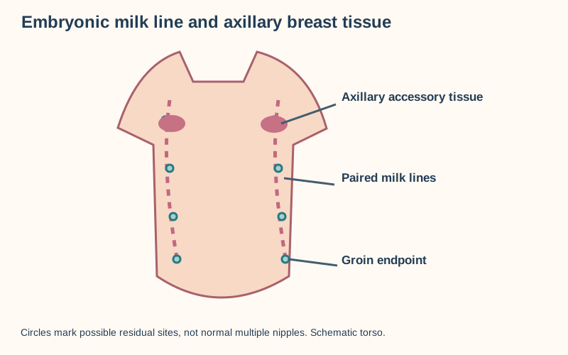
A soft axillary fullness enlarges during pregnancy and has the same texture as neighboring breast. Before calling it a lymph node, trace the mammary ridge: embryonic paired epithelial ridges extend from axilla to inguinal region; most regress except the pectoral buds. Persistent accessory glandular breast tissue is called polymastia, whereas an accessory nipple is called polythelia; either can occur along this route. The route is not a rigid vertical line under the nipple, and an extra nipple is not required for accessory tissue. [8]
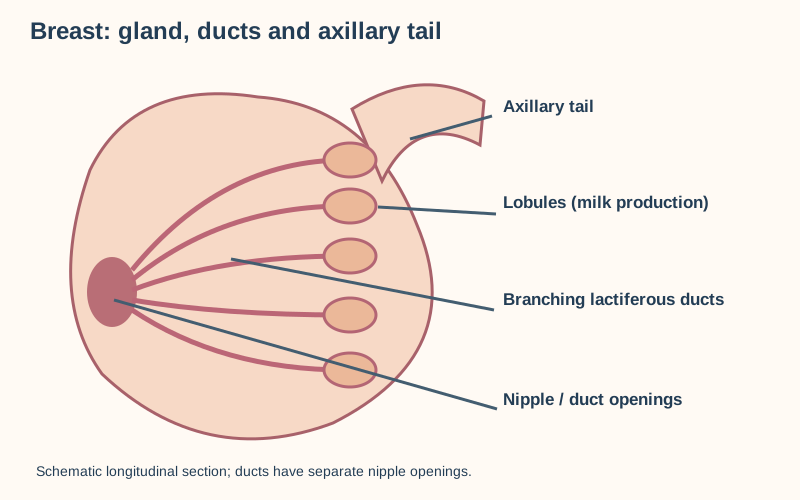
Lobules connect through ducts to the nipple; glandular tissue extends into the axillary tail. [8]Paired embryonic mammary ridges descend from axilla toward groin; persistent tissue in the axilla can form a mass. [8]
Breast parenchyma includes branching ducts and milk-producing lobules within adipose and fibrous stroma. Pregnancy and lactation can make accessory glandular tissue more evident, sometimes with milk secretion. A localized, firm node, abscess with inflammation, lipoma, or breast lesion within accessory tissue remains possible; ultrasound and assessment of suspicious change still matter. Accessory tissue can develop the same benign and malignant lesions as ordinary breast. [8][11]
Trace the axillary tissue pattern: a broad field that enlarges with pregnancy and matches breast on ultrasound favors accessory tissue; a discrete node draining an inflamed arm favors reactive adenopathy. If the pregnancy-related fullness develops a new hard focal component, do not dismiss that component as normal accessory tissue. [5]
Predict: if the lactation response enlarges during lactation and secretes milk, which origin becomes likelier? Milk-producing lobules explain the change; a lymph node cannot make milk. If a separate fixed nodule appears inside that field, reassess the nodule independently.
Check the tissue trace
Follow the mammary ridge from embryo to adult: mammary ridge persists at an ectopic site, ducts and lobules respond to pregnancy, and ultrasound can show tissue resembling normal breast. A firm focal lesion remains a separate diagnostic problem.
Which route does the axilla collect?
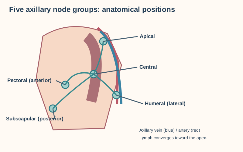
Follow the flow rather than memorize five names in isolation. The pectoral group follows lateral thoracic vessels, the subscapular group follows subscapular vessels, central nodes occupy axillary fat near its base, and apical nodes lie partly behind and above the upper edge of pectoralis minor. Pectoral (anterior) nodes receive much of the breast and anterior chest wall; subscapular (posterior) nodes receive posterior thorax; humeral (lateral) nodes receive most upper-limb lymph.
Many efferents reach central nodes and then apical nodes; some pectoral and humeral efferents bypass central nodes to reach apical nodes directly. Thus central obstruction impedes a major route without necessarily abolishing every collateral route. Apical efferents form the subclavian lymphatic trunk, which may enter the venous junction directly or join a larger duct, including the right lymphatic duct on the right or thoracic duct on the left.
Some lymph accompanying the cephalic vein reaches apical nodes directly. The lateral group also lies beside the medial or posterior aspect of the axillary vein, so the apical group is not the only group near that vessel. The axillary vein becomes subclavian at the lateral border of the first rib, not simply beneath the clavicle.
The medial breast can also drain to parasternal (internal mammary) nodes. The subclavian trunk may terminate directly at the venous angle or join the jugular trunk; on the left it may enter the thoracic duct. These variations prevent a single compulsory endpoint. [5]
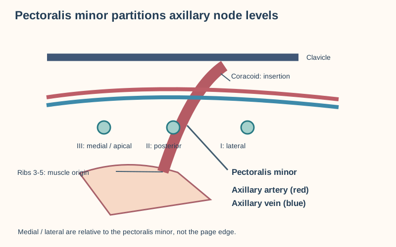
Pectoralis minor separates levels I, II and III; the axillary artery and vein course toward the apex. [11]Pectoral, subscapular and humeral groups drain toward central then apical nodes beside axillary vessels. [5][11]
Try tracing a hand infection and a breast lesion separately: hand to humeral, then central and apical; breast to pectoral, then central and apical. Both converge proximally but not at the same first station. The apical group lies close to the axillary vein and artery, yet proximity is not proof of venous compression. A newly swollen arm can reflect lymphatic injury after surgery or radiation, venous thrombosis or obstruction, infection, or tumor. Assess the pattern and investigate rather than diagnosing an apical node from swelling alone. [5][6]
Transfer: after axillary dissection an arm swells without prominent superficial venous distension; interrupted lymphatic drainage is plausible. Rapid unilateral swelling with prominent veins needs urgent evaluation for venous obstruction, not a lymphatic massage trial. [5]
For surgical staging, pectoralis minor supplies a different coordinate system: level I nodes are lateral to it, level II behind it, and level III medial to it. These levels are not interchangeable with the five named drainage groups. [11] Predict which named group near the apex most closely maps to level III, then remember that an individual node is assigned by its actual position.
Check the two routes
Breast lymph: pectoral to central to apical. Most arm lymph: humeral to central to apical. Their convergence explains shared proximal stations, not a unique diagnosis from swelling.
Try it here · Checkpoint 1 of 3
Make your prediction before reading the choices. A first attempt is just a starting point.
Case 4
Show answer and explanations for case 4
A. Pectoral group, which receives much of the breast and follows lateral thoracic vessels (Best answer)
Lateral breast lymph commonly first enters pectoral (anterior) axillary nodes, which accompany lateral thoracic vessels. Both the primary drainage territory and vessel landmark support this assignment; surgical level is a different location-based classification.
Reasoning steps for option A
What territory supplies the node expected from this primary?
Much of the lateral breast drains toward anterior or pectoral axillary nodes.
What landmark confirms the named group?
Pectoral nodes lie along lateral thoracic vessels at the anterior axillary wall.
B. Humeral group, which collects most upper-limb lymph beside the axillary vein (Why this does not fit)
Humeral nodes do occupy the axilla and relate to the axillary vein, which can make a nearby vascular node tempting. Their usual afferents come from the upper limb, whereas this lesion is breast-derived and the imaged node follows lateral thoracic rather than axillary vessels.
Reasoning steps for option B
Why does the vessel-adjacent description invite a humeral answer?
Humeral or lateral nodes run along the axillary vein and are prominent upper-limb collectors.
Which route and vessel disqualify them here?
A lateral breast lesion favors pectoral drainage, and the reported vessel is lateral thoracic, not axillary vein.
C. Subscapular group, which receives posterior thorax along subscapular vessels (Why this does not fit)
The subscapular chain is another peripheral axillary group and can eventually join the same central collectors. It lies on the posterior wall near subscapular vessels and normally receives posterior thoracic lymph, not the anterior-fold lateral thoracic drainage described.
Reasoning steps for option C
What afferents ordinarily feed the subscapular chain?
Posterior trunk and posterior shoulder-region lymph reaches posterior axillary nodes.
Why does the observed location favor another chain?
The node is at the anterior fold on lateral thoracic vessels, not posteriorly on subscapular vessels.
D. Central group, which receives several peripheral chains in the axillary fat (Why this does not fit)
A breast tumor can reach central nodes after peripheral drainage, and central nodes may become abnormal in established disease. A node explicitly following lateral thoracic vessels at the anterior fold identifies the pectoral first station rather than the convergent central group in axillary fat.
Reasoning steps for option D
Why can central nodes become involved in breast cancer?
Pectoral efferents often join central collectors before reaching the apex.
What makes central an incorrect first-group label for this node?
Its lateral thoracic vessel association and anterior-wall position specify the peripheral pectoral group.
Takeaway: Match both afferent territory and vessel landmark: lateral breast to pectoral nodes along lateral thoracic vessels, then central and apical.
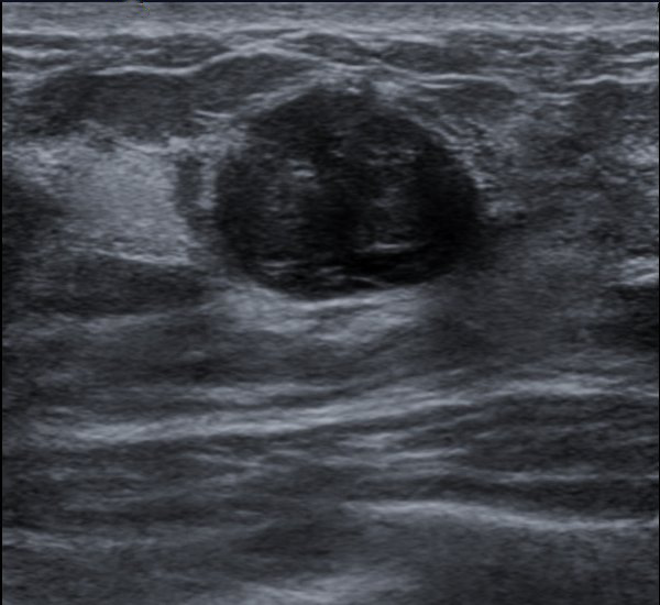
Breast pain and mobility are informative, not exclusion tests for cancer. A fibroadenoma is often smooth and mobile in reproductive years, commonly appearing as an oval, circumscribed, hypoechoic solid lesion oriented parallel to the skin; it is not reliably encapsulated. A simple cyst has a thin wall, anechoic fluid and posterior acoustic enhancement. Fibrocystic change commonly causes bilateral diffuse nodularity and tenderness that worsens before menstruation. A focal mass still deserves evaluation rather than automatic reassurance based on age or tenderness. [3]
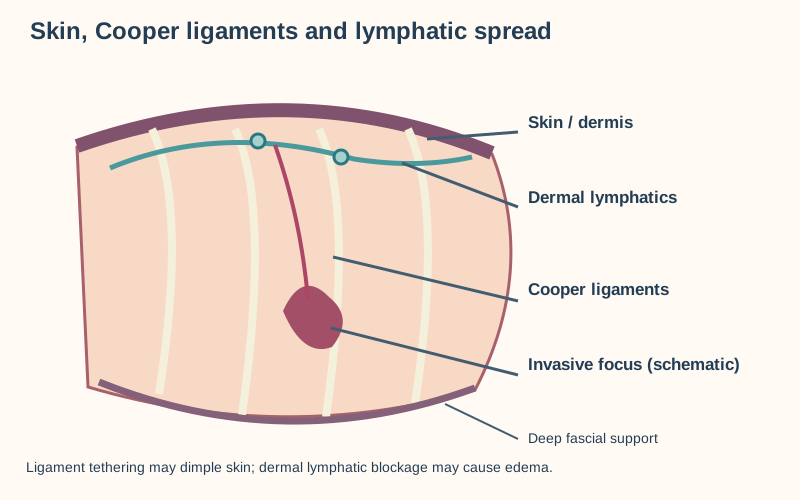
Cooper ligament tethering can cause dimpling; dermal lymphatic obstruction can cause skin edema (peau d’orange). Schematic invasive focus and deep fascial support are shown. [8][14]
Compare ultrasound descriptions: a circumscribed oval solid mass and an anechoic fluid space have different tissue contents; a growing irregular solid lesion requires a different diagnostic decision. A symptomatic cyst may be aspirated, but bloody fluid, residual mass, recurrence, or discordant examination calls for further evaluation. A core biopsy-proven concordant fibroadenoma without atypia need not be excised or undergo dedicated serial imaging; return to age-appropriate screening.
Symptoms, patient preference, size or substantive growth can favor excision. Suspicion for a phyllodes tumor or concerning fibroepithelial lesion instead calls for complete surgical excisional biopsy. An unbiopsied imaging finding categorized probably benign is a different situation and may need short-interval imaging surveillance. [9][3]
Fat necrosis follows injury, surgery or radiation and can resemble carcinoma through firmness, retraction or calcification; an oil cyst with rim calcification can help; injured fat attracts foamy lipid-laden macrophages, but a suspicious or discordant lesion requires tissue confirmation. [13] Infection or abscess is more likely with warmth, erythema, fever and focal tenderness. Cancer can be hard, irregular, fixed or accompanied by skin dimpling, edema, nipple retraction or fixed nodes, but absence of these signs cannot exclude it.
A spiculated mass, pleomorphic microcalcifications or a taller-than-wide irregular ultrasound lesion requires diagnostic assessment, not a diagnosis from texture alone. Ligament tethering can dimple skin; dermal lymphatic obstruction can produce peau d’orange. Rapid erythema and edema affecting at least one-third of the breast raises concern for inflammatory breast cancer and warrants prompt assessment even without a discrete lump.
Neither appearance alone identifies the histologic subtype. [14][3]
Predict what changes when the oval mass becomes rapidly enlarging and pathology shows increased stromal cellularity: a phyllodes tumor enters the differential, and a simple fibroadenoma label is insufficient. A concerning fibroepithelial lesion requires surgical excisional biopsy rather than routine follow-up. [9] A radial scar is a benign sclerosing lesion with a central fibroelastic core and peripheral compressed glands; its architectural distortion can resemble malignancy. Atypia, sampling adequacy and imaging-pathology concordance guide individualized management rather than a reassuring name alone. [15]
Check the tissue comparison
Anechoic fluid with enhancement points toward a simple cyst; irregular solid architecture needs tissue assessment. Trauma can explain fat necrosis but does not prove that a suspicious lesion is benign.
Does discharge implicate one duct or a systemic process?
Compare spontaneous, unilateral, single-duct bloody or clear discharge with bilateral multiduct milky discharge expressed only on squeezing. The former is pathologic even without a mass; the latter suggests a physiologic, medication-related or endocrine process. Ask about pregnancy, medicines, menstrual change, headaches and vision symptoms when galactorrhea is suspected; prolactin and thyroid-stimulating hormone testing can be appropriate. Do not infer safety from discharge color alone. [2][4]
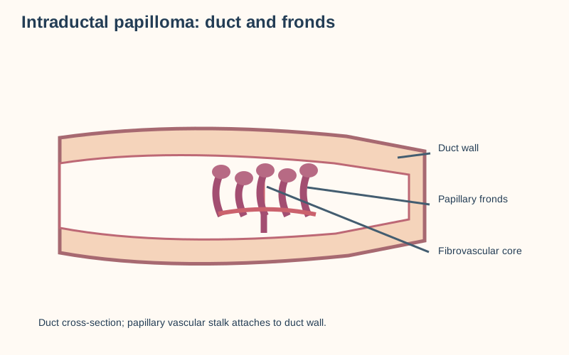
Papillary projections arise within a duct, with a schematic fibrovascular core. [10]
A benign intraductal papilloma has fibrovascular papillary fronds lined by epithelial and myoepithelial cells; its vessels can bleed. It is a common cause of bloody discharge without a mass. Duct ectasia can produce sticky green, gray or multicolored discharge from more than one duct; carcinoma remains an alternative to a papilloma in pathologic single-duct discharge.
Eczematous unilateral nipple change that persists raises concern for mammary Paget disease and warrants evaluation even if no mass is felt. Ductal carcinoma in situ need not cause a palpable mass. Predict the next action for a 45-year-old with spontaneous discharge from one duct: diagnostic mammography or tomosynthesis plus targeted ultrasound, with biopsy of an identified suspicious lesion.
Initial blind duct excision is not obligatory. If conventional imaging is negative but discharge persists, specialist reassessment and individualized additional imaging, often contrast-enhanced breast magnetic resonance imaging, or duct surgery may be appropriate; negative imaging is not an absolute exclusion of cancer. [10][2][4]
For a radiologic-pathologic concordant papilloma without atypia discovered on core biopsy, routine excision is not invariably necessary. A prospective registry of selected asymptomatic concordant lesions found two local upgrades among 116 excisions (1.7%); this does not settle symptomatic, atypical or discordant lesions. Persistent symptomatic discharge, atypia or a palpable papilloma favors surgical evaluation. [7][4]
Check your discharge prediction
One spontaneous duct suggests a localized duct lesion and age-appropriate diagnostic imaging; bilateral milky secretion from multiple ducts suggests endocrine or medication assessment. A bloody single duct can still be malignant without a palpable mass.
Try it here · Checkpoint 2 of 3
Make your prediction before reading the choices. A first attempt is just a starting point.
Case 19
Show answer and explanations for case 19
A. Ductal carcinoma in situ (Why this does not fit)
Ductal carcinoma in situ requires a neoplastic epithelial population in the ducts. Bland fronds containing intact myoepithelial cells instead support a benign papillary proliferation.
Reasoning steps for option A
What would in situ ductal carcinoma show?
Atypical malignant epithelial proliferation within ducts.
What argues against it here?
Preserved myoepithelium and bland epithelium over vascular fronds favor papilloma.
B. Intraductal papilloma (Best answer)
A stalk of vessels and connective tissue lined by both cell layers identifies a papilloma. Its vascularity offers a mechanism for bleeding from the single involved duct.
Reasoning steps for option B
What forms the frond core?
A fibrovascular axis invested by epithelial and myoepithelial cells.
How does that relate to the bloody single-duct symptom?
A vascular intraductal papilloma can bleed from its involved duct.
C. Mammary duct ectasia (Why this does not fit)
Duct ectasia produces enlarged ducts and thick secretions, not an organized epithelial nodule built around branching vascular stalks.
Reasoning steps for option C
What morphology characterizes ectasia?
Dilated ducts with inspissated secretions and periductal inflammation.
Are branching papillary fronds its defining feature?
No; the sampled nodule has organized fibrovascular projections.
D. Invasive papillary carcinoma (Why this does not fit)
An invasive papillary malignancy would raise concern for absent myoepithelium and infiltration beyond the duct. The preserved cellular layer in the specimen argues against invasion.
Reasoning steps for option D
What makes papillary invasion concerning?
Loss of the peripheral myoepithelial barrier with infiltrative tumor.
Is that barrier lost?
No; the biopsy reports a preserved myoepithelial layer.
Takeaway: A papilloma has vascular fibroconnective fronds with a retained myoepithelial layer; bloody single-duct discharge does not by itself establish carcinoma. Age-appropriate diagnostic imaging precedes sampling of the localized lesion.
Begin with history and examination of both breasts and regional nodes, and localization of the finding. Clinical features adjust concern but cannot establish benignity. For a palpable mass in an adult woman younger than 30, targeted ultrasound is usually the first imaging study. From 30 to 39, ultrasound and diagnostic mammography or digital breast tomosynthesis are usually appropriate options; at 40 or older, diagnostic mammography or tomosynthesis is usually the initial study, with targeted ultrasound as indicated.
Screening mammography is not the diagnostic test for a new palpable lump. In pregnancy, targeted ultrasound is the initial imaging examination for a palpable abnormality; a suspicious finding still needs biopsy and appropriate further diagnostic evaluation rather than waiting until after delivery. [12] These age variants do not replace individualized evaluation for other patient populations. [1]
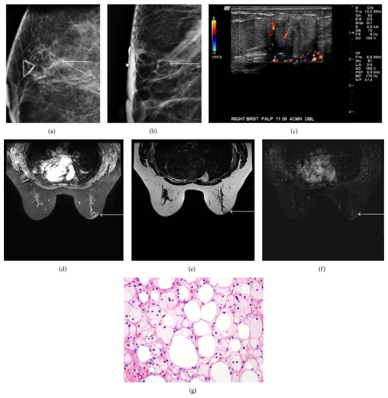
Original uncropped figure panels include mammography, ultrasound, MRI, and histology. Fat necrosis can mimic malignancy; this particular histology is not papilloma or carcinoma.Example breast ultrasound supplied as fibroadenoma by its author; ultrasound appearance alone does not establish histology. Original image preserved without overlays. [1][9]
For a palpable mass in a 43-year-old with a negative diagnostic mammogram, targeted ultrasound is usually appropriate. If imaging and a distinctly suspicious examination disagree, negative imaging is not permission to abandon evaluation; arrange specialist review and tissue sampling when clinically indicated. An image-guided core needle biopsy provides architecture for a suspicious solid lesion. The American College of Radiology initial-imaging table does not imply that every palpable mass needs immediate biopsy before imaging. [1][3]
For a young patient with a simple cyst, fluid and symptom response can support conservative care; persistent or bloody aspirate requires follow-up. Compare a probably benign imaging assessment with a suspicious imaging finding: follow the radiologist's recommended surveillance for an appropriately concordant probably benign lesion versus tissue diagnosis for suspicion. If the breast changes or imaging conflicts with examination, revisit the plan rather than relying on a reassuring label. [1][3]
Check the age and concordance branches
Age-specific imaging: at 27, targeted ultrasound first; at 35, ultrasound or diagnostic mammography/tomosynthesis is appropriate; at 43, diagnostic mammography/tomosynthesis first, and ultrasound follows a negative mammogram at the palpable site. A suspicious examination overrides falsely reassuring discordant imaging.
Try it here · Checkpoint 3 of 3
Make your prediction before reading the choices. A first attempt is just a starting point.
Case 17
Show answer and explanations for case 17
A. Screening mammography for both (Why this does not fit)
A symptomatic lesion calls for a diagnostic rather than a screening examination in both women. Their ages also change which diagnostic modality is appropriate first.
Reasoning steps for option A
What is screening missing?
Both women have symptoms requiring diagnostic assessment.
Can one screening order address both masses?
No; the age-specific diagnostic choices differ.
B. Ultrasound or diagnostic mammography for the 35-year-old; diagnostic mammography for the 57-year-old (Best answer)
At 35, either focused ultrasound or diagnostic mammography or tomosynthesis may initiate the workup. At 57, diagnostic mammography or tomosynthesis is the recommended initial examination for the palpable concern.
Reasoning steps for option B
Which modalities are appropriate at 35?
Ultrasound or diagnostic mammography or tomosynthesis may start evaluation.
What changes at 57?
Diagnostic mammography or tomosynthesis is initial rather than screening.
C. Wait until age 40 for the younger woman; screening mammography for the older (Why this does not fit)
The younger woman's mass cannot be ignored until screening age; the older woman's symptoms make her ineligible for an ordinary screening-only approach.
Reasoning steps for option C
Does a woman at 35 need to wait?
No; a palpable lesion requires prompt diagnostic evaluation.
Is screening the right 57-year-old order?
No; the palpable finding requires a diagnostic examination.
D. Magnetic resonance imaging for both as the only first test (Why this does not fit)
Breast magnetic resonance imaging alone bypasses the established age-specific initial studies and is not the usual starting point for either newly palpated lesion.
Reasoning steps for option D
What role does breast magnetic resonance imaging have?
It is not the routine first-line test for either initial palpable-mass workup.
What is missing if it is used alone?
Age-appropriate diagnostic mammography and targeted ultrasound assessment.
Takeaway: For ages 30 to 39 either ultrasound or diagnostic mammography or tomosynthesis is appropriate; at 40 or older diagnostic mammography or tomosynthesis is initial, not screening.
The first decision for a new mass, bloody discharge, or new unilateral arm swelling remains diagnostic evaluation. An osteopathic structural examination may document associated chest-wall or shoulder somatic dysfunction, but palpated restriction or a proposed thoracic inlet mechanism cannot distinguish cancer, venous thrombosis, or lymphatic obstruction. Do not use osteopathic manipulative treatment to delay imaging, biopsy, or vascular assessment. [1][5]
In established breast cancer-related lymphedema, specialized manual lymphatic drainage is studied as one component of complex decongestive therapy, which also includes compression, exercise and skin care. A 2015 Cochrane review found a possible additional swelling reduction when manual drainage was added to compression bandaging, based on small trials; sleeve comparisons were inconsistent. It did not test thoracic inlet release or an osteopathic lymphatic pump, and it does not establish that such techniques prevent metastasis or reliably resolve swelling.
A claim that touching malignancy necessarily causes metastatic spread is unsupported; defer elective manual treatment of an unexplained mass while it is assessed, and individualize care with the treating team. [6]
Predict the consequence of confusing postoperative lymphatic swelling with an acute venous event: elective manual care could delay urgent vascular assessment. In contrast, a stable patient with established lymphedema may discuss evidence-limited adjuncts with the treating team while continuing compression and skin care.
Check the evidence boundary
Manual lymphatic drainage in the Cochrane trials was not an osteopathic pump or inlet release; a positive effect in one compression comparison cannot be transferred to an untested technique.
Apply the pathways
Three checkpoints: trace breast versus upper-limb drainage, compare unilateral single-duct discharge with multiduct secretion, and choose age-appropriate imaging while checking examination-imaging concordance. Each answer should explain what new information could reverse the decision. [1][2][5]
Case 1
Show answer and explanations for case 1
A. Reactive enlargement of axillary lymph nodes (Why this does not fit)
Nodes can enlarge in the axilla during infection and may be mistaken for breast tissue. Here the diffuse branching ductal pattern and absence of a discrete node or inflammatory source argue against nodal hyperplasia.
Reasoning steps for option A
What sonographic structure would support nodal enlargement?
A discrete oval or abnormal node with a cortex and hilum would support this route.
Why does the observed field not behave like a reactive node?
It contains branching mammary ducts and expands in parallel with breast tissue without an infectious trigger.
B. Expansion of a preexisting axillary lipoma (Why this does not fit)
A longstanding soft axillary swelling could be a lipoma, particularly when pregnancy makes it more noticeable. A lipoma is a localized fatty lesion rather than hormone-responsive ductal and lobular parenchyma under an accessory nipple.
Reasoning steps for option B
Why might lipoma initially enter the differential?
Its soft texture and axillary location can mimic a painless accessory breast mound.
Which ultrasound detail specifically defeats a lipoma?
Visible ducts and lobules indicate glandular parenchyma rather than an encapsulated adipose mass.
C. Pregnancy-stimulated accessory mammary lobules (Best answer)
A persistent mammary-ridge remnant can contain functional lobules; gestational hormonal stimulation enlarges both ordinary and ectopic glandular tissue. The childhood nipple and ductal ultrasound pattern locate the response outside the usual breast.
Reasoning steps for option C
What tissue does a childhood axillary nipple suggest beneath the skin?
It marks a possible persistent mammary-ridge site that can contain ducts and lobules.
Why does the third-trimester enlargement favor this mechanism?
Hormonal stimulation enlarges glandular breast tissue in pregnancy, including ectopic lobules.
D. Obstruction of an apical lymphatic collector (Why this does not fit)
Proximal axillary lymphatic obstruction can cause regional swelling, so location alone cannot exclude it. Obstruction would not create mammary ducts beneath a congenital nipple or selectively reproduce physiologic breast enlargement during pregnancy.
Reasoning steps for option D
What distribution is expected with a blocked apical collector?
Because several distal axillary groups converge proximally, upstream arm or chest-wall edema may appear.
Why is this patient’s swelling not explained by that blockage?
A focal glandular field changes with gestation rather than widespread upstream lymphatic congestion.
Takeaway: Accessory breast lobules retain pregnancy responsiveness; imaging of a diffuse ductal field separates physiologic enlargement from a new focal lesion.
A. Independent pigmented nevi and a normal axillary tail; the findings have unrelated origins (Why this does not fit)
Stable pigmented papules could be melanocytic nevi, and the axillary tail of the ordinary breast can extend toward the axilla. Yet two central nipple-like projections along the mammary-ridge trajectory favor accessory nipples; the separate duct-bearing axillary field can likewise reflect retained ridge tissue.
Reasoning steps for option A
Why might these findings appear unrelated at first?
Small pigmented spots can resemble nevi, while the ordinary breast can extend into the axilla.
Which combination instead supports shared mammary development?
Nipple-like centers along the chest-to-groin ridge and breast-type ducts in axillary tissue support persistent mammary structures.
B. Persistent mammary-ridge segments can form nipples and functional glandular tissue (Best answer)
The paired embryonic mammary ridges extend from axilla toward groin and normally regress outside the pectoral region. Persistence can produce nipple-only remnants or accessory gland; glandular tissue can undergo the same benign and malignant processes as usual breast.
Reasoning steps for option B
Why does the chest-to-groin distribution implicate a developmental ridge?
The embryonic mammary ridges traverse that territory before most of their length regresses.
What does the separate duct-like axillary field add?
A retained ridge can form parenchyma as well as a nipple, so usual breast pathology remains possible there.
C. Cutaneous mammary remnants without ectopic gland; the axillary field cannot be breast tissue (Why this does not fit)
Accessory nipples may occur without an underlying accessory gland, making a skin-only remnant plausible for either papule. But a separate axillary field with duct-like parenchyma supports actual glandular tissue; nipple-only morphology elsewhere cannot exclude it.
Reasoning steps for option C
Can an accessory nipple exist without underlying gland?
Yes; polythelia can represent a cutaneous remnant without functional parenchyma.
Why is a skin-only explanation insufficient for the axillary finding?
A distinct axillary duct-bearing field provides evidence of parenchyma, which may share ordinary breast pathology.
D. Persistent mammary ridge forms the axillary gland, but ectopic gland cannot develop breast carcinoma (Why this does not fit)
The ridge origin correctly connects nipple-like papules with accessory gland, so this answer captures the embryology. It fails the clinical prediction: retained ducts and lobules remain mammary tissue and can develop benign lesions or carcinoma despite their ectopic position.
Reasoning steps for option D
Which part of this answer matches the developmental distribution?
Persistent mammary-ridge segments can account for accessory nipples and ectopic axillary gland.
What misconception does the disease prediction reveal?
Ectopic location does not protect mammary parenchyma from the same pathologic processes as usual breast.
Takeaway: Persistent mammary ridge can yield polythelia or accessory gland; gland, when present, can acquire ordinary breast pathology.
A. Reassure her that all axillary tissue is responding to lactation (Why this does not fit)
Accessory lobules often engorge with milk and enlarge during pregnancy and lactation, which explains the bilateral diffuse fullness. A new unilateral hard focus that persists independently of diffuse fullness and receives a suspicious imaging assessment is a separate diagnostic problem.
Reasoning steps for option A
Which component can lactation plausibly explain?
The bilateral soft fields that fluctuate with feeding can reflect functioning accessory lobules.
What makes reassurance unsafe for the right side?
The solid irregular nodule remains after feeding and is independently classified BI-RADS 4.
B. Repeat ultrasound after weaning without tissue sampling now (Why this does not fit)
Transient milk-related nodularity can settle after weaning, so follow-up may seem convenient. Waiting months is inappropriate for a persistent irregular category 4 lesion; lactation does not prevent indicated imaging-guided biopsy.
Reasoning steps for option B
What observation makes a short physiologic fluctuation less likely?
The firm right-sided nodule persists despite feeds while the soft surrounding tissue changes.
Why does planned weaning not resolve the diagnostic decision?
A suspicious solid ultrasound morphology demands sampling before a prolonged delay can be justified.
C. Treat empirically for an axillary abscess and reassess (Why this does not fit)
Lactating patients can develop infection, and a painful inflammatory collection might require antibiotics and drainage. This lesion is hard and solid without erythema, fever, fluctuance or a fluid collection, so empiric infection treatment would postpone tissue diagnosis.
Reasoning steps for option C
Why is infection initially plausible in this patient?
Lactation increases the clinical relevance of mastitis and abscess in breast-bearing tissue.
Which features point away from an abscess here?
No systemic or local inflammatory signs are present, and ultrasound shows an irregular solid rather than fluid-filled focus.
D. Arrange image-guided core needle biopsy of the focal lesion (Best answer)
Category 4 signifies a suspicious imaging abnormality for which tissue diagnosis is indicated. Ultrasound guidance samples the discrete solid focus and preserves architecture; lactational change in the surrounding accessory gland cannot explain away discordant focal morphology.
Reasoning steps for option D
What does a BI-RADS 4 designation imply for this focus?
The irregular nonparallel solid focus warrants tissue diagnosis rather than routine observation.
Why target the focus rather than the whole accessory tissue field?
The changing diffuse field may be physiologic, but the persistent hard focal component is the suspicious structure.
Takeaway: A suspicious focal lesion inside accessory breast tissue requires its own imaging-pathology assessment even when surrounding tissue behaves physiologically.
A. Pectoral (anterior) axillary nodes (Why this does not fit)
Pectoral nodes are a plausible first stop for infection on the anterior chest wall or breast. The focus is on posterior trunk, and the palpable node follows subscapular rather than lateral thoracic vessels at the anterior wall.
Reasoning steps for option A
What nearby infection territory would favor pectoral nodes?
An anterior chest-wall or lateral breast lesion generally sends lymph toward the anterior axillary chain.
Which observed localization defeats that option?
Both the infected skin and the enlarged vessel-associated node are posterior, not anterior.
B. Humeral (lateral) axillary nodes (Why this does not fit)
Most upper-limb lymph travels through humeral nodes, so an infection near the shoulder might make them appealing. This source is explicitly posterior thoracic skin well below the scapula, without an arm source; its node lies near subscapular vessels rather than the axillary vein.
Reasoning steps for option B
What source would instead predict the humeral group?
An infection of much of the upper limb would ordinarily enter lateral or humeral collectors.
Why does this source not follow that route?
The lesion is on the posterior trunk below the scapula, and its node is on the posterior axillary wall.
C. Subscapular (posterior) axillary nodes (Best answer)
Posterior thoracic wall lymph reaches the subscapular chain near subscapular vessels. The clearly trunk-based infection and posterior-wall node identify its first peripheral station; efferents then pass toward central and apical nodes.
Reasoning steps for option C
Why does specifying posterior thoracic wall matter?
Posterior trunk drainage commonly enters posterior or subscapular axillary nodes.
How does the physical node location strengthen that prediction?
A node beside subscapular vessels on the posterior axillary wall matches the subscapular chain.
D. Central axillary nodes as the first peripheral station (Why this does not fit)
Central nodes do receive efferents from several axillary peripheral groups and can enlarge with infection. They are a downstream convergence point, whereas the subscapular-vessel node is the identifiable posterior first station in this case.
Reasoning steps for option D
Why might central nodes eventually enlarge?
Subscapular efferents join central collectors as lymph proceeds proximally.
What timing error does selecting central first make?
It skips the peripheral posterior chain already identified beside subscapular vessels.
Takeaway: Posterior thoracic wall to subscapular nodes beside subscapular vessels, then central and apical collectors.
A. Axillary-vein thrombosis sends retrograde lymph to epitrochlear nodes (Why this does not fit)
A tender axillary region and arm symptoms can raise concern for venous thrombosis. Venous clot causes impaired blood return rather than a normal retrograde nodal chain; duplex patency and lack of diffuse venous congestion oppose this explanation.
Reasoning steps for option A
What findings would increase concern for a proximal venous clot?
Diffuse arm swelling, cyanosis or collateral veins would prompt vascular assessment.
How do the examination and duplex change the inference?
Patent axillary flow plus ordered tender nodes favor infection-related lymphatic drainage over venous obstruction.
B. Epitrochlear nodes can feed humeral axillary nodes beside a patent axillary vein (Best answer)
Medial forearm superficial lymph may first reach epitrochlear nodes, then travel to humeral (lateral) axillary collectors associated with the axillary vein. Adjacent anatomy does not prove thrombosis; a patent vein and absent venous congestion support reactive lymphatic spread.
Reasoning steps for option B
Why is the medial-elbow node informative?
Epitrochlear nodes intercept some superficial lymph from the medial hand and forearm.
What explains the proximal node without diagnosing a clot?
That lymph can enter humeral axillary nodes alongside a patent axillary vein.
C. Forearm lymph first reaches pectoral nodes along lateral thoracic vessels (Why this does not fit)
Pectoral nodes and humeral nodes ultimately converge on central nodes, so both may appear in an axillary map. The lateral thoracic chain primarily collects breast and anterior chest-wall lymph, not the medial forearm route traced through the elbow.
Reasoning steps for option C
What tissue drains preferentially to pectoral nodes?
The lateral breast and anterior thoracic wall commonly send afferents along the lateral thoracic vessels.
Which intermediate station rules against that first route?
The medial-elbow epitrochlear node marks upper-limb lymph moving toward humeral collectors.
D. The infection bypasses humeral nodes and enters parasternal nodes (Why this does not fit)
Parasternal nodes offer an alternate drainage pathway for the medial breast, so they are important when mapping breast cancer. They do not explain a medial-elbow chain followed by a node beside the axillary vein after forearm cellulitis.
Reasoning steps for option D
Why are parasternal nodes a plausible named alternative in breast anatomy?
Some medial breast lymph drains independently to nodes beside the internal thoracic vessels.
Why is that pathway irrelevant to this source?
The source is forearm skin with epitrochlear involvement, an upper-limb rather than medial-breast route.
Takeaway: Medial forearm lymph can pass through epitrochlear to humeral axillary nodes; adjacency to a vein is not evidence of venous thrombosis.
A. Preserved apical nodes guarantee normal arm drainage; parasternal drainage is abolished (Why this does not fit)
Apical nodes are downstream collectors and their preservation can sound protective. But removing central nodes interrupts many incoming humeral efferents; a separate medial-breast parasternal pathway was not removed by axillary surgery.
Reasoning steps for option A
Why does an intact apex not guarantee normal peripheral flow?
Proximal recipients cannot restore severed inflow from humeral nodes through excised central collectors.
What contradicts loss of the internal mammary route?
Parasternal nodes drain some medial breast tissue independently of the axillary central chain.
B. Central removal diverts all upper-limb lymph obligatorily through parasternal nodes (Why this does not fit)
Collateral lymphatic drainage may develop after surgery, making rerouting sound plausible. Yet the internal mammary chain primarily serves medial breast and adjacent chest-wall territories; no obligatory arm-to-parasternal bypass follows central-node excision.
Reasoning steps for option B
What might change after collector disruption?
Individual collateral pathways may adapt, so drainage is not perfectly rigid after surgery.
Why is the proposed universal bypass unjustified?
The parasternal chain is not the normal mandatory destination for upper-limb humeral efferents.
C. Normal venous duplex excludes a lymphatic cause; the swelling proves apical thrombosis (Why this does not fit)
Unilateral arm swelling after cancer treatment warrants consideration of vascular obstruction. Patent venous flow and no collateral venous distension do not exclude lymphatic injury; apical nodes were preserved, and a clot is not established by edema alone.
Reasoning steps for option C
What does a normal venous duplex directly assess?
It tests patency of the examined veins, not continuity of surgically interrupted lymphatic collectors.
What explains the gradual nonpitting arm swelling better?
Central dissection can interrupt humeral lymphatic efferents even when the apical nodes and veins remain intact.
D. Arm and pectoral lymph can be impeded before the apex; medial-breast parasternal drainage can persist (Best answer)
Central nodes receive humeral and pectoral efferents, so removing them can disrupt proximal arm and lateral-breast drainage despite preserved apical nodes. The separate medial-breast path toward parasternal nodes need not pass through central axillary nodes.
Reasoning steps for option D
How can preserved apical nodes coexist with arm lymphedema?
Humeral drainage may be interrupted upstream at the resected central collectors before reaching the apex.
Why need parasternal drainage not vanish?
Some medial breast lymph reaches internal mammary nodes along a separate route that does not require central axillary transit.
Takeaway: Central axillary resection can impede humeral and pectoral drainage before apical transit without necessarily disrupting medial-breast parasternal drainage.
Level I nodes lie lateral to pectoralis minor, farther from the apex than this medial node. A lateral breast location does not make every draining axillary node level I.
Reasoning steps for option A
What boundary separates level I from the other two axillary levels?
The lateral border of pectoralis minor: nodes on its lateral side belong to level I.
Can the quadrant of the primary breast tumor assign this level?
No; level designation follows the node’s physical location, not the primary tumor quadrant.
B. Level II axillary nodes behind the pectoralis minor muscle (Why this does not fit)
Level II describes nodes behind pectoralis minor; that description applies to the second node, not the one beyond its medial edge. Superior position alone does not establish level II.
Reasoning steps for option B
Why might a posterior node be confused with the target?
Both may appear near the upper axilla on an axial image, but depth behind the muscle defines level II.
What landmark prevents that confusion?
Follow the muscle to its medial edge on consecutive images instead of assigning a level from image height alone.
C. Level III axillary nodes (Best answer)
Level III lies medial to the medial border of pectoralis minor at the apical axilla. The described node satisfies that spatial definition regardless of whether metastasis is subsequently confirmed.
Reasoning steps for option C
Does level III require a specific histologic diagnosis?
No; level III is a positional designation that applies whether a node is malignant or benign.
How do apical nodes relate to this level?
Apical nodes commonly occupy this medial, proximal compartment, although each node must be localized individually.
D. Internal mammary nodes (Why this does not fit)
Internal mammary nodes track the internal thoracic vessels beside the sternum, not the axillary apex. Medial to pectoralis minor is not synonymous with parasternal.
Reasoning steps for option D
What vessel course identifies the proposed extra-axillary chain?
Parasternal nodes follow the internal thoracic vessels close to the sternum.
Can a medial axillary node be classified as parasternal solely because it is medial?
No; medial is relative to pectoralis minor here, whereas parasternal is relative to the sternum.
Takeaway: Assign surgical axillary levels by position relative to pectoralis minor: lateral I, posterior II, medial III.
A patent vein can still have functionally important outflow resistance when compressed. Proximal narrowing raises distal venous pressure and filtration; shoulder collaterals reflect rerouting of venous return.
Reasoning steps for option A
How can a nonoccluded vein produce edema?
Narrowing raises resistance even when contrast still traverses the lumen.
Why do superficial chest veins enlarge?
Collateral channels carry blood around a proximal outflow bottleneck.
B. Lymphatic interruption from axillary surgery causing protein-rich interstitial accumulation (Why this does not fit)
Nodal disease can affect lymph drainage, but this option specifically requires an operation that has not occurred. Prominent superficial veins and venogram-demonstrated impaired venous emptying identify a hemodynamic pathway here.
Reasoning steps for option B
Which drainage function would postoperative lymph injury impair?
It would prevent clearance of interstitial proteins and lymph from the upper limb.
Why is an operative cause temporally untenable here?
There has been no intervention capable of severing the axillary collecting channels.
C. Occlusive axillary vein thrombosis causing abrupt loss of venous flow (Why this does not fit)
Thrombosis could cause acute unilateral edema and venous collaterals, so it merits consideration clinically. Venography shows external narrowing without a filling defect, separating compression from intraluminal occlusion.
Reasoning steps for option C
What finding would establish a thrombotic rather than compressive mechanism?
An intraluminal filling defect in the venous segment would support thrombosis.
Does a negative thrombus assessment eliminate all venous causes?
No; extrinsic stenosis still creates venous hypertension.
D. Isolated obstruction of breast lymph drainage through pectoral nodes (Why this does not fit)
Pectoral nodes chiefly receive breast and anterior chest-wall lymph. Their isolated obstruction would not explain the dilated shoulder veins or venogram-demonstrated axillary-vein bottleneck.
Reasoning steps for option D
Which territory primarily enters pectoral nodes?
Much of the breast and anterior chest wall drains to the anterior axillary group.
What independently establishes the arm mechanism?
The contrast study directly demonstrates restricted venous egress and collateral formation.
Takeaway: Apical nodal compression of a patent vein can produce arm edema by venous backpressure without thrombosis.
A. Reduced clearance of interstitial proteins after interruption of upper-limb lymphatic channels (Best answer)
Axillary dissection can sever collecting lymphatics that return interstitial proteins and fluid to the circulation. Retained proteins favor sustained fluid accumulation in the affected limb despite open veins.
Reasoning steps for option A
Why is the edema fluid protein-rich?
Lymphatics normally remove macromolecules that cannot readily return directly through capillary walls.
What maintains swelling over time?
Trapped interstitial protein increases local oncotic attraction for water until alternative lymphatic drainage compensates.
B. Elevated venous capillary pressure from an occult obstructing axillary vein thrombus (Why this does not fit)
A proximal venous thrombus can increase filtration pressure, but the duplex demonstrates an open axillary-subclavian outflow path. The gradual postoperative course without distended collateral veins supports impaired lymph clearance instead.
Reasoning steps for option B
What process makes venous-obstruction edema physiologically different?
A downstream vein blockage increases upstream capillary hydrostatic pressure and fluid filtration.
Which test interrogates the proposed obstruction?
Duplex assesses venous patency and flow in the proximal arm veins.
C. Increased arterial inflow from postoperative arteriovenous shunting (Why this does not fit)
An arteriovenous fistula typically produces local warmth, bruit, or pulsatility and increases flow. Node removal disrupts drainage rather than creating a high-flow arterial connection.
Reasoning steps for option C
What bedside finding would make an arteriovenous connection more plausible?
A palpable thrill or audible bruit would suggest abnormal high-flow communication.
Why does axillary dissection instead implicate a low-flow return pathway?
Surgery removes or transects lymphatic collectors rather than directly connecting artery and vein.
D. Reduced plasma oncotic pressure from systemic hypoalbuminemia (Why this does not fit)
Low circulating albumin reduces oncotic reabsorption throughout the body and tends to cause generalized or bilateral edema. It does not selectively follow the dissected axillary drainage territory.
Reasoning steps for option D
How would systemic low oncotic pressure generally present?
Dependent edema at multiple body sites is more likely than isolated unilateral hand and arm enlargement.
Which circulating component chiefly determines plasma oncotic pressure?
Albumin supplies the major colloid osmotic contribution.
Takeaway: Post-dissection lymphedema reflects impaired clearance of protein-rich interstitial fluid, not presumed venous thrombosis.
A. Repeat targeted breast ultrasound in six months (Best answer)
For an appropriately characterized unbiopsied a probably benign imaging assessment mass with concordant examination, six-month imaging documents stability before longer-term disposition. New growth or discordance would change the biopsy threshold.
Reasoning steps for option A
What does BI-RADS 3 communicate?
After complete diagnostic evaluation, the imaging appearance is probably benign and eligible for planned surveillance.
What event should override this interval plan?
Meaningful growth or a newly suspicious examination warrants reassessment and potentially sampling.
B. Proceed immediately to ultrasound-guided core biopsy of this concordant probably benign mass (Why this does not fit)
Core biopsy supplies histology when imaging is suspicious, the mass grows, or clinical and imaging findings disagree. A stable, explicitly a probably benign imaging assessment lesion may instead enter the recommended imaging surveillance pathway.
Reasoning steps for option B
When does tissue sampling become preferable to observation?
Imaging suspicion, interval growth, or examination-imaging discordance raises the need for histology.
Is a solid mass automatically a biopsy indication?
C. Discharge from follow-up as though core biopsy had proved fibroadenoma (Why this does not fit)
No dedicated imaging follow-up is a guideline option for a core-biopsy-proven, concordant fibroadenoma without atypia. This patient lacks histologic confirmation, so that postbiopsy rule cannot replace a probably benign imaging assessment surveillance.
Reasoning steps for option C
Which population is addressed by the 2025 no-dedicated-follow-up statement?
Patients with core-confirmed concordant fibroadenoma without atypia.
What crucial evidence is missing for applying it here?
Neither the tissue diagnosis nor radiologic-pathologic concordance exists without biopsy.
D. Perform needle aspiration to confirm a simple cyst (Why this does not fit)
Aspiration is useful for selected symptomatic fluid-filled cysts, not a homogeneous solid lesion. A needle yielding no fluid would not resolve its imaging classification.
Reasoning steps for option D
How does a cyst differ sonographically from this mass?
A simple cyst is anechoic and fluid-filled, with posterior enhancement.
What is the usual purpose of aspirating a simple cyst?
Drainage primarily relieves symptoms or clarifies a fluid lesion rather than characterizing an established solid mass.
Takeaway: Unbiopsied category 3 probably benign solid masses warrant indicated short-interval surveillance; postbiopsy fibroadenoma guidance is a separate pathway.
A. Complete surgical excisional biopsy of the lesion (Best answer)
Rapid enlargement and a core report suspicious for phyllodes call for complete surgical excisional biopsy to assess the whole lesion. Limited sampling can miss the architecture or heterogeneous stromal features needed for classification.
Reasoning steps for option A
Why can cores leave uncertainty in fibroepithelial lesions?
Stromal features can vary across a large mass, while needle cores sample only small regions.
What information does complete excision add?
The full architecture and margins can be assessed for definitive classification and subsequent treatment planning.
B. Six-month ultrasound surveillance as for an unbiopsied category 3 mass (Why this does not fit)
a probably benign imaging assessment surveillance applies to appropriately probably benign imaging findings without concerning pathology. A growing mass with a phyllodes-suspicious core has crossed that decision boundary.
Reasoning steps for option B
What assumption underlies short-interval BI-RADS 3 observation?
The lesion has no suspicious clinical or pathologic feature after diagnostic assessment.
Which independent signal violates that assumption?
Rapid growth coupled with stromal hypercellularity raises concern for phyllodes.
C. Repeat fine-needle aspiration to establish stromal architecture (Why this does not fit)
Fine-needle aspiration yields cells but poorly preserves stromal architecture and cannot reliably resolve a fibroepithelial lesion that was indeterminate even on core sampling.
Reasoning steps for option C
What does aspiration principally sample?
It collects fluid or dispersed cells, not an intact architectural cross-section.
Which feature is especially important here?
Assessment of the stromal component and broader fibroepithelial architecture requires tissue.
D. Routine screening only because a fibroadenoma has already been proven (Why this does not fit)
The report does not establish a concordant fibroadenoma without atypia. Applying routine-screening guidance for proven fibroadenoma would overlook the stated stromal concern.
Reasoning steps for option D
What diagnosis must precede routine fibroadenoma advice?
A concordant core-confirmed fibroadenoma without atypia is the relevant guideline population.
Why does the current pathology not meet that category?
It explicitly raises phyllodes and does not conclusively establish fibroadenoma.
Takeaway: A core showing a concerning fibroepithelial lesion or possible phyllodes merits complete surgical excisional biopsy.
A. Reassure and arrange clinical reexamination in four to eight weeks, with earlier review if the lump recurs (Best answer)
Classic simple-cyst imaging plus complete collapse after nonbloody aspiration supports benign care without routine cytology or immediate biopsy. Recurrent, bloody, or residually palpable findings require fresh evaluation.
Reasoning steps for option A
What confirms that the palpable target was the cyst?
The palpable lump disappears when the imaged fluid space is drained.
What change should trigger a new assessment?
Recurrence, bloody fluid, or persistent palpable tissue calls for examination and targeted workup.
B. Submit clear fluid for cytology and biopsy the collapsed site regardless of findings (Why this does not fit)
Routine cytology of clear, nonbloody simple-cyst fluid has little diagnostic value after complete resolution. Bloody fluid or a persisting mass would justify a different diagnostic pathway.
Reasoning steps for option B
When does aspirated material change management?
Bloody fluid or a solid residual finding raises concern and should prompt further diagnostic evaluation.
Why is routine cytology of clear fluid low-yield?
A completely resolved uncomplicated cyst has no suspicious remaining target and routine fluid cytology rarely changes care.
C. Treat as a persistent solid mass and arrange immediate core biopsy (Why this does not fit)
Core sampling targets a suspicious or persistent tissue lesion; here imaging shows fluid and examination confirms complete disappearance after drainage. A residual solid component would reverse that judgment.
Reasoning steps for option C
When would core biopsy become pertinent after drainage?
A residual solid component or persistent suspicious examination would provide a tissue target.
Why does posterior enhancement matter before aspiration?
Sound passes efficiently through fluid, producing increased echoes behind an uncomplicated cyst.
D. Ignore any future recurrence because aspiration proved permanent benignity (Why this does not fit)
Aspiration treats symptoms but does not immunize against a new or recurrent lesion. Recurrent focal findings must be reexamined, particularly if bloody aspirate or incomplete collapse occurs.
Reasoning steps for option D
Does drainage prevent the formation of other cysts?
No; aspiration empties this cavity but does not alter future cyst formation.
How should a later focal lump be handled?
It deserves a fresh clinical and imaging assessment rather than automatic attribution to the previously drained cyst.
Takeaway: A symptomatic simple cyst that collapses after nonbloody aspiration can receive benign follow-up; bloody aspirate or residual mass triggers evaluation.
A. Fat necrosis after adipocyte injury (Best answer)
Necrotic fat cells release lipid, provoking foamy histiocytes and a giant-cell reaction. The resulting scar can feel firm and irregular even though no cancer cells are present.
Reasoning steps for option A
Which cells follow disrupted fat?
Foamy lipid-laden macrophages and giant cells accompany necrotic adipocytes.
Does this fit the proposed process?
Yes; injured fat undergoes a foreign-body response and fibrosis.
B. Invasive ductal carcinoma with desmoplasia (Why this does not fit)
Desmoplastic cancer can produce an irregular palpable lesion, but it requires infiltrating malignant epithelium. The biopsy instead documents destroyed adipocytes and lipid-clearing inflammatory cells.
Reasoning steps for option B
What histology would demonstrate invasion?
Malignant epithelial cells infiltrating stroma would be needed.
Are such cells reported?
No; the observed anucleate fat and macrophages instead favor fat necrosis.
C. Ductal carcinoma in situ with calcification (Why this does not fit)
In situ carcinoma is an epithelial process confined to ducts; its calcifications cannot explain a specimen dominated by necrotic fat and foreign-body inflammation.
Reasoning steps for option C
Where does in situ carcinoma reside?
Neoplastic epithelial cells fill ducts while respecting the basement membrane.
Does the sample show an epithelial duct lesion?
No; the tissue description centers on necrotic adipocytes.
D. Acute bacterial mastitis (Why this does not fit)
Bacterial mastitis typically features acute neutrophilic inflammation and sometimes an abscess. This months-old post-traumatic lesion instead shows a chronic response to released fat.
Reasoning steps for option D
What dominates an acute infection?
Neutrophils, often with organisms or abscess formation.
Do the chronic lipid reaction and fibrosis match that pattern?
No; macrophages and giant cells indicate an organizing fat injury.
Takeaway: Injured fat releases lipid and elicits foamy macrophages and giant cells; fibrosis can mimic carcinoma clinically.
A. Repeat screening mammography in one year (Why this does not fit)
A palpable, clinically concerning lesion needs a diagnostic resolution rather than a routine population screening appointment next year. Negative images do not cancel the examination finding.
Reasoning steps for option A
What is the purpose of screening?
Screening assesses asymptomatic people.
Is this patient asymptomatic?
No; a hard persistent lump requires a diagnostic tissue decision now.
B. Obtain palpation-guided core biopsy (Best answer)
The examiner can locate the persistent suspicious target directly. Core sampling under palpation can establish its histology even when mammography and ultrasound show no corresponding focus.
Reasoning steps for option B
Can the clinician target the abnormality?
The hard lump is reproducibly palpable despite absent imaging correlate.
Which sampling route remains available?
Palpation-guided core biopsy directly samples the clinical target.
C. Observe until the lump becomes visible on imaging (Why this does not fit)
Waiting for a radiographic correlate assumes the suspicious examination is falsely positive. Clinically and radiologically discordant findings require tissue evaluation now.
Reasoning steps for option C
What does negative imaging exclude?
It does not negate a suspicious physical finding.
Would observation settle discordance?
No; obtaining tissue is required to investigate the palpable lesion.
D. Treat empirically with antibiotics (Why this does not fit)
Without warmth, erythema, systemic illness or another infectious sign, antibiotics have no identified target and could postpone a necessary biopsy.
Reasoning steps for option D
What points toward infection?
Inflammatory pain, erythema, warmth, or systemic features would support mastitis.
Are those features provided?
No; antibiotics would leave a concerning solid mass unevaluated.
Takeaway: A suspicious examination is discordant with negative images; palpation-guided tissue sampling resolves the discordance.
Screening is intended for patients without symptoms. This 27-year-old has a defined clinical target that needs age-appropriate diagnostic characterization.
Reasoning steps for option A
What group is routine screening designed for?
It is not the initial assessment of a symptomatic 27-year-old.
What change is needed?
Choose targeted diagnostic imaging instead of screening.
B. Breast magnetic resonance imaging (Why this does not fit)
Magnetic resonance imaging is not the routine opening examination of an uncomplicated new lump in this age group; sonography can characterize its contents at the reported location.
Reasoning steps for option B
When is magnetic resonance commonly used?
It can answer selected questions after conventional workup.
Is it the first test for this young patient's new lump?
No; ultrasound resolves solid versus cystic structure first.
C. Targeted breast ultrasound (Best answer)
A discrete mass in someone younger than 30 is first localized and characterized by targeted ultrasound. A mobile examination finding does not eliminate the need to investigate.
Reasoning steps for option C
What age threshold applies?
A symptomatic woman younger than 30 starts with targeted ultrasound.
Does mobility waive imaging?
No; palpation alone cannot establish histology.
D. Whole-body positron emission tomography (Why this does not fit)
Whole-body metabolic imaging serves selected staging indications, not first-line characterization of a newly palpated breast lesion with no established cancer.
Reasoning steps for option D
What does whole-body metabolic imaging assess?
It is primarily used for selected oncologic staging questions.
Has cancer been diagnosed here?
No; characterize the palpable breast target locally first.
Takeaway: For a palpable mass before age 30, targeted ultrasound is the preferred initial examination; mobility does not establish benignity.
A. Wait until after delivery for all testing (Why this does not fit)
Pregnancy is not a reason to postpone investigation of an enlarging lesion with suspicious ultrasound morphology. Waiting until delivery could delay a cancer diagnosis.
Reasoning steps for option A
Does pregnancy prohibit assessment?
No; a growing suspicious mass should be evaluated promptly.
What would waiting risk?
Delaying diagnosis of a potentially malignant lesion.
B. Core needle biopsy (Best answer)
The irregular solid target is already visualized on ultrasound. Image-guided core sampling supplies histology without postponement solely because the patient is pregnant.
Reasoning steps for option B
What imaging finding requires tissue?
Irregular suspicious margins in a solid ultrasound-visible mass.
Which route targets it?
Ultrasound-guided core sampling can establish histology during pregnancy.
C. Order screening mammography alone (Why this does not fit)
Screening mammography is an asymptomatic surveillance service; it does not resolve a suspicious sonographic target. The next decision is tissue sampling.
Reasoning steps for option C
What is screening designed to do?
It evaluates asymptomatic populations, not a known suspicious target.
Does it sample this lesion?
No; core biopsy is needed after the suspicious ultrasound.
D. Presume a lactating adenoma and observe (Why this does not fit)
Benign masses can enlarge during pregnancy, but the irregular ultrasound appearance prevents a confident clinical diagnosis of lactating adenoma without biopsy.
Reasoning steps for option D
Can pregnancy cause a benign adenoma?
Yes, but benign pregnancy-associated masses can resemble cancer.
Does irregular suspicious imaging justify presumption?
No; histologic confirmation is necessary.
Takeaway: Ultrasound evaluates a mass during pregnancy without ionizing radiation; suspicious morphology warrants image-guided core biopsy rather than delay.
A solitary papilloma more often causes spontaneous discharge from one duct, sometimes bloody. Expression-dependent green secretion from several ducts is a different localization pattern.
Reasoning steps for option A
How does papilloma discharge usually localize?
A solitary duct can produce spontaneous serous or bloody fluid.
How does this patient's expression pattern differ?
Several ducts produce thick green fluid only with compression.
B. Duct ectasia (Best answer)
Multiple widened ducts beneath the areola provide the structural source for viscous green fluid expressed from several openings. This combination is characteristic of duct ectasia.
Reasoning steps for option B
Which anatomy is observed?
Multiple dilated subareolar ducts are present.
What secretion pattern fits?
Expressed sticky green discharge supports duct ectasia.
C. Paget disease of the nipple (Why this does not fit)
Paget disease primarily produces a persistent abnormal nipple skin surface, rather than a multinodular pattern of expressed colored duct fluid with dilated ducts.
Reasoning steps for option C
What clinical site is Paget disease centered on?
Persistent eczematous change of the nipple epidermis.
Is a nipple skin lesion described?
No; the findings are ductal dilation and multiduct fluid.
D. Prolactinoma (Why this does not fit)
Prolactin excess classically causes milky galactorrhea, often bilateral. Thick green fluid arising from dilated subareolar ducts is not that endocrine phenotype.
Reasoning steps for option D
What pattern suggests excess prolactin?
Typically bilateral milk-like galactorrhea outside lactation.
Does this fluid resemble milk?
No; it is green, viscous, and associated with dilated ducts.
Takeaway: Multiduct, expressed, sticky green discharge with dilated ducts supports duct ectasia rather than spontaneous single-duct pathologic discharge.
A. Continue topical steroid indefinitely (Why this does not fit)
A temporary eczema treatment cannot indefinitely substitute for tissue diagnosis when only one nipple remains crusted after therapy. Persistence changes the risk assessment.
Reasoning steps for option A
When can dermatitis be treated empirically?
A short course may be reasonable for a typical benign eruption.
Paget disease can imitate stubborn unilateral nipple dermatitis even when breast palpation is normal. Sampling the affected skin can reveal malignant cells and prompt evaluation for an underlying tumor.
Reasoning steps for option B
What can Paget disease resemble?
It may look like eczema limited to one nipple.
Does absence of a lump rule out associated carcinoma?
No; biopsy the skin lesion despite no palpable mass.
C. Reassure because there is no lump (Why this does not fit)
Absence of a palpable breast mass does not exclude associated in situ or invasive disease beneath a Paget lesion. Reassurance would leave the persistent skin abnormality unexplained.
Reasoning steps for option C
Can an occult underlying lesion exist?
Yes; Paget disease may accompany in situ or invasive cancer.
D. Check serum prolactin alone (Why this does not fit)
Serum prolactin investigates galactorrhea, not a treatment-resistant localized nipple eruption. The visible lesion itself needs histologic assessment.
Reasoning steps for option D
What symptom would motivate a prolactin test?
Milk-like discharge or reproductive endocrine symptoms.
Does prolactin testing address a crusted nipple lesion?
No; tissue evaluation is the priority.
Takeaway: Persistent unilateral eczematous nipple change warrants biopsy for Paget disease, which can accompany underlying carcinoma without a palpable mass.
A. Mandatory excision for every papilloma (Why this does not fit)
Universal excision overgeneralizes risk from papillomas with atypia, symptoms or discordant sampling. This lesion meets the narrower criteria in which observation can be discussed.
Reasoning steps for option A
What is the evidentiary population?
The low upgrade registry involved asymptomatic, concordant papillomas without atypia.
Would mandatory excision reflect that risk?
No; observation is a reasonable selected alternative.
B. Imaging surveillance with follow-up (Best answer)
The patient has neither discharge nor atypia, and imaging agrees with biopsy. For such selected lesions, a shared surveillance plan with interval imaging is a defensible alternative to immediate removal.
Reasoning steps for option B
What makes surveillance selectable?
No discharge, no atypia, and imaging-pathology concordance.
What must accompany observation?
Shared decisions and planned imaging follow-up rather than dismissal.
C. Ignore the result without follow-up (Why this does not fit)
Concordance supports an imaging follow-up strategy, not abandonment of care. Without surveillance, a change or missed sampling problem may go undetected.
Reasoning steps for option C
What does concordance mean?
The core pathology plausibly accounts for the imaged target.
Does that permit abandoning follow-up?
No; surveillance is an active management plan.
D. Apply the same surveillance rule to atypical papillomas (Why this does not fit)
The low upgrade experience in the cited registry was restricted to asymptomatic concordant lesions without atypia. Atypical papillomas require a separate excision discussion rather than automatic extrapolation.
Reasoning steps for option D
What changes with atypia?
Atypia raises concern for nearby higher-grade disease.
Can this low-risk registry be extrapolated?
No; its asymptomatic nonatypical selection excludes atypical lesions.
Takeaway: Selected asymptomatic concordant papillomas without atypia may undergo surveillance; the prospective registry found 2 of 116 local ductal carcinoma in situ upgrades, not evidence for symptomatic or atypical lesions.
A. Dermal lymphatic obstruction; prompt biopsy (Best answer)
Tumor within skin lymphatics blocks drainage and produces diffuse edema accentuated around hair follicles. Extensive rapid erythema that fails antibiotics warrants urgent breast assessment and tissue diagnosis.
Reasoning steps for option A
What causes peau d orange?
Dermal lymphatic obstruction produces skin edema around tethered follicles.
What does failure of antibiotics signal?
Prompt evaluation and biopsy for inflammatory carcinoma are needed.
B. Simple duct ectasia; routine annual screening (Why this does not fit)
Duct ectasia is centered on widened ducts and often colored discharge; it does not account for broad rapidly worsening skin edema after unsuccessful antibiotic therapy.
Reasoning steps for option B
What symptom is central to ectasia?
Duct-related discharge, often thick and colored.
Can it explain diffuse rapid erythema and edema?
No; the widespread skin change calls for urgent malignancy assessment.
C. Stable postoperative lymphedema; compression alone (Why this does not fit)
Stable treatment-related lymphedema presupposes prior axillary treatment and a compatible chronic course. This new rapidly progressive breast presentation has neither.
Reasoning steps for option C
What is required for postoperative lymphedema?
A history of axillary treatment would support it.
Is such treatment described?
No; rapidly progressive breast skin changes are not stable postsurgical edema.
D. Uncomplicated cellulitis; indefinite antibiotics (Why this does not fit)
Cellulitis can resemble inflammatory breast cancer initially, but persistent extensive peau d orange without a drainable focus after antibiotics cannot safely be assigned to infection indefinitely.
Reasoning steps for option D
When would uncomplicated cellulitis improve?
An effective antibiotic regimen should reduce infectious inflammation.
What does nonresponse plus extensive peau d orange require?
Reconsider the diagnosis and obtain tissue rather than extend antibiotics indefinitely.
Takeaway: Inflammatory breast carcinoma can obstruct dermal lymphatics and cause peau d orange; persistent extensive inflammation requires prompt tissue diagnosis, not repeated empiric treatment.
A. Stop follow-up after antibiotics (Why this does not fit)
Initial infection management does not explain a firm red focus that remains after drainage and antimicrobial therapy. Closing the case now risks missing another underlying lesion.
Reasoning steps for option A
What is accomplished by abscess treatment?
Drainage evacuates purulence and antibiotics treat infection.
What remains unexplained?
A residual firm erythematous area needs follow-up rather than closure.
B. Repeat antibiotics indefinitely without imaging (Why this does not fit)
Repeated antibiotics assume ongoing infection despite a treated collection and a persistent localized abnormality. Imaging reassessment and possible sampling are needed instead.
Reasoning steps for option B
What would repeated antibiotics presuppose?
That persistent firmness is still uncomplicated infection.
Has another cause been excluded?
No; persistent changes warrant imaging and possible sampling.
C. Reassess and consider biopsy (Best answer)
The abscess required both evacuation and antimicrobial treatment, but residual firmness and erythema demand a new evaluation. A persisting mass or inflammatory cancer must be considered and biopsied when indicated.
Reasoning steps for option C
What finding triggers reassessment?
Firm focal erythema remaining after abscess drainage and antibiotic treatment.
Why investigate again?
An underlying malignant or residual focal process must not be missed.
D. Begin compression for presumed arm lymphedema (Why this does not fit)
Compression addresses established extremity lymphedema, not unresolved focal inflammation in the breast following an abscess. There is no arm swelling in the presentation.
Reasoning steps for option D
What body region is compression aimed at?
Arm swelling from established lymphedema.
Is that the presenting unresolved problem?
No; she has a persistent breast finding after infection treatment.
Takeaway: A true abscess needs drainage plus antibiotics; persistent inflammatory or focal findings after treatment require reassessment and possible biopsy.
A. Compression and lymphedema rehabilitation (Best answer)
Prior node surgery and radiation can damage lymphatic outflow even without recurrent tumor. Compression combined with specialist rehabilitation treats the established stable arm syndrome.
Reasoning steps for option A
What explains chronic post-treatment arm swelling?
Axillary dissection and radiation can impair lymphatic drainage.
What treatment has a supported role?
Compression and supervised lymphedema rehabilitation.
B. Unproven osteopathic pump as sole treatment (Why this does not fit)
Research on manual lymphatic drainage does not validate an osteopathic pumping maneuver as a standalone replacement for compression-based lymphedema care.
Reasoning steps for option B
What has been studied for lymphedema?
Limited trials address manual lymphatic drainage, often with compression.
Do these trials validate an osteopathic pump alone?
No; replacing established care with an unsupported pump is unjustified.
C. Urgent empiric chemotherapy (Why this does not fit)
Chemotherapy is not indicated merely because a previously treated patient has stable swelling, particularly when evaluation identifies no new mass or thrombosis.
Reasoning steps for option C
What new finding would raise recurrence concern?
A new breast or axillary mass or unexplained change in swelling.
Is one reported here?
No; empiric chemotherapy without evidence of cancer is inappropriate.
D. No intervention because swelling is expected (Why this does not fit)
Even chronic treatment-associated swelling may impair comfort and function. Availability of compression and rehabilitation makes simple dismissal inappropriate.
Reasoning steps for option D
Is stable swelling harmless by definition?
No; it can limit function and cause discomfort.
What follows exclusion of thrombosis and new mass?
Offer compression and rehabilitation, not neglect.
Takeaway: Stable treatment-associated lymphedema is managed with compression and rehabilitation; available manual drainage studies do not establish osteopathic pump efficacy.
A. Perform a pump first to prevent metastasis (Why this does not fit)
There is no reliable basis to promise that a pumping maneuver prevents cancer spread. Such a claim distracts from the unassessed new breast finding and ipsilateral arm edema.
Reasoning steps for option A
Can a pump prevent metastasis?
No evidence establishes this claim or its reverse as a certainty.
What is the immediate problem?
An unassessed new breast mass and swollen arm need diagnosis first.
B. Defer evaluation until a manipulation trial is complete (Why this does not fit)
Manual treatment cannot identify whether the mass is malignant or whether arm swelling has a venous or lymphatic cause. A trial before workup creates avoidable diagnostic delay.
Reasoning steps for option B
What information does manipulation provide?
It cannot determine mass histology or identify venous obstruction.
What is the cost of waiting?
A potentially serious new process remains unevaluated.
C. Evaluate the mass and swelling first (Best answer)
A new breast abnormality together with same-side arm swelling requires prompt diagnostic assessment before optional manual therapy. This precaution does not assert that osteopathic treatment mechanically disseminates cancer.
Reasoning steps for option C
What findings demand evaluation?
New ipsilateral mass and arm swelling rather than stable known lymphedema.
What should precede elective manual care?
Diagnostic breast and arm assessment without claiming mechanical spread certainty.
D. Diagnose radiation lymphedema without prior treatment (Why this does not fit)
Radiation-associated lymphedema needs a relevant treatment history and clinical assessment. Neither prior irradiation nor an established benign explanation is provided here.
Reasoning steps for option D
What history supports radiation lymphedema?
Prior radiation or axillary surgery and a stable compatible course.
Does this patient have that history?
No prior treatment is provided; presumptive labeling is unsound.
Takeaway: New breast and arm findings require diagnostic assessment before elective osteopathic treatment; claims that a pump necessarily spreads cancer are unsupported.
A. Manage the concordant benign finding conservatively and assess family-history screening risk separately (Best answer)
Cyclic bilateral nodularity fits benign fibrocystic change, while the persistent target appropriately underwent biopsy. Concordant nonproliferative change without atypia is neither atypical hyperplasia nor carcinoma; family history still merits independent risk assessment.
Reasoning steps for option A
Why is the diffuse breast tenderness a separate issue from the sampled focus?
Cyclic bilateral nodularity fits benign fibrocystic change, while the persistent target appropriately underwent biopsy.
Which pathology finding governs future lesion-specific cancer risk?
Concordant nonproliferative change without atypia is neither atypical hyperplasia nor carcinoma; family history still merits independent risk assessment.
B. Offer chemoprevention solely because apocrine metaplasia equals atypical hyperplasia (Why this does not fit)
Apocrine metaplasia here occurs in nonproliferative tissue without atypia, unlike atypical hyperplasia. Her family history warrants individualized assessment but cannot transform this biopsy into a high-risk atypical lesion.
Reasoning steps for option B
Does apocrine metaplasia automatically imply cytologic atypia?
Apocrine metaplasia here occurs in nonproliferative tissue without atypia, unlike atypical hyperplasia.
What does the family history modify outside this biopsy result?
Her family history warrants individualized assessment but cannot transform this biopsy into a high-risk atypical lesion.
C. Start cancer staging because a persistent palpable focus proves invasion despite concordant benign biopsy (Why this does not fit)
Persistence required investigation rather than dismissal as cyclical discomfort. Concordant sampling without carcinoma does not establish invasion; a newly discordant or changing finding would instead require reassessment.
Reasoning steps for option C
Why did a focal target require sampling despite menstruation-related pain?
Persistence required investigation rather than dismissal as cyclical discomfort.
What would overturn the reassuring biopsy concordance?
A follow-up finding inconsistent with the benign sampled target would require renewed tissue correlation.
D. Excise both breasts because bilateral premenstrual pain predicts malignancy (Why this does not fit)
Bilateral cyclical tenderness is common in benign breast changes. A separately persistent focus was already sampled concordantly, and neither finding alone warrants bilateral excision.
Reasoning steps for option D
When could bilateral surgery be considered despite cyclic symptoms?
Bilateral cyclical tenderness is common in benign breast changes.
How does concordant sampling narrow the decision about excision?
Bilateral surgery offers no risk advantage for concordantly sampled nonproliferative change without a separate indication.
Takeaway: Concordant nonproliferative cystic change without atypia is not atypical hyperplasia or cancer; evaluate familial risk independently.
A. Send aspirated fluid for cytology alone and stop if negative (Why this does not fit)
Blood-stained fluid and a remaining vascular mural component exclude a fully resolved simple cyst. Cytology of fluid alone can miss disease in the persistent solid target.
Reasoning steps for option A
What did blood in the aspirate and a vascular nodule imply?
Blood-stained fluid and a remaining vascular mural component exclude a fully resolved simple cyst.
Why can fluid cytology miss the residual target?
Cytology of fluid alone can miss disease in the persistent solid target.
B. Obtain ultrasound-guided core samples of the mural tissue and check concordance (Best answer)
Initial fluid imaging explains why aspiration was reasonable, but bloody aspirate and persistent vascular tissue change the diagnostic pathway. Targeted core biopsy examines the residual solid component rather than relying on fluid removal.
Reasoning steps for option B
Why was initial aspiration justified by sonography?
Initial fluid imaging explains why aspiration was reasonable, but bloody aspirate and persistent vascular tissue change the diagnostic pathway.
Where should the needle sample after incomplete resolution?
Targeted core biopsy examines the residual solid component rather than relying on fluid removal.
C. Resume routine screening because the lump shrank and posterior enhancement proves benignity (Why this does not fit)
Enhancement supports a fluid component but does not establish that the mural tissue is benign. Partial collapse leaves a palpable sonographic solid target that needs tissue assessment.
Reasoning steps for option C
What does acoustic enhancement establish, and what does it not?
Enhancement supports a fluid component but does not establish that the mural tissue is benign.
What does the remaining palpable solid target require?
Partial collapse leaves a palpable sonographic solid target that needs tissue assessment.
D. Repeat blind aspiration until the residual mural component disappears (Why this does not fit)
Further aspiration may remove liquid but cannot reliably diagnose vascular solid tissue. Persistent mural tissue after a bloody tap needs targeted core sampling rather than repeated blind drainage.
Reasoning steps for option D
What information would a second drainage attempt fail to provide?
Further aspiration may remove liquid but cannot reliably diagnose vascular solid tissue.
How can the clinician assess persistent mural tissue instead?
Persistent mural tissue after a bloody tap needs targeted core sampling rather than repeated blind drainage.
Takeaway: A bloody cyst aspirate with residual vascular mural tissue requires targeted core biopsy, not fluid cytology alone.
A. Presume occult pregnancy and repeat a breast procedure despite the endocrine results (Why this does not fit)
Amenorrhea and milk secretion warrant pregnancy testing, which is negative here. A breast procedure would not address overt thyroid dysfunction or explain bilateral expressed multiduct secretion.
Reasoning steps for option A
Which physiological explanation must be excluded when menses cease and milk appears?
Amenorrhea and milk secretion warrant pregnancy testing, which is negative here.
Why would an operation on a duct fail to address this finding?
A breast procedure would not address overt thyroid dysfunction or explain bilateral expressed multiduct secretion.
B. Start prolactinoma-directed therapy now without addressing the thyroid findings (Why this does not fit)
Hyperprolactinemia and amenorrhea can occur with pituitary disease. High thyroid-stimulating hormone with low free thyroxine identifies primary hypothyroidism as an alternative cause; correct it and reassess before labeling persistent pituitary disease.
Reasoning steps for option B
Why could elevated prolactin suggest a pituitary process?
Hyperprolactinemia and amenorrhea can occur with pituitary disease.
How do thyroxine and thyroid-stimulating hormone redirect the evaluation?
Replacement of the deficient thyroid hormone can be followed by repeat prolactin testing before pursuing a separate pituitary diagnosis.
C. Treat hypothyroidism, then reassess the discharge and prolactin (Best answer)
Expressed bilateral multiduct milk supports systemic galactorrhea, while the negative pregnancy test removes the immediate physiologic explanation. High thyroid-stimulating hormone and low free thyroxine identify overt primary hypothyroidism; treatment followed by reassessment tests whether hyperprolactinemia resolves.
Reasoning steps for option C
How does multiduct expressed milk localize the process?
Expressed bilateral multiduct milk supports systemic galactorrhea, while the negative pregnancy test removes the immediate physiologic explanation.
Which endocrine treatment offers a way to test whether prolactin normalizes?
High thyroid-stimulating hormone and low free thyroxine identify overt primary hypothyroidism; treatment followed by reassessment tests whether hyperprolactinemia resolves.
D. Diagnose breast carcinoma from the milk and immediately excise a duct (Why this does not fit)
A spontaneous unilateral single-duct discharge or mass would heighten concern for local breast pathology. Bilateral expressed multiduct milk with amenorrhea and thyroid abnormalities instead favors an endocrine cause that duct excision would not correct.
Reasoning steps for option D
Which discharge pattern would instead localize concern to one duct?
A spontaneous unilateral single-duct discharge or mass would heighten concern for local breast pathology.
What finding directs evaluation away from blind duct excision?
Bilateral expressed multiduct milk with amenorrhea and thyroid abnormalities instead favors an endocrine cause that duct excision would not correct.
Takeaway: Bilateral expressed multiduct galactorrhea with amenorrhea merits pregnancy and endocrine evaluation; treat overt hypothyroidism and reassess prolactin.