Cancer Pathology: Lineage, Spread, and Driver Logic
Build cancer reasoning from lineage, spread, grade, stage, morphology, infection, markers and driver genetics, then apply it to board-style cases.
Cancer questions become manageable when you stop memorizing isolated tumor names and ask four things in order: what lineage is this, where has it spread, what behavior does the tissue show, and what molecular change explains the pattern? A diagnosis is strongest when morphology, location, spread and genetics agree. This lesson builds that reasoning from broad rules, then shows where the rules break.
Start with lineage, then separate grade from stage
The first misconception is that every malignant mass behaves the same. Carcinomas arise from epithelial tissues, while sarcomas arise from mesenchymal tissues such as bone, muscle and fat. Lymphatic spread is common for carcinomas and blood-borne spread is common for sarcomas, but these are tendencies rather than laws. Renal cell carcinoma, hepatocellular carcinoma, follicular thyroid carcinoma and choriocarcinoma are classic epithelial exceptions with prominent vascular spread.
Grade describes how abnormal and poorly differentiated tumor cells appear microscopically, while stage describes the anatomic extent of disease. A poorly differentiated small primary can be high grade but still localized, and a well-differentiated tumor can be lower grade yet already metastatic. Stage therefore asks where the tumor is, while grade asks what the cells look like and how aggressively they appear to be behaving. [1][2]
Epithelial path
Carcinoma often enters regional lymphatics first. Ask whether the named tumor is a vascular exception.
Mesenchymal path
Sarcoma commonly enters the bloodstream and seeds distant organs such as lung.
Use lineage as the starting probability, then let the named tumor and observed spread pattern revise it.
Now perform the learner action: when a stem gives a primary site and a distant site, name the route of spread before naming the tumor. Bone metastases from follicular thyroid carcinoma fit hematogenous dissemination. Regional cervical nodes from papillary thyroid carcinoma fit lymphatic dissemination. Tumor biology can override the default lineage rule, so the route is evidence, not a rigid shortcut. [3]
Predict a spread pattern before opening
A thyroid lesion shows a follicular pattern, then a destructive femoral lesion appears. The better prediction is hematogenous spread, because follicular thyroid carcinoma characteristically invades vessels and can seed bone. The same prediction would be weaker for papillary thyroid carcinoma, where regional nodal disease is more typical. [3]
For a new case, use this order: identify lineage, classify the observed route, then check whether the tumor is a known exception. Observed anatomy outranks a memorized default when they conflict.
Thyroid tumors become a four-pattern comparison
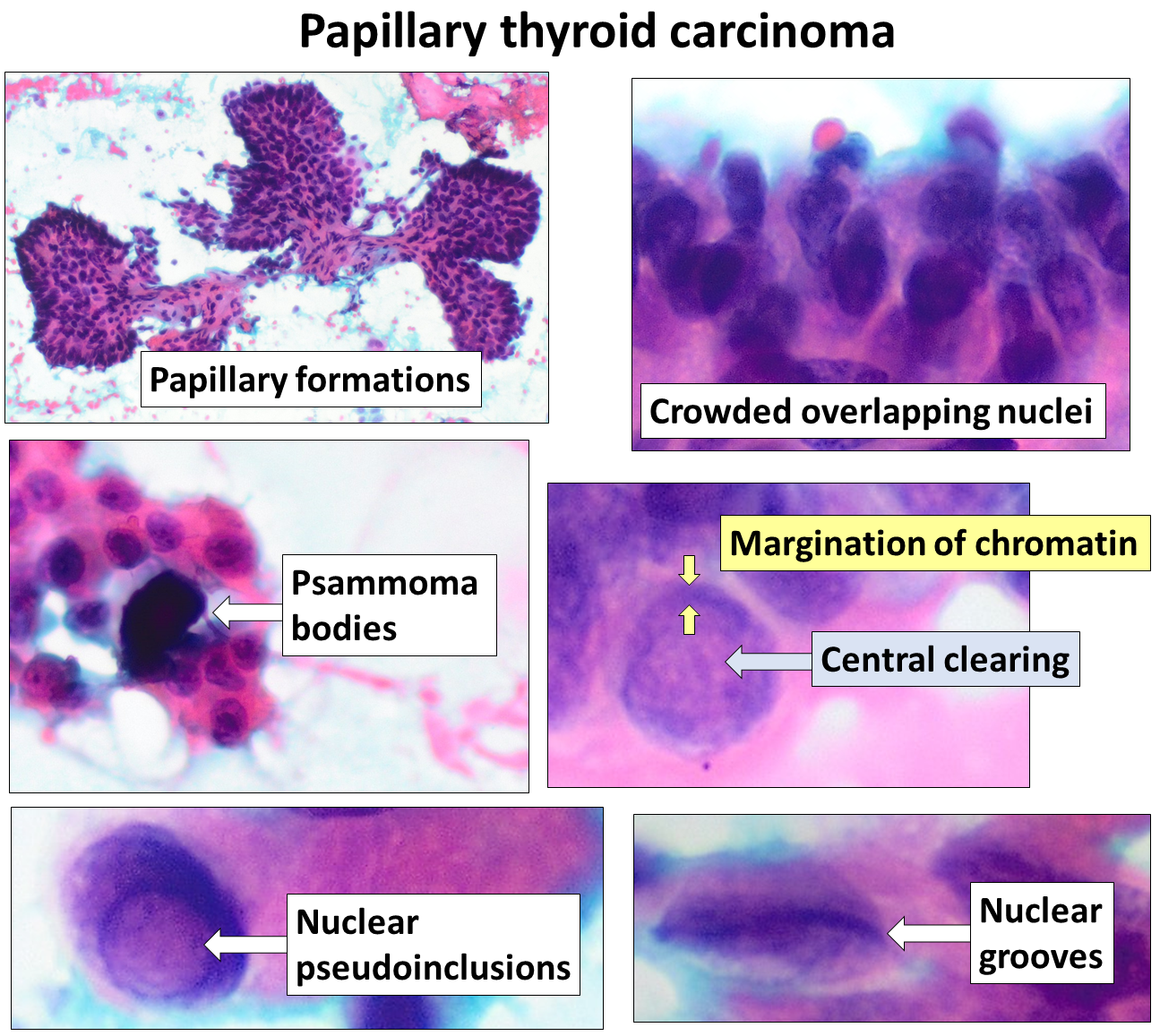
A fixed thyroid mass with enlarged cervical nodes can tempt you to jump directly to "thyroid cancer." Instead, ask which combination of morphology and spread identifies the subtype. Papillary thyroid carcinoma classically shows optically clear nuclei, nuclear grooves and intranuclear cytoplasmic inclusions, may contain psammoma bodies, and commonly reaches regional lymph nodes. RET rearrangements occur in a subset, but no single RET event defines every papillary tumor. [3][22]
Use the nuclear features as a recognition pattern rather than a single-feature diagnosis. Labels are part of the original image. Image: Mikael Häggström; original source; CC0 1.0.
Follicular thyroid carcinoma is different because the decisive finding is capsular or vascular invasion. A fine-needle aspirate can show follicular cells, but cytology alone cannot prove the invasion that separates carcinoma from adenoma. Once malignant, follicular carcinoma favors blood-borne spread to lung and bone. [3]
Medullary thyroid carcinoma arises from parafollicular C cells and commonly secretes calcitonin. Amyloid can be present in the stroma, and hereditary disease links to MEN2 through activating RET variants. MEN2A adds pheochromocytoma and parathyroid disease; MEN2B adds pheochromocytoma, mucosal neuromas and a marfanoid habitus. RET biology is context dependent: activating variants drive MEN2, while other germline loss-of-function variants can cause Hirschsprung disease. [3][22]
Anaplastic thyroid carcinoma typically presents in an older adult with rapidly progressive local symptoms such as dysphagia, hoarseness or airway compromise. It is biologically unlike the usually treatable differentiated papillary and follicular cancers. When a stem emphasizes speed, fixation and local destruction, do not let the shared thyroid location erase the behavior. [3]
Papillary
Nuclear pattern Regional nodes
Follicular
Capsular or vascular invasion Blood to bone or lung
Medullary
C cell and calcitonin RET and MEN2
Anaplastic
Rapid local destruction Older adult
Four thyroid tumors share an organ but separate cleanly by cell of origin, defining pathology and spread behavior.
Metastatic landmarks are signs, not diagnoses. A left supraclavicular node can receive lymph returning through the thoracic duct and therefore raises concern for an infradiaphragmatic or thoracic malignancy, including gastric cancer, but it is not specific to one organ. An umbilical metastatic nodule likewise suggests intra-abdominal or pelvic malignancy without identifying a single primary. [33]
Try it here · Checkpoint 1 of 3
Make your prediction before reading the choices. A first attempt is just a starting point.
Case 4
Show answer and explanations for case 4
A. Follicular carcinoma with hematogenous spread to bone (Why this does not fit)
Follicular carcinoma is defined by capsular or vascular invasion and more often spreads through blood.
Reasoning steps for option A
What in the history might suggest follicular carcinoma?
A thyroid malignancy in a young woman can be follicular, which is also a differentiated follicular-cell cancer.
Which histologic and spread findings argue against follicular carcinoma?
Clear nuclei with grooves and inclusions are papillary features, and lateral cervical nodes are lymphatic spread rather than the blood-borne pattern of follicular carcinoma.
B. Medullary carcinoma with lymphatic spread and calcitonin secretion (Why this does not fit)
Medullary carcinoma arises from C cells and is supported by calcitonin and neuroendocrine pathology, not papillary-type nuclei.
Reasoning steps for option B
Why could medullary carcinoma seem to fit enlarged cervical nodes?
Medullary carcinoma also spreads to cervical nodes and is a classic thyroid cancer to keep in the differential.
What histology is missing for a medullary diagnosis?
Medullary carcinoma shows neuroendocrine C cells, often with amyloid and calcitonin, not optically clear grooved nuclei with inclusions.
C. Anaplastic carcinoma with direct tracheal invasion (Why this does not fit)
Anaplastic carcinoma usually presents in an older adult with rapidly destructive local symptoms.
Reasoning steps for option C
What could make an aggressive local tumor seem possible in a neck mass with nodes?
Anaplastic carcinoma is a thyroid cancer that invades nearby structures such as the trachea.
Which features of this patient do not fit anaplastic carcinoma?
She is 34 without rapid destructive symptoms, and her tumor shows well-formed papillary nuclear features rather than undifferentiated pleomorphic cells.
D. Papillary carcinoma with regional lymphatic dissemination (Best answer)
Papillary thyroid carcinoma shows clear nuclei, grooves and inclusions and commonly reaches cervical lymph nodes.
Reasoning steps for option D
What does the combination of clear nuclei, grooves and inclusions identify?
These nuclear changes are the defining cytologic signature of papillary thyroid carcinoma.
How do the radiation history and lateral nodes support the same diagnosis?
Childhood neck radiation is a known risk factor for papillary carcinoma, and this tumor typically spreads through lymphatics to cervical nodes.
Takeaway: Papillary thyroid carcinoma pairs distinctive nuclei with a strong tendency for regional nodal spread.
The old habit was to identify a brain tumor from one microscopic phrase. Modern CNS diagnosis uses an integrated classification that combines histology with molecular information, so classic morphology remains useful but is not the whole diagnosis. Adult diffuse gliomas are now defined in a molecular framework: glioblastoma means glioblastoma, IDH-wildtype, which can be diagnosed from necrosis or microvascular proliferation, or from TERT promoter mutation, EGFR amplification or +7/−10, while IDH-mutant grade 4 tumors are classified as astrocytoma, IDH-mutant. [4]
For board-style localization, a destructive adult hemispheric mass crossing the corpus callosum can suggest glioblastoma, while pseudopalisading necrosis and microvascular proliferation support a high-grade astrocytic process. A child with a cerebellar cystic mass and mural nodule instead suggests pilocytic astrocytoma, a circumscribed glioma that commonly arises in the cerebellum and is often cystic. [5]
A child with a midline posterior fossa mass and dissemination through cerebrospinal fluid raises medulloblastoma, whereas a suprasellar calcified cystic lesion causing visual field loss and pituitary dysfunction points toward craniopharyngioma. Meningioma is generally extra-axial and can show whorls and psammoma bodies. These tumors are separated most efficiently by age, compartment and expected route of spread, not by memorizing one histologic noun. [5]
Outside the CNS, a painful diaphyseal bone tumor in an adolescent with a small round cell appearance and onion-skin periosteal reaction suggests Ewing sarcoma; most cases carry EWSR1::FLI1 from t(11;22). Neuroblastoma often arises in the adrenal or sympathetic trunk, and MYCN amplification is an adverse prognostic feature. [6][19]
Retinoblastoma illustrates the two-hit tumor suppressor model: hereditary RB1 disease predisposes to bilateral or multifocal retinal tumors and also increases later osteosarcoma risk. Rhabdomyosarcoma shows skeletal muscle differentiation and commonly expresses desmin, but desmin is a lineage marker rather than a unique tumor label. Wilms tumor is a renal embryonal tumor of young children; when aniridia and genitourinary anomalies accompany it, think of the WAGR spectrum rather than neuroblastoma. [36][34][27]
Apply the same approach to a new pediatric mass: use age plus compartment first, add morphology second, then use genetics when it changes the classification or prognosis. This keeps a "small round blue cell" description from becoming a false diagnosis by itself.
Try it here · Checkpoint 2 of 3
Make your prediction before reading the choices. A first attempt is just a starting point.
Case 12
Show answer and explanations for case 12
A. BCR::ABL1 from t(9;22) (Why this does not fit)
BCR::ABL1 is the hallmark fusion kinase of CML, not a primary diaphyseal bone sarcoma.
Reasoning steps for option A
Why could BCR::ABL1 be tempting for a malignant tumor with small cells?
It is the best-known translocation in oncology and is often the first fusion recalled.
Which tissue does BCR::ABL1 drive, and does it match?
It drives chronic myeloid leukemia in marrow, not a destructive sarcoma in the femoral diaphysis.
B. MYC juxtaposition from t(8;14) (Why this does not fit)
t(8;14) is characteristic of Burkitt lymphoma and does not define Ewing sarcoma.
Reasoning steps for option B
What could make t(8;14) seem to fit a small round cell tumor?
Burkitt lymphoma is also a small round blue cell tumor with a very high growth rate.
Why does the bone presentation argue against a MYC translocation?
t(8;14) defines Burkitt lymphoma, while a diaphyseal sarcoma with onion-skin periosteal reaction in a teenager fits Ewing sarcoma.
C. PML::RARA from t(15;17) (Why this does not fit)
PML::RARA defines acute promyelocytic leukemia and is associated with coagulopathy, not a bone sarcoma.
Reasoning steps for option C
Why might PML::RARA come up with fever and bone pain?
Acute leukemias can present with fever and bone pain from marrow expansion.
Which finding makes PML::RARA the wrong fusion?
The biopsy shows a sarcoma in bone, while PML::RARA defines acute promyelocytic leukemia with coagulopathy.
D. EWSR1::FLI1 from t(11;22) (Best answer)
Most Ewing sarcomas carry EWSR1::FLI1 from t(11;22), matching the age, diaphyseal location and small round cell phenotype.
Reasoning steps for option D
Which tumor fits an adolescent with a diaphyseal onion-skin lesion and small round sarcoma cells?
Ewing sarcoma, which also commonly causes fever and pain that can mimic infection.
Which fusion is found in most Ewing sarcomas?
EWSR1::FLI1 from t(11;22), a fusion transcription factor that drives the tumor.
Takeaway: Ewing sarcoma links an adolescent diaphyseal small round cell tumor to EWSR1::FLI1.
Organ patterns connect pathology to systemic effects
In lung cancer, anatomy and endocrine effects often identify the lineage before a biopsy result is shown. Squamous cell carcinoma is commonly central, can cavitate, and may produce PTH-related peptide with hypercalcemia. Adenocarcinoma is often peripheral and is common among patients who never smoked. The older label bronchioloalveolar carcinoma has been replaced by modern adenocarcinoma categories that include lepidic growth patterns. [7]
Small cell lung carcinoma is a high-grade neuroendocrine cancer with an early tendency to disseminate and can produce endocrine syndromes such as SIADH or ectopic ACTH. Lambert-Eaton myasthenic syndrome reflects presynaptic voltage-gated calcium-channel autoimmunity and may improve transiently with repeated activation, in contrast with the typical fatigability of myasthenia gravis. [8]
Stage can be encoded in a finding that seems secondary. In lung cancer, a malignant pleural effusion represents metastatic intrathoracic disease rather than a harmless accompaniment. In laryngeal squamous cancer, invasion through cartilage is evidence of advanced local extension. The general principle is to translate anatomic invasion into extent before deciding what the finding means for prognosis or treatment. [1][30]
Several abdominal tumors also have compact patterns. A pancreatic head adenocarcinoma can cause painless obstructive jaundice; smoking and chronic pancreatitis increase risk. Endometrioid endometrial carcinoma is associated with prolonged unopposed estrogen exposure, including chronic anovulation. Long-standing ulcerative colitis increases colorectal cancer risk, and high-grade dysplasia in chronically inflamed colon is a major management finding rather than a routine polyp. [28][29][31]
Breast oncology is best understood as both inherited repair biology and tumor-specific signaling. HER2 is a targetable receptor whose amplification or overexpression defines a treatment-relevant subset rather than a simple good-versus-bad prognosis label. BRCA1 and BRCA2 participate in homologous recombination DNA repair; pathogenic germline variants increase risks of breast, ovarian and several other cancers. [12][13]
For primary breast cancer risk reduction, medication choice depends on menopausal status and harms. Tamoxifen can be used in premenopausal or postmenopausal patients at increased risk, while raloxifene and aromatase inhibitors are used only after menopause for this purpose. SERMs increase venous thromboembolism risk, tamoxifen increases endometrial cancer risk, and aromatase inhibitors can worsen fracture risk. Risk-reducing surgery in selected hereditary syndromes is an individualized decision and does not reduce risk to zero. [14]
Infection can create risk, while markers need context
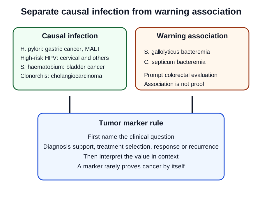
Infectious oncology is easiest when you separate direct carcinogenic infections from clinical warning associations. H. pylori increases risk of gastric adenocarcinoma and gastric MALT lymphoma; eradication can produce lymphoma regression in many localized H. pylori-associated MALT cases. Chronic HBV and HCV infection increase hepatocellular carcinoma risk, while high-risk HPV drives cervical, other anogenital and many oropharyngeal cancers. EBV is linked to selected lymphomas and nasopharyngeal carcinoma, HTLV-1 to adult T-cell leukemia/lymphoma, and KSHV/HHV-8 to Kaposi sarcoma. [9][10]
Separate causal carcinogens from clinical warning associations, then define what a tumor marker is being used to answer. [9][10][24][25]
Parasites are specific. Schistosoma haematobium is carcinogenic to humans and is linked to bladder cancer, especially squamous carcinoma in endemic settings. Clonorchis sinensis is linked to cholangiocarcinoma. By contrast, Schistosoma mansoni is not a validated cause of hepatocellular carcinoma, so the older species swap is unsafe. [24]
Some bacteria function as warning signals rather than direct proof of a tumor. Streptococcus gallolyticus bacteremia or endocarditis and Clostridium septicum bacteremia are associated with colorectal neoplasia and should trigger evaluation for an occult colorectal source when clinically appropriate. They do not mean every positive culture already proves cancer. [25]
Tumor markers are similarly contextual. An abnormal AFP can support HCC or a germ-cell tumor pattern, but AFP alone does not diagnose HCC. In an at-risk liver, surveillance commonly uses ultrasound plus AFP; a suspicious lesion or markedly abnormal marker prompts diagnostic multiphasic imaging. CEA is useful mainly for following treated colorectal cancer, CA-125 for selected ovarian cancer contexts, CA19-9 for pancreatic or biliary disease monitoring, and PSA can inform prostate evaluation and follow-up. [11][12][15]
Immunohistochemistry answers a lineage question, not a magic-word question. Cytokeratins support epithelial differentiation, desmin supports muscle differentiation, GFAP supports glial differentiation, and S-100 is found in several neural-crest and other tissues. Vimentin supports mesenchymal differentiation but is too nonspecific to mean "all sarcomas" or to identify a particular sarcoma by itself.
Marker present
Ask what it changes Diagnosis support Treatment selection Response or recurrence
Marker absent
Ask test sensitivity Absence may lower probability It rarely erases pathology by itself
A marker becomes useful only after you specify the clinical question it is being asked to answer.
Try it here · Checkpoint 3 of 3
Make your prediction before reading the choices. A first attempt is just a starting point.
Case 23
Show answer and explanations for case 23
A. AFP alone proves hepatocellular carcinoma, so imaging is unnecessary (Why this does not fit)
AFP is not sufficiently specific to establish HCC by itself and should not replace diagnostic imaging.
Reasoning steps for option A
Why might a raised AFP seem enough to diagnose HCC in chronic hepatitis B?
AFP is the familiar HCC marker and is raised in this at-risk patient.
Why is AFP alone insufficient?
AFP can rise in hepatitis and other conditions and is normal in many HCCs, so it supports context but cannot replace diagnostic imaging.
B. The lesion is most likely a hepatic adenoma because AFP is below 200 ng/mL (Why this does not fit)
An AFP threshold does not separate adenoma from HCC, and the vascular washout pattern in a high-risk liver argues for HCC.
Reasoning steps for option B
What could make an adenoma seem likely when AFP is only mildly raised?
A modest AFP sounds less alarming, and adenoma is a benign enhancing liver lesion.
Why does an AFP threshold not separate adenoma from HCC here?
No AFP cutoff excludes HCC, and arterial enhancement with washout in a chronic HBV liver is the HCC pattern.
C. The imaging pattern in an at-risk liver is strongly diagnostic of hepatocellular carcinoma (Best answer)
In an at-risk patient, arterial hyperenhancement with later washout is a characteristic noninvasive HCC imaging pattern; AFP supports context but is not the sole diagnostic test.
Reasoning steps for option C
What imaging pattern is expected of HCC on multiphasic MRI?
Arterial-phase hyperenhancement followed by washout in later phases, reflecting arterial blood supply.
Why can the diagnosis be made without biopsy in this patient?
In an at-risk liver such as chronic HBV, a 2.2-cm lesion with that pattern is diagnostic of HCC noninvasively, with AFP adding supportive context.
D. Normal liver enzymes would exclude hepatocellular carcinoma (Why this does not fit)
HCC can occur despite normal transaminases, especially in chronic HBV, and liver enzymes are not a rule-out test.
Reasoning steps for option D
Why might normal liver enzymes sound reassuring?
Enzymes are a routine liver test, and normal results suggest little active liver damage.
Can normal enzymes exclude HCC?
No. Chronic HBV can drive HCC while ALT and AST stay in the normal range, so a quiet enzyme panel cannot outweigh the enhancing, washing-out lesion already seen on MRI.
Takeaway: AFP supports HCC assessment but does not diagnose it alone; characteristic multiphasic imaging in an at-risk liver can establish the diagnosis.
Genetics explains why the phenotype behaves that way
Oncogenes usually represent increased or constitutive growth signaling, while tumor suppressor loss removes restraints on proliferation, genomic damage or survival. The useful question is not simply which gene is "on" or "off," but what cell behavior changes after that alteration. Cancer biology therefore links signaling, DNA repair, cell-cycle control, apoptosis, invasion and immune escape rather than treating gene names as a separate vocabulary list. [23]
Five translocations remain high-yield because the rearrangement explains the disease. BCR::ABL1 from the Philadelphia chromosome t(9;22) creates a constitutively active tyrosine kinase in CML. PML::RARA from t(15;17) blocks promyelocyte maturation in APL and is associated with dangerous coagulopathy; ATRA-based therapy directly addresses the differentiation block. [16][35]
MYC juxtaposition to an immunoglobulin locus, classically t(8;14), drives Burkitt lymphoma. EWSR1::FLI1 from t(11;22) defines most Ewing sarcomas. BCL2 rearrangement, classically t(14;18), supports follicular lymphoma by increasing anti-apoptotic signaling. The point is to pair chromosome change with protein consequence and phenotype, not to memorize numbers without a result. [18][6]
Amplification can also matter. MYCN amplification is an adverse prognostic feature in neuroblastoma [19]. ERBB2/HER2 amplification or overexpression identifies targetable breast and gastric cancer subsets [12]. In small cell lung cancer, MYC-family alterations occur in subsets, but an "L-MYC equals lung" rule is not a universal diagnostic marker. [8]
Tumor suppressors provide inheritance patterns. RB1 regulates the G1/S checkpoint and illustrates the two-hit concept. Germline TP53 variants cause Li-Fraumeni syndrome, with a spectrum that prominently includes early breast cancer, soft-tissue and bone sarcomas, CNS tumors and adrenocortical carcinoma. APC-associated FAP produces extensive colorectal adenomas and can include osteomas and desmoid tumors. [20][21]
For a new genetics stem, translate the alteration into function before naming the cancer. A fusion kinase suggests constitutive signaling, a defective repair gene predicts genomic instability, and a lost checkpoint removes a brake on proliferation. Function first makes t(9;22), TP53, BRCA1/2, RB1 and APC part of one causal model rather than five disconnected facts.
Translate the gene into a cellular consequence, then use the tissue pattern to identify the disease.
Apply the framework
For each case, commit to the diagnosis or process only after you identify the two most discriminating findings. Then test every option against both findings rather than choosing the first familiar association.
Case 1
Show answer and explanations for case 1
A. Hematogenous spread to the lungs (Best answer)
Malignant osteoid identifies osteosarcoma, a sarcoma that characteristically seeds the lungs through the bloodstream.
Reasoning steps for option A
What does malignant osteoid tell you about the tumor lineage?
Osteoid made by malignant cells identifies osteosarcoma, a mesenchymal tumor of bone.
How do mesenchymal tumors usually reach distant organs such as lung?
Sarcomas usually invade vessels and travel in the bloodstream, and the lung is the first capillary bed they meet. The new pulmonary nodules fit that route.
B. Lymphatic spread to hilar nodes before lung parenchyma (Why this does not fit)
Osteosarcoma is mesenchymal and commonly disseminates through blood rather than requiring a regional nodal step.
Reasoning steps for option B
Why could hilar nodal spread seem reasonable for a tumor with lung nodules?
Many lung-bound cancers pass through regional nodes first, so a nodal step feels like a natural bridge to the chest.
What stops a nodal route from fitting this adolescent's femoral tumor?
The osteoid-producing bone tumor is a sarcoma, and sarcomas seldom need a lymphatic step before seeding lung parenchyma.
C. Transcoelomic spread across the pleural surface (Why this does not fit)
Pleural seeding does not explain a primary metaphyseal osteoid-producing tumor with discrete lung nodules.
Reasoning steps for option C
What could make pleural surface spread tempting when the chest is involved?
Tumors in a body cavity can shed cells across serosal surfaces, and the metastases here are in the thorax.
Where are the chest lesions, and why does that matter for transcoelomic spread?
They are discrete nodules within lung parenchyma, and the primary is in the femur, far from any pleural space it could seed across.
D. Perineural spread along the sciatic nerve (Why this does not fit)
Perineural extension is a local pattern of selected carcinomas and does not explain multiple pulmonary metastases.
Reasoning steps for option D
Why might nerve involvement come to mind with distal femoral pain?
A painful thigh mass could suggest a tumor tracking along the sciatic nerve.
What finding makes perineural extension irrelevant to the question asked?
Perineural spread is local, mostly seen in selected carcinomas, and cannot carry tumor to several separate pulmonary nodules.
Takeaway: Malignant osteoid establishes osteosarcoma, and pulmonary metastases fit hematogenous sarcoma spread.
A. Urothelial carcinoma with lymphatic spread and ectopic ACTH (Why this does not fit)
Urothelial carcinoma arises from collecting-system epithelium and does not best explain a cortical renal mass with venous extension and erythrocytosis.
Reasoning steps for option A
What makes a urothelial tumor attractive when a man has gross hematuria?
Painless gross hematuria in an older man is a classic urothelial carcinoma presentation.
Which findings point away from a collecting-system tumor with nodal spread and ACTH excess?
The mass sits in the renal cortex, grows into the renal vein and IVC, and the paraneoplastic clue is erythrocytosis rather than Cushing features.
B. Renal cell carcinoma with venous invasion and paraneoplastic erythropoietin (Best answer)
Renal cell carcinoma can invade the renal vein and IVC and can secrete erythropoietin inappropriately as a paraneoplastic effect, linking the anatomy and the high red-cell mass.
Reasoning steps for option B
Which primary tumor arises in the renal cortex of an older adult and grows into veins?
Renal cell carcinoma is the cortical epithelial tumor famous for extending into the renal vein and inferior vena cava, an exception to the carcinoma lymphatic rule.
How does renal cell carcinoma explain the high red-cell mass?
Tumor cells can secrete erythropoietin without the normal hypoxic signal, so erythrocytosis is a paraneoplastic effect of the cancer.
C. Wilms tumor with renal-vein invasion and renin suppression (Why this does not fit)
Wilms tumor is primarily a childhood renal embryonal tumor, and renin suppression would not account for erythrocytosis.
Reasoning steps for option C
Why could Wilms tumor come up when a renal mass invades the renal vein?
Wilms tumor is a well-known renal malignancy that can also extend into the renal vein.
Which details rule Wilms tumor out in this patient?
He is 61, while Wilms tumor is a childhood embryonal tumor, and renin suppression would do nothing to raise red-cell mass.
D. Prostate adenocarcinoma with retrograde venous spread and secondary polycythemia (Why this does not fit)
Prostate cancer does not present as a renal cortical mass extending from the kidney into the renal vein.
Reasoning steps for option D
What could draw a reader toward prostate cancer in an older man with polycythemia?
Prostate cancer is common at this age and spreads through venous plexuses, so venous spread plus a high hematocrit sounds connected.
What anatomic finding excludes a prostate primary?
The tumor starts in the renal cortex and extends outward into the renal vein and IVC, which is the path of a kidney primary, not a pelvic one.
Takeaway: RCC is an epithelial exception with prominent venous invasion and can cause paraneoplastic (inappropriate) erythropoietin production.
A. Calcitonin production by tumor cells (Why this does not fit)
Calcitonin supports medullary thyroid carcinoma, not a follicular neoplasm.
Reasoning steps for option A
Why might calcitonin seem like a useful extra test for a thyroid nodule?
Calcitonin is a familiar thyroid tumor marker and is measured when a nodule is being classified.
What does calcitonin actually identify, and does it fit a follicular aspirate?
It marks C-cell derived medullary carcinoma, whereas a uniform follicular pattern comes from follicular epithelium.
B. Regional cervical lymph-node metastasis (Why this does not fit)
Nodal spread can occur in thyroid cancer but is not the defining histologic criterion separating follicular adenoma from carcinoma.
Reasoning steps for option B
Why could lateral neck nodes seem to prove malignancy in a thyroid nodule?
Nodal metastasis shows a tumor has spread, and spread implies cancer.
Why is nodal disease not the defining criterion for follicular carcinoma?
Follicular carcinoma is diagnosed on architecture and it favors blood-borne spread, so nodes are neither typical nor the criterion that separates it from adenoma.
C. Capsular or vascular invasion on tissue architecture (Best answer)
Follicular carcinoma requires demonstration of invasion, which aspiration cytology cannot assess because architecture is not preserved.
Reasoning steps for option C
Why can the aspirate not settle adenoma versus carcinoma?
Both lesions are made of similar bland follicular cells, and aspiration breaks up the tissue so the lesion edge and its vessels cannot be examined.
What must the excised specimen show to call the lesion carcinoma?
It must show tumor penetrating the capsule or invading vessels; that invasion is the defining feature of follicular carcinoma.
D. Psammoma bodies within the aspirate (Why this does not fit)
Psammoma bodies are associated with papillary thyroid carcinoma and would shift the diagnostic pattern away from follicular carcinoma.
Reasoning steps for option D
Why could psammoma bodies sound like a sign of thyroid malignancy?
Psammoma bodies are a classic calcified finding in a thyroid cancer and are often memorized as a cancer clue.
Which thyroid tumor do psammoma bodies point to, and how does that fit the aspirate?
They point to papillary carcinoma, and this aspirate specifically lacks papillary nuclear features.
Takeaway: Follicular thyroid carcinoma is diagnosed by capsular or vascular invasion, not by follicular cytology alone.
A. Hematogenous tumor emboli traveling through the azygos system (Why this does not fit)
A left supraclavicular node is a lymphatic finding rather than a blood-borne pulmonary or systemic embolic pattern.
Reasoning steps for option A
Why could blood-borne spread seem plausible for a gastric cancer with a distant finding?
Gastric cancer can spread through veins, and a lesion far from the stomach sounds like distant metastasis.
What kind of structure is the supraclavicular finding?
It is a lymph node, so it reflects lymphatic drainage rather than tumor emboli in the azygos venous system.
B. Direct extension of the gastric mass through the diaphragm (Why this does not fit)
Direct extension requires contiguous anatomic invasion and cannot account for a separate supraclavicular node.
Reasoning steps for option B
What makes direct extension tempting for a large gastric wall mass?
Advanced gastric cancer can invade neighboring structures, including the diaphragm.
Why can contiguous invasion not produce the neck finding?
The supraclavicular node is separate from the stomach, and direct extension requires continuous tumor growth into adjacent tissue.
C. Lymphatic dissemination through thoracic-duct drainage (Best answer)
The left supraclavicular region receives lymph returning through the thoracic duct, so a hard node can signal thoracic or abdominal malignancy.
Reasoning steps for option C
Where does lymph from the abdomen enter the venous system?
Abdominal lymph ascends through the thoracic duct and empties near the left subclavian and internal jugular junction, beside the left supraclavicular nodes.
How does that anatomy explain a hard left supraclavicular node with a gastric mass?
Tumor cells carried in thoracic-duct lymph can lodge in that node, so it marks lymphatic dissemination rather than naming a single primary.
D. Peritoneal seeding to an umbilical implant (Why this does not fit)
Peritoneal dissemination can produce an umbilical metastatic nodule, but that is a different anatomic landmark.
Reasoning steps for option D
Why might an umbilical nodule come to mind with gastric cancer?
Gastric cancer can spread through the peritoneum and produce an umbilical metastasis, another well-known landmark.
Why does peritoneal seeding not answer the question?
The finding in question is a supraclavicular node, and peritoneal implants do not reach the neck.
Takeaway: A left supraclavicular metastatic node is a lymphatic landing site, not a stomach-specific diagnosis.
A. The imaging pattern alone establishes glioblastoma without molecular classification (Why this does not fit)
The butterfly pattern is suggestive but modern CNS diagnosis integrates molecular data with histology.
Reasoning steps for option A
Why is a butterfly lesion crossing the corpus callosum so suggestive?
An infiltrative adult hemispheric mass crossing the corpus callosum is the classic imaging pattern of glioblastoma.
Why does imaging alone still fall short of the final diagnosis?
Current WHO CNS classification integrates histology with molecular features such as IDH status, so a picture cannot complete the diagnosis.
B. The lesion is most consistent with a benign extra-axial meningioma (Why this does not fit)
Meningioma is generally extra-axial and does not usually cross the corpus callosum as an infiltrative parenchymal mass.
Reasoning steps for option B
What could make meningioma seem possible in an older adult with a brain mass?
Meningioma is a common adult intracranial tumor and can cause personality change and weakness.
Which imaging and histology findings exclude meningioma?
Meningioma is extra-axial and dural-based, while this tumor infiltrates brain across the corpus callosum and shows necrosis with microvascular proliferation.
C. The lesion is a pilocytic astrocytoma because GFAP is expected (Why this does not fit)
Pilocytic astrocytoma is usually circumscribed, commonly pediatric and often cystic with a mural nodule.
Reasoning steps for option C
Why could GFAP expression seem to support pilocytic astrocytoma?
Pilocytic astrocytoma is a glial tumor, and GFAP is a glial marker.
Why does the patient profile argue against pilocytic astrocytoma?
Pilocytic tumors are usually circumscribed, cystic and pediatric, not infiltrative high-grade masses with necrosis in a 64-year-old.
D. The findings support a high-grade diffuse glioma that requires integrated histologic and molecular classification (Best answer)
The infiltrative hemispheric pattern, necrosis and microvascular proliferation support a high-grade diffuse glioma, while current WHO practice integrates molecular features.
Reasoning steps for option D
What do pseudopalisading necrosis and microvascular proliferation indicate?
They are high-grade features of a diffuse astrocytic glioma in this infiltrative adult hemispheric mass.
Why must the final label still include molecular testing?
Under WHO CNS5, glioblastoma means an IDH-wildtype tumor, and IDH-mutant high-grade tumors are classified as astrocytoma, IDH-mutant.
Takeaway: Classic glioblastoma morphology is useful, but current CNS diagnosis is integrated with molecular classification.
A cerebellar cyst with a mural nodule in a child is a classic circumscribed pilocytic astrocytoma pattern.
Reasoning steps for option A
What does a well-circumscribed cerebellar cyst with an enhancing mural nodule in a school-age child suggest?
It is the classic appearance of pilocytic astrocytoma.
How do the slow symptoms fit a circumscribed glioma?
Months of headache and imbalance fit a slowly growing cerebellar tumor that causes mass effect rather than rapid infiltration.
B. Glioblastoma (Why this does not fit)
Glioblastoma is usually an infiltrative high-grade adult diffuse glioma rather than a circumscribed pediatric cerebellar cyst.
Reasoning steps for option B
Why could glioblastoma come up when a child has an enhancing brain tumor?
Glioblastoma is the best-known enhancing malignant glioma.
Which features separate this lesion from glioblastoma?
Glioblastoma is typically an infiltrative adult hemispheric tumor, while this is a well-circumscribed cerebellar cyst in a child.
C. Medulloblastoma (Why this does not fit)
Medulloblastoma is a posterior fossa embryonal tumor, often midline in younger children, but the cyst-with-mural-nodule architecture is more typical of pilocytic astrocytoma.
Reasoning steps for option C
What makes medulloblastoma a real consideration here?
It is a common malignant posterior fossa tumor of childhood and causes gait imbalance.
Which imaging detail favors a different tumor?
Medulloblastoma is usually a solid midline mass near the fourth ventricle, whereas a cyst with a mural nodule is the pilocytic pattern.
D. Craniopharyngioma (Why this does not fit)
Craniopharyngioma is usually suprasellar near the pituitary and optic chiasm rather than cerebellar.
Reasoning steps for option D
Why might craniopharyngioma be considered for a cystic pediatric brain tumor?
Craniopharyngioma is a cystic childhood tumor that also causes headache.
Where is craniopharyngioma found, and does that match?
It sits in the suprasellar region near the pituitary and chiasm, not in the cerebellum.
Takeaway: A pediatric cerebellar cyst with a mural nodule strongly favors pilocytic astrocytoma.
Medulloblastoma arises in the posterior fossa and causes cerebellar or obstructive symptoms, not a calcified suprasellar syndrome.
Reasoning steps for option A
Why might medulloblastoma be considered in a child with a brain tumor?
It is one of the most common malignant pediatric brain tumors.
Which findings point away from a posterior fossa tumor?
Growth failure, polyuria and bitemporal field loss localize to the sella and chiasm, not the cerebellum.
B. Meningioma (Why this does not fit)
A meningioma can occur near the sella, but the pediatric calcified cystic pattern with endocrine dysfunction is more typical of craniopharyngioma.
Reasoning steps for option B
What makes meningioma a possible suprasellar lesion?
Meningiomas can arise near the sella and can calcify.
Which features favor another diagnosis in this girl?
Meningioma is uncommon in children, and a calcified cystic suprasellar mass with pituitary failure is the craniopharyngioma pattern.
C. Craniopharyngioma (Best answer)
A calcified suprasellar cystic lesion with pituitary and optic-chiasm effects strongly supports craniopharyngioma.
Reasoning steps for option C
What do growth failure, polyuria and bitemporal field loss localize to?
Growth hormone deficiency and diabetes insipidus point to the pituitary and hypothalamus, and bitemporal loss points to the optic chiasm above the sella.
Which imaging finding identifies the tumor?
A calcified cystic suprasellar mass in a child is characteristic of craniopharyngioma.
D. Pilocytic astrocytoma (Why this does not fit)
Optic-pathway pilocytic tumors occur in children but a calcified suprasellar cystic lesion with pituitary dysfunction is less typical.
Reasoning steps for option D
Why might an optic pathway glioma seem to fit visual field loss?
Pilocytic tumors of the optic pathway occur in children and damage vision.
Which features fit pilocytic astrocytoma less well?
Prominent calcification with a cyst and pituitary dysfunction are more typical of craniopharyngioma than of an optic pathway glioma.
Takeaway: Suprasellar localization plus calcification and pituitary dysfunction is the classic craniopharyngioma pattern.
A. Ectopic calcitonin secretion by the tumor (Why this does not fit)
Calcitonin lowers serum calcium and is associated with medullary thyroid carcinoma, not hypercalcemia from squamous lung cancer.
Reasoning steps for option A
Why might calcitonin come up when a patient has a calcium problem?
Calcitonin is a calcium-regulating hormone that tumors can secrete.
In which direction does calcitonin move serum calcium?
Calcitonin lowers calcium, so it cannot explain a level of 12.8 mg/dL.
B. Excess ACTH causing increased bone resorption (Why this does not fit)
Ectopic ACTH is associated with small cell lung cancer and does not directly produce this classic hypercalcemia pattern.
Reasoning steps for option B
What could link ACTH to a smoker with a lung tumor?
Lung cancers, especially small cell carcinoma, can secrete ectopic ACTH.
Why does ACTH not fit this tumor and this calcium level?
Ectopic ACTH is typical of small cell, not keratinizing squamous carcinoma, and it causes Cushing features rather than this hypercalcemia pattern.
C. Ectopic PTH-related peptide secretion by the tumor (Best answer)
Squamous lung carcinoma can secrete PTHrP, producing hypercalcemia with suppressed endogenous PTH.
Reasoning steps for option C
What does high calcium with suppressed PTH and no bone metastases suggest?
A circulating factor other than PTH is driving calcium up, which is humoral hypercalcemia of malignancy.
Which lung tumor secretes that factor?
Squamous cell carcinoma, a central tumor in heavy smokers, secretes PTH-related peptide, which acts on the PTH receptor while native PTH is suppressed.
D. Primary hyperparathyroidism from a parathyroid adenoma (Why this does not fit)
Primary hyperparathyroidism would usually have inappropriately high or non-suppressed PTH.
Reasoning steps for option D
Why is primary hyperparathyroidism the most common cause of hypercalcemia in general?
Parathyroid adenoma is the leading outpatient cause of high calcium.
Which laboratory value excludes primary hyperparathyroidism here?
PTH is suppressed; a parathyroid adenoma would give an inappropriately normal or high PTH.
Takeaway: Squamous lung carcinoma can cause PTH-independent hypercalcemia through PTHrP.
A. Peripheral lung adenocarcinoma (Why this does not fit)
Adenocarcinoma can occur in smokers or never-smokers but is not the classic source of SIADH plus Lambert-Eaton syndrome.
Reasoning steps for option A
Why is adenocarcinoma a reasonable first thought for lung cancer?
It is the most common lung cancer and occurs in smokers too.
Which clinical findings fit a different histology?
Adenocarcinoma is usually peripheral and is not the classic cause of SIADH together with Lambert-Eaton syndrome.
B. Squamous cell carcinoma (Why this does not fit)
Squamous carcinoma is central and can cavitate, but its classic endocrine association is PTHrP-mediated hypercalcemia rather than this combination.
Reasoning steps for option B
What makes squamous cell carcinoma tempting in a smoker with a central mass?
Squamous carcinoma is central, strongly smoking-related and known for paraneoplastic effects.
Which paraneoplastic effect does squamous carcinoma produce instead?
It classically secretes PTH-related peptide and causes hypercalcemia, not hyponatremia with a neuromuscular syndrome.
C. Pleural mesothelioma (Why this does not fit)
Mesothelioma is a pleural malignancy linked to asbestos exposure and does not typically produce this paraneoplastic combination.
Reasoning steps for option C
Why could mesothelioma be considered for a thoracic malignancy?
Mesothelioma is a thoracic cancer in older adults with occupational exposures.
What separates mesothelioma from this presentation?
It arises from pleura, links to asbestos, and does not typically produce SIADH or Lambert-Eaton syndrome.
D. Small cell lung carcinoma (Best answer)
Small cell carcinoma is a central neuroendocrine lung cancer associated with SIADH and Lambert-Eaton myasthenic syndrome.
Reasoning steps for option D
What do hyponatremia with concentrated urine and weakness that improves with repeated effort represent?
SIADH and Lambert-Eaton myasthenic syndrome, two paraneoplastic syndromes.
Which lung tumor classically produces both?
Small cell carcinoma, a central neuroendocrine tumor of smokers, can secrete ADH and trigger antibodies against presynaptic voltage-gated calcium channels.
Takeaway: Central small cell lung cancer can produce both SIADH and Lambert-Eaton syndrome.
A. Bronchoscopy to search for small cell lung cancer (Why this does not fit)
S. gallolyticus is not a classic signal for an occult pulmonary neuroendocrine tumor.
Reasoning steps for option A
Why might a lung search be considered in an older man with a serious infection?
Occult lung cancer is common in older adults and can present indirectly.
Why does the organism not point to the lung?
Streptococcus gallolyticus is linked to colorectal neoplasia, not to small cell lung cancer.
B. Colonoscopy to evaluate for colorectal neoplasia (Best answer)
S. gallolyticus bacteremia and endocarditis are associated with colorectal neoplasia, so colon evaluation is warranted even without bowel symptoms.
Reasoning steps for option B
What is the significance of Streptococcus gallolyticus bacteremia or endocarditis?
This organism is associated with colorectal adenomas and carcinoma, likely reaching the blood through abnormal colonic mucosa.
Why is colonoscopy warranted even without bowel symptoms?
Colorectal neoplasia is often silent, and he has never been screened, so the association justifies direct colon evaluation once the infection is controlled.
C. Thyroid ultrasound to evaluate for medullary carcinoma (Why this does not fit)
Medullary thyroid carcinoma is linked to RET and calcitonin, not S. gallolyticus endocarditis.
Reasoning steps for option C
Why might thyroid imaging be considered in a patient with a cancer warning sign?
Thyroid nodules are common, and ultrasound is an easy screening test.
Does Streptococcus gallolyticus have any link to thyroid cancer?
No; medullary carcinoma is associated with RET and calcitonin, not this organism.
D. No cancer evaluation because the bacteremia is diagnostic of infection only (Why this does not fit)
Treating the infection is essential, but the organism is a recognized clinical warning association for colorectal neoplasia.
Reasoning steps for option D
Why is it tempting to treat the endocarditis and stop there?
Endocarditis is the urgent problem, and many bacteremias have no deeper cause.
What makes stopping after antibiotics inadequate here?
This organism is a recognized warning association for colorectal neoplasia, so ignoring it could miss an occult cancer.
Takeaway: S. gallolyticus bacteremia or endocarditis should prompt colorectal evaluation; it is an association, not proof of cancer.
A. Immediate total gastrectomy because infection does not affect lymphoma biology (Why this does not fit)
Gastric MALT lymphoma can be infection-driven, so surgery is not automatically the first step in a localized H. pylori-positive case.
Reasoning steps for option A
Why might surgery seem appropriate for a gastric lymphoma?
Gastrectomy removes the tumor and was used for gastric lymphoma in the past.
What biology makes surgery unnecessary as the first step?
Localized gastric MALT lymphoma is driven by chronic H. pylori antigen, so removing the infection can make it regress.
B. High-dose corticosteroids alone to suppress gastric inflammation (Why this does not fit)
Steroids do not eradicate H. pylori and do not address the chronic antigenic driver of this lymphoma.
Reasoning steps for option B
Why might corticosteroids seem to help a lymphoma with gastritis?
Steroids suppress inflammation and are part of some lymphoma regimens.
Why do steroids alone fail to address the cause?
They do not eradicate H. pylori, which is the chronic stimulus sustaining the lymphoma.
C. H. pylori eradication therapy with response assessment (Best answer)
Many localized H. pylori-associated gastric MALT lymphomas regress after eradication of the chronic antigenic stimulus.
Reasoning steps for option C
What drives a localized low-grade gastric MALT lymphoma with active H. pylori?
Chronic antigenic stimulation from H. pylori drives B-cell proliferation in gastric mucosa.
Why do eradication therapy and follow-up biopsies fit that biology?
Many localized H. pylori-positive MALT lymphomas regress after eradication, and response assessment detects the ones that persist and need other therapy.
D. Observation only because MALT lymphoma never progresses (Why this does not fit)
MALT lymphoma can persist or progress, and treatment plus response assessment is required.
Reasoning steps for option D
Why could observation seem reasonable for a low-grade lymphoma?
Low-grade lymphomas can be indolent, and some are observed without treatment.
Why is observation alone wrong for this patient?
The lymphoma can persist or progress, and an easily treatable driver is present, so eradication plus reassessment is expected.
Takeaway: Localized H. pylori-associated gastric MALT lymphoma may regress after eradication therapy, but response still requires follow-up.
A. Tamoxifen because it has no endometrial or thrombotic harms (Why this does not fit)
Tamoxifen reduces breast-cancer risk but can increase venous thromboembolism and endometrial cancer risk, so the stated rationale is wrong.
Reasoning steps for option A
Why is tamoxifen a natural first choice for breast-cancer risk reduction?
Tamoxifen is a well-established SERM that lowers invasive breast-cancer risk.
What is wrong with the justification given for tamoxifen?
Tamoxifen does raise venous thromboembolism and endometrial cancer risk, so a claim that it has no such harms is false.
B. Anastrozole because it increases bone mineral density (Why this does not fit)
Aromatase inhibitors can reduce breast-cancer risk after menopause but can worsen fracture risk rather than improve osteoporosis.
Reasoning steps for option B
Why could an aromatase inhibitor be considered in a postmenopausal woman at high risk?
Aromatase inhibitors reduce breast-cancer risk after menopause.
How do aromatase inhibitors affect her osteoporosis?
They lower estrogen further and reduce bone density, so they can worsen fracture risk instead of helping bone.
C. Trastuzumab because HER2 blockade prevents primary breast cancer in high-risk women (Why this does not fit)
Trastuzumab treats HER2-positive cancers; it is not used as primary chemoprevention based only on risk assessment.
Reasoning steps for option C
What could make HER2 blockade sound like prevention?
Trastuzumab is an effective targeted breast-cancer drug.
Why is trastuzumab not used for primary risk reduction?
It treats established HER2-positive cancers and has no role as chemoprevention based on risk assessment alone.
D. Raloxifene (Best answer)
Raloxifene is a SERM used for breast-cancer risk reduction in postmenopausal patients and has estrogen-agonist effects on bone, although it increases venous-thromboembolism risk.
Reasoning steps for option D
Which drug lowers invasive breast-cancer risk after menopause and acts like estrogen on bone?
Raloxifene, a SERM that blocks estrogen in breast tissue but acts as an agonist in bone.
Why do her other features make raloxifene a good fit?
She is postmenopausal with osteoporosis and has no venous thromboembolism history, which matters because raloxifene raises clot risk.
Takeaway: Risk-reduction medication depends on menopausal status and harms; raloxifene can pair postmenopausal breast-risk reduction with bone benefit.
A. The rising CEA is diagnostic of recurrence without any further evaluation (Why this does not fit)
CEA can signal recurrence but is not specific enough to define the site or establish recurrence without corroborating assessment.
Reasoning steps for option A
Why might a steadily rising CEA seem to prove recurrence?
CEA is the standard colorectal cancer follow-up marker, and a consistent rise after normalization is worrying.
Why is more evaluation still needed?
CEA is not specific, cannot locate disease and can rise for benign reasons such as smoking, so imaging and other assessment are needed to confirm recurrence.
B. The serial rise warrants evaluation for recurrence, but CEA alone is not definitive (Best answer)
A rising CEA after prior normalization can prompt evaluation for recurrent disease, while benign causes and imperfect sensitivity and specificity prevent a marker-only diagnosis.
Reasoning steps for option B
What does a CEA that normalized after surgery and then keeps rising suggest?
It raises concern for recurrent colorectal cancer in a patient treated for stage III disease.
How should that concern be acted on?
The rise should prompt imaging and clinical evaluation for recurrence, because a marker alone cannot establish the diagnosis.
C. CEA has no role after colorectal cancer treatment (Why this does not fit)
CEA is commonly used to help monitor response and detect recurrence in colorectal cancer follow-up.
Reasoning steps for option C
Why could someone think CEA has no use after treatment?
CEA lacks accuracy and is not a screening test, so its value can seem limited.
How is CEA actually used after colorectal cancer surgery?
Serial CEA is commonly used in surveillance to help detect recurrence in treated colorectal cancer.
D. CEA is useful only for screening people who have never had cancer (Why this does not fit)
CEA is not recommended as a population screening test; its better-established role is monitoring known colorectal cancer.
Reasoning steps for option D
Why might CEA seem like a screening test?
It is a well-known colorectal cancer blood test, and screening seeks cancer before symptoms.
What is CEA's accepted role, and does screening fit it?
Screening fits poorly because CEA is seldom raised in early, curable colorectal cancer; its value lies in tracking patients already treated for the disease, such as this man.
Takeaway: Tumor markers are most useful when tied to a defined clinical question and interpreted as trends rather than stand-alone proof.
A. Familial adenomatous polyposis from a germline APC variant (Why this does not fit)
FAP primarily causes extensive colorectal adenomas with selected extracolonic findings, not this classic Li-Fraumeni spectrum.
Reasoning steps for option A
Why could FAP be considered when a family has several cancers?
FAP is a well-known dominant cancer syndrome with extracolonic tumors.
Which tumors define FAP, and do they match this family?
FAP causes hundreds of colorectal adenomas, with osteomas and desmoids, not early breast cancer, sarcoma, brain tumor and adrenocortical carcinoma.
B. Li-Fraumeni syndrome from a germline TP53 pathogenic variant (Best answer)
The combination of early breast cancer, sarcoma, CNS tumor and adrenocortical carcinoma is characteristic of Li-Fraumeni syndrome.
Reasoning steps for option B
Which tumors appear in this family at young ages?
Osteosarcoma, breast cancer at 31, a brain tumor at 37 and adrenocortical carcinoma.
Which syndrome explains that spectrum?
Li-Fraumeni syndrome from a germline TP53 variant, which predisposes to early breast cancer, sarcomas, CNS tumors and adrenocortical carcinoma.
C. MEN2 from an activating RET variant (Why this does not fit)
MEN2 centers on medullary thyroid carcinoma, pheochromocytoma and selected parathyroid disease.
Reasoning steps for option C
What makes MEN2 a possible match for a family with endocrine tumors?
MEN2 is a dominant syndrome that includes an adrenal tumor.
Which adrenal tumor does MEN2 cause, and does the family fit?
MEN2 causes pheochromocytoma of the adrenal medulla with medullary thyroid carcinoma, not adrenocortical carcinoma, sarcoma or breast cancer.
D. Hereditary retinoblastoma from a germline RB1 variant (Why this does not fit)
RB1 can predispose to retinoblastoma and later osteosarcoma, but it does not explain this multigenerational spectrum of early breast, CNS and adrenal cortical tumors.
Reasoning steps for option D
Why could hereditary retinoblastoma be tempting with osteosarcoma in the proband?
Germline RB1 variants increase the risk of later osteosarcoma.
Which part of the family history does RB1 not explain?
It does not explain early breast cancer, a brain tumor and adrenocortical carcinoma across generations, and no one has retinoblastoma.
Takeaway: Li-Fraumeni syndrome is an autosomal dominant TP53 cancer-predisposition syndrome with a broad early-onset tumor spectrum.
TP53 causes Li-Fraumeni syndrome, which produces a broad early-onset cancer spectrum rather than diffuse adenomatous polyposis with osteomas.
Reasoning steps for option A
Why might TP53 be considered for a young man with many tumors?
Germline TP53 causes a dominant syndrome with multiple early-onset cancers.
Which part of the phenotype does TP53 not explain?
Li-Fraumeni does not produce hundreds of colorectal adenomas with osteomas and desmoids.
B. RET (Why this does not fit)
RET gain-of-function causes MEN2, centered on medullary thyroid carcinoma and pheochromocytoma.
Reasoning steps for option B
What could make RET a possible answer for an inherited tumor syndrome?
RET causes a dominant multiple-tumor syndrome.
Which tumors does RET cause?
Germline activating RET variants cause MEN2 with medullary thyroid carcinoma and pheochromocytoma, not colonic polyposis.
C. APC (Best answer)
A germline APC pathogenic variant causes familial adenomatous polyposis and can be associated with osteomas and desmoid tumors.
Reasoning steps for option C
What does a young man with hundreds of colorectal adenomas and an affected father suggest?
Familial adenomatous polyposis, a dominant polyposis syndrome.
How do the osteomas and desmoid tumor point to the gene?
Germline APC variants cause FAP, and osteomas and desmoid tumors are its characteristic extracolonic features.
D. BRCA2 (Why this does not fit)
BRCA2 predisposes to breast, ovarian, pancreatic, prostate and other cancers but does not cause hundreds of colorectal adenomas.
Reasoning steps for option D
Why could BRCA2 be considered in a family with early cancer?
BRCA2 is a common inherited cancer gene with a broad tumor spectrum.
Which cancers does BRCA2 raise, and does polyposis fit?
Mainly breast, ovarian, pancreatic and prostate cancers; none of those explains a carpet of colonic adenomas with jaw osteomas and a desmoid in a young man whose father needed colectomy.
Takeaway: Extensive adenomatous polyposis plus osteomas and desmoid tumor strongly supports APC-associated FAP.