Follow heat from core to skin, interpret shivering and ECG changes, and choose safe rewarming from temperature, airway, circulation and the underlying cause.
A cold surface and a cold core are different problems. Learn how the body limits heat loss, when it can still generate heat, and why warmer hands do not prove recovery. Use mental status, breathing, circulation and a reliable core temperature together to distinguish heat conservation from the need for active rewarming and emergency support. [1][2][3]
Find the surface that is losing heat
Why can a wet person keep cooling after leaving the water? Rescue stops immersion, but wet clothing, wind and contact with a cold surface can continue transferring heat away. Begin with the interface rather than memorizing a percentage assigned to each route. [1][2]
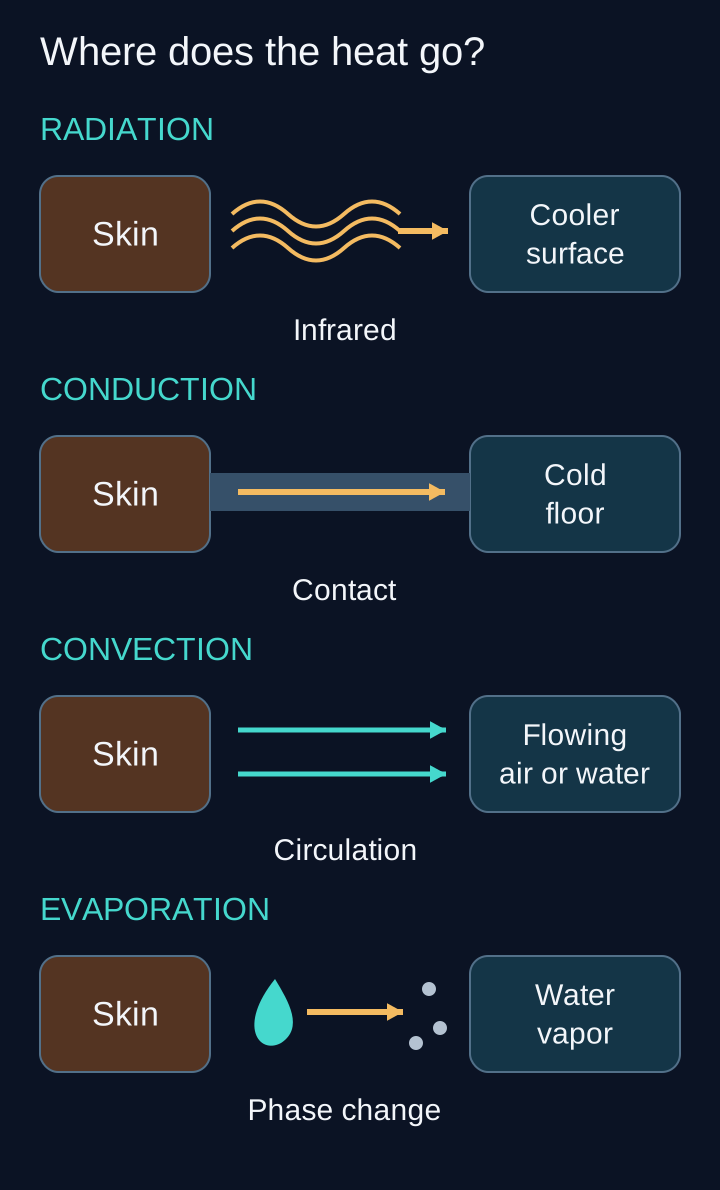
Radiation transfers infrared energy between surfaces without contact. Conduction transfers energy through direct contact, such as skin against a cold floor. Convection carries away warmed air or water as the surrounding fluid circulates. Evaporation consumes heat when liquid water becomes vapor. [1][2]
Use the heat-pathways diagram to trace each destination. Point to the route most directly reduced by putting a dry pad under a patient, then point to the route most directly reduced by a windproof outer layer. The pad limits contact heat transfer; the wind barrier limits replacement of warmed air next to the body.
Each route has a different physical interface. Several can operate together; no fixed fractions are assigned. [1][2]
A dry, resting person in a cool room can lose substantial heat by radiation. A person immersed in cold water loses heat through contact with water and its circulation; the submerged skin surface is not evaporating into air. After rescue, wet exposed surfaces can evaporate, and wind increases both convective loss and drying. These routes coexist, and clothing, exposure, activity and humidity change their relative importance. [1][2]
Water conducts heat much more effectively than air, but a material's conductivity ratio is not a patient's cooling-rate multiplier. Do not apply fixed resting percentages or a universal water-versus-air multiplier to a rescue. The useful question is which losses remain active and which available barriers interrupt them. [1]
Consider a canoeist lying in wet clothes on a metal dock. Say which three changes address three different interfaces before reading on. Shelter limits wind exposure, a pad separates the body from the dock, and replacing soaked clothes with dry insulation in shelter reduces ongoing wet-surface losses. Protect against further exposure while arranging appropriate medical help. [2]
Change the setting to a dry person on a cold floor
Dry clothes do not eliminate conduction into the floor. Insulation beneath the body still matters, even when evaporation is no longer the main correctable route. A blanket only above the person does not fully address that contact surface.
Separate a cold surface from a cold core
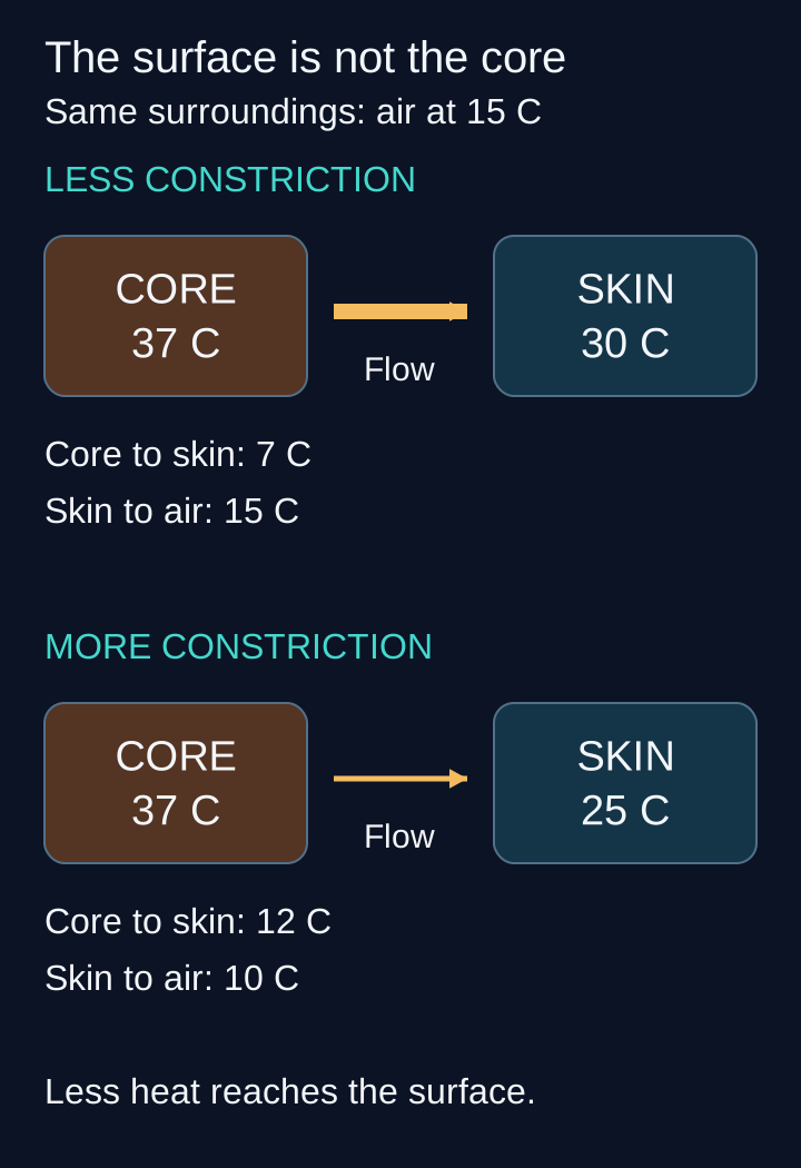
Cold fingers can accompany successful defense of core temperature. Cold-sensitive skin signals and central temperature information contribute to temperature regulation. Sympathetic vasoconstriction decreases warm blood delivery to the skin, so the surface cools and less heat reaches the environment. The core-to-skin temperature difference can increase while the skin-to-environment difference decreases. A skin reading is therefore not a substitute for a core measurement. [1][5][11]
Trace the core-to-skin arrow in the perfusion diagram. Narrowing this route conserves existing heat; it does not manufacture heat. Alpha-adrenergic signaling contributes, but receptor effects depend on the vessel and cooling conditions. Human finger studies emphasize alpha-2 responses during local cooling, whereas isolated human skin arteries show contributions from both alpha-1 and alpha-2 receptors. Neither experiment establishes that every cutaneous response uses alpha-2 alone. [5][11]
The arrow represents warm blood delivery toward skin. Values illustrate a controlled comparison, not a patient forecast or a diagnostic threshold. [1][5][11]
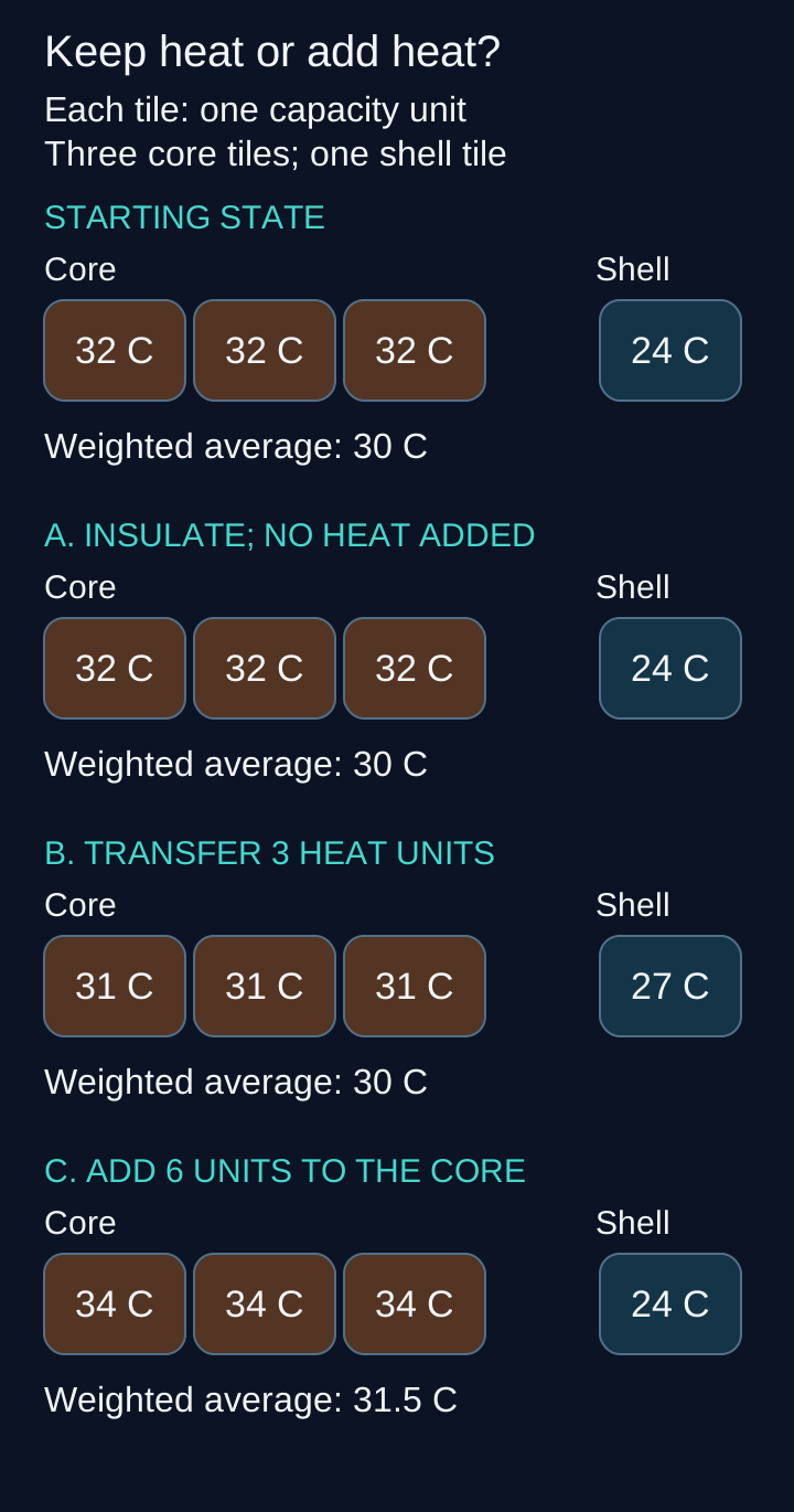
Now use a deliberately simplified heat ledger. The model has a core with three heat-capacity units and a shell with one. One heat unit raises one capacity unit by 1 degree C. Start at core 32 degrees C and shell 24 degrees C, giving a capacity-weighted average of 30 degrees C. These invented quantities explain heat accounting, not a patient's temperature, anatomy or rewarming rate.
Each tile contributes one heat-capacity unit; compare total heat change between the two branches. Compare branch B with C before checking the explanations. [1]
Choose a branch and predict the direction of core temperature before checking it. Keep the starting state fixed for each branch. There is no required answer or score, and the complete comparison remains readable below.
Branch A. Stop outside heat loss without adding heat
With no internal transfer during this model step, the core remains 32 and the shell remains 24 degrees C. The average stays 30. Insulation prevented a loss; it supplied no energy. A real shivering patient can warm because metabolism adds energy, not because the blanket creates it.
Branch B. Transfer three heat units from core to shell
The core falls by 1 degree to 31, and the shell rises by 3 degrees to 27. The weighted average remains 30. A warmer surface can coexist with a colder core even without additional environmental heat loss.
Branch C. Add six heat units directly to the core
With internal exchange temporarily held fixed, the core rises by 2 degrees to 34 while the shell remains 24. The weighted average becomes 31.5. This branch adds energy to the system rather than only redistributing it.
The openly readable result is A, unchanged temperatures; B, a colder core and warmer shell with unchanged total heat; C, increased total heat. Real bodies simultaneously produce, redistribute and lose heat, so these isolated branches are explanations, not treatment settings. For a transfer challenge, halve branch B to 1.5 heat units. The core becomes 31.5 and the shell 25.5 degrees C, with the same weighted average of 30.
Brief increases in finger blood flow during prolonged local cooling are called cold-induced vasodilation. The timing and magnitude vary with the whole-body thermal state; a finger becoming pink is not proof that the core is safe. This intermittent response does not guarantee protection from frostbite or begin at a universal skin-temperature cutoff. [6]
Assess function before trusting a temperature label
Two patients can have the same measured temperature but different immediate needs. An alert person who shivers vigorously and breathes normally differs from someone who is confused, exhausted or barely breathing. Core hypothermia means a core temperature below 35 degrees C, but consciousness, breathing, circulation and the direction of change determine urgency. [1][3]
Temperature bands for orientation, not independent treatment commands
Core temperature
Common pattern
Core temperature32 to less than 35 degrees C
Common patternMild. Shivering and impaired coordination can occur; early heart rate and ventilation may increase.
Core temperature28 to less than 32 degrees C
Common patternModerate. Confusion, reduced reflex responses, slowing pulse and breathing, and weakening shivering become more likely.
Core temperatureBelow 28 degrees C
Common patternSevere. Coma, apnea, absent shivering and ventricular arrhythmias or asystole are major concerns.
These nonoverlapping bands are a teaching convention. Symptoms overlap: vigorous shivering can persist around 31 degrees C, and drugs, illness or exhaustion can suppress it at higher temperatures. Do not infer recovery from shivering stopping, and do not infer an exact core temperature from appearance. [1]
Compare an alert hiker at 34.2 degrees C with a person at the same temperature who cannot stay awake. Identify what changed without changing the thermometer value. Reduced responsiveness requires emergency assessment and attention to the airway; it also raises concern for another contributor such as hypoglycemia, intoxication, infection or injury. [1][2]
Use a thermometer capable of reading low core temperatures. Forehead and exposed-skin measurements are affected by the environment. In an intubated patient, a correctly placed lower-esophageal temperature probe can track central temperature; rectal and bladder readings can lag during rapid thermal change. Airway protection and appropriate technique matter before esophageal placement. [1][4]
In a profoundly cold unconscious person, breathing and pulses may be extremely slow and difficult to detect. Trained rescuers may assess vital signs for up to a minute under hypothermia protocols. If cardiac arrest is identified, begin appropriate resuscitation with rewarming; fixed pupils alone do not establish irreversible death. This is not permission to delay help in an ordinary collapse. [3][4]
Apply the contrast to a patient with a pulse of 34 per minute and six shallow breaths per minute. Breathing support cannot wait for warmer skin. Oral food or fluid is unsafe when responsiveness or swallowing is impaired, regardless of how attractive a warm drink sounds. [2][3]
Conserve heat, add heat and support circulation
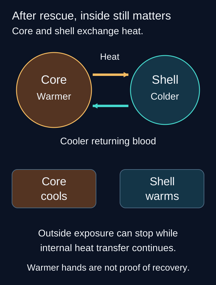
Why might core temperature fall after rescue while the hands feel warmer? Afterdrop is continued core cooling after removal from cold exposure. Heat can pass from warmer central tissues into colder peripheral tissues, and blood circulating through cold regions can return cooler. The afterdrop diagram separates this redistribution from fresh heat loss to the environment. [1]
Heat transfer through tissues and blood can continue after rescue. The diagram isolates redistribution; real patients also produce and lose heat. [1][2]
Use the heat ledger again: branch B explains why warming at the surface is not proof of improved core temperature. Increased limb perfusion or exertion can increase redistribution. Gentle handling, limiting unnecessary exertion and keeping an impaired patient horizontal when feasible are precautions, not reasons to postpone airway care, CPR or transport. [1][3]
Passive rewarming uses shelter and insulation to conserve the patient's own metabolic heat. An alert, stable, mildly hypothermic person with vigorous shivering may improve after wet-clothing management, dry insulation and appropriate monitoring. Calories and warm nonalcoholic drinks are only for someone alert enough to swallow safely. Active external heat can also support a mildly hypothermic patient; it is not reserved exclusively for collapse. [1][2]
Active external rewarming adds heat, for example with a properly used forced-air system or protected heat sources focused on the trunk. It is especially important when shivering is inadequate or consciousness is impaired. Protect cold skin from burns and reassess core temperature, breathing and perfusion. Concern about afterdrop does not make monitored trunk warming contraindicated. [1][2]
Choose between a blanket and a heat source for a confused, nonshivering patient after shelter is established. A blanket reduces losses but cannot replace missing heat production. Add active warming with emergency care rather than waiting for the blanket alone to reverse the problem. Do not rub the limbs, immerse an impaired patient in a warm bath or require exercise to produce heat. [1][2]
Warmed intravenous fluid treats clinically important volume depletion and avoids adding a cold load. It is not an adequate sole rewarming technique for a severely hypothermic patient. Warmed humidified oxygen is also an adjunct, not a substitute for effective rewarming or ventilatory support. Fluid amounts must follow perfusion and reassessment rather than an unlimited replacement rule. [1][3]
Extracorporeal life support circulates blood through a device that can provide rewarming and organ support. Venoarterial extracorporeal membrane oxygenation, or VA-ECMO, supports circulation as well as gas exchange and is favored for suitable hypothermic cardiac-arrest patients where available. Early expert referral matters before arrest too. RCUK 2025 identifies core temperature below 30 degrees C, systolic pressure below 90 mmHg, pulse below 45 per minute or ventricular arrhythmia as referral risk features. These are alternatives, not four conditions that must all be present. [3][4]
Assess potentially reversible hypothermic arrest using the circumstances and an appropriate prognostic tool such as HOPE with the receiving team. A single potassium value, prolonged resuscitation or fixed pupils is not a universal futility rule. In avalanche incidents, airway obstruction and whether asphyxia preceded cooling matter; cold exposure does not guarantee brain protection. [3][4]
Keep rhythm decisions specific. Asystole is not shockable. For ventricular fibrillation below 30 degrees C, AHA 2025 permits one defibrillation attempt before deferring further attempts until at least 30 degrees C if it fails; RCUK 2025 permits three attempts before deferral. Follow the named local protocol rather than combining algorithms. CPR and rewarming continue while that temperature-dependent decision is made. [3][4]
Rewarming rate depends on the method and the patient. Do not promise one fixed hourly increase or wait a preset observation interval before responding to deteriorating function. [1][3]
For a new situation, consider a patient who still has a pulse but is becoming hypotensive during warming. That is a circulation problem requiring reassessment and possible escalation, even if core temperature is increasing. Temperature improvement does not by itself establish physiological recovery. [1][3][4]
Decide whether the patient can still generate heat
Vasoconstriction saves heat; shivering generates it. Hypothalamic temperature regulation activates repeated skeletal-muscle contractions. ATP turnover in those contractions releases heat and raises metabolic demand, so the response requires both fuel and adequate oxygen delivery. Strong shivering can increase heat production severalfold, but not by one fixed multiplier in every patient. [1]
Compare a fed, alert person who shivers vigorously with an exhausted person who has eaten almost nothing and shivers weakly. Trace fuel into muscle work, then heat into the core. The second person may be unable to replace ongoing losses even after receiving the same insulation. [1]
Choose which observation would make passive insulation alone less dependable: persistent vigorous shivering or declining responsiveness with absent shivering. The second combination requires emergency care and active rewarming rather than waiting for spontaneous heat production. The important distinction is remaining function, not whether a thermometer has crossed an exact shivering cutoff. [1][2]
Behavioral defenses such as seeking shelter can occur early and alongside vasoconstriction and shivering. They are not the last stage of a mandatory sequence. Alcohol, sedating drugs, impaired mobility, malnutrition and illness can interfere with these defenses; apparent warmth or sleepiness is not reassurance after cold exposure. [1][2]
Apply the principle to a drowsy patient with glucose 38 mg/dL (usual fasting reference 70 to 99). Hypoglycemia can contribute to both impaired consciousness and inadequate heat production. Correct it through an appropriate nonoral route while supporting the airway and rewarming; a warm sugary drink is not safe when swallowing is impaired. Check again because glucose and treatment needs can change during recovery. [1][2][12]
Read the ECG and laboratory results in context
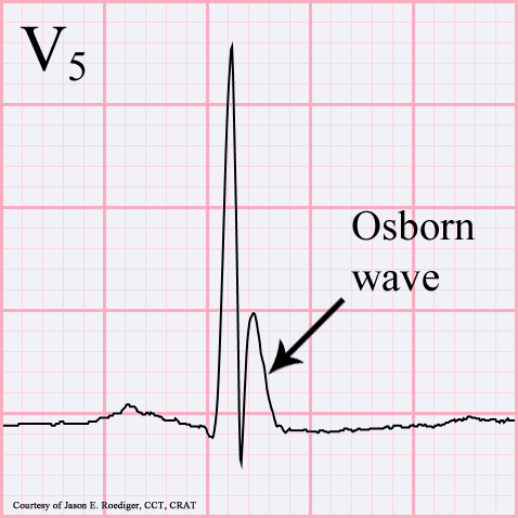
What does a hump at the end of the QRS mean in a cold patient? An Osborn or J wave is a deflection near the QRS-ST junction. The accompanying annotated V5 tracing shows its location; it is a teaching image, not a blinded diagnostic test. The original clinical image is credited to its uploader and carries a CC BY-SA 3.0 license. [8][9][10]
Annotated clinical ECG, not a blinded test. Source: Jer5150, Osborn wave.gif, Wikimedia Commons, CC BY-SA 3.0. Frame zero extracted as a static PNG; the original arrow, label and embedded courtesy credit are retained. The label teaches location and does not establish a diagnosis without clinical context. Image: Jer5150; embedded courtesy credit in the source image; original source; CC BY-SA 3.0 Unported. [8][9][10].
Point first to the QRS and then to the extra deflection. In experimental ventricular tissue, differences between epicardial and endocardial early repolarization create a voltage gradient that can produce a J wave. This is an electrical gradient, not proof of a physical temperature difference across the heart wall. The experiment supports a mechanism, not a patient-specific treatment rule. [7]
Hypothermia can also slow the rhythm and prolong PR, QRS and QT intervals. J waves are neither required for diagnosis nor unique to hypothermia; early repolarization and hypercalcemia are among competing settings. Other J-point abnormalities, including Brugada patterns, require their own ECG and clinical context. Compare temperature, symptoms, electrolytes and the full ECG instead of assigning the cause from the hump alone. Its size is not a standalone prediction of ventricular fibrillation, and a J wave alone neither proves nor excludes myocardial infarction. Stable atrial fibrillation during cooling often improves with rewarming and does not automatically require cardioversion. [1][8][9]
Now compare urine output with blood concentration. Peripheral vasoconstriction initially shifts blood centrally and can contribute to cold diuresis. Urinary losses, poor intake and fluid shifts can leave the circulating volume depleted. Hemoconcentration means less plasma around existing red cells, not rapid creation of new cells. During rewarming, expanding peripheral vascular capacity can expose that deficit and worsen blood pressure. [1]
Glucose can be high during cold stress and impaired insulin secretion, or low after inadequate intake and depleted fuel. Check the actual value. A reported case demonstrated reversible hyperglycemia after severe hypothermia; it does not justify withholding indicated diabetes treatment from every cold patient. When insulin is needed, reassess closely as temperature and glucose regulation change. [1][12]
For transfer, an older adult is confused and hypothermic in a heated room. Do not attribute everything to the room temperature. Infection, endocrine disease, hypoglycemia, medication effects and injury need assessment while rewarming proceeds. A cold patient with a new pulmonary infiltrate needs evaluation for infection, not warming as the entire plan. [1]
Judge the core, the airway, the circulation and the cause together. Warmer skin or one improved number cannot stand in for all four.
Independent practice
Case 1
Show answer and explanations for case 1
A. Greater conduction into the chair plus less blood-borne heat delivery to skin (Why this does not fit)
A change in contact or its thermal resistance could increase direct-contact heat loss. Contact is held constant and skin flow increases rather than decreases. [1] [2]
Reasoning steps for option A
Which change would increase conduction into a support surface?
A change in contact or its thermal resistance could increase direct-contact heat loss.
Do either proposed changes match the supplied comparison?
Contact is held constant and skin flow increases rather than decreases.
B. Greater convection plus less blood-borne heat delivery to skin (Why this does not fit)
Greater airflow increases convective removal of warmed air. Skin blood flow increases, so blood-borne delivery toward skin increases rather than decreases. [1] [2]
Reasoning steps for option B
Which part of this explanation correctly matches the fan?
Greater airflow increases convective removal of warmed air.
Which direction contradicts the measured local perfusion?
Skin blood flow increases, so blood-borne delivery toward skin increases rather than decreases.
C. Greater convection plus greater blood-borne heat delivery to skin (Best answer)
It increases replacement of warmed air near the clothing, increasing convective loss. More skin blood flow delivers more central heat toward the surface without adding metabolic heat. [1] [2]
Reasoning steps for option C
What does faster airflow change when ambient temperature is fixed?
It increases replacement of warmed air near the clothing, increasing convective loss.
How does the measured perfusion change reinforce that effect?
More skin blood flow delivers more central heat toward the surface without adding metabolic heat.
D. Greater radiation from warmer surroundings plus greater skin heat delivery (Why this does not fit)
Warmer surroundings generally reduce the radiative gradient from the patient. Ambient and surrounding conditions are held constant; airflow, not a warmer radiative environment, is the changing external factor. [1] [2]
Reasoning steps for option D
How would warmer surroundings affect net outward radiation?
Warmer surroundings generally reduce the radiative gradient from the patient.
What external condition actually changed here?
Ambient and surrounding conditions are held constant; airflow, not a warmer radiative environment, is the changing external factor.
Takeaway: Convection removes heat at the surface; greater skin perfusion supplies more central heat to that interface.
A. Greater contact thermal resistance with less conductive heat loss (Best answer)
It increases resistance to heat transfer across that contact. The floor is the heat sink, and the compressed layer beneath the patient provides little insulation. [1] [2]
Reasoning steps for option A
What does an insulating layer do between a body and a cold floor?
It increases resistance to heat transfer across that contact.
Why does the pad address the stated dominant loss?
The floor is the heat sink, and the compressed layer beneath the patient provides little insulation.
B. Lower cutaneous vascular resistance with greater core heat retention (Why this does not fit)
It allows more warm blood to reach the skin surface. No. The pad changes the contact barrier, while increased skin perfusion can increase heat delivery toward the cold surface. [1] [2]
Reasoning steps for option B
What does lower skin vascular resistance tend to do in a cold setting?
It allows more warm blood to reach the skin surface.
Would that explain the insulating pad effect?
No. The pad changes the contact barrier, while increased skin perfusion can increase heat delivery toward the cold surface.
C. Greater metabolic heat production with less oxygen consumption (Why this does not fit)
Shivering increases metabolic activity and generally increases oxygen demand. No. It conserves heat; it does not create metabolic heat while lowering its oxygen cost. [1] [2]
Reasoning steps for option C
How does producing heat through shivering affect metabolic demand?
Shivering increases metabolic activity and generally increases oxygen demand.
Does placement of the pad itself supply muscular work?
No. It conserves heat; it does not create metabolic heat while lowering its oxygen cost.
D. Lower surrounding humidity with less evaporative heat loss (Why this does not fit)
Drier air generally favors evaporation rather than suppressing it. No. The stated problem is contact with the cold floor. [1] [2]
Reasoning steps for option D
What does lower humidity tend to do to evaporation from a wet surface?
Drier air generally favors evaporation rather than suppressing it.
Is wet-surface evaporation the dominant interface supplied here?
No. The stated problem is contact with the cold floor.
Takeaway: A dry barrier beneath a cold patient addresses conduction; insulation is not a heat source.
A. Conduction as warm blood contacts cold skin (Why this does not fit)
Internal conduction and blood flow can redistribute heat within the body. No. That observed liquid-to-vapor change identifies evaporation, even if redistribution also contributes to afterdrop. [1] [2]
Reasoning steps for option A
How can heat pass from warmer central tissues toward a colder shell?
Internal conduction and blood flow can redistribute heat within the body.
Does internal redistribution explain the shirt losing liquid to air?
No. That observed liquid-to-vapor change identifies evaporation, even if redistribution also contributes to afterdrop.
B. Convection as river water circulates over skin (Why this does not fit)
It carries away water warmed at the skin surface. No. Water convection mattered during immersion, but the question asks about the shirt drying after rescue. [1] [2]
Reasoning steps for option B
How does circulating cold water remove body heat?
It carries away water warmed at the skin surface.
Is circulating river water still at the skin on the bank?
No. Water convection mattered during immersion, but the question asks about the shirt drying after rescue.
C. Radiation as infrared energy leaves the shirt (Why this does not fit)
Yes. Its surface exchanges radiant energy with its surroundings. The disappearance of liquid into air is vaporization, not infrared radiation. [1] [2]
Reasoning steps for option C
Can wet clothing radiate infrared energy?
Yes. Its surface exchanges radiant energy with its surroundings.
Which supplied observation requires a different explanation?
The disappearance of liquid into air is vaporization, not infrared radiation.
D. Evaporation as liquid water becomes vapor (Best answer)
Vaporization requires energy that can be drawn from the wet clothing and underlying body. The shirt is drying into the surrounding air after removal from the river. [1] [2]
Reasoning steps for option D
What energy change accompanies liquid water becoming vapor?
Vaporization requires energy that can be drawn from the wet clothing and underlying body.
Where is that transition explicitly occurring now?
The shirt is drying into the surrounding air after removal from the river.
Takeaway: Evaporation requires a phase change into vapor; it is not the explanation for submerged skin cooling.
A. Core-to-skin difference decreases; skin-to-air difference increases (Why this does not fit)
It would narrow the core-to-skin difference and widen the skin-to-air difference. No. Skin cooled by 5 degrees, giving the opposite pair of changes. [1] [5] [11]
Reasoning steps for option A
What would warmer skin at a fixed core and ambient temperature do?
It would narrow the core-to-skin difference and widen the skin-to-air difference.
Did this volunteer develop warmer skin?
No. Skin cooled by 5 degrees, giving the opposite pair of changes.
B. Core-to-skin difference increases; skin-to-air difference decreases (Best answer)
It increases from 7.9 to 12.9 degrees C. It falls from 13 to 8 degrees C while reduced perfusion limits heat carried to the surface. [1] [5] [11]
Reasoning steps for option B
How does the core-to-skin difference change numerically?
It increases from 7.9 to 12.9 degrees C.
What happens to the skin-to-air difference and surface heat delivery?
It falls from 13 to 8 degrees C while reduced perfusion limits heat carried to the surface.
C. Core-to-skin difference increases; skin-to-air difference increases (Why this does not fit)
Independent changes in all three temperatures can increase both differences. Core and ambient temperatures are fixed; a lower skin temperature cannot increase its difference above the unchanged ambient temperature. [1] [5] [11]
Reasoning steps for option C
What can widen both differences if core and ambient temperatures also change?
Independent changes in all three temperatures can increase both differences.
Are those independent changes present here?
Core and ambient temperatures are fixed; a lower skin temperature cannot increase its difference above the unchanged ambient temperature.
D. Core-to-skin difference decreases; skin-to-air difference decreases (Why this does not fit)
The core could cool while the surroundings warm enough to reduce both gaps. Neither occurred. Only skin cooled, which increases its separation from the unchanged core. [1] [5] [11]
Reasoning steps for option D
When could both differences fall together?
The core could cool while the surroundings warm enough to reduce both gaps.
Which of those changes occurred in this comparison?
Neither occurred. Only skin cooled, which increases its separation from the unchanged core.
Takeaway: A colder shell can widen the core-to-skin gap while narrowing the skin-to-environment gap.
A. Central alpha-2 receptor signaling increases sympathetic outflow to skin (Why this does not fit)
It reduces sympathetic outflow rather than increasing it. Arterial pressure and untreated-site flow remain unchanged after a local intervention. [5] [11]
Reasoning steps for option A
What is a usual central effect of alpha-2 receptor stimulation?
It reduces sympathetic outflow rather than increasing it.
What additional feature argues against a generalized central explanation?
Arterial pressure and untreated-site flow remain unchanged after a local intervention.
B. Local beta-2 receptor signaling directly contracts finger arterioles (Why this does not fit)
It favors smooth-muscle relaxation in responsive vascular beds. No. The clonidine-yohimbine pattern supports alpha-2 signaling in this local experiment. [5] [11]
Reasoning steps for option B
What vascular effect does beta-2 stimulation usually favor?
It favors smooth-muscle relaxation in responsive vascular beds.
Does the supplied antagonist pattern identify beta-2 receptors?
No. The clonidine-yohimbine pattern supports alpha-2 signaling in this local experiment.
C. Local alpha-2 receptor signaling contributes to finger vasoconstriction (Best answer)
Clonidine stimulates alpha-2 receptors and yohimbine can antagonize that effect. The local treatment changes local flow without the supplied systemic or untreated-site changes. [5] [11]
Reasoning steps for option C
What receptor interaction is supported by clonidine and yohimbine?
Clonidine stimulates alpha-2 receptors and yohimbine can antagonize that effect.
What confines the supported conclusion to the finger site?
The local treatment changes local flow without the supplied systemic or untreated-site changes.
D. Local muscarinic receptor signaling suppresses all cutaneous blood flow (Why this does not fit)
Muscarinic receptors mediate cholinergic responses, including sympathetic sweating. No. It tests adrenergic signaling, and one local response cannot establish suppression of all skin blood flow. [5] [11]
Reasoning steps for option D
Which autonomic system uses muscarinic receptors as an effector pathway?
Muscarinic receptors mediate cholinergic responses, including sympathetic sweating.
Does the experimental drug pair establish a muscarinic vascular mechanism?
No. It tests adrenergic signaling, and one local response cannot establish suppression of all skin blood flow.
Takeaway: A local finger experiment supports a local receptor contribution, not one universal skin-vessel rule.
A. A fixed local temperature determines the same perfusion cycle in both sessions (Why this does not fit)
The same local cooling condition would produce a similar response independent of body state. No. Whole-body cooling changes both the timing and size of the flow recovery. [6]
Reasoning steps for option A
What would a strictly local fixed-threshold explanation predict?
The same local cooling condition would produce a similar response independent of body state.
Does the observed response stay the same?
No. Whole-body cooling changes both the timing and size of the flow recovery.
B. The later flow rises demonstrate that core temperature has normalized (Why this does not fit)
Yes. Cold-induced vasodilation can be intermittent during ongoing local cooling. The whole-body-cooled session still shows some later flow rises despite the different thermal state. [6]
Reasoning steps for option B
Can a rise in finger blood flow occur during continued cold exposure?
Yes. Cold-induced vasodilation can be intermittent during ongoing local cooling.
Which supplied observation prevents treating it as core-temperature proof?
The whole-body-cooled session still shows some later flow rises despite the different thermal state.
C. Smaller perfusion rises demonstrate that the finger is protected from freezing injury (Why this does not fit)
Flow contributes to heat delivery, but injury also depends on exposure and tissue temperature. No. The data show a limited response, not tissue-safety outcomes or guaranteed frostbite prevention. [6]
Reasoning steps for option C
What does blood flow tell us about local heat delivery?
Flow contributes to heat delivery, but injury also depends on exposure and tissue temperature.
Would smaller delayed flow recovery establish protection?
No. The data show a limited response, not tissue-safety outcomes or guaranteed frostbite prevention.
D. Whole-body thermal state modifies intermittent local perfusion recovery (Best answer)
The overall body thermal state changes while the finger-cooling condition stays fixed. The intermittent local vasodilator response depends on more than the local cold stimulus alone. [6]
Reasoning steps for option D
What changes between the two sessions?
The overall body thermal state changes while the finger-cooling condition stays fixed.
What does the weaker delayed recovery imply?
The intermittent local vasodilator response depends on more than the local cold stimulus alone.
Takeaway: Intermittent finger reperfusion is neither a core thermometer nor a guarantee against tissue injury.
A. Reduced environmental loss plus shivering-generated metabolic heat (Best answer)
No. It reduces transfer of existing heat to the surroundings. His vigorous muscular shivering continues to generate metabolic heat. [1] [2]
Reasoning steps for option A
Does dry insulation add thermal energy by itself?
No. It reduces transfer of existing heat to the surroundings.
What supplies heat while this patient is insulated?
His vigorous muscular shivering continues to generate metabolic heat.
B. Increased skin blood flow plus heat production by the blankets (Why this does not fit)
It brings more central heat toward the surface. No external heater is present; the patient metabolic activity supplies the heat. [1] [2]
Reasoning steps for option B
What effect can greater skin perfusion have in a cold environment?
It brings more central heat toward the surface.
Are the blankets generating energy in this case?
No external heater is present; the patient metabolic activity supplies the heat.
C. Reduced muscular ATP use plus increased heat production at rest (Why this does not fit)
Repeated contractions increase ATP turnover and metabolic heat production. No. The supplied vigorous shivering indicates the opposite metabolic direction. [1] [2]
Reasoning steps for option C
What happens to ATP turnover during vigorous shivering?
Repeated contractions increase ATP turnover and metabolic heat production.
Would reduced muscular ATP use explain the observed response?
No. The supplied vigorous shivering indicates the opposite metabolic direction.
D. Increased evaporative cooling plus conversion of skin heat into core heat (Why this does not fit)
Evaporation removes heat when body energy supports vaporization. No. Wet-clothing replacement reduces the loss while continued metabolism adds heat. [1] [2]
Reasoning steps for option D
What does evaporation do to body heat balance?
Evaporation removes heat when body energy supports vaporization.
Would increasing that loss explain successful warming after drying?
No. Wet-clothing replacement reduces the loss while continued metabolism adds heat.
Takeaway: Passive rewarming works when retained metabolic heat exceeds the remaining losses.
A. Continue dry blankets and observe until shivering has stopped (Why this does not fit)
It can be suitable for an alert, stable, mildly hypothermic patient generating adequate heat. No. His neurological impairment already warrants escalation, and loss of shivering may represent deterioration. [1] [2]
Reasoning steps for option A
When can insulation with observation be sufficient initially?
It can be suitable for an alert, stable, mildly hypothermic patient generating adequate heat.
Would shivering stopping be a safe endpoint in this patient?
No. His neurological impairment already warrants escalation, and loss of shivering may represent deterioration.
B. Give warm oral calories and have him walk to the waiting vehicle (Why this does not fit)
They require adequate alertness, swallowing and functional stability. No. Disorientation and inability to sit safely make oral intake and required exertion inappropriate. [1] [2]
Reasoning steps for option B
When are oral calories and independent activity safer to consider?
They require adequate alertness, swallowing and functional stability.
Do the supplied neurological findings meet that condition?
No. Disorientation and inability to sit safely make oral intake and required exertion inappropriate.
C. Add external trunk heat; arrange gentle, monitored transport (Best answer)
Yes. Shivering may persist around 31 degrees C and does not reliably assign severity by itself. Disorientation and impaired postural control require emergency assessment and supported rewarming. [1] [2]
Reasoning steps for option C
Can vigorous shivering persist near this core temperature?
Yes. Shivering may persist around 31 degrees C and does not reliably assign severity by itself.
What finding makes simple observation inadequate here?
Disorientation and impaired postural control require emergency assessment and supported rewarming.
D. Use warm-water immersion before reassessing the confusion (Why this does not fit)
Water warming requires an appropriate conscious, safely managed person and a suitable setting. Impaired consciousness creates drowning and handling risks; monitored trunk warming and transport address the systemic problem. [1] [2]
Reasoning steps for option D
When might immersion be considered for an isolated cold-exposure problem?
Water warming requires an appropriate conscious, safely managed person and a suitable setting.
Why does this patient need a different approach?
Impaired consciousness creates drowning and handling risks; monitored trunk warming and transport address the systemic problem.
Takeaway: Persistent shivering does not neutralize confusion or impaired coordination.
A. Core temperature is probably below 28; stage severity from absent shivering (Why this does not fit)
Shivering is commonly absent as central and muscular function deteriorate. No. Drugs and illness can suppress shivering at higher temperatures; it is not an exact thermometer. [1] [2]
Reasoning steps for option A
What pattern often occurs in profound hypothermia?
Shivering is commonly absent as central and muscular function deteriorate.
Can that symptom override the supplied reliable core measurement?
No. Drugs and illness can suppress shivering at higher temperatures; it is not an exact thermometer.
B. Heat production may be too low; assess medication effects while actively warming (Best answer)
They can impair awareness, behavior and thermoregulatory responses. No. The measured hypothermia and impaired responsiveness require rewarming and evaluation of contributing causes. [1] [2]
Reasoning steps for option B
What can sedating drugs change during cold exposure?
They can impair awareness, behavior and thermoregulatory responses.
Does a core temperature of 33.7 with drowsiness support recovery?
No. The measured hypothermia and impaired responsiveness require rewarming and evaluation of contributing causes.
C. Heat balance has normalized; judge recovery from the quiet muscles (Why this does not fit)
Shivering may stop once cold stress resolves in an otherwise recovered person. Core temperature remains low and the patient cannot maintain alertness. [1] [2]
Reasoning steps for option C
What could quiet muscles mean after successful recovery?
Shivering may stop once cold stress resolves in an otherwise recovered person.
What evidence contradicts that interpretation here?
Core temperature remains low and the patient cannot maintain alertness.
D. Fuel depletion is established; give oral glucose before further evaluation (Why this does not fit)
A low measured glucose would support hypoglycemia as a treatable contributor. No. Glucose is not low, and impaired responsiveness makes oral intake unsafe. [1] [2]
Reasoning steps for option D
What finding would establish a current low blood glucose?
A low measured glucose would support hypoglycemia as a treatable contributor.
Do glucose 94 and drowsiness justify that oral plan?
No. Glucose is not low, and impaired responsiveness makes oral intake unsafe.
Takeaway: Use measured temperature and function; absent shivering has several possible causes.
A. Ventilate with a bag and mask and prepare appropriate airway support (Best answer)
Ventilation is inadequate despite the presence of a pulse. Assisted ventilation and appropriate airway management address gas exchange while rewarming continues. [1] [2] [3]
Reasoning steps for option A
What does poor chest excursion with five shallow breaths suggest?
Ventilation is inadequate despite the presence of a pulse.
Which immediate support addresses that failure?
Assisted ventilation and appropriate airway management address gas exchange while rewarming continues.
B. Increase oxygen flow by mask and observe spontaneous ventilation (Why this does not fit)
It can raise inspired oxygen concentration. It does not provide the missing minute ventilation or protect the unresponsive patient airway. [1] [2] [3]
Reasoning steps for option B
What can an oxygen mask improve when breathing is adequate?
It can raise inspired oxygen concentration.
What problem would an oxygen mask alone leave untreated here?
It does not provide the missing minute ventilation or protect the unresponsive patient airway.
C. Begin chest compressions for the low measured heart rate (Why this does not fit)
They are indicated when cardiac arrest is identified after an appropriate assessment. A central pulse is present; the immediately demonstrated failure is ventilation. [1] [2] [3]
Reasoning steps for option C
When are chest compressions indicated in this setting?
They are indicated when cardiac arrest is identified after an appropriate assessment.
What finding distinguishes this patient from established arrest?
A central pulse is present; the immediately demonstrated failure is ventilation.
D. Administer a warm carbohydrate drink to stimulate heat production (Why this does not fit)
It can supply fuel when the person is alert and can swallow safely. He is unresponsive with inadequate breathing, making oral intake an aspiration risk. [1] [2] [3]
Reasoning steps for option D
When can oral carbohydrate support a cold patient?
It can supply fuel when the person is alert and can swallow safely.
Why does this patient not meet that condition?
He is unresponsive with inadequate breathing, making oral intake an aspiration risk.
Takeaway: A profoundly cold patient with a pulse can still need immediate ventilation and airway support.
A. Perform synchronized cardioversion before continuing the warming plan (Why this does not fit)
An unstable tachyarrhythmia causing major circulatory compromise may require cardioversion. No. The current findings favor supported rewarming rather than immediate electrical conversion. [1] [2]
Reasoning steps for option A
When can synchronized cardioversion be warranted?
An unstable tachyarrhythmia causing major circulatory compromise may require cardioversion.
Does the supplied slow rhythm with preserved pressure establish that indication?
No. The current findings favor supported rewarming rather than immediate electrical conversion.
B. Administer a beta blocker to control the ventricular response (Why this does not fit)
It can slow an excessively rapid ventricular response in suitable patients. No. The ventricular rate is already slow, so additional slowing does not address the thermal deficit. [1] [2]
Reasoning steps for option B
What atrial-fibrillation problem can a beta blocker address?
It can slow an excessively rapid ventricular response in suitable patients.
Is an excessive ventricular rate present here?
No. The ventricular rate is already slow, so additional slowing does not address the thermal deficit.
C. Continue passive insulation until coordinated shivering returns (Why this does not fit)
It depends on the patient producing more heat than the remaining losses. He is already nonshivering and confused, so external heat is needed instead of waiting for spontaneous recovery. [1] [2]
Reasoning steps for option C
What does passive insulation depend on for net warming?
It depends on the patient producing more heat than the remaining losses.
What supplied finding makes waiting for that response unreliable?
He is already nonshivering and confused, so external heat is needed instead of waiting for spontaneous recovery.
D. Start active trunk warming and monitor rhythm and circulation (Best answer)
Rewarming and monitoring are generally prioritized rather than immediate rhythm-specific treatment. Confusion and absent shivering make reliance on his own heat production inadequate. [1] [2]
Reasoning steps for option D
How is stable atrial fibrillation associated with hypothermia usually approached initially?
Rewarming and monitoring are generally prioritized rather than immediate rhythm-specific treatment.
What makes active warming especially important here?
Confusion and absent shivering make reliance on his own heat production inadequate.
Takeaway: Treat the thermal and physiological problem before reacting to a stable cold-associated atrial rhythm.
A. Evaporative loss from continued immersion of the skin in river water (Why this does not fit)
They can lose heat after water vaporizes into surrounding air. No. The patient is dry and sheltered; the paired central and peripheral trend instead supports redistribution. [1] [2]
Reasoning steps for option A
When can wet exposed surfaces lose heat through evaporation?
They can lose heat after water vaporizes into surrounding air.
Are the proposed continued immersion and wet exposure present?
No. The patient is dry and sheltered; the paired central and peripheral trend instead supports redistribution.
B. Heat passes into cold peripheral tissues as cooler blood returns to the core (Best answer)
Heat can leave central tissues for a colder shell, including through increased blood circulation. The hands warm while the measured core cools after activity, despite protection from further exposure. [1] [2]
Reasoning steps for option B
How can peripheral warming coexist with continued core cooling?
Heat can leave central tissues for a colder shell, including through increased blood circulation.
Which supplied trend supports that redistribution?
The hands warm while the measured core cools after activity, despite protection from further exposure.
C. Direct central cooling from infusion of low-temperature crystalloid (Why this does not fit)
It can add a cold thermal load directly to the circulation. No cold fluid has been infused, whereas skin temperature is rising during the central fall. [1] [2]
Reasoning steps for option C
How could a cold intravenous infusion affect core temperature?
It can add a cold thermal load directly to the circulation.
What supplied detail excludes that explanation?
No cold fluid has been infused, whereas skin temperature is rising during the central fall.
D. Reduced net radiation because the ambulance air temperature is higher (Why this does not fit)
It reduces the thermal difference driving outward heat transfer. No. Reduced outward loss is protective and does not account for simultaneous warming of cold peripheral tissues. [1] [2]
Reasoning steps for option D
What does a warmer environment generally do to outward heat loss?
It reduces the thermal difference driving outward heat transfer.
Would that reduction explain a new central temperature fall by itself?
No. Reduced outward loss is protective and does not account for simultaneous warming of cold peripheral tissues.
Takeaway: A warmer periphery can accompany continued central cooling through afterdrop.
A. Give room-temperature crystalloid as the sole method of thermal recovery (Why this does not fit)
It can support circulating volume and cardiac filling. Room-temperature fluid can add a cold load, and intravenous fluid alone is not sufficient rewarming for this impaired patient. [1] [3] [4]
Reasoning steps for option A
What can intravenous crystalloid restore when filling is low?
It can support circulating volume and cardiac filling.
Why is this proposed thermal plan incomplete?
Room-temperature fluid can add a cold load, and intravenous fluid alone is not sufficient rewarming for this impaired patient.
B. Give a large fixed volume of warmed crystalloid without interim reassessment (Why this does not fit)
Yes. It treats a deficit and avoids infusion-related cooling. Fluid needs can change during rewarming, and excessive replacement can cause overload; repeated assessment is necessary. [1] [3] [4]
Reasoning steps for option B
Can warmed fluid help a hypothermic patient who is volume depleted?
Yes. It treats a deficit and avoids infusion-related cooling.
Why is an unreassessed fixed large volume inappropriate?
Fluid needs can change during rewarming, and excessive replacement can cause overload; repeated assessment is necessary.
C. Give warmed isotonic crystalloid in increments, reassess each, and continue active warming (Best answer)
A depleted circulating volume must fill a larger vascular space. Warmed isotonic fluid with frequent perfusion and congestion reassessment addresses the deficit without replacing active warming. [1] [3] [4]
Reasoning steps for option C
Why can pressure fall when peripheral circulation opens during rewarming?
A depleted circulating volume must fill a larger vascular space.
What fluid strategy matches the supplied low filling and clear lungs?
Warmed isotonic fluid with frequent perfusion and congestion reassessment addresses the deficit without replacing active warming.
D. Withhold intravenous fluid until peripheral vasoconstriction has returned (Why this does not fit)
It may be avoided when filling is adequate or fluid overload is present. No. Hypotension, low filling and substantial losses support monitored replacement during ongoing rewarming. [1] [3] [4]
Reasoning steps for option D
When might additional fluid be withheld?
It may be avoided when filling is adequate or fluid overload is present.
Do the current perfusion and filling findings support withholding here?
No. Hypotension, low filling and substantial losses support monitored replacement during ongoing rewarming.
Takeaway: Warmed fluid supports perfusion; it neither replaces active heat delivery nor removes the need to reassess.
A. Arrange transfer to an extracorporeal center while maintaining warming (Best answer)
No. RCUK lists slow heart rate, low pressure, low core temperature or ventricular arrhythmia as alternative risk features. Heart rate 38 is below the listed 45-per-minute threshold, and an appropriate center is readily accessible. [4]
Reasoning steps for option A
Must all the referral risk features be present simultaneously?
No. RCUK lists slow heart rate, low pressure, low core temperature or ventricular arrhythmia as alternative risk features.
Which feature changes this destination decision?
Heart rate 38 is below the listed 45-per-minute threshold, and an appropriate center is readily accessible.
B. Use the nearer general hospital because systolic pressure remains above 90 mmHg (Why this does not fit)
It can be one reassuring part of an otherwise low-risk assessment. No. Marked bradycardia remains an independent referral feature under the named pathway. [4]
Reasoning steps for option B
When can preserved circulation support a lower-intensity destination?
It can be one reassuring part of an otherwise low-risk assessment.
Does preserved pressure remove the supplied rhythm-rate risk?
No. Marked bradycardia remains an independent referral feature under the named pathway.
C. Observe in the vehicle until core temperature is below 30 degrees C (Why this does not fit)
It is one referral risk feature. Another listed risk feature is already present, and observation would delay access to advanced support. [4]
Reasoning steps for option C
What role does a core temperature below 30 have in the named pathway?
It is one referral risk feature.
Why would waiting for that particular threshold be inappropriate?
Another listed risk feature is already present, and observation would delay access to advanced support.
D. Reserve the extracorporeal-capable center for documented cardiac arrest (Why this does not fit)
It can be lifesaving for suitable hypothermic cardiac-arrest patients. No. The pathway also directs at-risk patients toward a capable center before arrest occurs. [4]
Reasoning steps for option D
When is extracorporeal rewarming especially important?
It can be lifesaving for suitable hypothermic cardiac-arrest patients.
Does that restrict referral to patients already in arrest?
No. The pathway also directs at-risk patients toward a capable center before arrest occurs.
Takeaway: An individual arrest-risk feature can justify early referral even while a pulse and blood pressure persist.
A. Deliver repeated defibrillation attempts before arranging rewarming transfer (Why this does not fit)
Ventricular fibrillation and pulseless ventricular tachycardia are shockable rhythms. No. Confirmed asystole does not become shockable because the patient is cold. [3] [4]
Reasoning steps for option A
Which rhythms are treated by defibrillation during arrest?
Ventricular fibrillation and pulseless ventricular tachycardia are shockable rhythms.
Is one of those rhythms documented here?
No. Confirmed asystole does not become shockable because the patient is cold.
B. Stop CPR and use external heat until a spontaneous rhythm appears (Why this does not fit)
Yes. Temperature correction is central in hypothermic arrest. No. CPR and appropriate rewarming proceed together rather than leaving the patient without perfusion. [3] [4]
Reasoning steps for option B
Can rewarming be necessary to restore effective cardiac activity?
Yes. Temperature correction is central in hypothermic arrest.
Does that permit withholding circulatory support while warming?
No. CPR and appropriate rewarming proceed together rather than leaving the patient without perfusion.
C. Conclude irreversible death from the low temperature and absent spontaneous circulation (Why this does not fit)
It can produce profound depression of breathing, circulation and neurological responses. No. The arrest circumstances and validated expert prognostic assessment must guide decisions. [3] [4]
Reasoning steps for option C
What may very low temperature cause clinically?
It can produce profound depression of breathing, circulation and neurological responses.
Do those reversible effects alone establish irreversible death?
No. The arrest circumstances and validated expert prognostic assessment must guide decisions.
D. Continue CPR and arrange extracorporeal rewarming assessment (Best answer)
Confirmed asystole is nonshockable. Severe hypothermia can be reversible, and no supplied unequivocally fatal injury establishes futility. [3] [4]
Reasoning steps for option D
Which electrical rhythm is present?
Confirmed asystole is nonshockable.
What makes continued resuscitation with rewarming assessment appropriate?
Severe hypothermia can be reversible, and no supplied unequivocally fatal injury establishes futility.
Takeaway: Asystole remains nonshockable; severe hypothermia changes the rewarming and resuscitation pathway.
A. Give two additional attempts before applying the 30-degree deferral threshold (Why this does not fit)
RCUK 2025 permits up to three attempts below 30 degrees C. The stem explicitly selects AHA 2025, whose recommendation describes a single initial attempt. [3] [4]
Reasoning steps for option A
Which current pathway permits three attempts before deferral?
RCUK 2025 permits up to three attempts below 30 degrees C.
Why is that not the specified response here?
The stem explicitly selects AHA 2025, whose recommendation describes a single initial attempt.
B. Continue the normothermic shock sequence without a temperature-dependent modification (Why this does not fit)
It uses rhythm reassessment and indicated shocks during ongoing high-quality CPR. The named AHA hypothermia pathway provides a temperature-dependent approach after a failed initial attempt. [3] [4]
Reasoning steps for option B
What does ordinary shockable-arrest care emphasize?
It uses rhythm reassessment and indicated shocks during ongoing high-quality CPR.
Which special circumstance modifies that approach here?
The named AHA hypothermia pathway provides a temperature-dependent approach after a failed initial attempt.
C. Defer further attempts until the core reaches 30 degrees C (Best answer)
One defibrillation attempt may be performed in hypothermic cardiac arrest. Additional attempts may be deferred until core temperature reaches at least 30 degrees C while CPR and rewarming continue. [3] [4]
Reasoning steps for option C
What does AHA 2025 permit before 30 degrees C?
One defibrillation attempt may be performed in hypothermic cardiac arrest.
What follows if that attempt does not terminate the rhythm?
Additional attempts may be deferred until core temperature reaches at least 30 degrees C while CPR and rewarming continue.
D. Defer additional attempts until core temperature has returned to 35 degrees C (Why this does not fit)
The cold myocardium may respond poorly until rewarming progresses. No. Its threshold for reconsidering additional attempts is at least 30 degrees C. [3] [4]
Reasoning steps for option D
Why might a team defer shocks in profound hypothermia?
The cold myocardium may respond poorly until rewarming progresses.
Does the specified guideline require waiting to 35 degrees C?
No. Its threshold for reconsidering additional attempts is at least 30 degrees C.
Takeaway: State which guideline is being applied; the AHA and RCUK initial shock counts differ.
A. Decline extracorporeal support because potassium exceeds 6 mmol/L (Why this does not fit)
Very high values can reflect major cellular injury or adverse circumstances. No. A fixed single-value rule ignores the missing circumstances and the multivariable assessment recommended for selection. [3] [4]
Reasoning steps for option A
Why has potassium been used in hypothermia prognosis?
Very high values can reflect major cellular injury or adverse circumstances.
Does this isolated value establish a universal exclusion in the supplied case?
No. A fixed single-value rule ignores the missing circumstances and the multivariable assessment recommended for selection.
B. Complete a validated hypothermia score using the missing history (Best answer)
It is one physiological variable considered within the overall arrest context. The team should clarify the circumstances and use a validated tool such as HOPE rather than treating this value as a universal stopping threshold. [3] [4]
Reasoning steps for option B
What can a potassium value contribute to prognostication?
It is one physiological variable considered within the overall arrest context.
What is still needed for a defensible extracorporeal decision?
The team should clarify the circumstances and use a validated tool such as HOPE rather than treating this value as a universal stopping threshold.
C. Select extracorporeal support from core temperature without clarifying the history (Why this does not fit)
Cooling may be a major reversible contributor to the arrest. The mechanism and sequence of hypoxia, cooling and arrest also affect the chance of meaningful recovery. [3] [4]
Reasoning steps for option C
Why can a very low core temperature support a rewarming strategy?
Cooling may be a major reversible contributor to the arrest.
Why is temperature alone insufficient for selection?
The mechanism and sequence of hypoxia, cooling and arrest also affect the chance of meaningful recovery.
D. Use absent pupillary responses as the decisive neurological stopping criterion (Why this does not fit)
It can markedly suppress neurological responses. Cold-related suppression can be reversible, so isolated pupil findings do not establish irreversible neurological injury. [3] [4]
Reasoning steps for option D
What can profound hypothermia do to the pupillary examination?
It can markedly suppress neurological responses.
Why should that examination not decide this case by itself?
Cold-related suppression can be reversible, so isolated pupil findings do not establish irreversible neurological injury.
Takeaway: Use the circumstances and validated prognostic assessment rather than one temperature, potassium or pupil finding.
A. J deflections tend to enlarge as calcium falls from an initially high level (Why this does not fit)
An abnormally high calcium level would support that alternative in an appropriate ECG context. Corrected calcium is within the stated reference range, while profound cold exposure supplies a coherent alternative. [1] [7] [8] [9]
Reasoning steps for option A
What laboratory pattern would support hypercalcemia as a competing cause?
An abnormally high calcium level would support that alternative in an appropriate ECG context.
What measured finding makes that explanation less suitable here?
Corrected calcium is within the stated reference range, while profound cold exposure supplies a coherent alternative.
B. QRS widening tends to increase as potassium falls from an initially high level (Why this does not fit)
Significant hyperkalemia can slow ventricular conduction and widen the complex. Potassium is normal, and correction of hyperkalemia would not be expected to progressively widen the QRS. [1] [7] [8] [9]
Reasoning steps for option B
What electrolyte problem can markedly widen the QRS?
Significant hyperkalemia can slow ventricular conduction and widen the complex.
Do the supplied potassium and predicted direction fit that mechanism?
Potassium is normal, and correction of hyperkalemia would not be expected to progressively widen the QRS.
C. The ventricular rate tends to fall further as cold-induced conduction slowing resolves (Why this does not fit)
Cooling can slow impulse formation and conduction. No. In uncomplicated thermal recovery, the direction is generally toward improved rate and conduction. [1] [7] [8] [9]
Reasoning steps for option C
What happens to cardiac electrophysiology during substantial cooling?
Cooling can slow impulse formation and conduction.
Does reversal of that slowing predict an additional rate fall?
No. In uncomplicated thermal recovery, the direction is generally toward improved rate and conduction.
D. J deflections tend to diminish as rate and conduction recover (Best answer)
Bradycardia, interval prolongation and QRS-ST J deflections can occur together. The deflections often become smaller as temperature and conduction recover, although disappearance is not guaranteed. [1] [7] [8] [9]
Reasoning steps for option D
What ECG combination can accompany systemic hypothermia?
Bradycardia, interval prolongation and QRS-ST J deflections can occur together.
What trend supports a thermal component during recovery?
The deflections often become smaller as temperature and conduction recover, although disappearance is not guaranteed.
Takeaway: Use the whole ECG, core temperature and electrolytes; a J deflection alone does not identify the cause.
A. A transmural voltage difference during early ventricular repolarization (Best answer)
The early epicardial repolarization notch is more prominent than the endocardial notch. The electrical difference across the ventricular wall creates a voltage gradient; a physical wall-temperature difference is not required. [7]
Reasoning steps for option A
What differs between the two action-potential recordings?
The early epicardial repolarization notch is more prominent than the endocardial notch.
How can that produce a post-QRS deflection at uniform tissue temperature?
The electrical difference across the ventricular wall creates a voltage gradient; a physical wall-temperature difference is not required.
B. A physical temperature gradient between the inner and outer ventricular wall (Why this does not fit)
Different temperatures can influence cellular electrophysiology. Both layers have the same measured temperature; the observed difference is in their action-potential shape. [7]
Reasoning steps for option B
Could uneven tissue temperatures modify local electrical behavior?
Different temperatures can influence cellular electrophysiology.
Why is a physical temperature gradient not the supplied explanation?
Both layers have the same measured temperature; the observed difference is in their action-potential shape.
C. A delay in atrioventricular nodal conduction before ventricular activation (Why this does not fit)
It contributes to prolongation of the interval before the QRS. No. The stated difference is early ventricular repolarization after the QRS, not transmission through the AV node. [7]
Reasoning steps for option C
Where does delayed AV-nodal conduction primarily appear on an ECG?
It contributes to prolongation of the interval before the QRS.
Does that timing match the measured cellular event and extra deflection?
No. The stated difference is early ventricular repolarization after the QRS, not transmission through the AV node.
D. A change in sinoatrial firing frequency during atrial depolarization (Why this does not fit)
It determines the frequency of initiated cardiac cycles. No. The experiment links the deflection to ventricular action-potential shape rather than cycle initiation. [7]
Reasoning steps for option D
What aspect of the ECG is strongly influenced by sinus-node firing frequency?
It determines the frequency of initiated cardiac cycles.
Does a rate change explain the wall-specific early repolarization difference?
No. The experiment links the deflection to ventricular action-potential shape rather than cycle initiation.
Takeaway: A J wave reflects an electrical difference; do not replace that with an assumed thermal gradient across the wall.
A. New erythrocyte production expands red-cell mass over the same few hours (Why this does not fit)
It can increase production over a longer biological time course. The rapid change follows fluid loss and reduced filling, not a time course of substantial new erythrocyte production. [1]
Reasoning steps for option A
How does sustained erythropoietic stimulation change red-cell mass?
It can increase production over a longer biological time course.
Why is it less suitable for this immediate pattern?
The rapid change follows fluid loss and reduced filling, not a time course of substantial new erythrocyte production.
B. Plasma water loss increases the existing red-cell fraction (Best answer)
It is the fraction of blood volume occupied by red cells. Reduced plasma volume increases that fraction and fits the supplied low-volume perfusion assessment. [1]
Reasoning steps for option B
What does hematocrit measure?
It is the fraction of blood volume occupied by red cells.
How do the urinary losses explain an increase over hours?
Reduced plasma volume increases that fraction and fits the supplied low-volume perfusion assessment.
C. Acute intravascular hemolysis increases the fraction of intact red cells (Why this does not fit)
It destroys them and releases their contents into plasma. No. The supplied losses and low volume favor hemoconcentration rather than red-cell destruction. [1]
Reasoning steps for option C
What does intravascular hemolysis do to intact circulating red cells?
It destroys them and releases their contents into plasma.
Would that explain a higher intact-cell fraction with documented fluid loss?
No. The supplied losses and low volume favor hemoconcentration rather than red-cell destruction.
D. Retention of free water reduces plasma osmolality while raising hematocrit (Why this does not fit)
It dilutes the red-cell fraction when red-cell mass is otherwise unchanged. No. The observed direction and volume findings instead support plasma water depletion. [1]
Reasoning steps for option D
What effect does adding water to the vascular plasma tend to have on hematocrit?
It dilutes the red-cell fraction when red-cell mass is otherwise unchanged.
Does water retention fit the measured urine loss and higher hematocrit?
No. The observed direction and volume findings instead support plasma water depletion.
Takeaway: A rising hematocrit during fluid loss can mean less plasma, not more red cells.
A. Give warm sweetened fluid by mouth and reassess shivering (Why this does not fit)
It can provide fuel for an alert patient who can swallow safely. The patient is drowsy and cannot reliably swallow, creating an aspiration risk. [1] [2] [12]
Reasoning steps for option A
When can a warm carbohydrate drink be helpful?
It can provide fuel for an alert patient who can swallow safely.
Which supplied finding prevents that route here?
The patient is drowsy and cannot reliably swallow, creating an aspiration risk.
B. Wait for core warming to correct glucose before giving carbohydrate (Why this does not fit)
Rewarming can improve disturbed metabolism and must continue. A severe, treatable glucose deficit is present now and may itself worsen consciousness and thermal defense. [1] [2] [12]
Reasoning steps for option B
Can temperature correction improve some metabolic abnormalities?
Rewarming can improve disturbed metabolism and must continue.
Why should correction of this measured glucose not be deferred?
A severe, treatable glucose deficit is present now and may itself worsen consciousness and thermal defense.
C. Give glucose intravenously, then reassess both blood glucose and mental status (Best answer)
It establishes hypoglycemia, which can impair consciousness and limit available fuel. The patient cannot reliably swallow, so a nonoral route addresses the deficit without requiring unsafe oral intake. [1] [2] [12]
Reasoning steps for option C
What does glucose 39 establish in this setting?
It establishes hypoglycemia, which can impair consciousness and limit available fuel.
Why is intravenous rather than oral replacement appropriate?
The patient cannot reliably swallow, so a nonoral route addresses the deficit without requiring unsafe oral intake.
D. Suppress the residual shivering with a sedative to conserve glucose (Why this does not fit)
Muscular contractions require metabolic fuel and oxygen. No. Sedation can worsen responsiveness and heat production; the measured fuel deficit needs replacement. [1] [2] [12]
Reasoning steps for option D
Why might shivering draw attention to fuel use?
Muscular contractions require metabolic fuel and oxygen.
Would suppressing this patient residual defense treat the primary deficit?
No. Sedation can worsen responsiveness and heat production; the measured fuel deficit needs replacement.
Takeaway: Treat measured hypoglycemia promptly through a route the patient can safely receive.
A. Check glucose often; titrate insulin by the glucose-management protocol (Best answer)
Endogenous regulation and insulin requirements can change with rewarming and recovery from stress. Glucose is falling at an unchanged infusion rate, so frequent checks and protocol-based titration are needed to avoid hypoglycemia. [1] [12]
Reasoning steps for option A
Why may a fixed insulin rate become excessive during recovery?
Endogenous regulation and insulin requirements can change with rewarming and recovery from stress.
What supplied trend calls for active reassessment?
Glucose is falling at an unchanged infusion rate, so frequent checks and protocol-based titration are needed to avoid hypoglycemia.
B. Keep the same infusion rate until core temperature reaches the normal range (Why this does not fit)
Clinically important hyperglycemia can require treatment under a monitored protocol. The current downward trend shows that the treatment balance is changing before core temperature has fully normalized. [1] [12]
Reasoning steps for option B
Why might a previous high glucose value justify starting insulin?
Clinically important hyperglycemia can require treatment under a monitored protocol.
Why should that earlier value not determine an unchanged rate through rewarming?
The current downward trend shows that the treatment balance is changing before core temperature has fully normalized.
C. Increase the infusion because warming increases metabolic heat demand (Why this does not fit)
Metabolic activity can change as body temperature and function recover. No. The measured glucose is already falling rapidly on the existing rate, so escalating it risks overshooting. [1] [12]
Reasoning steps for option C
What physiological demand can increase as a cold patient recovers?
Metabolic activity can change as body temperature and function recover.
Does that establish a need for more insulin despite these measurements?
No. The measured glucose is already falling rapidly on the existing rate, so escalating it risks overshooting.
D. Stop glucose surveillance because the initial abnormality was temperature related (Why this does not fit)
Yes. Reversible hyperglycemia has been reported after severe hypothermia. No. An insulin-treated patient remains at risk of hypoglycemia as regulation recovers. [1] [12]
Reasoning steps for option D
Can cold-related dysregulation be reversible?
Yes. Reversible hyperglycemia has been reported after severe hypothermia.
Does reversibility eliminate the need for monitoring during treatment?
No. An insulin-treated patient remains at risk of hypoglycemia as regulation recovers.
Takeaway: An earlier high glucose value is not a reason to hold insulin delivery fixed while glucose falls during rewarming.
A. Use passive warming and reassess the infiltrate after core temperature normalizes (Why this does not fit)
An otherwise stable exposure-related case may respond to appropriate warming. The infectious respiratory findings and hypoperfusion cannot be deferred until temperature is normal. [1]
Reasoning steps for option A
When can a primarily environmental cold problem improve with thermal support?
An otherwise stable exposure-related case may respond to appropriate warming.
Which supplied findings require parallel cause-directed care now?
The infectious respiratory findings and hypoperfusion cannot be deferred until temperature is normal.
B. Prioritize thyroid replacement before investigating the pulmonary findings (Why this does not fit)
Severe hypothyroidism is one possible contributor. The new cough, focal infiltrate and inflammatory findings give direct evidence of an acute pulmonary infection. [1]
Reasoning steps for option B
What endocrine disorder can contribute to hypothermia and impaired consciousness?
Severe hypothyroidism is one possible contributor.
What directs the immediate differential in this particular presentation?
The new cough, focal infiltrate and inflammatory findings give direct evidence of an acute pulmonary infection.
C. Treat presumed hypoglycemia as the explanation for the abnormal temperature and confusion (Why this does not fit)
Yes. It is an important reversible cause to check. Glucose is 108, while infection and hypoperfusion findings remain unexplained by that proposal. [1]
Reasoning steps for option C
Can low glucose contribute to both confusion and hypothermia?
Yes. It is an important reversible cause to check.
What measured result makes it insufficient here?
Glucose is 108, while infection and hypoperfusion findings remain unexplained by that proposal.
D. Urgently assess and treat infection while supporting perfusion and rewarming (Best answer)
Productive cough, leukocytosis and a new focal infiltrate support pulmonary infection. Confusion, low pressure and increased lactate indicate a systemic illness with impaired perfusion that also needs treatment. [1]
Reasoning steps for option D
What cluster supports a pulmonary infectious source?
Productive cough, leukocytosis and a new focal infiltrate support pulmonary infection.
What makes warming alone inadequate?
Confusion, low pressure and increased lactate indicate a systemic illness with impaired perfusion that also needs treatment.
Takeaway: Secondary hypothermia may accompany serious infection; rewarming and treatment of the cause proceed together.
A. Acute environmental cooling with an appropriate intact thyroid response (Why this does not fit)
Yes, especially when exposure overwhelms normal defenses. The heated home, chronic symptoms and markedly abnormal thyroid tests identify an important intrinsic contributor. [1] [13]
Reasoning steps for option A
Can environmental cold cause hypothermia despite normal endocrine function?
Yes, especially when exposure overwhelms normal defenses.
Why is exposure alone insufficient for this patient?
The heated home, chronic symptoms and markedly abnormal thyroid tests identify an important intrinsic contributor.
B. Severe primary hypothyroidism with impaired metabolic heat production (Best answer)
The pattern supports primary hypothyroidism. Chronic fatigue, constipation, bradycardia and delayed reflex relaxation fit reduced thyroid effect and impaired heat production. [1] [13]
Reasoning steps for option B
How should low free T4 with markedly increased TSH be classified?
The pattern supports primary hypothyroidism.
Which clinical features connect it to the thermal presentation?
Chronic fatigue, constipation, bradycardia and delayed reflex relaxation fit reduced thyroid effect and impaired heat production.
C. Central hypothyroidism from deficient pituitary TSH secretion (Why this does not fit)
Free T4 is low with a TSH that is low or inappropriately normal rather than appropriately high. TSH is markedly increased, supporting failure at the thyroid gland rather than deficient TSH secretion. [1] [13]
Reasoning steps for option C
What thyroid-test pattern is expected with inadequate pituitary stimulation?
Free T4 is low with a TSH that is low or inappropriately normal rather than appropriately high.
Which supplied result points away from that localization?
TSH is markedly increased, supporting failure at the thyroid gland rather than deficient TSH secretion.
D. Thyroid hormone excess with peripheral exhaustion of thermogenesis (Why this does not fit)
It increases metabolic activity rather than explaining a low measured free T4. Free T4 is very low and TSH is high, the opposite of primary thyroid hormone excess. [1] [13]
Reasoning steps for option D
What broad metabolic effect does thyroid hormone excess produce?
It increases metabolic activity rather than explaining a low measured free T4.
Which biochemical direction contradicts that proposal?
Free T4 is very low and TSH is high, the opposite of primary thyroid hormone excess.
Takeaway: A cold patient in a warm environment needs assessment of intrinsic causes; thyroid tests must be interpreted as a pair.
A. Allow for rectal lag; use the verified central trend and reassess clinically (Best answer)
Rectal measurements can respond more slowly than more central measurements. Placement and connections are verified, the patient is intubated and perfusion is improving alongside a reproducible central rise. [1] [4]
Reasoning steps for option A
Why can rectal and central temperatures diverge during rapid thermal change?
Rectal measurements can respond more slowly than more central measurements.
What supports using the esophageal trend in this patient?
Placement and connections are verified, the patient is intubated and perfusion is improving alongside a reproducible central rise.
B. The lower rectal value proves that central rewarming has failed (Why this does not fit)
A reliable relevant measurement with poor clinical progress would warrant reassessment. No. Site lag can account for the difference, and the verified central trend and perfusion are improving. [1] [4]
Reasoning steps for option B
When would persistent low temperature raise concern about inadequate warming?
A reliable relevant measurement with poor clinical progress would warrant reassessment.
Does the lower rectal reading alone prove failure here?
No. Site lag can account for the difference, and the verified central trend and perfusion are improving.
C. Average the two values and use the mean as the true core temperature (Why this does not fit)
It can seem to reduce random measurement error. The discrepancy can be systematic during rewarming rather than two equally timed estimates of one temperature. [1] [4]
Reasoning steps for option C
Why might averaging appear useful for disagreeing measurements?
It can seem to reduce random measurement error.
Why is a simple mean not the solution to known site lag?
The discrepancy can be systematic during rewarming rather than two equally timed estimates of one temperature.
D. Replace both probes with a forehead reading to avoid invasive-site disagreement (Why this does not fit)
It is influenced by the surface and the surrounding environment. No. It would substitute a less suitable surface estimate for a correctly placed central measurement. [1] [4]
Reasoning steps for option D
What does a forehead reading sample?
It is influenced by the surface and the surrounding environment.
Would that resolve the central-temperature question during rewarming?
No. It would substitute a less suitable surface estimate for a correctly placed central measurement.
Takeaway: Know the measurement site and its response time before declaring rewarming failure.