Oncogenes and tumor suppressors: added drive versus lost restraint
Distinguish oncogene activation from tumor suppressor loss, then apply RB1, TP53, RAS, BRCA, VHL, NF1/NF2 and fusion patterns to cancer cases.
Cancer genetics becomes easier when you ask one question first: did the alteration create an active growth signal, or did it disable a safeguard? After this lesson, you should be able to predict allele behavior, connect a driver alteration to its pathway, and distinguish inherited first-hit risk from the later tumor event.
Start with direction: added drive versus lost restraint
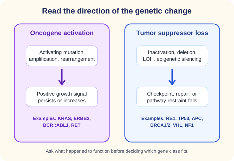
A proto-oncogene normally supports regulated growth, survival, or differentiation. An activating alteration can convert it into an oncogene. At the cellular level, one activated allele can be sufficient because the altered product supplies an abnormal positive signal. Point mutation, copy-number amplification, and gene rearrangement are common routes to this state. [1]
A tumor suppressor normally limits proliferation, preserves genome integrity, or restrains a signaling pathway. Cancer can be favored when enough suppressor function is lost. The classic cell-level pattern is recessive because the remaining normal allele can retain useful function, but the two-hit rule is a model rather than a requirement that every suppressor acquire exactly two sequence mutations. Deletion, loss of heterozygosity, epigenetic silencing, dominant-negative effects, and dosage sensitivity can change the pattern. [1][2][3]
Classify the functional direction first: oncogene activation adds positive signaling, while suppressor loss reduces protection. [1][3][4]
Read the alteration before memorizing the gene name
Alteration
Typical direction
Example
Predicted result
AlterationActivating point mutation
Typical directionAdded signaling
ExampleKRAS
Predicted resultRAS remains active longer
AlterationAmplification
Typical directionAdded signaling
ExampleERBB2
Predicted resultMore HER2 receptor is produced
AlterationRearrangement
Typical directionAdded signaling
ExampleBCR::ABL1
Predicted resultConstitutive kinase signaling
AlterationDeletion or inactivating variant
Typical directionLost restraint
ExampleRB1
Predicted resultG1/S control is weakened
Use the table as a direction test. If a change makes a signaling protein active without its usual input, think oncogene activation. If the change abolishes checkpoint, repair, degradation, or inhibitory activity, think suppressor loss. This distinction is more reliable than assuming a mutation is activating or inactivating from the word mutation alone. [1]
RAS stays in its active GTP-bound state for longer, so this is an activating oncogenic alteration. The useful observation is biochemical direction, not merely the gene name.
Prediction exercise: the only functional RB1 copy is deleted
Cell-cycle restraint falls because RB1 activity is lost. This is suppressor logic, even though the DNA event is a deletion rather than a point mutation.
Promoter CpG hypermethylation can reduce transcription and silence a tumor suppressor without changing its coding sequence. Aging tissues show complex methylome changes, including broad hypomethylation, focal hypermethylation, and epigenetic drift. [4]
TP53 is altered in roughly half of human cancers, which makes it important but not a default answer whenever a cancer gene is uncertain. The case still has to fit TP53 biology, such as impaired DNA-damage response or a Li-Fraumeni pattern. Mutant p53 can show loss of function, dominant-negative effects, and context-dependent acquired activities. [3]
Try it here · Checkpoint 1 of 3
Make your prediction before reading the choices. A first attempt is just a starting point.
Case 1
Show answer and explanations for case 1
A. Impaired RAS GTP hydrolysis (Best answer)
The assay directly shows failure to return efficiently to the GDP-bound state, which is the activating direction expected for an oncogenic RAS variant.
Reasoning steps for option A
What does the assay show about the mutant protein after the growth-factor signal is removed?
Mutant RAS stays mostly GTP-bound, while wild-type RAS returns to the GDP-bound resting state.
Why does a persistent GTP-bound state point to reduced GTP hydrolysis?
RAS switches itself off by hydrolyzing GTP to GDP, so a G12D protein that stays GTP-bound keeps signaling without input, which is oncogene activation.
B. Loss of β-catenin degradation after biallelic APC inactivation (Why this does not fit)
APC loss can activate Wnt signaling, but it does not explain the measured persistence of GTP-bound RAS in this tumor.
Reasoning steps for option B
Why could Wnt pathway activation tempt someone in a pancreatic carcinoma?
APC loss is a classic suppressor route to uncontrolled epithelial growth in the gastrointestinal tract.
Which assay result does APC loss fail to explain?
APC regulates β-catenin degradation, and it has no role in whether KRAS stays GTP-bound after the signal is withdrawn.
C. Failure of homologous recombination after BRCA1 loss (Why this does not fit)
BRCA1 deficiency impairs DNA double-strand break repair rather than the GTPase cycle measured in the assay.
Reasoning steps for option C
What makes a DNA-repair defect attractive in an aggressive adenocarcinoma?
BRCA1-associated repair failure does occur in some pancreatic cancers and drives genomic instability.
Why does repair failure not match the biochemical readout?
The measured defect is in the GTPase cycle of a single protein, not in repair of double-strand breaks.
D. Stabilization of HIF after loss of VHL protein (Why this does not fit)
VHL loss changes oxygen-responsive protein degradation, not the GDP-GTP state of KRAS.
Reasoning steps for option D
Why might HIF stabilization seem relevant to a solid tumor with growth signaling?
HIF activity drives angiogenic programs that many tumors exploit.
What separates VHL loss from the KRAS result?
VHL governs oxygen-dependent HIF degradation, whereas the assay tracks the GDP-GTP state of a mutant KRAS protein.
Takeaway: An activating RAS variant can prolong the GTP-bound signaling state, an oncogene gain-of-function pattern.
RB1 shows how inherited risk and tumor formation are different events
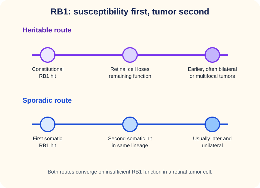
Knudson inferred the two-hit model from the age and laterality of retinoblastoma. A child with a constitutional pathogenic RB1 variant already carries one altered copy in every cell. A susceptible retinal cell needs an additional somatic event affecting the remaining functional copy, so tumors tend to occur earlier and can be bilateral or multifocal. A sporadic tumor generally requires both relevant events to arise in the same retinal lineage. [2][5]
Both routes converge on insufficient RB1 function in a retinal tumor cell, but heritable susceptibility starts one event earlier. [2][5]
RB protein restrains the G1/S transition by limiting E2F-dependent transcription. When functional RB is absent, E2F-dependent S-phase programs are less restrained. The practical sequence is inherited first hit, tissue-specific second hit, then clonal expansion. The inheritance of susceptibility can therefore look dominant in a pedigree even though loss of RB1 function in an individual cell follows suppressor logic. [1][5]
Heritable pattern
One pathogenic RB1 copy is constitutional. A later retinal event affects the remaining functional copy. Early onset, bilateral disease, or multiple tumors should increase concern for heritable retinoblastoma. [5]
Sporadic pattern
Blood testing can be negative while the retinal tumor contains two somatic RB1-disrupting events. A later unilateral presentation is more compatible with this route, although clinical evaluation still determines the individual diagnosis. [5]
A second hit is not restricted to a second small mutation. Loss of a chromosome segment, mitotic recombination, or another event that eliminates the remaining functional allele can create loss of heterozygosity. Read what happened to function, not only the mutation label. [1][2]
Prediction exercise: bilateral retinal tumors at 18 months plus an affected parent
The family history and bilateral early presentation favor a constitutional RB1 first hit followed by independent somatic events in retinal cells. The tumor still requires loss of sufficient RB function.
Leukocoria is an important presentation of retinoblastoma, but a white pupillary reflex is not itself a gene test. Gene interpretation belongs beside the eye findings, family history, laterality, and molecular data. [5]
Try it here · Checkpoint 2 of 3
Make your prediction before reading the choices. A first attempt is just a starting point.
Case 4
Show answer and explanations for case 4
A. Two independent activating RB1 variants in the same retinal cell (Why this does not fit)
RB1 is a tumor suppressor, so retinoblastoma is linked to loss of RB function rather than activating RB1 variants.
Reasoning steps for option A
Why might two RB1 variants in one cell seem to fit bilateral tumors?
Two RB1 events are indeed needed within a retinal cell for the tumor to form.
What is wrong with calling the RB1 changes activating?
RB1 is a tumor suppressor, and the tumor lost the normal allele, so the pathway works through loss of RB function.
B. A somatic BCR::ABL1 fusion followed by germline transmission (Why this does not fit)
BCR::ABL1 is an acquired leukemia driver and does not explain this familial bilateral retinal tumor pattern.
Reasoning steps for option B
Why might a fusion gene be considered for an inherited cancer?
BCR::ABL1 is the best-known example of a single genetic event that drives cancer.
Which findings exclude a Philadelphia fusion here?
BCR::ABL1 is an acquired leukemia driver, and this family shows inherited bilateral retinal tumors with an RB1 variant.
C. A constitutional MYC rearrangement followed by ERBB2 amplification (Why this does not fit)
Those oncogenic events do not match the inherited RB1 result or the tumor-specific loss of the normal RB1 allele.
Reasoning steps for option C
Why could oncogene events attract attention in an aggressive childhood tumor?
MYC and ERBB2 changes are powerful drivers of rapidly growing cancers.
Which test results point away from these oncogenes?
Blood shows a pathogenic RB1 variant, and the tumor lost the remaining normal RB1 allele, which is suppressor loss.
D. Constitutional RB1 hit plus a retinal somatic hit (Best answer)
The inherited variant supplies the first hit throughout the body, and loss of the remaining functional allele in retinal cells completes the classic suppressor pattern.
Reasoning steps for option D
What do the father's history and the blood test show about the first event?
The pathogenic RB1 variant is constitutional and inherited, so every retinal cell starts with one hit.
How does the tumor finding complete the explanation for bilateral multifocal disease?
A retinal cell that loses the remaining normal RB1 allele has no functional RB, and because every cell carries the first hit, several tumors can arise early in both eyes.
Takeaway: Heritable retinoblastoma supplies one RB1 hit constitutionally, so fewer additional events are needed in each susceptible retinal cell.
Oncogenes can be activated by mutation, amplification, or rearrangement
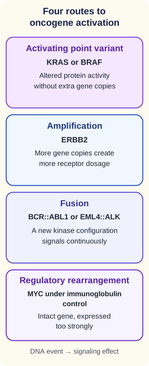
RAS proteins are small GTPases. Activating variants can impair the transition back toward the GDP-bound inactive state, extending proliferative signaling. BRAF is a downstream kinase in the same broad MAPK signaling axis, and an activating BRAF variant can drive signaling without requiring a lost second allele. [1]
ERBB2 illustrates amplification. A breast tumor can carry many additional copies of the HER2-encoding locus, increasing receptor abundance and signaling. Copy-number gain is therefore an oncogenic mechanism even when the receptor coding sequence is unchanged. [1]
BCR::ABL1 illustrates a fusion oncogene. The t(9;22) Philadelphia rearrangement creates a constitutively active tyrosine kinase central to chronic myeloid leukemia. Marked leukocytosis with a spectrum of myeloid maturation and basophilia should make the cytogenetic result mechanistically meaningful rather than decorative. [11]
MYC in Burkitt lymphoma shows a different rearrangement principle. The classic t(8;14) places MYC under strong immunoglobulin regulatory influence, increasing MYC expression in a B cell. The same broad category, chromosomal rearrangement, can therefore activate cancer through a new fusion protein or through abnormal control of an intact gene. [12]
Different DNA events can converge on the same functional direction: increased positive signaling. [1][11][12][14]
ALK rearrangements define a molecular subset of lung adenocarcinoma in which kinase signaling can become constitutive. The alteration matters because it identifies a driver pathway rather than merely a chromosome abnormality. [14]
RET is a receptor tyrosine kinase. Germline activating RET variants cause MEN2, with medullary thyroid carcinoma as a central feature and pheochromocytoma in MEN2A and MEN2B. MEN2A also includes risk of primary hyperparathyroidism. This is inherited oncogene activation, not a hereditary tumor suppressor syndrome. [13]
High-risk HPV demonstrates that cancer can also arise when viral proteins disable host safeguards. HPV E6 promotes p53 degradation, while E7 disrupts pRB control of E2F. The viral genes are oncogenic because their products reduce the function of host TP53 and RB1 pathways. [16]
Prediction exercise: a tumor has many ERBB2 copies but no coding variant
The copy-number change can itself increase receptor abundance. Classify the event as amplification-based oncogene activation rather than requiring an activating sequence variant.
Inherited tumor suppressor syndromes point to the pathway that lost protection
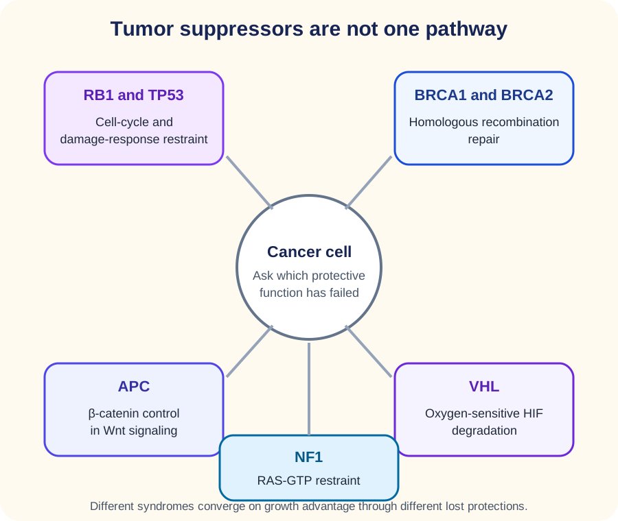
BRCA1 and BRCA2 support homologous recombination repair of DNA double-strand breaks. Germline pathogenic variants increase risks for breast and ovarian cancer; BRCA2 is also strongly associated with male breast, prostate, and pancreatic cancer. Tumors can become highly dependent on alternate DNA-repair processes once homologous recombination is deficient. [9]
Inherited syndromes become easier to distinguish when each gene is linked to the protective process that fails. [3][6][8][9][10]
APC helps control β-catenin in the Wnt pathway. In familial adenomatous polyposis, germline APC loss creates a field of susceptible colonic epithelium. Additional somatic loss in individual clones permits β-catenin accumulation and contributes to the development of numerous adenomas and very high colorectal cancer risk without preventive management. [10]
VHL protein participates in oxygen-sensitive degradation of HIF-α subunits under normoxic conditions. Loss of VHL permits HIF-dependent transcription, including angiogenic programs such as VEGF. The syndrome links clear cell renal cell carcinoma with retinal and central nervous system hemangioblastomas and pheochromocytoma or paraganglioma. [8]
NF1 encodes neurofibromin, a RAS GTPase-activating protein that helps restrain RAS signaling. Café-au-lait macules, axillary or inguinal freckling, neurofibromas, Lisch nodules, and optic pathway glioma fit NF1. The gene is at 17q11.2. [6]
NF2 encodes merlin at 22q12.2. Bilateral vestibular schwannomas are characteristic, and meningiomas and other schwannomas can occur. Current GeneReviews terminology is NF2-related schwannomatosis, replacing older wording such as central neurofibromatosis. [7]
WT1 belongs in a Wilms tumor predisposition framework, but WAGR and hemihyperplasia should not be fused into one mnemonic. WAGR is associated with a constitutional 11p13 deletion involving WT1 and PAX6 and can include Wilms tumor risk, aniridia, genitourinary abnormalities, and developmental disability. Isolated hemihyperplasia and Beckwith-Wiedemann spectrum are instead linked to the 11p15 region. [15]
Use phenotype to identify the missing safeguard
Pattern
Safeguard
Pathway consequence
PatternHundreds to thousands of colorectal adenomas
SafeguardAPC
Pathway consequenceβ-catenin control is lost
PatternBreast and ovarian family pattern
SafeguardBRCA1/2
Pathway consequenceHomologous recombination is impaired
PatternClear cell RCC plus hemangioblastomas
SafeguardVHL
Pathway consequenceHIF signaling persists in normoxia
PatternCafé-au-lait macules plus neurofibromas
SafeguardNF1
Pathway consequenceRAS restraint is reduced
PatternBilateral vestibular schwannomas
SafeguardNF2
Pathway consequenceMerlin-dependent growth restraint is lost
Prediction exercise: normal oxygen but high HIF and VEGF in a clear cell renal tumor
Loss of VHL function can prevent normal HIF degradation, so a normoxic cell behaves as though a hypoxic transcriptional program should remain active.
Try it here · Checkpoint 3 of 3
Make your prediction before reading the choices. A first attempt is just a starting point.
Case 15
Show answer and explanations for case 15
A. Activating RB1 mutation that increases E2F restraint (Why this does not fit)
RB1 is a suppressor and the proposed direction is internally inconsistent; it also does not fit the VHL syndrome phenotype.
Reasoning steps for option A
Why might an RB1 change be considered in a young adult with several tumors?
RB1 is a well-known inherited tumor suppressor.
What is internally inconsistent about an activating RB1 change here?
Tumor suppressors cause cancer by losing function, and RB1 does not explain renal cancer, retinal angiomas and hemangioblastoma.
B. Loss of MYC expression after t(8;14) (Why this does not fit)
t(8;14) increases MYC expression in Burkitt lymphoma rather than producing this renal and hemangioblastoma pattern.
Reasoning steps for option B
Why might MYC be linked to a tumor with high target-gene expression?
MYC is a strong transcriptional driver of growth programs.
What is wrong with MYC loss after t(8;14)?
t(8;14) increases MYC in Burkitt lymphoma, and MYC does not regulate normoxic HIF and VEGF.
C. Increased neurofibromin activity that suppresses RAS (Why this does not fit)
NF1 biology does not account for normoxic HIF stabilization and the VHL-associated tumor spectrum.
Reasoning steps for option C
Why could a RAS regulator be considered in an inherited tumor syndrome?
NF1 is another suppressor syndrome with tumors of the nervous system.
Which findings point away from neurofibromin?
Increased neurofibromin would lower RAS signaling and cannot stabilize HIF in normal oxygen, and the tumor spectrum is that of VHL.
D. Loss of VHL-dependent HIF degradation (Best answer)
The syndrome and normoxic HIF-VEGF activation fit loss of pVHL-mediated degradation of HIF-α subunits.
Reasoning steps for option D
What do clear cell renal cancer, retinal angiomas and a cerebellar hemangioblastoma suggest together?
They form the von Hippel-Lindau tumor spectrum.
Why are HIF targets and VEGF high despite normal oxygen?
pVHL normally tags HIF-α for degradation when oxygen is present, so its loss lets HIF-α accumulate and drive VEGF as if the cell were hypoxic.
Takeaway: VHL loss permits HIF-dependent transcription to persist in normoxia, promoting angiogenic programs.
Tumor names and metastatic sites need context, not shortcuts
The suffix -oma does not guarantee benign behavior. Lymphoma, melanoma, mesothelioma, and seminoma are malignant. Hepatoma is an older term commonly used for hepatocellular carcinoma. Teratoma behavior depends strongly on site, age, and histology, so it should not be treated as a universal benign-or-malignant suffix exception. [18]
Metastatic cancer can spread to almost any organ, and the preferred destinations depend on the primary tumor. Bone, liver, and lung are frequent metastatic sites across common cancers, with brain and adrenal involvement important for selected primaries. It is safer to identify the known primary, lesion distribution, and pathology than to memorize a universal six-site rule or assume every lesion at one organ is more likely metastatic than primary. [17]
For example, multiple liver lesions in a patient with colorectal adenocarcinoma can represent metastatic colorectal carcinoma, especially when morphology and immunophenotype match the colon primary. A solitary liver mass in a cirrhotic patient raises a different differential. The organ name alone does not determine whether a tumor is primary or metastatic. [17]
The practical synthesis is consistent across the lesson: identify whether the genetic event adds signaling or subtracts protection, then connect the event to the phenotype. A gene name is useful only when its functional direction explains the clinical and molecular findings.
Prediction exercise: prior colon cancer plus multiple CDX2-positive liver lesions
The known primary, multifocal distribution, matching morphology, and intestinal immunophenotype support metastatic colorectal carcinoma rather than a liver-origin diagnosis based only on location.
Apply the direction and pathway logic
Case 2
Show answer and explanations for case 2
A. Loss of heterozygosity at a tumor suppressor locus (Why this does not fit)
Loss of heterozygosity decreases functional suppressor dosage; this tumor instead has a large increase in ERBB2 copy number.
Reasoning steps for option A
Why might loss of heterozygosity come to mind for a breast carcinoma?
Loss of heterozygosity is common in breast tumors, especially at suppressor loci such as BRCA1 and BRCA2.
Which sequencing result points in the opposite direction?
The tumor gained ERBB2 copies to 14 per cell, and loss of heterozygosity removes an allele rather than adding copies.
B. Promoter hypermethylation that silences transcription (Why this does not fit)
Promoter silencing would decrease transcript abundance, opposite the markedly increased ERBB2 messenger RNA reported here.
Reasoning steps for option B
What could make an epigenetic mechanism seem plausible when no coding variant is found?
Epigenetic changes can alter gene activity without any change in the coding sequence.
Why does the ERBB2 transcript level rule out promoter silencing?
Silencing lowers messenger RNA, but ERBB2 messenger RNA is markedly increased here.
C. Gene amplification that increases receptor dosage (Best answer)
The concordant high copy number, transcript abundance, and protein expression support oncogenic amplification rather than coding-sequence activation.
Reasoning steps for option C
What do the copy number, transcript and protein results have in common?
All three are increased together: 14 ERBB2 copies, abundant ERBB2 messenger RNA and strong HER2 membrane staining.
How can a normal coding sequence still drive oncogenic signaling?
Extra gene copies raise receptor dosage, and the excess HER2 increases signaling without any activating variant, so amplification is the driver.
D. A germline first hit followed by retinal somatic loss (Why this does not fit)
That sequence describes heritable RB1 predisposition and does not explain ERBB2 copy-number gain in a breast tumor.
Reasoning steps for option D
Why might a two-hit sequence be considered for a woman with breast cancer?
Inherited first-hit syndromes such as BRCA-related cancer predispose to breast tumors.
What in the tumor data does a retinal two-hit sequence fail to address?
It describes heritable RB1 loss in the retina and cannot produce a gain of ERBB2 copies in breast tissue.
Takeaway: Oncogenes can be activated by copy-number amplification even when the coding sequence is unchanged.
A. Oxygen-dependent HIF degradation (Why this does not fit)
That response depends on VHL and would not explain the Li-Fraumeni pedigree or absent p21 induction after radiation.
Reasoning steps for option A
Why might an oxygen-sensing defect be considered in a family with many early cancers?
VHL loss is an inherited suppressor defect that also causes cancer in young adults.
Which findings argue against the VHL pathway?
The pedigree of early breast cancer, childhood adrenocortical carcinoma and sarcoma, plus absent p21 after radiation, fits TP53 rather than HIF regulation.
B. p53-dependent p21 induction with DNA-damage cell-cycle arrest (Best answer)
The pedigree fits Li-Fraumeni syndrome and the failed p21 response indicates impaired p53-mediated arrest after DNA damage.
Reasoning steps for option B
What does the family tumor spectrum suggest before the genetic result is read?
Osteosarcoma, breast cancer at 29 and childhood adrenocortical carcinoma form the classic Li-Fraumeni pattern.
How does the failed p21 response connect to the TP53 variant?
After ionizing radiation, p53 normally induces p21 to halt the cell cycle, so absent p21 shows the lost p53 damage response.
C. RAS conversion from GTP-bound to GDP-bound state (Why this does not fit)
Neurofibromin and RAS GTPase activity regulate this process, not TP53-dependent p21 induction.
Reasoning steps for option C
Why could a RAS-control defect seem relevant in a hereditary sarcoma family?
NF1 loss raises RAS signaling and can predispose to sarcomas, including malignant nerve sheath tumors.
What result places the defect outside RAS regulation?
The cells fail specifically to induce p21 after DNA damage, which is a p53 transcriptional response, not a RAS GTPase function.
D. β-catenin destruction in resting colonic epithelium (Why this does not fit)
APC regulates β-catenin; the supplied family tumor spectrum and radiation-response defect point to TP53.
Reasoning steps for option D
Why might β-catenin control be considered in a family with inherited tumors?
APC loss is a well-known inherited suppressor defect that causes early cancer.
Which features separate the family from an APC pattern?
There is no colonic polyposis, and the radiation-response defect with a pathogenic TP53 variant points to p53.
Takeaway: TP53 loss impairs DNA-damage responses such as p21-mediated cell-cycle arrest, and the clinical setting must fit the gene.
A. One RB1 hit must have been inherited but was missed because the tumor is unilateral (Why this does not fit)
An inherited variant is possible in some unilateral cases, but the supplied testing instead demonstrates two tumor-restricted events and no blood variant.
Reasoning steps for option A
Why is an inherited RB1 variant a reasonable concern in any child with retinoblastoma?
Some children with unilateral disease still carry a constitutional RB1 variant.
Which results make an inherited first hit unlikely here?
Blood testing is negative, there is no family history, and both RB1 events are confined to the tumor.
B. Two somatic RB1 hits in one retinal lineage (Best answer)
A unilateral later presentation with negative blood testing and two tumor-restricted RB1 events fits a sporadic two-somatic-hit route.
Reasoning steps for option B
What do the negative blood test and single tumor suggest about the route?
A unilateral tumor in an older child without a family history fits the sporadic pattern.
How do the two tumor-restricted events fit the two-hit model?
Both RB1 copies were inactivated somatically in the same retinal lineage, so RB function was lost without an inherited first hit.
C. One activating RB1 allele was sufficient to create the tumor (Why this does not fit)
RB1 is a suppressor, so gain of RB1 activity does not fit the loss-of-function events reported.
Reasoning steps for option C
Why might a single gene event seem enough to start this tumor?
One activated allele is enough for an oncogene, and the boy has only one tumor.
Why does single-allele sufficiency not apply to RB1?
RB1 is a suppressor, and sequencing found two inactivating events, not a gain of RB activity.
D. The tumor is best explained by inherited RET activation (Why this does not fit)
RET activation is linked to MEN2, not the two inactivating RB1 events documented in this retinal tumor.
Reasoning steps for option D
Why might an inherited activating variant come to mind for a childhood tumor?
Germline RET activation is a classic cause of inherited cancer in young people.
What in the tumor points away from RET?
RET activation causes MEN2 thyroid and adrenal tumors, while this retinal tumor carries two inactivating RB1 events.
Takeaway: Sporadic retinoblastoma can acquire both relevant RB1-disrupting events within the tumor lineage.
A. Both alleles of every cancer gene must be lost before signaling changes (Why this does not fit)
That oversimplifies the classic tumor suppressor model and conflicts with the activating BRAF result.
Reasoning steps for option A
Why is a two-allele requirement tempting when one BRAF allele is still normal?
The two-hit rule is the most familiar model of how cancer genes lose control.
What does the high phospho-ERK show about that requirement?
Signaling is already abnormal with one intact allele, so the altered gene acts as an oncogene and does not need both copies changed.
B. One activated BRAF allele supplies the abnormal signal (Best answer)
Persistent downstream MAPK activity despite one intact allele is the expected cellular-dominant behavior of an activated oncogene.
Reasoning steps for option B
What does the persistent phospho-ERK show about the MAPK pathway?
ERK stays phosphorylated without upstream stimulation, so the pathway is active on its own.
Why is one V600E allele enough to produce that signal?
The mutant kinase is constitutively active, so its product supplies the abnormal signal even while the normal allele keeps making normal BRAF.
C. The intact allele is necessarily methylated even though no methylation data are given (Why this does not fit)
No evidence of epigenetic silencing is supplied, and an activating oncogene does not require loss of the normal allele.
Reasoning steps for option C
Why might silencing of the normal allele be proposed?
Epigenetic silencing is a real way to remove the remaining allele of a cancer gene.
What evidence is missing for silencing, and why is it unnecessary?
No methylation data are given, and an activated oncogene drives signaling even with a working normal allele.
D. BRAF is acting as a DNA-repair tumor suppressor in this tumor (Why this does not fit)
BRAF is a signaling kinase; the observed high phospho-ERK supports pathway activation rather than loss of DNA repair.
Reasoning steps for option D
Why could a suppressor label be considered for a common melanoma gene?
Melanomas carry many DNA-damage changes from ultraviolet light.
Which result shows BRAF is acting in the opposite direction?
Phospho-ERK is high without upstream stimulation, which is a gain of kinase signaling; a lost repair gene would raise mutation burden, not switch on the MAPK pathway.
Takeaway: Activated oncogenes can show cellular-dominant behavior because one altered allele can provide an abnormal positive signal.
A. A second somatic hit abolishes a DNA-repair suppressor (Why this does not fit)
The reported lesion is a fusion driver with kinase activity, not biallelic loss of a repair gene.
Reasoning steps for option A
Why could a lost repair suppressor be considered in lung adenocarcinoma?
Lung cancers often carry repair and checkpoint defects that allow genomic instability.
What does the molecular report actually show?
The tumor has an active ALK fusion kinase, a gain of signaling, not biallelic loss of a repair gene.
B. Promoter methylation suppresses ALK transcript production (Why this does not fit)
The tumor has active ALK signaling; silencing ALK transcription would predict the opposite direction.
Reasoning steps for option B
Why might a transcriptional mechanism be proposed for a gene rearrangement?
Rearrangements can change how strongly a gene is transcribed.
Why does silencing contradict the ALK finding?
The tumor shows ALK kinase activity, and silencing ALK transcription would reduce, not create, that activity.
C. Loss of HIF degradation creates the ALK fusion (Why this does not fit)
VHL-HIF regulation is a separate oxygen-sensing pathway and does not generate EML4::ALK.
Reasoning steps for option C
Why might HIF signaling be linked to a lung tumor that grows without its normal stimulus?
HIF stabilization lets tumor cells act as if hypoxic and drives angiogenic growth.
What separates oxygen sensing from how an EML4::ALK fusion forms?
VHL-HIF regulation does not rearrange chromosomes, and the fusion arises from a structural rearrangement of chromosome 2.
D. The ALK fusion creates constitutive oncogenic kinase signaling (Best answer)
EML4::ALK is an oncogenic fusion that can drive ligand-independent kinase activity in a molecular subset of lung adenocarcinoma.
Reasoning steps for option D
What does kinase activity without the normal stimulus indicate?
ALK is signaling constitutively, which is the gain-of-function direction of an oncogene.
How does the EML4::ALK fusion produce that activity?
The EML4 partner promotes clustering of the ALK kinase domain, which activates it without ligand and defines a treatable subset of never-smoker adenocarcinoma.
Takeaway: Gene fusions can create constitutively active oncogenic kinases.
A. Biallelic tumor suppressor deletion (Why this does not fit)
The reported variant increases RET signaling rather than abolishing a suppressive function.
Reasoning steps for option A
Why might a suppressor deletion be considered in a family with several endocrine tumors?
Many inherited cancer syndromes arise from suppressor loss passed through families.
What does the genetic report say about the direction of change?
The RET variant increases receptor signaling, so it adds function rather than deleting a suppressor.
B. Homologous recombination repair defect (Why this does not fit)
That category fits BRCA1/2 loss, not an activating RET receptor variant.
Reasoning steps for option B
Why could a repair defect be considered in a family with early cancers?
BRCA1 and BRCA2 repair defects cause strong autosomal dominant family patterns.
Which tumor combination points away from a repair defect?
Medullary thyroid carcinoma with pheochromocytoma is the MEN2 pattern of RET activation, not a BRCA tumor spectrum.
C. Activating oncogene variant (Best answer)
MEN2 results from germline activating RET variants, so the inherited lesion adds receptor tyrosine kinase signaling.
Reasoning steps for option C
What do medullary thyroid carcinoma and pheochromocytoma in three relatives suggest?
They form the multiple endocrine neoplasia type 2 pattern, inherited as an autosomal dominant trait.
How does the RET result define the genetic category?
A germline variant that increases RET receptor tyrosine kinase signaling is inherited oncogene activation, even though the syndrome runs in families like a suppressor disorder.
D. Constitutional RB1 first hit (Why this does not fit)
RB1 predisposition causes retinoblastoma and does not explain medullary thyroid carcinoma with pheochromocytoma.
Reasoning steps for option D
Why might an inherited first hit seem attractive in a familial tumor syndrome?
Constitutional RB1 variants are the model of inherited cancer susceptibility.
Which tumor types rule out RB1 predisposition here?
Carriers of an RB1 first hit develop retinoblastoma in early childhood and later osteosarcoma, whereas this family has medullary thyroid carcinoma and pheochromocytoma with a RET gain.
Takeaway: Hereditary cancer can result from germline oncogene activation, as in RET-associated MEN2.
A. Neurofibromatosis type 1 with loss of neurofibromin (Why this does not fit)
NF1 is at 17q11.2 and typically features café-au-lait macules, neurofibromas, and other NF1 manifestations rather than bilateral vestibular schwannomas.
Reasoning steps for option A
Why might NF1 be considered in a patient with nerve sheath tumors?
Both neurofibromatosis types produce tumors of nerve sheath origin.
Which locus and tumor pattern rule out NF1?
NF1 is at 17q11.2 with café-au-lait macules and neurofibromas, while this patient has bilateral vestibular schwannomas and a 22q12.2 variant.
B. Von Hippel-Lindau syndrome with loss of pVHL (Why this does not fit)
VHL is associated with clear cell renal cancer and hemangioblastomas, not the specified NF2 locus and bilateral vestibular tumors.
Reasoning steps for option B
Why might VHL be considered for a young adult with intracranial tumors?
VHL causes cerebellar hemangioblastomas, which are also posterior fossa tumors.
What separates VHL from this presentation?
VHL tumors are hemangioblastomas and renal cancers, not bilateral vestibular schwannomas with meningiomas.
C. NF2-related schwannomatosis with impaired merlin function (Best answer)
Bilateral vestibular schwannomas plus meningiomas and an NF2 variant at 22q12.2 identify NF2-related schwannomatosis and merlin loss.
Reasoning steps for option C
What do bilateral vestibular schwannomas and meningiomas point to?
This combination is the hallmark of NF2-related schwannomatosis.
Which protein is lost when the 22q12.2 suppressor is inactivated?
Merlin, the NF2 product, whose loss removes growth restraint in Schwann and meningeal cells.
D. Li-Fraumeni syndrome with loss of p53 (Why this does not fit)
Li-Fraumeni has a broad early cancer spectrum and TP53 variants, not the 22q12.2 vestibular schwannoma pattern.
Reasoning steps for option D
Why could Li-Fraumeni syndrome be considered in a man with several tumors?
Li-Fraumeni also causes multiple early tumors, including brain tumors.
Which features point away from TP53?
The variant is at 22q12.2, and bilateral vestibular schwannomas are not part of the TP53 spectrum.
Takeaway: Bilateral vestibular schwannomas are characteristic of NF2-related schwannomatosis caused by loss of merlin.
A. Every tumor ending in -oma is benign unless AFP is increased (Why this does not fit)
Seminoma is malignant and may have normal AFP, so neither the suffix nor AFP establishes benignity.
Reasoning steps for option A
Why might a normal AFP seem reassuring about this testicular mass?
AFP is a familiar marker of nonseminomatous germ cell tumors.
Why do neither the suffix nor a normal AFP show that the tumor is benign?
Pure seminoma does not make AFP, and seminoma is malignant regardless of how its name ends.
B. Seminoma is a malignant testicular germ cell tumor despite the suffix (Best answer)
NCI classifies seminoma among malignant testicular germ cell tumors; the suffix alone cannot determine biologic behavior.
Reasoning steps for option B
What does orchiectomy show about the tumor type?
The pathology is pure seminoma, a testicular germ cell tumor.
How should the student's suffix argument be answered?
Seminoma is malignant at diagnosis, like lymphoma and melanoma, so the ending of a tumor name does not define its behavior.
C. Seminoma becomes malignant only after it metastasizes (Why this does not fit)
Seminoma is already a malignant germ cell tumor at diagnosis; metastasis changes stage, not the basic malignant classification.
Reasoning steps for option C
Why might metastasis seem to be the marker of malignancy?
Spread to distant sites is the most obvious sign of malignant behavior.
How does metastasis relate to the classification of seminoma?
Seminoma is malignant when it is diagnosed, and spread changes the stage rather than the diagnosis.
D. The mass is malignant only if β-hCG is increased (Why this does not fit)
Seminoma can be malignant with or without increased β-hCG, and the pathology diagnosis determines the tumor type.
Reasoning steps for option D
Why could a raised hormone marker seem to prove malignancy in a testicular mass?
Some seminomas and other germ cell tumors secrete β-hCG, which clinicians follow.
Why is a normal or raised β-hCG not the deciding factor?
Markers help with staging and follow-up, but many pure seminomas secrete no β-hCG; the orchiectomy histology has already made this a malignant germ cell tumor.
Takeaway: Tumor nomenclature has historical exceptions; -oma cannot be used as a universal benignity rule.
A. New primary hepatocellular carcinoma in a noncirrhotic liver (Why this does not fit)
Hepatocellular carcinoma is possible without cirrhosis, but the matching colon morphology and CDX2 positivity strongly support metastatic colorectal origin.
Reasoning steps for option A
Why is hepatocellular carcinoma still worth considering without cirrhosis?
It can arise in a noncirrhotic liver, and a new liver mass always raises the question.
Which biopsy findings favor colorectal origin instead?
The lesion resembles the prior colon tumor and is CDX2 positive, an intestinal marker not expected in hepatocellular carcinoma.
B. Benign focal nodular hyperplasia in multiple sites (Why this does not fit)
Focal nodular hyperplasia would not reproduce the prior adenocarcinoma morphology or the intestinal tumor immunophenotype.
Reasoning steps for option B
Why might a benign lesion be considered for several liver masses?
Focal nodular hyperplasia is a common benign liver mass that can be multiple.
What in the biopsy excludes focal nodular hyperplasia?
The tissue is malignant adenocarcinoma matching the prior colon tumor, with intestinal CDX2 staining.
C. Metastatic colorectal adenocarcinoma to the liver (Best answer)
The known colon primary, multifocal liver distribution, matching morphology, and intestinal immunophenotype together support metastatic colorectal cancer.
Reasoning steps for option C
What do the history and imaging suggest before the biopsy result?
A resected colon adenocarcinoma followed by six new liver lesions is a common pattern of portal venous metastasis.
How does the biopsy confirm the source?
Matching colon morphology and CDX2 positivity show intestinal origin, so the lesions are metastatic colorectal adenocarcinoma.
D. Primary seminoma of the liver (Why this does not fit)
Seminoma is a germ cell tumor and does not match the prior colon adenocarcinoma or CDX2-positive morphology.
Reasoning steps for option D
Why might a germ cell tumor be considered in an unusual site?
Germ cell tumors can occasionally arise outside the gonads.
Why does seminoma fail to fit the biopsy?
The lesion is an adenocarcinoma resembling the colon primary and is CDX2 positive, not a germ cell tumor.
Takeaway: Classify a distant lesion using the known primary, distribution, morphology, and molecular or immunophenotypic evidence rather than an organ shortcut.
The tumor already lacks effective BRCA1-dependent homologous recombination, so blocking a compensatory repair process creates a combination the cancer cell tolerates poorly.
Reasoning steps for option A
What does the tumor already lack before treatment?
Its BRCA1 deficiency leaves little homologous recombination to repair double-strand breaks.
Why does PARP inhibition harm tumor cells more than normal cells?
Blocking PARP adds a second repair failure that the BRCA1-deficient cells cannot survive, while normal cells use intact homologous recombination, which is synthetic lethality.
B. Activation of RET signaling by the PARP inhibitor (Why this does not fit)
RET is a receptor tyrosine kinase linked to MEN2 and does not explain the repair-selective response.
Reasoning steps for option B
Why might a signaling change be proposed for a drug response?
Some cancer drugs work through receptor tyrosine kinase pathways.
What result points away from RET?
The selective effect depends on a repair defect in BRCA1-deficient cells, and RET is an MEN2 receptor unrelated to PARP.
C. Restoration of APC function through β-catenin accumulation (Why this does not fit)
β-catenin accumulation reflects loss of APC function with Wnt pathway activation, not restoration of a missing DNA-repair pathway.
Reasoning steps for option C
Why might a suppressor pathway be linked to a selective drug effect?
APC is a familiar suppressor, and β-catenin changes are common in tumors.
Why is restoring APC through β-catenin backwards?
β-catenin accumulates when APC is lost, and neither event repairs DNA in a BRCA1-deficient tumor.
D. Creation of the Philadelphia chromosome during treatment (Why this does not fit)
BCR::ABL1 arises from a specific chromosomal rearrangement and is unrelated to PARP inhibition in a BRCA1-deficient tumor.
Reasoning steps for option D
Why might a new chromosomal rearrangement be blamed for a treatment effect?
Therapy can sometimes induce new genetic changes in cells.
Why does the Philadelphia chromosome not explain the tumor vulnerability?
BCR::ABL1 is a myeloid leukemia rearrangement and has no role in the repair-selective response to PARP inhibition.
Takeaway: BRCA-deficient tumors can be vulnerable to PARP inhibition through synthetic lethality, reflecting dependence on alternate repair processes.