Ionizing radiation: from energy deposition to cancer
Connect source, route, absorbed dose, tissue, and time to DNA injury, acute syndromes, late tissue effects, and later cancer risk.
Radiation questions become manageable when you stop naming a cancer from the symbol alone. The central task is to connect source and route to the tissue that absorbs energy, then connect dose and time to cell death, mutation, or recovery. After this lesson, you should be able to trace an exposure from source to target, separate acute tissue injury from later cancer risk, and explain why the same radionuclide can be minor outside the body yet dangerous after inhalation or ingestion.
Start with four questions, not one radiation label
A vial can have high activity while a distant organ receives little absorbed dose. Conversely, a small amount of material lodged in one tissue can deliver a concentrated local dose. Activity is not dose, and dose is not a diagnosis. Ask what is emitting, where the source is, what tissue receives energy, and over what interval. The answers give a tissue-dose distribution; only then predict acute injury or later cancer risk. [1][2]
The four-gate exposure trace
Source: machine-generated photons or a radionuclide?
Route: outside the body, inhaled, swallowed, injected, or placed in tissue?
Target: which tissue can the radiation reach, and where does the material concentrate?
Time: brief high dose, repeated exposure, or retained internal source?
The units answer different questions. A becquerel counts nuclear decays per second. A gray measures energy absorbed per kilogram of tissue. A sievert weights absorbed dose for radiation type and tissue sensitivity when estimating protection-related risk. Bq, Gy, and Sv therefore cannot be substituted for one another.
Quantity
Unit
Question answered
QuantityActivity
UnitBq
Question answeredHow often does the source decay?
QuantityAbsorbed dose
UnitGy
Question answeredHow much energy entered this tissue?
QuantityEquivalent or effective dose
UnitSv
Question answeredHow is the dose weighted for protection estimates?
External irradiation ends when the person leaves the beam or the source is shielded. Contamination means radioactive material is on or in the person and can continue emitting until it decays, is cleared, or is physically taken away. Irradiation and contamination are related but not interchangeable.
Apply the four gates to a sealed source
A worker briefly stands near a sealed photon source, then leaves. The source remained outside, no material entered the body, and exposure stopped with distance. This is external irradiation, not internal contamination.
Transfer the same reasoning to a swallowed radionuclide. Its activity matters, but the biologic pattern also depends on absorption, organ uptake, physical half-life, biologic clearance, and radiation range. The useful prediction is the absorbed tissue dose, not the isotope name by itself.
Trace the energy path before predicting harm
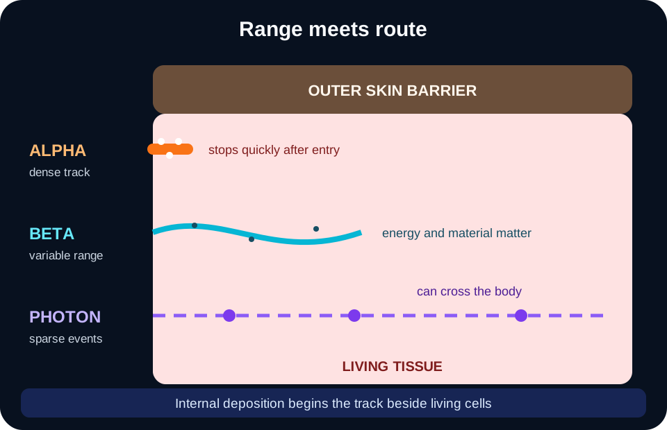
The common shortcut says alpha, beta, or gamma and jumps directly to a cancer. That skips the decisive step. Range meets route: a short-range particle outside the body may never reach living cells, while the same emitter deposited beside living cells can create dense ionization. [1][3]
Track density and reach are separate. Internal deposition can place a short-range source beside living cells. [1][3]
Alpha particles
Alpha particles are heavy, doubly charged, and deposit energy densely over a very short path. Intact outer skin blocks external alpha radiation, but inhaled, swallowed, injected, or wound contamination can place an alpha emitter next to living tissue. Internal alpha is the clinically important pattern.
Beta particles
Beta particles have a broader energy range and usually travel farther than alpha particles. Some can injure skin from outside; internal beta emitters irradiate the tissue in which they distribute. Energy and location determine depth, so one fixed penetration distance is misleading.
Photons
X-rays and gamma rays are photons. They can cross the body and deposit energy along their path, making external exposure important. Attenuation depends on photon energy, material, thickness, distance, and geometry. Penetrating photons can expose marrow and multiple organs.
Shielding must fit the radiation and the job. Paper or intact skin can stop alpha particles. Low-density material is often used for beta sources, with additional design for secondary x-rays when needed. Dense material, concrete, water, distance, and shorter time all help reduce photon dose. No single shield is correct for every source.
Route-and-range trace
Mark the source as external or internal.
Estimate whether its emissions can reach living target cells.
Identify chemical or physiologic organ uptake.
Predict the tissue receiving dose, then consider dose rate and duration.
Compare two alpha scenarios
A sealed alpha source on intact skin deposits essentially no dose in viable organs. Radon progeny inhaled into airways place alpha emitters beside bronchial epithelium. The radiation is the same class, but route changes target.
A useful transfer test is to swap only the route. If an external alpha source becomes inhaled dust, the important target changes from no meaningful deep tissue exposure to the deposition site. If an external photon beam becomes a localized internal seed, the spatial dose distribution changes even though the radiation remains ionizing energy.
Try it here · Checkpoint 1 of 3
Make your prediction before reading the choices. A first attempt is just a starting point.
Case 6
Show answer and explanations for case 6
A. The worker who touched a sealed alpha source with intact skin (Why this does not fit)
External alpha particles stop before reaching living deep tissue. Sealing and intact skin eliminate the route needed for marrow dose.
Reasoning steps for option A
Why can an alpha source seem the most dangerous of the four?
Alpha particles cause dense, highly damaging ionization tracks.
Can a sealed alpha source touched with intact skin reach marrow?
No; the particles stop in the outer skin, so marrow receives no meaningful dose.
B. The worker with the brief whole-body photon exposure (Best answer)
Penetrating photons can traverse the body and deposit energy in marrow across many bones. Whole-body geometry makes broad hematopoietic exposure plausible.
Reasoning steps for option B
Which radiation can travel through the body to many bones at once?
Penetrating photons such as gamma rays or x-rays.
Why does whole-body geometry matter for marrow dose?
Active marrow is spread through the skeleton, so only broad penetrating exposure doses most of it.
C. The worker with a localized beta spill confined to a glove (Why this does not fit)
A glove-limited beta source mainly threatens nearby superficial tissue until it is discarded. It does not create a broad whole-body marrow dose.
Reasoning steps for option C
Why might a spilled beta emitter seem to threaten marrow?
Beta emitters can irradiate marrow when they are taken inside and deposit in bone.
What limits this spill to superficial tissue?
It stayed on a glove, so the beta range reached only nearby skin until the glove was discarded.
D. The worker with no internal uptake after a negative survey (Why this does not fit)
A negative uptake assessment provides no retained source and no specified external field. There is no mechanism for a substantial marrow dose in the described option.
Reasoning steps for option D
Why might a worker with an exposure record be picked by default?
Any radiation incident can seem capable of causing marrow injury.
Does a negative uptake assessment supply any dose pathway?
No; it shows no retained source and names no external field, so there is no route to marrow.
Takeaway: Deep penetration plus whole-body geometry, not radiation name alone, determines broad marrow dose.
From ionization to either cell loss or a surviving clone
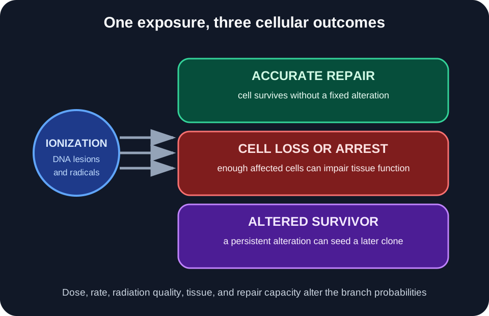
Ionizing radiation can damage DNA directly or through reactive chemical species formed from water. Closely spaced lesions and double-strand breaks are harder to restore accurately than isolated lesions. The outcome depends on dose, dose rate, radiation quality, cell state, and repair capacity. DNA damage is not destiny: many lesions are restored, some cells die, and a small fraction may survive with a heritable alteration. [3][4]
Radiation injury does not guarantee cancer. Repair, cell loss, and survival with alteration are competing outcomes. [3][4]
High linear energy transfer describes dense energy deposition along a track, not deep penetration. Alpha particles are a classic high-LET exposure. Photons are lower-LET and usually produce more widely spaced ionizations. LET and range answer different questions and often vary in opposite directions.
A tissue reaction requires enough cells to be injured before the organ shows a clinical effect. Its probability rises around a threshold, and severity generally increases with dose. Examples include skin injury, marrow failure after large whole-body exposure, cataract, fibrosis, and mucosal injury. Tissue reactions are not the same as radiation-induced cancer. [2]
Cancer is treated as a stochastic outcome for protection purposes: increasing dose increases probability, while a tumor's severity is not graded by the initiating dose. At low doses, an individual's added risk is difficult to measure directly. Regulators use the linear no-threshold model as a prudent protection model, not as proof that a particular future cancer was caused by a particular low-dose event. [2]
Sort the outcomes
Pancytopenia after a large, brief whole-body dose is a dose-dependent tissue reaction. A leukemia years after exposure is a probabilistic late outcome. The distinction is cell depletion versus clonal survival.
Apply the same distinction to a treated organ. Fibrosis within a high-dose field reflects progressive normal-tissue injury. A new, histologically distinct malignancy years later may qualify as a second primary cancer, but attribution requires population evidence, dose context, latency, and competing causes. Timing alone is insufficient.
Recognize exposure signatures without turning them into absolutes
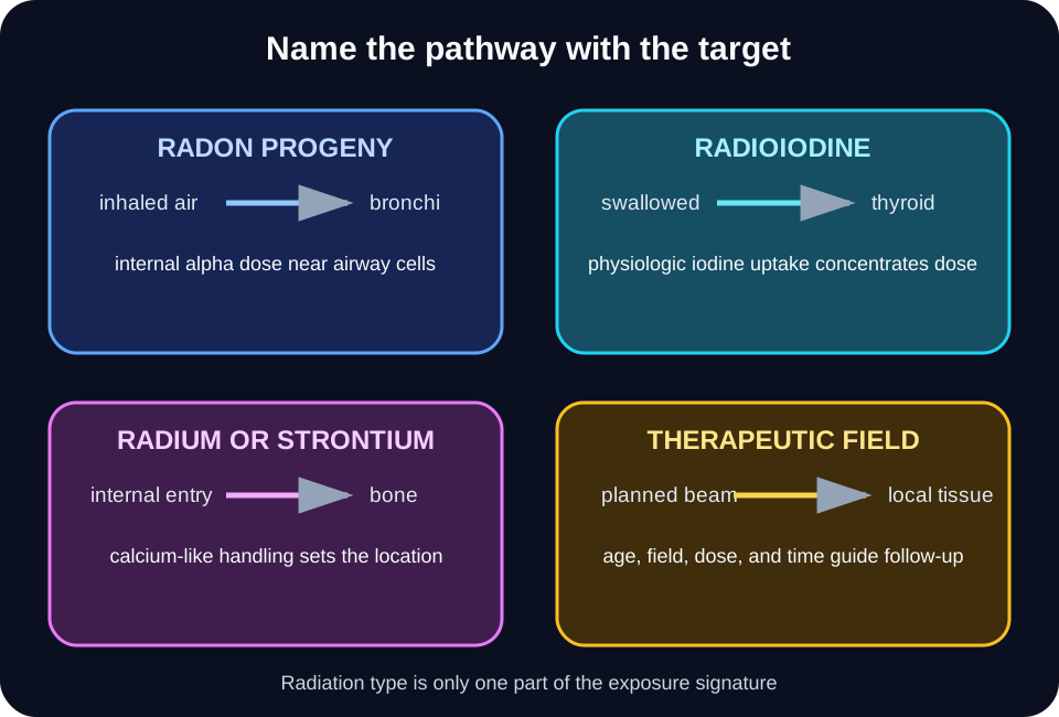
Radon is a gas produced in uranium decay. Its short-lived progeny can deposit in airways and emit alpha particles near bronchial epithelium. The established outcome is increased lung cancer risk, especially when combined with smoking. Radon means internal airway dose, not external alpha reaching the lung. [5]
After the Chernobyl accident, radioiodine entered food pathways and was concentrated by the thyroid. A clear increase in thyroid cancer was observed among people exposed as children or adolescents. That evidence does not justify claiming that every reactor event produces the same population pattern or that every later thyroid tumor is attributable to radiation. Age, dose, and pathway matter. [6]
Potassium iodide saturates thyroid iodine uptake and can reduce thyroid dose from radioactive iodine when public-health authorities advise it at the appropriate time. It does not protect the rest of the body, does not block external radiation, and does not treat exposure to other radionuclides. KI protects only the thyroid from radioiodine uptake. [7]
Chemistry directs some internal sources. Radium can behave like calcium and deposit in bone. Strontium-90 can also enter bone and deliver beta dose near skeletal and marrow tissues. Thorium dioxide contrast historically remained in reticuloendothelial organs and was linked to hepatic malignancies. Biokinetics selects the target before radiation quality shapes microscopic injury. [3]
Therapeutic radiation is deliberately localized, yet nearby normal tissue receives some dose. Years later, survivors can have organ-specific late effects and second primary cancers whose pattern depends on age at treatment, field, dose, other therapies, and baseline risk. Field and age guide risk-based follow-up rather than a single universal schedule. [11][12]
Test a treatment-field inference
A breast tumor years after chest radiation in adolescence deserves evaluation as a new primary cancer and activates risk-based survivorship guidance. It should not be labeled treatment-caused from history alone, and it should not automatically be called recurrent lymphoma. Classify first, attribute cautiously.
The safe transfer rule is to name the pathway with the disease. Say inhaled radon progeny and lung dose, swallowed radioiodine and thyroid dose, or a prior treatment field and exposed normal tissue. Specific exposure language prevents false shortcuts such as “alpha always means skin” or “photons always mean leukemia.”
Pair the pathway with the target organ. Radiation type alone is insufficient. [3][5][6][11][12]
Try it here · Checkpoint 2 of 3
Make your prediction before reading the choices. A first attempt is just a starting point.
Case 15
Show answer and explanations for case 15
A. Every leukemia appears before every solid cancer after radiation (Why this does not fit)
Population curves overlap, and individual diagnoses vary by dose, age, disease subtype, and competing causes. An average earlier excess is not a strict personal timetable.
Reasoning steps for option A
Why does the graph make leukemia-first seem like a rule?
The leukemia excess appears earlier than the solid-cancer excess.
Why is an earlier average not a fixed order for each person?
The time distributions overlap and vary with dose, age, and subtype.
B. All lymphoma subtypes respond identically to radiation exposure (Why this does not fit)
Hematologic malignancies have distinct biology and epidemiologic patterns. Combining every lymphoma into one response erases meaningful differences.
Reasoning steps for option B
Why might grouping all lymphomas seem reasonable?
Lymphomas share a lymphoid origin and are often tabulated together.
Why does lumping them hide important patterns?
Hematologic malignancies differ in biology and in how clearly they are associated with radiation.
C. A cancer absent from one survivor dataset can never be radiation-associated in any other population, exposure route, or dose range (Why this does not fit)
Lack of a detected increase may reflect power, classification, dose, age, or exposure context. It does not create a universal biological exemption.
Reasoning steps for option C
Why might a null finding in one cohort seem conclusive?
Atomic bomb survivor data are among the strongest radiation evidence available.
Why does one null result not create a universal exemption?
Power, classification, dose range, age, and exposure route can all hide an association.
D. The graph supports different average time patterns, not an invariant sequence for every person or cancer (Best answer)
Cohort data can show that some leukemias have shorter average latency than many solid cancers. It cannot guarantee the first diagnosis in each individual or establish a universal exempt malignancy across every exposure setting.
Reasoning steps for option D
What can cohort curves legitimately show?
Average patterns, such as a shorter mean latency for some leukemias than for many solid cancers.
What can they not show?
A guaranteed first diagnosis for each person or a cancer that is exempt in every exposure setting.
Takeaway: Cohort data describe disease-specific risk patterns and distributions, not a fixed personal timetable or universal exception.
Use time to separate acute syndromes, late tissue injury, and cancer
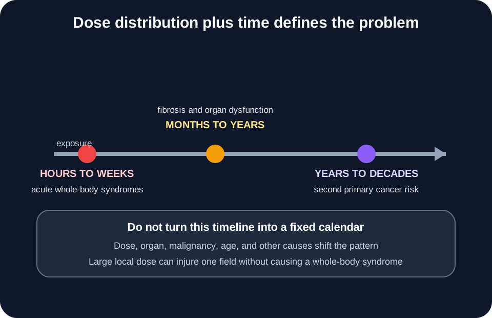
Acute radiation syndrome requires a substantial penetrating dose to most or all of the body over a short interval. A typical sequence is prodromal symptoms, a variable latent phase, and then organ-specific illness. The dominant syndrome depends on dose and distribution. Whole-body dose and timing are essential; a local skin exposure is not acute radiation syndrome. [10]
Pattern
Dominant injury
Typical clinical direction
PatternHematopoietic
Dominant injuryMarrow stem and progenitor cell loss
Dominant injuryCrypt and barrier failure at higher whole-body dose
Typical clinical directionSevere diarrhea, fluid loss, sepsis plus marrow injury
PatternNeurovascular
Dominant injuryExtreme whole-body exposure
Typical clinical directionRapid neurologic and circulatory collapse
The latent phase is not a fixed two- or three-week interval. Its duration shortens as dose rises and can be minimal after extreme exposure. Lymphocyte kinetics, serial blood counts, symptoms, physical dosimetry, and exposure reconstruction are interpreted together. A quiet interval can be deceptive, but its duration alone cannot assign an exact dose.
Long-term cohort data show excess leukemia and solid cancers after acute radiation exposure, but the patterns differ by disease, age, sex, dose, and time. Many leukemias show a shorter average latency than many solid tumors, yet there is no reliable rule that leukemia always appears first or that one named malignancy is universally exempt. Population patterns are not clocks. [8][9]
Time helps classify the problem, but dose distribution, organ, age, and disease type shift the pattern. [8][9][10][11]
Radiotherapy divides a prescribed course when that schedule best balances tumor control and normal-tissue injury. Intervals can permit repair in normal tissue, while tumor oxygenation, cell-cycle distribution, and repopulation also influence response. The clinical schedule is site-specific and planned, not a generic “smaller is always safer” rule. Fractionation changes biologic effect. [13]
Integrate dose distribution and time
A patient with local rectal telangiectasia and fibrosis years after pelvic treatment has a late tissue reaction in the field. A different new cancer in that field years later is a second primary candidate. The decisive split is progressive tissue injury versus malignant clone.
Finish every vignette with the same synthesis: identify the source and route, estimate the tissue dose distribution, decide whether the immediate problem is cell depletion or surviving mutation, and place the event on an acute or late timeline. That source-to-tissue framework is more reliable than memorizing one cancer for each radiation symbol.
Try it here · Checkpoint 3 of 3
Make your prediction before reading the choices. A first attempt is just a starting point.
Case 21
Show answer and explanations for case 21
A. Recurrent pelvic cancer invading the rectum (Why this does not fit)
Recurrence can cause bleeding, but the endoscopic and histologic findings show diffuse field injury without malignant cells. No invasive mass is described.
Reasoning steps for option A
Why might bleeding after pelvic cancer treatment suggest recurrence?
Recurrent tumor can invade the rectum and bleed.
What in the endoscopy and biopsy argues against recurrence?
Diffuse telangiectatic mucosa with fibrosis and no malignant cells or mass.
B. A radiation-associated rectal adenocarcinoma (Why this does not fit)
A second primary cancer requires malignant cells or a tumor, neither of which is present. Radiation history does not convert every late symptom into cancer.
Reasoning steps for option B
Why might a new rectal cancer be suspected in an irradiated field?
Second primary cancers can arise in treated tissue.
What finding is missing for that diagnosis?
Malignant cells or a tumor; the biopsy shows only fibrosis and vascular injury.
C. Chronic radiation proctopathy (Best answer)
Late mucosal and microvascular injury in the treated field can produce ischemia, fibrosis, telangiectasia, and bleeding. The biopsy shows those changes without a malignant population.
Reasoning steps for option C
What do telangiectasia, fibrosis, and obliterative vascular injury indicate?
Late microvascular damage and ischemia in the irradiated rectal mucosa.
Why does that pattern fit chronic radiation proctopathy?
It is a field-limited late tissue reaction that causes bleeding, found 3 years after pelvic treatment.
D. Acute gastrointestinal radiation syndrome (Why this does not fit)
Acute syndrome follows a large near-whole-body dose over a short interval and presents within days. This lesion is local, delayed for years, and confined to the prior pelvic field.
Reasoning steps for option D
Why might gut radiation injury point to the acute syndrome?
Gastrointestinal radiation syndrome also involves bowel injury.
What separates this lesion from that syndrome?
The acute syndrome follows near whole-body dose within days, while this is local and years later.
Takeaway: Late radiation proctopathy is a field-limited tissue reaction with vascular injury and fibrosis, not cancer by default.
Becquerels count decays per second. They describe the source activity on the glove, not energy absorbed by the skin.
Reasoning steps for option A
Why is the megabecquerel value on the glove the first number that catches the eye?
It is the largest figure in the spill report and it describes the radioactive material itself.
What does a becquerel count, and is that the skin's absorbed energy?
It counts decays per second in the source, so it describes activity on the glove rather than energy deposited in skin.
B. 0.02 mGy (Best answer)
Gray units quantify absorbed energy per kilogram. The dosimeter estimate therefore directly describes the skin absorbed dose.
Reasoning steps for option B
Which unit expresses energy deposited per kilogram of tissue?
The gray, one joule per kilogram, so a milligray value is an absorbed-dose figure.
Which reported number was measured for the skin beneath the glove?
The dosimeter estimate of 0.02 mGy, which directly answers the absorbed-energy question.
C. 2 mSv (Why this does not fit)
Sievert units apply radiation and tissue weighting for protection estimates. No 2 mSv value was measured, and it is not the direct absorbed-energy quantity requested.
Reasoning steps for option C
Why does a sievert value sound right for a worker's exposure record?
Occupational dose reports are often written in millisieverts, so the unit feels familiar.
What does the sievert add that the question did not ask for?
It weights dose for radiation type and tissue sensitivity for protection estimates, and no 2 mSv value was measured here.
D. 1.71 MeV (maximum beta energy) (Why this does not fit)
The maximum beta energy of phosphorus-32 characterizes each emission and its range in tissue, but it does not state the total energy absorbed per kilogram of this skin. The needed quantity is the measured absorbed dose.
Reasoning steps for option D
Why does the phosphorus-32 emission energy seem relevant to skin injury?
A maximum beta energy of 1.71 MeV explains why these electrons can reach viable skin layers.
Why can an emission energy not serve as the skin dose?
It describes one decay's particle energy, not the total energy absorbed per kilogram of skin during the spill.
Takeaway: Activity describes the source; absorbed dose in gray describes energy deposited in tissue.
A. He remains internally contaminated until absorbed cesium is excreted from blood and soft tissues (Why this does not fit)
Internal contamination requires cesium to enter the body. The sealed source and negative surveys provide no evidence that radioactive material entered him.
Reasoning steps for option A
Why might cesium-137 raise concern about internal contamination?
Dispersed cesium can be inhaled or swallowed and then distributes through soft tissues.
What evidence shows that no cesium entered this worker?
The source stayed sealed and repeated skin and clothing surveys found no radioactive material.
B. He is externally contaminated but not irradiated (Why this does not fit)
External contamination means radioactive material is present on the body. The surveys are negative, while the badge proves irradiation occurred.
Reasoning steps for option B
Why could a nearby unshielded source suggest contamination on the skin?
The worker was physically close to radioactive material, which can blur the idea of material on him.
What do the negative surveys and the badge reading establish together?
No material is on his body, yet photons reached him, which is irradiation without contamination.
C. He was externally irradiated, and that exposure ended when he left the source (Best answer)
Photons from the sealed source crossed his body while he was nearby. Once distance separated him from the source, irradiation stopped because no radioactive material accompanied him.
Reasoning steps for option C
What does the badge reading show about the photons from the sealed source?
Penetrating photons crossed his body during the 40 seconds he stood near it.
Why did his exposure stop once he walked away?
Irradiation depends on the external source; with no radioactive material carried on or in him, distance ended it.
D. He will continue emitting photons at the recorded dose rate (Why this does not fit)
A person exposed to an external beam does not become a photon source from that exposure. Continued emission would require retained radioactive material, which was not detected.
Reasoning steps for option D
Why might someone expect an exposed person to keep emitting radiation?
Patients given therapeutic radionuclides do emit radiation for a period after treatment.
Why does that expectation not apply to this worker?
He never retained a radionuclide; passing through an external photon field does not make the body radioactive.
Takeaway: External irradiation can occur without contamination and ends when the external source is no longer present.
A. A measurable increase in leukemia from marrow irradiation (Why this does not fit)
Leukemia would require meaningful marrow dose. External alpha particles cannot cross intact skin to reach marrow in this scenario.
Reasoning steps for option A
Why does leukemia come to mind after any radiation incident?
Leukemia is one of the best-documented cancers after marrow irradiation.
Could alpha particles from a sealed source on a gloved hand reach marrow?
No; external alpha particles stop in the outer skin and never reach bone marrow.
B. A predictable future lung cancer from bronchial deposition (Why this does not fit)
Bronchial dose requires inhalation of an alpha emitter or its progeny. No material escaped the sealed source or entered the airway.
Reasoning steps for option B
Why does polonium-210 suggest a lung hazard?
Inhaled alpha emitters deposited in airways irradiate bronchial epithelium.
What in this event rules out airway deposition?
The source remained sealed and recovered, and surveys found no escaped material to inhale.
C. A localized thyroid cancer from alpha uptake (Why this does not fit)
Thyroid uptake is relevant to radioiodine, not sealed polonium on a gloved hand. The source did not enter the body.
Reasoning steps for option C
Why might thyroid cancer be proposed as a radiation outcome here?
Thyroid cancer is a well-known late effect after some radiation releases.
Which element actually drives thyroid uptake?
Iodine; sealed polonium on a gloved hand never entered the body or the thyroid.
D. No meaningful deep-organ dose from the alpha particles (Best answer)
Intact outer skin blocks external alpha particles. With a sealed source, no wound, and negative surveys, deep organs did not receive a meaningful alpha dose.
Reasoning steps for option D
How far do alpha particles from an external source penetrate?
They stop within the dead outer layer of skin and glove material.
What do the sealed source, lack of a wound, and negative surveys add?
They exclude internal entry, so deep organs received no meaningful alpha dose.
Takeaway: External alpha radiation does not reach deep organs through intact skin; internal entry changes the hazard.
A. Inhaled radon progeny deposited near bronchial epithelium (Best answer)
Radon decay products can be inhaled and deposited in airways, where alpha emissions irradiate nearby bronchial cells. This pathway is linked to increased lung cancer risk.
Reasoning steps for option A
How does radon in house air reach bronchial cells?
Its short-lived decay products are inhaled and deposit on the airway lining.
Why does that deposition explain lung cancer in a lifelong nonsmoker?
Alpha emissions from deposited progeny irradiate bronchial epithelium over decades, raising lung cancer risk.
B. External alpha particles crossed the chest wall and reached the lung (Why this does not fit)
Alpha particles in room air do not cross skin and chest wall to irradiate the lung. The risk arises after inhalation and airway deposition.
Reasoning steps for option B
Why could the long residence make an external source seem enough?
Thirty years in a radon-filled house suggests the whole body was bathed in radiation.
Can alpha particles in room air cross the chest wall?
No; they stop in the outer skin, so lung dose requires inhalation and airway deposition.
C. Ingested radon selectively concentrated in thyroid follicles (Why this does not fit)
The thyroid concentrates iodine, not radon progeny as a defining pathway. The epidemiologic association in this setting is lung cancer after inhalation.
Reasoning steps for option C
Why might a gland-targeting route come up for an environmental radionuclide?
Radioiodine releases are famous for concentrating in the thyroid.
Does radon behave like iodine, and where is this tumor?
No; radon is an inert gas whose progeny are inhaled, and the mass is in the right upper lobe.
D. Whole-body gamma radiation chronically suppressed marrow immunity (Why this does not fit)
Residential radon risk is not explained by a persistent whole-body gamma field or immune suppression. It is a local airway alpha-dose problem.
Reasoning steps for option D
Why might chronic whole-body exposure seem to explain a cancer years later?
Low-level background radiation reaches everyone over a lifetime.
What makes residential radon a local rather than a whole-body problem?
Its risk comes from alpha dose to airway cells after inhalation, not from marrow suppression by gamma rays.
Takeaway: Radon risk comes from inhaled progeny delivering alpha dose near bronchial epithelium.
A. All beta particles stop in dead skin and cannot injure viable epidermis (Why this does not fit)
Beta energy varies, and some beta particles can penetrate into viable skin and cause injury. Treating every beta source like an alpha source ignores that wider range.
Reasoning steps for option A
Why might the forearm spill seem harmless?
Beta particles are often taught as stopping in the outer skin.
Why is that generalization unsafe for a phosphorus-32 spill?
Its energetic beta particles can pass the dead layer and injure viable epidermis.
B. Beta radiation selectively concentrates in marrow even when the source stays outside (Why this does not fit)
Organ concentration requires internal entry and biokinetics. The vignette describes external skin contamination that was washed away, not systemic uptake.
Reasoning steps for option B
Why might marrow seem to be the organ at risk?
Phosphorus-32 given internally concentrates in bone and marrow.
What in this case prevents that internal pattern?
The material stayed on the skin and was washed off, so there was no systemic uptake.
C. Beta particles can deposit dose in superficial tissue but have limited reach from the surface (Best answer)
Some beta emissions can penetrate the outer skin and injure superficial tissue. From an external forearm source, their range is limited compared with penetrating photons, so deep marrow receives far less dose.
Reasoning steps for option C
Which tissue sits within range of beta particles from a forearm spill?
The superficial skin layers directly beneath the contamination.
Why does the marrow receive far less dose than the skin?
Beta range from the surface is limited, unlike penetrating photons, and whole-body dosimetry is low.
D. The washing increased beta penetration by hydrating the skin (Why this does not fit)
Prompt decontamination lowers the time that radioactive material remains on the skin. Washing is a protective action when performed according to contamination procedures.
Reasoning steps for option D
Why might wet skin seem to change the radiation hazard?
Water on the skin could appear to help the contamination spread or soak in.
What does prompt washing actually achieve?
It removes radioactive material, shortening the time it irradiates the skin, as the falling survey shows.
Takeaway: External beta contamination can injure superficial tissue; prompt decontamination reduces duration and dose.
A. Alpha particles penetrate farther through tissue because dense ionization allows them to retain energy over a longer path (Why this does not fit)
Dense ionization causes alpha particles to lose energy quickly, which limits their range. High LET does not mean deep penetration.
Reasoning steps for option A
Why might dense ionization suggest deep travel?
More ionization per track can sound like more energy carried farther.
What happens to an alpha particle's energy as it ionizes densely?
It is spent quickly, so dense ionization shortens the range rather than extending it.
B. X-rays are nonionizing because their tracks are less dense (Why this does not fit)
X-rays are ionizing photons even though their energy deposition is more sparsely distributed than alpha tracks. Lower LET is not the same as nonionizing.
Reasoning steps for option B
Why might sparse tracks seem to mean x-rays are not ionizing?
X-ray tracks show far fewer clustered ionizations than alpha tracks.
Are x-rays ionizing even with sparse tracks?
Yes; they are ionizing photons with low linear energy transfer, not nonionizing radiation.
C. Equal gray values guarantee identical biologic effect (Why this does not fit)
Gray measures absorbed energy, but microscopic distribution and radiation quality can alter biologic effect. Equal absorbed dose does not erase LET differences.
Reasoning steps for option C
Why does equal gray sound like equal effect?
Both cultures absorbed the same energy per kilogram.
What differs at the microscopic level despite equal gray?
Alpha energy is clustered along tracks, producing complex DNA damage, so biologic effect differs.
D. Alpha particles have higher linear energy transfer along their short tracks (Best answer)
Dense ionizations per unit path define higher linear energy transfer. Alpha particles characteristically deposit energy intensely over a short range.
Reasoning steps for option D
What does linear energy transfer measure?
Energy deposited per unit length of a particle's track.
How do the observed alpha tracks fit that definition?
Densely clustered ionizations over a short path are the signature of high linear energy transfer.
Takeaway: LET describes ionization density along a track; penetration and absorbed dose are separate properties.
A. Iodine-131 was absorbed and concentrated by thyroid follicular cells (Best answer)
Dietary radioiodine enters the bloodstream and follows normal iodine handling into the thyroid. Emissions then deliver a concentrated gland dose, with young age increasing susceptibility.
Reasoning steps for option A
How does iodine-131 in milk reach the thyroid?
It is absorbed from the gut and handled like dietary iodine.
Why does that pathway concentrate dose in follicular cells?
The thyroid actively traps iodine, so the gland receives most of the emitted dose, and children are most susceptible.
B. External alpha particles from the milk crossed the neck skin (Why this does not fit)
The relevant isotope emits beta particles and photons after internal uptake, not external alpha particles crossing skin. The milk was swallowed, so the pathway is internal.
Reasoning steps for option B
Why might contaminated milk seem to irradiate from outside the neck?
The child was near contaminated food during the accident.
What emissions does iodine-131 produce, and how did it enter?
It emits beta particles and gamma rays, and it was swallowed, so the exposure was internal.
C. Iodine-131 behaved like calcium and deposited in bone (Why this does not fit)
Bone-seeking behavior is associated with elements such as strontium or radium. Iodine is handled primarily by the thyroid.
Reasoning steps for option C
Why might a bone-seeking pattern come to mind after a reactor accident?
Strontium-90 released in the same accidents deposits in bone.
Which organ handles iodine, and which fact links the tumor to it?
The thyroid; papillary carcinoma arose in the organ that concentrates iodine.
D. Whole-body photon dose selectively mutated thyroid cells while sparing every other organ exposed during the accident (Why this does not fit)
A reactor accident can involve several exposure pathways, but this vignette specifically supplies contaminated milk and iodine-131. Organ concentration, not selective whole-body photon action, explains the pattern.
Reasoning steps for option D
Why might general reactor photon exposure seem a sufficient explanation?
Accidents can expose people to external gamma radiation as well as contaminated food.
What in the vignette points to organ concentration instead?
The supplied pathway is iodine-131 in milk, which delivers a concentrated thyroid dose that a uniform field cannot.
Takeaway: Radioiodine follows normal iodine physiology, concentrating dose in thyroid tissue, especially relevant after childhood exposure.
A. It neutralizes every radionuclide in the bloodstream (Why this does not fit)
Potassium iodide does not chemically neutralize cesium, strontium, or other radionuclides. Its protective effect depends on thyroid iodine transport.
Reasoning steps for option A
Why might a parent hope the tablets are a general antidote?
They are handed out during a radiation emergency, which suggests broad protection.
How does potassium iodide actually work?
It saturates thyroid iodine uptake; it does not neutralize cesium or other radionuclides in blood.
B. It blocks thyroid uptake of radioactive iodine but does not shield other organs or radionuclides (Best answer)
Stable iodine reduces thyroid uptake of radioactive iodine when taken at the advised time. It does not create whole-body shielding and does not protect against cesium or external photons.
Reasoning steps for option B
What does stable iodide do to thyroid uptake of radioiodine?
It fills the thyroid's iodine uptake so radioactive iodine is taken up far less.
What does this release include that the tablets cannot address?
Cesium and any external photon exposure, which potassium iodide does not block.
C. It prevents acute radiation syndrome from an external photon dose (Why this does not fit)
Acute radiation syndrome depends on absorbed whole-body dose. KI cannot attenuate an external photon field or reverse tissue energy deposition.
Reasoning steps for option C
Why might the tablets seem to protect against acute radiation illness?
They are the best-known protective drug used in radiation emergencies.
What causes acute radiation syndrome, and can potassium iodide change it?
A large whole-body absorbed dose; iodide neither shields external photons nor reverses energy already deposited.
D. It should be taken routinely whenever any radiation detector alarms (Why this does not fit)
KI has risks and should be used under public-health or clinical direction when radioiodine exposure is relevant. A detector alarm alone does not identify the radionuclide or justify thyroid blockade.
Reasoning steps for option D
Why might taking tablets at any alarm seem prudent?
Early use is important when radioiodine exposure is expected.
Why is an alarm alone not an indication?
It does not identify radioiodine, and potassium iodide carries risks, so use follows public-health direction.
Takeaway: Potassium iodide is a time-sensitive thyroid blocker for radioiodine, not a general radiation antidote.
A. Only the skin receives dose because beta radiation is always superficial (Why this does not fit)
Superficial injury describes many external beta exposures. Once the emitter is inside bone, its range begins beside living skeletal and marrow tissue.
Reasoning steps for option A
Why might beta radiation seem to injure only skin?
External beta sources mainly cause superficial skin injury.
What changes once strontium-90 is retained in bone?
The emitter now sits inside the skeleton, so its range begins beside bone and marrow.
B. The thyroid receives the dominant dose through iodine transport (Why this does not fit)
The thyroid transporter concentrates iodine, not strontium as its defining substrate. The measured activity is in bone.
Reasoning steps for option B
Why might a gland-concentrating route come to mind for an ingested radionuclide?
Swallowed radioiodine concentrates in the thyroid.
Where do the follow-up measurements place the activity?
In mineralized bone, because strontium follows calcium rather than iodine.
C. Bone surfaces and adjacent marrow receive internal beta irradiation (Best answer)
Strontium can follow calcium pathways and become incorporated into bone. A retained beta emitter then deposits energy in nearby skeletal tissue and marrow rather than from the body surface.
Reasoning steps for option C
Why does strontium end up in mineralized bone?
It behaves chemically like calcium and is incorporated into the skeleton.
Which tissues then receive beta dose over time?
Bone surfaces and adjacent marrow, irradiated from within by the retained emitter.
D. No tissue receives dose after the material enters bone (Why this does not fit)
Radioactive decay continues while the isotope remains active and retained. Incorporation into bone changes location; it does not stop emission.
Reasoning steps for option D
Why might incorporation into bone seem to neutralize the material?
Mineral binding could look like safe storage.
Does binding in bone stop radioactive decay?
No; decay continues while the isotope is retained, so nearby tissue keeps receiving dose.
Takeaway: Internal radionuclide chemistry determines deposition; retained strontium can irradiate bone and nearby marrow.
A. External alpha particles penetrated the face to reach the mandible (Why this does not fit)
Alpha particles outside the body cannot cross skin and soft tissue to reach the jaw. The brush-to-mouth behavior matters because it creates internal contamination.
Reasoning steps for option A
Why might radium on a brush seem to irradiate the face from outside?
The painter held the radium-coated brush close to her face every day.
Can external alpha particles cross skin to reach the jaw?
No; lip-pointing the brush mattered because she swallowed radium, creating internal contamination.
B. Radium selectively accumulated in thyroid follicles (Why this does not fit)
Thyroid concentration is characteristic of iodine handling. Radium's relevant biologic resemblance is to calcium and bone deposition.
Reasoning steps for option B
Why might a gland-concentrating route be considered for an ingested radionuclide?
Radioiodine famously concentrates in the thyroid after ingestion.
Which chemical behavior of radium explains the jaw lesion?
Radium mimics calcium and deposits in bone, not in thyroid follicles.
C. The lesion proves every radium exposure produces cancer (Why this does not fit)
An exposure history can increase risk without making disease inevitable or proving individual causation by itself. Dose, retention, latency, and alternative causes still matter.
Reasoning steps for option C
Why might a destructive lesion seem to prove inevitable harm?
The occupational history and the tumor fit the well-known dial painter story.
Why is the absolute claim still unjustified?
Exposure raises risk without making cancer certain; dose, retention, latency, and other causes still matter.
D. Radium entered through ingestion and deposited in bone (Best answer)
Radium can follow calcium pathways after internal entry and remain near skeletal tissue. Its emissions then deliver local dose over time, providing a coherent exposure-to-target pathway.
Reasoning steps for option D
How did radium get inside the dial painter?
She shaped radium-coated brushes with her lips and swallowed small amounts repeatedly.
Why does that route explain a jaw lesion decades later?
Radium deposits in bone like calcium, and its retained alpha emissions irradiate nearby skeletal tissue for years.
Takeaway: Radium became dangerous after ingestion and skeletal deposition, not by external alpha penetration.
A. Long-lived thorium deposits delivered internal alpha dose to hepatic tissue (Best answer)
Thorium dioxide was retained in reticuloendothelial organs for years. Alpha emissions from deposits beside liver cells created prolonged local irradiation associated with hepatic malignancies.
Reasoning steps for option A
Where did thorium dioxide contrast stay after injection?
In reticuloendothelial organs such as the liver and spleen, seen here as retained dense material.
How do those deposits lead to hepatic angiosarcoma decades later?
The thorium emits alpha particles beside liver cells for the rest of life, creating prolonged local irradiation.
B. A brief external x-ray exposure during each angiogram created the entire later hepatic malignancy risk (Why this does not fit)
Diagnostic x-rays may add a small photon dose, but they do not explain retained dense material or the distinctive historic thorium association. The persistent internal source dominates this vignette.
Reasoning steps for option B
Why might the imaging procedure itself seem responsible?
Angiography uses x-ray beams, which are a known radiation exposure.
What does the retained liver material show that brief x-rays cannot explain?
A persistent internal alpha source that keeps irradiating the liver long after any imaging beam.
C. Thorium caused only chemical liver toxicity without radiation (Why this does not fit)
Thorium dioxide's historic carcinogenicity is tied to radioactive decay and tissue irradiation. A purely chemical mechanism does not explain the alpha-emitter evidence.
Reasoning steps for option C
Why might a purely toxic mechanism seem plausible?
Retained foreign material can cause inflammation and fibrosis.
What makes thorium contrast carcinogenic beyond chemistry?
Its radioactive decay emits alpha particles that irradiate the tissues retaining it.
D. The tumor arose because alpha particles crossed the abdominal wall (Why this does not fit)
External alpha particles cannot reach the liver through intact tissue. The injection bypassed that barrier and placed the emitter inside the target organ.
Reasoning steps for option D
Why might an external path seem possible for a deep tumor?
Thorium is an alpha emitter and the liver lies close under the abdominal wall.
How did the emitter actually reach the liver?
It was injected and retained inside the organ, bypassing skin that stops external alpha particles.
Takeaway: Historic thorium contrast produced prolonged internal alpha irradiation in the organs that retained it.
A. Both are stochastic because neither clinical effect becomes visible immediately after the radiation exposure (Why this does not fit)
Delayed timing does not define stochasticity. Fibrosis can be delayed yet still show threshold-like dose dependence and increasing severity.
Reasoning steps for option A
Why might delayed onset suggest a stochastic effect?
Both fibrosis and cancer appear months to years after exposure.
Does timing define the category?
No; fibrosis is delayed yet threshold-like, with severity that rises with dose.
B. Both are tissue reactions because radiation damaged DNA (Why this does not fit)
DNA damage contributes to both, but cancer arises from a surviving altered clone rather than broad cell depletion. Shared initiation does not make the outcomes the same category.
Reasoning steps for option B
Why might shared DNA damage suggest a single category?
Both outcomes start with radiation injury to DNA.
How do the outcomes differ after that shared start?
Fibrosis reflects loss of many cells, while cancer arises from one surviving altered clone.
C. Fibrosis is a tissue reaction; cancer is a stochastic outcome (Best answer)
Fibrosis reflects injury to enough normal cells and supporting structures to produce a field effect whose severity rises with dose. Cancer risk is modeled probabilistically because a surviving altered cell can found a clone.
Reasoning steps for option C
Which features of the fibrosis make it a tissue reaction?
It needs injury to many cells and its severity increases with dose in the treated field.
Why is the later cancer classified as stochastic?
Its probability, not severity, rises with dose because a single altered cell can found a tumor.
D. Fibrosis is stochastic; cancer has a fixed threshold (Why this does not fit)
This reverses the standard protection framework. Fibrosis is the threshold-like tissue reaction, while cancer is modeled without a risk-free threshold for protection.
Reasoning steps for option D
Why might the categories be reversed by mistake?
Cancer seems severe and dose-driven, while fibrosis can look variable.
Which outcome shows a threshold in the protection framework?
Fibrosis does; cancer risk is modeled without a risk-free threshold.
Takeaway: Tissue reactions reflect sufficient cell injury; cancer risk is treated as a probabilistic clonal outcome.
A. The reading proves any future cancer was caused by occupational radiation (Why this does not fit)
A badge dose can inform population-level risk estimates but cannot identify the cause of one future cancer. Baseline cancer incidence and other exposures prevent that certainty.
Reasoning steps for option A
Why might a documented badge reading seem to settle causation?
It is a precise, personal record of occupational dose.
What prevents assigning a future cancer to 38 mSv?
Baseline cancer is common and a small added risk cannot be separated in one person.
B. Protection models treat added cancer risk as small and dose-related, while individual causation remains uncertain (Best answer)
Radiation protection uses dose-related risk models to guide minimization of avoidable exposure. At this dose range, any added individual risk is small and a later cancer could not be uniquely attributed from the badge value alone.
Reasoning steps for option B
How do protection models treat risk at this cumulative dose?
As a small added probability that rises with dose, which guides keeping exposure low.
Why does individual causation remain uncertain?
At tens of millisieverts the added risk is small beside baseline cancer risk, so one later tumor cannot be traced to the badge.
C. The dose is below acute-syndrome levels, so cancer risk is exactly zero (Why this does not fit)
Acute tissue reactions and late cancer risk are different endpoints. Absence of acute illness does not establish zero added long-term risk.
Reasoning steps for option C
Why might the absence of acute illness sound reassuring about cancer?
The dose is far below levels that cause acute radiation syndrome.
Why does that not make cancer risk exactly zero?
Acute syndromes and late cancer are separate endpoints; protection models assume no risk-free threshold.
D. The effective dose predicts the exact organ and date of a future malignancy (Why this does not fit)
Effective dose is a protection quantity for comparing risk across exposures, not a personalized forecast. It cannot specify which organ will develop cancer or when.
Reasoning steps for option D
Why might effective dose sound like a personal forecast?
It is a single number calculated for this clinician's exposure.
What is effective dose designed to do?
Compare exposures for protection purposes; it cannot predict which organ or when cancer appears.
Takeaway: Low-dose protection models guide risk reduction but cannot prove the cause of an individual future cancer.
A. Hematopoietic acute radiation syndrome (Best answer)
A several-gray whole-body dose can destroy marrow progenitors while mature circulating cells persist temporarily. The latent interval followed by pancytopenia, infection, and bleeding is the defining hematopoietic pattern.
Reasoning steps for option A
What does 3 Gy of penetrating whole-body dose do to marrow?
It destroys stem and progenitor cells, while mature circulating cells last for a while.
How does the 12-day course fit that marrow injury?
Vomiting, a quiet interval, then fever, petechiae, and pancytopenia is the hematopoietic pattern.
B. Gastrointestinal acute radiation syndrome (Why this does not fit)
Gastrointestinal syndrome at higher doses features severe diarrhea, barrier failure, fluid loss, and sepsis, usually with a shorter latent interval. This patient instead has dominant marrow failure without severe enteric findings.
Reasoning steps for option B
Why might the early vomiting suggest gut injury?
Gastrointestinal symptoms appear on the evening of exposure.
What distinguishes true gastrointestinal syndrome from this course?
It needs higher doses and causes severe diarrhea, fluid loss, and sepsis after a short latent period.
C. Neurovascular acute radiation syndrome (Why this does not fit)
Neurovascular syndrome follows extreme exposure and progresses rapidly with severe neurologic and circulatory collapse. A 12-day interval of relative wellness is incompatible with that course.
Reasoning steps for option C
Why might severity of exposure suggest the neurovascular form?
The dose is large and the worker becomes seriously ill.
Why is a 12-day quiet interval incompatible with it?
Neurovascular syndrome follows extreme doses and progresses to collapse within hours to days.
D. Localized radiation dermatitis (Why this does not fit)
Localized skin injury does not explain simultaneous neutropenia, anemia, and thrombocytopenia. The dosimetry documents whole-body rather than field-limited exposure.
Reasoning steps for option D
Why might an erythema-type diagnosis be considered?
Skin injury is a common radiation complaint in workers.
What findings cannot come from local skin injury?
Simultaneous neutropenia, anemia, and thrombocytopenia after a documented whole-body dose.
Takeaway: A penetrating whole-body dose followed by a latent interval and pancytopenia indicates hematopoietic acute radiation syndrome.
A. Isolated thyroid follicular-cell mutation (Why this does not fit)
A thyroid mutation could contribute to a late clonal outcome but cannot produce rapid diarrhea, dehydration, and bacteremia. The time course and organ pattern are acute.
Reasoning steps for option A
Why might a mutation-type answer seem attractive after heavy exposure?
Radiation causes DNA mutations, and thyroid mutation is a known late effect.
Why can it not drive this illness?
A mutation is a late clonal event and cannot cause diarrhea, dehydration, and bacteremia within days.
B. Slow pulmonary fibrosis from scattered photon dose (Why this does not fit)
Fibrosis is a late normal-tissue reaction developing over months or years. It does not explain an acute gastrointestinal barrier collapse.
Reasoning steps for option B
Why might lung fibrosis be considered after a large photon dose?
Fibrosis is a known normal-tissue reaction to high radiation doses.
Why does the timing exclude it?
Fibrosis develops over months to years, not as an enteric collapse within days.
C. Intestinal crypt stem-cell loss with mucosal barrier failure (Best answer)
At this high near-whole-body dose, rapidly dividing crypt cells are depleted. The resulting denuded barrier causes diarrhea, fluid loss, and bacterial translocation, while marrow injury occurs concurrently.
Reasoning steps for option C
What does 12 Gy to nearly the whole body do to the intestine?
It depletes rapidly dividing crypt stem cells so the lining is not replaced.
How does that loss produce the clinical picture?
The denuded mucosa leaks fluid and lets bacteria enter blood, while marrow failure develops alongside.
D. Selective destruction of mature erythrocytes in circulation alone (Why this does not fit)
Radiation does not primarily create this syndrome by instantly clearing red cells. Stem and progenitor loss plus intestinal injury drive the delayed cytopenias and acute enteric findings.
Reasoning steps for option D
Why might falling blood counts suggest direct red-cell destruction?
Anemia and cytopenias appear in the same illness.
Why is that mechanism wrong?
Radiation kills stem and progenitor cells, and counts fall later; the dominant problem here is intestinal barrier failure.
Takeaway: At very high whole-body dose, intestinal crypt failure causes severe diarrhea and sepsis while marrow injury also develops.
A. Hematopoietic acute radiation syndrome caused by extensive marrow stem-cell depletion (Why this does not fit)
Marrow syndrome would produce lymphocyte decline and later multilineage cytopenias after a broad penetrating exposure. A hand-only field cannot injure enough active marrow to cause that systemic pattern.
Reasoning steps for option A
Why might a high-dose beam suggest marrow failure?
The trainee received a large dose from a strong beam.
Why can a hand-only field not cause hematopoietic syndrome?
It exposes too little active marrow, and blood counts are normal.
B. A localized radiation tissue reaction, not acute radiation syndrome (Best answer)
The beam delivered a high dose to one hand and produced a local skin response. Acute radiation syndrome requires a substantial penetrating dose to most or all of the body, which the dosimetry excludes.
Reasoning steps for option B
What did the dosimetry show about dose distribution?
A substantial dose to one hand with negligible whole-body dose.
How should erythema and tenderness of that hand be classified?
As a localized tissue reaction in the exposed field, not acute radiation syndrome.
C. Gastrointestinal acute radiation syndrome (Why this does not fit)
Gastrointestinal syndrome requires high dose to a large portion of the body and presents with severe enteric symptoms. The trainee has normal gastrointestinal function.
Reasoning steps for option C
Why might any severe radiation injury suggest gut involvement?
Gastrointestinal syndrome is a classic high-dose radiation illness.
What excludes it here?
It needs high dose to much of the body with severe enteric symptoms, and gut function is normal.
D. A stochastic cancer endpoint (Why this does not fit)
The current erythema is a dose-dependent tissue reaction appearing in the exposed field. Cancer risk is a separate late probabilistic issue, not the diagnosis of this immediate lesion.
Reasoning steps for option D
Why might the trainee worry about cancer?
Any radiation exposure carries a possible late cancer risk.
What is the current hand lesion, and how does it differ from cancer risk?
It is a dose-dependent tissue reaction now; cancer risk is a separate probabilistic late question.
Takeaway: Acute radiation syndrome depends on large-body penetrating exposure; a high local dose can instead cause a field-limited tissue reaction.
A. No breast imaging is needed unless a mass appears (Why this does not fit)
Waiting for symptoms abandons surveillance in a group with recognized excess risk after young-age chest exposure. Screening aims to detect disease before a palpable mass develops.
Reasoning steps for option A
Why might waiting for a lump seem acceptable in a well woman?
She is 34 and asymptomatic, and screening is often thought to start later.
Why is symptom-triggered imaging inadequate for her?
Chest radiation at 15 raises later breast cancer risk, and surveillance aims to find cancer before a mass.
B. Routine population screening is always sufficient after any form, dose, field, or age of therapeutic radiation exposure (Why this does not fit)
Radiotherapy fields and age at exposure alter later organ risk. Applying one average-risk schedule ignores the direct chest-treatment history.
Reasoning steps for option B
Why might ordinary screening seem enough?
It is the standard schedule most women follow.
What part of her history changes that?
Chest irradiation during adolescence places her in a higher-risk group needing earlier or added imaging.
C. Annual whole-body scanning is required for every possible second cancer (Why this does not fit)
Whole-body scanning is not a universal survivorship strategy and adds radiation, false positives, and downstream procedures. Surveillance should be organ- and exposure-specific.
Reasoning steps for option C
Why might scanning everything seem thorough?
Survivors can develop second cancers in several organs.
Why is whole-body scanning not recommended?
It adds radiation, false positives, and procedures; surveillance is matched to organ and exposure.
D. Use specialized risk-based breast surveillance based on age at treatment and chest dose (Best answer)
Young breast tissue within a therapeutic field is associated with elevated later breast-cancer risk. Survivorship guidance uses treatment age, dose, and interval to define earlier or additional imaging rather than relying only on average-risk schedules.
Reasoning steps for option D
Which facts define her breast cancer risk?
Chest radiation at age 15 for lymphoma, now 19 years later.
How does survivorship guidance use those facts?
It sets earlier or additional breast imaging by treatment age, chest dose, and time since therapy.
Takeaway: Survivorship surveillance after therapeutic radiation should be matched to field, dose, age at exposure, and time since treatment.
A. Field concordance, plausible latency, and histologic distinction from the first cancer (Best answer)
A new sarcoma within irradiated tissue after a substantial interval and with different histology fits standard clinical reasoning for a radiation-associated second primary. The classification remains an association, not proof from one feature alone.
Reasoning steps for option A
Which three features make a sarcoma look radiation-associated?
It lies in the prior field, appears after a long interval, and has histology distinct from the first tumor.
Why do these features together support a second primary?
They fit exposure of normal tissue and argue against recurrence, though they show association rather than proof.
B. Any new mass occurring after radiotherapy, regardless of location (Why this does not fit)
A mass outside the field lacks the direct normal-tissue dose relationship central to this classification. Temporal sequence alone is too broad.
Reasoning steps for option B
Why might any later mass seem connected to prior treatment?
Radiotherapy is known to cause second cancers.
Why does location matter?
A mass outside the field lacks the normal-tissue dose that the classification depends on.
C. Identical histology to the original tumor within one year (Why this does not fit)
Identical early disease is more consistent with persistence or recurrence than a radiation-associated new sarcoma. A meaningful interval and distinct histology help establish a second primary.
Reasoning steps for option C
Why might a tumor in the same site suggest radiation cause?
It arose where the original cancer was treated.
What do identical histology and a one-year interval suggest instead?
Persistence or recurrence of the original tumor, not a new radiation-associated sarcoma.
D. A positive family history of cancer without information about field, dose, latency, or the original tumor histology (Why this does not fit)
Inherited susceptibility may explain multiple cancers but does not by itself classify a tumor as radiation-associated. The necessary exposure geometry and tissue-dose history are missing.
Reasoning steps for option D
Why might family history seem to explain multiple cancers?
Inherited syndromes predispose to several tumors, including sarcomas.
Why is it insufficient for this classification?
It says nothing about field, dose, latency, or histology, which are needed to call a tumor radiation-associated.
Takeaway: A second primary sarcoma is supported by location in the treated field, plausible latency, and histology distinct from the original tumor.
A. Central hypothyroidism from isolated pituitary failure (Why this does not fit)
Central hypothyroidism usually has a low or inappropriately normal TSH with low free T4. The markedly elevated TSH shows preserved pituitary signaling.
Cranial exposure can damage the pituitary and cause central hypothyroidism.
What does a TSH of 32 mIU/L show about the pituitary?
It is responding strongly to low thyroid hormone, which argues against central failure.
B. Primary hypothyroidism from thyroid gland injury (Best answer)
A high TSH with low free T4 means the pituitary is responding but the thyroid cannot produce adequate hormone. Prior neck and total-body exposure can cause late thyroid dysfunction.
Reasoning steps for option B
What does high TSH with low free T4 localize?
Failure of the thyroid gland with an intact pituitary response.
How does the patient's history explain the gland failure?
Neck and total-body irradiation in childhood can cause late primary hypothyroidism.
C. Acute radiation syndrome of the thyroid (Why this does not fit)
Acute radiation syndrome is a systemic illness after a large short-duration whole-body dose, not a decades-later endocrine diagnosis. Thyroid dysfunction is a late organ effect.
Reasoning steps for option C
Why might an acute radiation label be applied to a radiation-linked organ problem?
The thyroid dysfunction is a direct consequence of prior radiation.
Why is acute radiation syndrome the wrong frame?
It is a systemic illness within weeks of a large dose, not an endocrine problem decades later.
D. Radioiodine contamination still circulating from childhood (Why this does not fit)
A radionuclide exposure from childhood would not remain in circulation indefinitely. The current problem is late tissue dysfunction after past dose, not persistent blood-borne iodine.
Reasoning steps for option D
Why might persistent radioactive material be suspected?
Radionuclide exposures can leave material retained in the body.
Why is circulating radioiodine implausible here?
The exposure was external irradiation, and iodine-131 decays with an 8-day half-life, so none would remain decades later.
Takeaway: High TSH with low free T4 localizes primary thyroid failure, a recognized late effect after thyroid exposure.
A. The sessions are divided only to make the machine schedule easier (Why this does not fit)
Operational scheduling does not explain the therapeutic ratio. Dose per session and overall treatment time are selected for biologic and clinical reasons.
Reasoning steps for option A
Why might repeated sessions look like a scheduling choice?
Patients come back daily, which can resemble machine booking.
What actually sets the number of sessions?
Dose per session and treatment time change biologic effect, so the schedule reflects biology, not convenience.
B. A single large dose and divided treatment are always biologically identical (Why this does not fit)
Cell survival and normal-tissue injury depend on dose per session and time between sessions. Equal physical totals can have different biologic effects.
Reasoning steps for option B
Why might splitting a dose seem to make no difference?
The total physical dose in gray can be the same either way.
Why does dose per session change the result?
Cell survival and normal-tissue injury depend on fraction size and the interval between sessions.
C. Fractionation prevents all late normal-tissue effects (Why this does not fit)
Divided schedules can reduce selected toxicities but cannot eliminate late injury, and some tumors or sites use different fraction sizes. Treatment planning balances risks rather than guaranteeing safety.
Reasoning steps for option C
Why might fractionation seem to remove all late injury?
Dividing dose spares normal tissue compared with one large exposure.
What limit remains despite fractionation?
It lowers some toxicities but cannot eliminate late injury, and some sites use large fractions.
D. Intervals permit normal-tissue repair while tumor oxygenation, cell-cycle distribution, and repopulation also shape response (Best answer)
Dividing dose changes biologic effect rather than merely changing the calendar. Normal tissue can restore some sublethal injury between sessions, while tumor reoxygenation, cell-cycle distribution, and repopulation influence the chosen total schedule.
Reasoning steps for option D
What happens in normal tissue between sessions?
Cells repair much sublethal DNA damage before the next dose.
Which tumor processes also shape the schedule?
Reoxygenation, redistribution through the cell cycle, and repopulation influence the total course.
Takeaway: Fractionation changes biologic effect through time-dependent normal-tissue repair and tumor-response processes.
A. Evaluate the nodule normally; prior iodine-131 informs risk but does not prove its cause (Best answer)
Therapeutic radioiodine delivered thyroid dose and belongs in the risk history. However, a later nodule may be benign or malignant for many reasons, so ultrasound and indicated sampling determine diagnosis while individual causation remains uncertain.
Reasoning steps for option A
How does her iodine-131 history bear on the new nodule?
Therapeutic radioiodine gave the thyroid a substantial dose, so it belongs in her risk history.
How should the nodule be assessed?
Like any nodule, with ultrasound and sampling if indicated, since the history informs risk but does not prove cause.
B. Every thyroid nodule after iodine-131 is a treatment-caused cancer (Why this does not fit)
Most thyroid nodules are not established as treatment-caused cancers, and a nodule is not itself a malignant diagnosis. This wording confuses risk with certainty.
Reasoning steps for option B
Why might radioiodine treatment seem to guarantee a cancer?
It delivered a large radiation dose directly to the gland.
What two errors does the absolute claim make?
Most nodules are benign, and a history of exposure cannot prove the cause of one tumor.
C. Prior iodine-131 is irrelevant because internal emitters cannot affect thyroid tissue after treatment (Why this does not fit)
Radioiodine is specifically taken up by thyroid tissue and is used therapeutically for that reason. The exposure is biologically relevant even though it does not prove causation.
Reasoning steps for option C
Why might a completed treatment seem irrelevant now?
The radioiodine decayed long ago and the hyperthyroidism was treated.
Why does the exposure still matter?
The thyroid concentrated iodine-131 and received dose, so it remains part of her risk history.
D. The nodule should be assumed to be recurrent hyperthyroidism without imaging (Why this does not fit)
A focal nodule requires structural assessment rather than an endocrine label based only on past treatment. Thyroid function and nodule malignancy risk are separate questions.
Reasoning steps for option D
Why might a nodule seem like returning hyperthyroidism?
She was treated for an overactive thyroid in the past.
What does a focal nodule require instead?
Structural evaluation; thyroid function and malignancy risk are separate questions.
Takeaway: A relevant radiation history informs risk, but it neither diagnoses a nodule nor proves the cause of an individual tumor.
A. Both exposures produce identical organ doses because activity and isotope are equal despite different routes and distances from living cells (Why this does not fit)
Activity alone does not specify distance, absorption, or target tissue. The two geometries create very different energy deposition.
Reasoning steps for option A
Why might equal activity seem to guarantee equal dose?
The vignette holds the isotope and activity constant in both containers.
What does activity leave out?
Where the emitter sits relative to living cells, which decides how much energy tissue absorbs.
B. Container A gives the larger lung dose because its source is outside (Why this does not fit)
An outside alpha source lacks the range to cross skin and chest wall. Proximity in the room is less important than deposition beside airway cells.
Reasoning steps for option B
Why might the intact container seem the bigger lung hazard?
Penetrating external sources can dose deep organs from outside.
Why does that not apply to an alpha emitter?
Alpha particles cannot cross skin and chest wall, so the sealed source gives the lung essentially no dose.
C. Container B can deliver much greater bronchial dose because the source is beside living airway cells (Best answer)
Equal activity means equal decay rate, not equal tissue dose. Inhalation places the short-range alpha source next to bronchial epithelium, while the sealed external source cannot cross intact skin to reach that target.
Reasoning steps for option C
Where does the material from the ruptured container end up?
It is inhaled and deposits on the bronchial lining.
Why does that position make the bronchial dose much greater?
The short alpha range now begins beside living airway epithelium, concentrating energy there.
D. Neither exposure can damage living tissue because alpha particles have short range (Why this does not fit)
Short range protects against an external sealed source but intensifies local energy deposition after internal contamination. The statement ignores the inhaled source's position.
Reasoning steps for option D
Why might the short alpha range seem to protect everyone?
Alpha particles travel only a few cell diameters in tissue.
When does short range become a hazard rather than a protection?
After inhalation, because the energy is deposited densely in the airway cells right beside the source.
Takeaway: Equal activity does not mean equal tissue dose; internal deposition can transform a short-range emitter into a focused local hazard.