Trace umbilical blood flow, identify fetal shunts and adult remnants, and use cord anatomy to distinguish isolated findings from urgent fetal bleeding.
A cord has two arteries and one vein, yet the vein delivers the most oxygenated blood to the fetus. Why? Start with direction, then ask what happens when a vessel closes, loses its protective tissue, or lies across the cervix. By the end, you should be able to trace fetal flow, identify adult remnants, and distinguish a finding that needs assessment from an obstetric emergency.
Why can an artery carry oxygen-poor blood?
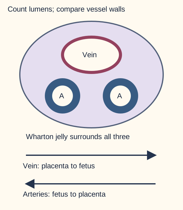
Arteries carry blood away from the fetal heart; veins return it. The placenta supplies oxygen, so two umbilical arteries carry relatively oxygen-poor fetal blood toward the placenta, while one umbilical vein returns oxygen and nutrients. Neither vessel contains maternal blood in the normal circulation. Exchange occurs across the placental barrier between maternal blood outside the villi and fetal blood inside villous capillaries. [1][2]
Use wall thickness and direction together. Colors identify vessels, not a Doppler color convention.
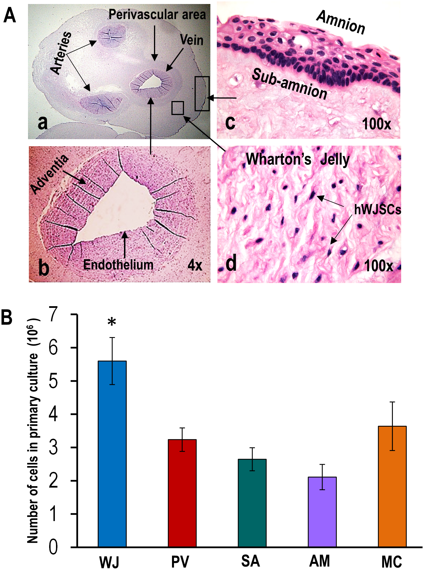
The arteries have relatively thick muscular walls and smaller lumens. The vein generally has a larger lumen and thinner wall. Both are embedded in Wharton jelly, hydrated connective tissue beneath the amniotic covering. It cushions bending and compression; it is neither a fourth vessel nor a lymphatic channel. A normal cord does not have a separate major lymphatic vessel to add to the count. [1][2][15]
Trace an arterial lumen back into the fetus: it leads toward an internal iliac artery. Do not substitute superior vesical artery for that fetal origin. Bladder branches are relevant to the patent proximal segment after birth.
Predict what compression changes
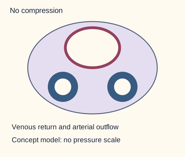
Compare the open lumens below. Before opening either optional answer, predict which return pathway is more vulnerable to gentle external compression. Each answer adds a visual state; close it to retry.
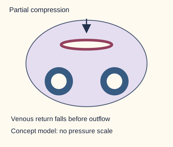
Compare lumen shapes under a hypothetical external force. This qualitative model does not predict a fetal tracing or a clinical pressure threshold. Which lumen narrows first? Compare partial compression.Compare lumen shapes under a hypothetical external force. This qualitative model does not predict a fetal tracing or a clinical pressure threshold.
The thinner venous wall is more readily compressed in this simplified model. Placental return to the fetus falls while arterial outflow may continue. That is a loss of circulating volume returning to the heart, even before both arteries are occluded.
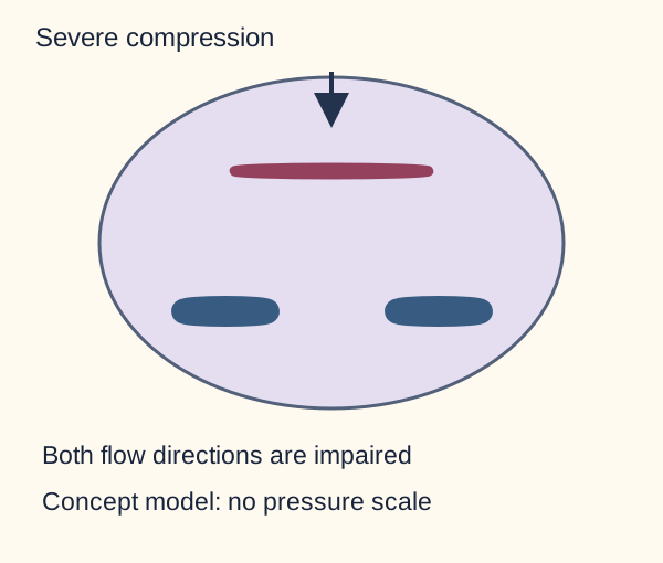
What changes with stronger compression? Compare severe compression.Compare lumen shapes under a hypothetical external force. This qualitative model does not predict a fetal tracing or a clinical pressure threshold.
Severe compression can impair all three vessels. Both exchange delivery and return are compromised; prolonged interruption can cause hypoxemia and acidosis.
Worked comparison: partial venous obstruction reduces fetal venous return; more severe obstruction can interrupt arterial flow too. Wharton jelly reduces vulnerability but cannot guarantee patency. The drawings are qualitative, without force thresholds or predicted heart-rate patterns.
Now apply the relationship: a vessel traveling through membranes without Wharton jelly has lost an important layer of protection, even when its lumen and oxygen content were initially normal.
In panel A, compare vascular walls with the surrounding tissue labeled WJ. These are real H&E tissue images, with the publication's original labels. Panel B reports cell-culture yield and is not a measure of fetal blood flow. The original label “Adventia” means adventitia, the vessel outer connective-tissue coat; it is not an additional named cord compartment. Image: Subramanian, Fong, Biswas and Bongso, 2015, Figure 1, PLOS ONE, CC BY 4.0. [14].
Use a catheter endpoint to predict the tissue at risk. Arterial access descends into the pelvic iliac route before turning toward the aorta; an occlusion threatens territories downstream of its actual branch level. Venous access takes a hepatic route toward the ductus venosus and cava. A catheter ending in a portal branch is not a completed central placement. Hyperosmolar infusion there can injure the liver and leak into hepatic tissue or the abdomen; stop using a malpositioned line and obtain urgent neonatal assessment. [20][21]
Try it here · Checkpoint 1 of 3
Make your prediction before reading the choices. A first attempt is just a starting point.
Case 2
Show answer and explanations for case 2
A. Preserved femoral pulses; renal flow reduced bilaterally (Why this does not fit)
Renal arterial compromise requires a lesion involving the renal origins or their inflow. This obstruction is below them in the right iliac pathway and directly threatens right-leg perfusion instead.
Reasoning steps for option A
What route explains the catheter's initial descent into the pelvis?
An umbilical arterial catheter passes caudally through the pelvic iliac route before reaching the aorta.
Which supplied vascular territory lies below the occlusion rather than above it?
The right common iliac lesion is upstream of right-leg supply but below the renal arterial origins.
How does this option fit the supplied findings?
Renal arterial compromise requires a lesion involving the renal origins or their inflow. This obstruction is below them in the right iliac pathway and directly threatens right-leg perfusion instead.
B. Weak femoral pulses bilaterally; renal flow preserved (Why this does not fit)
An obstruction above the aortic bifurcation could impair both legs. The imaged lesion lies on the right after the split, leaving the left iliac supply open.
Reasoning steps for option B
What route explains the catheter's initial descent into the pelvis?
An umbilical arterial catheter passes caudally through the pelvic iliac route before reaching the aorta.
Which supplied vascular territory lies below the occlusion rather than above it?
The right common iliac lesion is upstream of right-leg supply but below the renal arterial origins.
How does this option fit the supplied findings?
An obstruction above the aortic bifurcation could impair both legs. The imaged lesion lies on the right after the split, leaving the left iliac supply open.
C. Weak right femoral pulse; preserved renal arterial flow (Best answer)
The caudal pelvic course identifies an umbilical arterial route through the internal iliac system. The new lesion is in the right common iliac artery, upstream of right external iliac and femoral flow but below the renal origins. Distribution of the lesion, not the catheter entry name alone, predicts the examination.
Reasoning steps for option C
What route explains the catheter's initial descent into the pelvis?
An umbilical arterial catheter passes caudally through the pelvic iliac route before reaching the aorta.
Which supplied vascular territory lies below the occlusion rather than above it?
The right common iliac lesion is upstream of right-leg supply but below the renal arterial origins.
How does this option fit the supplied findings?
The caudal pelvic course identifies an umbilical arterial route through the internal iliac system. The new lesion is in the right common iliac artery, upstream of right external iliac and femoral flow but below the renal origins. Distribution of the lesion, not the catheter entry name alone, predicts the examination.
D. Weak left femoral pulse; reduced right renal flow (Why this does not fit)
Left-leg pulse loss would require left iliac involvement, while right renal flow originates upstream of the lesion. Neither distribution matches the localized right common iliac obstruction.
Reasoning steps for option D
What route explains the catheter's initial descent into the pelvis?
An umbilical arterial catheter passes caudally through the pelvic iliac route before reaching the aorta.
Which supplied vascular territory lies below the occlusion rather than above it?
The right common iliac lesion is upstream of right-leg supply but below the renal arterial origins.
How does this option fit the supplied findings?
Left-leg pulse loss would require left iliac involvement, while right renal flow originates upstream of the lesion. Neither distribution matches the localized right common iliac obstruction.
Takeaway: Trace the arterial route, then use the obstruction's branch level to predict threatened perfusion.
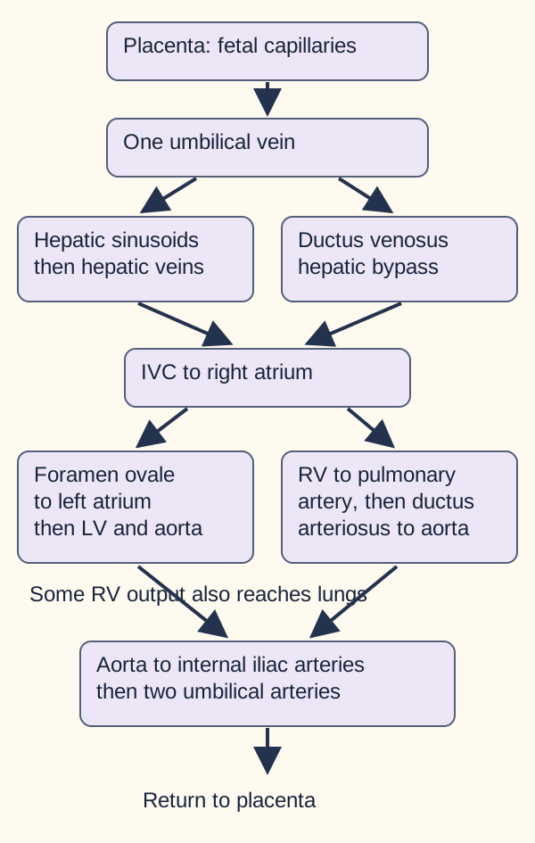
No. The umbilical vein reaches the liver and supplies the hepatic circulation. A portion passes through the ductus venosus, connecting the umbilical/portal venous region to the inferior vena cava near the hepatic venous confluence. This bypasses hepatic sinusoids for that portion. It is not a connection from the left hepatic vein to the IVC, and it does not mean the fetal liver receives no placental blood. Human flow fractions vary with gestation and physiological conditions. [1][18]
Trace the hepatic split before the cardiac split. The simplified routes do not specify flow percentages or imply absent fetal lung perfusion.
Put a finger on the umbilical vein in the diagram. Trace one route through hepatic tissue, then the other through the ductus venosus. Both can reach the IVC. The consequence of the split is simultaneous liver perfusion and delivery of relatively oxygen-rich blood toward the heart.
In the right atrium, relatively oxygen-rich IVC blood is preferentially directed across the foramen ovale to the left atrium, left ventricle and ascending aorta. This supports oxygen delivery to the heart and brain. Streaming is preferential, not perfect separation: oxygen content changes as streams mix. The umbilical vein has the highest oxygen saturation in the normal fetal circuit before this admixture. [3][18]
Superior vena caval return preferentially enters the right ventricle and pulmonary artery. Because fetal pulmonary vascular resistance is high, much of this output reaches the descending aorta through the ductus arteriosus. Some blood still perfuses the lungs. Descending aortic blood returns through common iliac, internal iliac and umbilical arteries to the placenta. The three shunts are inside the fetus, not extra vessels in the cord. [3]
Predict a local effect: if the ductus venosus route is unavailable but venous inflow still reaches the liver, blood must use another available hepatic pathway. That does not make the umbilical vein an artery, nor does it reverse its normal direction. In a new diagram, identify each shunt by its two endpoints before assigning the organ it bypasses.
Does one pressure change close every fetal channel?
Birth changes the circuit in several ways. Lung aeration reduces pulmonary vascular resistance and increases pulmonary venous return. Left atrial pressure then exceeds right atrial pressure, pressing the foramen ovale flap closed. After cord clamping, placental venous return ends and the low-resistance placental circuit is removed. This describes physiology, not a direction to clamp immediately.
For most newborns undergoing normal transition without a need for immediate resuscitation, current AHA/AAP guidance supports deferral for at least 60 seconds; timely effective ventilation remains the priority when resuscitation is needed. [29] These events interact, but they are not one universal closure signal. Functional closure and later anatomical fusion are distinct. [3]
Predict the pressure result before checking the example: if left atrial pressure is 9 mmHg and right atrial pressure is 5 mmHg, the flap is pressed shut from the left. If pulmonary vascular resistance remains abnormally high, fetal-type right-to-left flow can persist. Oxygen does not close the atrial septum by directly contracting it.
Match the channel to its closure mechanism and later landmark
Fetal channel
Main transition
Adult landmark
Fetal channelDuctus venosus
Main transitionPlacental inflow ends; the channel closes
Adult landmarkLigamentum venosum
Fetal channelForamen ovale
Main transitionLeft atrial pressure exceeds right
Adult landmarkFossa ovalis region
Fetal channelDuctus arteriosus
Main transitionHigher oxygen tension and lower prostaglandin signaling favor constriction
Adult landmarkLigamentum arteriosum
Prostaglandin E2 helps maintain fetal ductal patency. Indomethacin reduces prostaglandin synthesis and can close a hemodynamically significant PDA in selected preterm infants. A duct-dependent cardiac lesion changes the objective: preserving patency with prostaglandin E1 may be lifesaving. A murmur alone does not mandate treatment. Renal impairment, bleeding, intestinal disease and cardiac anatomy affect drug selection. [3][11][17]
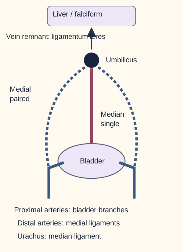
Localize first: toward liver, paired beside bladder, or central above bladder. Dashed arterial paths mark obliterated distal segments.
Follow location to identify the remnant. The umbilical vein becomes the ligamentum teres hepatis in the free edge of the falciform ligament. Distal umbilical arteries become paired medial umbilical ligaments; their proximal segments remain patent and give rise to superior vesical branches. The single median umbilical ligament is the urachus, between the bladder dome and umbilicus. Lateral umbilical folds cover inferior epigastric vessels and are another structure. [1][15]
Early paired umbilical veins remodel, normally leaving the left while the right regresses. Umbilical vessels develop in mesoderm around the allantoic region of the connecting stalk; the endodermal allantoic tube is not transformed directly into arterial walls. Its intraembryonic connection is associated with the urachus. The vitelline duct instead connects the midgut and yolk sac; persistence can produce intestinal communication at the umbilicus or a Meckel diverticulum. [1][15]
Cover the labels in the remnant diagram and trace from the bladder dome to the umbilicus. Urine draining along that central route suggests a patent urachus. Now trace a paired route beside the bladder: this follows an arterial remnant, so it does not explain urine leakage. Location separates similarly named structures.
Test a communication by its endpoint and effect. Decompressing the bladder can reduce leakage through a patent urachus without proving that the tract has closed. A distal ileal pouch with an umbilical attachment instead points to a vitelline remnant. Ectopic gastric mucosa in a Meckel diverticulum can cause ulceration and bleeding; pertechnetate scintigraphy targets that tissue. [22][23][30]
In portal hypertension, paraumbilical collateral veins near the round ligament can carry blood from the portal system toward the abdominal wall. Their direction follows the pressure difference; do not assume every collateral is the reopened fetal vein. A persistent ductus venosus is a different route: it diverts portal blood toward the cava without full sinusoidal exposure and can alter postnatal ammonia handling. [24][25]
Try it here · Checkpoint 2 of 3
Make your prediction before reading the choices. A first attempt is just a starting point.
Case 8
Show answer and explanations for case 8
A. Less sinusoidal exposure; systemic ammonia decreases (Why this does not fit)
The reduced sinusoidal exposure is correct. With production and enzyme capacity fixed, allowing more portal blood to avoid hepatic processing does not predict lower systemic ammonia.
Reasoning steps for option A
Which normally closing channel has these endpoints?
The left portal-to-caval channel in this location is a persistent ductus venosus.
How does a larger bypass fraction change first-pass hepatic processing?
More bypass at fixed inflow reduces first-pass exposure to hepatocytes and tends to increase systemic ammonia.
How does this option fit the supplied findings?
The reduced sinusoidal exposure is correct. With production and enzyme capacity fixed, allowing more portal blood to avoid hepatic processing does not predict lower systemic ammonia.
B. More sinusoidal exposure; systemic ammonia decreases (Why this does not fit)
Greater sinusoidal exposure would follow a smaller bypass fraction at the same total inflow. The measured change is in the opposite direction, so it does not favor greater first-pass clearance.
Reasoning steps for option B
Which normally closing channel has these endpoints?
The left portal-to-caval channel in this location is a persistent ductus venosus.
How does a larger bypass fraction change first-pass hepatic processing?
More bypass at fixed inflow reduces first-pass exposure to hepatocytes and tends to increase systemic ammonia.
How does this option fit the supplied findings?
Greater sinusoidal exposure would follow a smaller bypass fraction at the same total inflow. The measured change is in the opposite direction, so it does not favor greater first-pass clearance.
C. More sinusoidal exposure; systemic ammonia increases (Why this does not fit)
An increase in a direct portal-to-caval shunt reduces the share reaching hepatocytes. Increased production or a separate clearance defect could raise ammonia, but neither explains the claimed increase in sinusoidal exposure here.
Reasoning steps for option C
Which normally closing channel has these endpoints?
The left portal-to-caval channel in this location is a persistent ductus venosus.
How does a larger bypass fraction change first-pass hepatic processing?
More bypass at fixed inflow reduces first-pass exposure to hepatocytes and tends to increase systemic ammonia.
How does this option fit the supplied findings?
An increase in a direct portal-to-caval shunt reduces the share reaching hepatocytes. Increased production or a separate clearance defect could raise ammonia, but neither explains the claimed increase in sinusoidal exposure here.
D. Less sinusoidal exposure; systemic ammonia increases (Best answer)
The location identifies persistent ductus venosus flow, a portosystemic bypass after birth. At fixed total portal inflow, a larger shunt fraction exposes less blood to hepatocytes and reduces first-pass ammonia handling. The expected direction is higher systemic ammonia, not a universal concentration or proof that every affected child has hyperammonemia.
Reasoning steps for option D
Which normally closing channel has these endpoints?
The left portal-to-caval channel in this location is a persistent ductus venosus.
How does a larger bypass fraction change first-pass hepatic processing?
More bypass at fixed inflow reduces first-pass exposure to hepatocytes and tends to increase systemic ammonia.
How does this option fit the supplied findings?
The location identifies persistent ductus venosus flow, a portosystemic bypass after birth. At fixed total portal inflow, a larger shunt fraction exposes less blood to hepatocytes and reduces first-pass ammonia handling. The expected direction is higher systemic ammonia, not a universal concentration or proof that every affected child has hyperammonemia.
Takeaway: A persistent fetal bypass can alter postnatal liver processing even when the liver's intrinsic enzyme capacity is unchanged.
A single umbilical artery leaves one artery and one vein. It occurs in roughly 0.5% to 1% of singleton pregnancies, with estimates varying by population. It can occur alone or with renal, cardiac and other malformations, including patterns seen in trisomy 18 and VACTERL association. The cord finding prompts careful anatomical assessment; it does not diagnose renal agenesis or a chromosomal disorder. [1][4][12]
Compare two scans: both show a single artery. In one, a detailed survey shows normal anatomy and growth. In the other, growth restriction, a cardiac defect and overlapping fingers are present. Decide which fetus fits the word isolated. The first does; the second needs evaluation of a broader pattern. A reassuring four-chamber view alone is not a complete anatomical survey.
For an isolated single artery, SMFM recommends no additional aneuploidy evaluation solely because of this finding, even if earlier screening was declined. It recommends third-trimester ultrasound for growth and consideration of weekly antenatal surveillance from 36 weeks. Additional malformations or growth restriction require their own evaluation and counseling. [4]
After birth, examine the infant and review the quality of prenatal renal imaging. An automatic renal ultrasound for every otherwise normal infant is not supported by all evidence. Incomplete prenatal views, abnormal examination or other malformations support targeted imaging according to local practice. A cohort study found no excess of significant renal malformations among infants with isolated single artery, but this is not a universal exemption from renal assessment. [5]
The renal association is not explained by the ureteric bud sharing a lateral plate mesoderm origin with cord vessels. Kidney development depends on intermediate mesoderm and reciprocal signaling between ureteric bud and metanephric tissue. Association alone does not establish a shared developmental cause. [13]
Transfer the decision: if an initially isolated finding is followed by growth restriction, reassess the pregnancy using the new information. A label applied at 20 weeks cannot substitute for the current scan.
A new structural-anomaly pattern is not canceled by a previous low-risk cell-free DNA result. Offer diagnostic genetic evaluation with chromosomal microarray and counseling when a structural abnormality is identified. Ultrasound suspicion is not chromosome confirmation, and testing remains the patient's choice. [26]
Which matters more: insertion, or the vessel crossing the cervix?
A normal cord reaches the placental disc with its vessels protected by Wharton jelly. With velamentous insertion, the cord attaches to membranes and vessels travel within them before reaching the disc. Marginal insertion is at the placental edge and is not the same anatomical arrangement. Velamentous vessels are vulnerable even when they do not cross the cervix. [1][6]
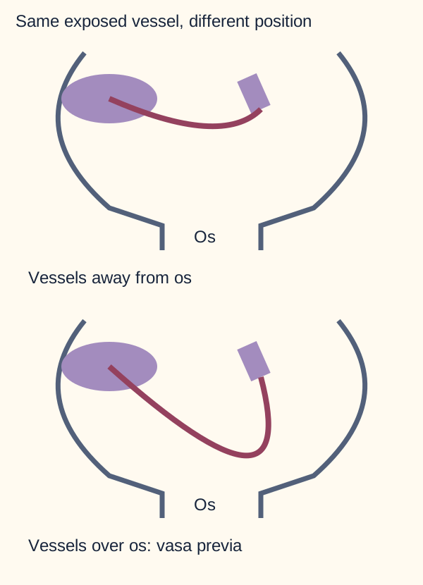
The pink line represents exposed fetal vessels, not maternal placental tissue. Trace the exposed fetal vessels relative to the internal cervical os.
Trace the exposed vessel in each panel and point to the internal cervical os. Only the lower panel places a fetal vessel in the path of cervical dilation and membrane rupture. That relationship is vasa previa. Vessels connecting a main placental disc and an accessory or second lobe can also cross the os, even without velamentous cord insertion. Prenatal transvaginal ultrasound with color and pulsed Doppler establishes location and fetal vascular flow. A low-lying placenta that later appears clear of the os does not by itself exclude persistent exposed vessels. [6]
For confirmed vasa previa, plan cesarean delivery before labor or membrane rupture. SMFM gives a 34 to 37 week window; timing is individualized to symptoms and risk of earlier delivery. A single mandatory week is not appropriate for every patient. New labor, membrane rupture, bleeding or fetal compromise changes the plan. Avoid amniotomy across known exposed vessels. [6][7]
Bleeding after membrane rupture with acute fetal deterioration is an emergency. Ruptured vasa previa loses fetal blood, so maternal blood pressure may remain normal while the fetus becomes profoundly anemic. Bradycardia or a sinusoidal pattern may occur; both are not required together. Urgent obstetric delivery and neonatal resuscitation with transfusion readiness take priority over waiting for a confirmatory blood assay. [7]
The Apt alkali-denaturation test exploits relative resistance of fetal hemoglobin: it remains pink while adult hemoglobin denatures toward yellow-brown under the test conditions. Sodium hydroxide is the conventional reagent; potassium hydroxide has been studied in modified methods. This is a laboratory principle, not a bedside prerequisite for emergency delivery, and mixtures or unsuitable samples limit interpretation. [8][16]
Apply the distinction to another bleed: placenta previa concerns placental tissue at the cervix and usually maternal blood loss; abruption concerns placental separation. Pain, tone, imaging and fetal findings help, but absence of pain alone cannot exclude abruption. Cord prolapse primarily threatens flow by compression, whereas disrupted exposed vessels can rapidly deplete fetal blood volume.
When a complete cervical Doppler assessment shows no vasa previa, a velamentous insertion still calls for follow-up, including serial fetal growth assessment. It does not by itself establish the earlier cesarean schedule used for cervical fetal vessels. Reassess when symptoms or new findings develop. [27]
Try it here · Checkpoint 3 of 3
Make your prediction before reading the choices. A first attempt is just a starting point.
Case 18
Show answer and explanations for case 18
A. Induce labor after cervical ripening to achieve a controlled vaginal birth (Why this does not fit)
A fetal vessel crossing the os is vulnerable during cervical dilation and membrane rupture. A controlled induction does not remove that anatomical risk.
Reasoning steps for option A
Does the waveform and interlobar course identify fetal or maternal circulation?
Fetal-rate interlobar flow over the os identifies type II vasa previa.
How does stable status within the delivery window differ from ruptured bleeding vessels?
Stable status at 35 weeks calls for planned prelabor cesarean coordination, not waiting for rupture to create an emergency.
How does this option fit the supplied findings?
A fetal vessel crossing the os is vulnerable during cervical dilation and membrane rupture. A controlled induction does not remove that anatomical risk.
B. Await spontaneous labor because the cord inserts into a placental lobe (Why this does not fit)
Insertion into a placental lobe does not exclude exposed interlobar vessels over the cervix. The cervical vessel course, not the insertion alone, determines the need to avoid labor and rupture.
Reasoning steps for option B
Does the waveform and interlobar course identify fetal or maternal circulation?
Fetal-rate interlobar flow over the os identifies type II vasa previa.
How does stable status within the delivery window differ from ruptured bleeding vessels?
Stable status at 35 weeks calls for planned prelabor cesarean coordination, not waiting for rupture to create an emergency.
How does this option fit the supplied findings?
Insertion into a placental lobe does not exclude exposed interlobar vessels over the cervix. The cervical vessel course, not the insertion alone, determines the need to avoid labor and rupture.
C. Repeat Doppler at term because the stable waveform identifies maternal flow (Why this does not fit)
Matching the fetal heart rate supports fetal rather than maternal circulation. Stable flow today does not justify deferring planning until term when the vessel remains over the os.
Reasoning steps for option C
Does the waveform and interlobar course identify fetal or maternal circulation?
Fetal-rate interlobar flow over the os identifies type II vasa previa.
How does stable status within the delivery window differ from ruptured bleeding vessels?
Stable status at 35 weeks calls for planned prelabor cesarean coordination, not waiting for rupture to create an emergency.
How does this option fit the supplied findings?
Matching the fetal heart rate supports fetal rather than maternal circulation. Stable flow today does not justify deferring planning until term when the vessel remains over the os.
D. Arrange prelabor cesarean with neonatal transfusion capability available (Best answer)
A fetal-rate interlobar vessel over the os establishes type II vasa previa even without velamentous insertion. At 35 weeks, coordinate a planned cesarean before labor or rupture within the individualized SMFM delivery window, at a center prepared for neonatal transfusion.
Reasoning steps for option D
Does the waveform and interlobar course identify fetal or maternal circulation?
Fetal-rate interlobar flow over the os identifies type II vasa previa.
How does stable status within the delivery window differ from ruptured bleeding vessels?
Stable status at 35 weeks calls for planned prelabor cesarean coordination, not waiting for rupture to create an emergency.
How does this option fit the supplied findings?
A fetal-rate interlobar vessel over the os establishes type II vasa previa even without velamentous insertion. At 35 weeks, coordinate a planned cesarean before labor or rupture within the individualized SMFM delivery window, at a center prepared for neonatal transfusion.
Takeaway: Identify whose vessel crosses the cervix, then plan delivery around the risk created by that location.
Does an unusual cord prove the cause of fetal compromise?
Wharton jelly helps the cord tolerate bending. Coiling has been proposed to distribute mechanical stress, but its independent protective effect is uncertain. Left-directed coiling is common, but the number of turns varies; a fixed count such as 11 is not a diagnostic requirement. The postnatal coiling index is complete coils divided by cord length in centimeters. Studies often define hypocoiling below the 10th percentile and hypercoiling above the 90th. A cutoff near 0.1 coils/cm appears in some references, but methods and populations differ. [2][10][19]
Calculate before interpreting: four complete coils in a 50 cm cord gives 4 / 50 = 0.08 coils/cm. If the study's lower limit is 0.10, that cord is hypocoiled by that study's rule. The same four coils in a 25 cm specimen gives 0.16. The consequence is a different index, despite an identical turn count; incomplete specimens and antenatal estimates cannot be interchanged casually.
Abnormal coiling is associated with adverse outcomes, including fetal heart-rate abnormalities and growth problems. Compression is a plausible contributor, but a coiling measurement alone cannot establish why a fetus died or prescribe delivery timing. Evaluate the fetal course and placental findings. [10]
A true knot is an actual knot in the cord and can restrict flow if it tightens. A false knot is a focal prominence from redundant vessels or connective tissue, without an actual cord knot. A nuchal cord passes around the fetal neck. Single loops are common and often uncomplicated; their presence alone is not proof of hypoxic injury. A meta-analysis found true knots associated with greater stillbirth risk, while any nuchal cord as a group did not show a clear association. [1][9]
Predict two outcomes: an untightened knot with maintained flow may coexist with a vigorous infant; a tightened knot with prolonged obstruction can impair gas exchange. The anatomical label tells you what could happen. Evidence of obstruction and the clinical course tell you what did happen. In a new delivery with a loose neck loop and reassuring status, do not infer injury from the loop alone.
Use physiology before attributing injury. Low cord pH with high carbon dioxide and a modest base deficit points to a predominantly respiratory pattern rather than a large metabolic acid burden. This is not a guarantee of no morbidity or proof of which cord feature caused the change. During labor, integrate decelerations with contraction frequency and the clinical picture. Excessive contractions during oxytocin use call for reducing or stopping stimulation, appropriate repositioning and prompt reassessment; a known neck loop does not replace that assessment. [28][31]
Apply the lesson
Case 1
Show answer and explanations for case 1
A. Reduced hepatic inflow; increased ductus venosus inflow (Why this does not fit)
Preferential diversion away from sinusoids could increase the bypass fraction if common inflow were maintained. An obstruction before the hepatic split instead reduces the placental inlet to both pathways.
Reasoning steps for option A
Which circulation does the fetal-directed vessel supply?
The fetal-directed vessel is the umbilical vein, which supplies the common inlet to the hepatic and bypass routes.
Is the blockage before the two routes separate or within only one route?
The blockage is before the split, so both routes lose placental inflow.
How does this option fit the supplied findings?
Preferential diversion away from sinusoids could increase the bypass fraction if common inflow were maintained. An obstruction before the hepatic split instead reduces the placental inlet to both pathways.
B. Increased hepatic inflow; reduced ductus venosus inflow (Why this does not fit)
A blocked ductus venosus with maintained common inflow could redirect blood toward the liver. Here the obstruction is upstream of both patent routes, so an increased hepatic share cannot replace the lost common supply.
Reasoning steps for option B
Which circulation does the fetal-directed vessel supply?
The fetal-directed vessel is the umbilical vein, which supplies the common inlet to the hepatic and bypass routes.
Is the blockage before the two routes separate or within only one route?
The blockage is before the split, so both routes lose placental inflow.
How does this option fit the supplied findings?
A blocked ductus venosus with maintained common inflow could redirect blood toward the liver. Here the obstruction is upstream of both patent routes, so an increased hepatic share cannot replace the lost common supply.
C. Reduced hepatic inflow; reduced ductus venosus inflow (Best answer)
The large fetal-directed vessel is the umbilical vein. Blocking it before hepatic branching limits the placental supply to both the sinusoidal and ductus venosus routes, despite their anatomical patency.
Reasoning steps for option C
Which circulation does the fetal-directed vessel supply?
The fetal-directed vessel is the umbilical vein, which supplies the common inlet to the hepatic and bypass routes.
Is the blockage before the two routes separate or within only one route?
The blockage is before the split, so both routes lose placental inflow.
How does this option fit the supplied findings?
The large fetal-directed vessel is the umbilical vein. Blocking it before hepatic branching limits the placental supply to both the sinusoidal and ductus venosus routes, despite their anatomical patency.
D. Increased hepatic inflow; increased ductus venosus inflow (Why this does not fit)
The continued patency of two downstream routes does not create a new placental source. Reduced flow through their shared upstream inlet cannot explain increased placental delivery through both routes.
Reasoning steps for option D
Which circulation does the fetal-directed vessel supply?
The fetal-directed vessel is the umbilical vein, which supplies the common inlet to the hepatic and bypass routes.
Is the blockage before the two routes separate or within only one route?
The blockage is before the split, so both routes lose placental inflow.
How does this option fit the supplied findings?
The continued patency of two downstream routes does not create a new placental source. Reduced flow through their shared upstream inlet cannot explain increased placental delivery through both routes.
Takeaway: Localize an obstruction before or after a branch point before predicting which routes lose inflow.
A. Portal venous malposition; parenteral nutrition extravasation (Best answer)
The large-lumen hepatic course identifies venous access that has entered a portal branch rather than completing the central route. Peritoneal glucose far above serum supports leakage of glucose-rich parenteral nutrition instead of ordinary blood. The precise contribution of direct perforation versus chemical tissue injury is not established by these findings. Stop using the malpositioned line and obtain urgent neonatal assessment.
Reasoning steps for option A
Does the observed route lead through the hepatic venous system or the pelvic arterial system?
The large-lumen hepatic course identifies venous access ending in a portal branch, not iliac arterial placement.
Which fluid source accounts for glucose far above the simultaneous serum value?
Glucose-rich parenteral nutrition leakage explains the peritoneal enrichment better than blood from a puncture injury.
How does this option fit the supplied findings?
The large-lumen hepatic course identifies venous access that has entered a portal branch rather than completing the central route. Peritoneal glucose far above serum supports leakage of glucose-rich parenteral nutrition instead of ordinary blood. The precise contribution of direct perforation versus chemical tissue injury is not established by these findings. Stop using the malpositioned line and obtain urgent neonatal assessment.
B. Portal venous malposition; hemorrhage from puncture injury (Why this does not fit)
The portal-system location fits the observed route, and puncture-related bleeding is a possible catheter complication. However, blood leakage does not account for peritoneal glucose far above the simultaneous serum value; direct loss of infused nutrition does.
Reasoning steps for option B
Does the observed route lead through the hepatic venous system or the pelvic arterial system?
The large-lumen hepatic course identifies venous access ending in a portal branch, not iliac arterial placement.
Which fluid source accounts for glucose far above the simultaneous serum value?
Glucose-rich parenteral nutrition leakage explains the peritoneal enrichment better than blood from a puncture injury.
How does this option fit the supplied findings?
The portal-system location fits the observed route, and puncture-related bleeding is a possible catheter complication. However, blood leakage does not account for peritoneal glucose far above the simultaneous serum value; direct loss of infused nutrition does.
C. Iliac arterial malposition; parenteral nutrition extravasation (Why this does not fit)
The peritoneal fluid chemistry fits parenteral nutrition leakage. The catheter location does not fit: an umbilical arterial catheter takes a pelvic iliac route before ascending toward the aorta, rather than entering a lateral hepatic branch from the large cord lumen.
Reasoning steps for option C
Does the observed route lead through the hepatic venous system or the pelvic arterial system?
The large-lumen hepatic course identifies venous access ending in a portal branch, not iliac arterial placement.
Which fluid source accounts for glucose far above the simultaneous serum value?
Glucose-rich parenteral nutrition leakage explains the peritoneal enrichment better than blood from a puncture injury.
How does this option fit the supplied findings?
The peritoneal fluid chemistry fits parenteral nutrition leakage. The catheter location does not fit: an umbilical arterial catheter takes a pelvic iliac route before ascending toward the aorta, rather than entering a lateral hepatic branch from the large cord lumen.
D. Iliac arterial malposition; hemorrhage from puncture injury (Why this does not fit)
The proposed location conflicts with the hepatic venous course, and the proposed hemorrhage does not explain the marked peritoneal-to-serum glucose difference. Both the route and the fluid must be accounted for.
Reasoning steps for option D
Does the observed route lead through the hepatic venous system or the pelvic arterial system?
The large-lumen hepatic course identifies venous access ending in a portal branch, not iliac arterial placement.
Which fluid source accounts for glucose far above the simultaneous serum value?
Glucose-rich parenteral nutrition leakage explains the peritoneal enrichment better than blood from a puncture injury.
How does this option fit the supplied findings?
The proposed location conflicts with the hepatic venous course, and the proposed hemorrhage does not explain the marked peritoneal-to-serum glucose difference. Both the route and the fluid must be accounted for.
Takeaway: Confirm the catheter's actual endpoint, then connect the affected tissue to the infusion complication.
A. 10 mL gain; stroke volume increases (Why this does not fit)
An inflow exceeding outflow would increase the modeled blood volume. Here outflow exceeds return by 30 mL/min, so the volume change has the opposite sign.
Reasoning steps for option A
How much volume is lost during 20 seconds rather than a full minute?
The net loss is 30 mL/min multiplied by one-third of a minute, or 10 mL.
How does reduced return affect filling when the other determinants are fixed?
Reduced venous return lowers preload and, under the stated controls, stroke volume.
How does this option fit the supplied findings?
An inflow exceeding outflow would increase the modeled blood volume. Here outflow exceeds return by 30 mL/min, so the volume change has the opposite sign.
B. 30 mL loss; stroke volume decreases (Why this does not fit)
The 30 mL/min deficit would remove 30 mL over a full minute. The interval is only one-third of a minute, so the loss is 10 mL even though the predicted stroke-volume direction is correct.
Reasoning steps for option B
How much volume is lost during 20 seconds rather than a full minute?
The net loss is 30 mL/min multiplied by one-third of a minute, or 10 mL.
How does reduced return affect filling when the other determinants are fixed?
Reduced venous return lowers preload and, under the stated controls, stroke volume.
How does this option fit the supplied findings?
The 30 mL/min deficit would remove 30 mL over a full minute. The interval is only one-third of a minute, so the loss is 10 mL even though the predicted stroke-volume direction is correct.
C. 10 mL loss; stroke volume decreases (Best answer)
The net deficit is (100 - 70) x (20/60) = 10 mL. Continued outflow with less return reduces preload; with the stated controls, stroke volume falls. These are hypothetical teaching values, not clinical compression thresholds.
Reasoning steps for option C
How much volume is lost during 20 seconds rather than a full minute?
The net loss is 30 mL/min multiplied by one-third of a minute, or 10 mL.
How does reduced return affect filling when the other determinants are fixed?
Reduced venous return lowers preload and, under the stated controls, stroke volume.
How does this option fit the supplied findings?
The net deficit is (100 - 70) x (20/60) = 10 mL. Continued outflow with less return reduces preload; with the stated controls, stroke volume falls. These are hypothetical teaching values, not clinical compression thresholds.
D. 10 mL loss; stroke volume increases (Why this does not fit)
The arithmetic correctly gives a 10 mL deficit. With the other determinants held fixed, reduced venous return lowers filling and tends to reduce rather than increase stroke volume.
Reasoning steps for option D
How much volume is lost during 20 seconds rather than a full minute?
The net loss is 30 mL/min multiplied by one-third of a minute, or 10 mL.
How does reduced return affect filling when the other determinants are fixed?
Reduced venous return lowers preload and, under the stated controls, stroke volume.
How does this option fit the supplied findings?
The arithmetic correctly gives a 10 mL deficit. With the other determinants held fixed, reduced venous return lowers filling and tends to reduce rather than increase stroke volume.
Takeaway: Calculate inflow minus outflow over the actual interval, then connect the volume change to cardiac filling.
A. Lower bladder pressure reduces flow through a patent urinary tract (Best answer)
Voiding-linked drainage and a bladder-dome connection identify a patent urachus. Decompression reduces the pressure driving urine toward the umbilicus; the persistent visible lumen shows that reduced drainage is not proof of closure.
Reasoning steps for option A
Which endpoint identifies the source of the draining fluid?
The bladder-dome endpoint and voiding-linked clear drainage identify a patent urachus.
What changed after decompression: the driving pressure or the demonstrated anatomy?
Bladder pressure fell; the still-visible lumen does not demonstrate closure.
How does this option fit the supplied findings?
Voiding-linked drainage and a bladder-dome connection identify a patent urachus. Decompression reduces the pressure driving urine toward the umbilicus; the persistent visible lumen shows that reduced drainage is not proof of closure.
B. Lower portal pressure reduces flow through a paraumbilical vessel (Why this does not fit)
A paraumbilical vessel communicates with the hepatic portal circulation. This nonvascular tract joins the bladder, and the intervention changes bladder pressure rather than establishing portal decompression.
Reasoning steps for option B
Which endpoint identifies the source of the draining fluid?
The bladder-dome endpoint and voiding-linked clear drainage identify a patent urachus.
What changed after decompression: the driving pressure or the demonstrated anatomy?
Bladder pressure fell; the still-visible lumen does not demonstrate closure.
How does this option fit the supplied findings?
A paraumbilical vessel communicates with the hepatic portal circulation. This nonvascular tract joins the bladder, and the intervention changes bladder pressure rather than establishing portal decompression.
C. Bowel decompression reduces flow through a persistent enteric tract (Why this does not fit)
A persistent vitelline tract communicates with bowel and can carry enteric contents. The demonstrated bladder endpoint and response to urinary drainage do not fit an intestinal route.
Reasoning steps for option C
Which endpoint identifies the source of the draining fluid?
The bladder-dome endpoint and voiding-linked clear drainage identify a patent urachus.
What changed after decompression: the driving pressure or the demonstrated anatomy?
Bladder pressure fell; the still-visible lumen does not demonstrate closure.
How does this option fit the supplied findings?
A persistent vitelline tract communicates with bowel and can carry enteric contents. The demonstrated bladder endpoint and response to urinary drainage do not fit an intestinal route.
D. Bladder emptying produces immediate fibrous closure of the tract (Why this does not fit)
Closure would eliminate the communicating lumen rather than merely reduce drainage. The prompt change with bladder decompression and unchanged visible tract support a pressure effect, not immediate fibrosis.
Reasoning steps for option D
Which endpoint identifies the source of the draining fluid?
The bladder-dome endpoint and voiding-linked clear drainage identify a patent urachus.
What changed after decompression: the driving pressure or the demonstrated anatomy?
Bladder pressure fell; the still-visible lumen does not demonstrate closure.
How does this option fit the supplied findings?
Closure would eliminate the communicating lumen rather than merely reduce drainage. The prompt change with bladder decompression and unchanged visible tract support a pressure effect, not immediate fibrosis.
Takeaway: Reduced drainage can reflect reduced driving pressure even when an abnormal communication remains open.
A. Voiding cystourethrography to detect contrast entering a bladder diverticulum (Why this does not fit)
A urachal or bladder abnormality could explain a urinary communication near the umbilicus. The demonstrated ileal pouch and gastrointestinal bleeding instead localize a vitelline remnant.
Reasoning steps for option A
Does the remnant connect with bowel or with the bladder?
The ileal pouch and umbilical attachment identify a vitelline remnant, not a urinary connection.
Which tissue within that intestinal remnant can cause ulceration and be detected by the proposed study?
Ectopic gastric mucosa can cause ulceration and is the tissue targeted by pertechnetate scintigraphy.
How does this option fit the supplied findings?
A urachal or bladder abnormality could explain a urinary communication near the umbilicus. The demonstrated ileal pouch and gastrointestinal bleeding instead localize a vitelline remnant.
B. Technetium-99m pertechnetate scintigraphy to detect ectopic gastric mucosa (Best answer)
The ileal pouch and umbilical attachment identify a Meckel-type vitelline remnant. Ectopic gastric mucosa can cause acid-related ulceration and bleeding; pertechnetate imaging tests for that mucosa. A negative scan does not by itself exclude the remnant.
Reasoning steps for option B
Does the remnant connect with bowel or with the bladder?
The ileal pouch and umbilical attachment identify a vitelline remnant, not a urinary connection.
Which tissue within that intestinal remnant can cause ulceration and be detected by the proposed study?
Ectopic gastric mucosa can cause ulceration and is the tissue targeted by pertechnetate scintigraphy.
How does this option fit the supplied findings?
The ileal pouch and umbilical attachment identify a Meckel-type vitelline remnant. Ectopic gastric mucosa can cause acid-related ulceration and bleeding; pertechnetate imaging tests for that mucosa. A negative scan does not by itself exclude the remnant.
C. Portal Doppler ultrasonography to detect a hepatofugal abdominal-wall vein (Why this does not fit)
Portal collaterals can carry blood toward abdominal-wall veins. They do not explain a blind-ending ileal pouch, and this study does not test the suspected acid-secreting mucosa.
Reasoning steps for option C
Does the remnant connect with bowel or with the bladder?
The ileal pouch and umbilical attachment identify a vitelline remnant, not a urinary connection.
Which tissue within that intestinal remnant can cause ulceration and be detected by the proposed study?
Ectopic gastric mucosa can cause ulceration and is the tissue targeted by pertechnetate scintigraphy.
How does this option fit the supplied findings?
Portal collaterals can carry blood toward abdominal-wall veins. They do not explain a blind-ending ileal pouch, and this study does not test the suspected acid-secreting mucosa.
D. Renal cortical scintigraphy to detect reduced function of an ectopic kidney (Why this does not fit)
Renal cortical imaging evaluates functioning kidney tissue. The source localized here is an intestinal remnant rather than a urinary tract lesion.
Reasoning steps for option D
Does the remnant connect with bowel or with the bladder?
The ileal pouch and umbilical attachment identify a vitelline remnant, not a urinary connection.
Which tissue within that intestinal remnant can cause ulceration and be detected by the proposed study?
Ectopic gastric mucosa can cause ulceration and is the tissue targeted by pertechnetate scintigraphy.
How does this option fit the supplied findings?
Renal cortical imaging evaluates functioning kidney tissue. The source localized here is an intestinal remnant rather than a urinary tract lesion.
Takeaway: Locate the embryonic remnant, then choose a study that tests the proposed mechanism of its complication.
A. 30 mL/min collateral flow; 80% of tracer reaches systemic blood (Why this does not fit)
The pressure gradient halves from 14 to 7 mmHg, so collateral flow correctly halves to 30 mL/min. However, 80% tracer escape belongs to the original 60 mL bypass plus 40 mL sinusoidal split; the new split permits greater extraction.
Reasoning steps for option A
How does halving the driving gradient change the original collateral flow?
At fixed resistance, the gradient falls from 14 to 7 mmHg and collateral flow falls from 60 to 30 mL/min.
What fraction bypasses the liver, and how much of the remaining tracer survives sinusoidal extraction?
Thirty percent bypasses extraction; half of the remaining 70% survives, so 65% reaches the systemic circulation.
How does this option fit the supplied findings?
The pressure gradient halves from 14 to 7 mmHg, so collateral flow correctly halves to 30 mL/min. However, 80% tracer escape belongs to the original 60 mL bypass plus 40 mL sinusoidal split; the new split permits greater extraction.
B. 30 mL/min collateral flow; 65% of tracer reaches systemic blood (Best answer)
The gradient falls from 14 to 7 mmHg, halving collateral flow from 60 to 30 mL/min at fixed resistance. Conservation sends the remaining 70 mL/min through sinusoids. Thirty percent of tracer bypasses extraction and half of the remaining 70% survives, giving 30% + 35% = 65%. These are imposed model values, not clinical clearance measurements.
Reasoning steps for option B
How does halving the driving gradient change the original collateral flow?
At fixed resistance, the gradient falls from 14 to 7 mmHg and collateral flow falls from 60 to 30 mL/min.
What fraction bypasses the liver, and how much of the remaining tracer survives sinusoidal extraction?
Thirty percent bypasses extraction; half of the remaining 70% survives, so 65% reaches the systemic circulation.
How does this option fit the supplied findings?
The gradient falls from 14 to 7 mmHg, halving collateral flow from 60 to 30 mL/min at fixed resistance. Conservation sends the remaining 70 mL/min through sinusoids. Thirty percent of tracer bypasses extraction and half of the remaining 70% survives, giving 30% + 35% = 65%. These are imposed model values, not clinical clearance measurements.
C. 60 mL/min collateral flow; 80% of tracer reaches systemic blood (Why this does not fit)
This retains both the original bypass flow and original tracer escape. A halved driving gradient at unchanged resistance reduces bypass flow, so both the route distribution and extraction must be recalculated.
Reasoning steps for option C
How does halving the driving gradient change the original collateral flow?
At fixed resistance, the gradient falls from 14 to 7 mmHg and collateral flow falls from 60 to 30 mL/min.
What fraction bypasses the liver, and how much of the remaining tracer survives sinusoidal extraction?
Thirty percent bypasses extraction; half of the remaining 70% survives, so 65% reaches the systemic circulation.
How does this option fit the supplied findings?
This retains both the original bypass flow and original tracer escape. A halved driving gradient at unchanged resistance reduces bypass flow, so both the route distribution and extraction must be recalculated.
D. 60 mL/min collateral flow; 65% of tracer reaches systemic blood (Why this does not fit)
The tracer result matches the new 30/70 flow split, but the proposed 60 mL/min collateral flow retains the old gradient. Flow of 60 would instead leave 40 for sinusoids and produce 80% tracer escape under the stated extraction rule.
Reasoning steps for option D
How does halving the driving gradient change the original collateral flow?
At fixed resistance, the gradient falls from 14 to 7 mmHg and collateral flow falls from 60 to 30 mL/min.
What fraction bypasses the liver, and how much of the remaining tracer survives sinusoidal extraction?
Thirty percent bypasses extraction; half of the remaining 70% survives, so 65% reaches the systemic circulation.
How does this option fit the supplied findings?
The tracer result matches the new 30/70 flow split, but the proposed 60 mL/min collateral flow retains the old gradient. Flow of 60 would instead leave 40 for sinusoids and produce 80% tracer escape under the stated extraction rule.
Takeaway: Calculate flow through the collateral, then redistribute the remaining inflow before predicting hepatic first-pass extraction.
A. Higher oxygen tension directly contracts the interatrial flap tissue (Why this does not fit)
Higher oxygen tension promotes constriction of ductal smooth muscle. The atrial flap instead apposes because of a pressure difference, not direct oxygen-induced contraction.
Reasoning steps for option A
Which atrium receives the increased pulmonary venous return?
Increased pulmonary venous return raises left atrial filling.
Does flap apposition require the arterial duct to have closed first?
Left atrial pressure exceeding right presses the flap closed even before the arterial duct closes.
How does this option fit the supplied findings?
Higher oxygen tension promotes constriction of ductal smooth muscle. The atrial flap instead apposes because of a pressure difference, not direct oxygen-induced contraction.
B. Hepatic duct closure increases placental venous filling of the heart (Why this does not fit)
Closure of the hepatic bypass does not increase placental inflow. The observed rise in pulmonary return supplies the relevant increase in left atrial filling.
Reasoning steps for option B
Which atrium receives the increased pulmonary venous return?
Increased pulmonary venous return raises left atrial filling.
Does flap apposition require the arterial duct to have closed first?
Left atrial pressure exceeding right presses the flap closed even before the arterial duct closes.
How does this option fit the supplied findings?
Closure of the hepatic bypass does not increase placental inflow. The observed rise in pulmonary return supplies the relevant increase in left atrial filling.
C. Arterial duct closure redirects the entire right ventricular output (Why this does not fit)
The stem documents a still-patent arterial duct. Increased pulmonary venous return can produce atrial flap apposition without completed ductal closure.
Reasoning steps for option C
Which atrium receives the increased pulmonary venous return?
Increased pulmonary venous return raises left atrial filling.
Does flap apposition require the arterial duct to have closed first?
Left atrial pressure exceeding right presses the flap closed even before the arterial duct closes.
How does this option fit the supplied findings?
The stem documents a still-patent arterial duct. Increased pulmonary venous return can produce atrial flap apposition without completed ductal closure.
D. Pulmonary venous return raises left atrial pressure above right (Best answer)
Lung aeration increases pulmonary flow and return to the left atrium. The resulting left-to-right pressure difference presses the atrial flap against the septum even while the arterial duct remains open.
Reasoning steps for option D
Which atrium receives the increased pulmonary venous return?
Increased pulmonary venous return raises left atrial filling.
Does flap apposition require the arterial duct to have closed first?
Left atrial pressure exceeding right presses the flap closed even before the arterial duct closes.
How does this option fit the supplied findings?
Lung aeration increases pulmonary flow and return to the left atrium. The resulting left-to-right pressure difference presses the atrial flap against the septum even while the arterial duct remains open.
Takeaway: Foramen ovale apposition depends on atrial pressures, not prior anatomical closure of the arterial duct.
A. Right-to-left atrial flow preferentially supplies the distal descending aorta (Why this does not fit)
An atrial right-to-left shunt can lower systemic oxygenation. It does not selectively enter the descending aorta after the right-arm branches, which is the localization needed for the supplied difference.
Reasoning steps for option A
Which direction does the documented ductal flow take?
The observed pulmonary artery-to-aorta flow is a right-to-left ductal shunt.
Is the right-hand sample taken before or after the ductal aortic insertion?
Right-hand supply arises before the ductal insertion, whereas the foot receives postductal blood.
How does this option fit the supplied findings?
An atrial right-to-left shunt can lower systemic oxygenation. It does not selectively enter the descending aorta after the right-arm branches, which is the localization needed for the supplied difference.
B. Right-to-left ductal flow adds desaturated blood beyond the right-arm supply (Best answer)
High pulmonary resistance supports the observed pulmonary artery-to-aorta shunt. Its insertion is distal to the right-arm arterial supply, so less oxygenated blood lowers postductal foot saturation relative to the preductal right hand.
Reasoning steps for option B
Which direction does the documented ductal flow take?
The observed pulmonary artery-to-aorta flow is a right-to-left ductal shunt.
Is the right-hand sample taken before or after the ductal aortic insertion?
Right-hand supply arises before the ductal insertion, whereas the foot receives postductal blood.
How does this option fit the supplied findings?
High pulmonary resistance supports the observed pulmonary artery-to-aorta shunt. Its insertion is distal to the right-arm arterial supply, so less oxygenated blood lowers postductal foot saturation relative to the preductal right hand.
C. Left-to-right atrial flow directs desaturated blood into the descending aorta (Why this does not fit)
A left-to-right atrial shunt sends blood toward the right heart. It does not add pulmonary arterial blood specifically below the aortic branches supplying the right arm.
Reasoning steps for option C
Which direction does the documented ductal flow take?
The observed pulmonary artery-to-aorta flow is a right-to-left ductal shunt.
Is the right-hand sample taken before or after the ductal aortic insertion?
Right-hand supply arises before the ductal insertion, whereas the foot receives postductal blood.
How does this option fit the supplied findings?
A left-to-right atrial shunt sends blood toward the right heart. It does not add pulmonary arterial blood specifically below the aortic branches supplying the right arm.
D. Left-to-right ductal flow recirculates arterial blood toward the pulmonary bed (Why this does not fit)
This describes a possible postnatal PDA direction but not the direction documented here. Aortic-to-pulmonary recirculation does not explain the addition of less oxygenated blood to the descending aorta.
Reasoning steps for option D
Which direction does the documented ductal flow take?
The observed pulmonary artery-to-aorta flow is a right-to-left ductal shunt.
Is the right-hand sample taken before or after the ductal aortic insertion?
Right-hand supply arises before the ductal insertion, whereas the foot receives postductal blood.
How does this option fit the supplied findings?
This describes a possible postnatal PDA direction but not the direction documented here. Aortic-to-pulmonary recirculation does not explain the addition of less oxygenated blood to the descending aorta.
Takeaway: Use shunt direction and aortic insertion to predict regional oxygenation.
A. Prostaglandin synthesis rises; aortic-to-pulmonary runoff rises (Why this does not fit)
More prostaglandin signaling favors patency and can sustain the left-to-right runoff. Indomethacin is being selected to inhibit that pathway rather than increase it.
Reasoning steps for option A
Which circulation receives the excess aortic runoff?
Aortic blood is running into the pulmonary circulation through the patent duct.
How does inhibiting prostaglandin synthesis affect that channel?
Prostaglandin inhibition favors constriction, reducing the aortic-to-pulmonary runoff.
How does this option fit the supplied findings?
More prostaglandin signaling favors patency and can sustain the left-to-right runoff. Indomethacin is being selected to inhibit that pathway rather than increase it.
B. Prostaglandin synthesis rises; aortic-to-pulmonary runoff falls (Why this does not fit)
Reducing the shunt is the desired physiological direction. The proposed increase in prostaglandin synthesis would oppose the drug's constricting mechanism.
Reasoning steps for option B
Which circulation receives the excess aortic runoff?
Aortic blood is running into the pulmonary circulation through the patent duct.
How does inhibiting prostaglandin synthesis affect that channel?
Prostaglandin inhibition favors constriction, reducing the aortic-to-pulmonary runoff.
How does this option fit the supplied findings?
Reducing the shunt is the desired physiological direction. The proposed increase in prostaglandin synthesis would oppose the drug's constricting mechanism.
C. Prostaglandin synthesis falls; aortic-to-pulmonary runoff falls (Best answer)
Indomethacin inhibits prostaglandin synthesis, promoting ductal constriction. In the supplied aorta-to-pulmonary shunt, this reduces pulmonary overcirculation and systemic runoff. The decision remains specific to a significant PDA after contraindication assessment.
Reasoning steps for option C
Which circulation receives the excess aortic runoff?
Aortic blood is running into the pulmonary circulation through the patent duct.
How does inhibiting prostaglandin synthesis affect that channel?
Prostaglandin inhibition favors constriction, reducing the aortic-to-pulmonary runoff.
How does this option fit the supplied findings?
Indomethacin inhibits prostaglandin synthesis, promoting ductal constriction. In the supplied aorta-to-pulmonary shunt, this reduces pulmonary overcirculation and systemic runoff. The decision remains specific to a significant PDA after contraindication assessment.
D. Prostaglandin synthesis falls; aortic-to-pulmonary runoff rises (Why this does not fit)
The proposed drug mechanism is correct, but the predicted flow effect is reversed. Constricting this arterial duct reduces rather than increases the documented aortic-to-pulmonary runoff.
Reasoning steps for option D
Which circulation receives the excess aortic runoff?
Aortic blood is running into the pulmonary circulation through the patent duct.
How does inhibiting prostaglandin synthesis affect that channel?
Prostaglandin inhibition favors constriction, reducing the aortic-to-pulmonary runoff.
How does this option fit the supplied findings?
The proposed drug mechanism is correct, but the predicted flow effect is reversed. Constricting this arterial duct reduces rather than increases the documented aortic-to-pulmonary runoff.
Takeaway: The intended drug effect follows the current shunt direction and the infant's anatomy.
A. Defer the next dose until renal recovery and reassess the PDA plan (Best answer)
Urine output of 0.3 is below 0.6 mL/kg/hour, with rising creatinine supporting renal deterioration. Withhold the scheduled dose and reassess renal function and the PDA plan; further labeled dosing depends on renal recovery.
Reasoning steps for option A
Does current urine output cross the threshold for withholding?
Urine output of 0.3 is below the 0.6 mL/kg/hour withholding threshold, with rising creatinine.
Does reducing the dose or changing to another cyclooxygenase inhibitor resolve that safety condition?
Further doses are withheld pending renal recovery and reassessment, rather than simply reduced or replaced by a similar drug.
How does this option fit the supplied findings?
Urine output of 0.3 is below 0.6 mL/kg/hour, with rising creatinine supporting renal deterioration. Withhold the scheduled dose and reassess renal function and the PDA plan; further labeled dosing depends on renal recovery.
B. Give a reduced next dose and continue renal surveillance (Why this does not fit)
Dose reduction is not the specified response to marked oliguria before a scheduled second or third dose. The label directs withholding further doses pending renal recovery.
Reasoning steps for option B
Does current urine output cross the threshold for withholding?
Urine output of 0.3 is below the 0.6 mL/kg/hour withholding threshold, with rising creatinine.
Does reducing the dose or changing to another cyclooxygenase inhibitor resolve that safety condition?
Further doses are withheld pending renal recovery and reassessment, rather than simply reduced or replaced by a similar drug.
How does this option fit the supplied findings?
Dose reduction is not the specified response to marked oliguria before a scheduled second or third dose. The label directs withholding further doses pending renal recovery.
C. Give the next full dose and continue renal surveillance (Why this does not fit)
Closing a significant PDA can improve systemic perfusion, which makes proceeding tempting. However, the measured urine output is below the label's withholding threshold, and worsening renal function makes continued dosing inappropriate.
Reasoning steps for option C
Does current urine output cross the threshold for withholding?
Urine output of 0.3 is below the 0.6 mL/kg/hour withholding threshold, with rising creatinine.
Does reducing the dose or changing to another cyclooxygenase inhibitor resolve that safety condition?
Further doses are withheld pending renal recovery and reassessment, rather than simply reduced or replaced by a similar drug.
How does this option fit the supplied findings?
Closing a significant PDA can improve systemic perfusion, which makes proceeding tempting. However, the measured urine output is below the label's withholding threshold, and worsening renal function makes continued dosing inappropriate.
D. Substitute ibuprofen and continue renal surveillance (Why this does not fit)
Ibuprofen also inhibits prostaglandin synthesis and does not eliminate concern about active renal impairment. Switching agents is not a substitute for reassessing this infant's renal status.
Reasoning steps for option D
Does current urine output cross the threshold for withholding?
Urine output of 0.3 is below the 0.6 mL/kg/hour withholding threshold, with rising creatinine.
Does reducing the dose or changing to another cyclooxygenase inhibitor resolve that safety condition?
Further doses are withheld pending renal recovery and reassessment, rather than simply reduced or replaced by a similar drug.
How does this option fit the supplied findings?
Ibuprofen also inhibits prostaglandin synthesis and does not eliminate concern about active renal impairment. Switching agents is not a substitute for reassessing this infant's renal status.
Takeaway: A treatment that constricts the PDA may also compromise renal function.
A. Cesarean delivery at 34 weeks to prevent later single-artery flow limitation (Why this does not fit)
Isolated single artery with normal growth and anatomy does not establish a need for preterm operative delivery. Follow-up is directed toward growth and later fetal assessment, with delivery decisions based on the evolving clinical situation.
Reasoning steps for option A
Does this survey satisfy the definition of isolated single artery?
The complete survey and normal growth meet the isolated-SUA definition.
Does previous screening refusal change the specific isolated-SUA recommendation?
Prior screening refusal does not add a finding-driven aneuploidy indication; plan growth assessment and discuss surveillance.
How does this option fit the supplied findings?
Isolated single artery with normal growth and anatomy does not establish a need for preterm operative delivery. Follow-up is directed toward growth and later fetal assessment, with delivery decisions based on the evolving clinical situation.
B. Third-trimester growth scan; discuss weekly surveillance from 36 weeks (Best answer)
Normal growth and a complete survey without anomalies or other markers establish isolated single umbilical artery. SMFM recommends third-trimester growth assessment and consideration of weekly surveillance from 36 weeks, without extra aneuploidy evaluation solely for this finding, even after screening was declined.
Reasoning steps for option B
Does this survey satisfy the definition of isolated single artery?
The complete survey and normal growth meet the isolated-SUA definition.
Does previous screening refusal change the specific isolated-SUA recommendation?
Prior screening refusal does not add a finding-driven aneuploidy indication; plan growth assessment and discuss surveillance.
How does this option fit the supplied findings?
Normal growth and a complete survey without anomalies or other markers establish isolated single umbilical artery. SMFM recommends third-trimester growth assessment and consideration of weekly surveillance from 36 weeks, without extra aneuploidy evaluation solely for this finding, even after screening was declined.
C. Routine prenatal visits with growth scans reserved for abnormal fundal height (Why this does not fit)
A normal anatomy survey does not remove the recommendation for third-trimester growth imaging. Waiting for an abnormal fundal-height measurement omits that finding-specific assessment.
Reasoning steps for option C
Does this survey satisfy the definition of isolated single artery?
The complete survey and normal growth meet the isolated-SUA definition.
Does previous screening refusal change the specific isolated-SUA recommendation?
Prior screening refusal does not add a finding-driven aneuploidy indication; plan growth assessment and discuss surveillance.
How does this option fit the supplied findings?
A normal anatomy survey does not remove the recommendation for third-trimester growth imaging. Waiting for an abnormal fundal-height measurement omits that finding-specific assessment.
D. Amniocentesis for chromosome analysis because prior screening was declined (Why this does not fit)
Diagnostic testing remains a patient choice, but previous screening refusal does not turn isolated single artery into a new testing indication. The specific SMFM recommendation instead addresses growth and surveillance.
Reasoning steps for option D
Does this survey satisfy the definition of isolated single artery?
The complete survey and normal growth meet the isolated-SUA definition.
Does previous screening refusal change the specific isolated-SUA recommendation?
Prior screening refusal does not add a finding-driven aneuploidy indication; plan growth assessment and discuss surveillance.
How does this option fit the supplied findings?
Diagnostic testing remains a patient choice, but previous screening refusal does not turn isolated single artery into a new testing indication. The specific SMFM recommendation instead addresses growth and surveillance.
Takeaway: First establish whether the finding is isolated; then apply its specific surveillance guidance.
A. Serum creatinine with urinalysis (Why this does not fit)
Renal function tests can be reassuring even with one kidney or a structural anomaly. They cannot establish that the unvisualized kidney is present and structurally normal.
Reasoning steps for option A
What specific question was left unanswered by prenatal assessment?
The prenatal study never established the anatomy of one kidney.
Which test can show the kidney rather than only measure urinary function?
Ultrasound can resolve that structural uncertainty; urine output or creatinine cannot.
How does this option fit the supplied findings?
Renal function tests can be reassuring even with one kidney or a structural anomaly. They cannot establish that the unvisualized kidney is present and structurally normal.
B. Voiding cystourethrography (Why this does not fit)
A reflux or urethral study can address selected urinary problems. It is not the initial study for an unvisualized kidney when the immediate question is renal anatomy.
Reasoning steps for option B
What specific question was left unanswered by prenatal assessment?
The prenatal study never established the anatomy of one kidney.
Which test can show the kidney rather than only measure urinary function?
Ultrasound can resolve that structural uncertainty; urine output or creatinine cannot.
How does this option fit the supplied findings?
A reflux or urethral study can address selected urinary problems. It is not the initial study for an unvisualized kidney when the immediate question is renal anatomy.
C. Renal and bladder ultrasonography (Best answer)
The reassuring examination leaves an unresolved prenatal structural question. Ultrasound can assess both kidneys and the urinary tract; this is targeted imaging after incomplete prenatal visualization, not a rule to scan every isolated single-artery case.
Reasoning steps for option C
What specific question was left unanswered by prenatal assessment?
The prenatal study never established the anatomy of one kidney.
Which test can show the kidney rather than only measure urinary function?
Ultrasound can resolve that structural uncertainty; urine output or creatinine cannot.
How does this option fit the supplied findings?
The reassuring examination leaves an unresolved prenatal structural question. Ultrasound can assess both kidneys and the urinary tract; this is targeted imaging after incomplete prenatal visualization, not a rule to scan every isolated single-artery case.
D. Urine-output measurement over 24 hours (Why this does not fit)
Normal urine output does not establish the presence of two normal kidneys. Measuring output cannot resolve the missing anatomical information in the prenatal report.
Reasoning steps for option D
What specific question was left unanswered by prenatal assessment?
The prenatal study never established the anatomy of one kidney.
Which test can show the kidney rather than only measure urinary function?
Ultrasound can resolve that structural uncertainty; urine output or creatinine cannot.
How does this option fit the supplied findings?
Normal urine output does not establish the presence of two normal kidneys. Measuring output cannot resolve the missing anatomical information in the prenatal report.
Takeaway: Selective imaging depends on what was actually assessed before birth.
A. Assign a trisomy 18 diagnosis from the ultrasound phenotype (Why this does not fit)
The phenotype makes trisomy 18 a leading consideration, but ultrasound does not establish the chromosome result. Diagnostic testing is needed for genetic confirmation rather than assigning a karyotype from appearance.
Reasoning steps for option A
Is this an isolated marker or a pattern containing structural abnormalities?
Cardiac and hand malformations with growth restriction make the finding nonisolated.
Which test provides diagnostic information when a screening result conflicts with the anatomy?
Offer diagnostic testing with chromosomal microarray and counseling; another screen is not genetic confirmation.
How does this option fit the supplied findings?
The phenotype makes trisomy 18 a leading consideration, but ultrasound does not establish the chromosome result. Diagnostic testing is needed for genetic confirmation rather than assigning a karyotype from appearance.
B. Offer amniocentesis with chromosomal microarray and counseling (Best answer)
The growth, cardiac and hand pattern raises concern for trisomy 18 or another genetic disorder; the artery finding is not isolated. SMFM recommends offering diagnostic testing with chromosomal microarray when a structural abnormality is identified. Counseling must distinguish suspicion from confirmation and respect the patient's choice to accept or decline testing.
Reasoning steps for option B
Is this an isolated marker or a pattern containing structural abnormalities?
Cardiac and hand malformations with growth restriction make the finding nonisolated.
Which test provides diagnostic information when a screening result conflicts with the anatomy?
Offer diagnostic testing with chromosomal microarray and counseling; another screen is not genetic confirmation.
How does this option fit the supplied findings?
The growth, cardiac and hand pattern raises concern for trisomy 18 or another genetic disorder; the artery finding is not isolated. SMFM recommends offering diagnostic testing with chromosomal microarray when a structural abnormality is identified. Counseling must distinguish suspicion from confirmation and respect the patient's choice to accept or decline testing.
C. Repeat the anatomy scan before discussing genetic testing (Why this does not fit)
Further imaging can help define the anatomy, but the already established malformations justify offering diagnostic evaluation now. Deferring that discussion treats a nonisolated pattern as an unresolved screening marker.
Reasoning steps for option C
Is this an isolated marker or a pattern containing structural abnormalities?
Cardiac and hand malformations with growth restriction make the finding nonisolated.
Which test provides diagnostic information when a screening result conflicts with the anatomy?
Offer diagnostic testing with chromosomal microarray and counseling; another screen is not genetic confirmation.
How does this option fit the supplied findings?
Further imaging can help define the anatomy, but the already established malformations justify offering diagnostic evaluation now. Deferring that discussion treats a nonisolated pattern as an unresolved screening marker.
D. Repeat cell-free DNA screening for the common trisomies (Why this does not fit)
Repeating a screening test gives another risk estimate rather than diagnostic evaluation of the newly identified malformations. A low-risk screen does not override a structural-anomaly pattern.
Reasoning steps for option D
Is this an isolated marker or a pattern containing structural abnormalities?
Cardiac and hand malformations with growth restriction make the finding nonisolated.
Which test provides diagnostic information when a screening result conflicts with the anatomy?
Offer diagnostic testing with chromosomal microarray and counseling; another screen is not genetic confirmation.
How does this option fit the supplied findings?
Repeating a screening test gives another risk estimate rather than diagnostic evaluation of the newly identified malformations. A low-risk screen does not override a structural-anomaly pattern.
Takeaway: A reassuring screen does not cancel a new structural-anomaly pattern; offer diagnostic evaluation rather than relabeling the screen.
A. Reclassify as nonisolated and reassess diagnostic counseling (Best answer)
A newly confirmed structural abnormality defeats the original isolated classification. Reassess the phenotype and diagnostic options rather than extending the earlier isolated-SUA recommendation to a different clinical situation.
Reasoning steps for option A
Can normal growth cancel the significance of a structural malformation?
A newly confirmed structural cardiac defect prevents continued use of the isolated category.
What changes in counseling when the isolated-marker category no longer applies?
Reassess diagnostic counseling without assuming a causal mechanism or a chromosome result.
How does this option fit the supplied findings?
A newly confirmed structural abnormality defeats the original isolated classification. Reassess the phenotype and diagnostic options rather than extending the earlier isolated-SUA recommendation to a different clinical situation.
B. Diagnose trisomy 18 from the combined artery and cardiac findings (Why this does not fit)
Single artery with a cardiac defect is not a diagnostic chromosome result. The new information supports renewed evaluation and discussion of diagnostic testing rather than an assigned karyotype.
Reasoning steps for option B
Can normal growth cancel the significance of a structural malformation?
A newly confirmed structural cardiac defect prevents continued use of the isolated category.
What changes in counseling when the isolated-marker category no longer applies?
Reassess diagnostic counseling without assuming a causal mechanism or a chromosome result.
How does this option fit the supplied findings?
Single artery with a cardiac defect is not a diagnostic chromosome result. The new information supports renewed evaluation and discussion of diagnostic testing rather than an assigned karyotype.
C. Retain the isolated category because fetal growth remains appropriate (Why this does not fit)
Normal growth satisfies only one part of the isolation assessment. A structural cardiac abnormality means the finding no longer fits the category used for isolated-marker guidance.
Reasoning steps for option C
Can normal growth cancel the significance of a structural malformation?
A newly confirmed structural cardiac defect prevents continued use of the isolated category.
What changes in counseling when the isolated-marker category no longer applies?
Reassess diagnostic counseling without assuming a causal mechanism or a chromosome result.
How does this option fit the supplied findings?
Normal growth satisfies only one part of the isolation assessment. A structural cardiac abnormality means the finding no longer fits the category used for isolated-marker guidance.
D. Attribute the cardiac lesion to reduced flow from the missing artery (Why this does not fit)
The two abnormalities justify broader assessment, but their coexistence does not establish that the missing artery caused the cardiac lesion. Counseling should address the phenotype without inventing a causal link.
Reasoning steps for option D
Can normal growth cancel the significance of a structural malformation?
A newly confirmed structural cardiac defect prevents continued use of the isolated category.
What changes in counseling when the isolated-marker category no longer applies?
Reassess diagnostic counseling without assuming a causal mechanism or a chromosome result.
How does this option fit the supplied findings?
The two abnormalities justify broader assessment, but their coexistence does not establish that the missing artery caused the cardiac lesion. Counseling should address the phenotype without inventing a causal link.
Takeaway: Reassess classification when new anatomy becomes available.
A. Urgent Apt testing with delivery selected after fetal blood is confirmed (Why this does not fit)
An Apt result can support the origin of blood but is not required before emergency treatment. Waiting for confirmation prolongs a plausible fetal hemorrhage with persistent compromise.
Reasoning steps for option A
Whose circulation is endangered by the known cervical vessels?
The exposed vessels belong to the fetus, so maternal stability does not exclude major fetal blood loss.
Which action addresses ongoing fetal blood loss without waiting for confirmation?
Emergency cesarean and neonatal resuscitation with blood ready address the immediate threat without waiting for an assay.
How does this option fit the supplied findings?
An Apt result can support the origin of blood but is not required before emergency treatment. Waiting for confirmation prolongs a plausible fetal hemorrhage with persistent compromise.
B. Maternal hemodynamic observation with delivery after hypotension develops (Why this does not fit)
The bleeding can come from the fetal circulation while maternal blood pressure remains normal. Maternal hypotension is therefore the wrong trigger for responding to the fetus's immediate threat.
Reasoning steps for option B
Whose circulation is endangered by the known cervical vessels?
The exposed vessels belong to the fetus, so maternal stability does not exclude major fetal blood loss.
Which action addresses ongoing fetal blood loss without waiting for confirmation?
Emergency cesarean and neonatal resuscitation with blood ready address the immediate threat without waiting for an assay.
How does this option fit the supplied findings?
The bleeding can come from the fetal circulation while maternal blood pressure remains normal. Maternal hypotension is therefore the wrong trigger for responding to the fetus's immediate threat.
C. Antenatal corticosteroids with delivery after the course is completed (Why this does not fit)
A steroid course must not delay delivery for active bleeding with persistent fetal bradycardia. Lung-maturity planning does not take priority over this acute threat.
Reasoning steps for option C
Whose circulation is endangered by the known cervical vessels?
The exposed vessels belong to the fetus, so maternal stability does not exclude major fetal blood loss.
Which action addresses ongoing fetal blood loss without waiting for confirmation?
Emergency cesarean and neonatal resuscitation with blood ready address the immediate threat without waiting for an assay.
How does this option fit the supplied findings?
A steroid course must not delay delivery for active bleeding with persistent fetal bradycardia. Lung-maturity planning does not take priority over this acute threat.
D. Activate emergency cesarean with neonatal resuscitation and blood available (Best answer)
Known fetal membrane vessels over the os, bleeding after rupture and persistent fetal bradycardia indicate threatened fetal hemorrhage. Activate emergency cesarean delivery with neonatal resuscitation and transfusion readiness immediately. Do not wait for an Apt result or maternal deterioration.
Reasoning steps for option D
Whose circulation is endangered by the known cervical vessels?
The exposed vessels belong to the fetus, so maternal stability does not exclude major fetal blood loss.
Which action addresses ongoing fetal blood loss without waiting for confirmation?
Emergency cesarean and neonatal resuscitation with blood ready address the immediate threat without waiting for an assay.
How does this option fit the supplied findings?
Known fetal membrane vessels over the os, bleeding after rupture and persistent fetal bradycardia indicate threatened fetal hemorrhage. Activate emergency cesarean delivery with neonatal resuscitation and transfusion readiness immediately. Do not wait for an Apt result or maternal deterioration.
Takeaway: Fetal hemorrhage can be catastrophic despite normal maternal blood pressure.
A. Arrange prelabor cesarean during the 34-to-37-week window (Why this does not fit)
The earlier prelabor cesarean window applies to confirmed vasa previa. The complete examination has not demonstrated exposed cervical vessels, so velamentous insertion alone does not establish that delivery indication.
Reasoning steps for option A
Which insertion is present, and is a separate cervical vessel hazard demonstrated?
Velamentous insertion is present, but the complete examination does not demonstrate vasa previa.
Does that combination call for a vasa previa delivery plan or ongoing growth follow-up?
Continue pregnancy with serial growth assessment and an individualized obstetric plan.
How does this option fit the supplied findings?
The earlier prelabor cesarean window applies to confirmed vasa previa. The complete examination has not demonstrated exposed cervical vessels, so velamentous insertion alone does not establish that delivery indication.
B. Arrange serial growth assessment and continued obstetric follow-up (Best answer)
Membrane insertion establishes velamentous anatomy, but the complete cervical examination does not demonstrate vasa previa. Continue pregnancy with serial growth assessment and an individualized obstetric plan rather than automatically applying the vasa previa delivery schedule.
Reasoning steps for option B
Which insertion is present, and is a separate cervical vessel hazard demonstrated?
Velamentous insertion is present, but the complete examination does not demonstrate vasa previa.
Does that combination call for a vasa previa delivery plan or ongoing growth follow-up?
Continue pregnancy with serial growth assessment and an individualized obstetric plan.
How does this option fit the supplied findings?
Membrane insertion establishes velamentous anatomy, but the complete cervical examination does not demonstrate vasa previa. Continue pregnancy with serial growth assessment and an individualized obstetric plan rather than automatically applying the vasa previa delivery schedule.
C. Arrange cervical Doppler follow-up instead of serial growth imaging (Why this does not fit)
Further cervical assessment can be appropriate when new concerns arise. It does not replace growth follow-up for the demonstrated velamentous insertion; the supplied complete cervical study already addresses the immediate anatomical distinction.
Reasoning steps for option C
Which insertion is present, and is a separate cervical vessel hazard demonstrated?
Velamentous insertion is present, but the complete examination does not demonstrate vasa previa.
Does that combination call for a vasa previa delivery plan or ongoing growth follow-up?
Continue pregnancy with serial growth assessment and an individualized obstetric plan.
How does this option fit the supplied findings?
Further cervical assessment can be appropriate when new concerns arise. It does not replace growth follow-up for the demonstrated velamentous insertion; the supplied complete cervical study already addresses the immediate anatomical distinction.
D. Arrange routine obstetric visits with fundal-height screening (Why this does not fit)
Fundal-height screening alone does not provide the serial ultrasound growth assessment recommended for velamentous insertion. A clear os removes the demonstrated vasa previa hazard, not the need to follow fetal growth.
Reasoning steps for option D
Which insertion is present, and is a separate cervical vessel hazard demonstrated?
Velamentous insertion is present, but the complete examination does not demonstrate vasa previa.
Does that combination call for a vasa previa delivery plan or ongoing growth follow-up?
Continue pregnancy with serial growth assessment and an individualized obstetric plan.
How does this option fit the supplied findings?
Fundal-height screening alone does not provide the serial ultrasound growth assessment recommended for velamentous insertion. A clear os removes the demonstrated vasa previa hazard, not the need to follow fetal growth.
Takeaway: A clear cervical vessel assessment changes the delivery indication, not the need to follow a velamentous insertion.
A. Plan cesarean at 39 weeks to reduce respiratory morbidity (Why this does not fit)
Reducing neonatal respiratory morbidity matters, but waiting to 39 weeks adds exposure to spontaneous labor and rupture. The patient is already within the individualized earlier delivery window for confirmed vasa previa.
Reasoning steps for option A
Does current stability eliminate the future rupture hazard?
Confirmed cervical fetal vessels remain vulnerable even during reassuring monitoring.
Where does the current gestation fall within the recommended planning window?
At 36 weeks the patient is within the planning window for prompt cesarean before labor or rupture.
How does this option fit the supplied findings?
Reducing neonatal respiratory morbidity matters, but waiting to 39 weeks adds exposure to spontaneous labor and rupture. The patient is already within the individualized earlier delivery window for confirmed vasa previa.
B. Perform amniotomy, then select delivery based on the tracing (Why this does not fit)
Amniotomy can disrupt exposed fetal vessels at the cervix. The diagnosis already supplies a reason to avoid rupture rather than deliberately provoke it.
Reasoning steps for option B
Does current stability eliminate the future rupture hazard?
Confirmed cervical fetal vessels remain vulnerable even during reassuring monitoring.
Where does the current gestation fall within the recommended planning window?
At 36 weeks the patient is within the planning window for prompt cesarean before labor or rupture.
How does this option fit the supplied findings?
Amniotomy can disrupt exposed fetal vessels at the cervix. The diagnosis already supplies a reason to avoid rupture rather than deliberately provoke it.
C. Prompt planned cesarean before labor or spontaneous membrane rupture (Best answer)
At 36 weeks with confirmed vasa previa and stable status, coordinate prompt planned cesarean before labor or rupture. This fits the individualized 34 to 37 week window; new bleeding, labor, rupture or fetal compromise changes the urgency.
Reasoning steps for option C
Does current stability eliminate the future rupture hazard?
Confirmed cervical fetal vessels remain vulnerable even during reassuring monitoring.
Where does the current gestation fall within the recommended planning window?
At 36 weeks the patient is within the planning window for prompt cesarean before labor or rupture.
How does this option fit the supplied findings?
At 36 weeks with confirmed vasa previa and stable status, coordinate prompt planned cesarean before labor or rupture. This fits the individualized 34 to 37 week window; new bleeding, labor, rupture or fetal compromise changes the urgency.
D. Await spontaneous labor, then reassess the vessel location (Why this does not fit)
Reassuring monitoring does not remove the rupture hazard of fetal vessels over the os. Labor is not an appropriate test of whether that anatomy will remain safe.
Reasoning steps for option D
Does current stability eliminate the future rupture hazard?
Confirmed cervical fetal vessels remain vulnerable even during reassuring monitoring.
Where does the current gestation fall within the recommended planning window?
At 36 weeks the patient is within the planning window for prompt cesarean before labor or rupture.
How does this option fit the supplied findings?
Reassuring monitoring does not remove the rupture hazard of fetal vessels over the os. Labor is not an appropriate test of whether that anatomy will remain safe.
Takeaway: A management plan must fit the patient's current time point.
A. Fetal hemoglobin in the sample; velamentous insertion is established (Why this does not fit)
Fetal hemoglobin supports a blood-origin inference. It cannot identify an insertion type or distinguish velamentous vessels from an exposed interlobar vessel.
Reasoning steps for option A
What does resistance to alkali distinguish in this assay?
Alkali resistance supports a fetal hemoglobin contribution.
Do the clinical findings support a fetal source, and what stronger claims remain unsupported?
Newborn anemia supports fetal blood loss, but the color result does not locate the vessel or quantify a mixture.
How does this option fit the supplied findings?
Fetal hemoglobin supports a blood-origin inference. It cannot identify an insertion type or distinguish velamentous vessels from an exposed interlobar vessel.
B. Fetal hemoglobin in the sample; maternal admixture is quantified (Why this does not fit)
A qualitative color comparison does not measure the percentage of fetal and maternal blood. Mixture quantification requires a method designed for that purpose, not a stronger interpretation of this color result.
Reasoning steps for option B
What does resistance to alkali distinguish in this assay?
Alkali resistance supports a fetal hemoglobin contribution.
Do the clinical findings support a fetal source, and what stronger claims remain unsupported?
Newborn anemia supports fetal blood loss, but the color result does not locate the vessel or quantify a mixture.
How does this option fit the supplied findings?
A qualitative color comparison does not measure the percentage of fetal and maternal blood. Mixture quantification requires a method designed for that purpose, not a stronger interpretation of this color result.
C. Adult hemoglobin in the sample; a maternal bleeding source is supported (Why this does not fit)
Adult hemoglobin denatures under the supplied test conditions, as the adult control demonstrates. The pink sample and newborn rather than maternal anemia favor a fetal contribution.
Reasoning steps for option C
What does resistance to alkali distinguish in this assay?
Alkali resistance supports a fetal hemoglobin contribution.
Do the clinical findings support a fetal source, and what stronger claims remain unsupported?
Newborn anemia supports fetal blood loss, but the color result does not locate the vessel or quantify a mixture.
How does this option fit the supplied findings?
Adult hemoglobin denatures under the supplied test conditions, as the adult control demonstrates. The pink sample and newborn rather than maternal anemia favor a fetal contribution.
D. Fetal hemoglobin in the sample; a fetal bleeding source is supported (Best answer)
Relative alkali resistance supports fetal hemoglobin. Newborn anemia with unchanged maternal hemoglobin supports a fetal source, but the qualitative assay alone neither maps the damaged vessel nor quantifies a mixture.
Reasoning steps for option D
What does resistance to alkali distinguish in this assay?
Alkali resistance supports a fetal hemoglobin contribution.
Do the clinical findings support a fetal source, and what stronger claims remain unsupported?
Newborn anemia supports fetal blood loss, but the color result does not locate the vessel or quantify a mixture.
How does this option fit the supplied findings?
Relative alkali resistance supports fetal hemoglobin. Newborn anemia with unchanged maternal hemoglobin supports a fetal source, but the qualitative assay alone neither maps the damaged vessel nor quantifies a mixture.
Takeaway: A blood-origin test supports an interpretation but does not map vascular anatomy or quantify every mixture.
A. 0.08 coils/cm; below the laboratory's hypocoiling cutoff (Best answer)
The index is 4/50 = 0.08 coils/cm, below the supplied 0.10 cutoff. That supports classification as hypocoiled by this laboratory's rule. Its association with adverse outcomes does not establish that cord occlusion caused this particular tracing.
Reasoning steps for option A
Which quantity is divided by which, and what units result?
Complete coils divided by centimeters gives 4/50 = 0.08 coils/cm.
Does crossing the coiling cutoff establish an actual occlusive event?
The value crosses the supplied cutoff, but that classification does not prove an occlusive cause for the tracing.
How does this option fit the supplied findings?
The index is 4/50 = 0.08 coils/cm, below the supplied 0.10 cutoff. That supports classification as hypocoiled by this laboratory's rule. Its association with adverse outcomes does not establish that cord occlusion caused this particular tracing.
B. 0.125 coils/cm; cord occlusion caused the abnormal tracing (Why this does not fit)
The numerical result is incorrect and the causal attribution exceeds the evidence. Neither an erroneous quotient nor the observed turn count identifies an actual episode of cord occlusion.
Reasoning steps for option B
Which quantity is divided by which, and what units result?
Complete coils divided by centimeters gives 4/50 = 0.08 coils/cm.
Does crossing the coiling cutoff establish an actual occlusive event?
The value crosses the supplied cutoff, but that classification does not prove an occlusive cause for the tracing.
How does this option fit the supplied findings?
The numerical result is incorrect and the causal attribution exceeds the evidence. Neither an erroneous quotient nor the observed turn count identifies an actual episode of cord occlusion.
C. 0.08 coils/cm; cord occlusion caused the abnormal tracing (Why this does not fit)
The index is calculated correctly, but a low coiling index does not document an occlusive episode. Causation requires evidence beyond the cord measurement and a nonspecific abnormal tracing.
Reasoning steps for option C
Which quantity is divided by which, and what units result?
Complete coils divided by centimeters gives 4/50 = 0.08 coils/cm.
Does crossing the coiling cutoff establish an actual occlusive event?
The value crosses the supplied cutoff, but that classification does not prove an occlusive cause for the tracing.
How does this option fit the supplied findings?
The index is calculated correctly, but a low coiling index does not document an occlusive episode. Causation requires evidence beyond the cord measurement and a nonspecific abnormal tracing.
D. 0.125 coils/cm; above the laboratory's hypocoiling cutoff (Why this does not fit)
The defined calculation is complete coils divided by centimeters: 4/50 = 0.08. The value 0.125 is not that quotient; reversing the division would instead give 12.5 cm per coil.
Reasoning steps for option D
Which quantity is divided by which, and what units result?
Complete coils divided by centimeters gives 4/50 = 0.08 coils/cm.
Does crossing the coiling cutoff establish an actual occlusive event?
The value crosses the supplied cutoff, but that classification does not prove an occlusive cause for the tracing.
How does this option fit the supplied findings?
The defined calculation is complete coils divided by centimeters: 4/50 = 0.08. The value 0.125 is not that quotient; reversing the division would instead give 12.5 cm per coil.
Takeaway: Calculate the index correctly, then separate association from individual causation.
A. True knot; predominantly respiratory acidemia; assess the neonatal course (Best answer)
The cord geometry establishes a true knot independently of the infant's condition. The gas pattern is predominantly respiratory acidemia from carbon dioxide retention rather than a large metabolic deficit. Neither the knot nor this single sample proves that the knot caused prolonged hypoxic injury; interpret the neonatal course as well.
Reasoning steps for option A
Is the entire cord knotted, or is there only a focal vascular prominence?
The entire cord traversing its own loop identifies a true knot rather than a focal vessel prominence.
Does the gas pattern show mainly retained carbon dioxide or a large metabolic acid burden?
High carbon dioxide with a modest base deficit supports a predominantly respiratory pattern, not proof of prolonged metabolic injury.
How does this option fit the supplied findings?
The cord geometry establishes a true knot independently of the infant's condition. The gas pattern is predominantly respiratory acidemia from carbon dioxide retention rather than a large metabolic deficit. Neither the knot nor this single sample proves that the knot caused prolonged hypoxic injury; interpret the neonatal course as well.
B. True knot; predominantly metabolic acidemia; assess the neonatal course (Why this does not fit)
The geometry identifies a true knot. However, the low pH is accompanied by high carbon dioxide and a base deficit within the stated reference interval, not evidence of a large metabolic acid burden.
Reasoning steps for option B
Is the entire cord knotted, or is there only a focal vascular prominence?
The entire cord traversing its own loop identifies a true knot rather than a focal vessel prominence.
Does the gas pattern show mainly retained carbon dioxide or a large metabolic acid burden?
High carbon dioxide with a modest base deficit supports a predominantly respiratory pattern, not proof of prolonged metabolic injury.
How does this option fit the supplied findings?
The geometry identifies a true knot. However, the low pH is accompanied by high carbon dioxide and a base deficit within the stated reference interval, not evidence of a large metabolic acid burden.
C. False knot; predominantly respiratory acidemia; assess the neonatal course (Why this does not fit)
The gas interpretation fits elevated carbon dioxide without a large base deficit. The anatomy is still a true knot: reassuring status or a respiratory gas pattern does not turn an actual knot into a false knot.
Reasoning steps for option C
Is the entire cord knotted, or is there only a focal vascular prominence?
The entire cord traversing its own loop identifies a true knot rather than a focal vessel prominence.
Does the gas pattern show mainly retained carbon dioxide or a large metabolic acid burden?
High carbon dioxide with a modest base deficit supports a predominantly respiratory pattern, not proof of prolonged metabolic injury.
How does this option fit the supplied findings?
The gas interpretation fits elevated carbon dioxide without a large base deficit. The anatomy is still a true knot: reassuring status or a respiratory gas pattern does not turn an actual knot into a false knot.
D. False knot; predominantly metabolic acidemia; assess the neonatal course (Why this does not fit)
A false knot is a prominence of vessels or tissue, not a loop traversed by the cord itself. In addition, high carbon dioxide with a modest base deficit points to a respiratory rather than a predominantly metabolic contribution.
Reasoning steps for option D
Is the entire cord knotted, or is there only a focal vascular prominence?
The entire cord traversing its own loop identifies a true knot rather than a focal vessel prominence.
Does the gas pattern show mainly retained carbon dioxide or a large metabolic acid burden?
High carbon dioxide with a modest base deficit supports a predominantly respiratory pattern, not proof of prolonged metabolic injury.
How does this option fit the supplied findings?
A false knot is a prominence of vessels or tissue, not a loop traversed by the cord itself. In addition, high carbon dioxide with a modest base deficit points to a respiratory rather than a predominantly metabolic contribution.
Takeaway: Classify the cord anatomy and the acid-base disturbance separately before attributing injury or causation.
A. Continue the current oxytocin rate and reassess after cervical change (Why this does not fit)
A common anatomical finding does not make new decelerations and excessive contraction frequency benign. Continuing the same stimulation leaves a modifiable contributor unaddressed.
Reasoning steps for option A
Which new finding identifies a modifiable contributor to the tracing?
Excessive contractions during oxytocin are a modifiable contributor accompanying the new decelerations.
Which response reduces that contributor while preserving reassessment and escalation?
Stop stimulation, reposition and reassess promptly while continuing monitoring and escalating if abnormalities persist.
How does this option fit the supplied findings?
A common anatomical finding does not make new decelerations and excessive contraction frequency benign. Continuing the same stimulation leaves a modifiable contributor unaddressed.
B. Arrange immediate cesarean solely because the nuchal loop persists (Why this does not fit)
A nuchal loop by itself does not establish fetal injury or mandate cesarean. Urgent obstetric reassessment is appropriate, but the first response here addresses excessive contraction frequency while monitoring for deterioration or failure to improve.
Reasoning steps for option B
Which new finding identifies a modifiable contributor to the tracing?
Excessive contractions during oxytocin are a modifiable contributor accompanying the new decelerations.
Which response reduces that contributor while preserving reassessment and escalation?
Stop stimulation, reposition and reassess promptly while continuing monitoring and escalating if abnormalities persist.
How does this option fit the supplied findings?
A nuchal loop by itself does not establish fetal injury or mandate cesarean. Urgent obstetric reassessment is appropriate, but the first response here addresses excessive contraction frequency while monitoring for deterioration or failure to improve.
C. Increase oxytocin to shorten exposure to recurrent cord compression (Why this does not fit)
Increasing stimulation can further reduce recovery time between contractions. The immediate goal is to reduce excessive uterine activity and reassess fetal response, not intensify it.
Reasoning steps for option C
Which new finding identifies a modifiable contributor to the tracing?
Excessive contractions during oxytocin are a modifiable contributor accompanying the new decelerations.
Which response reduces that contributor while preserving reassessment and escalation?
Stop stimulation, reposition and reassess promptly while continuing monitoring and escalating if abnormalities persist.
How does this option fit the supplied findings?
Increasing stimulation can further reduce recovery time between contractions. The immediate goal is to reduce excessive uterine activity and reassess fetal response, not intensify it.
D. Stop oxytocin, reposition laterally and promptly reassess the tracing (Best answer)
The tracing must be interpreted with the excessive contraction rate during oxytocin use, not attributed to the loop alone. Stop oxytocin, use a lateral position and obtain prompt obstetric reassessment with continued monitoring. Persistent or worsening abnormalities require escalation according to the evolving tracing and clinical situation.
Reasoning steps for option D
Which new finding identifies a modifiable contributor to the tracing?
Excessive contractions during oxytocin are a modifiable contributor accompanying the new decelerations.
Which response reduces that contributor while preserving reassessment and escalation?
Stop stimulation, reposition and reassess promptly while continuing monitoring and escalating if abnormalities persist.
How does this option fit the supplied findings?
The tracing must be interpreted with the excessive contraction rate during oxytocin use, not attributed to the loop alone. Stop oxytocin, use a lateral position and obtain prompt obstetric reassessment with continued monitoring. Persistent or worsening abnormalities require escalation according to the evolving tracing and clinical situation.
Takeaway: Respond to the current tracing and reversible contributors rather than treating cord geometry as proof of injury.
Occluding the bypass leaves the sinusoidal route as the only open path for the fixed 300 mL/min inflow. Conservation of flow requires that amount to pass through the liver and return to the cava in the model. These imposed values do not represent universal fetal flow fractions.
Reasoning steps for option A
Which path remains after the bypass closes?
The sinusoidal route remains available after ductus venosus occlusion.
Where must the former bypass flow go under the model's conservation assumptions?
Conservation under the stated assumptions requires 300 mL/min through the liver and 300 mL/min of placental contribution to caval return.
How does this option fit the supplied findings?
Occluding the bypass leaves the sinusoidal route as the only open path for the fixed 300 mL/min inflow. Conservation of flow requires that amount to pass through the liver and return to the cava in the model. These imposed values do not represent universal fetal flow fractions.
B. Sinusoidal flow 200 mL/min; caval contribution 300 mL/min (Why this does not fit)
A total return of 300 with only 200 traversing sinusoids would require a remaining 100 mL/min bypass. The ductus venosus is closed and no alternative bypass is supplied.
Reasoning steps for option B
Which path remains after the bypass closes?
The sinusoidal route remains available after ductus venosus occlusion.
Where must the former bypass flow go under the model's conservation assumptions?
Conservation under the stated assumptions requires 300 mL/min through the liver and 300 mL/min of placental contribution to caval return.
How does this option fit the supplied findings?
A total return of 300 with only 200 traversing sinusoids would require a remaining 100 mL/min bypass. The ductus venosus is closed and no alternative bypass is supplied.
C. Sinusoidal flow 300 mL/min; caval contribution 200 mL/min (Why this does not fit)
Sinusoidal inflow of 300 with only 200 returning would require accumulation or an additional outlet. Both are excluded by the model, so the proposed caval contribution is inconsistent.
Reasoning steps for option C
Which path remains after the bypass closes?
The sinusoidal route remains available after ductus venosus occlusion.
Where must the former bypass flow go under the model's conservation assumptions?
Conservation under the stated assumptions requires 300 mL/min through the liver and 300 mL/min of placental contribution to caval return.
How does this option fit the supplied findings?
Sinusoidal inflow of 300 with only 200 returning would require accumulation or an additional outlet. Both are excluded by the model, so the proposed caval contribution is inconsistent.
D. Sinusoidal flow 200 mL/min; caval contribution 200 mL/min (Why this does not fit)
Keeping the former sinusoidal flow would leave 100 mL/min of supplied blood unaccounted for. The stem excludes falling inflow, accumulation and another route.
Reasoning steps for option D
Which path remains after the bypass closes?
The sinusoidal route remains available after ductus venosus occlusion.
Where must the former bypass flow go under the model's conservation assumptions?
Conservation under the stated assumptions requires 300 mL/min through the liver and 300 mL/min of placental contribution to caval return.
How does this option fit the supplied findings?
Keeping the former sinusoidal flow would leave 100 mL/min of supplied blood unaccounted for. The stem excludes falling inflow, accumulation and another route.
Takeaway: When a parallel bypass closes, conserve the specified inflow and trace the remaining patent route.