Across Levels 1 through 3 and Beyond, trace pampiniform anatomy and heat exchange; recognize presentation, Doppler workup, and first-line care.
Levels 1 through Beyond Level 1 explains venous anatomy, reflux, pressure, and heat exchange. Level 2 turns those mechanisms into presentation, examination, Doppler interpretation, workup, and first-line care. Level 3 follows testicular growth, fertility, pain, recurrence, and complications over time. Beyond applies current guideline thresholds, referral decisions, procedural tradeoffs, and counseling without treating a diameter or scan finding in isolation.
Start outside the testis and trace the drainage
A varicocele is a venous lesion of the pampiniform plexus, the network of veins surrounding the testicular artery within the spermatic cord. Dilated, tortuous extratesticular veins may feel like a bag of worms, become more apparent while standing or straining, and produce no symptoms or a dull dependent ache. The word describes the venous finding; it does not by itself establish infertility, a treatment indication, or an abdominal cause. [1 [1],2 [2]]
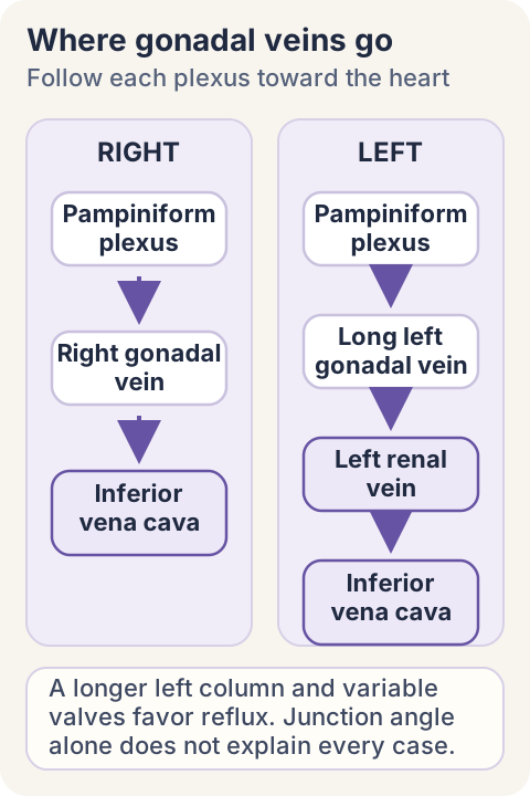
The right and left outlets differ. The right gonadal vein usually enters the inferior vena cava directly. The left gonadal vein is longer and usually enters the left renal vein before blood reaches the cava. Testicular lymphatics follow the gonadal vessels toward para-aortic nodes, a reminder that the testis and its vessels originate high in the posterior abdomen even though the organ descends into the scrotum. [1]
Left-sided predominance is multifactorial. A longer upright hydrostatic column, entry into the left renal vein, and variable valve competence favor reflux. The near-right-angle junction is a useful spatial feature, but angle alone does not produce every varicocele, and the terminal valve is not universally absent. Pressure plus anatomy plus valve function is the more accurate explanation. [1 [1],14 [14]]
Obstruction matters only where it lies. A lesion between the kidney and the gonadal confluence can impair renal drainage while leaving gonadal return able to enter a patent downstream segment. A lesion between that confluence and the cava obstructs the shared exit and can raise pressure in both renal and gonadal tributaries. Compression of the left renal vein between the superior mesenteric artery and aorta is nutcracker physiology; renal masses, thrombus, retroperitoneal disease, and caval obstruction are other possible secondary mechanisms. [1 [1],6 [6]]
Trace each tributary to its next major vessel before predicting which tissue experiences backpressure. Schematic, not to scale. [1]
Use posture as a pressure experiment
Examine the patient standing in a warm room, first at rest and then during Valsalva. Grade I is palpable only with Valsalva, grade II is palpable without Valsalva, and grade III is visible. A lesion detected only by imaging is subclinical, even if a vein is enlarged or reflux is recorded. Palpability and Doppler physiology answer different questions. [2 [2],4 [4]]
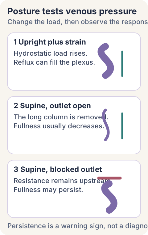
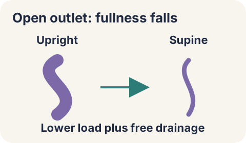
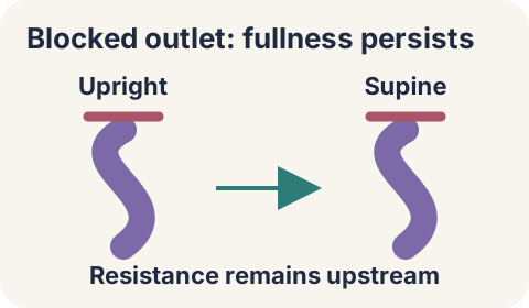
Recumbency removes hydrostatic load. Primary refluxing veins usually become less conspicuous when the patient lies flat because the long upright column is removed and blood can drain. Persistence while supine is not a cancer diagnosis, but it raises concern for sustained outflow resistance when it is new, large, nonreducible, or accompanied by renal, systemic, or lower-extremity findings. [1 [1],3 [3]]
Posture changes the pressure load. Persistence is a reason to reassess context, not proof of one diagnosis. [1 [1],3 [3]]
The testable prediction is simple: if the outlet remains open, venous fullness should fall after recumbency; if a proximal outlet remains obstructed, fullness may persist. This comparison teaches the mechanism even when the optional control is not opened.
Compare the predicted visual results
Free outflow: the supine plexus becomes less full.
Persistent resistance: the supine plexus stays full.
The limitation is that posture is a pressure probe, not a specific etiologic test. History, examination, targeted imaging, and the territories affected determine what comes next.
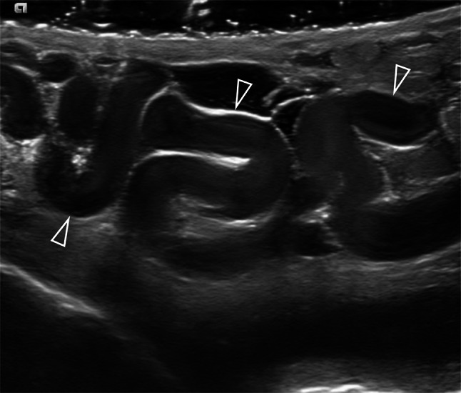
Doppler should be dynamic. When physical examination is uncertain, symptoms and examination conflict, or recurrence is suspected, report patient position, Valsalva, largest vein diameter, and reflux duration. ESUR-SPIWG uses a vein at least 3 mm while standing during Valsalva plus reflux lasting more than 2 seconds as a standardized ultrasound definition. These are diagnostic criteria, not automatic repair thresholds. [1]
Grayscale shows morphology. Direction and duration require color and spectral Doppler during a standardized maneuver. Bertolotto et al., Figure 1, CC BY 4.0. [1]
A clipped waveform is indeterminate. If recording ends while reverse flow continues, the observed interval is only a minimum. Capture the whole event before classifying it. After treatment, residual vein caliber can persist without pathologic reflux, so recurrence requires clinical correlation and an adequate dynamic study rather than a diameter alone. [1]
Try it here · Checkpoint 1 of 3
Make your prediction before reading the choices. A first attempt is just a starting point.
Case 4
Show answer and explanations for case 4
A. Brief reversal bilaterally; retain the negative reflux interpretation (Why this does not fit)
The complete right event lasts only 0.5 seconds and does not meet the greater-than-2-second duration criterion. The left recording stops during reversal, so total left duration is unknown and a bilateral negative interpretation is unsupported.
Reasoning steps for option A
Does the complete right recording demonstrate sustained reflux?
No. Its 0.5-second reversal does not reach the greater-than-two-second ESUR duration threshold.
Can the left clip establish equally brief reversal?
The left trace is censored while flow is still reversed, so total duration is unknown.
B. Brief right reversal, indeterminate left duration; complete left spectral acquisition (Best answer)
A complete 0.5-second event is brief despite the enlarged vein diameter. A complete standing-Valsalva spectral recording must capture its duration; a truncated 0.8-second clip neither proves nor excludes sustained reflux.
Reasoning steps for option B
How should the fully recorded right-sided reversal be classified?
A complete 0.5-second event is brief despite the enlarged vein diameter.
What must be obtained before classifying the left-sided event?
Record the entire left reversal during standing Valsalva before assigning duration.
C. Sustained right reflux, indeterminate left duration; complete left spectral acquisition (Why this does not fit)
A diameter of 3.5 mm describes caliber, not how long blood travels backward. The complete trace records only 0.5 seconds of reversal; completing the left study is appropriate, but labeling the right event sustained is not.
Reasoning steps for option C
What does the right vein diameter establish about time?
A diameter of 3.5 mm describes caliber, not how long blood travels backward.
Which measured fact defeats the right-sided classification?
A finished 0.5-second event is short, regardless of the 3.5-mm caliber.
D. Brief right reversal, sustained left reflux; confirm left pathological reflux (Why this does not fit)
The clip covers only 0.8 seconds, less than the greater-than-two-second criterion. The unseen remainder may or may not contain continued reversal; complete spectral acquisition is needed rather than assuming the missing duration.
Reasoning steps for option D
Does continuous reversal within the left clip meet the time threshold?
The clip covers only 0.8 seconds, less than the greater-than-two-second criterion.
Why is the final left diagnosis premature?
The missing tail of the waveform prevents labeling the event as sustained.
Takeaway: A completed short reversal and a recording cut off during reversal are different kinds of evidence.
Adult laterality is contextual. AUA and ASRM guidance advises against routine abdominal imaging solely because a small or moderate adult varicocele is isolated to the right. New onset, large size, failure to decompress, hematuria, flank symptoms, constitutional findings, lower-extremity edema, or another reason to suspect renal, retroperitoneal, or caval disease changes that decision. EAU guidance is more inclined to investigate an isolated clinical right varicocele, so identify which guideline and clinical context are being applied. [2 [2],3 [3]]
Pediatric screening is different. EAU pediatric guidance recommends abdominal ultrasound in prepubertal boys with varicocele and in isolated right pediatric varicocele to exclude a retroperitoneal or renal cause. Do not transfer the adult laterality-only exception to a child. [4]
Persistent left-sided fullness can localize upstream. The left gonadal vein joins the left renal vein, so left renal venous compression or obstruction can transmit backpressure into the plexus. Bilateral leg edema points toward iliac or caval territory rather than an isolated renal segment. Use the shared drainage territory to localize the level before assigning a cause. [1 [1],6 [6]]
Renal cell carcinoma is one possibility, not the definition of a secondary varicocele. Tumor thrombus, external compression, bland thrombosis, or other obstruction may be involved. Hematuria, flank pain, a renal mass, unexplained erythrocytosis, hypercalcemia, or constitutional symptoms can support renal evaluation, but the classic triad is uncommon and no single paraneoplastic pattern proves the tumor mechanism. [6 [6],9 [9],13 [13]]
Separate venous fullness from a time-sensitive scrotum
Torsion remains the emergency. Sudden severe unilateral pain, nausea or vomiting, a high-riding or horizontal testis, or an absent cremasteric reflex requires immediate surgical assessment when suspicion is high. Preserved arterial Doppler flow, temporary symptom resolution, a normal urinalysis, pain relief with support, or a known varicocele cannot safely exclude early, partial, or intermittent torsion. Do not delay urgent care to repeat an elective venous study. [5]
Epididymitis has a different pattern. Gradual posterior pain, epididymal tenderness or hyperemia, dysuria, fever, urethral symptoms, and exposure history support infection. For acute epididymitis most likely caused by gonorrhea or chlamydia, CDC guidance recommends ceftriaxone 500 mg intramuscularly once, or 1 g if body weight is at least 150 kg, plus doxycycline 100 mg orally twice daily for 10 days. Enteric-risk pathways use different regimens, and presumptive treatment begins while tests are pending. [7]
A complicated hernia outranks an incidental varicocele. Bowel in the inguinal canal with irreducibility, severe pain, vomiting, abdominal distension, or erythema raises concern for incarceration or strangulation and needs urgent surgical assessment. A reducible bulge that enlarges with cough is not the same compartment as dilated cord veins. [5]
Fluid and cysts occupy other compartments. A hydrocele surrounds the testis within the tunica vaginalis and may transilluminate; a communicating hydrocele can fluctuate through a patent processus vaginalis. A spermatocele is a cystic lesion related to the epididymis. Neither is diagnosed by calling every painless swelling a varicocele. [11]
A solid intratesticular mass follows a tumor pathway. Normal alpha-fetoprotein, beta-human chorionic gonadotropin, and lactate dehydrogenase do not exclude malignancy. Compartment and tissue character matter more than the coexistence of a benign venous lesion. [8]
Changed pain deserves relocalization. Typical varicocele discomfort is dull, dependent, and often worse after standing or exertion. Colicky flank-to-groin pain with hematuria suggests a urinary source; focal groin findings suggest hernia; neuropathic or musculoskeletal features suggest another pathway. Reassess a new pattern before attributing it to a familiar diagnosis. [10]
Treat the clinical consequence, not the vein alone
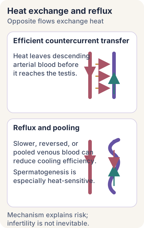
The pampiniform plexus is a heat exchanger. Cooler venous blood returning from the testis lies beside descending arterial blood and accepts heat before the artery reaches the gonad. Reflux, venous pooling, and dilation can impair this countercurrent system and contribute to heat stress, oxidative stress, inflammation, and germ-cell injury. These mechanisms can impair semen quality, but a varicocele does not guarantee infertility. [1 [1],14 [14],15 [15]]
Venous treatment changes the testicular environment, not the patency of the sperm transport ducts. [1 [1],14 [14]]
Spermatogenesis is especially temperature-sensitive. Sertoli-cell support and germ-cell development can be impaired, with changes in sperm concentration, motility, morphology, DNA integrity, and inhibin signaling. Leydig-cell testosterone production is often relatively preserved, but it is not invulnerable; severe disease and hypogonadism require direct endocrine assessment rather than assuming testosterone is normal. [2 [2],14 [14]]
Adult fertility repair has a defined target population. Discuss repair when a man is attempting conception, has a palpable clinical varicocele, has infertility with abnormal semen parameters, and the couple has otherwise unexplained or potentially correctable male-factor infertility. Do not recommend fertility-directed repair for an imaging-only lesion, and do not assume benefit when semen is normal. Nonobstructive azoospermia requires separate counseling because evidence is less certain. [2 [2],12 [12],15 [15]]
Evaluate the couple and the trend. One normal semen analysis can be sufficient under EAU guidance; an abnormal baseline should be confirmed on at least one additional analysis. Consider the partner's age, ovarian reserve, treatment timeline, and preferences because improvement may take up to two spermatogenic cycles and spontaneous pregnancy, when it occurs, is often observed six to twelve months after repair. [2]
Pain is a diagnosis of exclusion. Begin with scrotal support, activity modification when relevant, and appropriately labeled analgesia if safe. Consider repair for persistent typical pain after other causes are excluded and conservative care fails, while counseling that pain may persist. There is no medication that reverses gonadal venous reflux; drugs treat symptoms or an alternative diagnosis such as infection, not the valve and pressure problem itself. [10 [10],15 [15]]
Try it here · Checkpoint 2 of 3
Make your prediction before reading the choices. A first attempt is just a starting point.
Case 9
Show answer and explanations for case 9
A. Defer active fertility planning until repeat testing next year (Why this does not fit)
Seventeen months without conception and two low-motility analyses establish a reason for active couple-level assessment. A palpable potentially contributory lesion is already present, so deferring discussion ignores an available treatment option without a supplied reason for delay.
Reasoning steps for option A
Has the couple already supplied evidence of a persistent fertility problem?
Seventeen months without conception and two low-motility analyses establish a reason for active couple-level assessment.
Why does another year of observation not fit the next discussion?
A palpable potentially contributory lesion is already present, so deferring discussion ignores an available treatment option without a supplied reason for delay.
B. Prioritize assisted reproduction instead of considering venous repair (Why this does not fit)
Assisted reproduction is a legitimate option when timing or other couple factors favor it, but the stem does not make it categorically preferable. Infertility, reproducible semen impairment, and a palpable lesion support including repair in shared discussion.
Reasoning steps for option B
Can assisted reproduction belong in this couple's treatment plan?
Yes. It is a legitimate fertility option, particularly when reproductive timing or other factors favor it.
What prevents declaring it preferable from this stem?
Nothing in the stem makes assisted reproduction categorically preferable to discussing repair.
C. Require visible grade III disease before discussing intervention (Why this does not fit)
Clinical palpability, including a lesion detected during Valsalva, matters more than requiring visible grade III disease. The lesion is already palpable, so imposing a visibility threshold would withhold discussion despite infertility and reproducibly abnormal semen.
Reasoning steps for option C
Which examination category matters for the usual repair recommendation?
Clinical palpability, including a lesion detected during Valsalva, matters more than requiring visible grade III disease.
How does the given examination affect eligibility?
The lesion is already palpable, so imposing a visibility threshold would withhold discussion despite infertility and reproducibly abnormal semen.
D. Include varicocele repair among the couple's fertility options (Best answer)
The couple has infertility, a palpable varicocele and reproducibly abnormal motility rather than an imaging-only lesion. The absence of a major limiting factor permits consideration of repair alongside other fertility options; age, ovarian reserve, timing and preferences still guide the eventual choice.
Reasoning steps for option D
Which supplied findings support consideration of repair?
The couple has infertility, a palpable varicocele and reproducibly abnormal motility rather than an imaging-only lesion.
How should the partner evaluation shape the discussion?
With no dominant partner limitation, repair can be weighed beside other fertility strategies.
Takeaway: Fertility repair candidacy depends on a palpable lesion, reproducible semen impairment and the couple's reproductive context.
Follow growth, function, symptoms, and complications over time
Adolescent growth requires serial comparison. A testis more than 2 mL or 20 percent smaller than the other is considered hypotrophic under EAU pediatric guidance. Confirm persistence at two comparable visits six months apart because asynchronous pubertal growth and measurement variation can create a temporary difference. Persistent qualifying asymmetry supports offering pediatric urology consultation and a repair discussion without promising future paternity benefit. [4]
After repair, follow the original goal. For fertility, repeat semen testing after sufficient time for spermatogenesis and interpret the trend with the couple's plan. For pain, track the preoperative pain pattern rather than any scrotal sensation. For adolescents, compare bilateral testicular growth. A technically treated vein is not the only outcome that matters. [2 [2],4 [4],15 [15]]
Different postoperative findings imply different mechanisms. Persistent or recurrent reflux can reflect untreated collateral veins or recanalization. A new simple circumtesticular fluid collection can be a hydrocele from lymphatic disruption. Residual venous caliber without sustained reflux may be an anatomic remnant rather than failure. Dynamic Doppler and compartment localization prevent unnecessary retreatment. [1 [1],4 [4],11 [11]]
Procedural complications deserve explicit counseling. Discuss recurrence, hydrocele, hematoma, infection, arterial injury, testicular injury or atrophy, chronic or persistent pain, and the possibility that semen or pregnancy outcomes may not improve. Lymphatic-sparing and optical magnification reduce selected complications in pediatric guidance, while individual anatomy and expertise influence technique choice. [4 [4],15 [15]]
Beyond: apply thresholds without turning them into shortcuts
Observation is active management when the lesion is stable, symptoms are absent or mild, testicular size is symmetric, semen is normal, and no secondary warning sign is present. Document the examination, explain what changes should prompt reassessment, and avoid serial imaging that has no clinical question. [2 [2],3 [3],15 [15]]
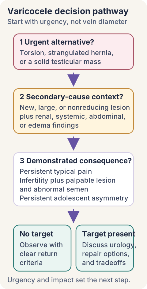
Referral urgency follows the competing diagnosis. Sudden high-risk acute scrotal pain needs immediate surgical assessment. A solid intratesticular mass needs prompt urologic evaluation. A new large nonreducing varicocele with renal, systemic, or edema findings needs targeted upstream assessment. Stable uncomplicated reflux can follow a routine urology or fertility pathway. [3 [3],5 [5],6 [6],8 [8]]
Choose the pathway from urgency, secondary-cause context, and demonstrated clinical consequence, not from venous diameter alone. [2 [2],3 [3],4 [4],5 [5],12 [12]]
Procedure choice is individualized. Microsurgical inguinal or subinguinal ligation, laparoscopic or open approaches, and radiologic embolization each have different access, anesthesia, recurrence, hydrocele, radiation, and complication profiles. Current reviews and guidelines generally favor magnification and lymphatic preservation where applicable, but the best choice depends on age, anatomy, prior procedures, local expertise, and the treatment goal. [2 [2],4 [4],15 [15]]
Common threshold errors are predictable. A 3-mm vein plus reflux longer than 2 seconds supports an ultrasound diagnosis, not surgery by itself. Right-sidedness in an adult is not an automatic computed tomography order. One abnormal adolescent volume measurement is not persistent hypotrophy. A normal tumor marker panel does not clear a solid mass. Preserved arterial flow does not clear a high-risk torsion presentation. [1 [1],3 [3],4 [4],5 [5],8 [8]]
Try it here · Checkpoint 3 of 3
Make your prediction before reading the choices. A first attempt is just a starting point.
Case 25
Show answer and explanations for case 25
A. Residual dilation; continue clinical outcome-based follow-up (Best answer)
Venous caliber can remain enlarged after successful treatment. The adequate dynamic study shows no sustained reflux and heaviness has resolved, supporting outcome-based follow-up rather than diameter-driven retreatment.
Reasoning steps for option A
Does a residual 3.5-mm vein itself establish recurrence?
No. Venous caliber can remain enlarged after treatment even when pathological reflux has resolved.
How do the functional and symptom findings guide the plan?
Resolved symptoms and absent dynamic reflux support observation despite residual caliber.
B. Recurrent reflux; plan another venous intervention (Why this does not fit)
Sustained reverse flow on an adequate dynamic study would provide hemodynamic evidence of recurrence. It does not, and symptoms have improved; residual caliber alone does not justify planning another venous intervention.
Reasoning steps for option B
What finding would be needed to support recurrent reflux?
Sustained reverse flow on an adequate dynamic study would provide hemodynamic evidence of recurrence.
Does the supplied study provide that evidence?
It does not, and symptoms have improved; residual caliber alone does not justify planning another venous intervention.
C. Secondary obstruction; investigate abdominal venous outflow (Why this does not fit)
The residual diameter is an anatomical finding rather than a direct measurement of proximal outflow resistance. Absent sustained reflux on adequate testing and resolved heaviness favor residual dilation; new upstream warning signs would be needed to justify the proposed obstruction pathway.
Reasoning steps for option C
What postoperative observation is being overinterpreted?
The residual diameter is an anatomical finding rather than a direct measurement of proximal outflow resistance.
What does the overall supplied course support instead?
The course fits harmless residual dilation unless new upstream warning signs appear.
D. Postoperative hydrocele; assess for fluid-directed treatment (Why this does not fit)
A hydrocele is a fluid collection in the space surrounding the testis rather than a measured venous lumen. No surrounding fluid collection is present, so a hydrocele-directed procedure is not supported by the demonstrated residual vein.
Reasoning steps for option D
How would a hydrocele differ from a residual vein on ultrasound?
A hydrocele is a fluid collection in the space surrounding the testis rather than a measured venous lumen.
Which supplied finding rules against that explanation of this scan?
No surrounding fluid collection is present, so a hydrocele-directed procedure is not supported by the demonstrated residual vein.
Takeaway: Persistent vein caliber is not the same as persistent reflux; judge follow-up against flow and the original clinical problem.
The cases below require one final decision. Trace the venous territory, classify the evidence as physical, dynamic, or longitudinal, rule out urgent alternatives, and then select the action supported by the demonstrated clinical consequence.
Case 1
Show answer and explanations for case 1
A. Left renal outflow impaired; left gonadal outflow impaired (Why this does not fit)
The hilar segment is in the renal route, so renal outflow is impaired. The gonadal vein joins beyond the block and retains a patent path to the cava, so impairment of both outlets is not predicted.
Reasoning steps for option A
Which part of the left renal outlet is occluded?
The occluded hilar segment lies in the route from the left kidney to the cava.
Does left gonadal blood traverse that occluded segment?
The tributary joins distal to the lesion, so its direct caval path stays available.
B. Left renal outflow spared; left gonadal outflow impaired (Why this does not fit)
Its usual main outflow crosses the occluded hilar segment, so renal outflow is not spared. The gonadal junction and its downstream caval connection are patent; this lesion does not block that main outlet.
Reasoning steps for option B
Where must blood leaving the left kidney pass?
Its usual main outflow crosses the occluded hilar segment, so renal outflow is not spared.
Which finding contradicts gonadal outlet obstruction?
The gonadal junction and its downstream caval connection are patent; this lesion does not block that main outlet.
C. Left renal outflow impaired; left gonadal outflow spared (Best answer)
The occlusion lies between the kidney and its venous junction, impairing the usual left renal outflow. It joins on the patent caval side of the occlusion, leaving its direct outlet open despite impaired renal drainage.
Reasoning steps for option C
Which supplied boundary determines the kidney effect?
The occlusion lies between the kidney and its venous junction, impairing the usual left renal outflow.
Where does the gonadal return join relative to this block?
It joins on the patent caval side of the occlusion, leaving its direct outlet open despite impaired renal drainage.
D. Left renal outflow spared; left gonadal outflow spared (Why this does not fit)
The left gonadal vein enters the patent downstream segment and retains its outlet. The kidney lies upstream of the occluded segment; downstream patency does not reopen its interrupted main route.
Reasoning steps for option D
Which pathway can reach the cava without crossing the block?
The left gonadal vein enters the patent downstream segment and retains its outlet.
Why cannot the same conclusion be applied to the kidney?
The kidney lies upstream of the occluded segment; downstream patency does not reopen its interrupted main route.
Takeaway: Place an obstruction relative to a tributary junction before predicting which upstream tissues lose their outlet.
A. Caval segment above the right gonadal junction (Best answer)
Both common iliac veins deliver lower-limb return into the inferior vena cava. Caval obstruction at or above the right gonadal entry can impede both pathways, unifying the new bilateral edema and right venous congestion.
Reasoning steps for option A
Which venous territory receives return from both legs?
Both common iliac veins deliver lower-limb return into the inferior vena cava.
What level also explains the persistent right cord fullness?
Only a caval lesion proximal to both entries explains congestion in the cord and both legs.
B. Left renal segment beyond the gonadal junction (Why this does not fit)
The left gonadal vein joins it, so an obstruction there primarily implicates left gonadal outflow. Right cord fullness and bilateral leg edema involve drainage into the cava, not an isolated left renal segment.
Reasoning steps for option B
Which gonadal vein usually joins the left renal vein?
The left gonadal vein joins it, so an obstruction there primarily implicates left gonadal outflow.
Which two supplied findings remain unexplained?
Right cord fullness and bilateral leg edema involve drainage into the cava, not an isolated left renal segment.
C. Right renal segment before the caval junction (Why this does not fit)
It usually enters the inferior vena cava directly, separately from the right renal vein. It does not interrupt both iliac outlets; relevant caval involvement would be needed to unify the scrotal and lower-limb findings.
Reasoning steps for option C
Where does the right gonadal vein usually terminate?
It usually enters the inferior vena cava directly, separately from the right renal vein.
Can isolated right renal obstruction explain both swollen legs?
It does not interrupt both iliac outlets; relevant caval involvement would be needed to unify the scrotal and lower-limb findings.
D. Right common iliac segment below the caval junction (Why this does not fit)
It would chiefly impair venous return from the right lower limb. The other leg is also swollen, and the right gonadal entry lies above the iliac confluence, favoring a more proximal shared obstruction.
Reasoning steps for option D
Which leg would an isolated right common iliac block chiefly affect?
It would chiefly impair venous return from the right lower limb.
Why is that insufficient for this presentation?
One iliac blockage cannot swell both legs or raise pressure at the gonadal entry above it.
Takeaway: Bilateral leg edema plus new right scrotal venous congestion localizes concern to their shared caval outlet.
A. Cystoscopy to inspect the bladder and urethra (Why this does not fit)
It evaluates the bladder and urethra and may be indicated within a complete hematuria assessment. It does not assess the renal or retroperitoneal process that could obstruct left gonadal return and maintain the newly distended veins.
Reasoning steps for option A
What part of hematuria evaluation can cystoscopy address?
It evaluates the bladder and urethra and may be indicated within a complete hematuria assessment.
What does it leave unanswered in this patient?
It does not assess the renal or retroperitoneal process that could obstruct left gonadal return and maintain the newly distended veins.
B. Interval scrotal examination after supportive care (Why this does not fit)
Stable uncomplicated positional fullness without concerning associated findings can be followed according to clinical impact. New supine persistence and hematuria introduce an upstream concern that a trial of scrotal support would not evaluate.
Reasoning steps for option B
When can conservative follow-up fit a varicocele?
Stable uncomplicated positional fullness without concerning associated findings can be followed according to clinical impact.
Which facts argue against deferring investigation here?
New supine persistence and hematuria introduce an upstream concern that a trial of scrotal support would not evaluate.
C. Repeat Doppler confined to the scrotal veins (Why this does not fit)
It could characterize the veins and quantify reverse flow in the scrotum. The new hematuria and persistent left distension implicate renal outflow, which lies outside the proposed examination.
Reasoning steps for option C
What could repeat scrotal Doppler establish?
It could characterize the veins and quantify reverse flow in the scrotum.
Why is a scrotum-only study incomplete here?
The suspected lesion is above the scrotum, in renal or retroperitoneal territory.
D. Renal and retroperitoneal imaging for outflow obstruction (Best answer)
Left gonadal return joins the left renal vein, allowing renal outflow disease to produce persistent scrotal venous congestion. It adds a renal or urinary warning sign, supporting targeted upper-tract and retroperitoneal imaging chosen for renal function and clinical context; other indicated hematuria evaluation remains necessary.
Reasoning steps for option D
Which route connects this left-sided swelling to the urinary system?
Left gonadal return joins the left renal vein, allowing renal outflow disease to produce persistent scrotal venous congestion.
How does hematuria affect the next investigation?
The urinary warning sign shifts imaging toward the upper tract and venous outflow.
Takeaway: A scrotal finding and a urinary finding may share a renal venous cause; investigate the connecting anatomy.
A. Sustained reflux; offer elective fertility-directed repair (Why this does not fit)
The Doppler findings establish sustained reflux, but the supplied clinical findings do not establish a fertility, growth, or pain target. Normal semen results, symmetric testes, and absent pain do not support fertility-directed repair.
Reasoning steps for option A
Do the Doppler values support a refluxing varicocele?
Standing-Valsalva diameter of 3.4 mm and reversal lasting 2.6 seconds satisfy the stated ESUR criteria.
What clinical benefit is established for repair in this case?
None is established by the supplied findings: repeated semen results are normal, testes are symmetric, and the patient has no pain.
B. Sustained reflux; observe and reassess clinical impact (Best answer)
They support a refluxing clinical varicocele because the lesion is palpable and meets the stated dynamic ultrasound criteria. Normal semen results, symmetric testes and absent pain leave no current demonstrated treatment target, making observation with clinical reassessment appropriate.
Reasoning steps for option B
What do the diameter and timed reverse flow demonstrate?
They support a refluxing clinical varicocele because the lesion is palpable and meets the stated dynamic ultrasound criteria.
Why does that finding not mandate treatment?
Normal semen results, symmetric testes and absent pain leave no current demonstrated treatment target, making observation with clinical reassessment appropriate.
C. Physiological reversal; document no venous abnormality (Why this does not fit)
It lasts 2.6 seconds, exceeding the ESUR threshold of more than two seconds during standing Valsalva. No. Reassuring function supports observation, but it does not negate the palpable refluxing venous abnormality.
Reasoning steps for option C
What separates the measured reversal from a brief strain response?
It lasts 2.6 seconds, exceeding the ESUR threshold of more than two seconds during standing Valsalva.
Does normal testicular function erase this diagnostic finding?
No. Reassuring function supports observation, but it does not negate the palpable refluxing venous abnormality.
D. Secondary obstruction; obtain upper-tract imaging first (Why this does not fit)
The swelling is longstanding and diminishes supine rather than presenting as new persistent venous congestion. It demonstrates reflux, not its cause; the supplied history and examination do not establish an upstream imaging indication.
Reasoning steps for option D
Which history supports uncomplicated positional reflux?
The swelling is longstanding and diminishes supine rather than presenting as new persistent venous congestion.
What does the positive Doppler study prove about the abdomen?
It demonstrates reflux, not its cause; the supplied history and examination do not establish an upstream imaging indication.
Takeaway: A diagnostic reflux threshold identifies a finding, not an automatic reason to intervene.
A. Repeat elective Doppler to quantify standing venous reflux (Why this does not fit)
It typically causes dependent heaviness or dull aching rather than abrupt nausea-associated attacks that end spontaneously. Quantifying the incidental varicocele would leave possible torsion-detorsion unaddressed and could delay urgent urologic assessment.
Reasoning steps for option A
What symptom pattern does positional venous reflux usually produce?
It typically causes dependent heaviness or dull aching rather than abrupt nausea-associated attacks that end spontaneously.
Why should elective venous testing not be the next step?
Quantifying the incidental varicocele would leave possible torsion-detorsion unaddressed and could delay urgent urologic assessment.
B. Arrange routine follow-up after a trial of scrotal support (Why this does not fit)
No. A twisted cord may untwist, allowing pain and perfusion to normalize between episodes. The abrupt recurrent attacks with nausea suggest a recurrent ischemic risk that warrants urgent assessment rather than a treatment trial.
Reasoning steps for option B
Does spontaneous resolution identify benign venous aching?
No. A twisted cord may untwist, allowing pain and perfusion to normalize between episodes.
What is unsafe about routine follow-up after support here?
Recurrent abrupt attacks with nausea can represent torsion-detorsion and need urgent review.
C. Start empiric antibiotics and reassess after 72 hours (Why this does not fit)
No fever, dysuria or discharge is reported, and the attacks are abrupt and self-resolving rather than progressively inflammatory. Waiting 72 hours could defer assessment of intermittent torsion; the history requires urgent urologic evaluation even with preserved interval flow.
Reasoning steps for option C
Which supplied findings support an inflammatory infection?
No fever, dysuria or discharge is reported, and the attacks are abrupt and self-resolving rather than progressively inflammatory.
Why is a response-to-antibiotics interval inappropriate?
Waiting 72 hours could defer assessment of intermittent torsion; the history requires urgent urologic evaluation even with preserved interval flow.
D. Obtain urgent urologic assessment for intermittent torsion (Best answer)
Torsion followed by detorsion can produce severe unilateral attacks with nausea and then apparent recovery. The scan was obtained between attacks, when flow may have returned; urgent urologic assessment is needed, with emergency reassessment for recurrent acute pain.
Reasoning steps for option D
What mechanism can produce abrupt pain followed by spontaneous relief?
Torsion followed by detorsion can produce severe unilateral attacks with nausea and then apparent recovery.
Why does symmetric arterial flow not end the assessment?
The scan was obtained between attacks, when flow may have returned; urgent urologic assessment is needed, with emergency reassessment for recurrent acute pain.
Takeaway: A normal scan between abrupt painful episodes cannot exclude a transient interruption of testicular blood flow.
A. Brief reversal; avoid repair based on diameter alone (Best answer)
The complete 0.5-second reversal is brief despite a 3.5-mm diameter. Nothing is palpable, so a clinical varicocele is not demonstrated and repair based on diameter alone is not justified.
Reasoning steps for option A
Does a complete 0.5-second reversal meet the stated reflux threshold?
No. It falls below the ESUR threshold of more than two seconds during standing Valsalva despite a diameter of 3.5 mm.
What does the examination add to the treatment decision?
No lesion is palpable, so a clinical varicocele is not demonstrated; these findings do not justify a diameter-based repair decision.
B. Clinical varicocele; select embolization using venous diameter (Why this does not fit)
A clinical varicocele is palpable on examination, including during Valsalva when appropriate. Nothing is palpable and the reversal is brief; neither a clinical lesion nor an embolization indication follows from the 3.5-mm diameter.
Reasoning steps for option B
What makes a varicocele clinical rather than imaging-only?
A clinical varicocele is palpable on examination, including during Valsalva when appropriate.
Why does this option misclassify and overtreat the finding?
Nothing is palpable and the reversal is brief; neither a clinical lesion nor an embolization indication follows from the 3.5-mm diameter.
C. Proximal obstruction; obtain renal and retroperitoneal imaging (Why this does not fit)
It documents a short directional change, not persistent pressure from an upstream obstruction. New persistent fullness, a large nondecompressing lesion or associated urinary or systemic findings would add concern; the recorded 0.5-second reversal alone does not.
Reasoning steps for option C
What does a brief response to strain establish about outflow?
It documents a short directional change, not persistent pressure from an upstream obstruction.
Which evidence would strengthen an abdominal concern?
Concern would rise with a new large nonreducible lesion or renal, systemic, or edema findings.
D. Subclinical sustained reflux; follow without fertility-directed repair (Why this does not fit)
No. A genuinely nonpalpable varicocele is not a standard fertility-directed repair indication. The observation plan avoids overtreatment, but the claimed sustained reflux contradicts the complete measured duration of only 0.5 seconds.
Reasoning steps for option D
Would imaging-only sustained reflux ordinarily justify fertility repair?
No. A genuinely nonpalpable varicocele is not a standard fertility-directed repair indication.
Why is this still not the best interpretation of the study?
The observation plan avoids overtreatment, but the claimed sustained reflux contradicts the complete measured duration of only 0.5 seconds.
Takeaway: Use measured reflux duration and clinical palpability separately; enlarged veins alone do not establish a repair indication.
A. More effective arterial cooling with newly restored duct patency (Why this does not fit)
Reducing reflux may improve the heat-exchange environment around testicular arterial inflow. No duct obstruction is demonstrated, and a procedure directed at veins does not directly reopen the vas or epididymal ducts.
Reasoning steps for option A
Which proposed effect of venous repair fits the reflux finding?
Reducing reflux may improve the heat-exchange environment around testicular arterial inflow.
Why is newly restored duct patency not supported?
No duct obstruction is demonstrated, and a procedure directed at veins does not directly reopen the vas or epididymal ducts.
B. Less effective arterial cooling with unchanged duct patency (Why this does not fit)
No. The supplied ducts have no demonstrated obstruction and are separate from the treated venous pathway. A beneficial heat-exchange effect would improve cooling rather than make it less effective; unchanged duct status does not rescue that incorrect thermal prediction.
Reasoning steps for option B
Would treating these veins necessarily alter duct patency?
No. The supplied ducts have no demonstrated obstruction and are separate from the treated venous pathway.
Which direction of thermal change conflicts with the question?
Successful venous treatment should not worsen cooling; duct status is a separate variable.
C. More effective arterial cooling with unchanged duct patency (Best answer)
Reflux can disturb countercurrent heat exchange and the testicular environment without blocking sperm transport. Cooling may improve while already patent ducts remain unchanged; the physiological rationale does not guarantee improved semen quality or pregnancy.
Reasoning steps for option C
Which process links venous reflux to sperm dysfunction here?
Reflux can disturb countercurrent heat exchange and the testicular environment without blocking sperm transport.
What paired response follows from a beneficial venous repair?
Cooling may improve while already patent ducts remain unchanged; the physiological rationale does not guarantee improved semen quality or pregnancy.
D. Unchanged arterial cooling with newly restored duct patency (Why this does not fit)
The intervention treats refluxing veins surrounding the testicular arterial supply, not a blocked sperm-transport duct. It attributes benefit to a newly opened duct despite absent obstruction and ignores the heat-exchange effect the question asks the learner to predict.
Reasoning steps for option D
What procedure target is actually described?
The intervention treats refluxing veins surrounding the testicular arterial supply, not a blocked sperm-transport duct.
Why does this prediction miss the relevant physiology?
It attributes benefit to a newly opened duct despite absent obstruction and ignores the heat-exchange effect the question asks the learner to predict.
Takeaway: Venous treatment targets the testicular environment, not a sperm-transport duct.
A. Begin couple-level fertility care without varicocele repair (Best answer)
A lesion not palpable at rest or with strain is subclinical even when ultrasound documents reflux. Avoid unsupported venous repair and proceed with timely couple-level fertility planning, because established infertility and a 39-year-old partner make delay consequential.
Reasoning steps for option A
How is Doppler reflux without palpability classified?
A lesion not palpable at rest or with strain is subclinical even when ultrasound documents reflux.
What plan accounts for both that classification and partner age?
Classify the vein as subclinical and proceed promptly with couple-level fertility care.
B. Complete venous repair before planning assisted reproduction (Why this does not fit)
No. The examination remains nonpalpable, so evidence for repair of a palpable varicocele cannot simply be transferred to it. It could postpone appropriate fertility care in a couple already infertile for 15 months with a time-sensitive partner assessment.
Reasoning steps for option B
Does persistent low concentration change an imaging-only lesion into a clinical varicocele?
No. The examination remains nonpalpable, so evidence for repair of a palpable varicocele cannot simply be transferred to it.
Why is making repair a prerequisite especially problematic?
It could postpone appropriate fertility care in a couple already infertile for 15 months with a time-sensitive partner assessment.
C. Repeat venous ultrasound in six months before fertility referral (Why this does not fit)
The couple already has 15 months without conception and repeated low sperm concentration. It would not establish a supported repair indication for the nonpalpable lesion and would defer needed couple-level planning at partner age 39.
Reasoning steps for option C
What unresolved problem needs attention now?
The couple already has 15 months without conception and repeated low sperm concentration.
Would waiting for another venous scan address that problem?
Another scan would not make the lesion palpable and would consume limited reproductive time.
D. Observe another year before repeating the couple's evaluation (Why this does not fit)
No. The duration without conception and reproducible semen abnormality already warrant evaluation and planning. The partner is 39, so another year of delay would disregard reproductive timing without a demonstrated benefit from observation.
Reasoning steps for option D
Does this couple need to wait longer to establish a fertility concern?
No. The duration without conception and reproducible semen abnormality already warrant evaluation and planning.
Which partner fact further argues against waiting?
The partner is 39, so another year of delay would disregard reproductive timing without a demonstrated benefit from observation.
Takeaway: An imaging-only lesion should not delay time-sensitive fertility care for the couple.
A. Offer repair using the first low sperm concentration (Why this does not fit)
Two subsequent samples returned to normal, so the initial abnormality was not reproducible after recovery. The current evidence lacks persistent semen impairment, pain or testicular asymmetry; the earlier low value does not establish an ongoing benefit from repair.
Reasoning steps for option A
What happened to semen concentration after recovery from the febrile illness?
Two subsequent samples returned to normal, so the initial abnormality was not reproducible after recovery.
Why does the first result not justify repair now?
The current evidence lacks persistent semen impairment, pain or testicular asymmetry; the earlier low value does not establish an ongoing benefit from repair.
B. Offer embolization to prevent another low sperm count (Why this does not fit)
No. The low concentration occurred after illness and was followed by normal repeat results. A current clinical treatment target is missing, and the supplied history does not establish that embolization would prevent another transient low result.
Reasoning steps for option B
Is ongoing venous injury established by this semen trajectory?
No. The low concentration occurred after illness and was followed by normal repeat results.
What is missing from the proposed preventive intervention?
There is no persistent abnormal semen result, pain, or asymmetry to prevent.
C. Observe and reassess when symptoms or goals change (Best answer)
The normal post-recovery measurements argue against persistent semen impairment rather than proving that all future fertility is assured. No pain, asymmetry or current conception concern is supplied, so observation and reassessment for changing symptoms or reproductive goals fit better than preventive intervention.
Reasoning steps for option C
How should the isolated abnormal result be weighed against the repeats?
The normal post-recovery measurements argue against persistent semen impairment rather than proving that all future fertility is assured.
What management follows from the present clinical state?
No pain, asymmetry or current conception concern is supplied, so observation and reassessment for changing symptoms or reproductive goals fit better than preventive intervention.
D. Obtain abdominal imaging for the transient semen change (Why this does not fit)
No. Semen concentration is a functional measurement, not anatomical evidence of renal or caval obstruction. New atypical venous fullness or other upstream findings would add an indication; a resolved post-illness semen abnormality alone does not supply one.
Reasoning steps for option D
Does the semen result identify where venous drainage is obstructed?
No. Semen concentration is a functional measurement, not anatomical evidence of renal or caval obstruction.
What else would be needed to support this proposed investigation?
New atypical venous fullness or other upstream findings would add an indication; a resolved post-illness semen abnormality alone does not supply one.
Takeaway: Interpret semen results over time before assigning a persistent treatment target to a varicocele.
A. Continue serial ultrasound observation because percentage asymmetry remains below twenty percent (Why this does not fit)
The difference is 17 minus 14, or 3 mL; 3 divided by 17 is about 17.6 percent. No. The EAU criterion uses an absolute difference greater than 2 mL or a 20 percent difference, and the qualifying absolute discrepancy persists across both visits.
Reasoning steps for option A
What is the volume difference and its percentage of the larger testis?
The difference is 17 minus 14, or 3 mL; 3 divided by 17 is about 17.6 percent.
Does the percentage below twenty negate the absolute threshold?
The persistent 3-mL deficit satisfies the absolute criterion even though 17.6 percent does not.
B. Offer pediatric urology consultation to discuss repair versus individualized follow-up (Best answer)
The 3-mL difference exceeds 2 mL even though the relative difference is about 17.6 percent. The same asymmetry is present at two comparable ultrasound assessments six months apart, rather than on a single variable measurement. Offer pediatric urology consultation with discussion of repair and individualized follow-up; EAU guidance recommends offering surgery for persistent small testis, while future paternity benefit remains uncertain.
Reasoning steps for option B
Does the absolute difference meet the pediatric size criterion?
The 3-mL difference exceeds 2 mL even though the relative difference is about 17.6 percent.
Has the required persistence been established?
The same asymmetry is present at two comparable ultrasound assessments six months apart, rather than on a single variable measurement.
What should the clinician offer rather than simply postpone?
Offer pediatric urology consultation with discussion of repair and individualized follow-up; EAU guidance recommends offering surgery for persistent small testis, while future paternity benefit remains uncertain.
C. Defer intervention discussion until the smaller testis shows an absolute loss of volume (Why this does not fit)
No. Persistent failure to match contralateral growth can be relevant even without an absolute decline. A greater-than-2-mL difference has already persisted across comparable visits six months apart, supporting an offer of treatment discussion now.
Reasoning steps for option C
Must the smaller testis lose volume to show a concerning discrepancy?
No. Persistent failure to match contralateral growth can be relevant even without an absolute decline.
What established finding makes further loss unnecessary here?
A greater-than-2-mL difference has already persisted across comparable visits six months apart, supporting an offer of treatment discussion now.
D. Select a repair threshold from vein diameter rather than serial testicular measurements (Why this does not fit)
Serial volumes compare the affected testis with its contralateral counterpart and establish persistent asymmetry. Venous caliber describes anatomy but does not replace the already documented persistent 3-mL testicular deficit used for pediatric repair counseling.
Reasoning steps for option D
Which measurement addresses the testicular-growth concern?
Serial volumes compare the affected testis with its contralateral counterpart and establish persistent asymmetry.
Why would substituting venous diameter miss the decision?
Venous caliber describes anatomy but does not replace the already documented persistent 3-mL testicular deficit used for pediatric repair counseling.
Takeaway: Persistent adolescent asymmetry can qualify through the absolute volume difference even when the percentage is below twenty percent.
A. Comparable bilateral testicular volumes six months later (Best answer)
The 16-mL and 13-mL testes differ by 3 mL, exceeding the absolute greater-than-2-mL criterion. A comparable assessment six months later is needed to determine persistence; a single asymptomatic measurement may reflect temporary asynchronous growth or measurement variation.
Reasoning steps for option A
What does the first measurement establish quantitatively?
The 16-mL and 13-mL testes differ by 3 mL, exceeding the absolute greater-than-2-mL criterion.
What important evidence is still missing?
A second comparable volume pair determines whether the asymmetry persists.
B. The initial absolute difference without another volume study (Why this does not fit)
A 3-mL difference exceeds the absolute size threshold, but it has been measured only once. Serial comparable assessment is needed before routine intervention in this asymptomatic adolescent.
Reasoning steps for option B
Does the first absolute difference deserve attention?
Yes. A 3-mL difference exceeds the absolute size threshold and should not be dismissed.
Why is this result alone insufficient for the stated decision?
One qualifying measurement cannot distinguish stable hypotrophy from temporary asynchronous growth.
C. The initial percentage difference without serial comparison (Why this does not fit)
Three divided by sixteen is 18.75 percent, below twenty percent. The absolute difference independently exceeds 2 mL, but persistence remains untested; percentage alone neither dismisses the difference nor establishes its duration.
Reasoning steps for option C
What is the initial percentage difference using the larger testis?
Three divided by sixteen is 18.75 percent, below twenty percent.
Why can that percentage not settle the management question?
The percentage is below 20, but the absolute 3-mL deficit still needs serial confirmation.
D. A repeat pampiniform vein diameter during standing strain (Why this does not fit)
It would assess venous caliber under a specified loading condition. No. The decision concerns comparative testicular growth, requiring bilateral volume measurements rather than a venous diameter substitute.
Reasoning steps for option D
What feature would repeating the vein diameter assess?
It would assess venous caliber under a specified loading condition.
Would it answer whether the testicular size difference persists?
Repeat bilateral testis volumes, not another vein diameter, answer the growth question.
Takeaway: A qualifying size difference on one scan needs comparable follow-up before it is called persistent.
A. Screening renal ultrasound with scheduled repeat imaging (Why this does not fit)
Some guidance favors further assessment of isolated clinical right varicocele, but the question specifies AUA and ASRM guidance. That guidance does not require routine imaging solely for a small stable right lesion that fully decompresses without warning signs.
Reasoning steps for option A
Could abdominal ultrasound be selected in some clinical contexts?
Yes. Other guidance favors further assessment of isolated clinical right varicocele, and individual findings can justify ultrasound.
Why is this bundled screening-and-surveillance plan not best under the specified guidance?
AUA/ASRM does not require routine imaging solely for a small stable right lesion, and the supplied reassuring history does not establish a need for scheduled abdominal surveillance.
B. Contrast abdominal CT for routine malignancy exclusion (Why this does not fit)
New onset, a large or nondecompressing lesion and associated urinary or systemic findings would heighten concern. The lesion is small, longstanding and fully decompresses, without hematuria or constitutional symptoms; right-sidedness alone does not justify routine CT under AUA/ASRM guidance.
Reasoning steps for option B
Which adult features would heighten upstream concern?
New onset, a large or nondecompressing lesion and associated urinary or systemic findings would heighten concern.
Which of those features are supplied here?
All supplied features are reassuring: small, longstanding, reducible, and without renal or systemic warnings.
C. Abdominal MRI followed by scheduled venous surveillance (Why this does not fit)
No. Modality choice follows the clinical reason for imaging, rather than supplying one. Longstanding small veins that fully decompress without warning signs do not alone establish proximal disease or a need for scheduled surveillance.
Reasoning steps for option C
Does avoiding CT radiation itself create an imaging indication?
No. Modality choice follows the clinical reason for imaging, rather than supplying one.
Why does this adult presentation not require the proposed MRI pathway?
MRI has no independent indication when the lesion is stable, reducible, and clinically uncomplicated.
D. Clinical assessment with symptom-directed follow-up (Best answer)
Longstanding small size, full supine decompression and absent urinary or constitutional symptoms favor uncomplicated reflux. AUA/ASRM advises against routine abdominal imaging solely for an isolated small or moderate right varicocele; reassess the need if the clinical pattern changes.
Reasoning steps for option D
Which features lower concern for a newly obstructed proximal outlet?
Longstanding small size, full supine decompression and absent urinary or constitutional symptoms favor uncomplicated reflux.
What does the specified adult guidance recommend about imaging on laterality alone?
AUA/ASRM advises against routine abdominal imaging solely for an isolated small or moderate right varicocele; reassess the need if the clinical pattern changes.
Takeaway: In an adult, laterality alone is not a reason for automatic abdominal imaging; onset, size, decompression and associated findings matter.
A. Boy renal ultrasound; adult routine renal ultrasound (Why this does not fit)
Prepubertal status and an isolated right varicocele warrant assessment for a renal or retroperitoneal cause under EAU pediatric guidance. AUA/ASRM does not require abdominal imaging solely for a small stable right varicocele that decompresses without warning signs.
Reasoning steps for option A
Why does the boy warrant abdominal or renal ultrasound?
Prepubertal status and an isolated right varicocele warrant assessment for a renal or retroperitoneal cause under EAU pediatric guidance.
Why does that not mandate the same routine study for this adult?
AUA/ASRM does not require abdominal imaging solely for a small stable right varicocele that decompresses without warning signs.
B. Boy renal ultrasound; adult clinical follow-up (Best answer)
A prepubertal boy with an isolated right lesion should receive abdominal ultrasound to assess the kidneys and retroperitoneum even without urinary symptoms. His small longstanding lesion fully decompresses and lacks warning signs, so AUA/ASRM supports clinical assessment and follow-up rather than routine imaging based on laterality alone.
Reasoning steps for option B
How does the pediatric context change the first patient's evaluation?
A prepubertal boy with an isolated right lesion should receive abdominal ultrasound to assess the kidneys and retroperitoneum even without urinary symptoms.
How should the adult's separate findings guide his assessment?
His small longstanding lesion fully decompresses and lacks warning signs, so AUA/ASRM supports clinical assessment and follow-up rather than routine imaging based on laterality alone.
C. Boy clinical follow-up; adult routine renal ultrasound (Why this does not fit)
No. Prepubertal or isolated right pediatric varicocele warrants abdominal ultrasound even without those symptoms. Not under the specified AUA/ASRM guidance for an otherwise small stable decompressing lesion; this choice applies both population rules in the wrong direction.
Reasoning steps for option C
Does the boy's lack of urinary symptoms remove the pediatric indication?
No. Prepubertal or isolated right pediatric varicocele warrants abdominal ultrasound even without those symptoms.
Does the adult's right-sidedness create an automatic indication?
Not under the specified AUA/ASRM guidance for an otherwise small stable decompressing lesion; this choice applies both population rules in the wrong direction.
D. Boy clinical follow-up; adult clinical follow-up (Why this does not fit)
The adult has a small stable decompressing lesion without warning signs, fitting the AUA/ASRM laterality-only exception. The boy is prepubertal and has an isolated right varicocele, so EAU guidance calls for abdominal ultrasound rather than follow-up alone.
Reasoning steps for option D
Which patient can reasonably avoid routine abdominal imaging under the named guidance?
The adult has a small stable decompressing lesion without warning signs, fitting the AUA/ASRM laterality-only exception.
Which independent pediatric finding still requires investigation?
The boy is prepubertal and has an isolated right varicocele, so EAU guidance calls for abdominal ultrasound rather than follow-up alone.
Takeaway: The pediatric screening rule and the context-dependent adult imaging rule must be applied separately.
A. Left renal impairment with neither gonadal outlet impaired (Why this does not fit)
It lies between that junction and the cava, on the shared downstream route. Left gonadal return must traverse the obstructed shared renal segment, so the left plexus can develop backpressure as well as the kidney.
Reasoning steps for option A
Where is the obstruction relative to the left gonadal junction?
It lies between that junction and the cava, on the shared downstream route.
Why does the left gonadal outlet not remain spared?
Because the block is distal to the confluence, kidney and gonadal tributary share the same obstructed exit.
B. Bilateral gonadal impairment with left renal outflow spared (Why this does not fit)
No. The shared left renal segment beyond the gonadal junction is obstructed. The right gonadal vein drains separately into the patent cava; the lesion impairs the left shared route rather than both gonadal outlets.
Reasoning steps for option B
Does left kidney return have a patent main route to the cava?
No. The shared left renal segment beyond the gonadal junction is obstructed.
Why is bilateral gonadal impairment not predicted by this localized block?
The right gonadal vein reaches the patent cava through a separate route.
C. Left renal impairment with left gonadal outflow impairment (Best answer)
The left kidney and left gonadal vein both drain through the left renal segment between their junction and the cava. It enters the patent inferior vena cava separately, so this localized left renal lesion does not directly obstruct that right-sided outlet.
Reasoning steps for option C
Which two upstream territories share the obstructed segment?
The left kidney and left gonadal vein both drain through the left renal segment between their junction and the cava.
Why is the usual right gonadal pathway spared?
It enters the patent inferior vena cava separately, so this localized left renal lesion does not directly obstruct that right-sided outlet.
D. Left renal impairment with bilateral gonadal outlet impairment (Why this does not fit)
The left kidney and left gonadal return both encounter the blocked shared renal segment. The cava remains patent, preserving the separate usual right gonadal outlet rather than transmitting this localized block into it.
Reasoning steps for option D
Which part of this prediction is supported by the obstruction location?
The left kidney and left gonadal return both encounter the blocked shared renal segment.
Which supplied patency finding contradicts the bilateral extension?
The cava remains patent, preserving the separate usual right gonadal outlet rather than transmitting this localized block into it.
Takeaway: A block after a venous junction affects both tributaries; a separate patent outlet can remain unaffected.
A. Left renal venous obstruction; excess renin (Why this does not fit)
Left renal venous obstruction can explain the local scrotal finding, but renin primarily affects blood pressure rather than red-cell production. The paired systemic mediator therefore does not fit the high hemoglobin.
Reasoning steps for option A
Can impaired left renal venous outflow explain the scrotal finding?
Yes. The left gonadal vein joins that route, so downstream obstruction can maintain left pampiniform distension.
Does renin explain the repeated high hemoglobin?
Renin chiefly drives the renin-angiotensin system and hypertension, not red-cell production; the local mechanism fits, but the systemic mediator does not.
B. Right renal venous obstruction; excess erythropoietin (Why this does not fit)
Excess erythropoietin can stimulate red-cell production and is a plausible renal-tumor-associated explanation for sustained high hemoglobin. Usual left gonadal drainage joins the left renal vein, not the right renal vein; an isolated right renal block does not obstruct that left outlet.
Reasoning steps for option B
Which part of this pair matches the blood-count pattern?
Excess erythropoietin can stimulate red-cell production and is a plausible renal-tumor-associated explanation for sustained high hemoglobin.
Why does the proposed venous site fail to explain the left scrotal finding?
Usual left gonadal drainage joins the left renal vein, not the right renal vein; an isolated right renal block does not obstruct that left outlet.
C. Left renal venous obstruction; excess erythropoietin (Best answer)
The left gonadal vein enters the left renal vein, so obstruction downstream of that junction can produce persistent left venous congestion. Tumor-associated erythropoietin is the most plausible listed mediator of erythrocytosis; venous imaging, hormone testing and evaluation of other causes are still needed rather than treating either mechanism as confirmed.
Reasoning steps for option C
Which proximal drainage route links the left plexus to a renal process?
The left gonadal vein enters the left renal vein, so obstruction downstream of that junction can produce persistent left venous congestion.
Which mediator fits high hemoglobin despite hydration and normal resting oxygenation?
Erythropoietin best links a renal mass to persistent erythrocytosis, but testing is still required.
D. Left renal venous obstruction; excess PTH-related peptide (Why this does not fit)
Left renal outflow obstruction can explain persistent left gonadal congestion. PTH-related peptide would more strongly suggest hypercalcemia, whereas this patient has normal calcium and persistent erythrocytosis.
Reasoning steps for option D
Does the local venous mechanism fit the affected side?
Yes. Impaired left renal outflow can impede the left gonadal tributary and maintain its distension.
Which laboratory variable distinguishes the proposed hormones?
Normal calcium argues against a PTHrP syndrome, whereas elevated hemoglobin points toward erythropoietic signaling.
Takeaway: Use the drainage map for a local sign and the affected laboratory variable for a systemic effect; one finding does not confirm the other.
A. PTH-related peptide secretion; increased renal phosphate reabsorption (Why this does not fit)
PTH-related peptide can create a PTH-like calcium signal while native PTH is suppressed, but it decreases proximal tubular phosphate reabsorption. Increased reabsorption is the wrong renal prediction.
Reasoning steps for option A
Is PTH-related peptide compatible with hypercalcemia and suppressed native PTH?
Yes. It can produce a PTH-like calcium signal while endogenous parathyroid secretion is suppressed.
Which direction of phosphate handling makes this pair incorrect?
PTH-related peptide reduces proximal tubular phosphate reabsorption, favoring phosphate loss rather than the increased reabsorption proposed here.
B. PTH-related peptide secretion; decreased renal phosphate reabsorption (Best answer)
The patient has hypercalcemia with normal albumin and suppressed native PTH, supporting a PTH-independent process rather than autonomous parathyroid secretion. PTH-related peptide can reduce proximal tubular phosphate reabsorption, causing phosphaturia; this is a predicted mechanism, not proof of an unmeasured peptide level or phosphate result.
Reasoning steps for option B
What do the calcium, albumin and intact PTH values establish?
The patient has hypercalcemia with normal albumin and suppressed native PTH, supporting a PTH-independent process rather than autonomous parathyroid secretion.
What effect follows from the most plausible listed tumor mediator?
PTH-related peptide can reduce proximal tubular phosphate reabsorption, causing phosphaturia; this is a predicted mechanism, not proof of an unmeasured peptide level or phosphate result.
C. Autonomous PTH secretion; decreased renal phosphate reabsorption (Why this does not fit)
Decreased phosphate reabsorption is compatible with PTH signaling, but autonomous parathyroid secretion conflicts with a clearly suppressed intact PTH during hypercalcemia.
Reasoning steps for option C
Could excess native PTH reduce phosphate reabsorption?
Yes. Its renal phosphate effect is compatible with the direction stated in this option.
Which measured value argues against that mediator here?
Autonomous parathyroid secretion is inconsistent with a clearly suppressed intact PTH.
D. Calcitriol excess; increased intestinal phosphate absorption (Why this does not fit)
It can increase intestinal phosphate absorption as part of its effect on mineral uptake. Calcitriol is measured as low, so the proposed excess contradicts the supplied data; a humoral PTH-like tumor mediator better fits the listed pattern.
Reasoning steps for option D
What phosphate effect could excess calcitriol produce?
It can increase intestinal phosphate absorption as part of its effect on mineral uptake.
Why is that not the most plausible mechanism in this patient?
Measured low calcitriol rules against calcitriol excess as the driver.
Takeaway: Suppressed intact PTH distinguishes a nonparathyroid calcium signal; its downstream renal effects must then be predicted separately.
A. Start epididymitis treatment and reassess after 72 hours (Why this does not fit)
Sudden severe pain, vomiting and a high-riding testis strongly raise concern for torsion. It delays urgent assessment of a time-sensitive process; normal urinalysis and preserved arterial signals do not exclude partial or early torsion.
Reasoning steps for option A
Which findings make ischemic torsion a priority over routine infection treatment?
Sudden severe pain, vomiting and a high-riding testis strongly raise concern for torsion.
Why is waiting for an antibiotic response unsafe here?
Waiting risks ischemic loss because preserved flow and normal urine do not rule out torsion.
B. Start scrotal support and plan elective venous treatment (Why this does not fit)
Abrupt severe pain with vomiting and abnormal testicular lie differs from chronic dependent aching. The current presentation requires immediate assessment for torsion, not attribution to the known varicocele or a trial of support.
Reasoning steps for option B
How does the new pain differ from typical varicocele discomfort?
Abrupt severe pain with vomiting and abnormal testicular lie differs from chronic dependent aching.
Why cannot elective venous care address the priority?
The current presentation requires immediate assessment for torsion, not attribution to the known varicocele or a trial of support.
C. Observe clinically and arrange a later repeat Doppler (Why this does not fit)
Early, partial, or intermittent torsion can preserve arterial signals. A later scheduled scan cannot replace immediate surgical assessment when the history and examination are strongly concerning.
Reasoning steps for option C
Can torsion coexist with recorded arterial flow?
Yes. Early, partial or intermittent torsion can preserve arterial Doppler signals.
What consequence follows for the timing of assessment?
The presentation warrants immediate surgical review, not a delayed repeat scan.
D. Obtain immediate surgical assessment for testicular torsion (Best answer)
Abrupt severe pain, vomiting and the high-riding testis indicate a high-risk acute scrotum. It does not safely exclude torsion, so immediate surgical assessment takes priority over observation, repeat scheduled imaging or venous treatment.
Reasoning steps for option D
Which three clinical findings determine the urgency?
Abrupt severe pain, vomiting and the high-riding testis indicate a high-risk acute scrotum.
How should preserved Doppler flow change that urgent decision?
Preserved arterial flow does not downgrade a high-risk acute scrotum.
Takeaway: A high-risk acute examination takes priority over reassuring arterial flow or a previous benign scrotal diagnosis.
Gradual posterior pain, fever, dysuria and epididymal hyperemia support acute epididymitis. A new unprotected sexual exposure without an enteric-risk context supports immediate gonorrhea and chlamydia coverage with ceftriaxone plus doxycycline while tests are pending.
Reasoning steps for option A
Which supplied findings favor infection rather than incidental venous aching?
Gradual posterior pain, fever, dysuria and epididymal hyperemia support acute epididymitis.
Which exposure pattern determines empiric coverage?
The exposure pattern calls for empiric gonococcal and chlamydial coverage at the visit.
B. Levofloxacin monotherapy now (Why this does not fit)
It can fit an enteric-only context after gonorrhea has been ruled out. The new sexual exposure leaves gonorrhea and chlamydia possible, while enteric-risk features and gonorrhea exclusion are absent.
Reasoning steps for option B
When can levofloxacin alone fit acute epididymitis?
It can fit an enteric-only context after gonorrhea has been ruled out.
Why do the supplied facts not establish that context?
Enteric-only therapy is unsupported because gonorrhea remains possible and no enteric risk is present.
C. Ceftriaxone monotherapy now (Why this does not fit)
It provides the gonorrhea component of the recommended empiric regimen. Possible chlamydial infection still requires accompanying coverage, so ceftriaxone alone is incomplete for this exposure pattern.
Reasoning steps for option C
Which relevant organism does ceftriaxone address?
It provides the gonorrhea component of the recommended empiric regimen.
What remains untreated by this proposed monotherapy?
D. Supportive care until microbiology returns (Why this does not fit)
Scrotal support and analgesia may accompany treatment, but suspected sexually transmitted epididymitis warrants presumptive antimicrobials at the visit rather than waiting for NAAT results.
Reasoning steps for option D
Can support and analgesia contribute to care?
Yes. They can accompany antimicrobial treatment to help relieve discomfort and inflammation.
Why should they not be the entire initial plan here?
Supportive measures may accompany therapy, but they cannot replace immediate empiric antimicrobials.
Takeaway: Empiric epididymitis treatment follows exposure risk and begins before pending microbiology returns.
A. Arrange elective hernia repair after outpatient analgesia (Why this does not fit)
The bowel-containing swelling is now irreducible and painful, with vomiting, distension and erythema. Those changes suggest incarceration with possible strangulation, requiring urgent surgical assessment rather than a delayed elective pathway.
Reasoning steps for option A
What has changed from the earlier reducible bulge?
The bowel-containing swelling is now irreducible and painful, with vomiting, distension and erythema.
Why does outpatient analgesia followed by elective repair not fit?
Those changes suggest incarceration with possible strangulation, requiring urgent surgical assessment rather than a delayed elective pathway.
B. Obtain urgent surgical assessment for complicated hernia (Best answer)
Ultrasound demonstrates bowel in the inguinal canal rather than only dilated scrotal veins. New irreducibility, pain, vomiting, distension and erythema raise concern for incarceration and possible strangulation, making urgent surgical assessment the priority.
Reasoning steps for option B
Which observation identifies the structure producing the dominant swelling?
Ultrasound demonstrates bowel in the inguinal canal rather than only dilated scrotal veins.
What changes its management from elective to urgent?
Irreducibility plus obstruction and erythema makes possible bowel ischemia an emergency.
C. Observe for reduction while repeating scrotal venous Doppler (Why this does not fit)
It does not account for irreducible bowel in the canal, vomiting or abdominal distension. The obstructive and inflammatory findings point to a complicated hernia with possible threatened bowel, which requires urgent surgical evaluation.
Reasoning steps for option C
Can scrotal venous reflux explain the bowel findings?
It does not account for irreducible bowel in the canal, vomiting or abdominal distension.
Why should more venous imaging not delay assessment?
Further venous characterization does not address threatened bowel.
D. Attempt bedside reduction before seeking surgical assessment (Why this does not fit)
It may be considered in selected incarcerated hernias when strangulation is not suspected. Pain, erythema and obstructive symptoms raise concern for strangulation, so urgent surgical assessment takes priority over an unassessed reduction attempt.
Reasoning steps for option D
When might nonoperative reduction be considered?
It may be considered in selected incarcerated hernias when strangulation is not suspected.
Which findings make that sequence inappropriate here?
Possible strangulation makes unassessed bedside reduction unsafe before surgical review.
Takeaway: Irreducible bowel with obstructive or ischemic warning signs is a surgical problem, not an incidental venous finding.
A. Peritoneal fluid entry through a patent processus vaginalis (Why this does not fit)
It would allow fluid to enter the tunica vaginalis from the peritoneal cavity, often producing positional or activity-related fluctuation. No fluctuation or communicating tract is demonstrated, and the new collection after venous surgery more strongly favors impaired local fluid drainage rather than a newly inferred abdominal connection.
Reasoning steps for option A
What would a patent processus vaginalis allow?
It would allow fluid to enter the tunica vaginalis from the peritoneal cavity, often producing positional or activity-related fluctuation.
Why is that less consistent with this postoperative presentation?
No fluctuation or communicating tract is demonstrated, and the new collection after venous surgery more strongly favors impaired local fluid drainage rather than a newly inferred abdominal connection.
B. Epididymal fluid retention after sperm-transport duct obstruction (Why this does not fit)
It would appear as a discrete cystic lesion related to the epididymis rather than fluid surrounding the testis. The ultrasound shows a circumtesticular collection and no separate epididymal cyst, pointing to the tunica vaginalis fluid space instead.
Reasoning steps for option B
Where would an epididymal retention cyst be centered?
It would appear as a discrete cystic lesion related to the epididymis rather than fluid surrounding the testis.
Which supplied localization argues against that mechanism?
The ultrasound shows a circumtesticular collection and no separate epididymal cyst, pointing to the tunica vaginalis fluid space instead.
C. Tunica vaginalis fluid accumulation after spermatic lymphatic injury (Best answer)
Simple fluid surrounding a normally perfused testis, rather than a separate cyst or refluxing vessel, identifies a hydrocele in the tunica vaginalis space. Disruption of spermatic lymphatic drainage during varicocele surgery can impair fluid clearance and cause a postoperative hydrocele; absent sustained reflux argues against recurrent varicocele as the new swelling.
Reasoning steps for option C
Which fluid compartment is identified by the ultrasound pattern?
Simple fluid surrounding a normally perfused testis, rather than a separate cyst or refluxing vessel, identifies a hydrocele in the tunica vaginalis space.
How does the recent operation explain its development?
Disruption of spermatic lymphatic drainage during varicocele surgery can impair fluid clearance and cause a postoperative hydrocele; absent sustained reflux argues against recurrent varicocele as the new swelling.
D. Pampiniform venous dilation after recurrent gonadal vein reflux (Why this does not fit)
Recurrent sustained reverse flow on a properly performed dynamic Doppler study would support recurrent reflux. It shows a surrounding fluid collection without sustained venous reflux, so residual vein size does not explain the new fluid-dominant enlargement.
Reasoning steps for option D
What would demonstrate a hemodynamic venous recurrence?
Recurrent sustained reverse flow on a properly performed dynamic Doppler study would support recurrent reflux.
What does the actual scan show instead?
It shows a surrounding fluid collection without sustained venous reflux, so residual vein size does not explain the new fluid-dominant enlargement.
Takeaway: After venous surgery, localize the new swelling before calling it recurrent reflux; lymphatic injury can produce a separate fluid complication.
A. Arrange prompt urologic evaluation of the solid lesion (Best answer)
The nodule is solid and intratesticular, unlike the extratesticular varicocele. Marker-negative testicular tumors occur, so normal AFP, hCG, and LDH do not justify reassurance.
Reasoning steps for option A
Which finding separates the nodule from the varicocele?
The nodule is solid and intratesticular, whereas the varicocele consists of extratesticular veins.
Do normal AFP, hCG and LDH permit dismissal of the nodule?
A marker-negative solid intratesticular mass still requires prompt specialist evaluation.
B. Reassess the nodule after elective venous repair (Why this does not fit)
No. The veins are extratesticular, while ultrasound locates the solid nodule within the testicular parenchyma. A procedure on the veins will not determine the cause of the separate solid lesion and could delay needed tumor evaluation.
Reasoning steps for option B
Are the vein abnormality and solid nodule in the same compartment?
No. The veins are extratesticular, while ultrasound locates the solid nodule within the testicular parenchyma.
Why should assessment not await venous repair?
A procedure on the veins will not determine the cause of the separate solid lesion and could delay needed tumor evaluation.
C. Follow the nodule as a routine epididymal cyst (Why this does not fit)
It would be extratesticular and cystic, related to the epididymis. The supplied nodule is solid and intratesticular, so normal serum markers cannot convert it into a routine benign-appearing epididymal cyst.
Reasoning steps for option C
Where would an epididymal cyst be located?
It would be extratesticular and cystic, related to the epididymis.
Which two ultrasound features contradict this classification?
Solid intratesticular tissue is the opposite of a cystic epididymal lesion.
D. Repeat tumor markers before specialist assessment (Why this does not fit)
No. Some malignant lesions do not produce an abnormal AFP, hCG or LDH result. It can postpone assessment of an already demonstrated solid intratesticular lesion; marker surveillance should not replace prompt urologic evaluation.
Reasoning steps for option D
Are the standard serum markers sensitive for every testicular tumor?
No. Some malignant lesions do not produce an abnormal AFP, hCG or LDH result.
What is the consequence of relying on a future marker rise here?
Waiting for marker conversion could delay care for an already visualized solid mass.
Takeaway: The compartment and solid character of a mass remain important when serum tumor markers are normal.
A. Repeat venous grading to plan a varicocele procedure (Why this does not fit)
The patient formerly had dull dependent aching after standing, fitting venous discomfort. Flank-to-groin colic, microscopic hematuria and pain persisting supine despite support suggest a new urinary source rather than merely a different venous grade.
Reasoning steps for option A
What symptom pattern was previously associated with the veins?
The patient formerly had dull dependent aching after standing, fitting venous discomfort.
Which current findings would venous grading fail to explain?
Venous grading cannot explain colicky flank radiation with hematuria and supine persistence.
B. Obtain lumbar imaging for radicular groin pain (Why this does not fit)
Radicular disease can refer pain to the groin, but this stem supplies no neurologic findings. Colicky flank-to-groin pain with microscopic hematuria more strongly supports urinary-tract evaluation.
Reasoning steps for option B
Can a spinal process produce pain referred to the groin?
It can, so groin pain is not automatically attributable to a scrotal lesion.
Which accompanying findings favor a different initial localization here?
Urinary localization is stronger than spinal localization without neurologic findings.
C. Image the inguinal canal for an occult hernia (Why this does not fit)
Hernia is an important alternative source of groin pain, but no bulge, irreducibility, or bowel symptoms are supplied. The new colicky flank radiation and hematuria support a ureteral source first.
Reasoning steps for option C
Can an inguinal hernia produce groin or scrotal discomfort?
Yes. It is an important alternative source of pain in this region.
Why is urinary evaluation better supported by this presentation?
Hematuria and flank-to-groin colic support a ureteral source; hernia features are absent.
D. Evaluate the urinary tract for referred ureteral pain (Best answer)
It now comes in waves from flank to groin, persists supine and accompanies microscopic hematuria despite a reassuring focal testicular examination. It favors a ureteral source such as a calculus, requiring urinary-tract evaluation before blaming the varicocele; the pattern suggests a cause but does not prove a stone.
Reasoning steps for option D
Which findings distinguish the current pain from the old venous ache?
It now comes in waves from flank to groin, persists supine and accompanies microscopic hematuria despite a reassuring focal testicular examination.
Where should that combination direct the initial assessment?
It favors a ureteral source such as a calculus, requiring urinary-tract evaluation before blaming the varicocele; the pattern suggests a cause but does not prove a stone.
Takeaway: A changed pain pattern deserves a new localization even when a previous diagnosis is still visible.