Trace amino nitrogen into urea, carbon skeletons into fuel, and AST, ALT, ALP, and GGT patterns into defensible liver-test interpretations.
Amino acid catabolism asks two linked questions: where does the amino nitrogen go, and where does the carbon skeleton go? The same chemistry also explains why AST and ALT rise in blood, but a laboratory pattern must be interpreted with tissue context rather than a memorized ratio.
First separate nitrogen from carbon
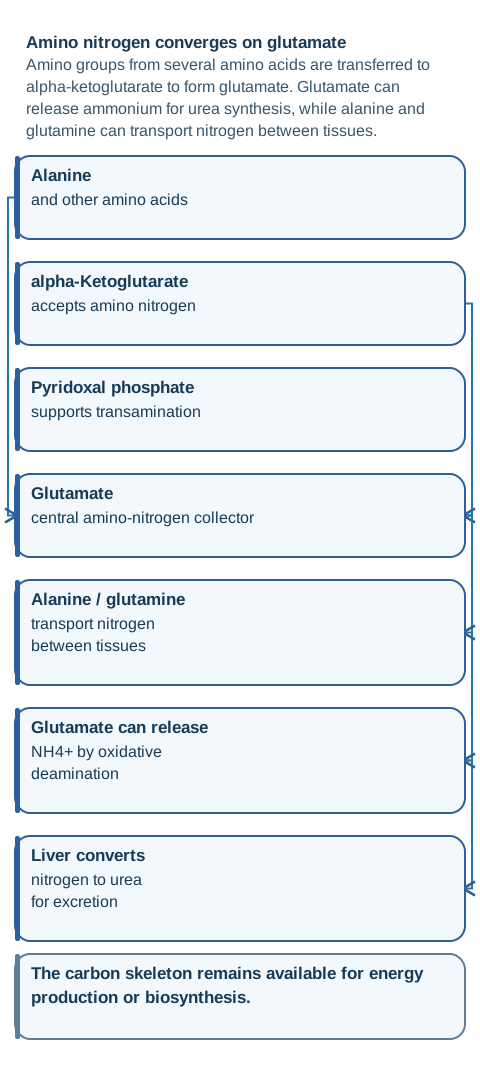
A common misconception is that protein catabolism means every amino acid has one dedicated clinically important transaminase. In reality, many amino groups are transferred through a shared network that commonly uses alpha-ketoglutarate as an acceptor and funnels nitrogen toward glutamate. Aminotransferases use pyridoxal phosphate, the active form of vitamin B6, to carry amino groups during these reversible reactions. [1]
Follow nitrogen separately from carbon. Transamination gathers much of the amino nitrogen on glutamate before ammonium release and urea disposal. [1][5]
The two reactions worth being able to reconstruct are not arbitrary names. ALT transfers the amino group of alanine to alpha-ketoglutarate, forming pyruvate plus glutamate. AST transfers the amino group of aspartate to alpha-ketoglutarate, forming oxaloacetate plus glutamate. The products explain the names: alanine aminotransferase connects alanine with pyruvate, while aspartate aminotransferase connects aspartate with oxaloacetate. [1]
Now follow the nitrogen. Glutamate can undergo oxidative deamination through glutamate dehydrogenase, releasing ammonium and regenerating alpha-ketoglutarate. The liver then disposes of nitrogen through urea synthesis, while alanine and glutamine help carry nitrogen safely between tissues. This is why transamination and deamination are related but not synonymous. [5]
Learner action: point to alanine and predict both products before checking the reaction. If you predict pyruvate plus glutamate, you have accounted for both halves of the molecule: carbon can support gluconeogenesis and nitrogen has joined the glutamate pool.
Checkpoint 1: alanine enters the liver during fasting. What two jobs can one ALT reaction support?
It creates pyruvate for carbon metabolism and glutamate for nitrogen handling. The transfer step itself does not directly release free ammonia.
The cofactor detail has a laboratory consequence. AST and ALT activity assays may differ in whether pyridoxal-5'-phosphate is added to the reagent, and cofactor supplementation can affect measured activity. This is another reason not to treat an enzyme number as a pure count of damaged cells. [7]
Try it here · Checkpoint 1 of 3
Make your prediction before reading the choices. A first attempt is just a starting point.
Case 2
Show answer and explanations for case 2
A. Alanine plus oxaloacetate forms aspartate plus pyruvate (Why this does not fit)
This combines products and substrates from different aminotransferase reactions. The key hepatic alanine reaction transfers nitrogen to alpha-ketoglutarate, not oxaloacetate.
Reasoning steps for option A
When the case states "Alanine carbon later appears in glucose", what does that indicate about "Alanine plus oxaloacetate forms aspartate plus pyruvate"?
The first discriminator is "Alanine carbon later appears in glucose". This combines products and substrates from different aminotransferase reactions.
How should "Alanine nitrogen later appears in urea" change the assessment of "Alanine plus oxaloacetate forms aspartate plus pyruvate"?
The second discriminator is "Alanine nitrogen later appears in urea". The key hepatic alanine reaction transfers nitrogen to alpha-ketoglutarate, not oxaloacetate.
After combining "Alanine carbon later appears in glucose" with "Alanine nitrogen later appears in urea", what is the final verdict on "Alanine plus oxaloacetate forms aspartate plus pyruvate"?
"Alanine plus oxaloacetate forms aspartate plus pyruvate" is not the best answer. This combines products and substrates from different aminotransferase reactions. The better fit is "Alanine plus alpha-ketoglutarate forms pyruvate plus glutamate" because it accounts for both supplied findings.
B. Alanine plus alpha-ketoglutarate forms pyruvate plus glutamate (Best answer)
ALT transfers the amino group of alanine to alpha-ketoglutarate, producing pyruvate and glutamate. Pyruvate can support gluconeogenesis, while glutamate funnels nitrogen toward ammonium and urea.
Reasoning steps for option B
When the case states "Alanine carbon later appears in glucose", what does that indicate about "Alanine plus alpha-ketoglutarate forms pyruvate plus glutamate"?
The first discriminator is "Alanine carbon later appears in glucose". ALT transfers the amino group of alanine to alpha-ketoglutarate, producing pyruvate and glutamate.
How should "Alanine nitrogen later appears in urea" change the assessment of "Alanine plus alpha-ketoglutarate forms pyruvate plus glutamate"?
The second discriminator is "Alanine nitrogen later appears in urea". Pyruvate can support gluconeogenesis, while glutamate funnels nitrogen toward ammonium and urea.
After combining "Alanine carbon later appears in glucose" with "Alanine nitrogen later appears in urea", what is the final verdict on "Alanine plus alpha-ketoglutarate forms pyruvate plus glutamate"?
"Alanine plus alpha-ketoglutarate forms pyruvate plus glutamate" is the best answer. ALT converts alanine and alpha-ketoglutarate to pyruvate and glutamate, linking gluconeogenesis to nitrogen transfer. It accounts for both "Alanine carbon later appears in glucose" and "Alanine nitrogen later appears in urea".
C. Alanine plus ammonia forms glutamine plus lactate (Why this does not fit)
Glutamine synthetase combines glutamate with ammonium; it does not convert alanine to lactate. This option therefore cannot explain the direct appearance of alanine carbon in the gluconeogenic pyruvate pool.
Reasoning steps for option C
When the case states "Alanine carbon later appears in glucose", what does that indicate about "Alanine plus ammonia forms glutamine plus lactate"?
The first discriminator is "Alanine carbon later appears in glucose". Glutamine synthetase combines glutamate with ammonium; it does not convert alanine to lactate.
How should "Alanine nitrogen later appears in urea" change the assessment of "Alanine plus ammonia forms glutamine plus lactate"?
The second discriminator is "Alanine nitrogen later appears in urea". This option therefore cannot explain the direct appearance of alanine carbon in the gluconeogenic pyruvate pool.
After combining "Alanine carbon later appears in glucose" with "Alanine nitrogen later appears in urea", what is the final verdict on "Alanine plus ammonia forms glutamine plus lactate"?
"Alanine plus ammonia forms glutamine plus lactate" is not the best answer. Glutamine synthetase combines glutamate with ammonium; it does not convert alanine to lactate. The better fit is "Alanine plus alpha-ketoglutarate forms pyruvate plus glutamate" because it accounts for both supplied findings.
D. Alanine plus acetyl-CoA forms citrate plus ammonium (Why this does not fit)
Citrate synthesis uses oxaloacetate and acetyl-CoA and is unrelated to direct alanine deamination. Alanine nitrogen is usually transferred first rather than released by this invented reaction.
Reasoning steps for option D
When the case states "Alanine carbon later appears in glucose", what does that indicate about "Alanine plus acetyl-CoA forms citrate plus ammonium"?
The first discriminator is "Alanine carbon later appears in glucose". Citrate synthesis uses oxaloacetate and acetyl-CoA and is unrelated to direct alanine deamination.
How should "Alanine nitrogen later appears in urea" change the assessment of "Alanine plus acetyl-CoA forms citrate plus ammonium"?
The second discriminator is "Alanine nitrogen later appears in urea". Alanine nitrogen is usually transferred first rather than released by this invented reaction.
After combining "Alanine carbon later appears in glucose" with "Alanine nitrogen later appears in urea", what is the final verdict on "Alanine plus acetyl-CoA forms citrate plus ammonium"?
"Alanine plus acetyl-CoA forms citrate plus ammonium" is not the best answer. Citrate synthesis uses oxaloacetate and acetyl-CoA and is unrelated to direct alanine deamination. The better fit is "Alanine plus alpha-ketoglutarate forms pyruvate plus glutamate" because it accounts for both supplied findings.
Takeaway: ALT converts alanine and alpha-ketoglutarate to pyruvate and glutamate, linking gluconeogenesis to nitrogen transfer.
Then ask where the carbon skeleton enters metabolism
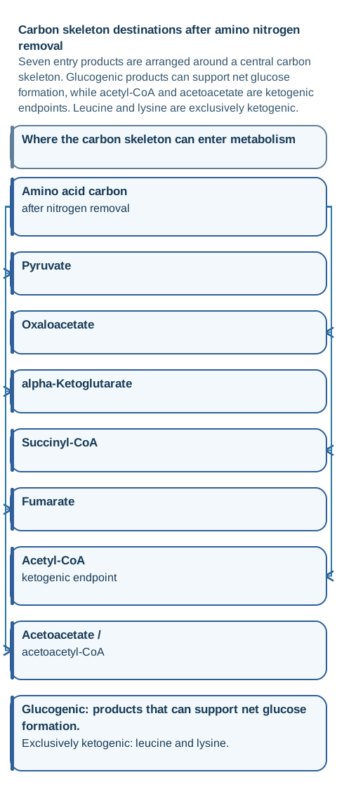
Removing nitrogen does not make the remaining carbon disappear. Amino acid carbon can enter central metabolism as pyruvate, oxaloacetate, alpha-ketoglutarate, succinyl-CoA, fumarate, acetyl-CoA, or acetoacetate-related products. The useful organizing question is whether those products can support net glucose formation, ketone or lipid production, or both. [4]
Use the endpoint to decide whether an amino acid can support net glucose formation, ketogenic metabolism, or both. [4]
A glucogenic amino acid yields carbon that can reach a gluconeogenic precursor. A ketogenic amino acid yields acetyl-CoA or acetoacetate-related carbon. Acetyl-CoA cannot produce net glucose in humans because its two carbons are lost as carbon dioxide through the citric acid cycle rather than creating a net oxaloacetate gain. Leucine and lysine are the exclusively ketogenic pair; several other amino acids have mixed fates. [4]
This corrects another oversimplification: protein does not become a universal emergency fuel only after glycogen reaches zero. Protein turnover occurs continuously. During fasting, amino acid carbon can contribute to gluconeogenesis and oxidation while nitrogen must still be transported and excreted. The destination depends on the amino acid and the metabolic state, not on one single entry point. [4]
Concrete example: valine and methionine can contribute propionyl-derived carbon that reaches succinyl-CoA, while leucine contributes only ketogenic carbon. If a case blocks methylmalonyl-CoA metabolism, predict a reduced succinyl-CoA entry rather than a direct acetyl-CoA defect.
Checkpoint 2: which pair cannot supply net carbon for gluconeogenesis?
Leucine and lysine are exclusively ketogenic. Phenylalanine and tyrosine are not, because they also yield fumarate.
Use the carbon map as a prediction tool rather than a list. If an enzyme defect traps a carbon skeleton before its entry product, the downstream pool receives less carbon. If an amino acid yields both a glucogenic and a ketogenic product, blocking one branch can alter the balance without changing the classification of every other amino acid.
Try it here · Checkpoint 2 of 3
Make your prediction before reading the choices. A first attempt is just a starting point.
Case 5
Show answer and explanations for case 5
A. Alanine and serine (Why this does not fit)
Alanine and serine can yield pyruvate. With intact gluconeogenesis, carbon from this pair could appear in newly synthesized glucose rather than remaining confined to ketogenic products.
Reasoning steps for option A
When the case states "Labeled carbon reaches acetyl-CoA and acetoacetate-derived ketone bodies but not newly synthesized glucose", what does that indicate about "Alanine and serine"?
The first discriminator is "Labeled carbon reaches acetyl-CoA and acetoacetate-derived ketone bodies but not newly synthesized glucose". Alanine and serine can yield pyruvate.
How should "Gluconeogenesis from lactate remains intact, excluding a global gluconeogenic failure" change the assessment of "Alanine and serine"?
The second discriminator is "Gluconeogenesis from lactate remains intact, excluding a global gluconeogenic failure". With intact gluconeogenesis, carbon from this pair could appear in newly synthesized glucose rather than remaining confined to ketogenic products.
After combining "Labeled carbon reaches acetyl-CoA and acetoacetate-derived ketone bodies but not newly synthesized glucose" with "Gluconeogenesis from lactate remains intact, excluding a global gluconeogenic failure", what is the final verdict on "Alanine and serine"?
"Alanine and serine" is not the best answer. Both can yield pyruvate, so intact gluconeogenesis would permit label to appear in glucose. The better fit is "Leucine and lysine" because it accounts for both supplied findings.
B. Aspartate and asparagine (Why this does not fit)
Aspartate and asparagine can yield oxaloacetate, a direct gluconeogenic intermediate. Their labeled carbon should not be absent from glucose when the gluconeogenic pathway is functioning.
Reasoning steps for option B
When the case states "Labeled carbon reaches acetyl-CoA and acetoacetate-derived ketone bodies but not newly synthesized glucose", what does that indicate about "Aspartate and asparagine"?
The first discriminator is "Labeled carbon reaches acetyl-CoA and acetoacetate-derived ketone bodies but not newly synthesized glucose". Aspartate and asparagine can yield oxaloacetate, a direct gluconeogenic intermediate.
How should "Gluconeogenesis from lactate remains intact, excluding a global gluconeogenic failure" change the assessment of "Aspartate and asparagine"?
The second discriminator is "Gluconeogenesis from lactate remains intact, excluding a global gluconeogenic failure". Their labeled carbon should not be absent from glucose when the gluconeogenic pathway is functioning.
After combining "Labeled carbon reaches acetyl-CoA and acetoacetate-derived ketone bodies but not newly synthesized glucose" with "Gluconeogenesis from lactate remains intact, excluding a global gluconeogenic failure", what is the final verdict on "Aspartate and asparagine"?
"Aspartate and asparagine" is not the best answer. Both can yield oxaloacetate, which can supply gluconeogenesis. The better fit is "Leucine and lysine" because it accounts for both supplied findings.
C. Valine and methionine (Why this does not fit)
Valine and methionine can contribute carbon through propionyl-CoA to succinyl-CoA. That citric acid cycle entry can support net glucose formation, so this pair is not exclusively ketogenic.
Reasoning steps for option C
When the case states "Labeled carbon reaches acetyl-CoA and acetoacetate-derived ketone bodies but not newly synthesized glucose", what does that indicate about "Valine and methionine"?
The first discriminator is "Labeled carbon reaches acetyl-CoA and acetoacetate-derived ketone bodies but not newly synthesized glucose". Valine and methionine can contribute carbon through propionyl-CoA to succinyl-CoA.
How should "Gluconeogenesis from lactate remains intact, excluding a global gluconeogenic failure" change the assessment of "Valine and methionine"?
The second discriminator is "Gluconeogenesis from lactate remains intact, excluding a global gluconeogenic failure". That citric acid cycle entry can support net glucose formation, so this pair is not exclusively ketogenic.
After combining "Labeled carbon reaches acetyl-CoA and acetoacetate-derived ketone bodies but not newly synthesized glucose" with "Gluconeogenesis from lactate remains intact, excluding a global gluconeogenic failure", what is the final verdict on "Valine and methionine"?
"Valine and methionine" is not the best answer. Their carbon can reach succinyl-CoA and thereby support net glucose formation. The better fit is "Leucine and lysine" because it accounts for both supplied findings.
D. Phenylalanine and tyrosine (Why this does not fit)
Phenylalanine and tyrosine yield both fumarate and acetoacetate. Their mixed glucogenic and ketogenic fate would permit some labeled carbon to reach glucose.
Reasoning steps for option D
When the case states "Labeled carbon reaches acetyl-CoA and acetoacetate-derived ketone bodies but not newly synthesized glucose", what does that indicate about "Phenylalanine and tyrosine"?
The first discriminator is "Labeled carbon reaches acetyl-CoA and acetoacetate-derived ketone bodies but not newly synthesized glucose". Phenylalanine and tyrosine yield both fumarate and acetoacetate.
How should "Gluconeogenesis from lactate remains intact, excluding a global gluconeogenic failure" change the assessment of "Phenylalanine and tyrosine"?
The second discriminator is "Gluconeogenesis from lactate remains intact, excluding a global gluconeogenic failure". Their mixed glucogenic and ketogenic fate would permit some labeled carbon to reach glucose.
After combining "Labeled carbon reaches acetyl-CoA and acetoacetate-derived ketone bodies but not newly synthesized glucose" with "Gluconeogenesis from lactate remains intact, excluding a global gluconeogenic failure", what is the final verdict on "Phenylalanine and tyrosine"?
"Phenylalanine and tyrosine" is not the best answer. They also generate fumarate, so their carbon fate is mixed rather than exclusively ketogenic. The better fit is "Leucine and lysine" because it accounts for both supplied findings.
E. Leucine and lysine (Best answer)
Leucine and lysine are exclusively ketogenic. Their carbon reaches acetyl-CoA or acetoacetate-related products without a pathway for net glucose production in humans.
Reasoning steps for option E
When the case states "Labeled carbon reaches acetyl-CoA and acetoacetate-derived ketone bodies but not newly synthesized glucose", what does that indicate about "Leucine and lysine"?
The first discriminator is "Labeled carbon reaches acetyl-CoA and acetoacetate-derived ketone bodies but not newly synthesized glucose". Leucine and lysine are exclusively ketogenic.
How should "Gluconeogenesis from lactate remains intact, excluding a global gluconeogenic failure" change the assessment of "Leucine and lysine"?
The second discriminator is "Gluconeogenesis from lactate remains intact, excluding a global gluconeogenic failure". Their carbon reaches acetyl-CoA or acetoacetate-related products without a pathway for net glucose production in humans.
After combining "Labeled carbon reaches acetyl-CoA and acetoacetate-derived ketone bodies but not newly synthesized glucose" with "Gluconeogenesis from lactate remains intact, excluding a global gluconeogenic failure", what is the final verdict on "Leucine and lysine"?
"Leucine and lysine" is the best answer. Tracer carbon confined to acetyl-CoA and acetoacetate-related products identifies leucine and lysine as the exclusively ketogenic pair. It accounts for both "Labeled carbon reaches acetyl-CoA and acetoacetate-derived ketone bodies but not newly synthesized glucose" and "Gluconeogenesis from lactate remains intact, excluding a global gluconeogenic failure".
Takeaway: Tracer carbon confined to acetyl-CoA and acetoacetate-related products identifies leucine and lysine as the exclusively ketogenic pair.
AST and ALT are reactions first, biomarkers second
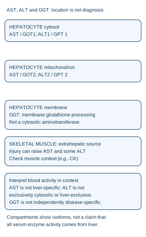
AST and ALT are often called liver enzymes, but their biology is broader. AST is found in liver and extrahepatic tissues such as skeletal muscle, and it has cytosolic and mitochondrial forms. ALT is more liver-enriched clinically, yet human ALT is not literally one cytosolic-only enzyme: ALT1 and ALT2 are encoded separately, and ALT2 has mitochondrial localization. [1][6]
Follow the labeled compartments and enzyme-specific conversions; interpret clinical patterns with tissue and precursor context. [1][6][14]
This matters because a blood value reports enzyme activity released into circulation, not a microscopic photograph of one organelle. A patient with crush injury and a very high creatine kinase can have substantial AST release from skeletal muscle. A modest ALT increase does not automatically prove simultaneous severe hepatic necrosis. The clinical context, CK, bilirubin, ALP, and other findings determine how persuasive a tissue source is. [2]
The reactions also participate in normal physiology. ALT is central to the glucose-alanine cycle, allowing muscle-derived alanine to deliver carbon and nitrogen to liver. AST participates in amino acid metabolism and the malate-aspartate shuttle. These roles explain why the enzymes exist before they ever become laboratory markers. [1]
Learner action: when AST is higher than ALT, do not name a diagnosis yet. First ask whether the case supplies a liver context, a muscle context, or both. Then use the rest of the panel to test that localization.
GGT belongs in a different biochemical family. Gamma-glutamyl transferase participates in gamma-glutamyl transfer and glutathione-related metabolism. It is sensitive to several hepatobiliary and exposure-related influences but has limited specificity for a particular etiology. [14]
Read the liver chemistry pattern before the ratio
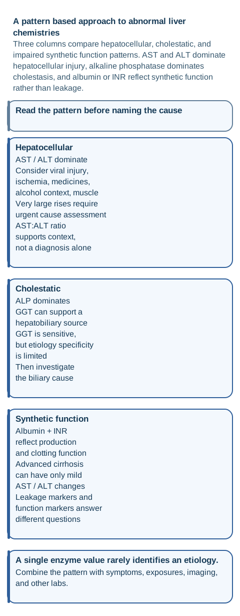
The first laboratory distinction is hepatocellular versus cholestatic injury. Hepatocellular injury has AST and ALT disproportionately high relative to alkaline phosphatase. Cholestatic injury has alkaline phosphatase disproportionately high relative to AST and ALT. This pattern classification is more dependable than treating any single enzyme as a diagnosis. [2]
Classify proportional enzyme patterns first, then use history, exposures, function markers, and imaging to determine cause. [2][3][14]
GGT can help when alkaline phosphatase is high because a concurrent GGT increase supports a hepatobiliary source. It is not a perfect liver-or-bone switch, and it does not identify the exact cause. A high ALP with normal GGT and a clear bone process makes bone more plausible, while a high ALP with GGT, conjugated bilirubin, pruritus, or biliary imaging strengthens a hepatobiliary interpretation. [2][14]
The AST:ALT ratio is also contextual evidence. In alcohol-associated hepatitis, an AST-predominant pattern with compatible alcohol exposure, recent jaundice, and moderate aminotransferase values can support the diagnosis. The ratio is not proof by itself, and the old story that alcohol simply releases two AST copies but one ALT copy is not a valid mechanism. Mitochondrial injury, tissue distribution, enzyme kinetics, and vitamin B6 biology contribute to the observed pattern. [3][7]
Very large aminotransferase increases, especially values in the thousands, should broaden urgency rather than collapse the differential to one word. Ischemic hepatopathy, acetaminophen or other severe toxic injury, and acute viral or other major hepatocellular injury are important considerations. Time course, exposure, hemodynamics, serology, bilirubin, INR, and imaging determine the cause. [2]
Cirrhosis creates the opposite trap. Advanced fibrosis can exist with only mild AST and ALT abnormalities. Albumin and INR provide information about hepatic synthetic capacity in context, while thrombocytopenia, splenomegaly, ascites, and varices can provide evidence of portal hypertension. Enzyme leakage and organ function are different questions. [2]
Checkpoint 3: AST and ALT are only mildly abnormal, but albumin is low and INR is prolonged in known cirrhosis. What changed?
The small enzyme rise does not exclude advanced disease. Synthetic function is impaired even though aminotransferase leakage is modest.
Application: for a new liver panel, classify the dominant pattern, look for extrahepatic sources, use GGT and ratios only as supporting evidence, then ask whether bilirubin, INR, albumin, platelets, symptoms, exposures, and imaging agree.
Try it here · Checkpoint 3 of 3
Make your prediction before reading the choices. A first attempt is just a starting point.
Case 15
Show answer and explanations for case 15
A. Mild AST release from injured hepatocytes into the circulation (Why this does not fit)
AST is primarily a leakage marker released from injured cells and is also present outside the liver. Its modest increase does not directly quantify hepatic protein synthesis.
Reasoning steps for option A
When the case states "Known cirrhosis is present with only mild AST and ALT abnormalities", what does that indicate about "Mild AST release from injured hepatocytes into the circulation"?
The first discriminator is "Known cirrhosis is present with only mild AST and ALT abnormalities". AST is primarily a leakage marker released from injured cells and is also present outside the liver.
How should "Albumin is low and INR is prolonged" change the assessment of "Mild AST release from injured hepatocytes into the circulation"?
The second discriminator is "Albumin is low and INR is prolonged". Its modest increase does not directly quantify hepatic protein synthesis.
After combining "Known cirrhosis is present with only mild AST and ALT abnormalities" with "Albumin is low and INR is prolonged", what is the final verdict on "Mild AST release from injured hepatocytes into the circulation"?
"Mild AST release from injured hepatocytes into the circulation" is not the best answer. AST is primarily a leakage marker released from injured cells and is also present outside the liver. The better fit is "Low albumin with prolonged INR" because it accounts for both supplied findings.
B. ALT of 44 U/L (Why this does not fit)
ALT is another aminotransferase leakage marker rather than a direct measure of hepatic protein production. Advanced cirrhosis can exist with only modest ALT abnormalities.
Reasoning steps for option B
When the case states "Known cirrhosis is present with only mild AST and ALT abnormalities", what does that indicate about "ALT of 44 U/L"?
The first discriminator is "Known cirrhosis is present with only mild AST and ALT abnormalities". ALT is another aminotransferase leakage marker rather than a direct measure of hepatic protein production.
How should "Albumin is low and INR is prolonged" change the assessment of "ALT of 44 U/L"?
The second discriminator is "Albumin is low and INR is prolonged". Advanced cirrhosis can exist with only modest ALT abnormalities.
After combining "Known cirrhosis is present with only mild AST and ALT abnormalities" with "Albumin is low and INR is prolonged", what is the final verdict on "ALT of 44 U/L"?
"ALT of 44 U/L" is not the best answer. ALT is another aminotransferase leakage marker rather than a direct measure of hepatic protein production. The better fit is "Low albumin with prolonged INR" because it accounts for both supplied findings.
C. Thrombocytopenia with splenomegaly (Why this does not fit)
Thrombocytopenia with splenomegaly can reflect portal hypertension and hypersplenism, which are important complications of cirrhosis. It does not directly measure hepatic protein synthesis.
Reasoning steps for option C
When the case states "Known cirrhosis is present with only mild AST and ALT abnormalities", what does that indicate about "Thrombocytopenia with splenomegaly"?
The first discriminator is "Known cirrhosis is present with only mild AST and ALT abnormalities". Thrombocytopenia with splenomegaly can reflect portal hypertension and hypersplenism, which are important complications of cirrhosis.
How should "Albumin is low and INR is prolonged" change the assessment of "Thrombocytopenia with splenomegaly"?
The second discriminator is "Albumin is low and INR is prolonged". It does not directly measure hepatic protein synthesis.
After combining "Known cirrhosis is present with only mild AST and ALT abnormalities" with "Albumin is low and INR is prolonged", what is the final verdict on "Thrombocytopenia with splenomegaly"?
"Thrombocytopenia with splenomegaly" is not the best answer. Thrombocytopenia with splenomegaly can reflect portal hypertension and hypersplenism, which are important complications of cirrhosis. The better fit is "Low albumin with prolonged INR" because it accounts for both supplied findings.
D. Low albumin with prolonged INR (Best answer)
Albumin production and synthesis of multiple clotting factors depend on the liver, so low albumin and a prolonged INR can reflect impaired synthetic function in the right context. These findings can be severe even when AST and ALT are only mildly abnormal.
Reasoning steps for option D
When the case states "Known cirrhosis is present with only mild AST and ALT abnormalities", what does that indicate about "Low albumin with prolonged INR"?
The first discriminator is "Known cirrhosis is present with only mild AST and ALT abnormalities". Albumin production and synthesis of multiple clotting factors depend on the liver, so low albumin and a prolonged INR can reflect impaired synthetic function in the right context.
How should "Albumin is low and INR is prolonged" change the assessment of "Low albumin with prolonged INR"?
The second discriminator is "Albumin is low and INR is prolonged". These findings can be severe even when AST and ALT are only mildly abnormal.
After combining "Known cirrhosis is present with only mild AST and ALT abnormalities" with "Albumin is low and INR is prolonged", what is the final verdict on "Low albumin with prolonged INR"?
"Low albumin with prolonged INR" is the best answer. Albumin and INR reflect hepatic synthetic capacity in context; AST and ALT primarily reflect enzyme release. It accounts for both "Known cirrhosis is present with only mild AST and ALT abnormalities" and "Albumin is low and INR is prolonged".
Takeaway: Albumin and INR reflect hepatic synthetic capacity in context; AST and ALT primarily reflect enzyme release.
Growth and severe illness both use amino acids, but their net direction differs
Normal growth, pregnancy, and tissue repair require protein synthesis, but high amino acid use does not mean the body is in the same metabolic state as a critically ill patient. Positive nitrogen balance means net nitrogen retention and is compatible with tissue accretion when intake is adequate. The important variable is net balance, not merely how quickly proteins are being synthesized and degraded.
Severe illness, major burns, and other systemic stress states can produce net protein catabolism, especially loss of skeletal muscle, even while whole-body protein synthesis and amino acid flux are also increased. Amino acids released from peripheral tissue can be redirected toward hepatic and immune needs. Nutritional support can limit protein loss but may not fully stop the acute catabolic response. [8]
This replaces the idea that cancer, chronic illness, burns, or crush injury simply reactivate a normal growth program. They can share high metabolic demand, but the net direction may be opposite. A growing child is building tissue; a patient with severe systemic stress may be losing it. Do not use the glycerol-3-phosphate shuttle as a general marker of rapid growth or amino acid catabolism.
Transfer task: if a vignette says a patient is losing muscle despite appropriate feeding, classify the state by the observed net protein loss. If a healthy adolescent is adding tissue, classify that as net anabolism even though both states have active protein turnover.
Several inherited metabolic diseases in the original practice material remain useful because they test the same habit: identify the abnormal substrate and product, then localize the enzyme or cofactor. Phenylalanine hydroxylase deficiency, HPRT deficiency, thiamine-dependent oxidative decarboxylation, cystathionine beta-synthase deficiency, and acquired hyperuricemia are retained as transfer problems with updated sourcing. [9][10][11][12][13]
Independent practice
Case 1
Show answer and explanations for case 1
A. AST and ALT require pyridoxal phosphate for amino-group transfer (Best answer)
AST and ALT are aminotransferases whose catalytic reactions require pyridoxal phosphate, the active form of vitamin B6. Supplying the cofactor in an assay can increase measured activity when apoenzyme lacks adequate bound cofactor.
Reasoning steps for option A
When the case states "AST and ALT activity increases when pyridoxal-5'-phosphate is supplied in the reagent", what does that indicate about "AST and ALT require pyridoxal phosphate for amino-group transfer"?
The first discriminator is "AST and ALT activity increases when pyridoxal-5'-phosphate is supplied in the reagent". AST and ALT are aminotransferases whose catalytic reactions require pyridoxal phosphate, the active form of vitamin B6.
How should "Poor nutrition and heavy alcohol exposure make vitamin status relevant" change the assessment of "AST and ALT require pyridoxal phosphate for amino-group transfer"?
The second discriminator is "Poor nutrition and heavy alcohol exposure make vitamin status relevant". Supplying the cofactor in an assay can increase measured activity when apoenzyme lacks adequate bound cofactor.
After combining "AST and ALT activity increases when pyridoxal-5'-phosphate is supplied in the reagent" with "Poor nutrition and heavy alcohol exposure make vitamin status relevant", what is the final verdict on "AST and ALT require pyridoxal phosphate for amino-group transfer"?
"AST and ALT require pyridoxal phosphate for amino-group transfer" is the best answer. AST and ALT are vitamin B6 dependent aminotransferases; assay design and vitamin status can affect measured activity. It accounts for both "AST and ALT activity increases when pyridoxal-5'-phosphate is supplied in the reagent" and "Poor nutrition and heavy alcohol exposure make vitamin status relevant".
B. GGT requires pyridoxal phosphate during glutathione-related membrane transfer reactions (Why this does not fit)
GGT participates in gamma-glutamyl transfer and glutathione metabolism, but it is not the aminotransferase reaction being measured here. The observed change occurs in AST and ALT after pyridoxal-5'-phosphate is added.
Reasoning steps for option B
When the case states "AST and ALT activity increases when pyridoxal-5'-phosphate is supplied in the reagent", what does that indicate about "GGT requires pyridoxal phosphate during glutathione-related membrane transfer reactions"?
The first discriminator is "AST and ALT activity increases when pyridoxal-5'-phosphate is supplied in the reagent". GGT participates in gamma-glutamyl transfer and glutathione metabolism, but it is not the aminotransferase reaction being measured here.
How should "Poor nutrition and heavy alcohol exposure make vitamin status relevant" change the assessment of "GGT requires pyridoxal phosphate during glutathione-related membrane transfer reactions"?
The second discriminator is "Poor nutrition and heavy alcohol exposure make vitamin status relevant". The observed change occurs in AST and ALT after pyridoxal-5'-phosphate is added.
After combining "AST and ALT activity increases when pyridoxal-5'-phosphate is supplied in the reagent" with "Poor nutrition and heavy alcohol exposure make vitamin status relevant", what is the final verdict on "GGT requires pyridoxal phosphate during glutathione-related membrane transfer reactions"?
"GGT requires pyridoxal phosphate during glutathione-related membrane transfer reactions" is not the best answer. GGT participates in gamma-glutamyl transfer and glutathione metabolism, but it is not the aminotransferase reaction being measured here. The better fit is "AST and ALT require pyridoxal phosphate for amino-group transfer" because it accounts for both supplied findings.
C. ALP requires vitamin B6 to hydrolyze phosphate esters (Why this does not fit)
Alkaline phosphatase is a phosphatase, not an aminotransferase, and the case does not report an ALP assay change. Vitamin B6 supplementation therefore does not explain the paired AST and ALT result.
Reasoning steps for option C
When the case states "AST and ALT activity increases when pyridoxal-5'-phosphate is supplied in the reagent", what does that indicate about "ALP requires vitamin B6 to hydrolyze phosphate esters"?
The first discriminator is "AST and ALT activity increases when pyridoxal-5'-phosphate is supplied in the reagent". Alkaline phosphatase is a phosphatase, not an aminotransferase, and the case does not report an ALP assay change.
How should "Poor nutrition and heavy alcohol exposure make vitamin status relevant" change the assessment of "ALP requires vitamin B6 to hydrolyze phosphate esters"?
The second discriminator is "Poor nutrition and heavy alcohol exposure make vitamin status relevant". Vitamin B6 supplementation therefore does not explain the paired AST and ALT result.
After combining "AST and ALT activity increases when pyridoxal-5'-phosphate is supplied in the reagent" with "Poor nutrition and heavy alcohol exposure make vitamin status relevant", what is the final verdict on "ALP requires vitamin B6 to hydrolyze phosphate esters"?
"ALP requires vitamin B6 to hydrolyze phosphate esters" is not the best answer. Alkaline phosphatase is a phosphatase, not an aminotransferase, and the case does not report an ALP assay change. The better fit is "AST and ALT require pyridoxal phosphate for amino-group transfer" because it accounts for both supplied findings.
D. Bilirubin conjugation directly consumes pyridoxal phosphate (Why this does not fit)
Bilirubin conjugation depends on UDP-glucuronosyltransferase rather than pyridoxal phosphate. Jaundice supplies clinical context but does not explain the assay-specific increase in AST and ALT activity.
Reasoning steps for option D
When the case states "AST and ALT activity increases when pyridoxal-5'-phosphate is supplied in the reagent", what does that indicate about "Bilirubin conjugation directly consumes pyridoxal phosphate"?
The first discriminator is "AST and ALT activity increases when pyridoxal-5'-phosphate is supplied in the reagent". Bilirubin conjugation depends on UDP-glucuronosyltransferase rather than pyridoxal phosphate.
How should "Poor nutrition and heavy alcohol exposure make vitamin status relevant" change the assessment of "Bilirubin conjugation directly consumes pyridoxal phosphate"?
The second discriminator is "Poor nutrition and heavy alcohol exposure make vitamin status relevant". Jaundice supplies clinical context but does not explain the assay-specific increase in AST and ALT activity.
After combining "AST and ALT activity increases when pyridoxal-5'-phosphate is supplied in the reagent" with "Poor nutrition and heavy alcohol exposure make vitamin status relevant", what is the final verdict on "Bilirubin conjugation directly consumes pyridoxal phosphate"?
"Bilirubin conjugation directly consumes pyridoxal phosphate" is not the best answer. Bilirubin conjugation depends on UDP-glucuronosyltransferase rather than pyridoxal phosphate. The better fit is "AST and ALT require pyridoxal phosphate for amino-group transfer" because it accounts for both supplied findings.
Takeaway: AST and ALT are vitamin B6 dependent aminotransferases; assay design and vitamin status can affect measured activity.
A. Acute biliary obstruction is the dominant source of both aminotransferase abnormalities here (Why this does not fit)
A biliary process would usually draw attention to alkaline phosphatase, bilirubin, symptoms, and imaging rather than a massive CK rise. The normal cholestatic markers weaken this explanation.
Reasoning steps for option A
When the case states "Creatine kinase is 12,000 U/L after prolonged crush injury", what does that indicate about "Acute biliary obstruction is the dominant source of both aminotransferase abnormalities here"?
The first discriminator is "Creatine kinase is 12,000 U/L after prolonged crush injury". A biliary process would usually draw attention to alkaline phosphatase, bilirubin, symptoms, and imaging rather than a massive CK rise.
How should "AST predominates while bilirubin and alkaline phosphatase remain normal" change the assessment of "Acute biliary obstruction is the dominant source of both aminotransferase abnormalities here"?
The second discriminator is "AST predominates while bilirubin and alkaline phosphatase remain normal". The normal cholestatic markers weaken this explanation.
After combining "Creatine kinase is 12,000 U/L after prolonged crush injury" with "AST predominates while bilirubin and alkaline phosphatase remain normal", what is the final verdict on "Acute biliary obstruction is the dominant source of both aminotransferase abnormalities here"?
"Acute biliary obstruction is the dominant source of both aminotransferase abnormalities here" is not the best answer. Normal bilirubin and alkaline phosphatase plus the massive CK increase oppose a dominant biliary process. The better fit is "Skeletal muscle injury is a major contributor to the AST rise" because it accounts for both supplied findings.
B. Alcohol-associated hepatitis is the dominant explanation for the AST-predominant pattern (Why this does not fit)
Alcohol-associated liver injury can produce AST predominance, but this patient has an immediate crush mechanism and a massive CK increase. Those findings make skeletal muscle the stronger source of the AST rise.
Reasoning steps for option B
When the case states "Creatine kinase is 12,000 U/L after prolonged crush injury", what does that indicate about "Alcohol-associated hepatitis is the dominant explanation for the AST-predominant pattern"?
The first discriminator is "Creatine kinase is 12,000 U/L after prolonged crush injury". Alcohol-associated liver injury can produce AST predominance, but this patient has an immediate crush mechanism and a massive CK increase.
How should "AST predominates while bilirubin and alkaline phosphatase remain normal" change the assessment of "Alcohol-associated hepatitis is the dominant explanation for the AST-predominant pattern"?
The second discriminator is "AST predominates while bilirubin and alkaline phosphatase remain normal". Those findings make skeletal muscle the stronger source of the AST rise.
After combining "Creatine kinase is 12,000 U/L after prolonged crush injury" with "AST predominates while bilirubin and alkaline phosphatase remain normal", what is the final verdict on "Alcohol-associated hepatitis is the dominant explanation for the AST-predominant pattern"?
"Alcohol-associated hepatitis is the dominant explanation for the AST-predominant pattern" is not the best answer. The immediate crush injury and CK of 12,000 U/L provide direct evidence of skeletal muscle release. The better fit is "Skeletal muscle injury is a major contributor to the AST rise" because it accounts for both supplied findings.
C. Skeletal muscle injury is a major contributor to the AST rise (Best answer)
AST is present in skeletal muscle, and severe muscle injury can release it into blood. The very high CK, crush mechanism, normal bilirubin, and normal ALP make muscle a major contributor to this pattern.
Reasoning steps for option C
When the case states "Creatine kinase is 12,000 U/L after prolonged crush injury", what does that indicate about "Skeletal muscle injury is a major contributor to the AST rise"?
The first discriminator is "Creatine kinase is 12,000 U/L after prolonged crush injury". AST is present in skeletal muscle, and severe muscle injury can release it into blood.
How should "AST predominates while bilirubin and alkaline phosphatase remain normal" change the assessment of "Skeletal muscle injury is a major contributor to the AST rise"?
The second discriminator is "AST predominates while bilirubin and alkaline phosphatase remain normal". The very high CK, crush mechanism, normal bilirubin, and normal ALP make muscle a major contributor to this pattern.
After combining "Creatine kinase is 12,000 U/L after prolonged crush injury" with "AST predominates while bilirubin and alkaline phosphatase remain normal", what is the final verdict on "Skeletal muscle injury is a major contributor to the AST rise"?
"Skeletal muscle injury is a major contributor to the AST rise" is the best answer. Aminotransferases are tissue enzymes; a very high CK can explain AST-predominant abnormalities after muscle injury. It accounts for both "Creatine kinase is 12,000 U/L after prolonged crush injury" and "AST predominates while bilirubin and alkaline phosphatase remain normal".
D. Severe hepatic necrosis is the dominant explanation for the ALT increase (Why this does not fit)
A modest ALT increase can accompany major muscle injury, and the normal bilirubin and alkaline phosphatase do not establish severe hepatic necrosis. The CK and trauma history provide a direct extrahepatic explanation.
Reasoning steps for option D
When the case states "Creatine kinase is 12,000 U/L after prolonged crush injury", what does that indicate about "Severe hepatic necrosis is the dominant explanation for the ALT increase"?
The first discriminator is "Creatine kinase is 12,000 U/L after prolonged crush injury". A modest ALT increase can accompany major muscle injury, and the normal bilirubin and alkaline phosphatase do not establish severe hepatic necrosis.
How should "AST predominates while bilirubin and alkaline phosphatase remain normal" change the assessment of "Severe hepatic necrosis is the dominant explanation for the ALT increase"?
The second discriminator is "AST predominates while bilirubin and alkaline phosphatase remain normal". The CK and trauma history provide a direct extrahepatic explanation.
After combining "Creatine kinase is 12,000 U/L after prolonged crush injury" with "AST predominates while bilirubin and alkaline phosphatase remain normal", what is the final verdict on "Severe hepatic necrosis is the dominant explanation for the ALT increase"?
"Severe hepatic necrosis is the dominant explanation for the ALT increase" is not the best answer. The ALT rise is modest, cholestatic markers are normal, and severe muscle injury already explains the pattern. The better fit is "Skeletal muscle injury is a major contributor to the AST rise" because it accounts for both supplied findings.
Takeaway: Aminotransferases are tissue enzymes; a very high CK can explain AST-predominant abnormalities after muscle injury.
A. Carboxylation of pyruvate to oxaloacetate (Why this does not fit)
Pyruvate carboxylase supports gluconeogenesis and anaplerosis, but it does not release the amino nitrogen collected on glutamate. The stem specifically asks how glutamate nitrogen becomes free ammonium.
Reasoning steps for option A
When the case states "Amino groups from several amino acids have already been transferred to glutamate", what does that indicate about "Carboxylation of pyruvate to oxaloacetate"?
The first discriminator is "Amino groups from several amino acids have already been transferred to glutamate". Pyruvate carboxylase supports gluconeogenesis and anaplerosis, but it does not release the amino nitrogen collected on glutamate.
How should "The liver must convert collected nitrogen into urea" change the assessment of "Carboxylation of pyruvate to oxaloacetate"?
The second discriminator is "The liver must convert collected nitrogen into urea". The stem specifically asks how glutamate nitrogen becomes free ammonium.
After combining "Amino groups from several amino acids have already been transferred to glutamate" with "The liver must convert collected nitrogen into urea", what is the final verdict on "Carboxylation of pyruvate to oxaloacetate"?
"Carboxylation of pyruvate to oxaloacetate" is not the best answer. Pyruvate carboxylase supports gluconeogenesis and anaplerosis, but it does not release the amino nitrogen collected on glutamate. The better fit is "Oxidative deamination of glutamate by glutamate dehydrogenase" because it accounts for both supplied findings.
B. Condensation of ammonia with glutamate to glutamine (Why this does not fit)
Glutamine synthesis captures rather than releases free ammonium. It is useful for nitrogen transport and detoxification, but it runs in the opposite direction from the step requested.
Reasoning steps for option B
When the case states "Amino groups from several amino acids have already been transferred to glutamate", what does that indicate about "Condensation of ammonia with glutamate to glutamine"?
The first discriminator is "Amino groups from several amino acids have already been transferred to glutamate". Glutamine synthesis captures rather than releases free ammonium.
How should "The liver must convert collected nitrogen into urea" change the assessment of "Condensation of ammonia with glutamate to glutamine"?
The second discriminator is "The liver must convert collected nitrogen into urea". It is useful for nitrogen transport and detoxification, but it runs in the opposite direction from the step requested.
After combining "Amino groups from several amino acids have already been transferred to glutamate" with "The liver must convert collected nitrogen into urea", what is the final verdict on "Condensation of ammonia with glutamate to glutamine"?
"Condensation of ammonia with glutamate to glutamine" is not the best answer. Glutamine synthesis captures rather than releases free ammonium. The better fit is "Oxidative deamination of glutamate by glutamate dehydrogenase" because it accounts for both supplied findings.
C. Transamination of glutamate back to an amino acid (Why this does not fit)
Transamination transfers an amino group between an amino acid and a keto acid without directly liberating free ammonium. It redistributes nitrogen rather than producing the requested ammonium.
Reasoning steps for option C
When the case states "Amino groups from several amino acids have already been transferred to glutamate", what does that indicate about "Transamination of glutamate back to an amino acid"?
The first discriminator is "Amino groups from several amino acids have already been transferred to glutamate". Transamination transfers an amino group between an amino acid and a keto acid without directly liberating free ammonium.
How should "The liver must convert collected nitrogen into urea" change the assessment of "Transamination of glutamate back to an amino acid"?
The second discriminator is "The liver must convert collected nitrogen into urea". It redistributes nitrogen rather than producing the requested ammonium.
After combining "Amino groups from several amino acids have already been transferred to glutamate" with "The liver must convert collected nitrogen into urea", what is the final verdict on "Transamination of glutamate back to an amino acid"?
"Transamination of glutamate back to an amino acid" is not the best answer. Transamination transfers an amino group between an amino acid and a keto acid without directly liberating free ammonium. The better fit is "Oxidative deamination of glutamate by glutamate dehydrogenase" because it accounts for both supplied findings.
D. Oxidative deamination of glutamate by glutamate dehydrogenase (Best answer)
Glutamate dehydrogenase can release ammonium from glutamate while regenerating alpha-ketoglutarate. This provides a direct connection between collected amino nitrogen and hepatic ammonia disposal.
Reasoning steps for option D
When the case states "Amino groups from several amino acids have already been transferred to glutamate", what does that indicate about "Oxidative deamination of glutamate by glutamate dehydrogenase"?
The first discriminator is "Amino groups from several amino acids have already been transferred to glutamate". Glutamate dehydrogenase can release ammonium from glutamate while regenerating alpha-ketoglutarate.
How should "The liver must convert collected nitrogen into urea" change the assessment of "Oxidative deamination of glutamate by glutamate dehydrogenase"?
The second discriminator is "The liver must convert collected nitrogen into urea". This provides a direct connection between collected amino nitrogen and hepatic ammonia disposal.
After combining "Amino groups from several amino acids have already been transferred to glutamate" with "The liver must convert collected nitrogen into urea", what is the final verdict on "Oxidative deamination of glutamate by glutamate dehydrogenase"?
"Oxidative deamination of glutamate by glutamate dehydrogenase" is the best answer. Many amino groups converge on glutamate, which can release ammonium through glutamate dehydrogenase. It accounts for both "Amino groups from several amino acids have already been transferred to glutamate" and "The liver must convert collected nitrogen into urea".
Takeaway: Many amino groups converge on glutamate, which can release ammonium through glutamate dehydrogenase.
Methylmalonyl-CoA mutase is vitamin B12 dependent and produces succinyl-CoA. Impairment therefore reduces entry of these propionyl-derived carbon skeletons at succinyl-CoA.
Reasoning steps for option A
When the case states "Vitamin B12 deficiency produces high methylmalonic acid", what does that indicate about "Succinyl-CoA"?
The first discriminator is "Vitamin B12 deficiency produces high methylmalonic acid". Methylmalonyl-CoA mutase is vitamin B12 dependent and produces succinyl-CoA.
How should "Valine and methionine carbon passes through propionyl-CoA and methylmalonyl-CoA" change the assessment of "Succinyl-CoA"?
The second discriminator is "Valine and methionine carbon passes through propionyl-CoA and methylmalonyl-CoA". Impairment therefore reduces entry of these propionyl-derived carbon skeletons at succinyl-CoA.
After combining "Vitamin B12 deficiency produces high methylmalonic acid" with "Valine and methionine carbon passes through propionyl-CoA and methylmalonyl-CoA", what is the final verdict on "Succinyl-CoA"?
"Succinyl-CoA" is the best answer. Propionyl-derived carbon reaches the citric acid cycle through the B12-dependent formation of succinyl-CoA. It accounts for both "Vitamin B12 deficiency produces high methylmalonic acid" and "Valine and methionine carbon passes through propionyl-CoA and methylmalonyl-CoA".
B. Oxaloacetate (Why this does not fit)
Oxaloacetate receives carbon from amino acids such as aspartate and asparagine, but it is not the direct product of methylmalonyl-CoA mutase. The high methylmalonic acid localizes a different entry point.
Reasoning steps for option B
When the case states "Vitamin B12 deficiency produces high methylmalonic acid", what does that indicate about "Oxaloacetate"?
The first discriminator is "Vitamin B12 deficiency produces high methylmalonic acid". Oxaloacetate receives carbon from amino acids such as aspartate and asparagine, but it is not the direct product of methylmalonyl-CoA mutase.
How should "Valine and methionine carbon passes through propionyl-CoA and methylmalonyl-CoA" change the assessment of "Oxaloacetate"?
The second discriminator is "Valine and methionine carbon passes through propionyl-CoA and methylmalonyl-CoA". The high methylmalonic acid localizes a different entry point.
After combining "Vitamin B12 deficiency produces high methylmalonic acid" with "Valine and methionine carbon passes through propionyl-CoA and methylmalonyl-CoA", what is the final verdict on "Oxaloacetate"?
"Oxaloacetate" is not the best answer. Oxaloacetate receives carbon from amino acids such as aspartate and asparagine, but it is not the direct product of methylmalonyl-CoA mutase. The better fit is "Succinyl-CoA" because it accounts for both supplied findings.
C. Fumarate (Why this does not fit)
Fumarate is an entry product for other amino acid pathways, including part of phenylalanine and tyrosine catabolism. It is not the immediate product of methylmalonyl-CoA metabolism.
Reasoning steps for option C
When the case states "Vitamin B12 deficiency produces high methylmalonic acid", what does that indicate about "Fumarate"?
The first discriminator is "Vitamin B12 deficiency produces high methylmalonic acid". Fumarate is an entry product for other amino acid pathways, including part of phenylalanine and tyrosine catabolism.
How should "Valine and methionine carbon passes through propionyl-CoA and methylmalonyl-CoA" change the assessment of "Fumarate"?
The second discriminator is "Valine and methionine carbon passes through propionyl-CoA and methylmalonyl-CoA". It is not the immediate product of methylmalonyl-CoA metabolism.
After combining "Vitamin B12 deficiency produces high methylmalonic acid" with "Valine and methionine carbon passes through propionyl-CoA and methylmalonyl-CoA", what is the final verdict on "Fumarate"?
"Fumarate" is not the best answer. Fumarate is an entry product for other amino acid pathways, including part of phenylalanine and tyrosine catabolism. The better fit is "Succinyl-CoA" because it accounts for both supplied findings.
D. Acetyl-CoA (Why this does not fit)
Acetyl-CoA is a ketogenic endpoint for several amino acids, but the propionyl-CoA to methylmalonyl-CoA route does not directly generate acetyl-CoA. The B12-dependent step points to succinyl-CoA instead.
Reasoning steps for option D
When the case states "Vitamin B12 deficiency produces high methylmalonic acid", what does that indicate about "Acetyl-CoA"?
The first discriminator is "Vitamin B12 deficiency produces high methylmalonic acid". Acetyl-CoA is a ketogenic endpoint for several amino acids, but the propionyl-CoA to methylmalonyl-CoA route does not directly generate acetyl-CoA.
How should "Valine and methionine carbon passes through propionyl-CoA and methylmalonyl-CoA" change the assessment of "Acetyl-CoA"?
The second discriminator is "Valine and methionine carbon passes through propionyl-CoA and methylmalonyl-CoA". The B12-dependent step points to succinyl-CoA instead.
After combining "Vitamin B12 deficiency produces high methylmalonic acid" with "Valine and methionine carbon passes through propionyl-CoA and methylmalonyl-CoA", what is the final verdict on "Acetyl-CoA"?
"Acetyl-CoA" is not the best answer. Acetyl-CoA is a ketogenic endpoint for several amino acids, but the propionyl-CoA to methylmalonyl-CoA route does not directly generate acetyl-CoA. The better fit is "Succinyl-CoA" because it accounts for both supplied findings.
Takeaway: Propionyl-derived carbon reaches the citric acid cycle through the B12-dependent formation of succinyl-CoA.
A. More alanine-derived pyruvate and more glutamate formation (Why this does not fit)
ALT is the reaction that produces both pyruvate and glutamate from alanine. Inhibiting it should reduce, not increase, both products.
Reasoning steps for option A
When the case states "Muscle continues releasing alanine during fasting", what does that indicate about "More alanine-derived pyruvate and more glutamate formation"?
The first discriminator is "Muscle continues releasing alanine during fasting". ALT is the reaction that produces both pyruvate and glutamate from alanine.
How should "Hepatic ALT activity is selectively reduced" change the assessment of "More alanine-derived pyruvate and more glutamate formation"?
The second discriminator is "Hepatic ALT activity is selectively reduced". Inhibiting it should reduce, not increase, both products.
After combining "Muscle continues releasing alanine during fasting" with "Hepatic ALT activity is selectively reduced", what is the final verdict on "More alanine-derived pyruvate and more glutamate formation"?
"More alanine-derived pyruvate and more glutamate formation" is not the best answer. ALT is the reaction that produces both pyruvate and glutamate from alanine. The better fit is "Less alanine-derived pyruvate and less glutamate formation" because it accounts for both supplied findings.
B. Less alanine-derived pyruvate and less glutamate formation (Best answer)
Reduced ALT limits conversion of alanine to pyruvate and transfer of its amino group to alpha-ketoglutarate. Both alanine-derived gluconeogenic carbon and glutamate formation therefore fall.
Reasoning steps for option B
When the case states "Muscle continues releasing alanine during fasting", what does that indicate about "Less alanine-derived pyruvate and less glutamate formation"?
The first discriminator is "Muscle continues releasing alanine during fasting". Reduced ALT limits conversion of alanine to pyruvate and transfer of its amino group to alpha-ketoglutarate.
How should "Hepatic ALT activity is selectively reduced" change the assessment of "Less alanine-derived pyruvate and less glutamate formation"?
The second discriminator is "Hepatic ALT activity is selectively reduced". Both alanine-derived gluconeogenic carbon and glutamate formation therefore fall.
After combining "Muscle continues releasing alanine during fasting" with "Hepatic ALT activity is selectively reduced", what is the final verdict on "Less alanine-derived pyruvate and less glutamate formation"?
"Less alanine-derived pyruvate and less glutamate formation" is the best answer. Hepatic ALT converts alanine to pyruvate while transferring nitrogen to glutamate. It accounts for both "Muscle continues releasing alanine during fasting" and "Hepatic ALT activity is selectively reduced".
C. Less acetyl-CoA formation with unchanged alanine nitrogen transfer (Why this does not fit)
ALT acts before any downstream pyruvate oxidation and directly transfers alanine nitrogen. A selective ALT block therefore affects nitrogen transfer as well as carbon flow.
Reasoning steps for option C
When the case states "Muscle continues releasing alanine during fasting", what does that indicate about "Less acetyl-CoA formation with unchanged alanine nitrogen transfer"?
The first discriminator is "Muscle continues releasing alanine during fasting". ALT acts before any downstream pyruvate oxidation and directly transfers alanine nitrogen.
How should "Hepatic ALT activity is selectively reduced" change the assessment of "Less acetyl-CoA formation with unchanged alanine nitrogen transfer"?
The second discriminator is "Hepatic ALT activity is selectively reduced". A selective ALT block therefore affects nitrogen transfer as well as carbon flow.
After combining "Muscle continues releasing alanine during fasting" with "Hepatic ALT activity is selectively reduced", what is the final verdict on "Less acetyl-CoA formation with unchanged alanine nitrogen transfer"?
"Less acetyl-CoA formation with unchanged alanine nitrogen transfer" is not the best answer. ALT acts before any downstream pyruvate oxidation and directly transfers alanine nitrogen. The better fit is "Less alanine-derived pyruvate and less glutamate formation" because it accounts for both supplied findings.
D. More urea formation with unchanged pyruvate production (Why this does not fit)
Less transfer of alanine nitrogen to glutamate would not directly increase urea production, and ALT inhibition also reduces pyruvate generation from alanine. Both parts of this option oppose the direct reaction.
Reasoning steps for option D
When the case states "Muscle continues releasing alanine during fasting", what does that indicate about "More urea formation with unchanged pyruvate production"?
The first discriminator is "Muscle continues releasing alanine during fasting". Less transfer of alanine nitrogen to glutamate would not directly increase urea production, and ALT inhibition also reduces pyruvate generation from alanine.
How should "Hepatic ALT activity is selectively reduced" change the assessment of "More urea formation with unchanged pyruvate production"?
The second discriminator is "Hepatic ALT activity is selectively reduced". Both parts of this option oppose the direct reaction.
After combining "Muscle continues releasing alanine during fasting" with "Hepatic ALT activity is selectively reduced", what is the final verdict on "More urea formation with unchanged pyruvate production"?
"More urea formation with unchanged pyruvate production" is not the best answer. Less transfer of alanine nitrogen to glutamate would not directly increase urea production, and ALT inhibition also reduces pyruvate generation from alanine. The better fit is "Less alanine-derived pyruvate and less glutamate formation" because it accounts for both supplied findings.
Takeaway: Hepatic ALT converts alanine to pyruvate while transferring nitrogen to glutamate.
A. High direct bilirubin with dark urine (Why this does not fit)
A conjugated bilirubin increase would redirect attention toward hepatobiliary disease rather than confirm muscle injury. It does not provide direct evidence that skeletal muscle released AST.
Reasoning steps for option A
When the case states "AST is disproportionately high after intense exertion", what does that indicate about "High direct bilirubin with dark urine"?
The first discriminator is "AST is disproportionately high after intense exertion". A conjugated bilirubin increase would redirect attention toward hepatobiliary disease rather than confirm muscle injury.
How should "The patient has diffuse muscle pain without jaundice" change the assessment of "High direct bilirubin with dark urine"?
The second discriminator is "The patient has diffuse muscle pain without jaundice". It does not provide direct evidence that skeletal muscle released AST.
After combining "AST is disproportionately high after intense exertion" with "The patient has diffuse muscle pain without jaundice", what is the final verdict on "High direct bilirubin with dark urine"?
"High direct bilirubin with dark urine" is not the best answer. A conjugated bilirubin increase would redirect attention toward hepatobiliary disease rather than confirm muscle injury. The better fit is "Creatine kinase of 9,500 U/L" because it accounts for both supplied findings.
B. High alkaline phosphatase with pruritus (Why this does not fit)
A marked alkaline phosphatase rise with pruritus suggests a cholestatic process. That pattern would not specifically support skeletal muscle as the source of AST.
Reasoning steps for option B
When the case states "AST is disproportionately high after intense exertion", what does that indicate about "High alkaline phosphatase with pruritus"?
The first discriminator is "AST is disproportionately high after intense exertion". A marked alkaline phosphatase rise with pruritus suggests a cholestatic process.
How should "The patient has diffuse muscle pain without jaundice" change the assessment of "High alkaline phosphatase with pruritus"?
The second discriminator is "The patient has diffuse muscle pain without jaundice". That pattern would not specifically support skeletal muscle as the source of AST.
After combining "AST is disproportionately high after intense exertion" with "The patient has diffuse muscle pain without jaundice", what is the final verdict on "High alkaline phosphatase with pruritus"?
"High alkaline phosphatase with pruritus" is not the best answer. A marked alkaline phosphatase rise with pruritus suggests a cholestatic process. The better fit is "Creatine kinase of 9,500 U/L" because it accounts for both supplied findings.
C. Creatine kinase of 9,500 U/L (Best answer)
A very high creatine kinase directly supports substantial skeletal muscle injury. In the setting of exertion, muscle pain, AST predominance, and normal bilirubin, it strongly favors muscle as a major AST source.
Reasoning steps for option C
When the case states "AST is disproportionately high after intense exertion", what does that indicate about "Creatine kinase of 9,500 U/L"?
The first discriminator is "AST is disproportionately high after intense exertion". A very high creatine kinase directly supports substantial skeletal muscle injury.
How should "The patient has diffuse muscle pain without jaundice" change the assessment of "Creatine kinase of 9,500 U/L"?
The second discriminator is "The patient has diffuse muscle pain without jaundice". In the setting of exertion, muscle pain, AST predominance, and normal bilirubin, it strongly favors muscle as a major AST source.
After combining "AST is disproportionately high after intense exertion" with "The patient has diffuse muscle pain without jaundice", what is the final verdict on "Creatine kinase of 9,500 U/L"?
"Creatine kinase of 9,500 U/L" is the best answer. AST can come from skeletal muscle; a high CK supplies direct evidence when the clinical context fits. It accounts for both "AST is disproportionately high after intense exertion" and "The patient has diffuse muscle pain without jaundice".
D. GGT of 260 U/L with normal creatine kinase (Why this does not fit)
A high GGT would support hepatobiliary enzyme induction or injury in context, not muscle release. A normal CK would also weaken the proposed muscle explanation.
Reasoning steps for option D
When the case states "AST is disproportionately high after intense exertion", what does that indicate about "GGT of 260 U/L with normal creatine kinase"?
The first discriminator is "AST is disproportionately high after intense exertion". A high GGT would support hepatobiliary enzyme induction or injury in context, not muscle release.
How should "The patient has diffuse muscle pain without jaundice" change the assessment of "GGT of 260 U/L with normal creatine kinase"?
The second discriminator is "The patient has diffuse muscle pain without jaundice". A normal CK would also weaken the proposed muscle explanation.
After combining "AST is disproportionately high after intense exertion" with "The patient has diffuse muscle pain without jaundice", what is the final verdict on "GGT of 260 U/L with normal creatine kinase"?
"GGT of 260 U/L with normal creatine kinase" is not the best answer. A high GGT would support hepatobiliary enzyme induction or injury in context, not muscle release. The better fit is "Creatine kinase of 9,500 U/L" because it accounts for both supplied findings.
Takeaway: AST can come from skeletal muscle; a high CK supplies direct evidence when the clinical context fits.
A. A cholestatic pattern from a hepatobiliary source (Best answer)
Alkaline phosphatase is disproportionately high compared with AST and ALT, which defines a cholestatic pattern. The concurrent GGT and direct bilirubin increases support a hepatobiliary source, while the specific cause still requires evaluation.
Reasoning steps for option A
When the case states "Alkaline phosphatase is disproportionately increased relative to AST and ALT", what does that indicate about "A cholestatic pattern from a hepatobiliary source"?
The first discriminator is "Alkaline phosphatase is disproportionately increased relative to AST and ALT". Alkaline phosphatase is disproportionately high compared with AST and ALT, which defines a cholestatic pattern.
How should "GGT and direct bilirubin are also high in a patient with pruritus" change the assessment of "A cholestatic pattern from a hepatobiliary source"?
The second discriminator is "GGT and direct bilirubin are also high in a patient with pruritus". The concurrent GGT and direct bilirubin increases support a hepatobiliary source, while the specific cause still requires evaluation.
After combining "Alkaline phosphatase is disproportionately increased relative to AST and ALT" with "GGT and direct bilirubin are also high in a patient with pruritus", what is the final verdict on "A cholestatic pattern from a hepatobiliary source"?
"A cholestatic pattern from a hepatobiliary source" is the best answer. High ALP with high GGT can support a hepatobiliary source, but the etiology still requires clinical evaluation. It accounts for both "Alkaline phosphatase is disproportionately increased relative to AST and ALT" and "GGT and direct bilirubin are also high in a patient with pruritus".
B. A hepatocellular pattern attributed to alcohol because GGT is elevated (Why this does not fit)
GGT has limited etiologic specificity. The dominant alkaline phosphatase increase, pruritus, and direct hyperbilirubinemia call for cholestatic evaluation rather than attribution to alcohol.
Reasoning steps for option B
When the case states "Alkaline phosphatase is disproportionately increased relative to AST and ALT", what does that indicate about "A hepatocellular pattern attributed to alcohol because GGT is elevated"?
The first discriminator is "Alkaline phosphatase is disproportionately increased relative to AST and ALT". GGT has limited etiologic specificity.
How should "GGT and direct bilirubin are also high in a patient with pruritus" change the assessment of "A hepatocellular pattern attributed to alcohol because GGT is elevated"?
The second discriminator is "GGT and direct bilirubin are also high in a patient with pruritus". The dominant alkaline phosphatase increase, pruritus, and direct hyperbilirubinemia call for cholestatic evaluation rather than attribution to alcohol.
After combining "Alkaline phosphatase is disproportionately increased relative to AST and ALT" with "GGT and direct bilirubin are also high in a patient with pruritus", what is the final verdict on "A hepatocellular pattern attributed to alcohol because GGT is elevated"?
"A hepatocellular pattern attributed to alcohol because GGT is elevated" is not the best answer. GGT is nonspecific, while the disproportionate alkaline phosphatase rise defines a cholestatic pattern. The better fit is "A cholestatic pattern from a hepatobiliary source" because it accounts for both supplied findings.
C. A bone-derived alkaline phosphatase pattern with unrelated direct hyperbilirubinemia (Why this does not fit)
A bone source would not account for the concurrent GGT increase and direct hyperbilirubinemia. Those abnormalities make a hepatobiliary source more coherent than two unrelated processes.
Reasoning steps for option C
When the case states "Alkaline phosphatase is disproportionately increased relative to AST and ALT", what does that indicate about "A bone-derived alkaline phosphatase pattern with unrelated direct hyperbilirubinemia"?
The first discriminator is "Alkaline phosphatase is disproportionately increased relative to AST and ALT". A bone source would not account for the concurrent GGT increase and direct hyperbilirubinemia.
How should "GGT and direct bilirubin are also high in a patient with pruritus" change the assessment of "A bone-derived alkaline phosphatase pattern with unrelated direct hyperbilirubinemia"?
The second discriminator is "GGT and direct bilirubin are also high in a patient with pruritus". Those abnormalities make a hepatobiliary source more coherent than two unrelated processes.
After combining "Alkaline phosphatase is disproportionately increased relative to AST and ALT" with "GGT and direct bilirubin are also high in a patient with pruritus", what is the final verdict on "A bone-derived alkaline phosphatase pattern with unrelated direct hyperbilirubinemia"?
"A bone-derived alkaline phosphatase pattern with unrelated direct hyperbilirubinemia" is not the best answer. Concurrent GGT elevation and direct hyperbilirubinemia make one hepatobiliary process more coherent than an unrelated bone process. The better fit is "A cholestatic pattern from a hepatobiliary source" because it accounts for both supplied findings.
D. A hepatocellular necrosis pattern with secondary alkaline phosphatase release (Why this does not fit)
AST and ALT are abnormal but are not the dominant findings. The much larger relative alkaline phosphatase increase defines a cholestatic rather than a hepatocellular pattern.
Reasoning steps for option D
When the case states "Alkaline phosphatase is disproportionately increased relative to AST and ALT", what does that indicate about "A hepatocellular necrosis pattern with secondary alkaline phosphatase release"?
The first discriminator is "Alkaline phosphatase is disproportionately increased relative to AST and ALT". AST and ALT are abnormal but are not the dominant findings.
How should "GGT and direct bilirubin are also high in a patient with pruritus" change the assessment of "A hepatocellular necrosis pattern with secondary alkaline phosphatase release"?
The second discriminator is "GGT and direct bilirubin are also high in a patient with pruritus". The much larger relative alkaline phosphatase increase defines a cholestatic rather than a hepatocellular pattern.
After combining "Alkaline phosphatase is disproportionately increased relative to AST and ALT" with "GGT and direct bilirubin are also high in a patient with pruritus", what is the final verdict on "A hepatocellular necrosis pattern with secondary alkaline phosphatase release"?
"A hepatocellular necrosis pattern with secondary alkaline phosphatase release" is not the best answer. Their relative increase is much smaller than that of alkaline phosphatase, so they do not define the dominant pattern. The better fit is "A cholestatic pattern from a hepatobiliary source" because it accounts for both supplied findings.
Takeaway: High ALP with high GGT can support a hepatobiliary source, but the etiology still requires clinical evaluation.
A. Acute hepatocellular necrosis from a toxic or ischemic process (Why this does not fit)
Severe hepatocellular injury would be expected to produce a prominent AST and ALT abnormality rather than an isolated ALP rise. Those aminotransferases are normal here.
Reasoning steps for option A
When the case states "Alkaline phosphatase is high during fracture healing", what does that indicate about "Acute hepatocellular necrosis from a toxic or ischemic process"?
The first discriminator is "Alkaline phosphatase is high during fracture healing". Severe hepatocellular injury would be expected to produce a prominent AST and ALT abnormality rather than an isolated ALP rise.
How should "GGT, bilirubin, AST, and ALT are normal" change the assessment of "Acute hepatocellular necrosis from a toxic or ischemic process"?
The second discriminator is "GGT, bilirubin, AST, and ALT are normal". Those aminotransferases are normal here.
After combining "Alkaline phosphatase is high during fracture healing" with "GGT, bilirubin, AST, and ALT are normal", what is the final verdict on "Acute hepatocellular necrosis from a toxic or ischemic process"?
"Acute hepatocellular necrosis from a toxic or ischemic process" is not the best answer. Severe hepatocellular injury would be expected to produce a prominent AST and ALT abnormality rather than an isolated ALP rise. The better fit is "Bone remodeling from fracture healing" because it accounts for both supplied findings.
B. Extrahepatic biliary obstruction (Why this does not fit)
Biliary obstruction often produces a cholestatic pattern with hepatobiliary support from GGT and bilirubin. The isolated ALP rise during fracture healing is better explained by bone turnover.
Reasoning steps for option B
When the case states "Alkaline phosphatase is high during fracture healing", what does that indicate about "Extrahepatic biliary obstruction"?
The first discriminator is "Alkaline phosphatase is high during fracture healing". Biliary obstruction often produces a cholestatic pattern with hepatobiliary support from GGT and bilirubin.
How should "GGT, bilirubin, AST, and ALT are normal" change the assessment of "Extrahepatic biliary obstruction"?
The second discriminator is "GGT, bilirubin, AST, and ALT are normal". The isolated ALP rise during fracture healing is better explained by bone turnover.
After combining "Alkaline phosphatase is high during fracture healing" with "GGT, bilirubin, AST, and ALT are normal", what is the final verdict on "Extrahepatic biliary obstruction"?
"Extrahepatic biliary obstruction" is not the best answer. Biliary obstruction often produces a cholestatic pattern with hepatobiliary support from GGT and bilirubin. The better fit is "Bone remodeling from fracture healing" because it accounts for both supplied findings.
C. Alcohol-associated hepatitis (Why this does not fit)
Alcohol-associated hepatitis is not established by an isolated ALP rise, particularly with normal AST, ALT, bilirubin, and GGT. No alcohol history is provided.
Reasoning steps for option C
When the case states "Alkaline phosphatase is high during fracture healing", what does that indicate about "Alcohol-associated hepatitis"?
The first discriminator is "Alkaline phosphatase is high during fracture healing". Alcohol-associated hepatitis is not established by an isolated ALP rise, particularly with normal AST, ALT, bilirubin, and GGT.
How should "GGT, bilirubin, AST, and ALT are normal" change the assessment of "Alcohol-associated hepatitis"?
The second discriminator is "GGT, bilirubin, AST, and ALT are normal". No alcohol history is provided.
After combining "Alkaline phosphatase is high during fracture healing" with "GGT, bilirubin, AST, and ALT are normal", what is the final verdict on "Alcohol-associated hepatitis"?
"Alcohol-associated hepatitis" is not the best answer. Alcohol-associated hepatitis is not established by an isolated ALP rise, particularly with normal AST, ALT, bilirubin, and GGT. The better fit is "Bone remodeling from fracture healing" because it accounts for both supplied findings.
D. Bone remodeling from fracture healing (Best answer)
Bone is an important source of alkaline phosphatase, and fracture healing can increase osteoblastic activity. Normal GGT and the rest of the liver panel, combined with a clear bone process, make bone the best fit.
Reasoning steps for option D
When the case states "Alkaline phosphatase is high during fracture healing", what does that indicate about "Bone remodeling from fracture healing"?
The first discriminator is "Alkaline phosphatase is high during fracture healing". Bone is an important source of alkaline phosphatase, and fracture healing can increase osteoblastic activity.
How should "GGT, bilirubin, AST, and ALT are normal" change the assessment of "Bone remodeling from fracture healing"?
The second discriminator is "GGT, bilirubin, AST, and ALT are normal". Normal GGT and the rest of the liver panel, combined with a clear bone process, make bone the best fit.
After combining "Alkaline phosphatase is high during fracture healing" with "GGT, bilirubin, AST, and ALT are normal", what is the final verdict on "Bone remodeling from fracture healing"?
"Bone remodeling from fracture healing" is the best answer. An isolated ALP rise with normal GGT and a clear bone process favors a skeletal source. It accounts for both "Alkaline phosphatase is high during fracture healing" and "GGT, bilirubin, AST, and ALT are normal".
Takeaway: An isolated ALP rise with normal GGT and a clear bone process favors a skeletal source.
The prolonged alcohol exposure, new jaundice, AST predominance above a 2:1 ratio, aminotransferase values below 400 U/L, and normal alkaline phosphatase form a compatible pattern. The diagnosis rests on the whole presentation rather than the ratio alone.
Reasoning steps for option A
When the case states "Prolonged heavy alcohol exposure is followed by new jaundice", what does that indicate about "Alcohol-associated hepatitis"?
The first discriminator is "Prolonged heavy alcohol exposure is followed by new jaundice". The prolonged alcohol exposure, new jaundice, AST predominance above a 2:1 ratio, aminotransferase values below 400 U/L, and normal alkaline phosphatase form a compatible pattern.
How should "AST is more than twice ALT, both are below 400 U/L, and alkaline phosphatase is normal" change the assessment of "Alcohol-associated hepatitis"?
The second discriminator is "AST is more than twice ALT, both are below 400 U/L, and alkaline phosphatase is normal". The diagnosis rests on the whole presentation rather than the ratio alone.
After combining "Prolonged heavy alcohol exposure is followed by new jaundice" with "AST is more than twice ALT, both are below 400 U/L, and alkaline phosphatase is normal", what is the final verdict on "Alcohol-associated hepatitis"?
"Alcohol-associated hepatitis" is the best answer. AST predominance can support alcohol-associated hepatitis when the history and presentation fit, but the ratio is not diagnostic alone. It accounts for both "Prolonged heavy alcohol exposure is followed by new jaundice" and "AST is more than twice ALT, both are below 400 U/L, and alkaline phosphatase is normal".
B. Acute viral hepatitis (Why this does not fit)
Acute viral hepatitis more often produces ALT-predominant hepatocellular injury and can generate much larger aminotransferase increases. The supplied alcohol history and characteristic pattern better fit alcohol-associated hepatitis.
Reasoning steps for option B
When the case states "Prolonged heavy alcohol exposure is followed by new jaundice", what does that indicate about "Acute viral hepatitis"?
The first discriminator is "Prolonged heavy alcohol exposure is followed by new jaundice". Acute viral hepatitis more often produces ALT-predominant hepatocellular injury and can generate much larger aminotransferase increases.
How should "AST is more than twice ALT, both are below 400 U/L, and alkaline phosphatase is normal" change the assessment of "Acute viral hepatitis"?
The second discriminator is "AST is more than twice ALT, both are below 400 U/L, and alkaline phosphatase is normal". The supplied alcohol history and characteristic pattern better fit alcohol-associated hepatitis.
After combining "Prolonged heavy alcohol exposure is followed by new jaundice" with "AST is more than twice ALT, both are below 400 U/L, and alkaline phosphatase is normal", what is the final verdict on "Acute viral hepatitis"?
"Acute viral hepatitis" is not the best answer. The supplied pattern is AST-predominant with prolonged heavy alcohol exposure rather than a typical ALT-predominant viral presentation. The better fit is "Alcohol-associated hepatitis" because it accounts for both supplied findings.
C. Ischemic hepatopathy (Why this does not fit)
Ischemic hepatopathy follows severe hypoperfusion and commonly causes an abrupt aminotransferase rise into the thousands. This patient has no hypotensive event and has a different magnitude and context.
Reasoning steps for option C
When the case states "Prolonged heavy alcohol exposure is followed by new jaundice", what does that indicate about "Ischemic hepatopathy"?
The first discriminator is "Prolonged heavy alcohol exposure is followed by new jaundice". Ischemic hepatopathy follows severe hypoperfusion and commonly causes an abrupt aminotransferase rise into the thousands.
How should "AST is more than twice ALT, both are below 400 U/L, and alkaline phosphatase is normal" change the assessment of "Ischemic hepatopathy"?
The second discriminator is "AST is more than twice ALT, both are below 400 U/L, and alkaline phosphatase is normal". This patient has no hypotensive event and has a different magnitude and context.
After combining "Prolonged heavy alcohol exposure is followed by new jaundice" with "AST is more than twice ALT, both are below 400 U/L, and alkaline phosphatase is normal", what is the final verdict on "Ischemic hepatopathy"?
"Ischemic hepatopathy" is not the best answer. There is no hypotensive event, and ischemic hepatopathy commonly causes aminotransferase values in the thousands. The better fit is "Alcohol-associated hepatitis" because it accounts for both supplied findings.
D. Extrahepatic biliary obstruction (Why this does not fit)
Extrahepatic obstruction usually produces a cholestatic pattern with a disproportionate alkaline phosphatase increase and often imaging evidence of ductal dilation. The alkaline phosphatase here is normal.
Reasoning steps for option D
When the case states "Prolonged heavy alcohol exposure is followed by new jaundice", what does that indicate about "Extrahepatic biliary obstruction"?
The first discriminator is "Prolonged heavy alcohol exposure is followed by new jaundice". Extrahepatic obstruction usually produces a cholestatic pattern with a disproportionate alkaline phosphatase increase and often imaging evidence of ductal dilation.
How should "AST is more than twice ALT, both are below 400 U/L, and alkaline phosphatase is normal" change the assessment of "Extrahepatic biliary obstruction"?
The second discriminator is "AST is more than twice ALT, both are below 400 U/L, and alkaline phosphatase is normal". The alkaline phosphatase here is normal.
After combining "Prolonged heavy alcohol exposure is followed by new jaundice" with "AST is more than twice ALT, both are below 400 U/L, and alkaline phosphatase is normal", what is the final verdict on "Extrahepatic biliary obstruction"?
"Extrahepatic biliary obstruction" is not the best answer. Normal alkaline phosphatase and AST-predominant aminotransferase abnormalities do not form a cholestatic obstructive pattern. The better fit is "Alcohol-associated hepatitis" because it accounts for both supplied findings.
Takeaway: AST predominance can support alcohol-associated hepatitis when the history and presentation fit, but the ratio is not diagnostic alone.
A. Predominantly cholestatic injury (Why this does not fit)
Cholestatic injury is characterized by disproportionate alkaline phosphatase increase compared with AST and ALT. Here the aminotransferases dominate strongly.
Reasoning steps for option A
When the case states "ALT and AST are much higher than alkaline phosphatase", what does that indicate about "Predominantly cholestatic injury"?
The first discriminator is "ALT and AST are much higher than alkaline phosphatase". Cholestatic injury is characterized by disproportionate alkaline phosphatase increase compared with AST and ALT.
How should "Acute hepatitis A testing is positive" change the assessment of "Predominantly cholestatic injury"?
The second discriminator is "Acute hepatitis A testing is positive". Here the aminotransferases dominate strongly.
After combining "ALT and AST are much higher than alkaline phosphatase" with "Acute hepatitis A testing is positive", what is the final verdict on "Predominantly cholestatic injury"?
"Predominantly cholestatic injury" is not the best answer. Cholestatic injury is characterized by disproportionate alkaline phosphatase increase compared with AST and ALT. The better fit is "Predominantly hepatocellular injury" because it accounts for both supplied findings.
B. Predominantly hepatocellular injury (Best answer)
The very large AST and ALT increases compared with alkaline phosphatase define a hepatocellular pattern. Positive acute hepatitis A testing supplies a compatible cause.
Reasoning steps for option B
When the case states "ALT and AST are much higher than alkaline phosphatase", what does that indicate about "Predominantly hepatocellular injury"?
The first discriminator is "ALT and AST are much higher than alkaline phosphatase". The very large AST and ALT increases compared with alkaline phosphatase define a hepatocellular pattern.
How should "Acute hepatitis A testing is positive" change the assessment of "Predominantly hepatocellular injury"?
The second discriminator is "Acute hepatitis A testing is positive". Positive acute hepatitis A testing supplies a compatible cause.
After combining "ALT and AST are much higher than alkaline phosphatase" with "Acute hepatitis A testing is positive", what is the final verdict on "Predominantly hepatocellular injury"?
"Predominantly hepatocellular injury" is the best answer. Aminotransferase dominance defines a hepatocellular pattern; a fixed AST:ALT ratio is not required. It accounts for both "ALT and AST are much higher than alkaline phosphatase" and "Acute hepatitis A testing is positive".
C. Isolated loss of hepatic synthetic function (Why this does not fit)
Synthetic function is assessed with measures such as INR and albumin in context, not by calling a marked aminotransferase rise an isolated synthetic defect. Those measures are not the dominant findings here.
Reasoning steps for option C
When the case states "ALT and AST are much higher than alkaline phosphatase", what does that indicate about "Isolated loss of hepatic synthetic function"?
The first discriminator is "ALT and AST are much higher than alkaline phosphatase". Synthetic function is assessed with measures such as INR and albumin in context, not by calling a marked aminotransferase rise an isolated synthetic defect.
How should "Acute hepatitis A testing is positive" change the assessment of "Isolated loss of hepatic synthetic function"?
The second discriminator is "Acute hepatitis A testing is positive". Those measures are not the dominant findings here.
After combining "ALT and AST are much higher than alkaline phosphatase" with "Acute hepatitis A testing is positive", what is the final verdict on "Isolated loss of hepatic synthetic function"?
"Isolated loss of hepatic synthetic function" is not the best answer. Synthetic function is assessed with measures such as INR and albumin in context, not by calling a marked aminotransferase rise an isolated synthetic defect. The better fit is "Predominantly hepatocellular injury" because it accounts for both supplied findings.
D. Nonhepatic skeletal muscle release (Why this does not fit)
Muscle injury can increase AST and some ALT, but the jaundice, positive hepatitis A IgM, and hepatocellular pattern provide direct hepatic evidence. No muscle symptoms or CK abnormality are supplied.
Reasoning steps for option D
When the case states "ALT and AST are much higher than alkaline phosphatase", what does that indicate about "Nonhepatic skeletal muscle release"?
The first discriminator is "ALT and AST are much higher than alkaline phosphatase". Muscle injury can increase AST and some ALT, but the jaundice, positive hepatitis A IgM, and hepatocellular pattern provide direct hepatic evidence.
How should "Acute hepatitis A testing is positive" change the assessment of "Nonhepatic skeletal muscle release"?
The second discriminator is "Acute hepatitis A testing is positive". No muscle symptoms or CK abnormality are supplied.
After combining "ALT and AST are much higher than alkaline phosphatase" with "Acute hepatitis A testing is positive", what is the final verdict on "Nonhepatic skeletal muscle release"?
"Nonhepatic skeletal muscle release" is not the best answer. Muscle injury can increase AST and some ALT, but the jaundice, positive hepatitis A IgM, and hepatocellular pattern provide direct hepatic evidence. The better fit is "Predominantly hepatocellular injury" because it accounts for both supplied findings.
Takeaway: Aminotransferase dominance defines a hepatocellular pattern; a fixed AST:ALT ratio is not required.
A. Acute-on-chronic decompensation of cirrhosis (Why this does not fit)
Cirrhosis can decompensate acutely, but that label does not explain an abrupt four-digit aminotransferase rise immediately after hours of profound shock. The timing identifies ischemic injury as the acute process.
Reasoning steps for option A
When the case states "Mean arterial pressure remains below 55 mm Hg for several hours with persistent lactate elevation", what does that indicate about "Acute-on-chronic decompensation of cirrhosis"?
The first discriminator is "Mean arterial pressure remains below 55 mm Hg for several hours with persistent lactate elevation". Cirrhosis can decompensate acutely, but that label does not explain an abrupt four-digit aminotransferase rise immediately after hours of profound shock.
How should "AST and ALT rise abruptly into the thousands while alkaline phosphatase is only mildly increased" change the assessment of "Acute-on-chronic decompensation of cirrhosis"?
The second discriminator is "AST and ALT rise abruptly into the thousands while alkaline phosphatase is only mildly increased". The timing identifies ischemic injury as the acute process.
After combining "Mean arterial pressure remains below 55 mm Hg for several hours with persistent lactate elevation" with "AST and ALT rise abruptly into the thousands while alkaline phosphatase is only mildly increased", what is the final verdict on "Acute-on-chronic decompensation of cirrhosis"?
"Acute-on-chronic decompensation of cirrhosis" is not the best answer. That label does not explain the immediate four-digit aminotransferase surge after prolonged shock; ischemia supplies the acute mechanism. The better fit is "Ischemic hepatopathy from severe hypoperfusion" because it accounts for both supplied findings.
B. Bone-derived alkaline phosphatase release (Why this does not fit)
Bone disease does not account for four-digit AST and ALT values. The alkaline phosphatase is not the dominant abnormality.
Reasoning steps for option B
When the case states "Mean arterial pressure remains below 55 mm Hg for several hours with persistent lactate elevation", what does that indicate about "Bone-derived alkaline phosphatase release"?
The first discriminator is "Mean arterial pressure remains below 55 mm Hg for several hours with persistent lactate elevation". Bone disease does not account for four-digit AST and ALT values.
How should "AST and ALT rise abruptly into the thousands while alkaline phosphatase is only mildly increased" change the assessment of "Bone-derived alkaline phosphatase release"?
The second discriminator is "AST and ALT rise abruptly into the thousands while alkaline phosphatase is only mildly increased". The alkaline phosphatase is not the dominant abnormality.
After combining "Mean arterial pressure remains below 55 mm Hg for several hours with persistent lactate elevation" with "AST and ALT rise abruptly into the thousands while alkaline phosphatase is only mildly increased", what is the final verdict on "Bone-derived alkaline phosphatase release"?
"Bone-derived alkaline phosphatase release" is not the best answer. Alkaline phosphatase is only mildly increased and cannot explain AST and ALT values in the thousands. The better fit is "Ischemic hepatopathy from severe hypoperfusion" because it accounts for both supplied findings.
C. Ischemic hepatopathy from severe hypoperfusion (Best answer)
Severe systemic hypoperfusion can cause an abrupt, very large aminotransferase increase from ischemic hepatocellular injury. The shock timing and lactate make ischemia the best fit.
Reasoning steps for option C
When the case states "Mean arterial pressure remains below 55 mm Hg for several hours with persistent lactate elevation", what does that indicate about "Ischemic hepatopathy from severe hypoperfusion"?
The first discriminator is "Mean arterial pressure remains below 55 mm Hg for several hours with persistent lactate elevation". Severe systemic hypoperfusion can cause an abrupt, very large aminotransferase increase from ischemic hepatocellular injury.
How should "AST and ALT rise abruptly into the thousands while alkaline phosphatase is only mildly increased" change the assessment of "Ischemic hepatopathy from severe hypoperfusion"?
The second discriminator is "AST and ALT rise abruptly into the thousands while alkaline phosphatase is only mildly increased". The shock timing and lactate make ischemia the best fit.
After combining "Mean arterial pressure remains below 55 mm Hg for several hours with persistent lactate elevation" with "AST and ALT rise abruptly into the thousands while alkaline phosphatase is only mildly increased", what is the final verdict on "Ischemic hepatopathy from severe hypoperfusion"?
"Ischemic hepatopathy from severe hypoperfusion" is the best answer. Very large AST and ALT increases after profound hypotension strongly suggest ischemic hepatocellular injury. It accounts for both "Mean arterial pressure remains below 55 mm Hg for several hours with persistent lactate elevation" and "AST and ALT rise abruptly into the thousands while alkaline phosphatase is only mildly increased".
D. Isolated GGT induction from alcohol exposure (Why this does not fit)
GGT induction would not explain the abrupt four-digit AST and ALT rise. No alcohol exposure is supplied, and the hemodynamic event already provides a direct mechanism.
Reasoning steps for option D
When the case states "Mean arterial pressure remains below 55 mm Hg for several hours with persistent lactate elevation", what does that indicate about "Isolated GGT induction from alcohol exposure"?
The first discriminator is "Mean arterial pressure remains below 55 mm Hg for several hours with persistent lactate elevation". GGT induction would not explain the abrupt four-digit AST and ALT rise.
How should "AST and ALT rise abruptly into the thousands while alkaline phosphatase is only mildly increased" change the assessment of "Isolated GGT induction from alcohol exposure"?
The second discriminator is "AST and ALT rise abruptly into the thousands while alkaline phosphatase is only mildly increased". No alcohol exposure is supplied, and the hemodynamic event already provides a direct mechanism.
After combining "Mean arterial pressure remains below 55 mm Hg for several hours with persistent lactate elevation" with "AST and ALT rise abruptly into the thousands while alkaline phosphatase is only mildly increased", what is the final verdict on "Isolated GGT induction from alcohol exposure"?
"Isolated GGT induction from alcohol exposure" is not the best answer. No isolated GGT pattern is supplied, and induction cannot account for an abrupt four-digit aminotransferase rise after shock. The better fit is "Ischemic hepatopathy from severe hypoperfusion" because it accounts for both supplied findings.
Takeaway: Very large AST and ALT increases after profound hypotension strongly suggest ischemic hepatocellular injury.
A. Primary bone turnover with incidental aminotransferase changes (Why this does not fit)
Bone turnover cannot explain the extreme AST and ALT values or the exposure history. The alkaline phosphatase is not the dominant abnormality.
Reasoning steps for option A
When the case states "A large acetaminophen ingestion occurred 30 hours earlier", what does that indicate about "Primary bone turnover with incidental aminotransferase changes"?
The first discriminator is "A large acetaminophen ingestion occurred 30 hours earlier". Bone turnover cannot explain the extreme AST and ALT values or the exposure history.
How should "AST and ALT are in the thousands while alkaline phosphatase is not proportionally high" change the assessment of "Primary bone turnover with incidental aminotransferase changes"?
The second discriminator is "AST and ALT are in the thousands while alkaline phosphatase is not proportionally high". The alkaline phosphatase is not the dominant abnormality.
After combining "A large acetaminophen ingestion occurred 30 hours earlier" with "AST and ALT are in the thousands while alkaline phosphatase is not proportionally high", what is the final verdict on "Primary bone turnover with incidental aminotransferase changes"?
"Primary bone turnover with incidental aminotransferase changes" is not the best answer. Bone turnover cannot explain the extreme AST and ALT values or the exposure history. The better fit is "Severe acute toxic hepatocellular injury" because it accounts for both supplied findings.
B. Uncomplicated chronic cholestasis (Why this does not fit)
Chronic cholestasis would usually make alkaline phosphatase disproportionately prominent. This case instead has extreme aminotransferase increases and a known hepatotoxic exposure.
Reasoning steps for option B
When the case states "A large acetaminophen ingestion occurred 30 hours earlier", what does that indicate about "Uncomplicated chronic cholestasis"?
The first discriminator is "A large acetaminophen ingestion occurred 30 hours earlier". Chronic cholestasis would usually make alkaline phosphatase disproportionately prominent.
How should "AST and ALT are in the thousands while alkaline phosphatase is not proportionally high" change the assessment of "Uncomplicated chronic cholestasis"?
The second discriminator is "AST and ALT are in the thousands while alkaline phosphatase is not proportionally high". This case instead has extreme aminotransferase increases and a known hepatotoxic exposure.
After combining "A large acetaminophen ingestion occurred 30 hours earlier" with "AST and ALT are in the thousands while alkaline phosphatase is not proportionally high", what is the final verdict on "Uncomplicated chronic cholestasis"?
"Uncomplicated chronic cholestasis" is not the best answer. Chronic cholestasis would usually make alkaline phosphatase disproportionately prominent. The better fit is "Severe acute toxic hepatocellular injury" because it accounts for both supplied findings.
C. Alcohol-associated hepatitis based only on the AST:ALT ratio (Why this does not fit)
ALT is actually slightly higher than AST, and alcohol-associated hepatitis usually does not require four-digit aminotransferase values. More importantly, a large acetaminophen ingestion directly explains the acute severe injury.
Reasoning steps for option C
When the case states "A large acetaminophen ingestion occurred 30 hours earlier", what does that indicate about "Alcohol-associated hepatitis based only on the AST:ALT ratio"?
The first discriminator is "A large acetaminophen ingestion occurred 30 hours earlier". ALT is actually slightly higher than AST, and alcohol-associated hepatitis usually does not require four-digit aminotransferase values.
How should "AST and ALT are in the thousands while alkaline phosphatase is not proportionally high" change the assessment of "Alcohol-associated hepatitis based only on the AST:ALT ratio"?
The second discriminator is "AST and ALT are in the thousands while alkaline phosphatase is not proportionally high". More importantly, a large acetaminophen ingestion directly explains the acute severe injury.
After combining "A large acetaminophen ingestion occurred 30 hours earlier" with "AST and ALT are in the thousands while alkaline phosphatase is not proportionally high", what is the final verdict on "Alcohol-associated hepatitis based only on the AST:ALT ratio"?
"Alcohol-associated hepatitis based only on the AST:ALT ratio" is not the best answer. ALT is actually slightly higher than AST, and alcohol-associated hepatitis usually does not require four-digit aminotransferase values. The better fit is "Severe acute toxic hepatocellular injury" because it accounts for both supplied findings.
D. Severe acute toxic hepatocellular injury (Best answer)
A large acetaminophen ingestion followed by AST and ALT in the thousands with relatively modest ALP is a severe hepatocellular pattern. The prolonged INR also raises concern about impaired hepatic function and urgency.
Reasoning steps for option D
When the case states "A large acetaminophen ingestion occurred 30 hours earlier", what does that indicate about "Severe acute toxic hepatocellular injury"?
The first discriminator is "A large acetaminophen ingestion occurred 30 hours earlier". A large acetaminophen ingestion followed by AST and ALT in the thousands with relatively modest ALP is a severe hepatocellular pattern.
How should "AST and ALT are in the thousands while alkaline phosphatase is not proportionally high" change the assessment of "Severe acute toxic hepatocellular injury"?
The second discriminator is "AST and ALT are in the thousands while alkaline phosphatase is not proportionally high". The prolonged INR also raises concern about impaired hepatic function and urgency.
After combining "A large acetaminophen ingestion occurred 30 hours earlier" with "AST and ALT are in the thousands while alkaline phosphatase is not proportionally high", what is the final verdict on "Severe acute toxic hepatocellular injury"?
"Severe acute toxic hepatocellular injury" is the best answer. Acetaminophen toxicity can cause extreme hepatocellular aminotransferase increases; enzyme height and INR signal urgency, not etiology by themselves. It accounts for both "A large acetaminophen ingestion occurred 30 hours earlier" and "AST and ALT are in the thousands while alkaline phosphatase is not proportionally high".
Takeaway: Acetaminophen toxicity can cause extreme hepatocellular aminotransferase increases; enzyme height and INR signal urgency, not etiology by themselves.
A. Medication-related microsomal enzyme induction (Best answer)
An enzyme-inducing medicine can increase GGT without producing a hepatocellular or cholestatic injury pattern. The otherwise normal liver chemistries make medication-related induction the best fit.
Reasoning steps for option A
When the case states "GGT is increased while AST, ALT, alkaline phosphatase, and bilirubin are normal", what does that indicate about "Medication-related microsomal enzyme induction"?
The first discriminator is "GGT is increased while AST, ALT, alkaline phosphatase, and bilirubin are normal". An enzyme-inducing medicine can increase GGT without producing a hepatocellular or cholestatic injury pattern.
How should "The patient takes an enzyme-inducing antiseizure medicine and reports only modest weekend alcohol intake" change the assessment of "Medication-related microsomal enzyme induction"?
The second discriminator is "The patient takes an enzyme-inducing antiseizure medicine and reports only modest weekend alcohol intake". The otherwise normal liver chemistries make medication-related induction the best fit.
After combining "GGT is increased while AST, ALT, alkaline phosphatase, and bilirubin are normal" with "The patient takes an enzyme-inducing antiseizure medicine and reports only modest weekend alcohol intake", what is the final verdict on "Medication-related microsomal enzyme induction"?
"Medication-related microsomal enzyme induction" is the best answer. An isolated GGT increase can reflect microsomal enzyme induction and does not identify a liver etiology by itself. It accounts for both "GGT is increased while AST, ALT, alkaline phosphatase, and bilirubin are normal" and "The patient takes an enzyme-inducing antiseizure medicine and reports only modest weekend alcohol intake".
B. Alcohol-associated hepatitis from weekend alcohol intake (Why this does not fit)
Alcohol-associated hepatitis requires a compatible clinical syndrome and broader chemistry pattern, commonly including jaundice and AST-predominant aminotransferase abnormalities. An isolated GGT increase does not provide that pattern.
Reasoning steps for option B
When the case states "GGT is increased while AST, ALT, alkaline phosphatase, and bilirubin are normal", what does that indicate about "Alcohol-associated hepatitis from weekend alcohol intake"?
The first discriminator is "GGT is increased while AST, ALT, alkaline phosphatase, and bilirubin are normal". Alcohol-associated hepatitis requires a compatible clinical syndrome and broader chemistry pattern, commonly including jaundice and AST-predominant aminotransferase abnormalities.
How should "The patient takes an enzyme-inducing antiseizure medicine and reports only modest weekend alcohol intake" change the assessment of "Alcohol-associated hepatitis from weekend alcohol intake"?
The second discriminator is "The patient takes an enzyme-inducing antiseizure medicine and reports only modest weekend alcohol intake". An isolated GGT increase does not provide that pattern.
After combining "GGT is increased while AST, ALT, alkaline phosphatase, and bilirubin are normal" with "The patient takes an enzyme-inducing antiseizure medicine and reports only modest weekend alcohol intake", what is the final verdict on "Alcohol-associated hepatitis from weekend alcohol intake"?
"Alcohol-associated hepatitis from weekend alcohol intake" is not the best answer. Alcohol-associated hepatitis requires a compatible syndrome and broader chemistry abnormalities, not normal AST, ALT, and bilirubin. The better fit is "Medication-related microsomal enzyme induction" because it accounts for both supplied findings.
C. Occult extrahepatic biliary obstruction (Why this does not fit)
Biliary obstruction is usually accompanied by a cholestatic alkaline phosphatase pattern, direct hyperbilirubinemia, symptoms, or imaging evidence. Normal alkaline phosphatase and bilirubin weaken this explanation.
Reasoning steps for option C
When the case states "GGT is increased while AST, ALT, alkaline phosphatase, and bilirubin are normal", what does that indicate about "Occult extrahepatic biliary obstruction"?
The first discriminator is "GGT is increased while AST, ALT, alkaline phosphatase, and bilirubin are normal". Biliary obstruction is usually accompanied by a cholestatic alkaline phosphatase pattern, direct hyperbilirubinemia, symptoms, or imaging evidence.
How should "The patient takes an enzyme-inducing antiseizure medicine and reports only modest weekend alcohol intake" change the assessment of "Occult extrahepatic biliary obstruction"?
The second discriminator is "The patient takes an enzyme-inducing antiseizure medicine and reports only modest weekend alcohol intake". Normal alkaline phosphatase and bilirubin weaken this explanation.
After combining "GGT is increased while AST, ALT, alkaline phosphatase, and bilirubin are normal" with "The patient takes an enzyme-inducing antiseizure medicine and reports only modest weekend alcohol intake", what is the final verdict on "Occult extrahepatic biliary obstruction"?
"Occult extrahepatic biliary obstruction" is not the best answer. Normal alkaline phosphatase and bilirubin provide no cholestatic pattern to support obstruction. The better fit is "Medication-related microsomal enzyme induction" because it accounts for both supplied findings.
D. Exercise-related skeletal muscle injury (Why this does not fit)
Muscle injury can raise CK and AST, but GGT is not a standard marker of skeletal muscle necrosis. The isolated GGT result and enzyme-inducing medicine point elsewhere.
Reasoning steps for option D
When the case states "GGT is increased while AST, ALT, alkaline phosphatase, and bilirubin are normal", what does that indicate about "Exercise-related skeletal muscle injury"?
The first discriminator is "GGT is increased while AST, ALT, alkaline phosphatase, and bilirubin are normal". Muscle injury can raise CK and AST, but GGT is not a standard marker of skeletal muscle necrosis.
How should "The patient takes an enzyme-inducing antiseizure medicine and reports only modest weekend alcohol intake" change the assessment of "Exercise-related skeletal muscle injury"?
The second discriminator is "The patient takes an enzyme-inducing antiseizure medicine and reports only modest weekend alcohol intake". The isolated GGT result and enzyme-inducing medicine point elsewhere.
After combining "GGT is increased while AST, ALT, alkaline phosphatase, and bilirubin are normal" with "The patient takes an enzyme-inducing antiseizure medicine and reports only modest weekend alcohol intake", what is the final verdict on "Exercise-related skeletal muscle injury"?
"Exercise-related skeletal muscle injury" is not the best answer. Skeletal muscle injury is better reflected by CK and AST, not an isolated GGT increase. The better fit is "Medication-related microsomal enzyme induction" because it accounts for both supplied findings.
Takeaway: An isolated GGT increase can reflect microsomal enzyme induction and does not identify a liver etiology by itself.
A. The silenced gene encodes mitochondrial ALT2, distinct from cytosolic ALT1 (Best answer)
Selective loss of mitochondrial ALT activity after silencing the ALT2 gene identifies that gene as the source of the mitochondrial enzyme. Preserved cytosolic activity is consistent with separately encoded ALT1.
Reasoning steps for option A
When the case states "ALT activity appears in an isolated mitochondrial fraction after mitochondrial stress", what does that indicate about "The silenced gene encodes mitochondrial ALT2, distinct from cytosolic ALT1"?
The first discriminator is "ALT activity appears in an isolated mitochondrial fraction after mitochondrial stress". Selective loss of mitochondrial ALT activity after silencing the ALT2 gene identifies that gene as the source of the mitochondrial enzyme.
How should "Silencing the gene that encodes mitochondrial ALT2 removes that signal while cytosolic ALT activity persists" change the assessment of "The silenced gene encodes mitochondrial ALT2, distinct from cytosolic ALT1"?
The second discriminator is "Silencing the gene that encodes mitochondrial ALT2 removes that signal while cytosolic ALT activity persists". Preserved cytosolic activity is consistent with separately encoded ALT1.
After combining "ALT activity appears in an isolated mitochondrial fraction after mitochondrial stress" with "Silencing the gene that encodes mitochondrial ALT2 removes that signal while cytosolic ALT activity persists", what is the final verdict on "The silenced gene encodes mitochondrial ALT2, distinct from cytosolic ALT1"?
"The silenced gene encodes mitochondrial ALT2, distinct from cytosolic ALT1" is the best answer. Selective loss of mitochondrial ALT after silencing its encoding gene supports a mitochondrial ALT2 isoform distinct from cytosolic ALT1. It accounts for both "ALT activity appears in an isolated mitochondrial fraction after mitochondrial stress" and "Silencing the gene that encodes mitochondrial ALT2 removes that signal while cytosolic ALT activity persists".
B. The silenced gene encodes membrane GGT that relocates during mitochondrial stress (Why this does not fit)
GGT is a different enzyme involved in gamma-glutamyl transfer at cell membranes. Silencing the mitochondrial ALT2 gene affects an alanine aminotransferase rather than causing GGT to relocate.
Reasoning steps for option B
When the case states "ALT activity appears in an isolated mitochondrial fraction after mitochondrial stress", what does that indicate about "The silenced gene encodes membrane GGT that relocates during mitochondrial stress"?
The first discriminator is "ALT activity appears in an isolated mitochondrial fraction after mitochondrial stress". GGT is a different enzyme involved in gamma-glutamyl transfer at cell membranes.
How should "Silencing the gene that encodes mitochondrial ALT2 removes that signal while cytosolic ALT activity persists" change the assessment of "The silenced gene encodes membrane GGT that relocates during mitochondrial stress"?
The second discriminator is "Silencing the gene that encodes mitochondrial ALT2 removes that signal while cytosolic ALT activity persists". Silencing the mitochondrial ALT2 gene affects an alanine aminotransferase rather than causing GGT to relocate.
After combining "ALT activity appears in an isolated mitochondrial fraction after mitochondrial stress" with "Silencing the gene that encodes mitochondrial ALT2 removes that signal while cytosolic ALT activity persists", what is the final verdict on "The silenced gene encodes membrane GGT that relocates during mitochondrial stress"?
"The silenced gene encodes membrane GGT that relocates during mitochondrial stress" is not the best answer. The targeted gene encodes an alanine aminotransferase isoform, whereas GGT is a different membrane transferase. The better fit is "The silenced gene encodes mitochondrial ALT2, distinct from cytosolic ALT1" because it accounts for both supplied findings.
C. The silenced gene encodes cytosolic AST that cross-reacts in the ALT assay (Why this does not fit)
The targeted gene encodes an alanine aminotransferase isoform, not cytosolic AST. Selective loss of mitochondrial ALT with preserved cytosolic ALT does not fit simple AST cross-reactivity.
Reasoning steps for option C
When the case states "ALT activity appears in an isolated mitochondrial fraction after mitochondrial stress", what does that indicate about "The silenced gene encodes cytosolic AST that cross-reacts in the ALT assay"?
The first discriminator is "ALT activity appears in an isolated mitochondrial fraction after mitochondrial stress". The targeted gene encodes an alanine aminotransferase isoform, not cytosolic AST.
How should "Silencing the gene that encodes mitochondrial ALT2 removes that signal while cytosolic ALT activity persists" change the assessment of "The silenced gene encodes cytosolic AST that cross-reacts in the ALT assay"?
The second discriminator is "Silencing the gene that encodes mitochondrial ALT2 removes that signal while cytosolic ALT activity persists". Selective loss of mitochondrial ALT with preserved cytosolic ALT does not fit simple AST cross-reactivity.
After combining "ALT activity appears in an isolated mitochondrial fraction after mitochondrial stress" with "Silencing the gene that encodes mitochondrial ALT2 removes that signal while cytosolic ALT activity persists", what is the final verdict on "The silenced gene encodes cytosolic AST that cross-reacts in the ALT assay"?
"The silenced gene encodes cytosolic AST that cross-reacts in the ALT assay" is not the best answer. Selective loss of mitochondrial ALT after targeted silencing and persistence of cytosolic ALT do not fit AST cross-reactivity. The better fit is "The silenced gene encodes mitochondrial ALT2, distinct from cytosolic ALT1" because it accounts for both supplied findings.
D. ALT has only a cytosolic isoform, so the mitochondrial signal is contamination (Why this does not fit)
Contamination would not predict selective disappearance of the signal after silencing the mitochondrial ALT2 gene. Human ALT2 has mitochondrial localization, while ALT1 provides a distinct cytosolic pool.
Reasoning steps for option D
When the case states "ALT activity appears in an isolated mitochondrial fraction after mitochondrial stress", what does that indicate about "ALT has only a cytosolic isoform, so the mitochondrial signal is contamination"?
The first discriminator is "ALT activity appears in an isolated mitochondrial fraction after mitochondrial stress". Contamination would not predict selective disappearance of the signal after silencing the mitochondrial ALT2 gene.
How should "Silencing the gene that encodes mitochondrial ALT2 removes that signal while cytosolic ALT activity persists" change the assessment of "ALT has only a cytosolic isoform, so the mitochondrial signal is contamination"?
The second discriminator is "Silencing the gene that encodes mitochondrial ALT2 removes that signal while cytosolic ALT activity persists". Human ALT2 has mitochondrial localization, while ALT1 provides a distinct cytosolic pool.
After combining "ALT activity appears in an isolated mitochondrial fraction after mitochondrial stress" with "Silencing the gene that encodes mitochondrial ALT2 removes that signal while cytosolic ALT activity persists", what is the final verdict on "ALT has only a cytosolic isoform, so the mitochondrial signal is contamination"?
"ALT has only a cytosolic isoform, so the mitochondrial signal is contamination" is not the best answer. Contamination would not predict selective disappearance of the signal after silencing the mitochondrial ALT2 gene while cytosolic ALT remains. The better fit is "The silenced gene encodes mitochondrial ALT2, distinct from cytosolic ALT1" because it accounts for both supplied findings.
Takeaway: Selective loss of mitochondrial ALT after silencing its encoding gene supports a mitochondrial ALT2 isoform distinct from cytosolic ALT1.
A. Primary skeletal muscle injury with secondary bilirubin retention (Why this does not fit)
Muscle injury can release AST and some ALT but does not explain a strongly ALP-dominant pattern, conjugated bilirubin, pale stools, or a dilated bile duct. No muscle injury evidence is supplied.
Reasoning steps for option A
When the case states "ALP is disproportionately high compared with AST and ALT", what does that indicate about "Primary skeletal muscle injury with secondary bilirubin retention"?
The first discriminator is "ALP is disproportionately high compared with AST and ALT". Muscle injury can release AST and some ALT but does not explain a strongly ALP-dominant pattern, conjugated bilirubin, pale stools, or a dilated bile duct.
How should "Pruritus, conjugated bilirubin, and duct dilation support impaired bile flow" change the assessment of "Primary skeletal muscle injury with secondary bilirubin retention"?
The second discriminator is "Pruritus, conjugated bilirubin, and duct dilation support impaired bile flow". No muscle injury evidence is supplied.
After combining "ALP is disproportionately high compared with AST and ALT" with "Pruritus, conjugated bilirubin, and duct dilation support impaired bile flow", what is the final verdict on "Primary skeletal muscle injury with secondary bilirubin retention"?
"Primary skeletal muscle injury with secondary bilirubin retention" is not the best answer. Muscle injury can release AST and some ALT but does not explain a strongly ALP-dominant pattern, conjugated bilirubin, pale stools, or a dilated bile duct. The better fit is "Cholestatic injury with imaging evidence of biliary obstruction" because it accounts for both supplied findings.
B. Isolated failure of hepatic albumin synthesis causing the entire laboratory pattern (Why this does not fit)
Albumin synthesis is a different aspect of hepatic function and is not represented by this acute ALP-dominant panel. The structural biliary evidence points elsewhere.
Reasoning steps for option B
When the case states "ALP is disproportionately high compared with AST and ALT", what does that indicate about "Isolated failure of hepatic albumin synthesis causing the entire laboratory pattern"?
The first discriminator is "ALP is disproportionately high compared with AST and ALT". Albumin synthesis is a different aspect of hepatic function and is not represented by this acute ALP-dominant panel.
How should "Pruritus, conjugated bilirubin, and duct dilation support impaired bile flow" change the assessment of "Isolated failure of hepatic albumin synthesis causing the entire laboratory pattern"?
The second discriminator is "Pruritus, conjugated bilirubin, and duct dilation support impaired bile flow". The structural biliary evidence points elsewhere.
After combining "ALP is disproportionately high compared with AST and ALT" with "Pruritus, conjugated bilirubin, and duct dilation support impaired bile flow", what is the final verdict on "Isolated failure of hepatic albumin synthesis causing the entire laboratory pattern"?
"Isolated failure of hepatic albumin synthesis causing the entire laboratory pattern" is not the best answer. Albumin synthesis is a different aspect of hepatic function and is not represented by this acute ALP-dominant panel. The better fit is "Cholestatic injury with imaging evidence of biliary obstruction" because it accounts for both supplied findings.
C. Cholestatic injury with imaging evidence of biliary obstruction (Best answer)
The disproportionate ALP increase defines a cholestatic pattern, and direct hyperbilirubinemia with duct dilation supports obstruction. Mild AST and ALT increases can accompany cholestasis without converting it into a primary hepatocellular pattern.
Reasoning steps for option C
When the case states "ALP is disproportionately high compared with AST and ALT", what does that indicate about "Cholestatic injury with imaging evidence of biliary obstruction"?
The first discriminator is "ALP is disproportionately high compared with AST and ALT". The disproportionate ALP increase defines a cholestatic pattern, and direct hyperbilirubinemia with duct dilation supports obstruction.
How should "Pruritus, conjugated bilirubin, and duct dilation support impaired bile flow" change the assessment of "Cholestatic injury with imaging evidence of biliary obstruction"?
The second discriminator is "Pruritus, conjugated bilirubin, and duct dilation support impaired bile flow". Mild AST and ALT increases can accompany cholestasis without converting it into a primary hepatocellular pattern.
After combining "ALP is disproportionately high compared with AST and ALT" with "Pruritus, conjugated bilirubin, and duct dilation support impaired bile flow", what is the final verdict on "Cholestatic injury with imaging evidence of biliary obstruction"?
"Cholestatic injury with imaging evidence of biliary obstruction" is the best answer. Disproportionate ALP increase plus conjugated bilirubin and duct dilation is a cholestatic obstructive pattern. It accounts for both "ALP is disproportionately high compared with AST and ALT" and "Pruritus, conjugated bilirubin, and duct dilation support impaired bile flow".
D. Alcohol-associated hepatitis established by AST and ALT abnormalities (Why this does not fit)
The aminotransferases are not the dominant abnormalities, AST does not predominate, and no alcohol history is supplied. The biliary symptoms and imaging provide a much more direct explanation.
Reasoning steps for option D
When the case states "ALP is disproportionately high compared with AST and ALT", what does that indicate about "Alcohol-associated hepatitis established by AST and ALT abnormalities"?
The first discriminator is "ALP is disproportionately high compared with AST and ALT". The aminotransferases are not the dominant abnormalities, AST does not predominate, and no alcohol history is supplied.
How should "Pruritus, conjugated bilirubin, and duct dilation support impaired bile flow" change the assessment of "Alcohol-associated hepatitis established by AST and ALT abnormalities"?
The second discriminator is "Pruritus, conjugated bilirubin, and duct dilation support impaired bile flow". The biliary symptoms and imaging provide a much more direct explanation.
After combining "ALP is disproportionately high compared with AST and ALT" with "Pruritus, conjugated bilirubin, and duct dilation support impaired bile flow", what is the final verdict on "Alcohol-associated hepatitis established by AST and ALT abnormalities"?
"Alcohol-associated hepatitis established by AST and ALT abnormalities" is not the best answer. The aminotransferases are not the dominant abnormalities, AST does not predominate, and no alcohol history is supplied. The better fit is "Cholestatic injury with imaging evidence of biliary obstruction" because it accounts for both supplied findings.
Takeaway: Disproportionate ALP increase plus conjugated bilirubin and duct dilation is a cholestatic obstructive pattern.
A. BCAA alpha-ketoacid dehydrogenase complex (Why this does not fit)
This defect causes maple syrup urine disease with accumulation of the BCAAs and their corresponding ketoacids. It does not specifically produce high phenylalanine with low tyrosine.
Reasoning steps for option A
When the case states "Phenylalanine is markedly high while tyrosine is low", what does that indicate about "BCAA alpha-ketoacid dehydrogenase complex"?
The first discriminator is "Phenylalanine is markedly high while tyrosine is low". This defect causes maple syrup urine disease with accumulation of the BCAAs and their corresponding ketoacids.
How should "Developmental delay and hypopigmentation follow a missed abnormal newborn screen" change the assessment of "BCAA alpha-ketoacid dehydrogenase complex"?
The second discriminator is "Developmental delay and hypopigmentation follow a missed abnormal newborn screen". It does not specifically produce high phenylalanine with low tyrosine.
After combining "Phenylalanine is markedly high while tyrosine is low" with "Developmental delay and hypopigmentation follow a missed abnormal newborn screen", what is the final verdict on "BCAA alpha-ketoacid dehydrogenase complex"?
"BCAA alpha-ketoacid dehydrogenase complex" is not the best answer. This defect causes maple syrup urine disease with accumulation of the BCAAs and their corresponding ketoacids. The better fit is "Phenylalanine hydroxylase" because it accounts for both supplied findings.
B. Homogentisate oxidase (Why this does not fit)
Homogentisate oxidase deficiency causes alkaptonuria with homogentisic acid accumulation and darkening urine. It occurs downstream in tyrosine degradation rather than at phenylalanine conversion.
Reasoning steps for option B
When the case states "Phenylalanine is markedly high while tyrosine is low", what does that indicate about "Homogentisate oxidase"?
The first discriminator is "Phenylalanine is markedly high while tyrosine is low". Homogentisate oxidase deficiency causes alkaptonuria with homogentisic acid accumulation and darkening urine.
How should "Developmental delay and hypopigmentation follow a missed abnormal newborn screen" change the assessment of "Homogentisate oxidase"?
The second discriminator is "Developmental delay and hypopigmentation follow a missed abnormal newborn screen". It occurs downstream in tyrosine degradation rather than at phenylalanine conversion.
After combining "Phenylalanine is markedly high while tyrosine is low" with "Developmental delay and hypopigmentation follow a missed abnormal newborn screen", what is the final verdict on "Homogentisate oxidase"?
"Homogentisate oxidase" is not the best answer. Homogentisate oxidase deficiency causes alkaptonuria with homogentisic acid accumulation and darkening urine. The better fit is "Phenylalanine hydroxylase" because it accounts for both supplied findings.
C. Cystathionine beta-synthase (Why this does not fit)
CBS deficiency causes homocysteine accumulation and methionine-related abnormalities, not isolated phenylalanine accumulation with low tyrosine. Its classic manifestations differ from this phenotype.
Reasoning steps for option C
When the case states "Phenylalanine is markedly high while tyrosine is low", what does that indicate about "Cystathionine beta-synthase"?
The first discriminator is "Phenylalanine is markedly high while tyrosine is low". CBS deficiency causes homocysteine accumulation and methionine-related abnormalities, not isolated phenylalanine accumulation with low tyrosine.
How should "Developmental delay and hypopigmentation follow a missed abnormal newborn screen" change the assessment of "Cystathionine beta-synthase"?
The second discriminator is "Developmental delay and hypopigmentation follow a missed abnormal newborn screen". Its classic manifestations differ from this phenotype.
After combining "Phenylalanine is markedly high while tyrosine is low" with "Developmental delay and hypopigmentation follow a missed abnormal newborn screen", what is the final verdict on "Cystathionine beta-synthase"?
"Cystathionine beta-synthase" is not the best answer. CBS deficiency causes homocysteine accumulation and methionine-related abnormalities, not isolated phenylalanine accumulation with low tyrosine. The better fit is "Phenylalanine hydroxylase" because it accounts for both supplied findings.
D. Phenylalanine hydroxylase (Best answer)
Phenylalanine hydroxylase converts phenylalanine to tyrosine using tetrahydrobiopterin. Deficiency causes phenylalanine accumulation, relative tyrosine deficiency, neurotoxicity, and reduced pigment production when untreated.
Reasoning steps for option D
When the case states "Phenylalanine is markedly high while tyrosine is low", what does that indicate about "Phenylalanine hydroxylase"?
The first discriminator is "Phenylalanine is markedly high while tyrosine is low". Phenylalanine hydroxylase converts phenylalanine to tyrosine using tetrahydrobiopterin.
How should "Developmental delay and hypopigmentation follow a missed abnormal newborn screen" change the assessment of "Phenylalanine hydroxylase"?
The second discriminator is "Developmental delay and hypopigmentation follow a missed abnormal newborn screen". Deficiency causes phenylalanine accumulation, relative tyrosine deficiency, neurotoxicity, and reduced pigment production when untreated.
After combining "Phenylalanine is markedly high while tyrosine is low" with "Developmental delay and hypopigmentation follow a missed abnormal newborn screen", what is the final verdict on "Phenylalanine hydroxylase"?
"Phenylalanine hydroxylase" is the best answer. Phenylalanine hydroxylase deficiency causes high phenylalanine with relative tyrosine deficiency and untreated neurologic toxicity. It accounts for both "Phenylalanine is markedly high while tyrosine is low" and "Developmental delay and hypopigmentation follow a missed abnormal newborn screen".
Takeaway: Phenylalanine hydroxylase deficiency causes high phenylalanine with relative tyrosine deficiency and untreated neurologic toxicity.
Adenosine deaminase deficiency causes severe combined immunodeficiency rather than the characteristic combination of uric acid overproduction and self-injury. The immune phenotype is not present.
Reasoning steps for option A
When the case states "A young boy has self-injurious behavior and dystonia", what does that indicate about "Adenosine deaminase"?
The first discriminator is "A young boy has self-injurious behavior and dystonia". Adenosine deaminase deficiency causes severe combined immunodeficiency rather than the characteristic combination of uric acid overproduction and self-injury.
How should "Uric acid is high with orange urate crystals" change the assessment of "Adenosine deaminase"?
The second discriminator is "Uric acid is high with orange urate crystals". The immune phenotype is not present.
After combining "A young boy has self-injurious behavior and dystonia" with "Uric acid is high with orange urate crystals", what is the final verdict on "Adenosine deaminase"?
"Adenosine deaminase" is not the best answer. Adenosine deaminase deficiency causes severe combined immunodeficiency rather than the characteristic combination of uric acid overproduction and self-injury. The better fit is "Hypoxanthine-guanine phosphoribosyltransferase" because it accounts for both supplied findings.
B. Xanthine oxidase (Why this does not fit)
Xanthine oxidase deficiency lowers uric acid and can increase xanthine, the opposite direction from this marked hyperuricemia. It does not explain the classic neurobehavioral syndrome.
Reasoning steps for option B
When the case states "A young boy has self-injurious behavior and dystonia", what does that indicate about "Xanthine oxidase"?
The first discriminator is "A young boy has self-injurious behavior and dystonia". Xanthine oxidase deficiency lowers uric acid and can increase xanthine, the opposite direction from this marked hyperuricemia.
How should "Uric acid is high with orange urate crystals" change the assessment of "Xanthine oxidase"?
The second discriminator is "Uric acid is high with orange urate crystals". It does not explain the classic neurobehavioral syndrome.
After combining "A young boy has self-injurious behavior and dystonia" with "Uric acid is high with orange urate crystals", what is the final verdict on "Xanthine oxidase"?
"Xanthine oxidase" is not the best answer. Xanthine oxidase deficiency lowers uric acid and can increase xanthine, the opposite direction from this marked hyperuricemia. The better fit is "Hypoxanthine-guanine phosphoribosyltransferase" because it accounts for both supplied findings.
C. Hypoxanthine-guanine phosphoribosyltransferase (Best answer)
HPRT deficiency impairs purine salvage and causes uric acid overproduction together with a characteristic neurologic and self-injury phenotype. The X-linked disorder classically presents in boys.
Reasoning steps for option C
When the case states "A young boy has self-injurious behavior and dystonia", what does that indicate about "Hypoxanthine-guanine phosphoribosyltransferase"?
The first discriminator is "A young boy has self-injurious behavior and dystonia". HPRT deficiency impairs purine salvage and causes uric acid overproduction together with a characteristic neurologic and self-injury phenotype.
How should "Uric acid is high with orange urate crystals" change the assessment of "Hypoxanthine-guanine phosphoribosyltransferase"?
The second discriminator is "Uric acid is high with orange urate crystals". The X-linked disorder classically presents in boys.
After combining "A young boy has self-injurious behavior and dystonia" with "Uric acid is high with orange urate crystals", what is the final verdict on "Hypoxanthine-guanine phosphoribosyltransferase"?
"Hypoxanthine-guanine phosphoribosyltransferase" is the best answer. HPRT deficiency causes purine salvage failure, uric acid overproduction, neurologic dysfunction, and self-injurious behavior. It accounts for both "A young boy has self-injurious behavior and dystonia" and "Uric acid is high with orange urate crystals".
D. Phenylalanine hydroxylase (Why this does not fit)
PAH deficiency causes hyperphenylalaninemia and untreated neurodevelopmental injury, but not uric acid overproduction with orange urate crystals and self-biting. The biochemical marker points to purine rather than phenylalanine metabolism.
Reasoning steps for option D
When the case states "A young boy has self-injurious behavior and dystonia", what does that indicate about "Phenylalanine hydroxylase"?
The first discriminator is "A young boy has self-injurious behavior and dystonia". PAH deficiency causes hyperphenylalaninemia and untreated neurodevelopmental injury, but not uric acid overproduction with orange urate crystals and self-biting.
How should "Uric acid is high with orange urate crystals" change the assessment of "Phenylalanine hydroxylase"?
The second discriminator is "Uric acid is high with orange urate crystals". The biochemical marker points to purine rather than phenylalanine metabolism.
After combining "A young boy has self-injurious behavior and dystonia" with "Uric acid is high with orange urate crystals", what is the final verdict on "Phenylalanine hydroxylase"?
"Phenylalanine hydroxylase" is not the best answer. PAH deficiency causes hyperphenylalaninemia and untreated neurodevelopmental injury, but not uric acid overproduction with orange urate crystals and self-biting. The better fit is "Hypoxanthine-guanine phosphoribosyltransferase" because it accounts for both supplied findings.
Pyruvate dehydrogenase requires thiamine pyrophosphate for oxidative decarboxylation of pyruvate. Reduced flux can divert more pyruvate toward lactate, while the neurologic syndrome supports thiamine deficiency.
Reasoning steps for option A
When the case states "Heavy alcohol exposure and poor nutrition accompany confusion, ocular abnormality, and ataxia", what does that indicate about "Pyruvate dehydrogenase complex"?
The first discriminator is "Heavy alcohol exposure and poor nutrition accompany confusion, ocular abnormality, and ataxia". Pyruvate dehydrogenase requires thiamine pyrophosphate for oxidative decarboxylation of pyruvate.
How should "Lactate is high, consistent with reduced pyruvate oxidation" change the assessment of "Pyruvate dehydrogenase complex"?
The second discriminator is "Lactate is high, consistent with reduced pyruvate oxidation". Reduced flux can divert more pyruvate toward lactate, while the neurologic syndrome supports thiamine deficiency.
After combining "Heavy alcohol exposure and poor nutrition accompany confusion, ocular abnormality, and ataxia" with "Lactate is high, consistent with reduced pyruvate oxidation", what is the final verdict on "Pyruvate dehydrogenase complex"?
"Pyruvate dehydrogenase complex" is the best answer. Thiamine pyrophosphate is required by pyruvate dehydrogenase; deficiency can impair pyruvate oxidation and contribute to lactate accumulation. It accounts for both "Heavy alcohol exposure and poor nutrition accompany confusion, ocular abnormality, and ataxia" and "Lactate is high, consistent with reduced pyruvate oxidation".
B. Pyruvate carboxylase (Why this does not fit)
Pyruvate carboxylase uses biotin rather than thiamine and produces oxaloacetate. Although it also handles pyruvate, it does not explain a thiamine-specific block.
Reasoning steps for option B
When the case states "Heavy alcohol exposure and poor nutrition accompany confusion, ocular abnormality, and ataxia", what does that indicate about "Pyruvate carboxylase"?
The first discriminator is "Heavy alcohol exposure and poor nutrition accompany confusion, ocular abnormality, and ataxia". Pyruvate carboxylase uses biotin rather than thiamine and produces oxaloacetate.
How should "Lactate is high, consistent with reduced pyruvate oxidation" change the assessment of "Pyruvate carboxylase"?
The second discriminator is "Lactate is high, consistent with reduced pyruvate oxidation". Although it also handles pyruvate, it does not explain a thiamine-specific block.
After combining "Heavy alcohol exposure and poor nutrition accompany confusion, ocular abnormality, and ataxia" with "Lactate is high, consistent with reduced pyruvate oxidation", what is the final verdict on "Pyruvate carboxylase"?
"Pyruvate carboxylase" is not the best answer. Pyruvate carboxylase uses biotin rather than thiamine and produces oxaloacetate. The better fit is "Pyruvate dehydrogenase complex" because it accounts for both supplied findings.
C. Succinate dehydrogenase (Why this does not fit)
Succinate dehydrogenase uses FAD and participates in the citric acid cycle and electron transport system. It is not a thiamine-dependent oxidative decarboxylase.
Reasoning steps for option C
When the case states "Heavy alcohol exposure and poor nutrition accompany confusion, ocular abnormality, and ataxia", what does that indicate about "Succinate dehydrogenase"?
The first discriminator is "Heavy alcohol exposure and poor nutrition accompany confusion, ocular abnormality, and ataxia". Succinate dehydrogenase uses FAD and participates in the citric acid cycle and electron transport system.
How should "Lactate is high, consistent with reduced pyruvate oxidation" change the assessment of "Succinate dehydrogenase"?
The second discriminator is "Lactate is high, consistent with reduced pyruvate oxidation". It is not a thiamine-dependent oxidative decarboxylase.
After combining "Heavy alcohol exposure and poor nutrition accompany confusion, ocular abnormality, and ataxia" with "Lactate is high, consistent with reduced pyruvate oxidation", what is the final verdict on "Succinate dehydrogenase"?
"Succinate dehydrogenase" is not the best answer. Succinate dehydrogenase uses FAD and participates in the citric acid cycle and electron transport system. The better fit is "Pyruvate dehydrogenase complex" because it accounts for both supplied findings.
D. Glutamine synthetase (Why this does not fit)
Glutamine synthetase uses ATP to incorporate ammonium into glutamine. It is important for nitrogen handling but not a thiamine-dependent explanation for this neurologic and lactate pattern.
Reasoning steps for option D
When the case states "Heavy alcohol exposure and poor nutrition accompany confusion, ocular abnormality, and ataxia", what does that indicate about "Glutamine synthetase"?
The first discriminator is "Heavy alcohol exposure and poor nutrition accompany confusion, ocular abnormality, and ataxia". Glutamine synthetase uses ATP to incorporate ammonium into glutamine.
How should "Lactate is high, consistent with reduced pyruvate oxidation" change the assessment of "Glutamine synthetase"?
The second discriminator is "Lactate is high, consistent with reduced pyruvate oxidation". It is important for nitrogen handling but not a thiamine-dependent explanation for this neurologic and lactate pattern.
After combining "Heavy alcohol exposure and poor nutrition accompany confusion, ocular abnormality, and ataxia" with "Lactate is high, consistent with reduced pyruvate oxidation", what is the final verdict on "Glutamine synthetase"?
"Glutamine synthetase" is not the best answer. Glutamine synthetase uses ATP to incorporate ammonium into glutamine. The better fit is "Pyruvate dehydrogenase complex" because it accounts for both supplied findings.
Takeaway: Thiamine pyrophosphate is required by pyruvate dehydrogenase; deficiency can impair pyruvate oxidation and contribute to lactate accumulation.
A. Inherited complete HPRT deficiency causing childhood purine salvage failure and neurologic disease (Why this does not fit)
Complete HPRT deficiency presents in childhood with a characteristic neurologic syndrome and uric acid overproduction. This adult presentation is far better explained by acquired underexcretion risk.
Reasoning steps for option A
When the case states "Chronic kidney disease reduces urate clearance", what does that indicate about "Inherited complete HPRT deficiency causing childhood purine salvage failure and neurologic disease"?
The first discriminator is "Chronic kidney disease reduces urate clearance". Complete HPRT deficiency presents in childhood with a characteristic neurologic syndrome and uric acid overproduction.
How should "Hydrochlorothiazide is associated with hyperuricemia" change the assessment of "Inherited complete HPRT deficiency causing childhood purine salvage failure and neurologic disease"?
The second discriminator is "Hydrochlorothiazide is associated with hyperuricemia". This adult presentation is far better explained by acquired underexcretion risk.
After combining "Chronic kidney disease reduces urate clearance" with "Hydrochlorothiazide is associated with hyperuricemia", what is the final verdict on "Inherited complete HPRT deficiency causing childhood purine salvage failure and neurologic disease"?
"Inherited complete HPRT deficiency causing childhood purine salvage failure and neurologic disease" is not the best answer. Complete HPRT deficiency presents in childhood with a characteristic neurologic syndrome and uric acid overproduction. The better fit is "Reduced renal urate excretion related to kidney disease and thiazide therapy" because it accounts for both supplied findings.
B. Reduced renal urate excretion related to kidney disease and thiazide therapy (Best answer)
Chronic kidney disease reduces renal clearance capacity, and thiazide therapy is a recognized contributor to hyperuricemia and gout. Together they provide a direct acquired underexcretion mechanism.
Reasoning steps for option B
When the case states "Chronic kidney disease reduces urate clearance", what does that indicate about "Reduced renal urate excretion related to kidney disease and thiazide therapy"?
The first discriminator is "Chronic kidney disease reduces urate clearance". Chronic kidney disease reduces renal clearance capacity, and thiazide therapy is a recognized contributor to hyperuricemia and gout.
How should "Hydrochlorothiazide is associated with hyperuricemia" change the assessment of "Reduced renal urate excretion related to kidney disease and thiazide therapy"?
The second discriminator is "Hydrochlorothiazide is associated with hyperuricemia". Together they provide a direct acquired underexcretion mechanism.
After combining "Chronic kidney disease reduces urate clearance" with "Hydrochlorothiazide is associated with hyperuricemia", what is the final verdict on "Reduced renal urate excretion related to kidney disease and thiazide therapy"?
"Reduced renal urate excretion related to kidney disease and thiazide therapy" is the best answer. CKD and thiazide therapy can promote hyperuricemia through reduced renal urate excretion. It accounts for both "Chronic kidney disease reduces urate clearance" and "Hydrochlorothiazide is associated with hyperuricemia".
C. Failure to convert phenylalanine to tyrosine (Why this does not fit)
PAH deficiency alters phenylalanine metabolism and does not cause this acquired hyperuricemia pattern. The adult renal and medication findings are unrelated to phenylalanine hydroxylation.
Reasoning steps for option C
When the case states "Chronic kidney disease reduces urate clearance", what does that indicate about "Failure to convert phenylalanine to tyrosine"?
The first discriminator is "Chronic kidney disease reduces urate clearance". PAH deficiency alters phenylalanine metabolism and does not cause this acquired hyperuricemia pattern.
How should "Hydrochlorothiazide is associated with hyperuricemia" change the assessment of "Failure to convert phenylalanine to tyrosine"?
The second discriminator is "Hydrochlorothiazide is associated with hyperuricemia". The adult renal and medication findings are unrelated to phenylalanine hydroxylation.
After combining "Chronic kidney disease reduces urate clearance" with "Hydrochlorothiazide is associated with hyperuricemia", what is the final verdict on "Failure to convert phenylalanine to tyrosine"?
"Failure to convert phenylalanine to tyrosine" is not the best answer. PAH deficiency alters phenylalanine metabolism and does not cause this acquired hyperuricemia pattern. The better fit is "Reduced renal urate excretion related to kidney disease and thiazide therapy" because it accounts for both supplied findings.
D. Increased bilirubin conjugation consuming renal transport capacity (Why this does not fit)
Bilirubin conjugation does not explain thiazide-associated hyperuricemia in chronic kidney disease. No bilirubin abnormality is supplied.
Reasoning steps for option D
When the case states "Chronic kidney disease reduces urate clearance", what does that indicate about "Increased bilirubin conjugation consuming renal transport capacity"?
The first discriminator is "Chronic kidney disease reduces urate clearance". Bilirubin conjugation does not explain thiazide-associated hyperuricemia in chronic kidney disease.
How should "Hydrochlorothiazide is associated with hyperuricemia" change the assessment of "Increased bilirubin conjugation consuming renal transport capacity"?
The second discriminator is "Hydrochlorothiazide is associated with hyperuricemia". No bilirubin abnormality is supplied.
After combining "Chronic kidney disease reduces urate clearance" with "Hydrochlorothiazide is associated with hyperuricemia", what is the final verdict on "Increased bilirubin conjugation consuming renal transport capacity"?
"Increased bilirubin conjugation consuming renal transport capacity" is not the best answer. Bilirubin conjugation does not explain thiazide-associated hyperuricemia in chronic kidney disease. The better fit is "Reduced renal urate excretion related to kidney disease and thiazide therapy" because it accounts for both supplied findings.
Takeaway: CKD and thiazide therapy can promote hyperuricemia through reduced renal urate excretion.
A. Pyridoxine replaces cobalamin in methionine synthase (Why this does not fit)
Methionine synthase uses vitamin B12, not vitamin B6. Normal methylmalonic acid and the biochemical pattern do not support a mechanism in which pyridoxine substitutes for cobalamin.
Reasoning steps for option A
When the case states "Downward lens subluxation accompanies high homocysteine and methionine with normal methylmalonic acid", what does that indicate about "Pyridoxine replaces cobalamin in methionine synthase"?
The first discriminator is "Downward lens subluxation accompanies high homocysteine and methionine with normal methylmalonic acid". Methionine synthase uses vitamin B12, not vitamin B6.
How should "Homocysteine falls substantially during a supervised high-dose pyridoxine trial" change the assessment of "Pyridoxine replaces cobalamin in methionine synthase"?
The second discriminator is "Homocysteine falls substantially during a supervised high-dose pyridoxine trial". Normal methylmalonic acid and the biochemical pattern do not support a mechanism in which pyridoxine substitutes for cobalamin.
After combining "Downward lens subluxation accompanies high homocysteine and methionine with normal methylmalonic acid" with "Homocysteine falls substantially during a supervised high-dose pyridoxine trial", what is the final verdict on "Pyridoxine replaces cobalamin in methionine synthase"?
"Pyridoxine replaces cobalamin in methionine synthase" is not the best answer. Methionine synthase requires cobalamin, and the high methionine plus normal methylmalonic acid pattern points away from that pathway. The better fit is "Residual cystathionine beta-synthase uses pyridoxal phosphate as its cofactor" because it accounts for both supplied findings.
B. Pyridoxine blocks methionine absorption from the intestine (Why this does not fit)
Pyridoxine responsiveness in CBS deficiency reflects enzyme cofactor biology, not intestinal blockade of methionine absorption. The response is assessed by biochemical change in homocysteine after cofactor therapy.
Reasoning steps for option B
When the case states "Downward lens subluxation accompanies high homocysteine and methionine with normal methylmalonic acid", what does that indicate about "Pyridoxine blocks methionine absorption from the intestine"?
The first discriminator is "Downward lens subluxation accompanies high homocysteine and methionine with normal methylmalonic acid". Pyridoxine responsiveness in CBS deficiency reflects enzyme cofactor biology, not intestinal blockade of methionine absorption.
How should "Homocysteine falls substantially during a supervised high-dose pyridoxine trial" change the assessment of "Pyridoxine blocks methionine absorption from the intestine"?
The second discriminator is "Homocysteine falls substantially during a supervised high-dose pyridoxine trial". The response is assessed by biochemical change in homocysteine after cofactor therapy.
After combining "Downward lens subluxation accompanies high homocysteine and methionine with normal methylmalonic acid" with "Homocysteine falls substantially during a supervised high-dose pyridoxine trial", what is the final verdict on "Pyridoxine blocks methionine absorption from the intestine"?
"Pyridoxine blocks methionine absorption from the intestine" is not the best answer. The supervised response tests an enzyme cofactor relationship rather than intestinal blockade, and pyridoxine does not prevent methionine absorption. The better fit is "Residual cystathionine beta-synthase uses pyridoxal phosphate as its cofactor" because it accounts for both supplied findings.
C. Residual cystathionine beta-synthase uses pyridoxal phosphate as its cofactor (Best answer)
CBS requires pyridoxal phosphate, the active form of vitamin B6. A substantial biochemical response to high-dose pyridoxine indicates enough residual enzyme activity to increase transsulfuration and lower homocysteine.
Reasoning steps for option C
When the case states "Downward lens subluxation accompanies high homocysteine and methionine with normal methylmalonic acid", what does that indicate about "Residual cystathionine beta-synthase uses pyridoxal phosphate as its cofactor"?
The first discriminator is "Downward lens subluxation accompanies high homocysteine and methionine with normal methylmalonic acid". CBS requires pyridoxal phosphate, the active form of vitamin B6.
How should "Homocysteine falls substantially during a supervised high-dose pyridoxine trial" change the assessment of "Residual cystathionine beta-synthase uses pyridoxal phosphate as its cofactor"?
The second discriminator is "Homocysteine falls substantially during a supervised high-dose pyridoxine trial". A substantial biochemical response to high-dose pyridoxine indicates enough residual enzyme activity to increase transsulfuration and lower homocysteine.
After combining "Downward lens subluxation accompanies high homocysteine and methionine with normal methylmalonic acid" with "Homocysteine falls substantially during a supervised high-dose pyridoxine trial", what is the final verdict on "Residual cystathionine beta-synthase uses pyridoxal phosphate as its cofactor"?
"Residual cystathionine beta-synthase uses pyridoxal phosphate as its cofactor" is the best answer. Cystathionine beta-synthase requires vitamin B6; some variants retain enough activity to respond to high-dose pyridoxine. It accounts for both "Downward lens subluxation accompanies high homocysteine and methionine with normal methylmalonic acid" and "Homocysteine falls substantially during a supervised high-dose pyridoxine trial".
D. Pyridoxine increases renal excretion of homocysteine as the primary effect (Why this does not fit)
The therapeutic response is not explained by converting the kidney into the main homocysteine disposal route. CBS normally directs homocysteine into transsulfuration using a B6-dependent reaction.
Reasoning steps for option D
When the case states "Downward lens subluxation accompanies high homocysteine and methionine with normal methylmalonic acid", what does that indicate about "Pyridoxine increases renal excretion of homocysteine as the primary effect"?
The first discriminator is "Downward lens subluxation accompanies high homocysteine and methionine with normal methylmalonic acid". The therapeutic response is not explained by converting the kidney into the main homocysteine disposal route.
How should "Homocysteine falls substantially during a supervised high-dose pyridoxine trial" change the assessment of "Pyridoxine increases renal excretion of homocysteine as the primary effect"?
The second discriminator is "Homocysteine falls substantially during a supervised high-dose pyridoxine trial". CBS normally directs homocysteine into transsulfuration using a B6-dependent reaction.
After combining "Downward lens subluxation accompanies high homocysteine and methionine with normal methylmalonic acid" with "Homocysteine falls substantially during a supervised high-dose pyridoxine trial", what is the final verdict on "Pyridoxine increases renal excretion of homocysteine as the primary effect"?
"Pyridoxine increases renal excretion of homocysteine as the primary effect" is not the best answer. CBS normally disposes of homocysteine through transsulfuration, and pyridoxine acts as its cofactor rather than a renal transport drug. The better fit is "Residual cystathionine beta-synthase uses pyridoxal phosphate as its cofactor" because it accounts for both supplied findings.
Takeaway: Cystathionine beta-synthase requires vitamin B6; some variants retain enough activity to respond to high-dose pyridoxine.
A. The patient has entered the same anabolic state as normal childhood growth (Why this does not fit)
Normal growth is characterized by net tissue accretion, whereas this patient is losing body protein despite nutrition. Severe illness can increase both turnover and amino acid flux while net balance remains catabolic.
Reasoning steps for option A
When the case states "A large burn is followed by rapid skeletal muscle loss and negative nitrogen balance", what does that indicate about "The patient has entered the same anabolic state as normal childhood growth"?
The first discriminator is "A large burn is followed by rapid skeletal muscle loss and negative nitrogen balance". Normal growth is characterized by net tissue accretion, whereas this patient is losing body protein despite nutrition.
How should "Appropriate nutrition does not fully prevent body protein loss" change the assessment of "The patient has entered the same anabolic state as normal childhood growth"?
The second discriminator is "Appropriate nutrition does not fully prevent body protein loss". Severe illness can increase both turnover and amino acid flux while net balance remains catabolic.
After combining "A large burn is followed by rapid skeletal muscle loss and negative nitrogen balance" with "Appropriate nutrition does not fully prevent body protein loss", what is the final verdict on "The patient has entered the same anabolic state as normal childhood growth"?
"The patient has entered the same anabolic state as normal childhood growth" is not the best answer. Normal growth is characterized by net tissue accretion, whereas this patient is losing body protein despite nutrition. The better fit is "Severe illness is driving net protein catabolism despite adequate nutrition" because it accounts for both supplied findings.
B. Protein breakdown must be normal because calories are adequate (Why this does not fit)
Adequate energy intake does not guarantee normal protein breakdown during severe illness. Stress responses can sustain muscle proteolysis despite nutritional support.
Reasoning steps for option B
When the case states "A large burn is followed by rapid skeletal muscle loss and negative nitrogen balance", what does that indicate about "Protein breakdown must be normal because calories are adequate"?
The first discriminator is "A large burn is followed by rapid skeletal muscle loss and negative nitrogen balance". Adequate energy intake does not guarantee normal protein breakdown during severe illness.
How should "Appropriate nutrition does not fully prevent body protein loss" change the assessment of "Protein breakdown must be normal because calories are adequate"?
The second discriminator is "Appropriate nutrition does not fully prevent body protein loss". Stress responses can sustain muscle proteolysis despite nutritional support.
After combining "A large burn is followed by rapid skeletal muscle loss and negative nitrogen balance" with "Appropriate nutrition does not fully prevent body protein loss", what is the final verdict on "Protein breakdown must be normal because calories are adequate"?
"Protein breakdown must be normal because calories are adequate" is not the best answer. Adequate energy intake does not guarantee normal protein breakdown during severe illness. The better fit is "Severe illness is driving net protein catabolism despite adequate nutrition" because it accounts for both supplied findings.
C. The main process is isolated failure of intestinal amino acid absorption (Why this does not fit)
An absorption disorder is not required to explain muscle protein loss after a major burn. Severe illness itself increases whole-body protein breakdown and amino acid flux.
Reasoning steps for option C
When the case states "A large burn is followed by rapid skeletal muscle loss and negative nitrogen balance", what does that indicate about "The main process is isolated failure of intestinal amino acid absorption"?
The first discriminator is "A large burn is followed by rapid skeletal muscle loss and negative nitrogen balance". An absorption disorder is not required to explain muscle protein loss after a major burn.
How should "Appropriate nutrition does not fully prevent body protein loss" change the assessment of "The main process is isolated failure of intestinal amino acid absorption"?
The second discriminator is "Appropriate nutrition does not fully prevent body protein loss". Severe illness itself increases whole-body protein breakdown and amino acid flux.
After combining "A large burn is followed by rapid skeletal muscle loss and negative nitrogen balance" with "Appropriate nutrition does not fully prevent body protein loss", what is the final verdict on "The main process is isolated failure of intestinal amino acid absorption"?
"The main process is isolated failure of intestinal amino acid absorption" is not the best answer. An absorption disorder is not required to explain muscle protein loss after a major burn. The better fit is "Severe illness is driving net protein catabolism despite adequate nutrition" because it accounts for both supplied findings.
D. Severe illness is driving net protein catabolism despite adequate nutrition (Best answer)
Extensive burns create a strong catabolic stress response that can sustain muscle protein breakdown despite adequate nutritional intake. Negative nitrogen balance documents net protein loss rather than normal growth.
Reasoning steps for option D
When the case states "A large burn is followed by rapid skeletal muscle loss and negative nitrogen balance", what does that indicate about "Severe illness is driving net protein catabolism despite adequate nutrition"?
The first discriminator is "A large burn is followed by rapid skeletal muscle loss and negative nitrogen balance". Extensive burns create a strong catabolic stress response that can sustain muscle protein breakdown despite adequate nutritional intake.
How should "Appropriate nutrition does not fully prevent body protein loss" change the assessment of "Severe illness is driving net protein catabolism despite adequate nutrition"?
The second discriminator is "Appropriate nutrition does not fully prevent body protein loss". Negative nitrogen balance documents net protein loss rather than normal growth.
After combining "A large burn is followed by rapid skeletal muscle loss and negative nitrogen balance" with "Appropriate nutrition does not fully prevent body protein loss", what is the final verdict on "Severe illness is driving net protein catabolism despite adequate nutrition"?
"Severe illness is driving net protein catabolism despite adequate nutrition" is the best answer. Severe illness can produce net muscle protein catabolism even with nutritional support; this differs from anabolic physiologic growth. It accounts for both "A large burn is followed by rapid skeletal muscle loss and negative nitrogen balance" and "Appropriate nutrition does not fully prevent body protein loss".
Takeaway: Severe illness can produce net muscle protein catabolism even with nutritional support; this differs from anabolic physiologic growth.
A. The growing adolescent is more likely to have sustained positive nitrogen balance (Best answer)
Physiologic growth requires net protein accretion and therefore favors nitrogen retention when intake is adequate. Severe sepsis often produces net protein loss despite increased protein turnover and nutritional support.
Reasoning steps for option A
When the case states "One scenario involves tissue accretion during normal growth", what does that indicate about "The growing adolescent is more likely to have sustained positive nitrogen balance"?
The first discriminator is "One scenario involves tissue accretion during normal growth". Physiologic growth requires net protein accretion and therefore favors nitrogen retention when intake is adequate.
How should "The competing scenario involves muscle loss during severe systemic stress" change the assessment of "The growing adolescent is more likely to have sustained positive nitrogen balance"?
The second discriminator is "The competing scenario involves muscle loss during severe systemic stress". Severe sepsis often produces net protein loss despite increased protein turnover and nutritional support.
After combining "One scenario involves tissue accretion during normal growth" with "The competing scenario involves muscle loss during severe systemic stress", what is the final verdict on "The growing adolescent is more likely to have sustained positive nitrogen balance"?
"The growing adolescent is more likely to have sustained positive nitrogen balance" is the best answer. Normal growth can support positive nitrogen balance; severe illness can remain net catabolic even with nutritional support. It accounts for both "One scenario involves tissue accretion during normal growth" and "The competing scenario involves muscle loss during severe systemic stress".
B. Both states are equally anabolic because increased amino acid turnover always means tissue gain (Why this does not fit)
High turnover can include simultaneous synthesis and breakdown and does not prove net anabolism. Severe sepsis can have high turnover while remaining net catabolic.
Reasoning steps for option B
When the case states "One scenario involves tissue accretion during normal growth", what does that indicate about "Both states are equally anabolic because increased amino acid turnover always means tissue gain"?
The first discriminator is "One scenario involves tissue accretion during normal growth". High turnover can include simultaneous synthesis and breakdown and does not prove net anabolism.
How should "The competing scenario involves muscle loss during severe systemic stress" change the assessment of "Both states are equally anabolic because increased amino acid turnover always means tissue gain"?
The second discriminator is "The competing scenario involves muscle loss during severe systemic stress". Severe sepsis can have high turnover while remaining net catabolic.
After combining "One scenario involves tissue accretion during normal growth" with "The competing scenario involves muscle loss during severe systemic stress", what is the final verdict on "Both states are equally anabolic because increased amino acid turnover always means tissue gain"?
"Both states are equally anabolic because increased amino acid turnover always means tissue gain" is not the best answer. High turnover can include simultaneous synthesis and breakdown and does not prove net anabolism. The better fit is "The growing adolescent is more likely to have sustained positive nitrogen balance" because it accounts for both supplied findings.
C. Severe sepsis is more likely to have positive nitrogen balance than growth (Why this does not fit)
Severe sepsis commonly causes net protein catabolism rather than sustained nitrogen retention. The growing adolescent is the state designed for tissue accretion.
Reasoning steps for option C
When the case states "One scenario involves tissue accretion during normal growth", what does that indicate about "Severe sepsis is more likely to have positive nitrogen balance than growth"?
The first discriminator is "One scenario involves tissue accretion during normal growth". Severe sepsis commonly causes net protein catabolism rather than sustained nitrogen retention.
How should "The competing scenario involves muscle loss during severe systemic stress" change the assessment of "Severe sepsis is more likely to have positive nitrogen balance than growth"?
The second discriminator is "The competing scenario involves muscle loss during severe systemic stress". The growing adolescent is the state designed for tissue accretion.
After combining "One scenario involves tissue accretion during normal growth" with "The competing scenario involves muscle loss during severe systemic stress", what is the final verdict on "Severe sepsis is more likely to have positive nitrogen balance than growth"?
"Severe sepsis is more likely to have positive nitrogen balance than growth" is not the best answer. Severe sepsis commonly causes net protein catabolism rather than sustained nitrogen retention. The better fit is "The growing adolescent is more likely to have sustained positive nitrogen balance" because it accounts for both supplied findings.
D. Neither state changes nitrogen balance if calorie intake is adequate (Why this does not fit)
Calorie adequacy does not eliminate biologic differences in growth and stress responses. Growth can retain nitrogen, while severe illness may still lose body protein.
Reasoning steps for option D
When the case states "One scenario involves tissue accretion during normal growth", what does that indicate about "Neither state changes nitrogen balance if calorie intake is adequate"?
The first discriminator is "One scenario involves tissue accretion during normal growth". Calorie adequacy does not eliminate biologic differences in growth and stress responses.
How should "The competing scenario involves muscle loss during severe systemic stress" change the assessment of "Neither state changes nitrogen balance if calorie intake is adequate"?
The second discriminator is "The competing scenario involves muscle loss during severe systemic stress". Growth can retain nitrogen, while severe illness may still lose body protein.
After combining "One scenario involves tissue accretion during normal growth" with "The competing scenario involves muscle loss during severe systemic stress", what is the final verdict on "Neither state changes nitrogen balance if calorie intake is adequate"?
"Neither state changes nitrogen balance if calorie intake is adequate" is not the best answer. Calorie adequacy does not eliminate biologic differences in growth and stress responses. The better fit is "The growing adolescent is more likely to have sustained positive nitrogen balance" because it accounts for both supplied findings.
Takeaway: Normal growth can support positive nitrogen balance; severe illness can remain net catabolic even with nutritional support.