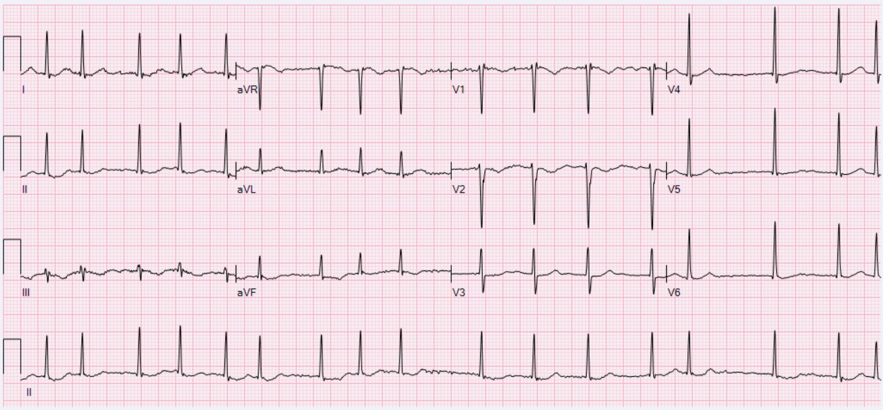
The atria stop squeezing and start quivering. That one fact drives the whole disease. An irregularly irregular pulse with no P waves, blood pooling into a clot in the left atrium, and three questions the boards ask over and over. Is the patient stable? What is the stroke risk on CHA2DS2-VASc? Can you shock them back without launching that clot? Miss the stable-versus-unstable fork and the rest of the answer does not matter. Start with the trap that catches the most students.
Medically reviewed by Fatima Ali, DO & Kaitlyn Cocuzzo, MD✦elite
Before you scroll
A 72-year-old man comes to the office because of a 3-day history of palpitations and mild breathlessness. He has hypertension and takes amlodipine. His blood pressure is 138/84 mmHg, heart rate is 96/min and irregular, and he is alert and comfortable. An ECG shows an irregularly irregular rhythm with no discrete P waves and a fibrillatory baseline. Which of the following is the most appropriate next step in management?
Is he stable or unstable?
Stable. His pressure is fine, he is alert, no chest pain, no crashing heart failure. Unstable means hypotension, active ischemia, decompensated heart failure, or altered mental status. None here. So you do NOT reach for the shock.
What is first line for a stable patient?
Control the rate. A beta-blocker or a non-dihydropyridine calcium channel blocker slows the ventricular response so the heart fills and the symptoms settle. Then you separately decide on stroke prevention by the stroke-risk score.
Why not just cardiovert him today?
His AFib has lasted 3 days, which is over 48 hours. Cardioverting now could fling a clot out of the left atrium and stroke him. You would need 3 weeks of anticoagulation first, OR a TEE to rule out a clot. Adenosine is for AV-nodal SVT, not AFib. Stable AFib over 48 hours. Rate control and anticoagulate, do not shock blindly.
Scroll ↓ watch the strip draw itself
The Signature Game · See What the Atria Are Doing
The Living Rhythm Strip
Watch the trace draw itself, one QRS at a time. Flip between a fibrillating atrium and a normal one. The readouts change as the rhythm changes. This is the picture behind every "irregularly irregular, no P waves" stem.
Lead II · monitor strip
ATRIAL FIBRILLATION
P waves
Absent · chaotic baseline
R-R interval
Variably irregular
Pulse
Irregularly irregular
SignalNo organized atrial depolarization, so no P wave forms.
AV nodeBombarded by random impulses, so it lets ventricles through at random intervals.
PearlNo P waves plus an irregularly irregular pulse equals atrial fibrillation.
The diagnosis in one line
Irregularly irregular rhythm, no discrete P waves, fibrillatory baseline, variably irregular R-R.
The atria are firing from hundreds of spots at once, so there is no single organized P wave, just a wavy line. The AV nodeThe electrical gatekeeper between atria and ventricles. It blocks most of the chaotic atrial impulses, which is why the ventricular rate is lower than the atrial rate and why slowing the AV node is how you rate-control AFib. gets hit by impulses at random, so it passes some through and blocks others, giving a ventricular rhythm with no pattern. That randomness at the wrist is what "irregularly irregular" means.
If the spaces between beats follow no pattern at all and the P waves are gone, you are looking at AFib. Atrial flutter is the organized cousin with sawtooth waves.
From the Attending
Do not overthink the strip. Cover the rate. Just ask two questions. Are the R waves marching to any pattern, and can you find a real P before each one? If the answer is no and no, it is fibrillation. No P, no pattern, that is the whole read. Bring that read to every stem.
Tap to reveal the flutter contrast
Atrial flutter is the disciplined sibling. A single reentry loop firing about 300 times a minute, giving sawtooth flutter waves and an AV block that is often a clean 2-to-1, so the ventricular rate is frequently a regular 150. AFib is chaos with no pattern; flutter is one loop with a steady ratio. If you see sawteeth, think flutter, not fibrillation.
The First Fork · Shock or Slow Down
Stable vs Unstable, Do You Cardiovert?
Every AFib question forks here first. One fork sends you straight to the defibrillator; the other sends you to rate control. Guess each branch before it opens. Get the fork right and the management writes itself.
Atrial fibrillation, no P waves, irregularly irregularAFib at 98/min, irregular R-R spacingNormal sinus rhythm, a P before every QRSSynchronized cardioversion (the unstable answer)
The fork that drives everything
Unstable = immediate synchronized cardioversion. Stable = rate control first, then sort out stroke risk.
Unstable means the rhythm is actively hurting the patient right now. Hypotension, active myocardial ischemia, acute decompensated heart failure, or altered mental status from poor perfusion. That patient gets shocked back without waiting for anticoagulation, because the immediate danger outweighs the clot risk. A stable patient has time, so you slow the rate and decide on blood thinners deliberately.
SHOCK the unstable. SLOW the stable. The four red flags are low pressure, chest pain from ischemia, crashing heart failure, and confusion.
From the Attending
When the stem hands you a soft blood pressure, chest pain with the rhythm, flash pulmonary edema, or a confused patient, stop reading and reach for synchronized cardioversion. Unstable does not wait for a clot workup. The danger in front of you outranks the danger you cannot see yet. Don't overthink it.
Tap to reveal the synchronized vs unsynchronized rule
For a patient who has a pulse but is unstable, you SYNCHRONIZE the shock to the R wave so you do not deliver energy during the vulnerable T-wave window and trigger ventricular fibrillation. Pulseless or a chaotic rhythm with no organized QRS to sync to gets unsynchronized defibrillation. AFib with a pulse, synchronized, every time.
First Aid for the clinical medicine. Cardiovascular, Atrial Fibrillation. Irregularly irregular rhythm, absent P waves, ABCD management (Anticoagulation, Beta-blockers, Calcium channel blockers, Digoxin), and left atrial appendage as the thrombus site.
Harrison's Principles of Internal Medicine. The supraventricular tachyarrhythmias, rate versus rhythm control and stroke prevention.
ACC/AHA/HRS Guideline for the Management of Atrial Fibrillation. CHA2DS2-VASc thresholds, DOAC versus warfarin, and periprocedural anticoagulation.
AFFIRM investigators. Rate versus rhythm control comparison. Roughly equivalent long-term mortality.
Reviewed by Fatima Ali DO and Kaitlyn Cocuzzo MD. Vignettes are original clinical teaching cases; demographics, values, and answer order are written for practice. Always confirm management against the current guideline at the point of care.
Bone Wizardry is an independent educational resource for visual learning in the medical sciences. It is not affiliated with, endorsed by, or sponsored by any licensing or examination board, contains no real or recalled examination questions, and does not guarantee any educational or examination outcome.
That was the free half
You can read the rhythm. Now make the management automatic.
The living rhythm strip and the stable-vs-unstable fork are yours free. Everything that turns "I get it" into a confident answer on exam day lives in Elite.
The CHA2DS2-VASc live calculator with the DOAC-vs-warfarin branch
Rate vs rhythm control and the non-DHP CCB heart-failure trap
The 48-hour cardioversion timing sequence (TEE and the 4-week rule)
The cause elimination drill (holiday heart, hyperthyroidism, mitral stenosis)
Five rapid calls plus board vignettes that save your progress
 Bone WizardryMEDICAL EDUCATION