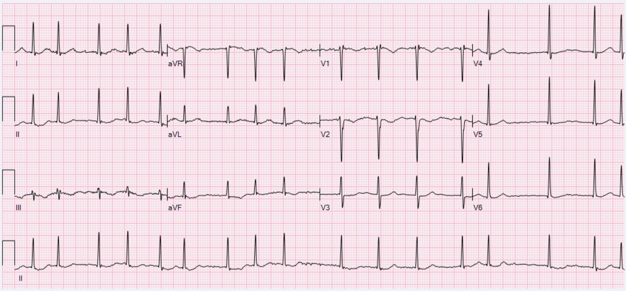
The atria stop squeezing and start quivering. That one fact drives the whole disease: an irregularly irregular pulse, blood pooling into a clot, and three forks the clinical medicine test over and over. Is the patient stable? What is the stroke risk? Can you safely shock them back? Start with the trap that catches the most students.
Medically reviewed by Fatima Ali, DO & Kaitlyn Cocuzzo, MD✦elite
Before you scroll
A 72-year-old man comes to the office because of a 3-day history of palpitations and mild breathlessness. He has hypertension and takes amlodipine. His blood pressure is 138/84 mmHg, heart rate is 96/min and irregular, and he is alert and comfortable. An ECG shows an irregularly irregular rhythm with no discrete P waves and a fibrillatory baseline. Which of the following is the most appropriate next step in management?
Is he stable or unstable?
Stable. His pressure is fine, he is alert, no chest pain, no crashing heart failure. Unstable means hypotension, active ischemia, decompensated heart failure, or altered mental status. None here. So you do NOT reach for the shock.
What is first line for a stable patient?
Control the rate. A beta-blocker or a non-dihydropyridine calcium channel blocker slows the ventricular response so the heart fills and the symptoms settle. Then you separately decide on stroke prevention by the stroke-risk score.
Why not just cardiovert him today?
His AFib has lasted 3 days, which is over 48 hours. Cardioverting now could fling a clot out of the left atrium and stroke him. You would need 3 weeks of anticoagulation first, OR a TEE to rule out a clot. Adenosine is for AV-nodal SVT, not AFib. Stable AFib over 48 hours: rate control and anticoagulate, do not shock blindly.
Scroll ↓ see the rhythm move
The Signature Game · See What the Atria Are Doing
The Living Rhythm Strip
Watch the trace draw itself, beat by beat. Flip between a fibrillating atrium and a normal one. The readouts change as the rhythm changes. This is the picture behind every "irregularly irregular, no P waves" stem.
Lead II · monitor strip
ATRIAL FIBRILLATION
P waves
Absent · chaotic baseline
R-R interval
Variably irregular
Pulse
Irregularly irregular
SignalNo organized atrial depolarization, so no P wave forms.
AV nodeBombarded by random impulses, so it lets ventricles through at random intervals.
PearlNo P waves plus an irregularly irregular pulse equals atrial fibrillation.
The diagnosis in one line
Irregularly irregular rhythm, no discrete P waves, fibrillatory baseline, variably irregular R-R.
The atria are firing from hundreds of spots at once, so there is no single organized P wave, just a wavy line. The AV nodeThe electrical gatekeeper between atria and ventricles. It blocks most of the chaotic atrial impulses, which is why the ventricular rate is lower than the atrial rate and why slowing the AV node is how you rate-control AFib. gets hit by impulses at random, so it passes some through and blocks others, giving a ventricular rhythm with no pattern. That randomness at the wrist is what "irregularly irregular" means.
If the spaces between beats follow no pattern at all and the P waves are gone, you are looking at AFib. Atrial flutter is the organized cousin with sawtooth waves.
From the Attending
Do not overthink the strip. Cover the rate. Just ask two questions. Are the R waves marching to any pattern, and can you find a real P before each one? If the answer is no and no, it is fibrillation. No P, no pattern, that is the whole read. Know your clues.
Tap to reveal the flutter contrast
Atrial flutter is the disciplined sibling: a single reentry loop firing about 300 times a minute, giving sawtooth flutter waves and an AV block that is often a clean 2-to-1, so the ventricular rate is frequently a regular 150. AFib is chaos with no pattern; flutter is one fast loop with a steady ratio. If you see sawteeth, think flutter, not fibrillation.
The First Fork · Shock or Slow Down
Stable vs Unstable: Do You Cardiovert?
Every AFib question forks here first. One fork sends you straight to the defibrillator; the other sends you to rate control. Guess each branch before it opens. Get the fork right and the management writes itself.
Atrial fibrillation: no P waves, irregularly irregularAFib at 98/min: irregular R-R spacingNormal sinus rhythm: a P before every QRSSynchronized cardioversion (the unstable answer)
The fork that drives everything
Unstable = immediate synchronized cardioversion. Stable = rate control first, then sort out stroke risk.
Unstable means the rhythm is actively hurting the patient right now: hypotension, active myocardial ischemia, acute decompensated heart failure, or altered mental status from poor perfusion. That patient gets shocked back without waiting for anticoagulation, because the immediate danger beats the clot risk. A stable patient has time, so you slow the rate and decide on blood thinners deliberately.
SHOCK the unstable. SLOW the stable. The four red flags are low pressure, chest pain from ischemia, crashing heart failure, and confusion.
From the Attending
When the stem hands you a soft blood pressure, chest pain with the rhythm, flash pulmonary edema, or a confused patient, stop reading and reach for synchronized cardioversion. Unstable does not wait for a clot workup. The danger in front of you outranks the danger you cannot see yet. Don't overthink it.
Tap to reveal the synchronized vs unsynchronized rule
For a patient who has a pulse but is unstable, you SYNCHRONIZE the shock to the R wave so you do not deliver energy during the vulnerable T-wave window and trigger ventricular fibrillation. Pulseless or a chaotic rhythm with no organized QRS to sync to gets unsynchronized defibrillation. AFib with a pulse: synchronized, every time.
The Second Fork · Who Needs a Blood Thinner
CHA₂DS₂-VASc Live Calculator
Tap the risk factors your patient has. The score tallies live, the recommendation appears, and then a branch decides DOAC versus warfarin. This is the exact engine the clinical medicine test when they ask "is this AFib patient anticoagulated?"
Congestive heart failureC · +1
HypertensionH · +1
Age 75 or olderA₂ · +2
Diabetes mellitusD · +1
Prior stroke, TIA, or clotS₂ · +2
Vascular diseaseV · +1
Age 65 to 74A · +1
0
Tap the risk factors above. The sex point only counts as a tiebreaker once another risk factor is present.
No risk factors selected yet.
This patient needs anticoagulation. Which agent?
Does this patient have a mechanical valve OR moderate-to-severe mitral stenosis?
The scoring you carry to the exam
CHF 1, HTN 1, Age 75+ = 2, Diabetes 1, prior Stroke/TIA = 2, Vascular disease 1, Age 65 to 74 = 1, Sex (female) 1. Anticoagulate at 2 or more in men and 3 or more in women. The two-pointers are the ones people forget: age 75+ and a prior stroke each count double.
The two-point flags are age 75 and a prior stroke. The female point is a tiebreaker, never the only reason to treat.
From the Attending
A DOAC is the default. It beats warfarin on convenience and on bleeding, and it needs no INR checks. Two situations still demand warfarin. A mechanical heart valve, and moderate-to-severe mitral stenosis. Mechanical valve or tight mitral stenosis means warfarin, every time. Everything else gets a DOAC. That distinction drives the whole answer.
Tap to reveal the HAS-BLED rule
HAS-BLED estimates bleeding risk, not whether to anticoagulate. A high HAS-BLED does NOT cancel a high CHA2DS2-VASc; it tells you to fix modifiable bleeding risks (control blood pressure, review alcohol and antiplatelets) and watch more closely. The board trap is using a high bleeding score as an excuse to withhold a blood thinner the stroke score clearly demands.
Long-Term Control · And the Shock Timing Rule
Rate Control, Rhythm Control, and the 48-Hour Rule
First the drug families that slow or reset the heart, with the one combination that is dangerous in heart failure. Then build the timing sequence that keeps a clot from launching when you cardiovert.
Mitral stenosis: valvular AFib, warfarin requiredHyperthyroidism (goiter): a reversible causeAtria and conduction system (left atrial enlargement)
Rate control · first line
Beta-Blocker
Examples
Metoprolol, esmolol
Action
Slows AV node conduction
Best for
Most stable patients, including heart failure
Watch
Bradycardia, bronchospasm in severe asthma
Board line
A reasonable first choice for rate control, and safe in reduced ejection fraction heart failure.
Beta-blocker: the default rate-control agent. Safe when the ejection fraction is low.
Rate control · first line
Non-Dihydropyridine CCB
Examples
DiltiazemA non-dihydropyridine calcium channel blocker. Unlike amlodipine (a dihydropyridine that acts on vessels), diltiazem and verapamil slow the AV node, which is what rate-controls AFib., verapamil
Action
Slows AV node conduction
Best for
Stable patients with preserved heart function
Avoid in
Reduced ejection fraction (HFrEF)
Board trap
Diltiazem and verapamil are negative inotropes. In systolic heart failure they can worsen the failure, so AVOID them in HFrEF and use a beta-blocker instead.
Diltiazem and verapamil slow the rate well, but keep them OUT of reduced-EF heart failure.
Rate control · adjunct
Digoxin
Action
Vagal AV node slowing
Best for
Hypotensive patients or heart failure, as add-on
Limit
Poor rate control with exertion
Watch
Narrow therapeutic window, toxicity
Board line
An adjunct, not a first choice. Useful when blood pressure is low or in heart failure because it slows rate without dropping pressure, but it controls rate poorly during activity.
Digoxin is the add-on for the hypotensive or heart-failure patient, not the lead agent.
Rhythm control · reset the rhythm
Rhythm Control
Tools
Electrical or pharmacologic cardioversion, antiarrhythmic, ablation
Drugs
Amiodarone, flecainide, sotalol
Choose when
Symptomatic despite rate control, young, new-onset, or heart failure
Procedure
Catheter ablation for refractory cases
Board line
Over the long run, rate and rhythm control give roughly equal mortality (the AFFIRM lesson), so rate control is the default. Favor rhythm control for ongoing symptoms, younger patients, new-onset AFib, or heart failure that cannot tolerate the arrhythmia.
Rhythm control for the symptomatic, young, new-onset, or heart-failure patient. Otherwise rate control wins on simplicity.
The 48-hour cardioversion sequence
AFib has lasted more than 48 hours (or unknown). The patient is stable and you want to cardiovert. Drag the four steps into the safe order so you do not launch a clot.
1
Drop the first safe step
2
Drop step 2
3
Drop step 3
4
Drop the last step
Exclude a clot: 3 weeks anticoagulation first, OR a TEE nowConfirm duration is over 48 hours or unknownPerform cardioversion once a clot is excludedAnticoagulate at least 4 weeks afterward
Drag (or tap a step, then a slot) to build the safe order. Fill all four to lock it in.
Why the order matters
Over 48 hours, a clot has had time to form in the left atrial appendage, the favorite hiding spot. Shock the heart back and that clot can fire to the brain. So before cardioversion you either give 3 weeks of therapeutic anticoagulation to let any clot organize and stabilize, OR do a TEE to look directly and confirm there is none. Afterward you keep anticoagulating for at least 4 weeks, because the stunned atrium does not contract normally right away and can still form clot even in sinus rhythm.
Over 48 hours: clear the clot before you shock (3 weeks of blood thinner OR a TEE), then 4 weeks after. Under 48 hours with low risk, you have more leeway.
From the Attending
The left atrial appendage is where the clot lives. That is why we either wait three weeks on a blood thinner or put the probe down the esophagus to look. And do not stop the anticoagulation the moment you restore sinus. The atrium is stunned, so the clot risk lingers, four weeks minimum after. Find the appendage, clear it, then shock. Every time.
What Started It · And Your Board Reps
Find the Trigger, Then Walk the Cases
First a quick elimination drill on what flips a heart into AFib. Then five quick calls and a full board-vignette bank. Cross out (right-click or long-press) and highlight (select text) as you read.
The causes worth burning in
HTN and valvular disease (especially mitral stenosis) are the structural heavyweights. Then the reversible flips: hyperthyroidism, alcohol binge ("holiday heart"), obstructive sleep apnea, pulmonary embolism, sepsis, post-operative state, and plain old aging. When AFib is new, check a TSH and ask about the weekend.
Old, hypertensive, leaky mitral valve, hyperthyroid, or hungover. New AFib means check the thyroid and the alcohol history.
Tap to reveal the "holiday heart" hook
Holiday heart is AFib triggered by a binge of alcohol, classically a young, otherwise healthy person who drank heavily over a weekend or holiday and shows up with palpitations. It often converts on its own once the alcohol clears. If a vignette gives you a young patient, new AFib, and a big night of drinking, that is the answer.
Don't Kill The Patient
Five Quick Calls
Five rapid questions pulled from a bigger pool, reshuffled every visit. Cross out (right-click / long-press) and highlight (select text) as you read.
clinical Practice
Walk the Cases
Full clinical vignettes, one at a time, in a shuffled order. Progress saves to your account on this device. Cross out (right-click / long-press) and highlight (select text) as you go.
From the Attending
These vignettes are written the way the real exam writes them. A patient, a rhythm, and one buried discriminator. Cover the choices. Ask the stable-or-unstable question first, then the stroke score, then the timing rule. Read every explanation, not just the one you missed. The fork you skip is the point you lose.
Tip: kill the wrong choices first, then read the explanation chain for every option.
First Aid for the clinical medicine. Cardiovascular: Atrial Fibrillation. Irregularly irregular rhythm, absent P waves, ABCD management (Anticoagulation, Beta-blockers, Calcium channel blockers, Digoxin), and left atrial appendage as the thrombus site.
Harrison's Principles of Internal Medicine. The supraventricular tachyarrhythmias: rate versus rhythm control and stroke prevention.
ACC/AHA/HRS Guideline for the Management of Atrial Fibrillation. CHA2DS2-VASc thresholds, DOAC versus warfarin, and periprocedural anticoagulation.
AFFIRM investigators. Rate versus rhythm control comparison. Roughly equivalent long-term mortality.
Reviewed by Fatima Ali DO and Kaitlyn Cocuzzo MD. Vignettes are original clinical teaching cases; demographics, values, and answer order are written for practice. Always confirm management against the current guideline at the point of care.
Bone Wizardry is an independent educational resource for visual learning in the medical sciences. It is not affiliated with, endorsed by, or sponsored by any licensing or examination board, contains no real or recalled examination questions, and does not guarantee any educational or examination outcome.
That was the free half
You can read the rhythm. Now make the management automatic.
The living rhythm strip and the stable-vs-unstable fork are yours free. Everything that turns "I get it" into a 10-second answer on exam day lives in Elite:
The CHA2DS2-VASc live calculator with the DOAC-vs-warfarin branch
Rate vs rhythm control and the non-DHP CCB heart-failure trap
The 48-hour cardioversion timing sequence (TEE and the 4-week rule)
The cause elimination drill (holiday heart, hyperthyroidism, mitral stenosis)
Five quick calls plus board vignettes that save your progress