Colon Polyps & Malignant Risk
Villous adenoma, Lynch, FAP, Peutz-Jeghers, Fearon-Vogelstein, surveillance intervals
The Adenoma to Carcinoma Sequence
Watch the same crypt drift toward cancer. Bigger, more villous, more dysplastic means more risk. Size and villous architecture are the two things you actually act on.
Normalflat mucosa
Tubularsmall, stalked, low risk
Tubulovillousmixed architecture
Villouslarge, sessile, highest risk
Carcinomainvades the basement membrane
Low malignant potentialHigh malignant potential
From the Attending
Three polyps walk into a colonoscopy. One is a tubular adenoma, one is tubulovillous, one is villous. Which one do you watch? The answer you pick tells me everything about whether you understand the actual biology here. Not the name. The biology.
The Trap
Most students pick tubular adenoma as the risky one because it is the most common. More common does not mean more dangerous. Villous architecture rewires the risk. Know the difference before you touch the walkthrough bank.
Polyp Risk Lineup
Tap a card to see the full profile.
Zero Risk
Hyperplastic Polyp
Small, pale, sigmoid/rectum. Sawtooth architecture. No dysplasia. Board answer: no cancer risk.
Hyperplastic polyp: saw-tooth glands, no dysplasia, no malignant potential. Found in sigmoid and rectum. Exception: large hyperplastic polyps in right colon may actually be sessile serrated adenomas misclassified. Small sigmoid hyperplastics = safe. Follow-up: resume 10-year standard screening.
Low Risk <5%
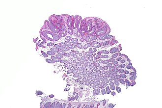
Tubular Adenoma
80% of all adenomas. Tubular glands, pedunculated. Low risk unless ≥1cm or HGD.
Tubular adenoma: most common adenoma. Round tubular glands. Often pedunculated (on a stalk). Risk under 5%. Becomes "advanced" if ≥1cm, ≥25% villous component, or high-grade dysplasia present. 1-2 small tubular adenomas: 5-7 year surveillance. 3+ adenomas: 3 years.
Moderate 7-10%
Tubulovillous Adenoma
Mixed architecture: 25-75% villous. Intermediate risk. Often larger and sessile.
Tubulovillous: mixed architecture, 25-75% villous component. Risk roughly 7-10%. Often found in rectosigmoid. Classified as advanced adenoma. Surveillance: 3 years after resection. Board pearl: if the question says "tubulovillous", the answer is probably 3-year surveillance.
Very High 10-40%
Villous Adenoma
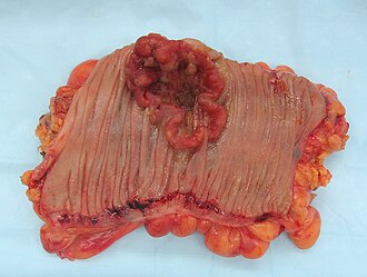
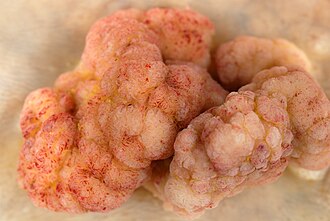
Frond-forming, sessile, large. Secretory diarrhea + hypokalemia. Highest adenoma risk.
Villous adenoma: the board villain. Finger-like fronds, sessile (no stalk), often in rectum. CRC risk 10-40%. Secretes sodium and potassium → massive watery diarrhea + hypokalemia (McKittrick-Wheelock if severe). Continues during fasting (secretory pattern). Surgical resection if not amenable to endoscopic removal.
Right-Sided Risk
Sessile Serrated Adenoma
Flat, right colon, serrated glands. Methylation pathway → sporadic MSI-H CRC.
Sessile serrated adenoma (SSA): right colon, flat/sessile, serrated architecture with crypt dilation. BRAF mutation → CpG island methylation → MLH1 silencing → MSI-H → sporadic right-sided CRC. Looks like hyperplastic on colonoscopy but is NOT. Board trap: sporadic MSI-H CRC without family history = SSA pathway, not Lynch germline.
No Adenoma Risk
Inflammatory Pseudopolyp
IBD-related. Granulation tissue regenerating after ulceration. No malignant potential by themselves.
Inflammatory pseudopolyp: not a true adenoma. Regenerative mucosa and granulation tissue forming during IBD flares. No inherent malignant potential. Note: UC itself carries CRC risk (surveillance colonoscopy every 1-2 years after 8 years of disease), but these polyps specifically are NOT precancerous.
Clinical Findings
Adenocarcinoma from adenoma
Tubular adenoma (histo)
Villous adenoma (histo)
Juvenile polyp (histo)
Medically reviewed by Fatima Ali, DO and Kaitlyn Cocuzzo, MD · Last reviewed June 2026
Bone Wizardry is an independent educational resource for visual learning in the medical sciences. It is not affiliated with, endorsed by, or sponsored by any licensing or examination board, contains no real or recalled examination questions, and does not guarantee any educational or examination outcome.