Localize infant obstruction from bile, timing and imaging; compare intestinal atresias with pyloric stenosis and choose safe next steps.
An infant who vomits may have a blocked lumen, an injured bowel segment, or a narrowing gastric outlet. The useful question is not just which diagnosis fits: it is where contents stop, what can still pass, and whether bowel perfusion is threatened.
Green vomiting needs urgent assessment. Treat bilious vomiting in a newborn or young infant as possible intestinal obstruction and obtain urgent pediatric surgical input. A comfortable appearance, a passed stool, or a nondiagnostic radiograph must not provide false reassurance. Resuscitation and investigation proceed together when the infant is unwell. [1][6]
By the end, you should be able to localize an obstruction from anatomy and gas distribution; distinguish duodenal recanalization failure from fetal vascular injury; and use age, ultrasound and chemistry to plan care for pyloric stenosis. Each comparison below includes a prediction to make before checking the explanation.
What failed: the lumen, the blood supply, or the muscle?
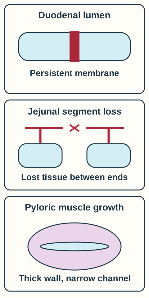
Two blocked intestines need not have the same developmental history. Atresia means complete interruption of the lumen. Stenosis means narrowing with some continuity. A web may completely occlude the lumen or retain an opening. That remaining opening changes the timing and the amount of gas that reaches downstream bowel. [2][3]
The conventional model for intrinsic duodenal atresia is failure to restore a lumen after epithelial proliferation temporarily fills it. This is the classic recanalization explanation, not proof that every duodenal obstruction has one cause. A pancreatic ring around the duodenum, an intrinsic web, and abnormal peritoneal bands can obstruct the same region through different anatomy. [2][6][9]
Follow the tissue that changes, rather than memorizing three names. Original Bone Wizardry schematic; the vascular example depicts segment loss, not every atresia subtype. [2][3][4][10]
Jejunoileal atresia is classically linked to impaired fetal mesenteric blood supply. Ischemic tissue can die and be resorbed, leaving blind ends and sometimes a mesenteric gap. Not every lesion has a visible gap: membranous and cord-connected forms also exist. Multiple or extensive lesions can reduce useful bowel length. This differs from a normally continuous duodenum whose lumen did not form properly. [3][10]
Hypertrophic pyloric stenosis is the comparison diagnosis, not an intestinal atresia. Pyloric muscle hypertrophy and hyperplasia progressively restrict gastric emptying after birth. The bowel is not missing, and the lesion is not simply temporary failure of muscle relaxation. [4]
Predict the surgical anatomy. One newborn has a duodenal membrane; another has separated jejunal ends and a mesenteric defect. Which observation supports loss of a previously formed segment?
Compare the structural explanations
The jejunal gap supports ischemic loss and resorption. A membrane alone describes an obstructed lumen and does not establish loss of a whole bowel segment. Location and tissue architecture must be interpreted together.
Transfer: A lesion detected in the first hours of life can be congenital, but its timing does not distinguish a web from segment loss. Ask what imaging or operative anatomy actually demonstrates.
Read the spaces that fill before the obstruction
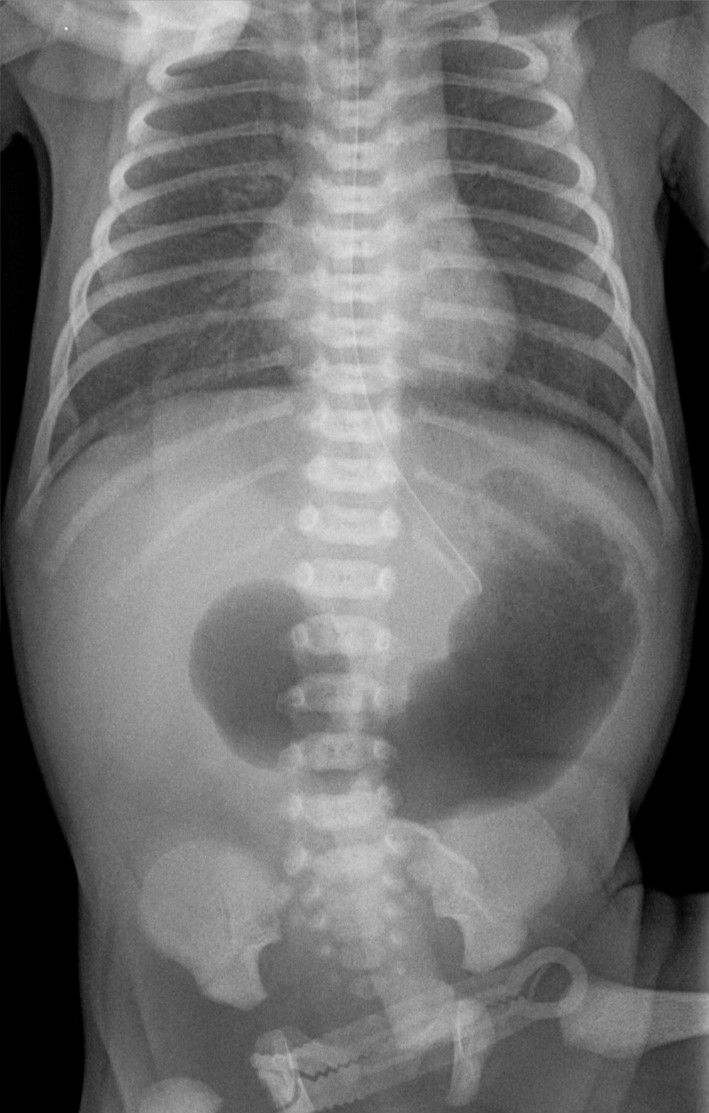
Does a double bubble identify a cause or a location? On a postnatal radiograph, swallowed air outlines compartments upstream of an obstruction. The first large lucency represents the stomach; the second represents the proximal duodenum. A classic double bubble with no distal gas strongly supports complete duodenal obstruction. It does not independently identify the tissue responsible. [2][5]
Real radiograph: identify the two upper abdominal gas spaces before reading their names in the text. The source reports surgically confirmed duodenal atresia with annular pancreas, illustrating why the gas pattern localizes better than it specifies cause. Image: Hellerhoff, 17 December 2021; original image, CC BY-SA 4.0. [8].
Extend the map downstream. A classic triple-bubble pattern can include stomach, duodenum and proximal jejunum, suggesting a proximal jejunal obstruction. More distal small-bowel obstruction often produces more dilated loops, air-fluid levels and more generalized distention. Counting bubbles is a localization aid, not a centimeter ruler or a reliable way to name every individual loop. [3][5]
Gas beyond a double bubble suggests incomplete obstruction, such as stenosis or a fenestrated web, or another process such as malrotation. Rare abnormal biliary connections can also permit distal gas despite atresia. Therefore, distal gas argues against a simple complete block but does not make green vomiting safe. Conversely, a normal or nearly gasless film cannot exclude volvulus. [2][5][9]
Make a comparison. Both infants have two enlarged upper abdominal compartments. Infant A has no distal gas; infant B has scattered gas throughout the remaining bowel. What question remains more open in infant B?
Check the interpretation
Whether there is partial intrinsic obstruction or abnormal rotation with intermittent obstruction. The presence of downstream gas demonstrates some route for gas passage, not normal rotation. Urgent evaluation of bilious vomiting remains necessary.
Transfer: The same stomach and duodenum can look fluid-filled on prenatal ultrasound and gas-filled after birth. Prenatal obstruction can reduce passage and absorption of swallowed amniotic fluid, contributing to polyhydramnios. Two fetal fluid spaces must be shown to communicate before treating them as a true double bubble. [2][9]
Decide what cannot wait
What changes if the infant becomes poorly perfused? A fixed atresia prevents passage. Midgut volvulus can obstruct both the lumen and its blood supply. Malrotation leaves an abnormally narrow mesenteric attachment around which bowel can twist. That difference changes the urgency: threatened bowel requires emergency surgical assessment, not a prolonged search for the prettiest radiographic pattern. [6]
For suspected obstruction, stop oral feeds, establish intravenous access, assess glucose and electrolytes, decompress the stomach with an appropriate gastric tube, and involve pediatric surgery. Support circulation and reassess perfusion. Bilious emesis with tenderness, bloody stool, lethargy or shock raises concern for ischemia. In an unstable infant with suspected volvulus or peritonitis, resuscitation and emergency surgical management must not be delayed for routine imaging or normalization of every laboratory value. [1][6][9]
Choose the test for the unanswered question
Early neonatal obstruction: an abdominal radiograph is usually the initial study. A classic complete proximal obstruction warrants prompt surgical discussion; an upper gastrointestinal contrast study is not automatically required for every classic double bubble.
Bilious vomiting with a normal or nonclassic radiograph: in a stable infant, an urgent upper gastrointestinal contrast study commonly evaluates rotation and duodenal passage. Experienced ultrasound assessment can contribute, according to local expertise. Neither a normal plain film nor a single normal vessel relationship should end a concerning evaluation.
Distal obstruction on radiography: a specialist-directed contrast enema can assess colonic caliber and distal anatomy. A small unused colon is not itself proof of one disease. Clinical context and the full study distinguish distal atresia from other obstructions. [3][5][6]
Reassess a changing patient. A newborn awaiting a contrast study develops abdominal tenderness, poor capillary refill and bloody stool. Would repeating the plain radiograph be the priority?
Check the priority
No. The new findings raise concern for compromised bowel perfusion. Activate emergency surgical care and resuscitation together; do not let routine imaging postpone time-critical treatment.
Transfer: Stabilization before repair is appropriate for an atresia or pyloric stenosis. It must not become a rule to defer treatment of a suspected ischemic volvulus until all chemistry is normal.
Use bile entry to test your localization
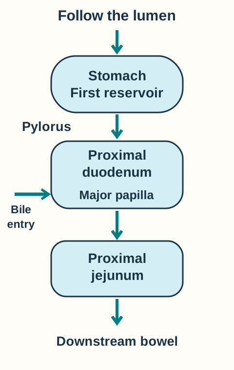
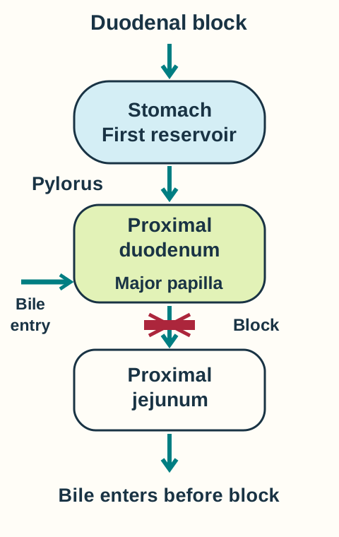
Does nonbilious vomiting exclude duodenal obstruction? No. Bile normally enters the descending duodenum at the major papilla. A block before that entry point can cause nonbilious vomiting, including the less common preampullary duodenal atresia. A block after bile entry commonly causes bilious vomiting. These are anatomic expectations, not guarantees about the cause or completeness of obstruction. [9]
Trace swallowed contents downward and locate the bile entry before predicting what can return upward. [9]
Ask the family to describe the actual color; bile-stained vomit is typically dark green, not just yellow milk. Absence of green color does not exclude serious illness or intestinal obstruction. Projectile force also fails to localize a lesion by itself. [1][9]
Predict, then reveal a different blocked-lumen state
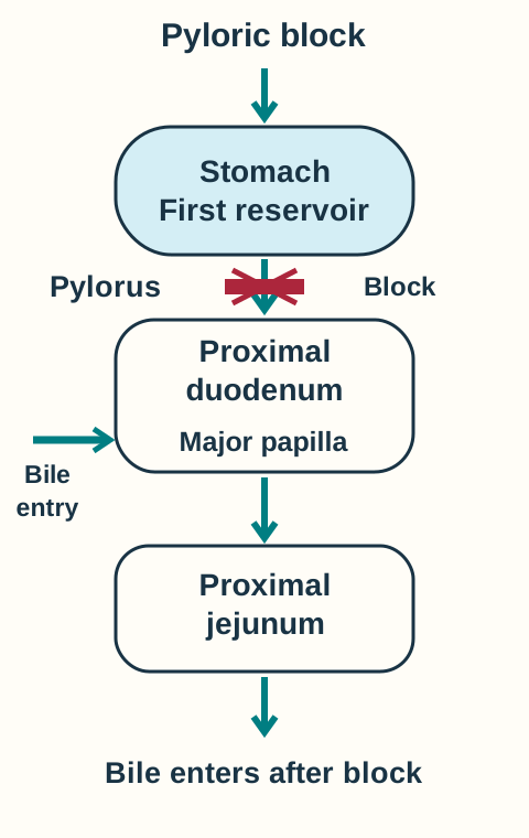
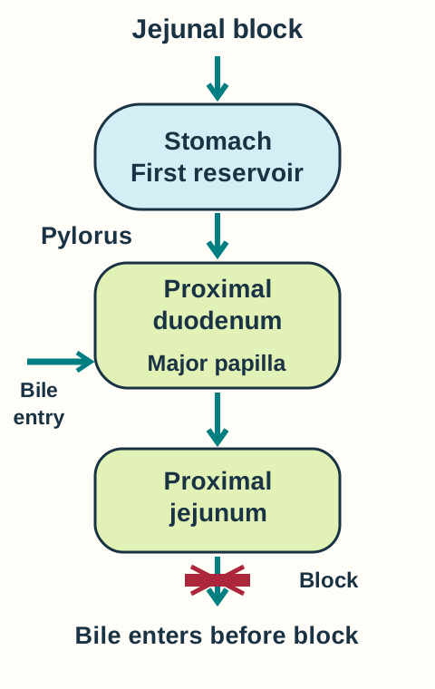
Use the normal map above. For each location, predict which compartments can fill and whether bile can enter the upstream reservoir. Open a state to compare your prediction with its diagram. Close it to reset; multiple states may stay open in the reading flow. Arrows show the normal forward route; shading marks compartments that can fill upstream of the complete block. The model assumes a complete block and ordinary biliary anatomy.
Place a complete block at the pyloric outlet
The stomach fills before the block; bile enters downstream. Predicted emesis is nonbilious. A newborn with a truly complete congenital pyloric obstruction differs from an older infant with gradually developing pyloric stenosis. [3][4]Place a complete block just after bile entry
Both upper compartments fill, and bile can join the retained contents. The model predicts a double bubble and bilious vomiting. It localizes a blockage without deciding whether the lesion is intrinsic or external. [9]Place a complete block in the proximal jejunum
An additional upstream compartment can fill, producing the classic triple-bubble concept. Bilious vomiting is expected, but the exact gas pattern varies with anatomy and timing. [3][5]
Transfer: Give the duodenal block a small opening. Now some gas can pass distally and presentation may be delayed, but the papilla has not changed location. Partial patency changes severity and gas distribution; it does not automatically change bile entry or exclude malrotation.
Let the time course and chemistry explain pyloric stenosis
Why can a hungry infant become dehydrated despite frequent feeds? Milk reaches the stomach, but progressive outlet narrowing prevents effective delivery to the intestine. Pyloric stenosis usually presents at 2 to 6 weeks with worsening nonbilious vomiting, sometimes projectile, and inadequate weight gain. A palpable olive or visible gastric peristalsis can support the diagnosis, but their absence does not exclude it. Complete intestinal atresias usually present much earlier; partial congenital lesions can present later. Age adjusts probability rather than replacing anatomy. [3][4]
Ultrasound is the preferred first study when pyloric stenosis is suspected. Persistent thickening and elongation of the pyloric muscle with impaired passage support the diagnosis. Borderline measurements require interpretation with infant size, the dynamic examination and the clinical course. Early disease may be equivocal; repeat expert ultrasound when concern continues. Transient pylorospasm can resemble fixed narrowing on a single observation. [4][5][11]
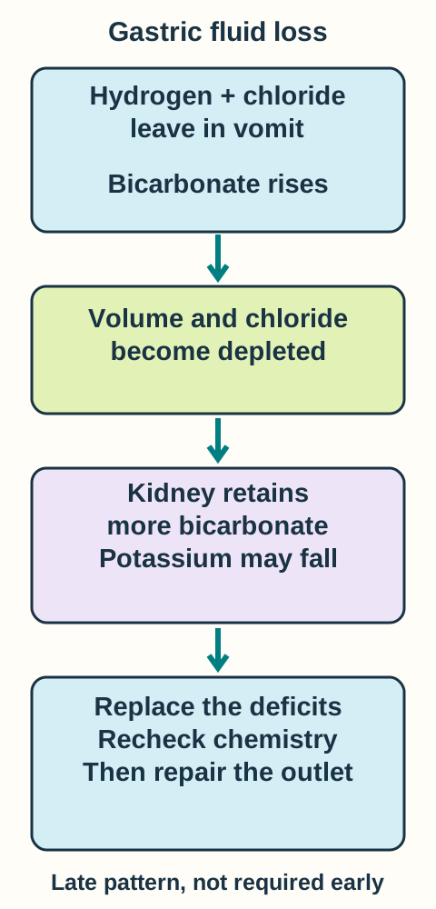
Explain each arrow from lost gastric contents to persistent alkalosis. Original Bone Wizardry physiological schematic. The classic chemistry follows prolonged vomiting and may be absent early. [4][12]
Loss of gastric hydrochloric acid produces hypochloremic metabolic alkalosis. Volume depletion reduces renal bicarbonate clearance, while chloride depletion impairs bicarbonate excretion. Secondary aldosterone effects and potassium depletion can reinforce hydrogen and potassium loss. Thus serum bicarbonate may stay high even after vomiting stops. In substantial depletion, urine may become acidic despite systemic alkalosis: preserving sodium and volume can coexist with continued hydrogen secretion. Do not mistake this late pattern for the initial cause of the alkalosis. [12]
The early priorities are stopping oral feeds and correcting dehydration, glucose needs, electrolytes and acid-base abnormalities. Chloride-containing intravenous fluid addresses the deficits; add potassium only after adequate urine output and renal assessment. Gastric decompression may be needed for persistent profuse vomiting. Definitive treatment is pyloromyotomy after physiological correction. Persistent alkalosis matters because it increases perioperative hypoventilation and apnea concern. A normal bicarbonate result early in the illness does not rule out pyloric stenosis. [4]
Read the trend. A 5-week-old has chloride 84 mmol/L and bicarbonate 38 mmol/L. Vomiting stops after feeds are withheld, but the repeat bicarbonate remains high. Should cessation of vomiting alone clear the infant for anesthesia?
Explain the remaining problem
No. Stopping new acid loss does not replace chloride, restore circulating volume or correct potassium depletion. Reassess physiology and repeat chemistry during correction before pyloromyotomy.
Transfer: A younger infant with progressive nonbilious vomiting, weight loss and normal electrolytes still needs evaluation. Conversely, green vomiting or new abdominal tenderness should redirect the assessment away from uncomplicated pyloric stenosis.
Use associations without letting them decide the diagnosis
Which associated finding changes the workup, rather than merely making a diagnosis sound familiar? Duodenal atresia is associated with trisomy 21 and congenital cardiac defects. Once the obstruction is recognized and stabilization begins, evaluate associated anomalies, including cardiac anatomy, with the neonatal and surgical teams. A chromosomal diagnosis neither proves the obstruction's level nor excludes another lesion. [2][9]
Jejunoileal atresia can accompany gastroschisis or other fetal intestinal injury. Gastroschisis exposes bowel through an abdominal wall defect, usually beside the umbilicus and without a protective sac. Compromised or twisted exposed bowel can have additional pathology beyond the visible wall defect. The surgical team assesses bowel continuity and viable length. Cystic fibrosis is also associated with jejunoileal obstruction and merits appropriate evaluation rather than being diagnosed from an unused small colon alone. [3][10][13]
Pyloric stenosis is more common in boys and firstborn infants. Early oral erythromycin or azithromycin exposure is associated with increased risk, particularly exposure in the first two weeks. The published evidence is observational: it does not mean that every exposed infant develops stenosis, that exposure proves the diagnosis, or that an indicated antibiotic should be withheld without clinical assessment. Symptoms and ultrasound still determine the evaluation. [4][7]
Compare two histories. Infant A is a firstborn boy with small effortless spit-ups and normal growth. Infant B is a girl with steadily worsening forceful vomiting, weight loss and persistently abnormal pyloric ultrasound. Which history should carry more diagnostic weight?
Check how probability is updated
The progressive illness and direct imaging in infant B outweigh the demographic pattern in infant A. Risk factors modify an assessment; they cannot substitute for evidence of impaired gastric emptying.
Before practice: Describe the level, completeness and likely tissue process separately. Then state the immediate safety priority and the test that answers the remaining uncertainty. Do not demand every classic feature before acting.
Extend the anatomical map
Esophageal atresia and tracheoesophageal fistula: a tube coiled in a proximal pouch supports atresia. In that setting, gastric gas supports a distal airway connection. Drooling, choking and cough with feeding direct attention above the stomach. An H-type fistula has a patent esophagus, so tube passage does not exclude a fistula. [14]
Ventral wall defects: uncovered bowel beside the umbilicus supports gastroschisis. A midline sac at the umbilical insertion supports omphalocele; its covering is normally present but can rupture. Inspect location and covering together. [13][16]
Pancreas and spleen embryology: the ventral pancreatic bud contributes the uncinate process and inferior head; the dorsal bud contributes the remaining pancreas. The spleen instead develops from mesodermal mesenchyme in the dorsal mesogastrium. These are distinct developmental tissues. [15][17]
Apply the anatomy to a new infant
These original educational cases test localization, changing physiology and safe decisions. Use the optional study controls for support; each case remains independent of the teaching predictions above.
Case 1
Show answer and explanations for case 1
A. Loss of fetal mesenteric blood supply (Why this does not fit)
Fetal vascular interruption can produce jejunoileal atresia through tissue loss. The specified lesion is a duodenal membrane in an externally continuous segment, not a jejunal gap. Interpret location and tissue architecture together.
Reasoning steps for option A
What structural consequence can fetal ischemia produce?
Fetal vascular interruption can produce jejunoileal atresia through tissue loss.
Which case finding argues against "Loss of fetal mesenteric blood supply"?
The specified lesion is a duodenal membrane in an externally continuous segment, not a jejunal gap.
What discriminator should be checked before choosing "Loss of fetal mesenteric blood supply"?
Interpret location and tissue architecture together.
B. Abnormal fixation of the midgut mesentery (Why this does not fit)
Malrotation permits twisting and can create obstructing peritoneal bands. The operation identifies an intrinsic occluding membrane rather than twisting or an external band. A similar obstruction pattern can arise from a different tissue process.
Reasoning steps for option B
How can abnormal fixation obstruct bowel?
Malrotation permits twisting and can create obstructing peritoneal bands.
Which case finding argues against "Abnormal fixation of the midgut mesentery"?
The operation identifies an intrinsic occluding membrane rather than twisting or an external band.
What discriminator should be checked before choosing "Abnormal fixation of the midgut mesentery"?
A similar obstruction pattern can arise from a different tissue process.
C. Failure to restore the duodenal lumen (Best answer)
The imaging localizes a complete duodenal obstruction. A continuous duodenum with an occluding membrane fits the classic recanalization explanation, rather than loss of an ischemic bowel segment.
Reasoning steps for option C
Where do the two gas spaces place the obstruction?
At the duodenal level, downstream from the stomach.
What does an occluding membrane add?
The lumen is interrupted without demonstrated loss of the whole segment.
Which developmental explanation fits that anatomy?
Classic failure of duodenal recanalization fits an intrinsic duodenal membrane; a double bubble alone would not establish the cause.
D. Progressive thickening of pyloric muscle (Why this does not fit)
Pyloric stenosis develops over the first weeks and restricts gastric emptying. This infant has a complete intrinsic duodenal lesion at birth and bile-stained vomiting. Match both the involved structure and the time course.
Reasoning steps for option D
When does pyloric muscle narrowing usually become symptomatic?
Pyloric stenosis develops over the first weeks and restricts gastric emptying.
Which case finding argues against "Progressive thickening of pyloric muscle"?
This infant has a complete intrinsic duodenal lesion at birth and bile-stained vomiting.
What discriminator should be checked before choosing "Progressive thickening of pyloric muscle"?
Match both the involved structure and the time course.
Takeaway: Classic failure of duodenal recanalization fits an intrinsic duodenal membrane; a double bubble alone would not establish the cause.
A. Epithelial occlusion followed by failure of luminal reopening (Why this does not fit)
Failed recanalization is the conventional explanation for intrinsic duodenal obstruction. The operation shows missing jejunal tissue and a mesenteric gap, not just persistent luminal occlusion. A missing segment and an occluded segment are not the same anatomical observation.
Reasoning steps for option A
What lesion classically follows failed recanalization?
Failed recanalization is the conventional explanation for intrinsic duodenal obstruction.
Which case finding argues against "Epithelial occlusion followed by failure of luminal reopening"?
The operation shows missing jejunal tissue and a mesenteric gap, not just persistent luminal occlusion.
What discriminator should be checked before choosing "Epithelial occlusion followed by failure of luminal reopening"?
A missing segment and an occluded segment are not the same anatomical observation.
B. Postnatal muscle growth followed by gastric outlet narrowing (Why this does not fit)
Pyloric muscle growth progressively restricts the stomach outlet. It does not account for blind jejunal ends and a missing mesenteric segment at birth. Locate the abnormal tissue before applying an age-based diagnosis.
Reasoning steps for option B
What outlet is affected by pyloric hypertrophy?
Pyloric muscle growth progressively restricts the stomach outlet.
Which case finding argues against "Postnatal muscle growth followed by gastric outlet narrowing"?
It does not account for blind jejunal ends and a missing mesenteric segment at birth.
What discriminator should be checked before choosing "Postnatal muscle growth followed by gastric outlet narrowing"?
Locate the abnormal tissue before applying an age-based diagnosis.
C. Fetal ischemia followed by bowel necrosis and resorption (Best answer)
Multiple proximal loops localize a small-bowel obstruction, and separated jejunal ends with a mesenteric defect support fetal ischemic segment loss.
Reasoning steps for option C
What do the dilated loops establish?
The obstruction is downstream of several small-bowel compartments.
What does the mesenteric defect suggest about the fetal event?
A segment and its supporting mesentery were injured or lost.
Which process connects that event to blind ends?
Fetal ischemia can cause necrosis and resorption, leaving jejunoileal atresia rather than a simple persistent epithelial plug.
D. Abnormal rotation followed by external duodenal compression (Why this does not fit)
Abnormal bands can compress the duodenum externally. External compression does not by itself explain the supplied jejunal tissue gap. Volvulus may cause fetal ischemia, but the final tissue-loss mechanism here is ischemia and resorption.
Reasoning steps for option D
What can peritoneal bands do in malrotation?
Abnormal bands can compress the duodenum externally.
Which case finding argues against "Abnormal rotation followed by external duodenal compression"?
External compression does not by itself explain the supplied jejunal tissue gap.
What discriminator should be checked before choosing "Abnormal rotation followed by external duodenal compression"?
Volvulus may cause fetal ischemia, but the final tissue-loss mechanism here is ischemia and resorption.
Takeaway: Fetal ischemia can cause necrosis and resorption, leaving jejunoileal atresia rather than a simple persistent epithelial plug.
A. Epithelial proliferation followed by failure of duodenal recanalization (Why this does not fit)
Intrinsic duodenal atresia is classically explained by failure to restore the duodenal lumen. The additional filled compartment is already beyond the duodenojejunal junction, placing the confirmed atresia in the jejunal region. Map the last filled compartment before assigning a developmental mechanism.
Reasoning steps for option A
Which location classically has a recanalization-related atresia?
Intrinsic duodenal atresia is classically explained by failure to restore the duodenal lumen.
Which case finding argues against "Epithelial proliferation followed by failure of duodenal recanalization"?
The additional filled compartment is already beyond the duodenojejunal junction, placing the confirmed atresia in the jejunal region.
What discriminator should be checked before choosing "Epithelial proliferation followed by failure of duodenal recanalization"?
Map the last filled compartment before assigning a developmental mechanism.
B. Fetal ischemia followed by intestinal necrosis and resorption (Best answer)
The third identified compartment is proximal jejunum, which places the obstruction beyond it rather than within the duodenum. Jejunal atresia is classically linked to fetal vascular injury with necrosis and resorption.
Reasoning steps for option B
Where does the third identified compartment localize the obstruction?
Beyond a short proximal jejunal segment, not within the duodenum.
Which mechanism is classically linked to an atresia at that level?
Fetal mesenteric vascular interruption.
How does that event produce intestinal discontinuity?
Ischemic necrosis and resorption can leave a jejunal atresia; localization must precede selection of the developmental explanation.
C. Postnatal muscle proliferation followed by pyloric channel narrowing (Why this does not fit)
The stomach lies before the pylorus, whose hypertrophy usually becomes symptomatic after birth over several weeks. Two additional intestinal compartments have filled and the obstruction is already present in a newborn. Combine anatomical level with time course rather than using vomiting alone.
Reasoning steps for option C
Which compartment lies upstream of a pyloric obstruction?
The stomach lies before the pylorus, whose hypertrophy usually becomes symptomatic after birth over several weeks.
Which case finding argues against "Postnatal muscle proliferation followed by pyloric channel narrowing"?
Two additional intestinal compartments have filled and the obstruction is already present in a newborn.
What discriminator should be checked before choosing "Postnatal muscle proliferation followed by pyloric channel narrowing"?
Combine anatomical level with time course rather than using vomiting alone.
D. Abnormal fixation followed by persistent external duodenal compression (Why this does not fit)
Bands associated with malrotation can compress the duodenum externally. The study identifies a jejunal-level obstruction and an atresia is confirmed, rather than demonstrating external duodenal compression. External compression and atresia are different structural lesions; fetal twisting can cause ischemia, but segment loss follows the vascular injury.
Reasoning steps for option D
What can abnormal peritoneal bands do?
Bands associated with malrotation can compress the duodenum externally.
Which case finding argues against "Abnormal fixation followed by persistent external duodenal compression"?
The study identifies a jejunal-level obstruction and an atresia is confirmed, rather than demonstrating external duodenal compression.
What discriminator should be checked before choosing "Abnormal fixation followed by persistent external duodenal compression"?
External compression and atresia are different structural lesions; fetal twisting can cause ischemia, but segment loss follows the vascular injury.
Takeaway: Ischemic necrosis and resorption can leave a jejunal atresia; localization must precede selection of the developmental explanation.
A. Upper gastrointestinal contrast examination (Best answer)
Distal gas makes a simple complete obstruction less typical but does not establish normal rotation. In this stable infant with bilious vomiting and a nonclassic proximal pattern, an urgent upper gastrointestinal study evaluates duodenal passage and rotation.
Reasoning steps for option A
Does distal gas establish normal passage and rotation?
No. Some gas passage can coexist with partial obstruction or malrotation.
Which dangerous question remains unresolved?
Whether the duodenum and midgut are abnormally rotated or intermittently obstructed.
What study fits the stable infant and available expertise?
Urgent upper gastrointestinal contrast assessment addresses duodenal passage and rotation; distal gas is not a reason to defer the evaluation.
B. Repeat abdominal radiography the following morning (Why this does not fit)
Repeat films can show changes in bowel dilation. They would delay evaluation of rotation in an infant with recurrent bilious vomiting. Choose a study that resolves the dangerous remaining possibility rather than merely repeating the pattern.
Reasoning steps for option B
What can serial radiographs document?
Repeat films can show changes in bowel dilation.
Which case finding argues against "Repeat abdominal radiography the following morning"?
They would delay evaluation of rotation in an infant with recurrent bilious vomiting.
What discriminator should be checked before choosing "Repeat abdominal radiography the following morning"?
Choose a study that resolves the dangerous remaining possibility rather than merely repeating the pattern.
C. Contrast enema as the first additional study (Why this does not fit)
A contrast enema helps characterize distal obstruction and colonic caliber. The supplied abnormality is proximal, and duodenal position and passage remain uncertain. Use the study directed at the unresolved anatomical level.
Reasoning steps for option C
When is contrast enema particularly useful?
A contrast enema helps characterize distal obstruction and colonic caliber.
Which case finding argues against "Contrast enema as the first additional study"?
The supplied abnormality is proximal, and duodenal position and passage remain uncertain.
What discriminator should be checked before choosing "Contrast enema as the first additional study"?
Use the study directed at the unresolved anatomical level.
D. Targeted pyloric ultrasound when available (Why this does not fit)
Progressive nonbilious vomiting in the first weeks often prompts pyloric ultrasound. This is bilious neonatal vomiting with duodenal dilation; assessing the pyloric muscle would not resolve rotation. Do not substitute a gastric-outlet test for a rotation assessment.
Reasoning steps for option D
Which presentation usually directs testing toward the pylorus?
Progressive nonbilious vomiting in the first weeks often prompts pyloric ultrasound.
Which case finding argues against "Targeted pyloric ultrasound when available"?
This is bilious neonatal vomiting with duodenal dilation; assessing the pyloric muscle would not resolve rotation.
What discriminator should be checked before choosing "Targeted pyloric ultrasound when available"?
Do not substitute a gastric-outlet test for a rotation assessment.
Takeaway: Urgent upper gastrointestinal contrast assessment addresses duodenal passage and rotation; distal gas is not a reason to defer the evaluation.
A. Resume feeding and reassess after another green episode (Why this does not fit)
Feeding observation may help characterize nonbilious regurgitation in an otherwise well infant. Green vomiting raises an obstructive concern that a normal radiograph has not excluded. Do not use a feeding trial to resolve an unexcluded surgical obstruction.
Reasoning steps for option A
What might an observed feed clarify in uncomplicated regurgitation?
Feeding observation may help characterize nonbilious regurgitation in an otherwise well infant.
Which case finding argues against "Resume feeding and reassess after another green episode"?
Green vomiting raises an obstructive concern that a normal radiograph has not excluded.
What discriminator should be checked before choosing "Resume feeding and reassess after another green episode"?
Do not use a feeding trial to resolve an unexcluded surgical obstruction.
B. Schedule pyloric sonography as an outpatient (Why this does not fit)
Some stable nonurgent symptoms can be reassessed after discharge. This newborn has bilious emesis and an unresolved rotation question, not a typical pyloric presentation. The urgency and target of imaging must match the symptom and anatomy.
Reasoning steps for option B
When does outpatient follow-up fit an imaging plan?
Some stable nonurgent symptoms can be reassessed after discharge.
Which case finding argues against "Schedule pyloric sonography as an outpatient"?
This newborn has bilious emesis and an unresolved rotation question, not a typical pyloric presentation.
What discriminator should be checked before choosing "Schedule pyloric sonography as an outpatient"?
The urgency and target of imaging must match the symptom and anatomy.
C. Proceed with an urgent upper gastrointestinal study (Best answer)
The normal radiograph does not exclude malrotation or volvulus. A stable infant with bilious emesis still needs timely assessment of rotation, commonly by an upper gastrointestinal contrast study.
Reasoning steps for option C
What does the normal radiograph exclude reliably?
It does not reliably exclude abnormal rotation or volvulus.
How does clinical stability affect the next step?
It allows prompt diagnostic assessment while the infant remains under urgent clinical and surgical care.
What should happen now?
Evaluate rotation urgently with the available upper gastrointestinal study rather than dismiss bilious vomiting because the film is normal.
D. Obtain abdominal CT before requesting contrast fluoroscopy (Why this does not fit)
CT can provide cross-sectional anatomical information in selected situations. An appropriate pediatric upper gastrointestinal study is available, without a stated need for CT. Use the established infant imaging pathway rather than escalating modality by default.
Reasoning steps for option D
What can CT show in intestinal disease?
CT can provide cross-sectional anatomical information in selected situations.
Which case finding argues against "Obtain abdominal CT before requesting contrast fluoroscopy"?
An appropriate pediatric upper gastrointestinal study is available, without a stated need for CT.
What discriminator should be checked before choosing "Obtain abdominal CT before requesting contrast fluoroscopy"?
Use the established infant imaging pathway rather than escalating modality by default.
Takeaway: Evaluate rotation urgently with the available upper gastrointestinal study rather than dismiss bilious vomiting because the film is normal.
A. Continue normal feeding with routine newborn follow-up (Why this does not fit)
A thriving infant with uncomplicated small-volume nonbilious regurgitation may need routine follow-up. Dark green vomiting is not that low-risk presentation. A reassuring appearance cannot replace evaluation of bilious vomiting.
Reasoning steps for option A
When is routine feeding follow-up reasonable?
A thriving infant with uncomplicated small-volume nonbilious regurgitation may need routine follow-up.
Which case finding argues against "Continue normal feeding with routine newborn follow-up"?
Dark green vomiting is not that low-risk presentation.
What discriminator should be checked before choosing "Continue normal feeding with routine newborn follow-up"?
A reassuring appearance cannot replace evaluation of bilious vomiting.
B. Urgent surgical assessment while withholding feeds (Best answer)
Passing material already present distally does not demonstrate that the entire intestine is currently patent. Bilious vomiting requires urgent assessment, even when the infant looks comfortable.
Reasoning steps for option B
Does one passed stool establish current intestinal patency?
No. Distal contents can be passed despite an upstream obstruction.
What feature determines the immediate priority?
Dark green vomiting in a newborn.
What should follow?
Withhold feeds and arrange urgent pediatric surgical assessment; stool passage and comfort do not settle the obstruction question.
C. Give an antiemetic and reassess if vomiting returns (Why this does not fit)
An antiemetic may reduce observed vomiting. It does not exclude an obstructive cause of the green emesis. Stopping the symptom is not the same as resolving its dangerous cause.
Reasoning steps for option C
What does symptomatic improvement after an antiemetic establish?
An antiemetic may reduce observed vomiting.
Which case finding argues against "Give an antiemetic and reassess if vomiting returns"?
It does not exclude an obstructive cause of the green emesis.
What discriminator should be checked before choosing "Give an antiemetic and reassess if vomiting returns"?
Stopping the symptom is not the same as resolving its dangerous cause.
D. Begin a trial of thickened feeds with weight monitoring (Why this does not fit)
Feeding modifications can be considered for selected uncomplicated regurgitation. The infant has possible intestinal obstruction rather than an established benign feeding problem. Do not begin a reflux treatment trial before addressing an obstructive warning sign.
Reasoning steps for option D
Which problem can feeding adjustments address?
Feeding modifications can be considered for selected uncomplicated regurgitation.
Which case finding argues against "Begin a trial of thickened feeds with weight monitoring"?
The infant has possible intestinal obstruction rather than an established benign feeding problem.
What discriminator should be checked before choosing "Begin a trial of thickened feeds with weight monitoring"?
Do not begin a reflux treatment trial before addressing an obstructive warning sign.
Takeaway: Withhold feeds and arrange urgent pediatric surgical assessment; stool passage and comfort do not settle the obstruction question.
A. Start resuscitation and emergency surgical assessment together (Best answer)
The new perfusion failure, tenderness and bleeding raise concern for ischemic bowel, including midgut volvulus. Resuscitation and emergency surgical management take priority over routine imaging or waiting for complete laboratory correction.
Reasoning steps for option A
Which new findings change the risk beyond luminal obstruction?
Tenderness, bloody stool, delayed refill and high lactate suggest impaired bowel perfusion.
What process could threaten both lumen and blood supply?
Midgut volvulus can twist the bowel and mesenteric vessels.
What is the time-critical response?
Resuscitate and obtain emergency surgical management together; a suspected ischemic volvulus should not wait for routine imaging or complete biochemical normalization.
B. Complete the scheduled contrast study before further escalation (Why this does not fit)
It can establish abnormal rotation and duodenal obstruction. This infant is deteriorating with signs of compromised bowel perfusion. A diagnostic sequence appropriate for stability must change when ischemia is suspected.
Reasoning steps for option B
Why is a contrast study valuable in a stable infant?
It can establish abnormal rotation and duodenal obstruction.
Which case finding argues against "Complete the scheduled contrast study before further escalation"?
This infant is deteriorating with signs of compromised bowel perfusion.
What discriminator should be checked before choosing "Complete the scheduled contrast study before further escalation"?
A diagnostic sequence appropriate for stability must change when ischemia is suspected.
C. Normalize lactate with fluids before requesting an operation (Why this does not fit)
Lactate can support assessment of impaired tissue perfusion and response to treatment. Waiting for normalization may leave the source of ongoing ischemia untreated. Resuscitate while addressing the source of perfusion failure, not before beginning source-directed care.
Reasoning steps for option C
What does lactate help assess?
Lactate can support assessment of impaired tissue perfusion and response to treatment.
Which case finding argues against "Normalize lactate with fluids before requesting an operation"?
Waiting for normalization may leave the source of ongoing ischemia untreated.
What discriminator should be checked before choosing "Normalize lactate with fluids before requesting an operation"?
Resuscitate while addressing the source of perfusion failure, not before beginning source-directed care.
D. Repeat the abdominal film after gastric decompression (Why this does not fit)
A gastric tube can reduce retained upper gastrointestinal contents during stabilization. Decompression does not reverse vascular compromise from a twist. Supportive measures must accompany, not postpone, emergency surgical assessment.
Reasoning steps for option D
What does decompression accomplish?
A gastric tube can reduce retained upper gastrointestinal contents during stabilization.
Which case finding argues against "Repeat the abdominal film after gastric decompression"?
Decompression does not reverse vascular compromise from a twist.
What discriminator should be checked before choosing "Repeat the abdominal film after gastric decompression"?
Supportive measures must accompany, not postpone, emergency surgical assessment.
Takeaway: Resuscitate and obtain emergency surgical management together; a suspected ischemic volvulus should not wait for routine imaging or complete biochemical normalization.
A. A can retain bile; B retains gastric contents (Why this does not fit)
The relationship to the major papilla determines whether bile enters upstream of a block. This answer reverses that relationship for the two infants. Trace bile entry before predicting emesis color.
Reasoning steps for option A
What matters more than simply being in the duodenum?
The relationship to the major papilla determines whether bile enters upstream of a block.
Which case finding argues against "A can retain bile; B retains gastric contents"?
This answer reverses that relationship for the two infants.
What discriminator should be checked before choosing "A can retain bile; B retains gastric contents"?
Trace bile entry before predicting emesis color.
B. Both retain bile within their upstream reservoirs (Why this does not fit)
Bile enters the descending duodenum at the major papilla. Infant A has a complete obstruction before that entry point. A duodenal location does not by itself guarantee bilious emesis.
Reasoning steps for option B
Where is bile delivered in normal anatomy?
Bile enters the descending duodenum at the major papilla.
Which case finding argues against "Both retain bile within their upstream reservoirs"?
Infant A has a complete obstruction before that entry point.
What discriminator should be checked before choosing "Both retain bile within their upstream reservoirs"?
A duodenal location does not by itself guarantee bilious emesis.
C. Both retain gastric contents without upstream bile (Why this does not fit)
The pylorus is proximal to the site of bile entry. Infant B has a patent pylorus and an obstruction after bile can enter the reservoir. Do not apply the pyloric relationship to a postampullary lesion.
Reasoning steps for option C
Why does pyloric obstruction usually cause nonbilious emesis?
The pylorus is proximal to the site of bile entry.
Which case finding argues against "Both retain gastric contents without upstream bile"?
Infant B has a patent pylorus and an obstruction after bile can enter the reservoir.
What discriminator should be checked before choosing "Both retain gastric contents without upstream bile"?
Do not apply the pyloric relationship to a postampullary lesion.
D. A retains gastric contents; B can also retain bile (Best answer)
Both infants can retain contents in the stomach and upstream duodenum. Bile enters downstream of A's block but upstream of B's block, so bile can mix with the retained contents in B.
Reasoning steps for option D
Where does bile enter relative to each block?
Downstream of A's obstruction and upstream of B's obstruction.
Which reservoir can therefore receive bile?
The retained upstream contents in infant B can include bile.
What clinical distinction follows?
A preampullary duodenal obstruction can cause nonbilious vomiting; a postampullary obstruction commonly produces bilious vomiting.
Takeaway: A preampullary duodenal obstruction can cause nonbilious vomiting; a postampullary obstruction commonly produces bilious vomiting.
A. High chloride, low potassium and low bicarbonate (Why this does not fit)
Loss of intestinal bicarbonate, such as substantial diarrhea, can produce hyperchloremic metabolic acidosis. The documented losses are gastric, and diarrhea is absent. Identify which gastrointestinal fluid is being lost before predicting the acid-base effect.
Reasoning steps for option A
What common gastrointestinal loss produces this pattern?
Loss of intestinal bicarbonate, such as substantial diarrhea, can produce hyperchloremic metabolic acidosis.
Which case finding argues against "High chloride, low potassium and low bicarbonate"?
The documented losses are gastric, and diarrhea is absent.
What discriminator should be checked before choosing "High chloride, low potassium and low bicarbonate"?
Identify which gastrointestinal fluid is being lost before predicting the acid-base effect.
B. Low chloride, high potassium and low bicarbonate (Why this does not fit)
Mineralocorticoid deficiency can cause potassium retention and metabolic acidosis. The history and ultrasound identify sustained gastric outlet obstruction with acid loss. Use the physiological source of loss, not vomiting alone, to select a chemistry pattern.
Reasoning steps for option B
What hormonal disorder can produce potassium retention with acidosis?
Mineralocorticoid deficiency can cause potassium retention and metabolic acidosis.
Which case finding argues against "Low chloride, high potassium and low bicarbonate"?
The history and ultrasound identify sustained gastric outlet obstruction with acid loss.
What discriminator should be checked before choosing "Low chloride, high potassium and low bicarbonate"?
Use the physiological source of loss, not vomiting alone, to select a chemistry pattern.
C. Low chloride, low potassium and high bicarbonate (Best answer)
Progressive gastric outlet obstruction causes repeated gastric acid and chloride loss. Substantial volume and potassium depletion can maintain a hypochloremic, hypokalemic metabolic alkalosis.
Reasoning steps for option C
Which obstruction process fits the age and ultrasound?
Volume, chloride and potassium deficits sustain high bicarbonate; the classic late pattern is hypochloremic, hypokalemic metabolic alkalosis.
D. Normal chloride, normal potassium and low bicarbonate (Why this does not fit)
Persistent hyperventilation can lower bicarbonate through renal adaptation. There is no respiratory disorder, while prolonged gastric losses and dehydration are documented. The primary disturbance follows the supplied organ process.
Reasoning steps for option D
How can primary respiratory alkalosis affect bicarbonate?
Persistent hyperventilation can lower bicarbonate through renal adaptation.
Which case finding argues against "Normal chloride, normal potassium and low bicarbonate"?
There is no respiratory disorder, while prolonged gastric losses and dehydration are documented.
What discriminator should be checked before choosing "Normal chloride, normal potassium and low bicarbonate"?
The primary disturbance follows the supplied organ process.
Takeaway: Volume, chloride and potassium deficits sustain high bicarbonate; the classic late pattern is hypochloremic, hypokalemic metabolic alkalosis.
A. Correct fluid and electrolytes before pyloromyotomy (Best answer)
Pyloric stenosis needs definitive pyloromyotomy, but dehydration and marked alkalosis require correction first. Chloride-containing fluid and monitored electrolyte replacement address the physiological risks of anesthesia.
Reasoning steps for option A
What immediate problem is demonstrated by the examination and chemistry?
Substantial volume depletion with hypochloremic, hypokalemic alkalosis.
Why is immediate anesthesia undesirable?
Persistent alkalosis and dehydration create avoidable perioperative risks, including hypoventilation and apnea concern.
What is the appropriate sequence?
Correct fluid, electrolyte and acid-base deficits before pyloromyotomy, with repeated physiological assessment.
B. Proceed to pyloromyotomy before correcting the alkalosis (Why this does not fit)
It relieves the narrowed gastric outlet. The infant still has severe dehydration and alkalosis that increase perioperative risk. Relieving the anatomical lesion does not instantly correct established electrolyte deficits.
Reasoning steps for option B
What does pyloromyotomy correct?
It relieves the narrowed gastric outlet.
Which case finding argues against "Proceed to pyloromyotomy before correcting the alkalosis"?
The infant still has severe dehydration and alkalosis that increase perioperative risk.
What discriminator should be checked before choosing "Proceed to pyloromyotomy before correcting the alkalosis"?
Relieving the anatomical lesion does not instantly correct established electrolyte deficits.
C. Give repeated oral rehydration feeds before reassessment (Why this does not fit)
Oral replacement is useful when fluid can be delivered and retained through a functioning gastrointestinal tract. The infant has a confirmed poorly emptying outlet with significant dehydration. Use intravenous correction when obstruction prevents reliable oral replacement.
Reasoning steps for option C
When can oral rehydration be effective?
Oral replacement is useful when fluid can be delivered and retained through a functioning gastrointestinal tract.
Which case finding argues against "Give repeated oral rehydration feeds before reassessment"?
The infant has a confirmed poorly emptying outlet with significant dehydration.
What discriminator should be checked before choosing "Give repeated oral rehydration feeds before reassessment"?
Use intravenous correction when obstruction prevents reliable oral replacement.
D. Give a loop diuretic to reduce serum bicarbonate (Why this does not fit)
Loop diuretics increase renal salt and potassium losses and can promote alkalosis. This infant is already volume, chloride and potassium depleted. Do not intensify the deficits maintaining gastric-loss alkalosis.
Reasoning steps for option D
What losses can a loop diuretic cause?
Loop diuretics increase renal salt and potassium losses and can promote alkalosis.
Which case finding argues against "Give a loop diuretic to reduce serum bicarbonate"?
This infant is already volume, chloride and potassium depleted.
What discriminator should be checked before choosing "Give a loop diuretic to reduce serum bicarbonate"?
Do not intensify the deficits maintaining gastric-loss alkalosis.
Takeaway: Correct fluid, electrolyte and acid-base deficits before pyloromyotomy, with repeated physiological assessment.
A. Increased bicarbonate secretion as tubular chloride delivery falls (Why this does not fit)
Distal bicarbonate secretion through chloride-bicarbonate exchange depends on chloride availability. Chloride depletion limits rather than increases this pathway. Check the direction of transport when explaining persistent alkalosis.
Reasoning steps for option A
What supports chloride-dependent bicarbonate secretion?
Distal bicarbonate secretion through chloride-bicarbonate exchange depends on chloride availability.
Which case finding argues against "Increased bicarbonate secretion as tubular chloride delivery falls"?
Chloride depletion limits rather than increases this pathway.
What discriminator should be checked before choosing "Increased bicarbonate secretion as tubular chloride delivery falls"?
Check the direction of transport when explaining persistent alkalosis.
B. Decreased proximal bicarbonate reclamation during volume depletion (Why this does not fit)
Volume depletion favors proximal salt and bicarbonate reabsorption and can reduce bicarbonate filtration. A decrease in reclamation would promote bicarbonate loss rather than explain its persistence. Renal adaptation to low volume can oppose correction of alkalosis.
Reasoning steps for option B
How does volume depletion influence proximal reabsorption?
Volume depletion favors proximal salt and bicarbonate reabsorption and can reduce bicarbonate filtration.
Which case finding argues against "Decreased proximal bicarbonate reclamation during volume depletion"?
A decrease in reclamation would promote bicarbonate loss rather than explain its persistence.
What discriminator should be checked before choosing "Decreased proximal bicarbonate reclamation during volume depletion"?
Renal adaptation to low volume can oppose correction of alkalosis.
C. Reduced bicarbonate elimination during volume and chloride depletion (Best answer)
Stopping gastric acid loss ends the main generating process, but low circulating volume and chloride depletion continue to limit renal bicarbonate elimination. The existing deficits must be corrected.
Reasoning steps for option C
Which process has diminished when vomiting stops?
The active generation of alkalosis through new gastric acid loss.
What renal constraints remain?
Low volume and chloride depletion continue to impair bicarbonate elimination.
Why must replacement continue?
Ending the loss does not correct its renal maintenance factors; volume and electrolyte deficits still require correction.
D. Decreased hydrogen secretion during secondary aldosterone activity (Why this does not fit)
Aldosterone can promote sodium retention with potassium and hydrogen secretion. Suppressed hydrogen secretion would not explain the continued bicarbonate retention in this depleted infant. Distinguish restoration of circulating volume from effects that maintain the acid-base disturbance.
Reasoning steps for option D
How does secondary aldosterone activity affect distal handling?
Aldosterone can promote sodium retention with potassium and hydrogen secretion.
Which case finding argues against "Decreased hydrogen secretion during secondary aldosterone activity"?
Suppressed hydrogen secretion would not explain the continued bicarbonate retention in this depleted infant.
What discriminator should be checked before choosing "Decreased hydrogen secretion during secondary aldosterone activity"?
Distinguish restoration of circulating volume from effects that maintain the acid-base disturbance.
Takeaway: Ending the loss does not correct its renal maintenance factors; volume and electrolyte deficits still require correction.
A. Depletion increases renal hydrogen secretion (Best answer)
Gastric acid loss creates systemic alkalosis, while substantial volume and potassium depletion can sustain renal hydrogen secretion and bicarbonate retention. Acidic urine can therefore coexist with systemic alkalemia.
Reasoning steps for option A
What is the primary systemic process?
High bicarbonate and alkalemia after gastric losses indicate metabolic alkalosis.
Why does acidic urine not refute that diagnosis?
Severe volume and potassium depletion can promote renal hydrogen secretion despite alkalemia.
What does this combination indicate for care?
The blood and urine reflect different consequences of depletion; correction of volume, chloride and potassium remains necessary.
B. Primary failure of distal urinary acidification (Why this does not fit)
Failure to acidify urine is characterized by impaired lowering of urine pH in a patient with metabolic acidosis. The urine is acidic and the blood has metabolic alkalosis. Interpret blood and urine acid-base findings together.
Reasoning steps for option B
What urine pattern is expected with a distal acidification defect?
Failure to acidify urine is characterized by impaired lowering of urine pH in a patient with metabolic acidosis.
Which case finding argues against "Primary failure of distal urinary acidification"?
The urine is acidic and the blood has metabolic alkalosis.
What discriminator should be checked before choosing "Primary failure of distal urinary acidification"?
Interpret blood and urine acid-base findings together.
C. Renal bicarbonate wasting as the dominant disturbance (Why this does not fit)
Loss of bicarbonate tends to produce systemic metabolic acidosis and can make urine alkaline while bicarbonate is being lost. This infant has high serum bicarbonate and acidic urine. A renal compensatory response is not automatically the dominant process in a depleted patient.
Reasoning steps for option C
What would dominant bicarbonate loss do to systemic pH?
Loss of bicarbonate tends to produce systemic metabolic acidosis and can make urine alkaline while bicarbonate is being lost.
Which case finding argues against "Renal bicarbonate wasting as the dominant disturbance"?
This infant has high serum bicarbonate and acidic urine.
What discriminator should be checked before choosing "Renal bicarbonate wasting as the dominant disturbance"?
A renal compensatory response is not automatically the dominant process in a depleted patient.
D. Primary respiratory alkalosis with renal compensation (Why this does not fit)
Renal compensation lowers bicarbonate retention and acid excretion. The supplied high bicarbonate and acidic urine do not fit that primary respiratory pattern. Find the primary disturbance before explaining renal adaptation.
Reasoning steps for option D
What renal adaptation follows sustained primary hyperventilation?
Renal compensation lowers bicarbonate retention and acid excretion.
Which case finding argues against "Primary respiratory alkalosis with renal compensation"?
The supplied high bicarbonate and acidic urine do not fit that primary respiratory pattern.
What discriminator should be checked before choosing "Primary respiratory alkalosis with renal compensation"?
Find the primary disturbance before explaining renal adaptation.
Takeaway: The blood and urine reflect different consequences of depletion; correction of volume, chloride and potassium remains necessary.
A. Exclude pyloric stenosis on the basis of normal chemistry (Why this does not fit)
Hypochloremic alkalosis is more likely after substantial or prolonged losses. This infant may be presenting early, before those deficits develop. Normal electrolytes do not exclude a structural outlet problem.
Reasoning steps for option A
When does the classic chemistry usually become apparent?
Hypochloremic alkalosis is more likely after substantial or prolonged losses.
Which case finding argues against "Exclude pyloric stenosis on the basis of normal chemistry"?
This infant may be presenting early, before those deficits develop.
What discriminator should be checked before choosing "Exclude pyloric stenosis on the basis of normal chemistry"?
Normal electrolytes do not exclude a structural outlet problem.
B. Begin long-term reflux treatment without further imaging (Why this does not fit)
Effortless regurgitation with preserved growth can favor uncomplicated reflux. Progressively forceful vomiting and poor growth persist after an inconclusive examination. Persistent concerning symptoms require resolution of an equivocal structural study.
Reasoning steps for option B
What supports uncomplicated reflux rather than obstruction?
Effortless regurgitation with preserved growth can favor uncomplicated reflux.
Which case finding argues against "Begin long-term reflux treatment without further imaging"?
Progressively forceful vomiting and poor growth persist after an inconclusive examination.
What discriminator should be checked before choosing "Begin long-term reflux treatment without further imaging"?
Persistent concerning symptoms require resolution of an equivocal structural study.
C. Proceed to pyloromyotomy on the first measurement alone (Why this does not fit)
A compatible clinical presentation with a diagnostic assessment of persistent pyloric obstruction supports repair. The initial study is explicitly limited and borderline rather than diagnostic. Repeat an unresolved examination before treating a single uncertain measurement as proof.
Reasoning steps for option C
What evidence supports definitive pyloric surgery?
A compatible clinical presentation with a diagnostic assessment of persistent pyloric obstruction supports repair.
Which case finding argues against "Proceed to pyloromyotomy on the first measurement alone"?
The initial study is explicitly limited and borderline rather than diagnostic.
What discriminator should be checked before choosing "Proceed to pyloromyotomy on the first measurement alone"?
Repeat an unresolved examination before treating a single uncertain measurement as proof.
D. Repeat expert pyloric ultrasound and reassess hydration (Best answer)
Early pyloric stenosis can lack the classic examination and electrolyte abnormalities. A technically limited, borderline ultrasound does not resolve persistent progressive symptoms; repeat expert imaging and hydration assessment are appropriate.
Reasoning steps for option D
Which features remain concerning despite normal electrolytes?
Progressive forceful vomiting and poor growth.
What did the first study fail to establish?
Whether the pylorus opens normally and allows sustained passage.
What resolves that limitation safely?
Repeat an expert dynamic ultrasound with continued clinical and hydration assessment; early normal chemistry and no palpable olive do not exclude pyloric stenosis.
Takeaway: Repeat an expert dynamic ultrasound with continued clinical and hydration assessment; early normal chemistry and no palpable olive do not exclude pyloric stenosis.
A. Fixed hypertrophic stenosis is established by the double-track appearance (Why this does not fit)
The double-track appearance can occur in hypertrophic pyloric stenosis. It can also occur in pylorospasm, and this examination repeatedly shows normal relaxation and passage. Interpret the dynamic examination rather than a single captured sign.
Reasoning steps for option A
How specific is a double-track sign?
The double-track appearance can occur in hypertrophic pyloric stenosis.
Which case finding argues against "Fixed hypertrophic stenosis is established by the double-track appearance"?
It can also occur in pylorospasm, and this examination repeatedly shows normal relaxation and passage.
What discriminator should be checked before choosing "Fixed hypertrophic stenosis is established by the double-track appearance"?
Interpret the dynamic examination rather than a single captured sign.
B. Complete duodenal atresia is favored by transient outlet narrowing (Why this does not fit)
Complete duodenal atresia causes persistent congenital obstruction beyond the stomach. Normal growth with transient pyloric narrowing and restored flow does not support a complete congenital block. A transient outlet observation should not be relabeled as an unrelated complete intestinal lesion.
Reasoning steps for option B
What would complete duodenal atresia imply about passage from birth?
Complete duodenal atresia causes persistent congenital obstruction beyond the stomach.
Which case finding argues against "Complete duodenal atresia is favored by transient outlet narrowing"?
Normal growth with transient pyloric narrowing and restored flow does not support a complete congenital block.
What discriminator should be checked before choosing "Complete duodenal atresia is favored by transient outlet narrowing"?
A transient outlet observation should not be relabeled as an unrelated complete intestinal lesion.
C. A persistent antral web is established by the paired echogenic tracks (Why this does not fit)
An antral web is a fixed structural membrane that can impede gastric emptying. The supplied image finding is nonspecific and resolves with normal pyloric opening; no web is demonstrated. Require anatomical evidence before naming a fixed membrane.
Reasoning steps for option C
What does an antral web represent?
An antral web is a fixed structural membrane that can impede gastric emptying.
Which case finding argues against "A persistent antral web is established by the paired echogenic tracks"?
The supplied image finding is nonspecific and resolves with normal pyloric opening; no web is demonstrated.
What discriminator should be checked before choosing "A persistent antral web is established by the paired echogenic tracks"?
Require anatomical evidence before naming a fixed membrane.
D. Transient pylorospasm is favored over fixed hypertrophic stenosis (Best answer)
The dynamic return of normal opening and passage, without sustained thickening, favors pylorospasm. A double-track appearance by itself is not specific for hypertrophic pyloric stenosis. Clinical follow-up remains appropriate if symptoms persist or progress.
Reasoning steps for option D
What happens to the apparent narrowing over time?
It relaxes repeatedly, with normal passage and no sustained thickening.
Does the double-track appearance override that observation?
No. The sign is not specific to hypertrophic stenosis.
What conclusion fits the full assessment?
Pylorospasm is favored by transient narrowing and normal dynamic passage; worsening symptoms would still warrant reassessment.
Takeaway: Pylorospasm is favored by transient narrowing and normal dynamic passage; worsening symptoms would still warrant reassessment.
A. It makes the ultrasound more consistent with uncomplicated reflux (Why this does not fit)
Preserved growth and small effortless regurgitation can support uncomplicated reflux. Neither being female nor being a later-born child explains persistent pyloric muscle abnormality with weight loss. Risk-factor absence does not erase direct disease evidence.
Reasoning steps for option A
What favors uncomplicated reflux?
Preserved growth and small effortless regurgitation can support uncomplicated reflux.
Which case finding argues against "It makes the ultrasound more consistent with uncomplicated reflux"?
Neither being female nor being a later-born child explains persistent pyloric muscle abnormality with weight loss.
What discriminator should be checked before choosing "It makes the ultrasound more consistent with uncomplicated reflux"?
Risk-factor absence does not erase direct disease evidence.
B. It favors duodenal atresia over pyloric stenosis (Why this does not fit)
Congenital duodenal obstruction usually presents early and is localized to the duodenum. The supplied lesion is persistent pyloric thickening after several weeks of life. Use the demonstrated level rather than demographic stereotypes.
Reasoning steps for option B
What features support duodenal atresia?
Congenital duodenal obstruction usually presents early and is localized to the duodenum.
Which case finding argues against "It favors duodenal atresia over pyloric stenosis"?
The supplied lesion is persistent pyloric thickening after several weeks of life.
What discriminator should be checked before choosing "It favors duodenal atresia over pyloric stenosis"?
Use the demonstrated level rather than demographic stereotypes.
C. Lower prior probability without negating diagnostic findings (Best answer)
Male sex and firstborn status are risk associations, not required criteria. Progressive symptoms and direct evidence of persistent pyloric obstruction remain compelling in this infant.
Reasoning steps for option C
Are sex and birth order required diagnostic criteria?
No. They change prior probability.
What evidence is more direct in this patient?
Progressive weight-losing vomiting with a persistently abnormal pylorus.
How should these findings be combined?
Demographic associations refine probability but do not overrule compatible clinical and imaging evidence.
D. It requires a macrolide exposure to validate the diagnosis (Why this does not fit)
Early macrolide exposure is one risk association for pyloric stenosis. The diagnosis does not require that exposure when the symptoms and imaging support it. An associated exposure is not a necessary diagnostic condition.
Reasoning steps for option D
What role does macrolide exposure play?
Early macrolide exposure is one risk association for pyloric stenosis.
Which case finding argues against "It requires a macrolide exposure to validate the diagnosis"?
The diagnosis does not require that exposure when the symptoms and imaging support it.
What discriminator should be checked before choosing "It requires a macrolide exposure to validate the diagnosis"?
An associated exposure is not a necessary diagnostic condition.
Takeaway: Demographic associations refine probability but do not overrule compatible clinical and imaging evidence.
Early macrolide exposure increases concern but does not diagnose the lesion. Progressive nonbilious vomiting at this age suggests a gastric-outlet process, for which pyloric ultrasound is the preferred initial examination.
Reasoning steps for option A
How should the antibiotic exposure be interpreted?
As a risk association, not as confirmation that stenosis has developed.
Where do age and progressive nonbilious symptoms direct the first investigation?
Toward the gastric outlet.
Which examination best answers that question first?
A targeted dynamic pyloric ultrasound is appropriate; exposure history does not replace diagnostic imaging.
B. Contrast enema (Why this does not fit)
Contrast enema can evaluate distal obstruction and colonic anatomy. The age and progressive nonbilious pattern point toward the gastric outlet rather than a demonstrated distal obstruction. Localize the problem before choosing the contrast route.
Reasoning steps for option B
Which anatomical question does a contrast enema address well?
Contrast enema can evaluate distal obstruction and colonic anatomy.
Which case finding argues against "Contrast enema"?
The age and progressive nonbilious pattern point toward the gastric outlet rather than a demonstrated distal obstruction.
What discriminator should be checked before choosing "Contrast enema"?
Localize the problem before choosing the contrast route.
C. Routine abdominal CT (Why this does not fit)
CT depicts cross-sectional abdominal anatomy in selected diagnostic settings. A focused ultrasound answers the leading pyloric question without a supplied indication for CT. Choose the least burdensome appropriate test that resolves the leading anatomical concern.
Reasoning steps for option C
What advantage can CT provide?
CT depicts cross-sectional abdominal anatomy in selected diagnostic settings.
Which case finding argues against "Routine abdominal CT"?
A focused ultrasound answers the leading pyloric question without a supplied indication for CT.
What discriminator should be checked before choosing "Routine abdominal CT"?
Choose the least burdensome appropriate test that resolves the leading anatomical concern.
D. Upper gastrointestinal contrast study (Why this does not fit)
It is important for questions such as abnormal rotation or some unresolved upper gastrointestinal obstructions. The supplied presentation is typical for suspected pyloric stenosis, with no bilious symptom or prior inconclusive ultrasound. Use pyloric ultrasound first for a typical suspected pyloric lesion; reserve other studies for a remaining question.
Reasoning steps for option D
When can an upper gastrointestinal study be particularly useful?
It is important for questions such as abnormal rotation or some unresolved upper gastrointestinal obstructions.
Which case finding argues against "Upper gastrointestinal contrast study"?
The supplied presentation is typical for suspected pyloric stenosis, with no bilious symptom or prior inconclusive ultrasound.
What discriminator should be checked before choosing "Upper gastrointestinal contrast study"?
Use pyloric ultrasound first for a typical suspected pyloric lesion; reserve other studies for a remaining question.
Takeaway: A targeted dynamic pyloric ultrasound is appropriate; exposure history does not replace diagnostic imaging.
A. Measured adjustment establishes an unconfounded causal effect of early prescribing (Why this does not fit)
It can account for differences in measured included variables. It does not eliminate unmeasured differences or make prescribing randomized. Statistical adjustment and randomized allocation answer different limitations.
Reasoning steps for option A
What can adjustment accomplish?
It can account for differences in measured included variables.
What case detail limits the inference implied by "Measured adjustment establishes an unconfounded causal effect of early prescribing"?
It does not eliminate unmeasured differences or make prescribing randomized.
What discriminator should be checked before choosing "Measured adjustment establishes an unconfounded causal effect of early prescribing"?
Statistical adjustment and randomized allocation answer different limitations.
B. The smaller later association establishes protection from later prescribing (Why this does not fit)
Protection would require an appropriate comparison showing less risk than the relevant unexposed group. A smaller positive association than early exposure does not itself establish protection. A decrease relative to a higher-risk group is not equivalent to a decrease below baseline risk.
Reasoning steps for option B
What comparison is needed to establish protection?
Protection would require an appropriate comparison showing less risk than the relevant unexposed group.
What case detail limits the inference implied by "The smaller later association establishes protection from later prescribing"?
A smaller positive association than early exposure does not itself establish protection.
What discriminator should be checked before choosing "The smaller later association establishes protection from later prescribing"?
A decrease relative to a higher-risk group is not equivalent to a decrease below baseline risk.
C. The early association can be applied unchanged to all infant exposure ages (Why this does not fit)
Age at exposure can affect the relevance of an association. The study reports different associations for early and later exposure. Apply evidence within the timing and population actually examined.
Reasoning steps for option C
Why should the exposure window be retained?
Age at exposure can affect the relevance of an association.
What case detail limits the inference implied by "The early association can be applied unchanged to all infant exposure ages"?
The study reports different associations for early and later exposure.
What discriminator should be checked before choosing "The early association can be applied unchanged to all infant exposure ages"?
Apply evidence within the timing and population actually examined.
D. Early prescribing is associated with risk, but residual confounding remains (Best answer)
The timing pattern supports an exposure-associated risk. Adjustment addresses selected measured differences, but nonrandom prescribing leaves causal uncertainty and possible residual confounding. The finding supports symptom awareness rather than a diagnosis in every exposed infant.
Reasoning steps for option D
What result depends on exposure age?
The observed association is stronger with the earliest prescribing window.
What does the nonrandom design leave unresolved?
Residual confounding and causal certainty.
What inference is supported without overstatement?
Early exposure is associated with increased concern; preserve the exposure window and evaluate compatible symptoms rather than treating adjusted association as proof of causation.
Takeaway: Early exposure is associated with increased concern; preserve the exposure window and evaluate compatible symptoms rather than treating adjusted association as proof of causation.
A. Reduced fetal urine production from absent renal tissue (Why this does not fit)
Reduced fetal urine production can lower amniotic-fluid volume. The kidneys and bladder are present, and the measured fluid volume is increased. Check whether a proposed mechanism predicts the observed direction of change.
Reasoning steps for option A
How does markedly reduced fetal urine production usually affect fluid volume?
Reduced fetal urine production can lower amniotic-fluid volume.
Which case finding argues against "Reduced fetal urine production from absent renal tissue"?
The kidneys and bladder are present, and the measured fluid volume is increased.
What discriminator should be checked before choosing "Reduced fetal urine production from absent renal tissue"?
Check whether a proposed mechanism predicts the observed direction of change.
B. Reduced passage and intestinal absorption of swallowed amniotic fluid (Best answer)
The communicating enlarged proximal structures support duodenal obstruction. Limiting intestinal delivery and absorption of swallowed fluid can contribute to polyhydramnios despite normal renal anatomy.
Reasoning steps for option B
What does communication between the two fluid spaces establish?
They represent consecutive enlarged proximal gastrointestinal compartments rather than two unrelated cysts.
How can a duodenal block affect swallowed amniotic fluid?
It limits delivery to intestine for absorption.
What fluid consequence can follow?
Polyhydramnios can accompany proximal fetal gastrointestinal obstruction; the prenatal spaces contain fluid rather than postnatal swallowed gas.
C. Increased loss of amniotic fluid through ruptured membranes (Why this does not fit)
Loss of fluid through ruptured membranes tends to reduce available amniotic fluid. That process does not explain increased fluid with enlarged communicating upper gastrointestinal compartments. Use both the anatomical finding and the direction of fluid imbalance.
Reasoning steps for option C
What direction does membrane leakage usually produce?
Loss of fluid through ruptured membranes tends to reduce available amniotic fluid.
Which case finding argues against "Increased loss of amniotic fluid through ruptured membranes"?
That process does not explain increased fluid with enlarged communicating upper gastrointestinal compartments.
What discriminator should be checked before choosing "Increased loss of amniotic fluid through ruptured membranes"?
Use both the anatomical finding and the direction of fluid imbalance.
D. Postnatal pyloric hypertrophy reducing gastric emptying (Why this does not fit)
It usually becomes symptomatic after birth as the pyloric muscle narrows the outlet. The obstruction is already visible prenatally and includes a dilated duodenum. A postnatal gastric-outlet process does not explain a prenatal duodenal obstruction pattern.
Reasoning steps for option D
When does typical hypertrophic pyloric stenosis develop clinically?
It usually becomes symptomatic after birth as the pyloric muscle narrows the outlet.
Which case finding argues against "Postnatal pyloric hypertrophy reducing gastric emptying"?
The obstruction is already visible prenatally and includes a dilated duodenum.
What discriminator should be checked before choosing "Postnatal pyloric hypertrophy reducing gastric emptying"?
A postnatal gastric-outlet process does not explain a prenatal duodenal obstruction pattern.
Takeaway: Polyhydramnios can accompany proximal fetal gastrointestinal obstruction; the prenatal spaces contain fluid rather than postnatal swallowed gas.
A. Progressive postnatal hypertrophy of the pyloric circular muscle (Why this does not fit)
It can produce progressive vomiting after several weeks of life. The pylorus is normal and the demonstrated narrowing is an intrinsic duodenal membrane. Age alone cannot override direct localization of the obstruction.
Reasoning steps for option A
What pattern can pyloric hypertrophy produce?
It can produce progressive vomiting after several weeks of life.
Which case finding argues against "Progressive postnatal hypertrophy of the pyloric circular muscle"?
The pylorus is normal and the demonstrated narrowing is an intrinsic duodenal membrane.
What discriminator should be checked before choosing "Progressive postnatal hypertrophy of the pyloric circular muscle"?
Age alone cannot override direct localization of the obstruction.
B. Complete prenatal loss of the obstructed intestinal segment (Why this does not fit)
Complete atresia prevents passage through the interrupted lumen. The study directly demonstrates passage through an opening in a membrane. Distinguish partial luminal patency from complete tissue discontinuity.
Reasoning steps for option B
What does complete segment discontinuity do to ordinary luminal passage?
Complete atresia prevents passage through the interrupted lumen.
Which case finding argues against "Complete prenatal loss of the obstructed intestinal segment"?
The study directly demonstrates passage through an opening in a membrane.
What discriminator should be checked before choosing "Complete prenatal loss of the obstructed intestinal segment"?
Distinguish partial luminal patency from complete tissue discontinuity.
C. Transient external compression caused by midgut twisting (Why this does not fit)
Twisting or abnormal bands may intermittently impede passage. The demonstrated lesion is an intrinsic membrane with a persistent opening, not an external rotational lesion. Use the observed anatomy to explain the course instead of substituting another intermittent process.
Reasoning steps for option C
Why can malrotation cause intermittent symptoms?
Twisting or abnormal bands may intermittently impede passage.
Which case finding argues against "Transient external compression caused by midgut twisting"?
The demonstrated lesion is an intrinsic membrane with a persistent opening, not an external rotational lesion.
What discriminator should be checked before choosing "Transient external compression caused by midgut twisting"?
Use the observed anatomy to explain the course instead of substituting another intermittent process.
D. Residual luminal patency through a congenital membrane (Best answer)
A small opening in a congenital duodenal web permits some flow. That partial patency explains distal gas and a less immediate presentation without requiring a postnatally acquired pyloric lesion.
Reasoning steps for option D
What does gas downstream establish in this study?
Some material can pass the obstructed region.
Which observed structure supplies that route?
The central opening in the duodenal membrane.
Why can diagnosis be delayed?
Partial congenital obstruction can permit enough early passage to delay presentation, so later age does not exclude a congenital web.
Takeaway: Partial congenital obstruction can permit enough early passage to delay presentation, so later age does not exclude a congenital web.
A. Targeted pyloric ultrasound (Why this does not fit)
It evaluates muscle thickness, channel behavior and gastric emptying at the pylorus. The radiographic abnormality extends through many small-bowel loops and the question concerns colon and distal anatomy. Choose the study for the demonstrated level and the question being asked.
Reasoning steps for option A
Which anatomical structure does pyloric ultrasound assess?
It evaluates muscle thickness, channel behavior and gastric emptying at the pylorus.
Which case finding argues against "Targeted pyloric ultrasound"?
The radiographic abnormality extends through many small-bowel loops and the question concerns colon and distal anatomy.
What discriminator should be checked before choosing "Targeted pyloric ultrasound"?
Choose the study for the demonstrated level and the question being asked.
B. Contrast enema (Best answer)
The radiograph suggests distal rather than isolated proximal obstruction. A specialist-directed contrast enema examines colonic caliber and distal anatomy, helping characterize the remaining obstruction question.
Reasoning steps for option B
Where does the gas distribution direct attention?
Toward a distal obstruction rather than an isolated gastric or duodenal block.
What information has the plain film not supplied?
The caliber and anatomy of the colon and distal passage.
Which examination targets that information?
A specialist-directed contrast enema is appropriate for the specified distal anatomical question.
C. Upper gastrointestinal contrast study (Why this does not fit)
It evaluates duodenal passage and rotation, especially when malrotation remains the main concern. The stated request is to characterize distal colonic anatomy after a distal obstructive pattern. The word bilious does not make every contrast investigation answer the same anatomical question.
Reasoning steps for option C
What is a principal role of an upper gastrointestinal study in a bilious infant?
It evaluates duodenal passage and rotation, especially when malrotation remains the main concern.
Which case finding argues against "Upper gastrointestinal contrast study"?
The stated request is to characterize distal colonic anatomy after a distal obstructive pattern.
What discriminator should be checked before choosing "Upper gastrointestinal contrast study"?
The word bilious does not make every contrast investigation answer the same anatomical question.
D. Repeat plain radiography alone (Why this does not fit)
It can show the extent and evolution of bowel dilation. It cannot directly delineate colonic caliber and luminal anatomy as requested. Use the next test to add missing information, not merely reproduce the initial finding.
Reasoning steps for option D
What can a plain film show in distal obstruction?
It can show the extent and evolution of bowel dilation.
Which case finding argues against "Repeat plain radiography alone"?
It cannot directly delineate colonic caliber and luminal anatomy as requested.
What discriminator should be checked before choosing "Repeat plain radiography alone"?
Use the next test to add missing information, not merely reproduce the initial finding.
Takeaway: A specialist-directed contrast enema is appropriate for the specified distal anatomical question.
A. Small colonic caliber establishes cystic fibrosis as the underlying cause (Why this does not fit)
Cystic fibrosis can cause meconium-related obstruction and is associated with some jejunoileal lesions. The enema has not demonstrated the specific cause, and a small unused colon is not a genetic diagnostic test. An association should prompt appropriate evaluation rather than become a diagnosis from one nonspecific feature.
Reasoning steps for option A
How can cystic fibrosis relate to neonatal obstruction?
Cystic fibrosis can cause meconium-related obstruction and is associated with some jejunoileal lesions.
Which case finding argues against "Small colonic caliber establishes cystic fibrosis as the underlying cause"?
The enema has not demonstrated the specific cause, and a small unused colon is not a genetic diagnostic test.
What discriminator should be checked before choosing "Small colonic caliber establishes cystic fibrosis as the underlying cause"?
An association should prompt appropriate evaluation rather than become a diagnosis from one nonspecific feature.
B. Reduced prenatal delivery to the colon can result from upstream obstruction (Best answer)
A small unused colon reflects reduced prenatal passage of intestinal contents. It can accompany distal small-bowel obstruction and does not, by itself, establish a specific cause such as cystic fibrosis or primary colonic disease.
Reasoning steps for option B
What does an unused small colon describe?
The downstream bowel has received little intestinal content.
Does that observation identify the upstream cause?
No. Several causes of impaired delivery remain possible.
How should the result be used?
Interpret colonic caliber with the complete study and clinical context; it describes disuse rather than proving cystic fibrosis or a primary colonic lesion.
C. Small colonic caliber establishes a complete primary colonic atresia (Why this does not fit)
A demonstrated focal interruption or other definitive anatomy would support that diagnosis. The report describes a diffusely small unused colon without a demonstrated colonic block. Distinguish disuse downstream from obstruction within the colon itself.
Reasoning steps for option C
What evidence would help establish a primary colonic interruption?
A demonstrated focal interruption or other definitive anatomy would support that diagnosis.
Which case finding argues against "Small colonic caliber establishes a complete primary colonic atresia"?
The report describes a diffusely small unused colon without a demonstrated colonic block.
What discriminator should be checked before choosing "Small colonic caliber establishes a complete primary colonic atresia"?
Distinguish disuse downstream from obstruction within the colon itself.
D. Small colonic caliber establishes normal small-bowel continuity (Why this does not fit)
A colon that receives little prenatal content can remain small downstream of an obstruction. Thus the observed small colon does not prove normal small-bowel continuity. Do not infer normal upstream anatomy from the appearance of an unused downstream segment.
Reasoning steps for option D
Can the colon be small while small-bowel passage is interrupted?
A colon that receives little prenatal content can remain small downstream of an obstruction.
Which case finding argues against "Small colonic caliber establishes normal small-bowel continuity"?
Thus the observed small colon does not prove normal small-bowel continuity.
What discriminator should be checked before choosing "Small colonic caliber establishes normal small-bowel continuity"?
Do not infer normal upstream anatomy from the appearance of an unused downstream segment.
Takeaway: Interpret colonic caliber with the complete study and clinical context; it describes disuse rather than proving cystic fibrosis or a primary colonic lesion.
A. Failure of epithelial reopening confined to the duodenum (Why this does not fit)
An intrinsic duodenal atresia or stenosis can result. The operation identifies missing jejunal tissue in association with exposed bowel, not an isolated duodenal lumen defect. Distinguish the tissue-loss pattern from a persistent intrinsic duodenal occlusion.
Reasoning steps for option A
What lesion classically follows failed duodenal recanalization?
An intrinsic duodenal atresia or stenosis can result.
Which case finding argues against "Failure of epithelial reopening confined to the duodenum"?
The operation identifies missing jejunal tissue in association with exposed bowel, not an isolated duodenal lumen defect.
What discriminator should be checked before choosing "Failure of epithelial reopening confined to the duodenum"?
Distinguish the tissue-loss pattern from a persistent intrinsic duodenal occlusion.
B. Persistence of a protective membrane around the umbilical contents (Why this does not fit)
A membrane-covered midline sac is the characteristic comparison in omphalocele. This infant has uncovered bowel beside the umbilicus and an actual intestinal gap. The covering and location distinguish wall defects, but the bowel must still be assessed independently.
Reasoning steps for option B
What distinguishes a membrane-covered abdominal wall sac?
A membrane-covered midline sac is the characteristic comparison in omphalocele.
Which case finding argues against "Persistence of a protective membrane around the umbilical contents"?
This infant has uncovered bowel beside the umbilicus and an actual intestinal gap.
What discriminator should be checked before choosing "Persistence of a protective membrane around the umbilical contents"?
The covering and location distinguish wall defects, but the bowel must still be assessed independently.
C. Postnatal gastric muscle hypertrophy limiting intestinal filling (Why this does not fit)
It narrows gastric emptying after birth. It does not cause a prenatal jejunal segment to be absent in exposed bowel. A reduction in delivered contents is not the same as loss of bowel tissue.
Reasoning steps for option C
What does pyloric hypertrophy obstruct?
It narrows gastric emptying after birth.
Which case finding argues against "Postnatal gastric muscle hypertrophy limiting intestinal filling"?
It does not cause a prenatal jejunal segment to be absent in exposed bowel.
What discriminator should be checked before choosing "Postnatal gastric muscle hypertrophy limiting intestinal filling"?
A reduction in delivered contents is not the same as loss of bowel tissue.
D. Fetal vascular compromise causing loss of a bowel segment (Best answer)
The uncovered right-sided defect identifies gastroschisis. Associated compromise of exposed bowel can include fetal ischemic injury and jejunoileal atresia, consistent with a missing segment.
Reasoning steps for option D
Which wall defect is indicated by the location and absent sac?
Gastroschisis.
What does jejunal discontinuity imply?
A segment was lost, consistent with fetal ischemic injury.
What should the association prompt the surgeon to assess?
Gastroschisis can coexist with intestinal atresia; viable bowel length and continuity require assessment beyond the visible wall defect.
Takeaway: Gastroschisis can coexist with intestinal atresia; viable bowel length and continuity require assessment beyond the visible wall defect.
A. A normally small unused distal colon (Why this does not fit)
Reduced prenatal delivery can leave downstream colon small in caliber. That finding alone does not establish a second structural interruption requiring another reconstruction. Separate an expected downstream effect from an additional complete lesion.
Reasoning steps for option A
Why can distal colon be small in an infant with upstream atresia?
Reduced prenatal delivery can leave downstream colon small in caliber.
Which case finding argues against "A normally small unused distal colon"?
That finding alone does not establish a second structural interruption requiring another reconstruction.
What discriminator should be checked before choosing "A normally small unused distal colon"?
Separate an expected downstream effect from an additional complete lesion.
B. Transiently reduced bowel sounds under anesthesia (Why this does not fit)
Anesthesia and surgery can reduce detectable intestinal activity. A transient activity change does not demonstrate a separate anatomical interruption of passage. Reconstruction planning depends on continuity and viability, not bowel sounds alone.
Reasoning steps for option B
How can anesthesia affect observed bowel activity?
Anesthesia and surgery can reduce detectable intestinal activity.
Which case finding argues against "Transiently reduced bowel sounds under anesthesia"?
A transient activity change does not demonstrate a separate anatomical interruption of passage.
What discriminator should be checked before choosing "Transiently reduced bowel sounds under anesthesia"?
Reconstruction planning depends on continuity and viability, not bowel sounds alone.
C. A dilated stomach from retained proximal contents (Why this does not fit)
Retained contents can distend structures upstream of an obstruction. Gastric dilation does not identify a second distal lesion that would defeat the planned anastomosis. Distinguish the consequences of the known obstruction from additional anatomy that changes repair.
Reasoning steps for option C
What can an upstream obstruction do to the stomach?
Retained contents can distend structures upstream of an obstruction.
Which case finding argues against "A dilated stomach from retained proximal contents"?
Gastric dilation does not identify a second distal lesion that would defeat the planned anastomosis.
What discriminator should be checked before choosing "A dilated stomach from retained proximal contents"?
Distinguish the consequences of the known obstruction from additional anatomy that changes repair.
D. A second atretic segment farther downstream (Best answer)
Jejunoileal atresia can be multiple. A technically successful proximal anastomosis would not restore intestinal passage if another complete obstruction remained distally, so bowel continuity and viable length must be assessed.
Reasoning steps for option D
Why might repairing the first lesion fail to restore passage?
Another complete atresia may remain downstream.
What type of jejunoileal anatomy makes that possible?
Multiple atretic segments can occur.
What operative assessment follows?
Inspect continuity and viable length of the remaining bowel so reconstruction addresses every relevant interruption while preserving useful bowel.
Takeaway: Inspect continuity and viable length of the remaining bowel so reconstruction addresses every relevant interruption while preserving useful bowel.
A. Evaluate cardiac anatomy for an associated congenital defect (Best answer)
Duodenal atresia is associated with trisomy 21 and congenital cardiac anomalies. Coordinated neonatal, anesthesia and surgical planning should include cardiac assessment without replacing current stabilization.
Reasoning steps for option A
Which important association accompanies duodenal atresia?
Congenital cardiac anomalies, particularly in the context of trisomy 21.
Why is that relevant before repair?
Cardiac anatomy can affect neonatal and anesthesia planning.
What should happen alongside obstruction care?
Include coordinated cardiac assessment without treating the chromosome result or absence of cyanosis as proof of normal anatomy.
B. Use the chromosome result to assign the bowel obstruction level (Why this does not fit)
It is associated with duodenal atresia. A chromosome result does not itself localize the obstruction or describe its anatomy. Associated conditions do not substitute for direct anatomical assessment.
Reasoning steps for option B
What does trisomy 21 contribute to the bowel differential?
It is associated with duodenal atresia.
Which case finding argues against "Use the chromosome result to assign the bowel obstruction level"?
A chromosome result does not itself localize the obstruction or describe its anatomy.
What discriminator should be checked before choosing "Use the chromosome result to assign the bowel obstruction level"?
Associated conditions do not substitute for direct anatomical assessment.
C. Use the absence of cyanosis to conclude cardiac anatomy is normal (Why this does not fit)
Important structural defects can be acyanotic. The lack of cyanosis would not complete the needed cardiac assessment. Assess associated anatomy directly rather than using one absent symptom as exclusion.
Reasoning steps for option C
Can congenital heart disease occur without obvious cyanosis?
Important structural defects can be acyanotic.
Which case finding argues against "Use the absence of cyanosis to conclude cardiac anatomy is normal"?
The lack of cyanosis would not complete the needed cardiac assessment.
What discriminator should be checked before choosing "Use the absence of cyanosis to conclude cardiac anatomy is normal"?
Assess associated anatomy directly rather than using one absent symptom as exclusion.
D. Postpone associated-anomaly assessment until routine outpatient follow-up (Why this does not fit)
Some nonurgent issues can be evaluated after acute care. An unassessed congenital cardiac defect can affect immediate perioperative planning. Coordinate relevant associated-anomaly assessment with stabilization and repair.
Reasoning steps for option D
When is outpatient evaluation reasonable?
Some nonurgent issues can be evaluated after acute care.
Which case finding argues against "Postpone associated-anomaly assessment until routine outpatient follow-up"?
An unassessed congenital cardiac defect can affect immediate perioperative planning.
What discriminator should be checked before choosing "Postpone associated-anomaly assessment until routine outpatient follow-up"?
Coordinate relevant associated-anomaly assessment with stabilization and repair.
Takeaway: Include coordinated cardiac assessment without treating the chromosome result or absence of cyanosis as proof of normal anatomy.
A. Complete duodenal obstruction is strongly supported; its tissue cause remains unresolved (Best answer)
The gas distribution strongly localizes a complete proximal obstruction to the duodenum. It cannot distinguish an intrinsic atresia from every other cause at that level; the documented teaching radiograph itself had atresia and annular pancreas together.
Reasoning steps for option A
Which structures are demonstrated to contain retained gas?
The stomach and proximal duodenum.
What do absent distal gas and green emesis support?
A complete duodenal-level obstruction downstream of bile entry is strongly suggested.
What cannot be assigned from this radiograph alone?
The precise tissue cause, including whether intrinsic and external abnormalities coexist; localization and etiology are separate conclusions.
B. Intrinsic duodenal atresia is established; an external lesion is excluded (Why this does not fit)
It classically produces a double bubble without distal gas. The radiograph depicts retained gas, not the tissue producing the blockage or possible coexisting anatomy. A characteristic distribution is not a complete tissue diagnosis.
Reasoning steps for option B
Why is intrinsic atresia a strong consideration?
It classically produces a double bubble without distal gas.
Which case finding argues against "Intrinsic duodenal atresia is established; an external lesion is excluded"?
The radiograph depicts retained gas, not the tissue producing the blockage or possible coexisting anatomy.
What discriminator should be checked before choosing "Intrinsic duodenal atresia is established; an external lesion is excluded"?
A characteristic distribution is not a complete tissue diagnosis.
C. Annular pancreas is established; an intrinsic membrane is excluded (Why this does not fit)
Pancreatic tissue surrounding the duodenum can contribute to obstruction. The image does not demonstrate pancreatic tissue, and external and intrinsic abnormalities can coexist. Require anatomical evidence before specifying an external cause.
Reasoning steps for option C
How can annular pancreas affect the duodenum?
Pancreatic tissue surrounding the duodenum can contribute to obstruction.
Which case finding argues against "Annular pancreas is established; an intrinsic membrane is excluded"?
The image does not demonstrate pancreatic tissue, and external and intrinsic abnormalities can coexist.
What discriminator should be checked before choosing "Annular pancreas is established; an intrinsic membrane is excluded"?
Require anatomical evidence before specifying an external cause.
D. Pyloric stenosis is established; the second bubble is incidental (Why this does not fit)
The stomach is upstream of the pylorus. A dilated proximal duodenum and neonatal bilious presentation support a lesion beyond the pylorus. Use the second compartment and bile entry to avoid mislocalizing a gastric-outlet lesion.
Reasoning steps for option D
Which compartments should an isolated pyloric block enlarge upstream?
The stomach is upstream of the pylorus.
Which case finding argues against "Pyloric stenosis is established; the second bubble is incidental"?
A dilated proximal duodenum and neonatal bilious presentation support a lesion beyond the pylorus.
What discriminator should be checked before choosing "Pyloric stenosis is established; the second bubble is incidental"?
Use the second compartment and bile entry to avoid mislocalizing a gastric-outlet lesion.
Takeaway: The precise tissue cause, including whether intrinsic and external abnormalities coexist; localization and etiology are separate conclusions.
A. Hypertrophic pyloric stenosis (Why this does not fit)
Its gastric-outlet location is upstream of bile entry. The infant has a complete congenital interruption without hypertrophy, not progressive muscle narrowing over weeks. Shared location does not mean shared structural pathology.
Reasoning steps for option A
Why can pyloric stenosis cause nonbilious vomiting?
Its gastric-outlet location is upstream of bile entry.
Which case finding argues against "Hypertrophic pyloric stenosis"?
The infant has a complete congenital interruption without hypertrophy, not progressive muscle narrowing over weeks.
What discriminator should be checked before choosing "Hypertrophic pyloric stenosis"?
Shared location does not mean shared structural pathology.
B. Congenital pyloric atresia (Best answer)
A complete pyloric interruption present with the first feeds explains a single upstream gastric reservoir and nonbilious vomiting. The lack of muscle hypertrophy and neonatal timing distinguish it from progressive hypertrophic pyloric stenosis.
Reasoning steps for option B
What does a single enlarged stomach initially suggest?
An obstruction at the gastric outlet, though further anatomy is needed.
What distinguishes this lesion from typical hypertrophic stenosis?
It is complete at birth and lacks muscle hypertrophy.
Which diagnosis combines these observations?
Congenital pyloric atresia fits; nonbilious vomiting is a localization aid, not a synonym for hypertrophic pyloric stenosis.
C. Postampullary duodenal atresia (Why this does not fit)
The stomach and proximal duodenum can fill, and bile enters before the obstruction. The demonstrated interruption is at the pylorus and the emesis is nonbilious. Combine direct anatomy, upstream compartments and bile entry.
Reasoning steps for option C
What can fill upstream of a postampullary duodenal block?
The stomach and proximal duodenum can fill, and bile enters before the obstruction.
Which case finding argues against "Postampullary duodenal atresia"?
The demonstrated interruption is at the pylorus and the emesis is nonbilious.
What discriminator should be checked before choosing "Postampullary duodenal atresia"?
Combine direct anatomy, upstream compartments and bile entry.
D. Proximal jejunal atresia (Why this does not fit)
The stomach, duodenum and proximal jejunum may fill. The anatomical interruption is at the gastric outlet, before those intestinal compartments. Do not assign a more distal lesion when the obstruction has been directly localized proximally.
Reasoning steps for option D
How much proximal intestine is available before a jejunal obstruction?
The stomach, duodenum and proximal jejunum may fill.
Which case finding argues against "Proximal jejunal atresia"?
The anatomical interruption is at the gastric outlet, before those intestinal compartments.
What discriminator should be checked before choosing "Proximal jejunal atresia"?
Do not assign a more distal lesion when the obstruction has been directly localized proximally.
Takeaway: Congenital pyloric atresia fits; nonbilious vomiting is a localization aid, not a synonym for hypertrophic pyloric stenosis.
A. Continue the pyloric pathway until repeat ultrasound is complete (Why this does not fit)
Persistent nonbilious symptoms after an equivocal study can justify repeat expert pyloric ultrasound. The new green emesis, tenderness and perfusion decline create a different immediate risk. A reasonable earlier plan must be revised when the clinical state changes.
Reasoning steps for option A
Why might repeat pyloric imaging have been reasonable earlier?
Persistent nonbilious symptoms after an equivocal study can justify repeat expert pyloric ultrasound.
Which case finding argues against "Continue the pyloric pathway until repeat ultrasound is complete"?
The new green emesis, tenderness and perfusion decline create a different immediate risk.
What discriminator should be checked before choosing "Continue the pyloric pathway until repeat ultrasound is complete"?
A reasonable earlier plan must be revised when the clinical state changes.
B. Treat the color change as expected progression of pyloric hypertrophy (Why this does not fit)
The pylorus lies before bile enters the duodenum. New bilious vomiting with tenderness and poor perfusion cannot be assigned to uncomplicated pyloric hypertrophy. Reconsider localization and urgency when a new feature conflicts with the initial diagnosis.
Reasoning steps for option B
What is the relationship between the pylorus and bile entry?
The pylorus lies before bile enters the duodenum.
Which case finding argues against "Treat the color change as expected progression of pyloric hypertrophy"?
New bilious vomiting with tenderness and poor perfusion cannot be assigned to uncomplicated pyloric hypertrophy.
What discriminator should be checked before choosing "Treat the color change as expected progression of pyloric hypertrophy"?
Reconsider localization and urgency when a new feature conflicts with the initial diagnosis.
C. Escalate to emergency surgical assessment for threatened intestinal obstruction (Best answer)
New bilious emesis, tenderness and impaired perfusion are not explained safely by an unconfirmed uncomplicated pyloric diagnosis. Resuscitation and emergency assessment for intestinal obstruction with possible ischemia take priority.
Reasoning steps for option C
Which new findings invalidate a routine repeat-pylorus plan?
Green emesis, abdominal tenderness, reduced responsiveness and worsening perfusion.
What dangerous process must now be considered?
Intestinal obstruction with compromised blood supply, including volvulus.
What is the appropriate response to the changed state?
Resuscitate and obtain emergency surgical assessment rather than remain anchored to an unconfirmed pyloric diagnosis.
D. Defer further assessment until bicarbonate returns to normal (Why this does not fit)
Correction reduces physiological and anesthetic risk in uncomplicated pyloric stenosis. Possible ischemic intestinal obstruction requires simultaneous resuscitation and emergency surgical action. Do not transfer the pyloric stabilization sequence to a deteriorating suspected volvulus.
Reasoning steps for option D
Why is alkalosis corrected before elective pyloric repair?
Correction reduces physiological and anesthetic risk in uncomplicated pyloric stenosis.
Which case finding argues against "Defer further assessment until bicarbonate returns to normal"?
Possible ischemic intestinal obstruction requires simultaneous resuscitation and emergency surgical action.
What discriminator should be checked before choosing "Defer further assessment until bicarbonate returns to normal"?
Do not transfer the pyloric stabilization sequence to a deteriorating suspected volvulus.
Takeaway: Resuscitate and obtain emergency surgical assessment rather than remain anchored to an unconfirmed pyloric diagnosis.
A. Begin potassium-containing replacement before reassessing urine output (Why this does not fit)
Potassium depletion contributes to physiological risk and persistence of alkalosis. The infant currently has prolonged absent urine output, so safe potassium handling has not been established. A low concentration does not remove the need to assess renal excretion before replacement.
Reasoning steps for option A
Why does the low serum potassium require attention?
Potassium depletion contributes to physiological risk and persistence of alkalosis.
Which case finding argues against "Begin potassium-containing replacement before reassessing urine output"?
The infant currently has prolonged absent urine output, so safe potassium handling has not been established.
What discriminator should be checked before choosing "Begin potassium-containing replacement before reassessing urine output"?
A low concentration does not remove the need to assess renal excretion before replacement.
B. Begin chloride-containing volume replacement, then add potassium after renal reassessment (Best answer)
Volume and chloride replacement are initial priorities. Potassium is deficient, but adding it requires assessment of renal function and adequate urine output so replacement can be monitored safely.
Reasoning steps for option B
What needs correction immediately?
Circulating volume and chloride depletion, with glucose needs and chemistry monitored.
What finding limits immediate potassium addition?
Absent recorded urine output leaves renal potassium handling uncertain.
How should replacement proceed?
Start appropriate chloride-containing volume replacement and add potassium after renal assessment and adequate urine output are established.
C. Begin bicarbonate-containing replacement, then reassess chloride (Why this does not fit)
High serum bicarbonate indicates metabolic alkalosis in this setting. Adding bicarbonate would not correct the chloride-depleted alkalosis. Replace the demonstrated deficits rather than supply an already excessive base.
Reasoning steps for option C
What acid-base problem is present?
High serum bicarbonate indicates metabolic alkalosis in this setting.
Which case finding argues against "Begin bicarbonate-containing replacement, then reassess chloride"?
Adding bicarbonate would not correct the chloride-depleted alkalosis.
What discriminator should be checked before choosing "Begin bicarbonate-containing replacement, then reassess chloride"?
Replace the demonstrated deficits rather than supply an already excessive base.
D. Begin free-water replacement, then defer electrolyte correction until repair (Why this does not fit)
Fluid loss is accompanied by important chloride and potassium deficits. Free water alone does not address the depleted extracellular volume and chloride or make anesthesia safe. Correct the physiological deficits before pyloromyotomy, with a fluid plan appropriate to the infant.
Reasoning steps for option D
What losses accompany repeated gastric vomiting?
Fluid loss is accompanied by important chloride and potassium deficits.
Which case finding argues against "Begin free-water replacement, then defer electrolyte correction until repair"?
Free water alone does not address the depleted extracellular volume and chloride or make anesthesia safe.
What discriminator should be checked before choosing "Begin free-water replacement, then defer electrolyte correction until repair"?
Correct the physiological deficits before pyloromyotomy, with a fluid plan appropriate to the infant.
Takeaway: Start appropriate chloride-containing volume replacement and add potassium after renal assessment and adequate urine output are established.