Hereditary polyposis: from tissue pattern to prevention
Connect inherited variants, polyp architecture and findings outside the bowel to family testing, surveillance and intervention in hereditary polyposis.
Two people can each have 30 colorectal polyps yet need different family testing and different surveillance outside the colon. The central question is not simply how many polyps are present. It is which inherited process explains the tissue pattern, family pattern and organs involved.
By the end, you should be able to distinguish the major syndromes, interpret an inherited test result, and decide when endoscopic control is no longer enough. Follow the family pattern, compare the tissue, then build a prevention plan. The examples are scenarios.
Does an unaffected parent exclude inherited disease?
No. Start with what must be inherited, not whether every generation looks affected. Classic and attenuated familial adenomatous polyposis are caused by a heterozygous pathogenic APC variant. Peutz-Jeghers syndrome involves STK11; juvenile polyposis is associated with SMAD4 or BMPR1A, although a causative variant is not identified in every clinically diagnosed person. These conditions are autosomal dominant. A person with a constitutional heterozygous variant has a 1 in 2 chance of transmitting that variant in each pregnancy. Severity and age of onset can differ between relatives. A negative family history can reflect a new variant, mosaicism or unrecognized disease. [1][2][3]
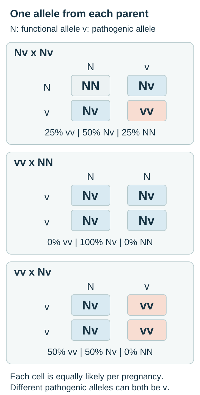
MUTYH-associated polyposis, abbreviated MAP, requires pathogenic variants affecting both gene copies. It is autosomal recessive. When both parents carry one pathogenic allele, a child has a 1 in 4 chance of biallelic disease, a 1 in 2 chance of carrying one allele, and a 1 in 4 chance of inheriting neither. An unaffected sibling does not change the probabilities for another pregnancy. The exact variants need not be identical: two different pathogenic variants on opposite gene copies (in trans) establish biallelic disease. Two changes on the same copy (in cis), with a functional second copy, do not. Count affected copies rather than variant names. [4]
Use the allele-pair diagram to trace one contribution from each parent. In the comparison table, N means a functional allele and v means a pathogenic allele. First predict the result for a person with biallelic MAP and a partner with two functional copies. Then compare that with a carrier partner.
Trace one allele from each parent. Predict how replacing NN with Nv changes the risk for a child of a person with biallelic MAP. The symbol v includes different pathogenic alleles; outcomes describe independent pregnancies. [4]
Compare every MUTYH parental pairing
Predict the result before reading across the row. Each pregnancy receives one allele from each parent. N is functional and v represents a pathogenic allele; vv can contain different variants on opposite copies.
Per-pregnancy probabilities under the stated complete genotypes
Parental pair
Biallelic MAP (vv)
One affected copy (Nv)
Neither (NN)
Parental pairNN with NN
Biallelic MAP (vv)0%
One affected copy (Nv)0%
Neither (NN)100%
Parental pairNN with Nv
Biallelic MAP (vv)0%
One affected copy (Nv)50%
Neither (NN)50%
Parental pairNN with vv
Biallelic MAP (vv)0%
One affected copy (Nv)100%
Neither (NN)0%
Parental pairNv with NN
Biallelic MAP (vv)0%
One affected copy (Nv)50%
Neither (NN)50%
Parental pairNv with Nv
Biallelic MAP (vv)25%
One affected copy (Nv)50%
Neither (NN)25%
Parental pairNv with vv
Biallelic MAP (vv)50%
One affected copy (Nv)50%
Neither (NN)0%
Parental pairvv with NN
Biallelic MAP (vv)0%
One affected copy (Nv)100%
Neither (NN)0%
Parental pairvv with Nv
Biallelic MAP (vv)50%
One affected copy (Nv)50%
Neither (NN)0%
Parental pairvv with vv
Biallelic MAP (vv)100%
One affected copy (Nv)0%
Neither (NN)0%
Trace one row: Nv with Nv produces NN, Nv, Nv and vv. Only one of these four equally likely combinations is biallelic. Exchanging the parents leaves the probabilities unchanged. Four combinations do not predict the exact composition of a four-child family.
Worked comparison: vv with NN produces Nv in all four equally likely cells: every child inherits one pathogenic allele, but none has biallelic MAP under these stated assumptions. Changing the partner to Nv changes two cells to vv. Biallelic risk becomes 50%, while the other 50% are carriers. These are genotype probabilities, not guarantees about the age of cancer onset. Real counseling incorporates test sensitivity and the partner's results. [4]
For transfer, ask why the sibling of a person with MAP can have a higher immediate chance of biallelic disease than that person's child. The sibling may have two carrier parents; the child's risk depends on the reproductive partner. Do not assign a dominant 50% disease risk merely because one parent has polyposis.
Make the test answer a clinical question
The phenotype determines what needs investigating; a test refines the explanation. Record cumulative adenoma count across examinations, histology, proximal versus distal distribution, age at presentation, cancers in relatives and findings outside the bowel. Twelve adenomas today plus twelve previously excised is not a twelve-adenoma lifetime history. Review the pathology report rather than treating every lesion called a polyp as equivalent.
The 2024 EHTG-ESCP guidance supports considering a germline panel above 20 cumulative colorectal adenomas, with a lower threshold of 10 when age is below 60, family history is relevant or extracolonic manifestations are suggestive. Such thresholds guide assessment; they are not a substitute for judgment in a young person with a striking phenotype. Overlapping APC and MUTYH presentations make appropriately selected multigene panels useful. Test the affected relative first when possible, then offer targeted testing for an established familial variant. [5]
Compare two reports: APC pathogenic variant and APC variant of uncertain significance. The first can establish an inherited explanation in the appropriate setting. The second does not establish or exclude the syndrome and should not determine irreversible surgery or predictive testing of relatives as though causative. Continue care based on the observed phenotype and family history while genetics professionals pursue clarification. A negative panel also does not erase convincing clinical disease. [7]
Here is a testing exercise: a patient has 70 adenomas and a negative blood panel; several separate adenomas contain the same pathogenic APC variant. Predict whether another tissue could help. The repeated variant raises the possibility of mosaic APC disease, in which only some cells carry the change. Specialist analysis of additional tissues and adenomas can investigate this possibility. A standard blood test can miss low-level mosaicism. It is not sufficient to dismiss the lesions as sporadic because the blood result was negative. [5]
Now transfer the approach to a relative with no symptoms. A known familial pathogenic result allows focused testing and age-appropriate prevention. An uninformative family test requires a phenotype-based plan rather than false reassurance. Bleeding, anemia, obstruction or suspected cancer needs assessment while genetics is being organized, not after sequencing returns. [1][3]
Architecture separates the polyp families
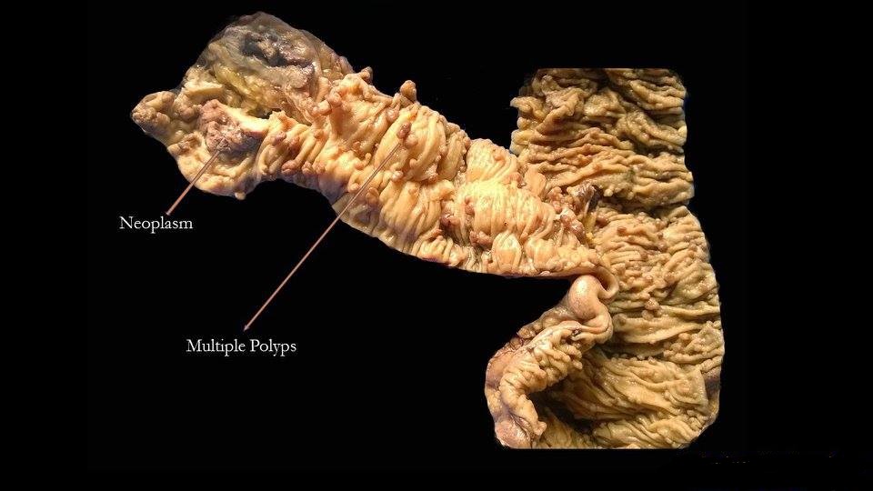
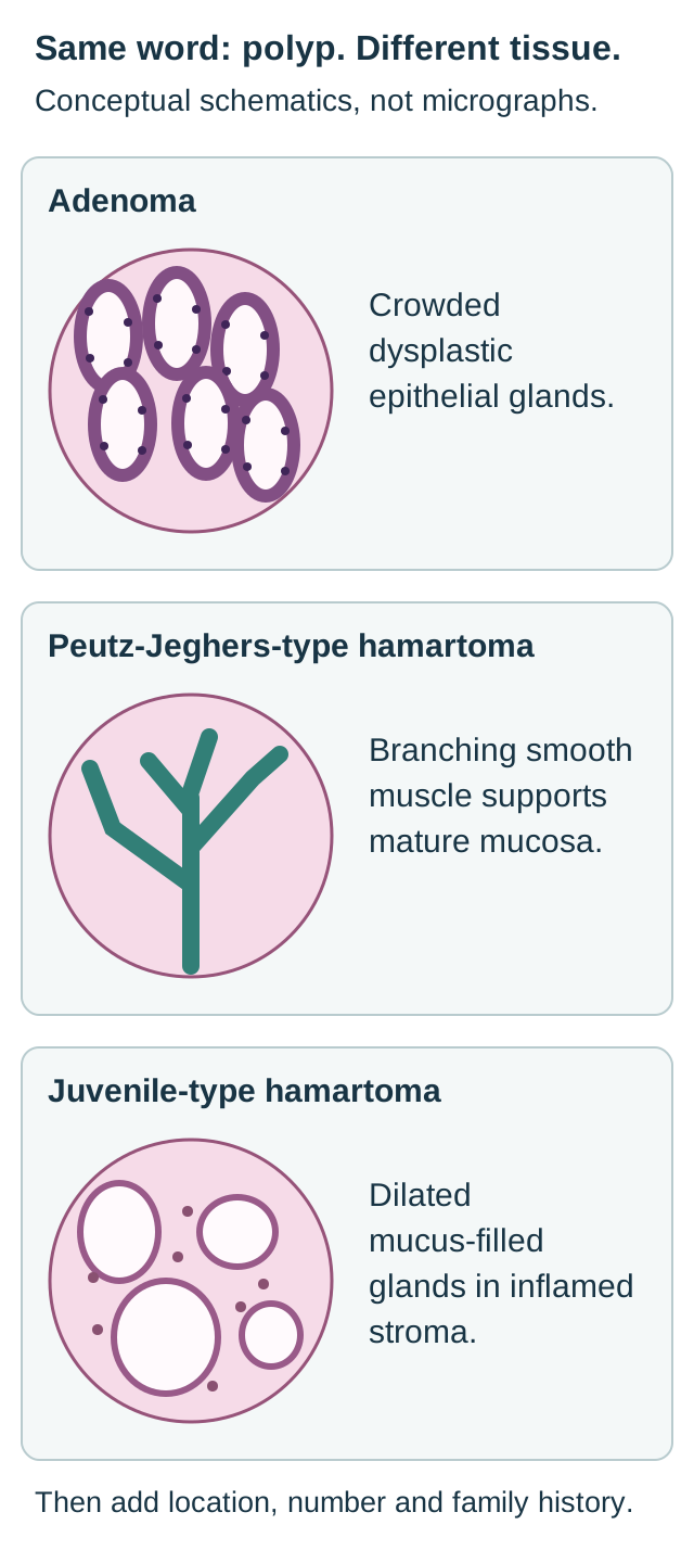
Adenoma describes dysplastic epithelial growth; hamartoma describes disorganized mature tissue. Neither word alone gives the patient's lifetime risk. Look at the gross photograph for the distribution of raised lesions, then at the architecture diagram for the microscopic distinctions. A photograph of many polyps cannot identify a germline gene or prove their histology. Tissue sampling and the clinical pattern are necessary. [1][2][3]
Connect the polyp pattern with inheritance and phenotype
Syndrome
Gene and inheritance
Pattern that matters
SyndromeClassic FAP
Gene and inheritanceAPC, dominant
Pattern that mattersUsually hundreds to thousands of colorectal adenomas, often beginning in adolescence.
SyndromeAttenuated FAP
Gene and inheritanceAPC, dominant
Pattern that mattersFewer adenomas, often proximal, and later colorectal cancer than classic FAP.
SyndromeMAP
Gene and inheritanceBiallelic MUTYH, recessive
Pattern that mattersOften tens to hundreds of adenomas; cancer can occur with a modest count or without overt polyposis.
SyndromePeutz-Jeghers
Gene and inheritanceSTK11, dominant
Pattern that mattersHamartomas, especially in small bowel, with branching smooth muscle supporting the mucosa.
SyndromeJuvenile polyposis
Gene and inheritanceSMAD4 or BMPR1A, dominant
Pattern that mattersHamartomas with dilated mucus-filled glands and inflamed stroma, commonly in colorectum and stomach.
APC helps regulate beta-catenin degradation. Loss of APC restraint permits Wnt-related proliferative signaling. A germline predisposition is present broadly, while additional changes in individual epithelial cells permit neoplasia. MUTYH instead participates in base-excision repair of oxidative DNA damage. Similar adenoma appearances can therefore result from different inherited failures. A microscopic adenoma is not itself proof of APC disease. [1][4]
In classic FAP, colorectal cancer is nearly inevitable without effective preventive management. Attenuated FAP has a later, smaller and often right-sided burden, not a harmless form of disease. With only a few rectal lesions, a normal sigmoidoscopy can miss extensive proximal adenomas. MAP overlaps the attenuated pattern; polyp count alone cannot reliably distinguish the two. [1][4]
Find the widespread polyp burden before considering the larger lesion. This photograph illustrates gross distribution, not a germline diagnosis or a substitute for histology. Image: Dr. Roshan Nasimudeen; original source; CC BY-SA 3.0.
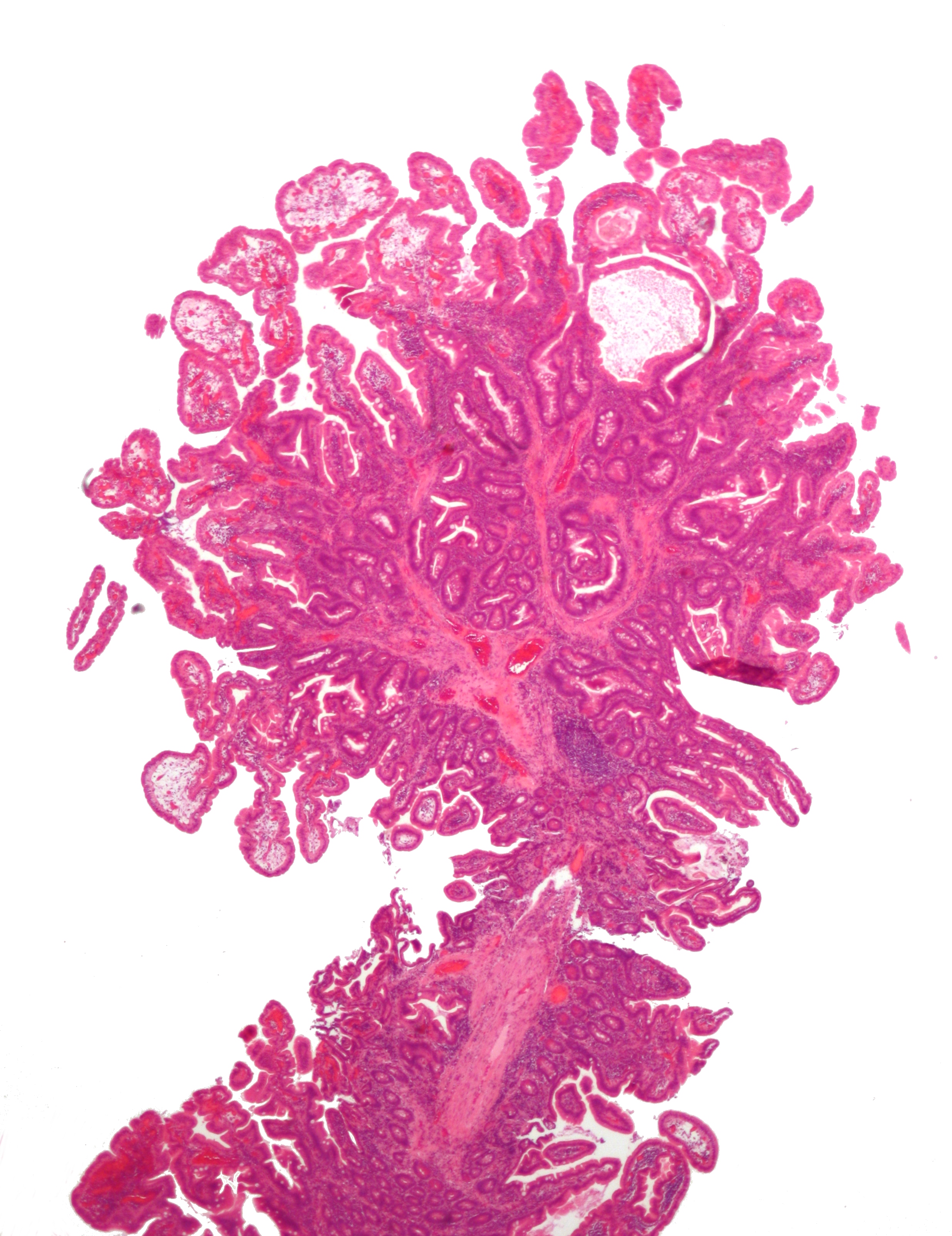
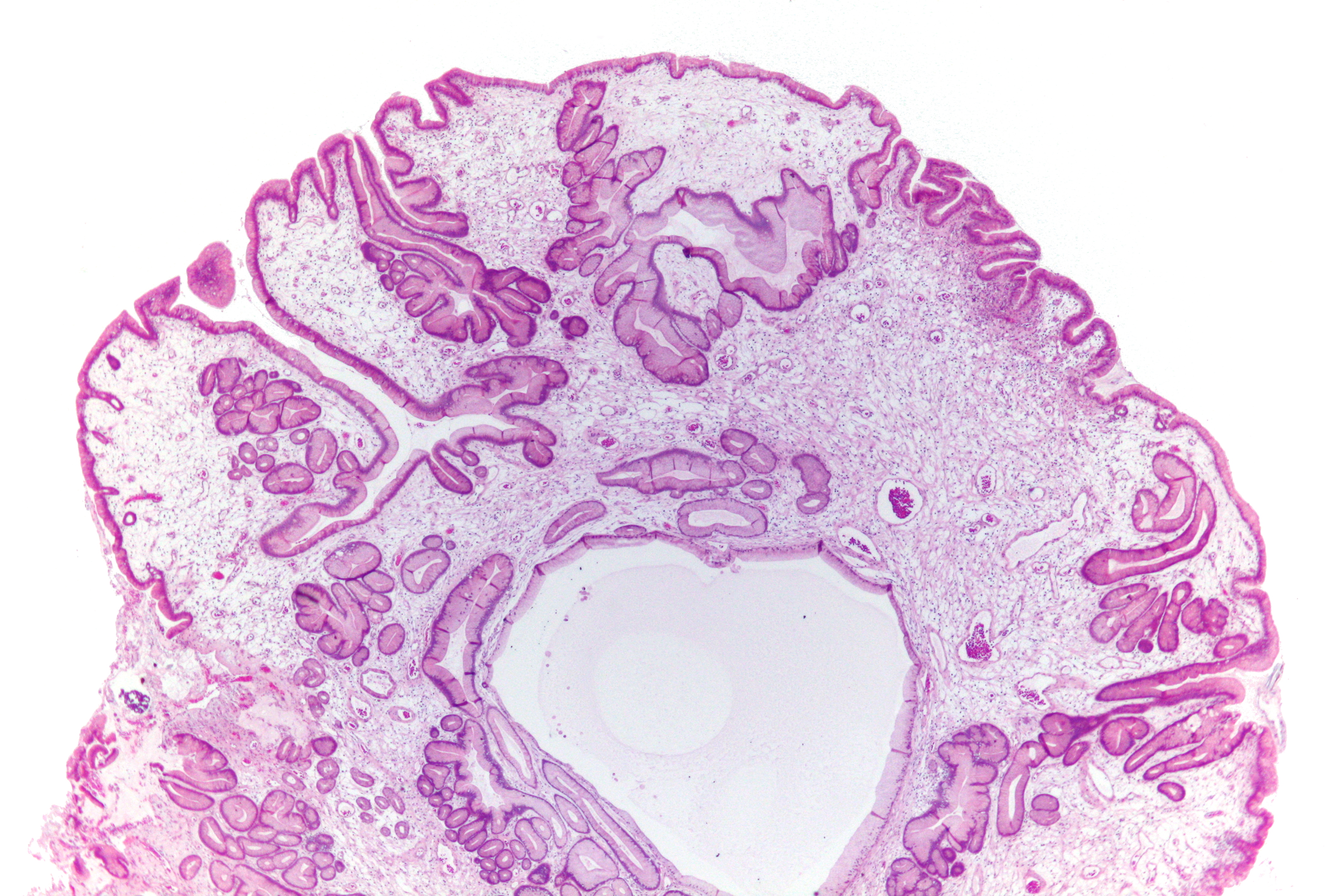
Trace the branching support in the Peutz-Jeghers schematic and real intestinal micrograph, then compare it with the expanded glands in the juvenile schematic and gastric micrograph. These photographs illustrate architecture; neither proves a germline syndrome on its own. In juvenile polyposis, juvenile describes architecture, not the patient's age. GeneReviews clinical criteria include more than five colorectal juvenile polyps, multiple juvenile polyps in upper and lower gastrointestinal sites, or any juvenile polyp with a family history of the syndrome. An isolated juvenile polyp without a supporting family history does not by itself establish juvenile polyposis. A later diagnosis in a parent changes that assessment: any juvenile polyp plus a family history of juvenile polyposis meets clinical criteria, even with fewer than six polyps. [3]
Compare these simplified architectures with the real intestinal and gastric micrographs. Architecture distinguishes polyp types; clinical context and genetic assessment establish a hereditary syndrome. [1][2][3]
Follow the branching support through this intestinal polyp and compare it with the schematic. This is an architecture example; the full clinical context is needed to diagnose the syndrome. Image: Nephron; original source; CC BY-SA 3.0.
Compare the enlarged glands with the branching framework in the intestinal PJS example. A juvenile-type polyp can occur in adults; one image alone does not establish juvenile polyposis syndrome. Image: Nephron; original source; CC BY-SA 3.0.
For transfer, consider two nondysplastic hamartomas: one solitary juvenile rectal polyp and one small-bowel polyp in a person with characteristic pigmentation and recurrent intussusception. The architecture and context lead to different syndrome assessments. Absence of dysplasia in the sampled polyp does not cancel a hereditary cancer predisposition. [2][3]
Which organ expands the diagnosis?
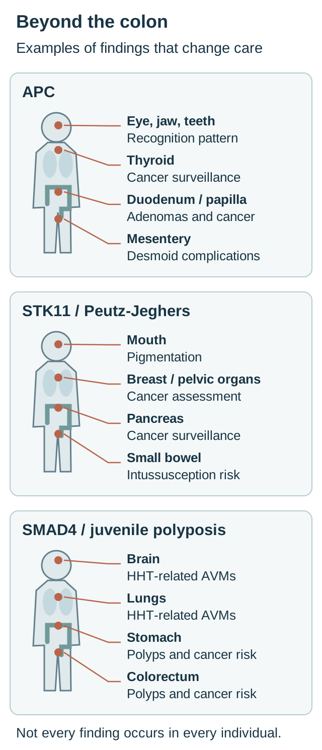
Findings outside the colon identify both a syndrome and additional prevention needs. Use the organ map to connect each site with its consequence. Jaw osteomas, dental abnormalities, epidermoid cysts and desmoid tumors can accompany APC-associated polyposis. The name Gardner describes this extracolonic phenotype, not a separate gene. Multifocal or bilateral congenital hypertrophy of the retinal pigment epithelium supports the pattern, but an isolated retinal lesion is not diagnostic of FAP. [1]
Choose an organ that remains at risk after colectomy. Then contrast pigment in Peutz-Jeghers with the vascular findings of SMAD4-related HHT. These are representative associations, not diagnostic checklists or complete screening schedules. [1][2][3][6][8]
APC disease also creates duodenal and ampullary adenomas and cancer risk, gastric neoplasia risk, thyroid cancer risk and selected central nervous system tumors. A desmoid is locally aggressive fibromatosis, not a distant metastasis from colorectal cancer. Treatment depends on growth, symptoms and threat to adjacent structures; an asymptomatic stable lesion does not automatically require surgery. [1][5]
Turcot is a historical description of inherited colorectal disease with a primary brain tumor. APC-associated disease is classically linked to medulloblastoma; mismatch-repair disorders are linked to gliomas. The label alone does not specify inheritance or molecular cause. Ask for the actual tumor type, bowel phenotype and genetic result instead of treating every recorded Turcot diagnosis as APC. [1]
Peutz-Jeghers combines hamartomatous polyps with dark mucocutaneous macules, particularly perioral and buccal pigmentation. External pigmentation can become less conspicuous with age. Small-bowel disease can cause bleeding and intussusception well before adult cancer surveillance begins. Cancer prevention extends to pancreas, breast and gynecologic organs as well as the gastrointestinal tract; gonadal tumors also warrant syndrome-specific assessment. [2][6]
In juvenile polyposis, compare a dark pigment spot with a vascular telangiectasia. Recurrent epistaxis, telangiectasias and pulmonary or cerebral arteriovenous malformations suggest SMAD4-associated overlap with hereditary hemorrhagic telangiectasia, or HHT. SMAD4 disease also tends toward substantial gastric involvement. HHT assessment should not wait for bowel symptoms. Pediatric guidance recommends pulmonary AVM screening at presentation even without epistaxis or breathlessness. [9] Pulmonary AVMs bypass the normal capillary filter and can permit paradoxical emboli or cerebral abscess; dedicated screening is appropriate even without respiratory symptoms. [3][8]
Apply the map: after an APC-related colectomy, cross out only the bowel that was excised, not the duodenum or thyroid. After treating a bleeding juvenile polyp in someone with SMAD4 disease, the vascular assessment remains necessary. Treating one manifestation does not delete the inherited predisposition elsewhere.
Surveillance begins when the relevant risk becomes actionable
Starting ages apply to asymptomatic people; symptoms require assessment earlier. Do not delay investigation of bleeding, iron deficiency or obstruction because a child has not reached a screening birthday. Nor is a negative baseline examination a promise that polyps will not develop later. [1][3][5]
Named starting-age frameworks, not a universal timetable
Condition
Lower or small-bowel assessment
Important qualification
ConditionClassic FAP
Lower or small-bowel assessmentGeneReviews: lower-GI surveillance at 10 to 15 years, usually every 1 to 2 years. EHTG-ESCP 2024: begin at 12.
Important qualificationShorten intervals according to burden, size, dysplasia and progression.
ConditionAttenuated FAP
Lower or small-bowel assessmentLate adolescence; EHTG-ESCP specifies no later than 18 to 20 years.
Important qualificationExamine the entire colon, including proximal regions.
ConditionMAP
Lower or small-bowel assessmentEHTG-ESCP 2024: generally start colonoscopy at 18, every 1 to 2 years.
Important qualificationOlder GeneReviews guidance begins at 25 to 30. State the framework being used; family phenotype can justify earlier assessment.
ConditionPeutz-Jeghers
Lower or small-bowel assessmentBaseline upper endoscopy, colonoscopy and small-bowel assessment at 8 years.
Important qualificationSmall-bowel MRI or capsule surveillance continues every 1 to 3 years. After negative baseline upper/lower examinations, those scopes resume at 18; detected polyps shorten that interval.
ConditionJuvenile polyposis
Lower or small-bowel assessmentGeneReviews: colonoscopy and upper endoscopy by 15, or earlier with symptoms.
Important qualificationAfter polyp treatment, annual examinations until no polyps are found, then generally every 3 years.
The table preserves distinctions between authorities rather than blending their numbers. The FAP, MAP and juvenile schedules depend on the phenotype and examination findings; the Peutz-Jeghers schedule distinguishes the small bowel from the stomach and colon. A question testing an exact guideline-dependent age should identify its framework. [1][3][4][5][6]
Upper-GI surveillance in APC disease must assess the duodenum and papilla as well as the stomach. The 2024 European guideline permits initiation after 18 but no later than 30, with intervals determined by the highest-risk findings. For MAP, it recommends upper endoscopy from 35, with intervals based on findings and not exceeding 3 years; it also recommends polypectomy regardless of polyp size. These examinations remain necessary after colorectal surgery. [5]
Predict the plan for an 11-year-old with Peutz-Jeghers and negative baseline upper and lower examinations at 8. The small bowel is still due for interval assessment; the negative scopes do not justify pausing all gastrointestinal surveillance until 18. New pain overrides the routine schedule. Pancreatic surveillance later belongs in an expert program within a study or registry, commonly using MRI/MRCP or endoscopic ultrasound, with explicit discussion of benefits, harms and remaining uncertainty. Eligibility in Peutz-Jeghers does not require an affected pancreatic-cancer relative. Breast, gynecologic and other evaluations need their own syndrome-specific plan. [6]
When is endoscopic control no longer enough?
Surgery addresses an organ's current risk, not a gene name in isolation. Known or suspected cancer, substantial symptoms, high-grade dysplasia, rapidly increasing adenomas, large lesions and inability to survey or clear the bowel can favor resection. Classic FAP often requires preventive colorectal surgery. Attenuated FAP and MAP can remain under endoscopic management when the burden is controllable and reliable follow-up is feasible. Chemoprevention is not a substitute for necessary surveillance or surgery. [1][5]
Compare two operative planning examples. One patient has an unmanageable colon but only a few small, completely treatable rectal adenomas. Colectomy with ileorectal anastomosis can preserve the rectum when its burden and follow-up permit. Another has extensive rectal polyposis with high-grade dysplasia that cannot be cleared. Proctocolectomy is more appropriate for that distribution. Cancer location, sphincter function, desmoid considerations, reproductive implications and patient preferences also matter. The retained rectum or ileal pouch still requires surveillance. [5]
Use the decision comparison to change one variable: keep the gene result fixed, but change rectal burden from three small adenomas to numerous dysplastic lesions. Predict the effect before reading on. The feasibility of retaining the rectum changes because the tissue left behind would carry an inadequately controlled risk. The germline diagnosis has not changed; the anatomy and ability to maintain surveillance have.
For Peutz-Jeghers, a large small-bowel polyp can be dangerous without containing cancer. It can pull adjacent bowel inward as a lead point for intussusception. The EHTG guideline supports elective treatment of small-bowel polyps larger than approximately 15 to 20 mm, and of smaller symptomatic lesions, usually through specialist enteroscopy. Acute severe abdominal pain, suspected intussusception or compromised bowel requires urgent surgical assessment rather than waiting for scheduled polypectomy. When safe, bowel preservation and treatment of the lead-point polyp help limit cumulative bowel loss. [6]
In juvenile polyposis, endoscopic treatment can control accessible polyps and bleeding. Persistent severe gastric or colorectal burden, dysplasia, cancer or symptoms despite treatment can require surgery directed at the involved organ. Iron replacement treats the consequence of bleeding, not the polyp source. HHT-related bleeding may coexist in SMAD4 disease, so not every episode of anemia has one explanation. [3]
For transfer, a person with MAP has twelve small colorectal adenomas that are completely excised and no cancer or high-grade dysplasia. The biallelic result supports close surveillance and family counseling, not automatic colectomy. Contrast that with an established cancer or a colon that cannot be reliably examined: the intervention threshold is different. Continue upper-GI and other indicated surveillance after any colorectal procedure. [5]
Apply the lesson
Case 1
Show answer and explanations for case 1
A. Accumulation of unrepaired oxidized DNA bases (Why this does not fit)
MUTYH-associated disease results from defective repair of oxidative DNA damage. The dominant pedigree and adolescent diffuse adenomas with osteoma and cysts favor APC-associated disease. Shared adenoma histology does not imply a shared inherited repair defect.
Reasoning steps for option A
Which repair defect could also produce multiple colorectal adenomas?
Biallelic MUTYH dysfunction impairs repair of oxidative DNA base damage.
What points away from recessive MUTYH polyposis here?
His mother had early colectomy, and hundreds of adolescent adenomas with osteoma and cysts fit dominant APC-associated FAP.
Does adenomatous histology identify the inherited mechanism?
No. APC loss alters Wnt signaling, whereas MUTYH loss disrupts oxidative base-excision repair.
B. Impaired correction of replication mismatches (Why this does not fit)
Mismatch-repair defects predispose to colorectal cancer and can involve other organs. The massive adolescent adenoma burden and Gardner phenotype point more specifically to APC. Identify the inherited pathway before predicting its cellular consequence.
Reasoning steps for option B
What does mismatch repair failure normally cause?
Uncorrected replication errors and microsatellite instability can predispose to colorectal cancer.
Why is mismatch repair not the best explanation for this phenotype?
More than 500 adenomas by age 17 plus mandibular osteoma and epidermoid cysts are classic APC-associated findings.
What happens after the second APC copy is lost?
Beta-catenin degradation is impaired, not replication mismatch correction.
C. Accumulation of transcriptionally active beta-catenin (Best answer)
APC normally contributes to regulation of beta-catenin degradation. The phenotype supports APC-associated FAP, so additional loss of APC permits Wnt-related proliferative signaling. Gardner findings describe an APC phenotype, not a separate molecular syndrome.
Reasoning steps for option C
Which gene unites adolescent diffuse adenomas, osteoma and epidermoid cysts?
APC; the extracolonic findings are Gardner features of APC-associated FAP.
How does loss of the remaining APC copy affect beta-catenin?
It impairs destruction-complex restraint, allowing beta-catenin to accumulate and drive proliferative transcription.
Do Gardner features imply a separate molecular pathway?
No. They are manifestations of APC-associated disease and predict the same beta-catenin consequence.
D. Loss of epithelial cell adhesion through E-cadherin (Why this does not fit)
E-cadherin defects can cause an inherited gastrointestinal cancer syndrome. Diffuse gastric cancer rather than this dominant adenomatous colorectal phenotype is its characteristic association. Use polyp architecture and organ distribution to distinguish inherited cancer mechanisms.
Reasoning steps for option D
Which inherited cancer mechanism involves E-cadherin?
CDH1-related loss of epithelial adhesion predisposes especially to diffuse gastric cancer.
Does this patient show the hallmark distribution of CDH1 disease?
No. His dominant familial burden is hundreds of colorectal adenomas with Gardner features.
Why does E-cadherin loss not answer the APC second-hit question?
The relevant APC second hit releases beta-catenin signaling rather than primarily abolishing epithelial adhesion.
E. Failure of homologous recombination repair (Why this does not fit)
Homologous recombination defects predispose to several epithelial cancers. They do not best explain hundreds of adolescent adenomas with the supplied APC-associated extracolonic pattern. Not every inherited cancer syndrome is a DNA repair disorder.
Reasoning steps for option E
What cellular process would homologous recombination loss disrupt?
Accurate repair of DNA double-strand breaks, a mechanism implicated in some hereditary epithelial cancers.
Which findings instead identify the pathway at issue?
Adolescent colorectal polyposis, maternal early colectomy, osteoma and cysts point to APC.
Is the predicted APC consequence another DNA repair defect?
No. Biallelic APC loss increases transcriptionally active beta-catenin.
Takeaway: Gardner findings describe an APC phenotype, not a separate molecular syndrome.
A. Postzygotic APC mosaicism involving several tissues (Best answer)
A postzygotic variant can be present in only a subset of cells. The repeated variant in separate adenomas and low-level non-neoplastic tissue explains why a routine blood assay was unrevealing. Negative blood testing does not exclude mosaic APC-associated disease.
Reasoning steps for option A
Why does one APC variant in three separate adenomas suggest more than a private tumor event?
Independent adenomas repeatedly harbor the same variant, suggesting it was already present in a shared precursor cell population.
What does reproducible low-level buccal detection add?
The variant extends into non-neoplastic tissue but occupies only a fraction of sampled cells, supporting postzygotic mosaicism.
Why could a routine blood panel miss this cause of polyposis?
Mosaic APC abundance may be below the blood assay detection threshold or underrepresented in blood.
B. Independent somatic APC events confined to the adenomas (Why this does not fit)
Individual sporadic adenomas can acquire somatic APC alterations. Detection in independently collected non-neoplastic buccal tissue argues against confinement to separate tumors. Compare neoplastic and non-neoplastic tissues when interpreting repeated variants.
Reasoning steps for option B
Can sporadic adenomas independently acquire APC mutations?
Yes. APC is a common somatic driver in individual colorectal adenomas.
What observation defeats a tumor-confined explanation?
The identical variant is reproducibly detected in independently collected non-neoplastic buccal tissue.
What tissue comparison establishes the relevant distinction?
A variant present outside tumors indicates a broader mosaic distribution, not merely separate somatic tumor events.
C. Constitutional APC heterozygosity with assay allele dropout (Why this does not fit)
Allele dropout can prevent an assay from detecting a constitutional variant. Reproducible low representation in a second assay across tissues fits mosaic distribution better than a single assay failure. A constitutional heterozygous variant and tissue mosaicism predict different allele distributions.
Reasoning steps for option C
How could allele dropout make a blood result falsely negative?
Failure to amplify one allele can conceal an otherwise constitutional APC heterozygous variant.
Why is ordinary heterozygosity less consistent with the buccal result?
A second assay repeatedly finds a low allele fraction rather than the approximately half-allele fraction expected in most cells.
What distribution favors mosaicism over a single failed assay?
Low-level detection in non-neoplastic tissue alongside matching variants in multiple adenomas.
D. Specimen cross-contamination during adenoma processing (Why this does not fit)
Contamination can introduce a low-level variant into a specimen. An independently collected buccal specimen reproduces the finding with a second assay, supporting a biological tissue distribution. Confirm low-level findings across independently assessed specimens before attributing them to mosaicism.
Reasoning steps for option D
Why must low-fraction APC findings raise a contamination concern?
Carryover of variant-bearing tumor material could create a spurious weak signal.
Which independent observation argues against processing contamination?
A separately collected buccal specimen tests positive with a second assay.
What does replication across specimens support?
A true low-level tissue distribution compatible with postzygotic APC mosaicism.
Takeaway: Negative blood testing does not exclude mosaic APC-associated disease.
A. Annual fecal immunochemical testing (Why this does not fit)
Stool testing can identify bleeding from some colorectal lesions. It neither surveys nor permits treatment of the documented proximal adenomas. Detection of occult bleeding is not equivalent to direct polyposis surveillance.
Reasoning steps for option A
What can annual fecal immunochemical testing detect?
Occult bleeding from some colorectal lesions.
Can it inspect or remove his 21 proximal adenomas?
No. Stool testing neither maps the proximal colon nor permits polypectomy.
Why is bleeding detection insufficient for this APC phenotype?
Documented recurrent adenomas require direct whole-colon inspection and removal, even if they are not bleeding.
B. Flexible sigmoidoscopy with rectal biopsies (Why this does not fit)
Sigmoidoscopy can assess distal colonic and rectal mucosa. Most lesions in this family are beyond its reach. A reassuring distal examination cannot exclude proximal disease.
Reasoning steps for option B
What part of the bowel does flexible sigmoidoscopy inspect?
The rectum and distal colon, which contained only one small adenoma in this patient.
Where were most of his documented lesions?
Twenty-one of 24 adenomas lay proximal to the splenic flexure, beyond routine sigmoidoscopy.
Would a normal distal examination exclude his principal risk?
No. His attenuated APC phenotype is predominantly proximal.
C. CT colonography followed by stool testing (Why this does not fit)
CT colonography can visualize the colon when colonoscopy is unsuitable. No barrier to colonoscopy is supplied, and repeated adenoma excision is expected. Choose an examination that can treat as well as detect a recurring polyp burden.
Reasoning steps for option C
When might CT colonography help assess the entire colon?
It can image the colon if conventional colonoscopy cannot be performed.
What treatment need makes CT imaging insufficient here?
He has a history of multiple proximal adenomas likely to require repeated endoscopic removal.
What advantage does colonoscopy offer beyond visualization?
It reaches the proximal lesions and allows polypectomy in the same examination.
D. Colonoscopy with endoscopic polypectomy (Best answer)
Attenuated APC disease often involves fewer, more proximal adenomas. The family pattern and observed distribution require inspection of the entire colon with treatment of accessible lesions. A smaller total polyp burden does not justify ignoring the proximal colon.
Reasoning steps for option D
What phenotype do 24 mostly proximal APC-associated adenomas suggest?
Attenuated APC-associated polyposis, which may spare much of the rectum.
Which examination covers the observed distribution and permits treatment?
Full colonoscopy with polypectomy of accessible adenomas.
Why does the normal sigmoidoscopy proposal fail?
A clear distal segment cannot exclude the many proximal adenomas already demonstrated.
E. Upper endoscopy with ampullary inspection (Why this does not fit)
Upper-GI assessment is important in APC-associated disease. It does not examine the colon where this patient's documented adenomas are located. Surveillance of one at-risk organ does not replace another organ's examination.
Reasoning steps for option E
Why is upper endoscopy relevant in APC-associated polyposis?
APC carriers can develop upper gastrointestinal and ampullary neoplasia.
Would ampullary inspection address the 21 proximal colorectal adenomas?
No. Upper endoscopy does not inspect or remove colonic adenomas.
How should surveillance of these separate sites be understood?
Upper-GI assessment may be needed independently, but it cannot replace full-colon surveillance.
Takeaway: A smaller total polyp burden does not justify ignoring the proximal colon.
The brother's genotype describes only that pregnancy. It does not remove either parent's ability to transmit an affected copy in later pregnancies. Prior outcomes do not consume an inheritance possibility.
Reasoning steps for option A
Does the unaffected brother prevent future transmission of either parental MUTYH variant?
No. Each carrier parent still has one affected and one functional allele.
What is the chance that both parents transmit affected copies in one new pregnancy?
One-half times one-half, or one-quarter.
Can the brother’s genotype reduce the two-child risk to zero?
No. Independent pregnancies retain the same transmission probabilities.
B. 6.25% (Best answer)
Each child of these two carriers has a one-half times one-half, or one-quarter, chance of biallelic disease. Both independent pregnancies meeting that condition has probability one-quarter times one-quarter, or one-sixteenth. Calculate per-pregnancy risk before combining independent outcomes.
Reasoning steps for option B
What is the biallelic risk for one child of two MUTYH carriers?
Each parent transmits the affected copy with probability one-half, giving one-quarter together.
What is the probability that both next children inherit two affected copies?
One-quarter times one-quarter is one-sixteenth, or 6.25%.
Does the younger brother inheriting neither variant alter either future pregnancy?
No. His genotype is a prior independent outcome, not a change in the parents’ alleles.
C. 25.00% (Why this does not fit)
One quarter is the biallelic probability for a single pregnancy from these carrier parents. The question requires that both of two further children be biallelic. A repeated-event probability is not the same as one child's risk.
Reasoning steps for option C
What event has a 25% probability in this carrier-by-carrier cross?
A single child receiving the affected MUTYH copy from each parent.
How many affected outcomes does the question require?
Both of the next two children must have biallelic variants.
What calculation replaces the single-child 25% figure?
Multiply one-quarter by one-quarter to obtain 6.25%.
D. 50.00% (Why this does not fit)
One half is the chance of inheriting the affected copy from one carrier parent. Biallelic MAP requires an affected contribution from both parents in each of two pregnancies. Identify the full event before assigning a familiar inheritance fraction.
Reasoning steps for option D
What does the familiar one-half transmission probability represent?
One carrier parent passing their affected MUTYH allele to one child.
Why is that not sufficient for MAP in one child?
Biallelic MAP also requires the other carrier parent to pass their affected allele.
What further condition applies to the question’s two children?
The joint one-quarter event must occur in each independent pregnancy, yielding one-sixteenth.
E. 75.00% (Why this does not fit)
Three quarters of children in a carrier-by-carrier cross inherit at least one affected copy. That includes carriers and is not the probability of biallelic disease in both further children. Separate any-variant inheritance from the specific biallelic outcome.
Reasoning steps for option E
What does a 75% figure count for one child of two carriers?
Receiving at least one affected allele, including unaffected heterozygous carriers.
Does that identify biallelic MAP in both future children?
No. One affected copy is not biallelic MAP, and the question asks about two pregnancies.
What probability applies to the narrowly defined event?
Biallelic inheritance is one-quarter per child and one-sixteenth for both.
Takeaway: Carrier-pair biallelic risk is 25% per pregnancy; two independent biallelic outcomes have probability 6.25%.
A. 0% biallelic; 100% carrier (Why this does not fit)
This pair applies when a biallelic parent has a partner with two functional alleles. The tested partner is a carrier and can transmit a pathogenic allele. A partner's carrier result changes a recessive offspring-risk calculation.
Reasoning steps for option A
When would every child of a biallelic father be only a carrier?
If the partner contributed a functional MUTYH copy in every pregnancy.
Does this partner have two functional copies?
No. Both changes lie on one inherited copy, making her a carrier.
What happens when she transmits that affected copy?
The child also receives a pathogenic copy from the father and is biallelic, so the all-carrier prediction fails.
B. 25% biallelic; 75% carrier (Why this does not fit)
One quarter is the biallelic probability in a carrier-by-carrier cross. The man has no functional allele to transmit, so the cross is not two carriers. Do not reuse the sibling-risk calculation for a different parental pairing.
Reasoning steps for option B
Which parental pairing gives a 25% biallelic child risk?
Two heterozygous carriers, each transmitting an affected copy with probability one-half.
Is the affected father a heterozygous carrier?
No. His two pathogenic variants are in trans, so every gamete carries an affected MUTYH copy.
How does his obligatory contribution change the calculation?
Biallelic risk depends only on the carrier partner’s one-half chance of transmitting her affected copy.
C. 75% biallelic; 25% carrier (Why this does not fit)
Seventy-five percent describes inheriting at least one variant in a carrier-by-carrier cross. It neither matches this parental pairing nor separates carriers correctly. Calculate genotype categories directly rather than relabeling a familiar fraction.
Reasoning steps for option C
Where does a 75% at-least-one-variant figure arise?
In a carrier-by-carrier cross, three of four child genotype classes have at least one affected copy.
Does this couple have a carrier-by-carrier cross?
No. The father has two affected copies and cannot transmit a functional one.
What fractions actually separate disease from carrier status?
Half inherit the partner’s affected copy and are biallelic; half inherit her functional copy and are carriers.
D. 100% biallelic; 0% carrier (Why this does not fit)
Two biallelic parents would make every child biallelic. The partner has a functional allele that is transmitted in half of pregnancies. One biallelic parent alone does not make biallelic disease inevitable.
Reasoning steps for option D
What parental genotype pairing would make biallelic disease inevitable?
Both parents would need to transmit an affected MUTYH copy in every pregnancy.
Can this partner transmit a functional allele?
Yes. Her two variants are in cis, leaving the other copy functional.
How often does that functional allele produce a carrier child?
In half of pregnancies it pairs with the father’s obligatory affected copy, yielding a carrier rather than biallelic MAP.
E. 50% biallelic; 50% carrier (Best answer)
The two changes in the partner affect one copy, leaving the other functional. The man always supplies a pathogenic allele, while the partner supplies the affected copy in half of pregnancies. Resolve variant phase before calculating biallelic and carrier probabilities.
Reasoning steps for option E
How many affected gene copies does the partner have despite two named variants?
One. The changes are in cis and the other inherited copy is functional.
What does the biallelic father contribute to every child?
An affected MUTYH allele because each of his copies carries a pathogenic variant.
What are the partner’s two equally likely contributions?
Her affected copy or her functional copy, each with probability one-half.
What offspring genotypes follow from those contributions?
Affected copy plus affected copy gives 50% biallelic MAP; functional plus affected gives 50% carriers.
Takeaway: Resolve variant phase before calculating biallelic and carrier probabilities.
A. Biallelic MAP; use the detected variant for family diagnosis (Why this does not fit)
A pathogenic MUTYH variant is relevant to reproductive counseling. One demonstrated affected copy does not establish the biallelic disorder. Separate a pathogenic allele from a molecular diagnosis requiring two affected copies.
Reasoning steps for option A
What does the identified pathogenic MUTYH allele establish?
He carries one affected copy, which can matter for reproductive counseling.
How many affected copies are required to diagnose MAP molecularly?
Two; the comprehensive panel identified no second pathogenic MUTYH allele.
Can his 28 adenomas alone turn the single detected allele into biallelic MAP?
No. The phenotype warrants surveillance but does not prove a missing second allele.
B. MUTYH carrier; continue phenotype-based adenoma surveillance (Best answer)
The test demonstrates one affected copy rather than two. The unexplained cumulative adenoma burden still needs surveillance and genetics follow-up. An incomplete molecular explanation does not erase observed colorectal risk.
Reasoning steps for option B
What molecular status does the panel establish?
A single-allele MUTYH carrier result, not confirmed biallelic MAP.
What observed finding still determines colorectal follow-up?
Twenty-eight cumulative adenomas despite no complete molecular explanation.
What counseling plan reconciles those facts?
Continue phenotype-based endoscopic surveillance and genetics follow-up without assigning a biallelic diagnosis.
C. Attenuated APC disease; offer APC-specific predictive testing (Why this does not fit)
The number of adenomas can resemble attenuated APC disease. No pathogenic APC variant has been identified for a targeted predictive family test. A compatible phenotype does not supply a missing familial molecular result.
Reasoning steps for option C
Why might attenuated APC disease enter the differential?
A few dozen cumulative adenomas can resemble an attenuated APC phenotype.
Was an APC variant found for predictive testing of relatives?
No. The comprehensive panel identified no explanatory variant in the other tested genes.
What must not be inferred solely from the adenoma count?
A specific familial APC variant suitable for targeted predictive testing.
D. Unexplained adenomas; return to population screening intervals (Why this does not fit)
An uninformative panel can leave multiple adenomas unexplained. Twenty-eight cumulative adenomas still require risk-based follow-up rather than population screening. Negative genetic testing does not normalize an abnormal phenotype.
Reasoning steps for option D
Can comprehensive testing leave multiple adenomas unexplained?
Yes. The panel found no established biallelic or alternative molecular explanation.
Does that permit population-risk screening for this patient?
No. Twenty-eight observed adenomas demonstrate a substantial colorectal phenotype.
What guides surveillance when the molecular diagnosis remains incomplete?
The documented adenoma burden and its manageability, not the absence of a second identified allele.
E. MUTYH carrier; use routine screening after polyp clearance (Why this does not fit)
A single-allele result identifies carrier status rather than established MAP. Removal of current adenomas does not remove the documented tendency to form multiple adenomas. Set follow-up from the phenotype as well as the genetic result.
Reasoning steps for option E
Why is calling him a MUTYH carrier reasonable?
Only one pathogenic MUTYH copy was detected after comprehensive testing.
Does clearing current polyps erase the 28-adenoma history?
No. Polypectomy removes existing lesions but not the demonstrated propensity to develop more.
What interval framework is more appropriate than routine screening?
Ongoing adenoma surveillance tailored to his polyposis phenotype.
Takeaway: A single MUTYH allele does not establish MAP or cancel phenotype-based surveillance.
A. Resume colonoscopy at the usual population screening age (Why this does not fit)
A normal colonoscopy can support long intervals in average-risk populations. This child has a confirmed predisposition to adolescent adenomatous polyposis. A negative examination does not convert a pathogenic-variant carrier to average risk.
Reasoning steps for option A
Why might a normal colonoscopy allow ordinary adults a long interval?
A negative high-quality examination reduces near-term concern in average-risk screening.
Is this 12-year-old at average inherited risk?
No. He carries the familial pathogenic APC variant, and his father had hundreds of adenomas by 18.
What does the normal examination not establish?
It does not rule out adenomas developing during the coming adolescent years.
B. Use annual stool testing until adenomas become symptomatic (Why this does not fit)
Stool testing can detect bleeding from some colorectal lesions. It does not provide direct surveillance of a childhood polyposis predisposition. Absence of bleeding does not show absence of emerging adenomas.
Reasoning steps for option B
What could stool testing detect if adenomas later bleed?
Blood shed into stool by some colorectal lesions.
Would lack of bleeding establish that this APC carrier remains polyp-free?
No. Adenomas can emerge without symptoms or detectable bleeding.
What examination is needed instead of waiting for symptomatic bleeding?
Scheduled direct lower-GI endoscopic surveillance at short intervals.
C. Continue lower-GI endoscopy at 1- to 2-year intervals (Best answer)
Classic FAP surveillance begins in childhood because adenomas can develop during adolescence. The baseline is clear, but the familial variant and classic parental phenotype remain present. A surveillance examination samples the present; it does not remove future inherited risk.
Reasoning steps for option C
What did the age-12 colonoscopy establish?
There are no detectable adenomas at this examination.
Why does his father’s age-18 burden still matter?
The pathogenic APC variant and classic familial phenotype predict adenoma development during adolescence.
What follow-up balances the clear baseline with continuing risk?
Repeat lower-GI endoscopy every 1 to 2 years rather than stopping surveillance.
D. Repeat colonoscopy after ten years if the child remains well (Why this does not fit)
A long interval may follow a normal examination in average-risk adults. Ten years would bypass the period when this family's adenoma burden became substantial. Match the interval to inherited risk and age rather than the absence of current symptoms.
Reasoning steps for option D
Why might ten years seem attractive after a negative scope?
Average-risk adults can receive a long interval after a normal high-quality screening colonoscopy.
What would a ten-year delay miss in this family?
The son would reach 22 without repeat examination despite his father having hundreds of adenomas by 18.
Why are symptoms not an adequate trigger for return?
APC-associated adenomas can accumulate before bleeding or anemia develops.
E. Plan immediate proctocolectomy before another examination (Why this does not fit)
Preventive colorectal surgery is often needed in classic FAP. No adenomas, symptoms or other urgent operative indication are supplied at this assessment. A pathogenic result starts prevention planning but does not mandate immediate childhood surgery.
Reasoning steps for option E
Why is preventive colectomy relevant to classic FAP eventually?
A heavy adenoma burden may become unmanageable endoscopically and carries substantial cancer risk.
What operative indication is absent at age 12?
His colonoscopy shows no adenomas, and he has no bleeding or anemia.
What should occur before selecting childhood surgery?
Continue close endoscopic surveillance and plan intervention according to emerging disease burden.
Takeaway: A surveillance examination samples the present; it does not remove future inherited risk.
A. Arrange diagnostic lower-GI evaluation now and assess the anemia (Best answer)
Screening starting ages apply to asymptomatic carriers. Bleeding and documented anemia require current evaluation rather than waiting for the previously planned age. Symptoms override a routine surveillance starting age.
Reasoning steps for option A
Does this nine-year-old still qualify as an asymptomatic surveillance patient?
No. Recurrent rectal bleeding and hemoglobin of 9.1 g/dL require diagnostic assessment.
Why does the previously suggested age-12 start not apply?
That starting age was for an asymptomatic APC carrier, not a child with bleeding and anemia.
What should be arranged now?
Prompt diagnostic lower-GI evaluation and assessment of the anemia; stable vital signs do not justify a three-year delay.
B. Give iron and retain the planned first colonoscopy at 12 (Why this does not fit)
Iron replacement can help replenish losses in iron-deficiency anemia. It does not identify or treat the source of recurrent bleeding in an APC carrier. Treating the consequence of bleeding does not replace investigation of its source.
Reasoning steps for option B
What problem could oral iron address?
It may replenish iron lost through repeated gastrointestinal bleeding.
What does iron not establish in this APC carrier?
It does not identify the source of recurrent rectal bleeding or explain the anemia.
Why should the age-12 colonoscopy not remain unchanged?
Active symptoms call for lower-GI diagnostic evaluation now, alongside anemia management.
C. Obtain another APC assay before deciding on endoscopy (Why this does not fit)
Genetic clarification is useful when the familial diagnosis is uncertain. The child already carries the established pathogenic variant and has active symptoms. Do not repeat an established genetic result while delaying symptomatic evaluation.
Reasoning steps for option C
When would repeating an APC assay be useful?
If the familial variant or this child’s carrier status were uncertain.
What genetic fact has already been established?
He carries his mother’s pathogenic APC variant.
What issue requires action independent of repeat genotyping?
Recurrent rectal bleeding with hemoglobin 9.1 g/dL needs current diagnostic evaluation.
D. Begin annual fecal occult blood testing until the first colonoscopy (Why this does not fit)
Stool testing can document occult bleeding. Visible recurrent bleeding and anemia have already demonstrated a problem requiring direct assessment. A screening test should not replace diagnostic evaluation of established symptoms.
Reasoning steps for option D
What can fecal occult blood testing add to a symptom-free screening assessment?
It can reveal bleeding not apparent to the patient.
Is occult bleeding the unanswered question here?
No. He has visible recurrent rectal bleeding and documented microcytic anemia.
What should replace annual stool testing as the immediate step?
Direct lower-GI evaluation of the bleeding source and assessment of the anemia.
E. Arrange prophylactic proctocolectomy without evaluating the bowel (Why this does not fit)
Colorectal surgery can prevent or treat major APC-related disease. The supplied information does not yet define polyp burden, cancer or the operative anatomy. Symptoms require timely assessment before choosing a specific elective operation.
Reasoning steps for option E
When might proctocolectomy become relevant for an APC carrier?
Extensive unmanageable polyposis, cancer or another defined operative indication could require surgery.
What essential information is missing before an operation is chosen?
The bowel has not yet been assessed for polyp burden, bleeding source or cancer.
What is the appropriate immediate response in this stable symptomatic child?
Arrange prompt diagnostic lower-GI evaluation and assess anemia before selecting a specific operation.
Takeaway: Symptoms override a routine surveillance starting age.
A. Small bowel at 18; upper and lower endoscopy at 18 (Why this does not fit)
Negative baseline upper and lower endoscopies can permit their routine resumption at 18. That interval does not apply to small-bowel assessment, which continues every 1 to 3 years. Keep small-bowel surveillance distinct from the upper and lower endoscopic timetable.
Reasoning steps for option A
Why might the normal upper and lower scopes at age 8 support an age-18 return?
Under the EHTG negative-baseline pathway, routine upper and lower endoscopy may resume at 18 if he stays asymptomatic.
Does the same age-18 deferral cover the small bowel?
No. Small-bowel assessment continues at 1- to 3-year intervals even after a normal childhood evaluation.
What does the elapsed time since his age-8 small-bowel MRI require?
At age 11, three years have passed, so small-bowel reassessment is due now, not at 18.
B. Small bowel at 18; upper and lower endoscopy now (Why this does not fit)
Detected upper or lower polyps can justify short-interval endoscopic follow-up. Those examinations were negative, while small-bowel surveillance is already due. A negative baseline does not transfer the longest interval to every bowel segment.
Reasoning steps for option B
What finding would make an early repeat upper or lower endoscopy more persuasive?
Polyps on the age-8 upper or lower examination or new symptoms could prompt earlier assessment.
Were upper or lower polyps found at the age-8 baseline?
No. Both examinations were normal, allowing routine upper and lower scopes to wait until 18.
Which compartment actually reaches its surveillance deadline at age 11?
The small bowel reaches the end of its 1- to 3-year interval after the age-8 MRI.
C. Small bowel annually; upper and lower endoscopy annually (Why this does not fit)
A substantial polyp phenotype can require frequent reassessment. No polyps or symptoms are supplied to justify replacing the negative-baseline pathway with yearly examination of every site. Surveillance frequency follows both the organ and its findings.
Reasoning steps for option C
When could frequent multisite examinations be justified in Peutz-Jeghers syndrome?
A substantial polyp burden or symptoms could require shorter, tailored intervals.
What do his age-8 examinations and current history show instead?
All three baseline examinations were normal and he is still asymptomatic at 11.
Why does an annual schedule for every site miss the EHTG distinction?
It ignores the permissible age-18 routine return for negative upper and lower scopes and imposes yearly small-bowel imaging without a supplied indication.
D. Small bowel now; upper and lower endoscopy at 18 (Best answer)
Small-bowel surveillance continues every 1 to 3 years despite negative upper and lower baseline examinations. Three years have elapsed, whereas routine upper and lower scopes can resume at 18 if he remains asymptomatic. A negative stomach and colon assessment does not pause small-bowel prevention.
Reasoning steps for option D
Which organ must be reassessed despite a negative age-8 baseline?
The small bowel remains on a 1- to 3-year surveillance cycle, making its next assessment due by age 11.
Why can routine upper and lower endoscopy wait?
His age-8 upper and lower examinations were negative and no intervening symptoms or polyps are reported, so their routine return is at 18.
What is the critical difference between the two schedules?
Negative stomach and colon findings do not extend the small-bowel interval to age 18.
E. Small bowel with symptoms; upper and lower endoscopy with symptoms (Why this does not fit)
New symptoms require assessment earlier than routine surveillance. The STK11-associated predisposition also requires surveillance before symptoms develop. Symptom-triggered assessment supplements rather than replaces hereditary surveillance.
Reasoning steps for option E
Would abdominal symptoms justify assessment ahead of a scheduled visit?
Yes. Symptoms would trigger diagnostic assessment before the next routine surveillance appointment.
Can an asymptomatic 11-year-old STK11 carrier wait for symptoms at all sites?
No. Inherited Peutz-Jeghers risk requires presymptomatic small-bowel surveillance, already due three years after his baseline.
What routine endoscopic timing remains after normal upper and lower examinations at eight?
Upper and lower scopes can routinely resume at 18, rather than being abandoned until symptoms arise.
Takeaway: A negative stomach and colon assessment does not pause small-bowel prevention.
A. Arrange elective device-assisted enteroscopic polypectomy (Why this does not fit)
Specialist enteroscopy can treat accessible large polyps before obstruction. Reduced enhancement and peritoneal signs raise concern for compromised bowel that cannot wait for an elective procedure. A preventive polypectomy pathway is different from treatment of suspected ischemic obstruction.
Reasoning steps for option A
Why is device-assisted polypectomy relevant to a Peutz-Jeghers jejunal polyp?
Elective removal of an accessible large polyp can prevent it from becoming an intussusception lead point.
What CT and examination findings make this no longer elective prevention?
The 25-mm polyp already anchors jejunojejunal intussusception, with reduced mural enhancement, guarding and rebound.
What takes precedence over scheduled endoscopic removal?
Resuscitation and urgent operative evaluation for threatened or ischemic bowel.
B. Resuscitate and obtain urgent operative evaluation (Best answer)
Intussusception can obstruct venous and arterial flow and progress to necrosis. The lead-point polyp, reduced enhancement and peritoneal findings make urgent surgical evaluation necessary. Suspected bowel compromise takes priority over routine surveillance or elective polypectomy.
Reasoning steps for option B
What mechanical event explains this patient's abrupt pain and vomiting?
A 25-mm jejunal polyp is the lead point for an acute jejunojejunal intussusception.
Why is bowel viability in question now?
Reduced mural enhancement suggests impaired perfusion, and guarding with rebound indicates peritoneal irritation.
What action responds to that immediate threat?
Resuscitate and obtain urgent surgical evaluation rather than delaying for elective polypectomy or surveillance.
C. Repeat small-bowel imaging after a short outpatient interval (Why this does not fit)
Repeat imaging can follow stable lesions during surveillance. An acute obstructive episode with peritoneal signs is not a stable surveillance finding. Interpret imaging together with the current abdominal examination.
Reasoning steps for option C
In what setting would interval small-bowel imaging be reasonable?
It may follow a stable polyp or surveillance finding without an acute abdomen.
Is this a stable finding suitable for outpatient follow-up?
No. CT demonstrates intussusception with reduced enhancement, while examination shows guarding and rebound.
What could a short outpatient delay miss?
Progression from compromised perfusion to bowel ischemia or necrosis requiring urgent operative assessment.
D. Obtain capsule endoscopy to characterize the remaining polyps (Why this does not fit)
Capsule endoscopy is useful for selected small-bowel surveillance. Known obstruction risks capsule retention and the immediate problem is already localized. Do not use a routine luminal survey to delay treatment of an established obstruction.
Reasoning steps for option D
What is capsule endoscopy designed to assess in Peutz-Jeghers syndrome?
It can survey selected small-bowel polyp burdens during nonurgent surveillance.
Why is swallowing a capsule unsafe or unhelpful during this episode?
An established jejunal obstruction risks capsule retention, and CT has already localized the intussusception and lead point.
Which finding demands action before characterizing other polyps?
Reduced mural enhancement plus peritoneal signs raises immediate concern for compromised bowel.
E. Complete germline testing before choosing an intervention (Why this does not fit)
STK11 testing can clarify the inherited diagnosis and family risks. The acute mechanical complication is demonstrated regardless of whether genetic confirmation is available. Treatment of an urgent complication does not wait for molecular confirmation.
Reasoning steps for option E
What could germline STK11 testing add after stabilization?
It could confirm inherited Peutz-Jeghers syndrome and guide relatives' risk assessment.
Does the acute intervention depend on molecular confirmation?
No. CT already identifies a 25-mm lead-point intussusception with reduced bowel enhancement.
What should happen despite pending genetic results?
Resuscitation and urgent operative evaluation are needed because guarding and rebound suggest threatened bowel.
Takeaway: Suspected bowel compromise takes priority over routine surveillance or elective polypectomy.
A. Resect the affected jejunal segment surgically (Why this does not fit)
Surgery can treat small-bowel lesions when endoscopic treatment is unsuitable. This accessible lesion has no obstruction or invasion requiring bowel resection. Preserve bowel when a large polyp can be safely removed endoscopically.
Reasoning steps for option A
When might jejunal segment resection be necessary for a Peutz-Jeghers polyp?
Surgery may be needed when endoscopic removal is unsuitable or there is obstruction, invasion or threatened bowel.
Which features of this 22-mm lesion argue against immediate resection?
MRI shows no obstruction or mural invasion, and device-assisted enteroscopy can reach the pedunculated polyp.
How can the lead point be treated while preserving bowel?
Remove the polyp endoscopically rather than excising a jejunal segment.
B. Repeat jejunal MRI during scheduled surveillance (Why this does not fit)
Interval imaging follows smaller lesions that do not yet require treatment. A 22-mm polyp and previous intussusception support removing this mechanical lead point. Surveillance alone does not control an already actionable lesion.
Reasoning steps for option B
For what sort of lesion would scheduled MRI alone be a reasonable approach?
Imaging can follow a smaller polyp below the elective removal threshold without a pressing mechanical risk.
Why is this particular 22-mm polyp actionable now?
It exceeds the approximate 15- to 20-mm treatment range, and this patient has already experienced intussusception.
What does repeat imaging fail to remove?
The accessible pedunculated lead point that may cause another intussusception.
C. Observe until polyp diameter reaches 40 mm (Why this does not fit)
A size threshold can guide elective intervention. This lesion already exceeds the guideline treatment range of approximately 15 to 20 mm. Do not substitute an unsupported larger threshold for the stated prevention framework.
Reasoning steps for option C
What size range commonly prompts elective small-bowel polyp removal in Peutz-Jeghers syndrome?
Approximately 15 to 20 mm, especially when preventing an intussusception lead point.
How does the MRI measurement compare with that range?
The jejunal polyp measures 22 mm, already above it.
Why is waiting for 40 mm particularly unattractive here?
It leaves a sizeable lead point in someone with a previous intussusception despite current endoscopic accessibility.
D. Sample the polyp by superficial biopsy (Why this does not fit)
Biopsy can investigate suspicious mucosal features. Sampling leaves the large lead point in place even when no dysplasia is found. Mechanical risk can justify treatment independently of malignant histology.
Reasoning steps for option D
What question might a superficial biopsy answer?
It might sample mucosal histology or investigate suspicious surface changes.
What risk persists even if that sample shows no dysplasia?
The intact 22-mm jejunal polyp can still act as a mechanical lead point for intussusception.
What intervention addresses that risk without bowel resection?
Device-assisted enteroscopic removal of the accessible polyp.
E. Remove the polyp by device-assisted enteroscopy (Best answer)
Large small-bowel polyps can precipitate intussusception. The accessible 22-mm lesion exceeds the elective treatment range without current evidence of compromised bowel. Remove a preventable lead point rather than waiting for obstruction or dysplasia.
Reasoning steps for option E
Which findings favor elective treatment rather than emergency surgery?
The 22-mm pedunculated polyp is accessible by device-assisted enteroscopy, with no current obstruction or mural invasion.
Why remove this nonobstructing 22-mm jejunal lesion now?
Its size exceeds the approximate 15- to 20-mm treatment range and prior intussusception establishes a relevant mechanical risk.
What does endoscopic polypectomy accomplish?
It removes the potential jejunal lead point while preserving bowel.
Takeaway: Treat a large accessible small-bowel lead point before intussusception develops.
A. Begin imaging after pancreatic cancer develops in a relative (Why this does not fit)
Family history influences eligibility in some inherited pancreatic-risk groups. This pigmentation and polyp pattern supports Peutz-Jeghers, whose eligibility does not require an affected relative. Identify the syndrome before applying another group's family-history rule.
Reasoning steps for option A
Why might family history affect pancreatic surveillance in another inherited risk group?
Some hereditary pancreatic-risk eligibility rules incorporate an affected relative.
Which findings establish a different syndrome here?
Childhood lip and buccal macules plus jejunal polyps with branching smooth muscle and intussusception indicate Peutz-Jeghers syndrome.
Does her lack of a pancreatic cancer relative justify postponing imaging?
No. Peutz-Jeghers confers syndrome-specific surveillance eligibility independently of an affected relative.
B. Obtain yearly CA 19-9 levels as the surveillance program (Why this does not fit)
CA 19-9 can contribute to assessment of concerning pancreatic findings. Serum-marker testing is not an adequate stand-alone surveillance strategy for this inherited risk. Use a structural surveillance program rather than a marker alone.
Reasoning steps for option B
When can CA 19-9 have a role in pancreatic assessment?
It may contribute to evaluation when a concerning lesion or clinical finding is already present.
Why is annual CA 19-9 alone inadequate for this 43-year-old?
A blood marker cannot replace structural pancreatic surveillance for Peutz-Jeghers-associated cancer risk.
What modality-based program fits the inherited risk?
Expert MRI/MRCP or EUS surveillance, preferably within a study or registry after discussing uncertainties.
C. Enter an expert MRI/MRCP or EUS surveillance program (Best answer)
The pigmentation, jejunal architecture and intussusception identify a Peutz-Jeghers phenotype. This syndrome supports expert imaging surveillance within a study or registry despite a clear colon and no pancreatic family history. Prevention must address the syndrome's organs beyond the treated bowel.
Reasoning steps for option C
What diagnosis follows from oral pigmentation and branching smooth muscle in jejunal hamartomas?
These findings, with prior intussusception, support Peutz-Jeghers syndrome.
Do a clear colon and no pancreatic cancer in relatives remove pancreatic eligibility?
No. Pancreatic risk follows the syndrome even when current colorectal examination and family history are negative.
What surveillance should be offered at age 43 after counseling?
Expert pancreatic MRI/MRCP or EUS in a study or registry-based program.
D. Begin imaging after new pancreatic symptoms develop (Why this does not fit)
Pancreatic symptoms require diagnostic evaluation. Waiting for symptoms omits presymptomatic surveillance for an eligible high-risk person. Distinguish a prevention program from investigation after disease becomes symptomatic.
Reasoning steps for option D
What would new pancreatic symptoms require?
Prompt diagnostic evaluation for a possible pancreatic lesion.
Why is waiting for symptoms the wrong surveillance strategy now?
Her Peutz-Jeghers phenotype already makes her eligible for presymptomatic pancreatic imaging at age 43.
How should pancreatic risk be followed before symptoms?
Offer an expert MRI/MRCP or EUS program after explaining benefits and uncertainty, rather than deferring until clinical disease appears.
Takeaway: Peutz-Jeghers pancreatic surveillance does not require pancreatic cancer in a relative.
A. BMPR1A or SMAD4; 50% transmission (Best answer)
Juvenile-type architecture with a dominant family pattern supports BMPR1A- or SMAD4-associated juvenile polyposis. A constitutional heterozygous affected parent transmits the variant in half of pregnancies. Histology identifies the syndrome, while inheritance determines the transmission probability.
Reasoning steps for option A
What does cystic mucus-filled glandular dilation with inflamed stroma identify?
Juvenile-type hamartomatous polyps, not the arborizing architecture of Peutz-Jeghers polyps.
Which inherited genes fit gastric and colorectal juvenile polyps in both father and son?
BMPR1A or SMAD4 can cause autosomal dominant juvenile polyposis.
What is the chance that this heterozygous parent passes the pathogenic variant in one pregnancy?
Each child independently has a 50% chance of inheriting the familial variant; severity is not predicted by transmission.
B. Biallelic MUTYH; 25% transmission (Why this does not fit)
MUTYH can explain inherited adenomatous polyposis. This is a juvenile hamartomatous phenotype, and 25% is a carrier-parent biallelic disease probability rather than an affected parent's variant-transmission probability. Distinguish polyp architecture and transmission from recessive disease risk.
Reasoning steps for option B
What polyp type is typical of biallelic MUTYH disease?
MUTYH-associated polyposis is predominantly adenomatous, unlike these cystically dilated juvenile hamartomas.
When does a 25% recessive disease probability arise?
It applies to an affected child of two heterozygous carriers, not transmission of one variant by this affected parent.
What inheritance model better matches an affected father and son with juvenile polyps?
Dominant BMPR1A or SMAD4 juvenile polyposis, with 50% variant transmission per pregnancy.
C. Heterozygous APC; 50% transmission (Why this does not fit)
A constitutional APC variant has a 50% transmission probability. The polyps here have juvenile rather than adenomatous architecture. A correct inheritance fraction does not rescue the wrong molecular syndrome.
Reasoning steps for option C
Why is the proposed APC transmission fraction tempting?
A constitutional heterozygous APC variant would also pass to half of an affected parent's children.
Which histology argues against APC-associated adenomatous polyposis?
The colorectal and gastric lesions contain cystically dilated mucus-filled glands and inflamed stroma characteristic of juvenile polyps.
Which gene group preserves both the architecture and 50% probability?
BMPR1A or SMAD4, rather than APC.
D. Heterozygous STK11; 50% transmission (Why this does not fit)
STK11-associated disease is also dominant and hamartomatous. The cystic glands and inflammatory stroma favor juvenile polyps rather than the contrasting arborizing pattern. Identify the hamartoma type before choosing the dominant gene.
Reasoning steps for option D
What inheritance feature makes STK11 seem plausible?
Peutz-Jeghers syndrome is dominant and produces hamartomatous polyps, with 50% variant transmission.
What specific polyp feature separates this case from STK11-associated Peutz-Jeghers?
Cystic mucus-filled glands with inflammatory stroma are juvenile architecture, not branching smooth-muscle cores.
Which dominant juvenile-polyposis genes better explain the father-son pattern?
BMPR1A or SMAD4.
E. BMPR1A or SMAD4; 25% transmission (Why this does not fit)
These genes fit the polyp architecture and family pattern. A constitutional heterozygous parent transmits the variant in 50%, not 25%, of pregnancies. Do not import a recessive carrier-pair probability into a dominant family.
Reasoning steps for option E
Why do BMPR1A or SMAD4 fit the patient's polyp histology?
They are associated with dominant juvenile polyposis featuring cystically dilated glands and inflamed stroma.
What does a heterozygous constitutional pathogenic variant imply for each pregnancy?
A parent passes the variant to 50% of offspring, irrespective of the father's exact polyp count.
Where does the offered 25% figure go wrong?
It substitutes a recessive two-carrier disease probability for transmission from a dominant affected parent.
A. Isolated juvenile polyp; defer testing unless bowel symptoms recur (Why this does not fit)
A single juvenile polyp without supporting family history can be sporadic. The newly established paternal diagnosis changes that context despite the child's current lack of symptoms. Reassess an old isolated-polyp classification when family evidence changes.
Reasoning steps for option A
Why was an isolated-polyp interpretation plausible when the child was five?
A solitary juvenile rectal polyp with an otherwise normal colonoscopy and no known family history can be sporadic.
Which new information changes that classification at age eight?
His father now has multiple juvenile colorectal and gastric polyps and a pathogenic BMPR1A variant.
Why not wait for renewed bleeding before genetic evaluation?
One juvenile polyp plus family history satisfies a clinical juvenile-polyposis criterion, and the known paternal variant enables targeted testing now.
B. Possible juvenile polyposis; defer testing until a sixth polyp appears (Why this does not fit)
More than five colorectal juvenile polyps is one diagnostic route. Any juvenile polyp plus a family history of juvenile polyposis is another route, so this child need not accumulate six. Diagnostic alternatives are not requirements that must all be met.
Reasoning steps for option B
What count-based criterion can establish juvenile polyposis?
More than five juvenile colorectal polyps is one possible clinical route.
Must this child develop a sixth polyp to qualify?
No. His previous juvenile polyp plus his father's established juvenile polyposis meets the alternative family-history criterion.
What test is available rather than waiting for additional polyps?
Targeted testing for the father's pathogenic BMPR1A variant.
C. Familial risk without clinical polyposis; offer targeted testing (Why this does not fit)
Targeted testing is appropriate for a known familial pathogenic variant. The prior juvenile polyp together with the paternal diagnosis already meets clinical criteria, rather than representing family risk alone. A low polyp count does not exclude a clinical syndrome when the family criterion is met.
Reasoning steps for option C
Why is targeted testing an appropriate part of this proposal?
The father has an identified pathogenic BMPR1A variant, allowing focused testing in the child.
What does the label 'familial risk without clinical polyposis' overlook?
The child already had a histologically juvenile rectal polyp, and his father now has juvenile polyposis.
Which clinical criterion does that combination satisfy?
Any juvenile polyp together with family history of juvenile polyposis, even without six polyps.
The child's previous juvenile polyp and the father's established syndrome satisfy a clinical diagnostic criterion. The known BMPR1A variant allows focused genetic evaluation with an age- and phenotype-based GI plan. New family evidence can change both classification and the testing strategy.
Reasoning steps for option D
Why did the old solitary-polyp assessment change after the father's diagnosis?
A juvenile rectal polyp now coexists with a first-degree relative with established juvenile polyposis.
What clinical conclusion follows without waiting for more lesions?
The child meets the family-history route to a clinical juvenile-polyposis diagnosis despite being asymptomatic at eight.
How should the known paternal BMPR1A result guide the next step?
Offer targeted familial-variant testing and an age- and phenotype-based gastrointestinal plan.
Takeaway: Any juvenile polyp plus a family history of juvenile polyposis warrants syndrome assessment.
A. Subarachnoid hemorrhage (Why this does not fit)
HHT can include cerebral vascular malformations that cause intracranial hemorrhage. Pulmonary vascular screening addresses a different compartment and its right-to-left shunt complications. Distinguish cerebral-vessel rupture from complications originating in a pulmonary shunt.
Reasoning steps for option A
How can HHT itself cause subarachnoid hemorrhage?
A cerebral vascular malformation may rupture and bleed into the subarachnoid space.
What lesion is the proposed pulmonary screening looking for instead?
A pulmonary arteriovenous malformation associated with the juvenile-polyposis and HHT overlap.
Which cerebral event does treating that pulmonary shunt particularly prevent?
Cerebral abscess from septic material bypassing lung capillary filtration, not rupture of a cerebral vessel.
B. Cerebral abscess (Best answer)
Juvenile polyps with telangiectasias and epistaxis suggest SMAD4-associated JPS-HHT. A pulmonary AVM can bypass capillary filtration and allow septic material to reach the brain. Recognize the overlap phenotype before translating the pulmonary lesion into its systemic complication.
Reasoning steps for option B
Which syndrome is suggested by twelve juvenile polyps, epistaxis and oral telangiectasias?
SMAD4-associated juvenile polyposis with hereditary hemorrhagic telangiectasia.
What does a pulmonary arteriovenous malformation do to venous blood?
It shunts blood past the pulmonary capillary filter into systemic arterial circulation, even without cough or breathlessness.
What neurologic infection can this allow?
Septic material can seed the brain and produce a cerebral abscess.
C. Cerebral venous thrombosis (Why this does not fit)
Venous sinus thrombosis can produce neurologic illness through impaired cerebral drainage. A pulmonary AVM instead permits material to enter systemic arterial blood without normal pulmonary filtration. Distinguish venous outflow obstruction from a right-to-left shunt.
Reasoning steps for option C
What mechanism produces cerebral venous sinus thrombosis?
Thrombus blocks venous drainage from the brain rather than allowing material to bypass the lungs.
What pulmonary lesion is predicted by juvenile polyps plus HHT signs?
A pulmonary arteriovenous malformation creating a right-to-left shunt.
How does its neurologic risk differ from venous thrombosis?
Unfiltered septic embolic material reaches the cerebral arterial circulation and can cause abscess.
D. Obstructive hydrocephalus (Why this does not fit)
A posterior fossa tumor can obstruct cerebrospinal fluid flow in an APC-associated brain-tumor phenotype. Juvenile polyps with telangiectasias point to a vascular overlap, not that tumor pattern. Separate inherited brain-tumor associations from pulmonary-shunt complications.
Reasoning steps for option D
What inherited setting could make obstructive hydrocephalus plausible?
An APC-associated posterior fossa tumor could block cerebrospinal fluid flow.
Do the twelve juvenile polyps and oral telangiectasias suggest that APC tumor pattern?
No. Together with spontaneous epistaxis they suggest SMAD4-associated juvenile polyposis with HHT.
Why does pulmonary AVM treatment target another complication?
Closing a shunt restores filtration of septic material and reduces cerebral abscess risk, rather than relieving cerebrospinal fluid obstruction.
E. Carotid atherosclerotic infarction (Why this does not fit)
Atherosclerotic carotid disease can cause cerebral infarction by stenosis or plaque embolization. The pulmonary lesion anticipated here causes a shunt, not carotid plaque formation. Identify the source of a neurologic risk rather than grouping all stroke mechanisms together.
Reasoning steps for option E
How does carotid atherosclerosis cause cerebral infarction?
Carotid stenosis or plaque emboli impair arterial blood flow to the brain.
What vascular abnormality is pulmonary screening seeking in this teenager?
An HHT-associated pulmonary AVM, whose shunt bypasses pulmonary capillary filtration.
What brain complication follows that unfiltered passage of infected material?
Cerebral abscess, not plaque-mediated carotid infarction.
Takeaway: SMAD4-associated JPS-HHT can connect a pulmonary shunt with cerebral abscess risk.
A. Wait for recurrent nosebleeds before starting vascular screening (Why this does not fit)
Nosebleeds can be an HHT manifestation. Serious vascular lesions can precede epistaxis, so its absence is not a screening rule. Do not require symptoms before assessing a genetically identified child.
Reasoning steps for option A
Why might epistaxis seem like a useful HHT trigger?
Recurrent spontaneous nosebleeds are a recognizable manifestation of hereditary hemorrhagic telangiectasia.
Does this asymptomatic SMAD4-positive six-year-old need to develop nosebleeds first?
No. His mother's pulmonary AVMs and their shared pathogenic variant establish vascular risk before symptoms.
When should the HHT assessment occur?
During childhood now, alongside a separate age- and phenotype-based GI surveillance plan.
B. Begin yearly chest CT as the pulmonary screening strategy (Why this does not fit)
CT can confirm and characterize pulmonary AVMs after a positive screen. Pediatric guidance does not recommend CT as the routine screening test. Select an established pediatric HHT screening pathway rather than repeated screening CT.
Reasoning steps for option B
What is chest CT useful for in pulmonary AVM care?
CT may confirm or define a pulmonary AVM after an appropriate positive screen.
Why is yearly CT a poor first-line screening plan for this child?
Pediatric HHT guidance does not recommend repeated CT as routine pulmonary screening, including its avoidable radiation exposure.
What should replace an automatic annual CT schedule?
An established pediatric HHT vascular assessment pathway now, separately from gastrointestinal surveillance.
C. Use intestinal endoscopy to screen for vascular involvement (Why this does not fit)
Endoscopy evaluates gastrointestinal polyps and sources of GI bleeding. It cannot screen the pulmonary vascular compartment implicated by the familial overlap. Different organ risks require different assessments.
Reasoning steps for option C
What can intestinal endoscopy evaluate in a SMAD4 carrier?
It can assess juvenile gastrointestinal polyps and investigate GI bleeding.
Which inherited vascular risk does endoscopy fail to screen?
The pulmonary AVMs seen in his mother, which can occur as part of SMAD4-associated HHT.
How should the organ-specific assessments be arranged?
Arrange childhood HHT evaluation now and a separate GI plan based on age and phenotype.
D. Start pulmonary screening at the routine GI surveillance age (Why this does not fit)
An age threshold helps organize asymptomatic juvenile-polyp endoscopy. Pulmonary HHT assessment is indicated earlier at presentation or diagnosis. The GI timetable does not determine when to assess vascular risk.
Reasoning steps for option D
What does a routine GI starting age govern in juvenile polyposis?
It helps time endoscopic surveillance for asymptomatic gastrointestinal polyps.
Can that age threshold delay pulmonary vascular screening in this six-year-old?
No. A confirmed familial SMAD4 variant and maternal pulmonary AVMs warrant childhood HHT assessment at diagnosis.
What timetable distinction does this option miss?
Vascular risk must be assessed now even if routine GI endoscopy follows a later age- or phenotype-based schedule.
E. Arrange childhood HHT assessment and a GI surveillance plan (Best answer)
The familial SMAD4 result identifies a risk extending beyond juvenile polyps. Pediatric HHT assessment is indicated now, while the GI plan follows age and phenotype. Separate early vascular assessment from later routine GI starting ages.
Reasoning steps for option E
What does sharing the mother's pathogenic SMAD4 variant imply?
This child is at risk for both juvenile polyposis and HHT-related vascular malformations, including pulmonary AVMs.
Do absent bowel symptoms and epistaxis remove either inherited risk?
No. In particular, serious pediatric vascular lesions may be present before nosebleeds or respiratory symptoms.
How should surveillance be separated?
Arrange childhood HHT assessment now and design GI surveillance according to age and phenotype rather than using the GI schedule to postpone vascular screening.
Takeaway: A routine GI starting age must not delay assessment of childhood HHT risk.
A. Colorectal surgery to remove the currently controllable colon (Why this does not fit)
Severe colorectal juvenile polyposis can require colectomy. The uncontrollable burden and high-grade dysplasia are gastric, whereas the colonic lesions have been cleared. Operate on the organ carrying the inadequately controlled risk.
Reasoning steps for option A
Why would colectomy enter the differential in juvenile polyposis?
Severe uncontrolled colorectal juvenile polyposis can warrant colectomy.
Where is high-grade dysplasia refractory to polypectomy in this patient?
It is in the carpeted stomach; three small nondysplastic colonic polyps were completely removed.
Which compartment should surgical referral target?
The stomach, not an adequately controlled colon.
B. Hematology to replace iron without changing polyp management (Why this does not fit)
Iron replacement treats the deficit produced by chronic bleeding. Persistent gastric burden and high-grade dysplasia require control of the source rather than replacement alone. Correcting anemia does not treat the underlying dysplastic polyposis.
Reasoning steps for option B
What does hematology address in this patient?
Iron replacement can correct the deficiency from ongoing blood loss.
Why does iron alone leave the principal risk unresolved?
Recurrent high-grade dysplasia persists in gastric polyps that cannot be cleared endoscopically.
What treatment problem remains after hemoglobin improves?
The bleeding and dysplastic gastric polyp source still needs definitive assessment.
C. Upper-GI surgery for the refractory gastric polyp burden (Best answer)
Extensive symptomatic gastric polyposis with dysplasia can require surgical treatment. Endoscopic treatment has failed in the stomach while colorectal control is adequate. Localization and feasibility of control determine which organ needs escalation.
Reasoning steps for option C
Which gastric findings make upper-GI surgery relevant?
Extensive persistent polyps, refractory anemia and recurrent high-grade dysplasia make gastric control urgent.
Why is another routine gastric polypectomy insufficient?
Repeated attempts have not completely treated the carpeted stomach.
Why not direct this referral at the colon?
Its three nondysplastic lesions were cleared, so the uncontrolled organ is the stomach.
D. Pancreatic surgery based on the hereditary cancer diagnosis (Why this does not fit)
Some hereditary gastrointestinal syndromes include pancreatic cancer risk. No pancreatic lesion is supplied, and the demonstrated refractory disease is gastric. A hereditary label is not an indication to resect an unaffected organ.
Reasoning steps for option D
Why might a hereditary polyposis patient need pancreatic assessment?
Some inherited polyposis syndromes have extracolonic pancreatic cancer risks.
What evidence here supports pancreatic resection?
None: no pancreatic lesion is described, while the stomach contains refractory dysplasia.
What determines the operative target in this case?
Demonstrated organ-specific disease, not an inherited label alone.
E. Small-bowel enteroscopy as the primary response to the anemia (Why this does not fit)
Small-bowel disease can be a source of bleeding in selected patients. The case already demonstrates an uncontrolled dysplastic gastric source after repeated treatment. Prioritize the established source before substituting a different anatomic target.
Reasoning steps for option E
When could enteroscopy help investigate iron-deficiency anemia?
It could evaluate a suspected small-bowel bleeding source.
A. Total abdominal colectomy with ileorectal anastomosis (Best answer)
A low, endoscopically controllable rectal burden can permit rectal preservation in selected APC patients. The unmanageable colon contrasts with three cleared small rectal adenomas and reliable follow-up. Rectal preservation requires both manageable rectal disease and ongoing surveillance.
Reasoning steps for option A
Which bowel segment has escaped staged endoscopic control?
About 300 colonic adenomas remain unmanageable, supporting colectomy.
What makes rectal preservation reasonable here?
Only three 3-mm rectal adenomas were fully excised without high-grade dysplasia, and follow-up is reliable.
What obligation follows ileorectal anastomosis?
The retained rectum requires continued endoscopic surveillance.
B. Segmental colectomy with ileocolonic anastomosis (Why this does not fit)
Segmental resection can treat a localized colorectal lesion. The adenomas are spread through the colon rather than confined to one resectable segment. Diffuse colonic polyposis requires a plan for the entire at-risk colonic burden.
Reasoning steps for option B
When does segmental colectomy make sense?
It can address a localized lesion limited to a resectable colonic segment.
How are this patient’s adenomas distributed?
Roughly 300 adenomas affect the colon diffusely, not one segment.
C. Proctocolectomy with ileal pouch-anal reconstruction (Why this does not fit)
Proctocolectomy is appropriate when rectal disease or cancer risk cannot be controlled. Three fully treated small lesions do not by themselves exclude rectal preservation in this selected setting. Assess rectal burden and follow-up feasibility rather than demanding a polyp-free lifetime history.
Reasoning steps for option C
What rectal finding would favor proctocolectomy over an ileorectal anastomosis?
Extensive or unmanageable rectal polyposis or high-grade dysplasia would make preservation unsafe.
How does the actual rectal examination differ?
Three tiny lesions were cleared completely, without high-grade dysplasia.
Why is pouch reconstruction not mandatory here?
The patient can preserve a manageable rectum with reliable ongoing surveillance.
D. Repeated stool testing instead of surgical planning (Why this does not fit)
Stool testing may detect bleeding from some colorectal lesions. It cannot control the extensive adenomas that staged endoscopy has failed to clear. A detection test is not treatment for an unmanageable polyposis burden.
Reasoning steps for option D
Can stool testing remove the approximately 300 adenomas?
No; it detects possible bleeding but does not clear adenomas.
What has staged colonoscopic therapy already shown?
The colonic burden cannot be kept clear despite repeated intervention.
What decision cannot be deferred to a stool result?
Planning resection of the uncontrollable colon.
E. Chemoprevention alone with deferred endoscopic reassessment (Why this does not fit)
Medication can influence adenoma burden in selected circumstances. It is not a substitute for indicated surgery or surveillance when the colon cannot be controlled. Drug-related polyp reduction is not equivalent to proven definitive cancer prevention.
Reasoning steps for option E
Can chemoprevention substitute for colectomy in this diffusely affected colon?
No; medication does not reliably eliminate the cancer risk of uncontrolled adenomas.
Why does deferred endoscopy compound the problem?
Neither medication alone nor delayed assessment controls the existing extensive burden.
What must still accompany any medical adjunct?
Appropriate surgery and continuing surveillance of retained at-risk tissue.
Takeaway: Rectal preservation requires both manageable rectal disease and ongoing surveillance.
A. Colectomy with ileorectal anastomosis (Why this does not fit)
Rectal preservation can be appropriate when rectal adenomas are controllable. The numerous rectal lesions with incompletely cleared high-grade dysplasia would leave an inadequately controlled rectum. The suitability of rectal preservation depends on rectal risk, not colonic burden alone.
Reasoning steps for option A
When can an APC patient retain the rectum after colectomy?
Only when rectal adenomas can be reliably cleared and monitored.
What would an ileorectal anastomosis leave behind here?
More than 40 rectal adenomas, including incompletely cleared high-grade dysplasia.
Which feature defeats rectal preservation?
Uncontrolled high-risk rectal mucosa, despite absence of invasive cancer.
B. Right hemicolectomy with ileocolonic anastomosis (Why this does not fit)
Segmental resection can address a localized right-sided lesion. The supplied disease is diffuse and includes high-risk rectal polyposis. A localized operation cannot control diffuse high-risk colorectal tissue.
Reasoning steps for option B
What disease could a right hemicolectomy remove?
Only adenomas in the right colon.
Where else is high-risk disease documented?
Diffuse colon involvement and numerous dysplastic rectal adenomas remain.
Why is a limited segmental resection inadequate?
It would retain much of the affected colon and rectum.
C. Low anterior resection with retained proximal colon (Why this does not fit)
Rectal resection can address disease localized to the rectum. This patient also has diffuse colonic adenomas needing a whole-colorectal plan. Both compartments matter when the colon and rectum are extensively involved.
Reasoning steps for option C
What does low anterior resection target?
It removes the diseased rectum while preserving proximal colon.
What would remain after that operation in this patient?
Diffuse colonic adenomas also require definitive management.
What distribution should guide the operation?
Both the colon and the rectum are extensively affected.
D. Proctocolectomy with ileal pouch-anal reconstruction (Best answer)
Removing colon and rectum addresses an unmanageable high-risk burden in both regions. The rectal high-grade dysplasia defeats a rectum-preserving approach, while intact sphincter function permits consideration of a pouch. Extensive dysplastic rectal disease can change the preferred colorectal operation.
Reasoning steps for option D
Why must both colon and rectum be removed?
Diffuse colonic adenomas coexist with over 40 rectal lesions and incompletely cleared high-grade dysplasia.
What supports considering an ileal pouch rather than a permanent stoma?
There is no invasive cancer, sphincter function is intact, and the patient wants to avoid a permanent stoma when feasible.
Why does the dysplastic rectal burden change the operation from colectomy alone?
Leaving the rectum would retain incompletely cleared high-grade dysplasia; proctocolectomy addresses the uncontrolled risk in both colorectal compartments.
E. Diverting ileostomy with the diseased colorectum retained (Why this does not fit)
Diversion can be useful for selected obstructive or perioperative problems. Diversion alone would leave the dysplastic colorectal tissue at risk. Changing the fecal route does not itself remove dysplastic mucosa.
Reasoning steps for option E
What does fecal diversion change?
It redirects the stream without removing diseased mucosa.
Which high-risk tissue would remain after diversion alone?
The diffuse colonic polyps and rectal high-grade dysplasia would remain in place.
Why is diversion not definitive prevention here?
Uncleared dysplastic colorectal tissue continues to carry risk.
Takeaway: Extensive dysplastic rectal disease can change the preferred colorectal operation.
A. Continue rectal surveillance and defer duodenal assessment to symptoms (Why this does not fit)
The retained rectum does require continuing surveillance. Known duodenal and papillary abnormalities require their own assessment before symptoms develop. An asymptomatic extracolonic lesion can still require risk-based evaluation.
Reasoning steps for option A
Why continue examining the rectum after ileorectal anastomosis?
Retained rectal mucosa remains susceptible to APC-associated adenomas.
What does a rectum-only plan miss now?
Multiple duodenal adenomas and an enlarged papillary lesion require assessment without waiting for symptoms.
Which asymptomatic region needs additional evaluation?
The duodenum and papilla, alongside the rectum.
B. Assess the duodenum and papilla while continuing rectal surveillance (Best answer)
The germline predisposition remains in gastrointestinal tissue outside the excised colon. The new duodenal and papillary findings need expert staging and management alongside surveillance of the retained rectum. Colectomy does not eliminate upper-GI or residual rectal risk.
Reasoning steps for option B
What at-risk mucosa was not removed by colectomy?
The rectum, duodenum and papilla remain exposed to APC-associated neoplasia.
How should the enlarged papillary lesion alter the plan?
Arrange expert upper-GI assessment and staging rather than stopping examinations.
What follow-up remains for the anastomosis?
Continue surveillance of the retained rectum.
C. Perform another APC blood test to determine whether surveillance is needed (Why this does not fit)
A genetic result can guide an initial family prevention plan. Colectomy does not reverse a constitutional APC variant or explain away new adenomas. Surgery changes anatomy, not the constitutional genotype.
Reasoning steps for option C
Would a repeat blood APC test change the constitutional variant?
No; removing the colon does not alter the inherited APC genotype.
What already demonstrates ongoing disease outside the colon?
Current duodenal adenomas and a visibly enlarged papilla.
What should determine assessment now?
The observed upper-GI lesions plus continuing rectal risk, not retesting the same gene.
D. Replace gastrointestinal surveillance with thyroid imaging (Why this does not fit)
Thyroid assessment can be part of APC-associated surveillance. It would not assess either of the currently affected gastrointestinal regions. Additional organ surveillance complements rather than substitutes for gastrointestinal care.
Reasoning steps for option D
Why could thyroid imaging belong in APC care?
APC-associated disease includes thyroid cancer risk.
Would thyroid imaging examine the current papillary and duodenal lesions?
No; it assesses a different organ and cannot replace upper endoscopy.
What is the relation between extracolonic and GI surveillance?
They complement each other rather than substitute for one another.
E. Excise the remaining rectum as the primary response to the papillary lesion (Why this does not fit)
Rectal resection can be needed when rectal disease becomes unmanageable. The rectum is controlled and the new concerning lesion is at the duodenal papilla. An intervention in one compartment cannot directly treat a lesion in another.
Reasoning steps for option E
When would completion proctectomy be considered?
Progressive, dysplastic or unmanageable rectal disease could require rectal removal.
Which site is currently concerning instead?
The rectum is controlled; the new lesion is at the duodenal papilla.
Why does rectal surgery not solve the present issue?
Removing the rectum does not assess or treat duodenal and papillary disease.
Takeaway: Colectomy does not eliminate upper-GI or residual rectal risk.
A. Colonoscopy from 25; upper endoscopy from 35 (Why this does not fit)
An older MAP reference begins colonoscopy at 25 to 30. The explicitly named 2024 European framework generally begins it at 18. Do not blend starting ages from different surveillance authorities.
Reasoning steps for option A
Which MAP colonoscopy starting age could explain choosing 25?
An older MAP reference starts colonoscopy at approximately 25 to 30 years, but the case specifies a different framework.
Which authority does the question specifically require?
The 2024 EHTG-ESCP framework generally starts MAP colonoscopy at age 18.
What should this asymptomatic 19-year-old do for lower-GI prevention?
Begin colonoscopy now rather than wait until age 25.
B. Colonoscopy now; upper endoscopy from 30 (Why this does not fit)
The current age is appropriate for lower-GI surveillance in the named framework. Its routine MAP upper-GI starting age is 35 rather than 30. Different organs within a syndrome can have different starting ages.
Reasoning steps for option B
Is colonoscopy now appropriate for biallelic MUTYH at 19?
Yes; the specified 2024 framework generally starts at age 18.
What is wrong with upper endoscopy at 30 as the routine threshold?
The same framework starts routine MAP upper endoscopy at age 35.
Why must the two age thresholds be distinguished?
Lower- and upper-GI surveillance begin at different ages in this guideline.
C. Colonoscopy from 35; upper endoscopy from 35 (Why this does not fit)
Age 35 is the named upper-GI starting point for MAP. It does not apply to the colon, where surveillance generally begins at 18. Do not apply one organ's later threshold to another organ.
Reasoning steps for option C
Which MAP examination begins routinely at 35?
Upper endoscopy begins at 35 under the named European guidance.
Can age 35 also be applied to the colon?
No; colonoscopy generally starts at 18, and this patient is already 19.
What error does this pairing make?
It delays lower-GI surveillance by transferring the upper-GI age to the colon.
D. Colonoscopy now; upper endoscopy with symptoms (Why this does not fit)
Early colonoscopy is appropriate for the biallelic result. MAP also calls for planned upper-GI surveillance before symptoms, starting at 35 in this framework. Symptom-triggered assessment does not replace scheduled prevention.
Reasoning steps for option D
Why is starting colonoscopy now justified?
Two pathogenic MUTYH variants inherited from different parents establish biallelic MAP at age 19.
Can absence of upper-GI symptoms replace scheduled endoscopy?
No; routine upper endoscopy begins at 35 even without symptoms in this framework.
What if symptoms appear before 35?
Investigate symptoms promptly rather than waiting for the routine starting age.
E. Colonoscopy now; upper endoscopy from 35 (Best answer)
Variants inherited on opposite copies establish biallelic MAP. The 2024 European framework starts colonoscopy generally at 18, every 1 to 2 years, and upper endoscopy from 35. Interpret the genotype before applying the separate organ timetables.
Reasoning steps for option E
What does inheritance of one MUTYH variant from each parent establish?
Pathogenic variants in trans establish biallelic MUTYH-associated polyposis.
When is colonoscopy due under 2024 EHTG-ESCP?
Generally from age 18 every 1 to 2 years, so this 19-year-old should start now.
When is first routine MAP upper endoscopy due here?
At 35, absent an earlier clinical indication.
Takeaway: In the named 2024 European MAP framework, lower-GI surveillance precedes routine upper-GI surveillance.
A. Continue colorectal polypectomy; arrange duodenal polypectomy (Best answer)
The colorectal disease is controllable endoscopically without a supplied surgical indication. The named MAP guideline also recommends treating duodenal polyps regardless of size. Integrate the burden and treatment needs of each gastrointestinal region.
Reasoning steps for option A
Why is continued colorectal polypectomy reasonable?
All twelve 2- to 5-mm adenomas were excised, with no cancer or high-grade dysplasia and reliable follow-up.
What does 2024 MAP guidance advise for the two small duodenal adenomas?
Polypectomy is recommended regardless of duodenal polyp size.
Why manage each region separately?
Controlled colonic polyps can stay under endoscopic care while known duodenal polyps are removed.
B. Plan colectomy; arrange duodenal polypectomy (Why this does not fit)
The duodenal lesions merit endoscopic treatment in this framework. Completely excised small colorectal adenomas without cancer or high-grade dysplasia do not supply an immediate colectomy indication. Escalate surgery according to control and risk, not biallelic status alone.
Reasoning steps for option B
Which part of this proposed plan correctly addresses the duodenum?
Duodenal polypectomy fits the MAP recommendation even for small adenomas.
What colorectal feature supports immediate colectomy?
None: twelve tiny adenomas were completely excised without advanced pathology.
When would colectomy become more plausible?
If colorectal disease became unmanageable, developed high-grade dysplasia or cancer, or could not be followed.
C. Continue colorectal polypectomy; observe the duodenal lesions (Why this does not fit)
Continued colorectal endoscopic control fits the manageable colonic burden. The named MAP recommendation does not use small size alone to leave the known duodenal polyps untreated. Apply the MAP-specific upper-GI recommendation as well as the colorectal plan.
Reasoning steps for option C
What is appropriate about continuing colorectal polypectomy?
The current small colorectal burden is endoscopically controlled.
Why not simply observe two small duodenal adenomas?
The named MAP framework recommends polypectomy regardless of size.
What does small duodenal size not imply?
It does not exempt these known adenomas from endoscopic treatment.
D. Plan colectomy; observe the duodenal lesions (Why this does not fit)
Colectomy can address unmanageable colorectal disease. Here the colon is controllable, while the untreated duodenal lesions still require their own plan. Neither colorectal resection nor its timing substitutes for upper-GI lesion management.
Reasoning steps for option D
Why does colectomy overshoot the colorectal findings?
Twelve completely excised tiny adenomas with no advanced pathology remain manageable.
Why does observation undershoot the duodenal findings?
Two duodenal adenomas need polypectomy under the stated MAP guideline.
Can a colonic operation treat the duodenum?
No; each affected compartment needs its own plan.
E. Continue colorectal polypectomy; arrange duodenal resection (Why this does not fit)
The colonic component fits continued endoscopic control. Two small duodenal adenomas without advanced features do not justify primary surgical duodenal resection. Match the intensity of intervention to the demonstrated lesion burden.
Reasoning steps for option E
Why preserve the current colorectal endoscopic strategy?
The small adenomas are fully cleared and follow-up is reliable.
What is excessive about duodenal resection here?
Two small adenomas without advanced features warrant endoscopic polypectomy, not primary surgery.
What would prompt surgical upper-GI discussion instead?
Advanced or uncontrollable duodenal disease rather than the limited lesions described.
Takeaway: Controllable colorectal MAP and small duodenal adenomas require separate endoscopic treatment plans.
A. Proceed to wide resection of the mesenteric mass (Why this does not fit)
Resection can be necessary for selected threatening desmoid disease. This lesion is stable and asymptomatic, and surgery would sacrifice substantial small bowel. Do not accept major operative morbidity without a current indication to intervene.
Reasoning steps for option A
When might mesenteric desmoid resection be necessary?
Progression, symptoms or threat to bowel, vessels or other structures can justify intervention.
What argues against wide resection in this patient?
MRI is unchanged for 12 months, there are no complications, and surgery would sacrifice substantial small bowel.
What is the likely consequence of unnecessary extensive bowel removal?
Major morbidity without evidence of benefit for this stable mass.
B. Begin systemic treatment for active desmoid disease (Why this does not fit)
Systemic treatment can be considered when desmoid behavior warrants intervention. No progression, symptoms or threatened structure is demonstrated here. Treatment selection follows current behavior rather than the tumor label alone.
Reasoning steps for option B
When would systemic desmoid therapy become relevant?
Progressive or symptomatic disease may warrant systemic treatment after specialist review.
What is the current behavior of this mass?
It has remained stable on MRI for a year without pain or threatened organs.
Why not treat based solely on the desmoid diagnosis?
The decision depends on behavior and complications, not the tumor label.
C. Continue surveillance for growth or local complications (Best answer)
The decision depends on behavior and threat to adjacent structures. Stability and absent complications favor specialist surveillance over a high-morbidity operation. A nonmetastatic tumor can still need follow-up without needing immediate treatment.
Reasoning steps for option C
Which MRI trend supports observation?
The mesenteric desmoid has shown no growth over 12 months.
Which complications are absent now?
There is no pain, obstruction, hydronephrosis or vascular compromise.
Why is follow-up still required?
A locally aggressive desmoid can later enlarge or compromise adjacent structures despite no metastatic behavior.
D. Arrange radiotherapy to control the mesenteric mass (Why this does not fit)
Local treatment can be considered for selected desmoid settings. This stable intra-abdominal lesion provides no current reason to expose adjacent bowel to treatment morbidity. Weigh local anatomy and natural history before initiating intervention.
Reasoning steps for option D
When could local radiotherapy be considered for desmoid disease?
Selected progressive or threatening lesions may need local treatment.
What makes radiotherapy poorly justified here?
The intra-abdominal mass is stable and asymptomatic, with adjacent bowel vulnerable to treatment morbidity.
What should precede a local intervention?
Evidence of progression or a clinically important threat and careful assessment of mesenteric anatomy.
E. Perform partial debulking to reduce the mesenteric mass (Why this does not fit)
An operation may be needed to relieve a desmoid-related complication. There is no obstruction or organ compromise to palliate, and reducing size alone does not establish a benefit over observation. Surgical action is not automatically preferable to surveillance of stable disease.
Reasoning steps for option E
What clinical problem might partial debulking relieve?
Obstruction or another local desmoid complication could justify an operation.
Does this patient have a complication to palliate?
No pain, obstruction, hydronephrosis or vascular compromise is present.
Why is shrinking the mass surgically not enough of a goal?
Stable asymptomatic disease can be watched without imposing substantial bowel loss.
Takeaway: Observe a stable asymptomatic desmoid with specialist follow-up; escalate for behavior or threat.
A. Mismatch repair in family A; APC in family B (Why this does not fit)
Both APC and mismatch-repair disorders can appear under the historical Turcot label. The proposed assignment reverses the classic medulloblastoma-polyposis and glioma-colorectal cancer patterns. Use actual brain histology and bowel phenotype rather than the historical label alone.
Reasoning steps for option A
Why are APC and mismatch repair both candidates under Turcot records?
The old label covers distinct inherited colorectal and primary brain tumor patterns.
What reverses the proposed assignment?
Family A has adolescent florid adenomas with medulloblastoma, while B has low-polyp cancers with glioblastoma.
Which pathway fits each observed pattern?
APC for A and mismatch repair for B, pending molecular evaluation.
B. STK11 in family A; SMAD4 in family B (Why this does not fit)
These genes cause inherited hamartomatous gastrointestinal syndromes. Neither family has the characteristic hamartomatous phenotype, pigmentation or HHT findings supporting that pair. A primary brain tumor association must be integrated with the polyp architecture.
Reasoning steps for option B
What GI polyp type points toward STK11 or SMAD4?
These genes are associated with hamartomatous rather than florid adenomatous patterns.
What is missing from the two families?
There is no characteristic pigmentation, juvenile-polyp or HHT phenotype; A instead has hundreds of adenomas.
Which additional evidence distinguishes these families?
Medulloblastoma with APC-type polyposis versus glioblastoma with low-polyp colorectal cancers.
C. MUTYH in both families (Why this does not fit)
Biallelic MUTYH disease can produce inherited colorectal adenomas and cancer. The two distinct brain and bowel patterns are not best unified as MAP on the supplied evidence. Do not force different familial phenotypes into one recessive diagnosis.
Reasoning steps for option C
Why could MUTYH initially enter an adenoma differential?
Biallelic MUTYH variants can cause multiple colorectal adenomas and cancer.
Why does one MAP explanation not capture both pedigrees well?
A pairs adolescent hundreds of adenomas with medulloblastoma; B pairs few polyps with glioblastoma.
What should investigation prioritize instead?
The distinct APC-associated and mismatch-repair-associated patterns, not a forced shared recessive diagnosis.
D. APC in family A; mismatch repair in family B (Best answer)
APC-associated polyposis is classically paired with medulloblastoma, whereas mismatch-repair disorders can involve gliomas. The diffuse adolescent adenomas in A and low-polyp colorectal cancer pattern in B reinforce that distinction. Turcot describes an association; it does not identify one gene or inheritance pattern.
Reasoning steps for option D
Which combination in family A favors APC?
Hundreds of adolescent colorectal adenomas accompanied by medulloblastoma.
Which combination in family B favors mismatch repair?
Early colorectal cancers with relatively few polyps and glioblastoma.
Does the Turcot label itself settle the genotype?
No; investigate each family using brain histology, bowel phenotype and molecular testing.
E. APC in both families (Why this does not fit)
APC is a recognized cause of one Turcot-associated phenotype. Family B's low-polyp cancer pattern with glioblastoma requires evaluation for mismatch-repair disease rather than an APC assumption. A historical syndrome name should not replace the discriminating pathology.
Reasoning steps for option E
Why does APC merit evaluation in family A?
Florid adolescent adenomatous polyposis and medulloblastoma match the APC-associated pattern.
What argues against assigning APC to family B automatically?
Its low-polyp early colorectal cancers plus glioblastoma point toward mismatch-repair disease.
What information outranks the shared historical label?
The different brain tumor histologies and colorectal presentations.
Takeaway: Turcot describes an association; it does not identify one gene or inheritance pattern.
A. Schedule colorectal surgery using the APC variant result (Why this does not fit)
An established pathogenic APC result can inform a prevention plan. The reported variant is uncertain, and the current phenotype does not independently establish a need for this operation. An uncertain variant should not determine irreversible surgery.
Reasoning steps for option A
What result would support APC-directed preventive decisions?
An established pathogenic APC variant could inform management, alongside the clinical findings.
What classification was actually reported?
The APC variant is of uncertain significance, with no pathogenic or likely pathogenic variant identified.
Why is colectomy unwarranted on this evidence?
The VUS cannot justify irreversible surgery, and 18 cleared adenomas have no cancer or high-grade dysplasia.
B. Continue phenotype-based surveillance and variant reassessment (Best answer)
An uncertain variant does not establish or exclude a hereditary explanation. The adenoma burden and young paternal cancer still require clinical follow-up while the variant is evaluated. Uncertain molecular evidence does not cancel demonstrated clinical risk.
Reasoning steps for option B
What does an APC VUS fail to prove?
It neither establishes nor excludes APC-associated polyposis.
What phenotype still warrants follow-up?
Eighteen cumulative adenomas at 34 and a father with colorectal cancer at 44.
How should uncertainty be managed?
Continue phenotype-based surveillance and pursue appropriate variant reassessment.
C. Use the APC variant for predictive testing of relatives (Why this does not fit)
Targeted testing is useful once a familial pathogenic variant is established. An uncertain result is not established as the explanation and should not be treated as a predictive pathogenic test. Research or segregation analysis is different from using an uncertain variant for predictive management.
Reasoning steps for option C
When is targeted predictive testing useful in relatives?
When a familial pathogenic or likely pathogenic variant is established.
Why is this APC result unsuitable as a predictive result?
Its clinical significance is uncertain and causality has not been established.
How is segregation investigation different from predictive testing?
It may clarify classification but should not treat the VUS as a confirmed familial diagnosis.
D. Return to population screening after complete adenoma clearance (Why this does not fit)
Some panels remain uninformative even after adequate analysis. Eighteen adenomas and a first-degree relative with early cancer still carry clinical significance. A negative or uncertain test must be interpreted alongside the phenotype.
Reasoning steps for option D
Why does complete removal of current adenomas not reset risk?
It removes existing lesions but does not erase a propensity to develop more.
Which facts argue against population screening intervals?
Eighteen cumulative adenomas and paternal colorectal cancer at 44 remain clinically significant.
Does an uninformative panel negate that phenotype?
No; continue surveillance according to the observed risk.
E. Base surveillance on family history rather than the adenoma count (Why this does not fit)
A first-degree relative with early colorectal cancer is relevant to follow-up. The patient's own cumulative adenoma burden also matters and cannot be replaced by family-history screening alone. Use the full clinical phenotype when molecular results are uncertain.
Reasoning steps for option E
What risk does the father’s cancer add?
A first-degree relative developed colorectal cancer at 44.
What does a family-history-only approach overlook?
The patient himself has accumulated 18 colorectal adenomas by 34.
How should surveillance be determined with an APC VUS?
Integrate personal adenoma burden and family history rather than using the uncertain variant or either finding alone.
Takeaway: Uncertain molecular evidence does not cancel demonstrated clinical risk.