Connect HCC risk and surveillance to imaging diagnosis, liver reserve, staging and treatment, with contrast-phase comparisons and applied clinical cases.
A liver mass has two clinical settings: the tumor and the liver supporting it. Hepatocellular carcinoma (HCC) can be diagnosed without biopsy in a defined at-risk population, but neither an AFP result nor a bright arterial-phase lesion is sufficient by itself. Work from risk to recall, then diagnosis, staging and treatment.
Can a normal AFP reassure you?
A patient with cirrhosis has AFP 6 ng/mL and a new 2.4 cm liver lesion. Choose what the blood test settles: absence of cancer, tumor type, or neither. Neither is correct. Many HCCs, especially small tumors, do not produce abnormal alpha-fetoprotein (AFP). The lesion still requires appropriate contrast imaging. Conversely, AFP can rise with active hepatitis, liver regeneration, pregnancy or germ cell malignancy. A positive result is not a tissue diagnosis. [1][7]
Separate three uses of the same measurement
Clinical setting
Useful interpretation
Unsafe conclusion
Clinical settingSurveillance in an at-risk adult
Useful interpretationAFP complements ultrasound and can trigger diagnostic imaging
Unsafe conclusionA normal value excludes HCC
Clinical settingA mass awaiting diagnosis
Useful interpretationInterpret alongside imaging, risk and sometimes pathology
Unsafe conclusionA high value proves HCC
Clinical settingEstablished HCC
Useful interpretationSerial values can contribute to prognosis, treatment assessment and transplant selection
Unsafe conclusionAFP alone replaces imaging or liver-function assessment
Now compare two patients with negative surveillance ultrasound. One has stable AFP 7 ng/mL. The other has successive values 12, 23 and 34 ng/mL. Identify who needs a diagnostic study rather than another routine surveillance appointment. The rising series is concerning despite the ultrasound: AFP at least 20 ng/mL, doubling, or consecutive increases can prompt multiphasic CT or MRI. The trajectory is a recall signal, not proof of malignancy. [1]
Apply this distinction to an active hepatitis flare: inflammation is a competing explanation, but it does not justify ignoring a concerning trend in someone at risk. Establish what the imaging shows before assigning the cause of the AFP result.
Who benefits from surveillance, and what ends routine follow-up?
Surveillance looks for treatable cancer before symptoms. Diagnosis characterizes an abnormal finding; staging asks how extensive the confirmed cancer is. These are different tasks. Cirrhosis of any cause is the major risk setting: viral hepatitis, alcohol-associated disease and metabolic dysfunction-associated steatotic liver disease all matter. Chronic hepatitis B can cause HCC without cirrhosis. Selected noncirrhotic HBV groups warrant surveillance based on age, origin, family history and risk assessment. Examples include men from endemic countries older than 40 and women older than 50; people from Africa may warrant earlier initiation. These are not universal thresholds for every person with HBV. [1]
Compare a person with cured hepatitis C and persistent cirrhosis with someone who has metabolic liver disease and confidently established stage F2 fibrosis. Viral cure reduces risk but does not erase cirrhosis-associated HCC risk. The first generally continues surveillance; the second does not routinely enter a cirrhosis surveillance program merely because steatosis is present. Reassess uncertain fibrosis rather than assuming either extreme.
Routine surveillance is ultrasound plus AFP every six months. Benefit also depends on whether HCC treatment is feasible. Child-Pugh A or B cirrhosis usually qualifies. Patients with Child-Pugh C cirrhosis should undergo HCC surveillance only if eligible for liver transplantation; routine surveillance is not recommended when they are transplant-ineligible. Severe life-limiting comorbidity can likewise eliminate benefit. [1]
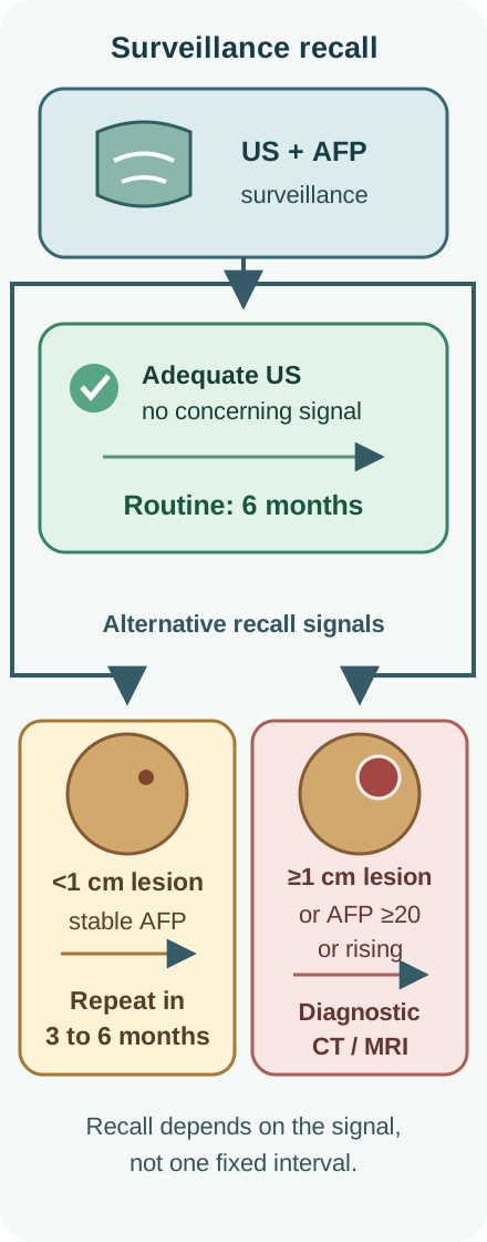
Use the recall diagram attached to this section to follow one surveillance encounter. Before reading the table, decide which finding changes the test and which changes only the interval.
The recall interval follows the surveillance signal: reassuring testing returns to six months, a subcentimeter finding with stable AFP gets short-interval reassessment, and a larger lesion or concerning AFP triggers diagnostic imaging. [1]
Recall after ultrasound and AFP
Finding
Next step
Why
FindingAdequate ultrasound, no lesion, reassuring AFP
Next stepContinue six-month surveillance
WhyNo current diagnostic signal
FindingLesion smaller than 1 cm, without a separate concerning AFP signal
Next stepRepeat ultrasound and AFP in 3 to 6 months
WhyCT and MRI are less reliable for characterizing very small lesions
FindingLesion at least 1 cm, or concerning AFP
Next stepDiagnostic multiphasic CT or contrast MRI
WhyAssess enhancement and the rest of the liver
FindingPersistently poor ultrasound visualization
Next stepConsider an MRI- or CT-based alternative surveillance strategy
WhyAn obscured liver is not a reassuring negative examination
A subcentimeter finding stable on at least two follow-up examinations may return to six-month surveillance. A lesion growing from 8 to 13 mm instead crosses the diagnostic-imaging threshold. Selected alternative surveillance is not a recommendation to expose everyone to serial CT radiation. [1]
Why does the lesion change relative to the liver?
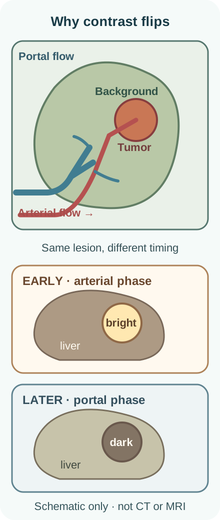
The background liver has dual inflow with a large portal contribution. During hepatocarcinogenesis, tumor supply often becomes predominantly arterial. Thus a lesion may enhance more than liver during the arterial phase and become relatively darker later. The perfusion diagram separates blood supply from image brightness. Trace the arterial supply to the tumor, then compare the lesion with the surrounding liver in each phase. Washout is a relative contrast observation, not simply a fall in the lesion's measured attenuation.[1][4]
Background liver receives substantial portal flow while HCC often becomes arterialized. Compare lesion contrast with the surrounding liver across phases. [1]
Phase comparison: change the background, not just the tumor
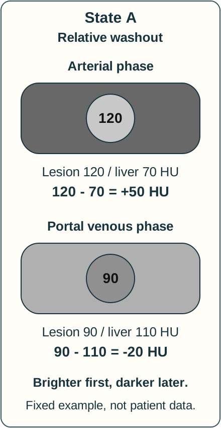
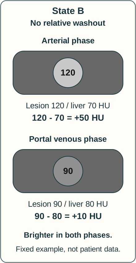
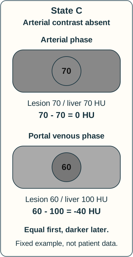
This numerical CT model uses fixed examples, not patient data or diagnostic cutoffs. In the three static figures accompanying this section, compare the lesion with its surrounding liver in each phase. Subtract liver attenuation from lesion attenuation before checking the visible results. All three states remain available together.
Attenuation in Hounsfield units (HU)
State
Arterial lesion / liver
Portal venous lesion / liver
StateA: arterialized lesion
Arterial lesion / liver120 / 70
Portal venous lesion / liver90 / 110
StateB: same lesion, different later background
Arterial lesion / liver120 / 70
Portal venous lesion / liver90 / 80
StateC: no relative arterial enhancement
Arterial lesion / liver70 / 70
Portal venous lesion / liver60 / 100
A: Arterial relative contrast is 120 - 70 = +50 HU; portal venous relative contrast is 90 - 110 = -20 HU. The lesion is brighter than liver first and darker later, illustrating relative washout after arterial hyperenhancement.
State A is a fixed numerical example. Brighter first, darker later. Compare lesion minus liver, not lesion attenuation alone. Use the supplied Hounsfield values to compare lesion and liver contrast. [4]
B: Arterial relative contrast is again +50 HU, but portal venous contrast is 90 - 80 = +10 HU. The lesion loses attenuation yet remains brighter than liver. A fall in lesion attenuation alone does not establish washout.
State B is a fixed numerical example. Brighter in both phases. Compare lesion minus liver, not lesion attenuation alone. Use the supplied Hounsfield values to compare lesion and liver contrast. [4]
C: Arterial relative contrast is 70 - 70 = 0 HU; portal venous contrast is 60 - 100 = -40 HU. Later relative darkness does not supply the missing arterial hyperenhancement. Washout can occur without arterial hyperenhancement when enhancement is otherwise present, but these are separate imaging features. [4]
State C is a fixed numerical example. Equal first, darker later. Compare lesion minus liver, not lesion attenuation alone. Use the supplied Hounsfield values to compare lesion and liver contrast. [4]
Worked A-versus-B comparison: Both lesions measure 120 HU early and 90 HU later. Only the later background changes, from 110 HU in A to 80 HU in B. Relative contrast therefore ends at -20 HU in A but +10 HU in B. The drawings use schematic gray values, not a calibrated CT display.
Check one calculation: why is A negative later?
Subtract background liver from lesion: 90 - 110 = -20 HU. A negative difference places the lesion below liver attenuation in that phase.
Check the comparison: why is B different?
The same 90 HU lesion is compared with an 80 HU liver, giving +10 HU. The background, not a different lesion value, explains the contrast difference.
Apply C: is later darkness arterial hyperenhancement?
No. The arterial-phase values are equal. Judge each feature in its own phase before applying the diagnostic criteria.
Transfer the rule to a report that says only that a lesion became less dense: ask how it compared with liver and which phases were obtained. No selection or disclosure is required to read the calculations or their explanations. [4]
Technique matters. Nonrim arterial-phase hyperenhancement differs from a peripheral rim. With an extracellular contrast agent, washout may be assessed in portal venous or delayed phases. With gadoxetate, LI-RADS washout is assessed in the portal venous phase; transitional-phase hypointensity alone can reflect uptake by the surrounding liver and is not equivalent. [4]
A typical hemangioma has peripheral discontinuous nodular enhancement with progressive centripetal filling and persistent blood-pool enhancement. Compare that spatial pattern with nonrim enhancement and nonperipheral washout. “Hemangiomas never appear to wash out” is too absolute, especially with hepatobiliary agents. In a patient with underlying liver disease, use appropriate contrast characterization rather than declaring every bright lesion HCC. A confidently typical hemangioma does not need lesion-specific cancer follow-up, but the patient's cirrhosis surveillance continues. [8]
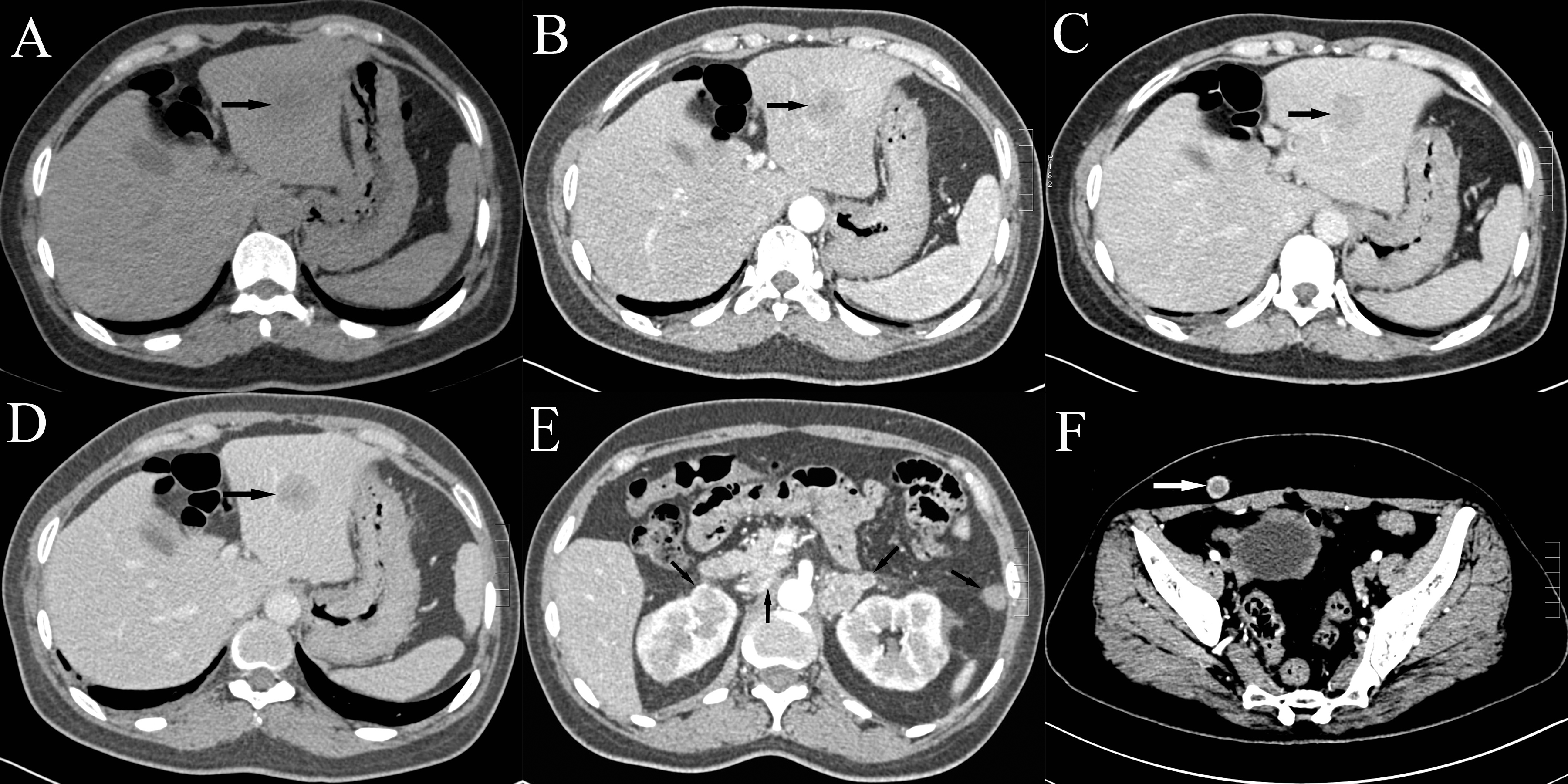
The clinical CT attached here is intentionally not a perfect textbook HCC. The published patient had multiple atypical lesions, normal AFP and no known chronic liver disease; biopsy established the diagnosis. A-D are the original plain and contrast-phase liver panels; E and F show additional reported lesions. Inspect the arrows, then ask what additional context is needed before assigning a noninvasive HCC category. This figure demonstrates why a real image cannot be detached from its clinical population and pathology. [5]
Pan and colleagues reported biopsy-confirmed HCC with normal AFP and atypical imaging in a patient without known chronic liver disease. Panels A to D are unenhanced and triphasic liver images; E shows adrenal and kidney lesions and F shows a subcutaneous lesion. This is not a stand-alone example establishing LR-5. Image: Zhenyu Pan, Guozi Yang, Tingting Yuan, Xiaochuan Pang, Yongxiang Wang, Limei Qu, Lihua Dong; original source; CC BY 2.0. [5].
When can imaging replace biopsy?
A familiar enhancement pattern is useful only within the population in which the diagnostic algorithm applies: principally cirrhosis or at-risk chronic HBV. Outside that setting, pathology is generally required. In the appropriate population, a lesion meeting definite-HCC criteria (LR-5) can establish the diagnosis without routine biopsy. AFP is not part of that imaging proof. [1]
Size and pattern must be interpreted together
Observation
Interpretation and next decision
ObservationAt least 2 cm, nonrim arterial hyperenhancement, plus nonperipheral washout, enhancing capsule or threshold growth
Interpretation and next decisionCan meet LR-5 criteria in the appropriate at-risk population
Observation10 to 19 mm with nonrim arterial hyperenhancement
Interpretation and next decisionMore stringent additional criteria apply; nonperipheral washout or LI-RADS threshold growth can establish LR-5, but capsule alone cannot
ObservationLR-3 observation
Interpretation and next decisionRepeat cross-sectional imaging in 3 to 6 months
ObservationLR-4 observation
Interpretation and next decisionMultidisciplinary review: biopsy or repeat contrast imaging within 3 months; favor biopsy if an immediate diagnosis changes care
ObservationLR-M, such as a targetoid malignant pattern
Interpretation and next decisionBiopsy to establish tumor type; malignant does not mean specifically HCC
In CT/MRI LI-RADS v2018, threshold growth means at least a 50% size increase within 6 months, assessed on comparable diagnostic examinations. Crossing 1 cm on surveillance ultrasound triggers diagnostic imaging; it does not by itself establish a CT/MRI category. Ancillary features can refine intermediate categories but cannot establish LR-5. [9]
This is a decision summary, not the complete LI-RADS categorization manual. Adequate technique and all major features remain necessary. A negative biopsy of a small suspicious lesion can be a sampling result, not proof of benignity; growth or evolving enhancement warrants reassessment. [1]
Compare a 15 mm lesion with arterial hyperenhancement and capsule but no washout against a 24 mm lesion with nonrim arterial hyperenhancement and washout. State why the smaller lesion cannot simply inherit the larger lesion's diagnosis. The first needs an indeterminate-observation pathway; the second can meet definite criteria if the population and technique are appropriate. Now put the second pattern in a person without cirrhosis or at-risk HBV and with prior colorectal cancer: the clinical population changes the need for tissue, even though the pixels look similar.
What must be known before choosing treatment?
“One 3 cm tumor” is not a complete stage or a treatment plan. Record number, size, distribution, macrovascular invasion and extrahepatic disease with high-quality liver imaging and appropriate chest assessment. Chest CT is recommended beyond very early BCLC stage; routine PET or bone scanning is not the default HCC staging strategy. At the same time, assess liver function, clinically significant portal hypertension, performance status, comorbidities and transplant feasibility. [1]
Two patients with the same solitary 3 cm HCC
Finding
Patient A
Patient B
FindingAlbumin / bilirubin / INR
Patient A4.0 g/dL / 0.8 mg/dL / 1.0
Patient B2.6 g/dL / 3.2 mg/dL / 1.6
FindingAscites or encephalopathy
Patient AAbsent
Patient BRecurrent ascites
FindingPortal hypertension
Patient ANo clinical evidence
Patient BVarices and thrombocytopenia
FindingImmediate planning implication
Patient AAssess resection feasibility and future liver remnant
Patient BAssess transplant eligibility and decompensation; do not assume resection is safe
Predict the consequence of a major hepatectomy in each setting before comparing the last row. A technically removable tumor does not guarantee that the remaining liver can sustain the patient. Child-Pugh incorporates bilirubin, albumin, INR, ascites and encephalopathy. Each component contributes 1 to 3 points: class A totals 5-6, class B 7-9 and class C 10-15. [11] It does not replace a separate portal-hypertension assessment. BCLC integrates tumor burden, liver function and performance status; it is a treatment framework, not a size-only label. [1][3]
An enhancing soft-tissue component in a portal vein, especially contiguous with tumor, raises concern for tumor in vein and changes staging and transplant planning. A nonenhancing filling defect can be bland thrombosis. Compare those patterns rather than treating every portal-vein defect as invasion. Resolve uncertainty with expert imaging review and multidisciplinary evaluation before excluding a potentially eligible transplant candidate. Imaging evidence of venous malignancy also does not, by itself, identify every tumor as HCC. [1]
Apply the same discipline to unexplained functional decline: distinguish cancer-related performance limitation from decompensated liver disease and other illness. The treatment team needs both causes, not just a numerical stage.
Match tumor control to the liver that must survive it
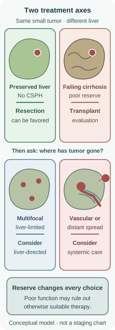
The treatment diagram compares similar tumor burdens in different livers. For each branch, first identify whether the limiting problem is tumor distribution, liver reserve or both. Then predict why a treatment appropriate for one patient can harm another. Decisions belong in a multidisciplinary team. [1]
Treatment depends on both tumor distribution and liver function. The four illustrated contexts show broad therapeutic directions, not an individual treatment algorithm or complete staging system. CSPH means clinically significant portal hypertension. [1]
Potentially curative approaches
Resection is favored for localized HCC without cirrhosis, and in selected patients with well-compensated cirrhosis, no clinically significant portal hypertension and an adequate future liver remnant. Thermal ablation offers curative-intent local treatment, especially for tumors no larger than 3 cm when surgery is unsuitable. Diameter is not the only constraint: proximity to major bile ducts or other vulnerable structures, inadequate margins and heat loss beside large vessels can impair safety or effectiveness. Radiation segmentectomy or external-beam radiation may be alternatives in selected cases. [1][3]
Transplantation treats both the cancer and the cirrhotic liver. Milan tumor criteria are one lesion no larger than 5 cm, or up to three lesions each no larger than 3 cm, without macrovascular invasion or extrahepatic spread. These are not a guarantee of listing: medical fitness, biology, AFP-related policies, organ allocation and center assessment also apply. A person with two 2 cm lesions and recurrent decompensation may be a better transplant candidate than a resection candidate. Selected patients beyond standard tumor limits may undergo downstaging and reassessment; response does not automatically confer eligibility. [1]
During an anticipated wait, bridging locoregional therapy can limit progression when hepatic reserve permits. Compare that purpose with downstaging: bridging maintains an existing tumor eligibility range, while downstaging attempts to reach an acceptable range from outside it. Neither justifies injuring an already failing liver indiscriminately.
Liver-limited multifocal disease and advanced disease
For selected multifocal, liver-limited HCC without macrovascular invasion or distant spread, transarterial chemoembolization (TACE) is a standard locoregional approach. Radioembolization is an alternative in selected settings. Arterial delivery exploits tumor supply, but nontumor liver can also be injured: selective treatment and adequate reserve matter. Progression or inadequate response after one or two adequately delivered sessions should prompt reconsideration, including systemic treatment, rather than automatic repetition until liver function fails. [1]
Macrovascular invasion, extrahepatic disease or disease unsuitable for effective locoregional treatment often calls for systemic therapy if liver function and performance status permit. Evidence is strongest in Child-Pugh A and good performance status. Selected Child-Pugh B patients require caution; results from fitter trial populations do not establish safety or benefit in severely decompensated Child-Pugh C disease. When transplantation and tolerable cancer-directed treatment are unavailable, symptom-focused care is active care, not abandonment. [1][6]
First-line systemic choices: the regimen and its constraints
Approach
Decision-relevant limitation
ApproachAtezolizumab plus bevacizumab
Decision-relevant limitationAssess gastrointestinal bleeding risk, including endoscopy for varices; treat high-risk varices and reconsider recent bleeding before anti-VEGF treatment
ApproachDurvalumab plus tremelimumab
Decision-relevant limitationThe standard regimen uses a single tremelimumab priming dose with durvalumab, followed by durvalumab; assess immune-related risks
ApproachNivolumab plus ipilimumab
Decision-relevant limitationFDA first-line approval in 2025; combination dosing is limited to four doses, then nivolumab alone, not indefinite dual treatment
ApproachSelected alternatives such as sorafenib or lenvatinib
Decision-relevant limitationRelevant when combination immunotherapy is unsuitable; these multikinase inhibitors include VEGF-pathway activity and are not substitutes for avoiding that pathway. Reserve and toxicity still govern selection
A recent variceal bleed can shift consideration toward a non-VEGF regimen, but does not make immunotherapy universally safe. Active severe autoimmune disease and prior liver transplantation need particular attention; immune checkpoint inhibitors can precipitate graft rejection. Assess these constraints before choosing among regimens, rather than presenting one regimen as best for every patient. The nivolumab-ipilimumab first-line trial enrolled Child-Pugh A patients with ECOG 0 or 1, not a general decompensated population. [1][2][6]
Transfer the two-part approach to follow-up: radiographic tumor response can coexist with worsening liver function. Reassess both before continuing treatment. A falling AFP alone cannot certify that the overall plan is helping. New jaundice, synthetic dysfunction, ascites or altered mentation before a scheduled infusion require prompt assessment, including treatment toxicity and other causes. Any treatment interruption, discontinuation or later resumption depends on the cause and severity, not tumor shrinkage alone. [10]
Practice the decisions
For each case, identify the current task before considering the options: surveillance, diagnosis, staging or treatment. Explain which finding makes the closest alternative less suitable. Optional answer explanations do not restrict access to the lesson.
Case 1
Show answer and explanations for case 1
A. Continue ultrasound and AFP every 6 months (Why this does not fit)
The obscured liver makes repeat ultrasound unreliable despite continued cirrhotic risk. [1]
Reasoning steps for option A
Does sustained HCV clearance erase this patient's established risk?
No. Prior biopsy-proven cirrhosis remains a surveillance indication; the inadequate ultrasound determines how to perform it.
How do the findings support or oppose this plan: Continue ultrasound and AFP every 6 months?
The obscured liver makes repeat ultrasound unreliable despite continued cirrhotic risk. [1]
B. Use an MRI-based surveillance strategy with AFP (Best answer)
Cirrhosis-associated risk persists after cure; an MRI-based alternative is reasonable when ultrasound remains severely limited. [1]
Reasoning steps for option B
Does sustained HCV clearance erase this patient's established risk?
No. Prior biopsy-proven cirrhosis remains a surveillance indication; the inadequate ultrasound determines how to perform it.
How do the findings support or oppose this plan: Use an MRI-based surveillance strategy with AFP?
Cirrhosis-associated risk persists after cure; an MRI-based alternative is reasonable when ultrasound remains severely limited. [1]
C. Stop HCC surveillance after sustained viral clearance (Why this does not fit)
Viral cure reduces HCC risk but does not eliminate the surveillance indication created by prior established cirrhosis. Residual cancer risk, not a claim that fibrosis can never regress, justifies continued surveillance. [1]
Reasoning steps for option C
Does sustained HCV clearance erase this patient's established risk?
No. Prior biopsy-proven cirrhosis remains a surveillance indication; the inadequate ultrasound determines how to perform it.
How do the findings support or oppose this plan: Stop HCC surveillance after sustained viral clearance?
Viral cure reduces HCC risk but does not eliminate the surveillance indication created by prior established cirrhosis. Residual cancer risk, not a claim that fibrosis can never regress, justifies continued surveillance. [1]
D. Use AFP surveillance without liver imaging (Why this does not fit)
Normal AFP does not compensate for an unevaluable liver. [1]
Reasoning steps for option D
Does sustained HCV clearance erase this patient's established risk?
No. Prior biopsy-proven cirrhosis remains a surveillance indication; the inadequate ultrasound determines how to perform it.
How do the findings support or oppose this plan: Use AFP surveillance without liver imaging?
Normal AFP does not compensate for an unevaluable liver. [1]
Takeaway: Cirrhosis-associated risk persists after cure; an MRI-based alternative is reasonable when ultrasound remains severely limited.
A. Obtain biopsy because cirrhosis is absent (Why this does not fit)
Pathology is generally needed outside eligible risk populations, but selected chronic HBV patients can qualify without cirrhosis. His age, sex and endemic origin support that setting. [1]
Reasoning steps for option A
Does absent cirrhosis prohibit imaging diagnosis in every HBV patient?
No. This man's chronic HBV risk setting permits the algorithm; the 18 mm enhancement combination then supports definite HCC.
How do the findings support or oppose this plan: Obtain biopsy because cirrhosis is absent?
Pathology is generally needed outside eligible risk populations, but selected chronic HBV patients can qualify without cirrhosis. His age, sex and endemic origin support that setting. [1]
B. Repeat MRI in three months because the lesion is under 2 cm (Why this does not fit)
In the appropriate population, a 10 to 19 mm lesion with nonrim arterial enhancement and nonperipheral washout can meet LR-5; 2 cm is not a universal minimum. [1,9]
Reasoning steps for option B
Does absent cirrhosis prohibit imaging diagnosis in every HBV patient?
No. This man's chronic HBV risk setting permits the algorithm; the 18 mm enhancement combination then supports definite HCC.
How do the findings support or oppose this plan: Repeat MRI in three months because the lesion is under 2 cm?
In the appropriate population, a 10 to 19 mm lesion with nonrim arterial enhancement and nonperipheral washout can meet LR-5; 2 cm is not a universal minimum. [1,9]
C. Use the imaging diagnosis and complete HCC staging (Best answer)
His chronic HBV risk permits application of noninvasive criteria despite absent cirrhosis. The 18 mm lesion has the required enhancement combination, so complete staging rather than require routine diagnostic biopsy. [1,9]
Reasoning steps for option C
Does absent cirrhosis prohibit imaging diagnosis in every HBV patient?
No. This man's chronic HBV risk setting permits the algorithm; the 18 mm enhancement combination then supports definite HCC.
How do the findings support or oppose this plan: Use the imaging diagnosis and complete HCC staging?
His chronic HBV risk permits application of noninvasive criteria despite absent cirrhosis. The 18 mm lesion has the required enhancement combination, so complete staging rather than require routine diagnostic biopsy. [1,9]
D. Return to six-month ultrasound because HBV is suppressed (Why this does not fit)
Antiviral suppression reduces risk without eliminating it. A diagnostic mass pattern cannot be returned to surveillance solely because viral replication is controlled. [1]
Reasoning steps for option D
Does absent cirrhosis prohibit imaging diagnosis in every HBV patient?
No. This man's chronic HBV risk setting permits the algorithm; the 18 mm enhancement combination then supports definite HCC.
How do the findings support or oppose this plan: Return to six-month ultrasound because HBV is suppressed?
Antiviral suppression reduces risk without eliminating it. A diagnostic mass pattern cannot be returned to surveillance solely because viral replication is controlled. [1]
Takeaway: First establish that the at-risk imaging algorithm applies, then interpret the size and enhancement combination.
A. No lesion follow-up or routine HCC surveillance (Best answer)
The completed MRI shows a confidently typical hemangioma, which does not need lesion-specific follow-up. Separately, F2 metabolic disease without cirrhosis or HBV does not establish an indication for routine HCC surveillance; ongoing liver-disease care continues. [1,8]
Reasoning steps for option A
Which two decisions follow from the completed MRI and fibrosis history?
The MRI establishes a typical hemangioma without lesion-specific follow-up. Independently, F2 metabolic fibrosis without HBV or cirrhosis does not justify routine HCC surveillance.
How do the findings support or oppose this plan: No lesion follow-up or routine HCC surveillance?
The completed MRI shows a confidently typical hemangioma, which does not need lesion-specific follow-up. Separately, F2 metabolic disease without cirrhosis or HBV does not establish an indication for routine HCC surveillance; ongoing liver-disease care continues. [1,8]
B. No lesion follow-up; ultrasound and AFP every six months (Why this does not fit)
This correctly recognizes that a typical hemangioma needs no dedicated follow-up. It incorrectly imports the cirrhosis surveillance schedule into a patient with established F2 fibrosis and no HBV. [1,8]
Reasoning steps for option B
Which two decisions follow from the completed MRI and fibrosis history?
The MRI establishes a typical hemangioma without lesion-specific follow-up. Independently, F2 metabolic fibrosis without HBV or cirrhosis does not justify routine HCC surveillance.
How do the findings support or oppose this plan: No lesion follow-up; ultrasound and AFP every six months?
This correctly recognizes that a typical hemangioma needs no dedicated follow-up. It incorrectly imports the cirrhosis surveillance schedule into a patient with established F2 fibrosis and no HBV. [1,8]
C. Liver MRI in three months for continued lesion surveillance (Why this does not fit)
Short-interval MRI can help characterize an indeterminate lesion. The dedicated examination here already demonstrates a typical benign vascular filling pattern, not an unresolved observation. [8]
Reasoning steps for option C
Which two decisions follow from the completed MRI and fibrosis history?
The MRI establishes a typical hemangioma without lesion-specific follow-up. Independently, F2 metabolic fibrosis without HBV or cirrhosis does not justify routine HCC surveillance.
How do the findings support or oppose this plan: Liver MRI in three months for continued lesion surveillance?
Short-interval MRI can help characterize an indeterminate lesion. The dedicated examination here already demonstrates a typical benign vascular filling pattern, not an unresolved observation. [8]
D. Core biopsy now with follow-up directed by histology (Why this does not fit)
Biopsy can establish the identity of an indeterminate or suspicious mass. The characteristic peripheral nodular filling and persistent blood-pool enhancement establish a typical hemangioma without routine sampling. [8]
Reasoning steps for option D
Which two decisions follow from the completed MRI and fibrosis history?
The MRI establishes a typical hemangioma without lesion-specific follow-up. Independently, F2 metabolic fibrosis without HBV or cirrhosis does not justify routine HCC surveillance.
How do the findings support or oppose this plan: Core biopsy now with follow-up directed by histology?
Biopsy can establish the identity of an indeterminate or suspicious mass. The characteristic peripheral nodular filling and persistent blood-pool enhancement establish a typical hemangioma without routine sampling. [8]
Takeaway: Identify the lesion from complete contrast imaging, then assess surveillance eligibility independently of that lesion.
A. Child-Pugh B; ultrasound and AFP every six months (Why this does not fit)
The laboratory and clinical abnormalities indicate Child-Pugh C, not B. The usual surveillance benefit in Child-Pugh A or B cannot be transferred to transplant-ineligible Child-Pugh C disease. [1,11]
Reasoning steps for option A
What reserve classification must be established before deciding on surveillance?
The specified laboratory and clinical components total 14 Child-Pugh points, class C. Because transplantation is unavailable, routine HCC surveillance is not recommended; decompensation and symptom care continue.
How do the findings support or oppose this plan: Child-Pugh B; ultrasound and AFP every six months?
The laboratory and clinical abnormalities indicate Child-Pugh C, not B. The usual surveillance benefit in Child-Pugh A or B cannot be transferred to transplant-ineligible Child-Pugh C disease. [1,11]
B. Child-Pugh C; omit routine HCC surveillance (Best answer)
The five components give 14 Child-Pugh points: 3 each for bilirubin, albumin, INR and poorly controlled ascites, plus 2 for grade-II encephalopathy. This is class C; without transplant eligibility, routine HCC surveillance is not recommended, while cirrhosis and symptom care continue. [1,11]
Reasoning steps for option B
What reserve classification must be established before deciding on surveillance?
The specified laboratory and clinical components total 14 Child-Pugh points, class C. Because transplantation is unavailable, routine HCC surveillance is not recommended; decompensation and symptom care continue.
How do the findings support or oppose this plan: Child-Pugh C; omit routine HCC surveillance?
The five components give 14 Child-Pugh points: 3 each for bilirubin, albumin, INR and poorly controlled ascites, plus 2 for grade-II encephalopathy. This is class C; without transplant eligibility, routine HCC surveillance is not recommended, while cirrhosis and symptom care continue. [1,11]
C. Child-Pugh C; ultrasound and AFP every six months (Why this does not fit)
The reserve classification is correct, but the surveillance plan ignores transplant ineligibility. Child-Pugh C patients receive HCC surveillance when transplantation is available, not simply because their cancer risk is high. [1]
Reasoning steps for option C
What reserve classification must be established before deciding on surveillance?
The specified laboratory and clinical components total 14 Child-Pugh points, class C. Because transplantation is unavailable, routine HCC surveillance is not recommended; decompensation and symptom care continue.
How do the findings support or oppose this plan: Child-Pugh C; ultrasound and AFP every six months?
The reserve classification is correct, but the surveillance plan ignores transplant ineligibility. Child-Pugh C patients receive HCC surveillance when transplantation is available, not simply because their cancer risk is high. [1]
D. Child-Pugh B; omit routine HCC surveillance (Why this does not fit)
The surveillance decision fits the stated transplant-ineligible severe disease, but the reserve classification does not. The combined laboratory impairment, poorly controlled ascites and encephalopathy place the patient in class C. [1,11]
Reasoning steps for option D
What reserve classification must be established before deciding on surveillance?
The specified laboratory and clinical components total 14 Child-Pugh points, class C. Because transplantation is unavailable, routine HCC surveillance is not recommended; decompensation and symptom care continue.
How do the findings support or oppose this plan: Child-Pugh B; omit routine HCC surveillance?
The surveillance decision fits the stated transplant-ineligible severe disease, but the reserve classification does not. The combined laboratory impairment, poorly controlled ascites and encephalopathy place the patient in class C. [1,11]
Takeaway: Determine reserve from all five Child-Pugh components, then apply the transplant-dependent surveillance exception for class C.
A. Definite HCC; complete staging for tumor treatment (Why this does not fit)
The lesion loses attenuation but remains brighter than liver: differences are +50, +10 and +7 HU. Thus the supplied data do not establish the washout needed to complete this LR-5 pattern. [1,4,9]
Reasoning steps for option A
Does the lesion become darker than background liver on either later phase?
No. It remains +10 HU and +7 HU relative to liver, so the falling absolute attenuation does not establish washout.
How do the findings support or oppose this plan: Definite HCC; complete staging for tumor treatment?
The lesion loses attenuation but remains brighter than liver: differences are +50, +10 and +7 HU. Thus the supplied data do not establish the washout needed to complete this LR-5 pattern. [1,4,9]
B. Typical hemangioma; return to routine surveillance (Why this does not fit)
The described homogeneous arterial enhancement is not the discontinuous peripheral nodular enhancement and progressive filling of a typical hemangioma. [8]
Reasoning steps for option B
Does the lesion become darker than background liver on either later phase?
No. It remains +10 HU and +7 HU relative to liver, so the falling absolute attenuation does not establish washout.
How do the findings support or oppose this plan: Typical hemangioma; return to routine surveillance?
The described homogeneous arterial enhancement is not the discontinuous peripheral nodular enhancement and progressive filling of a typical hemangioma. [8]
C. Probable HCC; multidisciplinary follow-up or biopsy (Best answer)
Subtracting liver from lesion shows persistent positive relative contrast, not washout. A mass at least 2 cm with nonrim arterial hyperenhancement alone is probable rather than definite HCC; multidisciplinary review selects imaging within three months or biopsy. [1,4,9]
Reasoning steps for option C
Does the lesion become darker than background liver on either later phase?
No. It remains +10 HU and +7 HU relative to liver, so the falling absolute attenuation does not establish washout.
How do the findings support or oppose this plan: Probable HCC; multidisciplinary follow-up or biopsy?
Subtracting liver from lesion shows persistent positive relative contrast, not washout. A mass at least 2 cm with nonrim arterial hyperenhancement alone is probable rather than definite HCC; multidisciplinary review selects imaging within three months or biopsy. [1,4,9]
D. Malignant-nonspecific pattern; obtain histologic typing (Why this does not fit)
No targetoid, rim or other LR-M feature is supplied. Missing washout does not by itself establish a malignant-nonspecific category. [1,9]
Reasoning steps for option D
Does the lesion become darker than background liver on either later phase?
No. It remains +10 HU and +7 HU relative to liver, so the falling absolute attenuation does not establish washout.
How do the findings support or oppose this plan: Malignant-nonspecific pattern; obtain histologic typing?
No targetoid, rim or other LR-M feature is supplied. Missing washout does not by itself establish a malignant-nonspecific category. [1,9]
Takeaway: Interpret lesion-to-liver contrast before applying size-dependent diagnostic categories and their management pathways.
A. Then: six-month surveillance; now: multiphasic CT or MRI (Best answer)
Two stable short-interval examinations with reassuring AFP permit return to six-month surveillance. At the later visit, the new AFP elevation above 20 ng/mL warrants diagnostic CT or MRI despite an unchanged subcentimeter observation and otherwise negative ultrasound. [1]
Reasoning steps for option A
What changes between the earlier stable visits and the current visit?
Two stable subcentimeter examinations with reassuring AFP permit six-month surveillance. The later AFP rise to 34 ng/mL independently requires diagnostic imaging despite unchanged lesion size.
How do the findings support or oppose this plan: Then: six-month surveillance; now: multiphasic CT or MRI?
Two stable short-interval examinations with reassuring AFP permit return to six-month surveillance. At the later visit, the new AFP elevation above 20 ng/mL warrants diagnostic CT or MRI despite an unchanged subcentimeter observation and otherwise negative ultrasound. [1]
B. Then: three-month surveillance; now: multiphasic CT or MRI (Why this does not fit)
The current diagnostic imaging choice is appropriate for the new AFP signal. Continuing three-month recall after the earlier two stable examinations was unnecessary when AFP was reassuring. [1]
Reasoning steps for option B
What changes between the earlier stable visits and the current visit?
Two stable subcentimeter examinations with reassuring AFP permit six-month surveillance. The later AFP rise to 34 ng/mL independently requires diagnostic imaging despite unchanged lesion size.
How do the findings support or oppose this plan: Then: three-month surveillance; now: multiphasic CT or MRI?
The current diagnostic imaging choice is appropriate for the new AFP signal. Continuing three-month recall after the earlier two stable examinations was unnecessary when AFP was reassuring. [1]
C. Then: six-month surveillance; now: repeat ultrasound in three months (Why this does not fit)
The earlier return to six-month surveillance is appropriate. The later AFP rise is an independent diagnostic recall signal; another short-interval ultrasound does not substitute for multiphasic characterization. [1]
Reasoning steps for option C
What changes between the earlier stable visits and the current visit?
Two stable subcentimeter examinations with reassuring AFP permit six-month surveillance. The later AFP rise to 34 ng/mL independently requires diagnostic imaging despite unchanged lesion size.
How do the findings support or oppose this plan: Then: six-month surveillance; now: repeat ultrasound in three months?
The earlier return to six-month surveillance is appropriate. The later AFP rise is an independent diagnostic recall signal; another short-interval ultrasound does not substitute for multiphasic characterization. [1]
D. Then: three-month surveillance; now: repeat ultrasound in three months (Why this does not fit)
This keeps the earlier short interval after sufficient stability and then fails to respond to the separate new AFP signal. Stability of one small observation cannot establish that the whole liver remains free of a developing lesion. [1]
Reasoning steps for option D
What changes between the earlier stable visits and the current visit?
Two stable subcentimeter examinations with reassuring AFP permit six-month surveillance. The later AFP rise to 34 ng/mL independently requires diagnostic imaging despite unchanged lesion size.
How do the findings support or oppose this plan: Then: three-month surveillance; now: repeat ultrasound in three months?
This keeps the earlier short interval after sufficient stability and then fails to respond to the separate new AFP signal. Stability of one small observation cannot establish that the whole liver remains free of a developing lesion. [1]
Takeaway: End short-interval recall when stability is established, but reassess the pathway when a separate surveillance signal changes.
A. Probable HCC; repeat contrast imaging within three months (Why this does not fit)
Short-interval reassessment fits some probable-HCC observations, but the 60% increase within four months meets threshold growth. Together with nonrim arterial enhancement at this size, it supports LR-5. [1,9]
Reasoning steps for option A
How much has the mass grown on comparable MRI?
The increase is (16-10)/10 = 60% in four months, meeting the at-least-50%-within-six-months diagnostic threshold.
How do the findings support or oppose this plan: Probable HCC; repeat contrast imaging within three months?
Short-interval reassessment fits some probable-HCC observations, but the 60% increase within four months meets threshold growth. Together with nonrim arterial enhancement at this size, it supports LR-5. [1,9]
B. Malignant-nonspecific lesion; obtain biopsy for tumor typing (Why this does not fit)
No malignant-nonspecific pattern is supplied. Threshold growth of a solid arterialized mass supports the HCC diagnostic pathway rather than automatically assigning LR-M. [1,9]
Reasoning steps for option B
How much has the mass grown on comparable MRI?
The increase is (16-10)/10 = 60% in four months, meeting the at-least-50%-within-six-months diagnostic threshold.
How do the findings support or oppose this plan: Malignant-nonspecific lesion; obtain biopsy for tumor typing?
No malignant-nonspecific pattern is supplied. Threshold growth of a solid arterialized mass supports the HCC diagnostic pathway rather than automatically assigning LR-M. [1,9]
C. Indeterminate observation; repeat contrast MRI in six months (Why this does not fit)
A 60% diameter increase on comparable MRI within four months meets threshold growth. In an eligible cirrhotic patient, that finding plus nonrim arterial hyperenhancement in a 16-mm mass supports LR-5, rather than a six-month indeterminate-observation pathway. [1,9]
Reasoning steps for option C
How much has the mass grown on comparable MRI?
The increase is (16-10)/10 = 60% in four months, meeting the at-least-50%-within-six-months diagnostic threshold.
How do the findings support or oppose this plan: Indeterminate observation; repeat contrast MRI in six months?
A 60% diameter increase on comparable MRI within four months meets threshold growth. In an eligible cirrhotic patient, that finding plus nonrim arterial hyperenhancement in a 16-mm mass supports LR-5, rather than a six-month indeterminate-observation pathway. [1,9]
D. Definite HCC; complete staging and assess treatment eligibility (Best answer)
Growth is (16-10)/10 = 60% within four months, exceeding the CT/MRI threshold of at least 50% within six months. In this cirrhotic patient, nonrim arterial enhancement plus qualifying growth at 16 mm supports LR-5 and subsequent staging. [1,9]
Reasoning steps for option D
How much has the mass grown on comparable MRI?
The increase is (16-10)/10 = 60% in four months, meeting the at-least-50%-within-six-months diagnostic threshold.
How do the findings support or oppose this plan: Definite HCC; complete staging and assess treatment eligibility?
Growth is (16-10)/10 = 60% within four months, exceeding the CT/MRI threshold of at least 50% within six months. In this cirrhotic patient, nonrim arterial enhancement plus qualifying growth at 16 mm supports LR-5 and subsequent staging. [1,9]
Takeaway: Calculate growth on comparable diagnostic studies, then combine it with population, size and enhancement before assigning a category.
A. AFP confirms HCC; biopsy assesses tumor differentiation (Why this does not fit)
The AFP trend justified diagnostic imaging but does not establish HCC, particularly with a competing inflammatory cause. The targetoid mass needs biopsy to determine tumor type, not merely to grade an already proven HCC. [1,7,9]
Reasoning steps for option A
Does the AFP series or MRI establish an HCC-specific diagnosis?
No. AFP prompts diagnostic recall but is not diagnostic; rim and targetoid enhancement identify a malignant-nonspecific pattern requiring tissue typing.
How do the findings support or oppose this plan: AFP confirms HCC; biopsy assesses tumor differentiation?
The AFP trend justified diagnostic imaging but does not establish HCC, particularly with a competing inflammatory cause. The targetoid mass needs biopsy to determine tumor type, not merely to grade an already proven HCC. [1,7,9]
B. AFP reflects hepatitis; repeat ultrasound after six months (Why this does not fit)
Hepatitis can raise AFP, but it cannot safely explain away the consecutive increases or the subsequently detected malignant-appearing mass. Six-month surveillance leaves the tumor identity unresolved. [1,7]
Reasoning steps for option B
Does the AFP series or MRI establish an HCC-specific diagnosis?
No. AFP prompts diagnostic recall but is not diagnostic; rim and targetoid enhancement identify a malignant-nonspecific pattern requiring tissue typing.
How do the findings support or oppose this plan: AFP reflects hepatitis; repeat ultrasound after six months?
Hepatitis can raise AFP, but it cannot safely explain away the consecutive increases or the subsequently detected malignant-appearing mass. Six-month surveillance leaves the tumor identity unresolved. [1,7]
C. AFP triggers recall; biopsy establishes the tumor type (Best answer)
The rising AFP is a diagnostic recall signal rather than proof of cancer type. Rim and targetoid enhancement support an LR-M-type observation; biopsy distinguishes HCC from other primary or secondary malignancies before selecting tumor-specific treatment. [1,9]
Reasoning steps for option C
Does the AFP series or MRI establish an HCC-specific diagnosis?
No. AFP prompts diagnostic recall but is not diagnostic; rim and targetoid enhancement identify a malignant-nonspecific pattern requiring tissue typing.
How do the findings support or oppose this plan: AFP triggers recall; biopsy establishes the tumor type?
The rising AFP is a diagnostic recall signal rather than proof of cancer type. Rim and targetoid enhancement support an LR-M-type observation; biopsy distinguishes HCC from other primary or secondary malignancies before selecting tumor-specific treatment. [1,9]
D. AFP triggers recall; ablation treats imaging-established HCC (Why this does not fit)
The interpretation of AFP as a recall signal is appropriate. The MRI is not an HCC-specific LR-5 pattern, so an HCC-directed ablation plan would assume the histologic identity that still needs to be established. [1,9]
Reasoning steps for option D
Does the AFP series or MRI establish an HCC-specific diagnosis?
No. AFP prompts diagnostic recall but is not diagnostic; rim and targetoid enhancement identify a malignant-nonspecific pattern requiring tissue typing.
How do the findings support or oppose this plan: AFP triggers recall; ablation treats imaging-established HCC?
The interpretation of AFP as a recall signal is appropriate. The MRI is not an HCC-specific LR-5 pattern, so an HCC-directed ablation plan would assume the histologic identity that still needs to be established. [1,9]
Takeaway: Interpret AFP as a reason to investigate, then use the actual imaging pattern to decide whether tumor typing requires tissue.
A. Assign definite imaging HCC and proceed to reserve assessment (Best answer)
The at-risk setting, size and major imaging features can establish HCC noninvasively; reserve still guides treatment. [1]
Reasoning steps for option A
Which combination establishes imaging specificity here?
Cirrhosis plus a lesion larger than 2 cm with nonrim arterial enhancement and nonperipheral washout supports definite HCC; normal AFP does not negate it.
How do the findings support or oppose this plan: Assign definite imaging HCC and proceed to reserve assessment?
The at-risk setting, size and major imaging features can establish HCC noninvasively; reserve still guides treatment. [1]
B. Classify as probably HCC and repeat MRI within three months (Why this does not fit)
Short-interval imaging is appropriate for some LR-4 observations, but this at-risk patient has a lesion larger than 2 cm with nonrim arterial hyperenhancement and nonperipheral washout, supporting LR-5. [1]
Reasoning steps for option B
Which combination establishes imaging specificity here?
Cirrhosis plus a lesion larger than 2 cm with nonrim arterial enhancement and nonperipheral washout supports definite HCC; normal AFP does not negate it.
How do the findings support or oppose this plan: Classify as probably HCC and repeat MRI within three months?
Short-interval imaging is appropriate for some LR-4 observations, but this at-risk patient has a lesion larger than 2 cm with nonrim arterial hyperenhancement and nonperipheral washout, supporting LR-5. [1]
C. Repeat ultrasound after six months to confirm persistence (Why this does not fit)
A diagnostic lesion needs staging and liver assessment, not routine surveillance. [1]
Reasoning steps for option C
Which combination establishes imaging specificity here?
Cirrhosis plus a lesion larger than 2 cm with nonrim arterial enhancement and nonperipheral washout supports definite HCC; normal AFP does not negate it.
How do the findings support or oppose this plan: Repeat ultrasound after six months to confirm persistence?
A diagnostic lesion needs staging and liver assessment, not routine surveillance. [1]
D. Require biopsy because the serum marker is normal (Why this does not fit)
Normal AFP does not invalidate qualifying LR-5 imaging features. [1]
Reasoning steps for option D
Which combination establishes imaging specificity here?
Cirrhosis plus a lesion larger than 2 cm with nonrim arterial enhancement and nonperipheral washout supports definite HCC; normal AFP does not negate it.
How do the findings support or oppose this plan: Require biopsy because the serum marker is normal?
Normal AFP does not invalidate qualifying LR-5 imaging features. [1]
Takeaway: The at-risk setting, size and major imaging features can establish HCC noninvasively; reserve still guides treatment.
A. Obtain core biopsy before choosing lesion follow-up (Why this does not fit)
Routine core biopsy is not needed for a confidently typical hemangioma on complete contrast imaging. Obtain tissue when the diagnosis remains uncertain, not solely because cirrhosis raises background HCC risk. [1,8]
Reasoning steps for option A
What spatial enhancement behavior distinguishes this lesion?
Discontinuous peripheral nodules with centripetal filling and persistent blood-pool brightness favor hemangioma, not the characteristic HCC pattern.
How do the findings support or oppose this plan: Obtain core biopsy before choosing lesion follow-up?
Routine core biopsy is not needed for a confidently typical hemangioma on complete contrast imaging. Obtain tissue when the diagnosis remains uncertain, not solely because cirrhosis raises background HCC risk. [1,8]
B. Treat the lesion as HCC and initiate staging (Why this does not fit)
Progressive centripetal blood-pool filling differs from HCC nonrim enhancement and washout. [1] [8]
Reasoning steps for option B
What spatial enhancement behavior distinguishes this lesion?
Discontinuous peripheral nodules with centripetal filling and persistent blood-pool brightness favor hemangioma, not the characteristic HCC pattern.
How do the findings support or oppose this plan: Treat the lesion as HCC and initiate staging?
Progressive centripetal blood-pool filling differs from HCC nonrim enhancement and washout. [1] [8]
C. Repeat contrast MRI in three months before assigning benignity (Why this does not fit)
Short-interval contrast imaging is appropriate for an unresolved observation. The clearly typical peripheral nodular filling pattern here already establishes a benign hemangioma; the separate cirrhosis risk still requires HCC surveillance. [1,8]
Reasoning steps for option C
What spatial enhancement behavior distinguishes this lesion?
Discontinuous peripheral nodules with centripetal filling and persistent blood-pool brightness favor hemangioma, not the characteristic HCC pattern.
How do the findings support or oppose this plan: Repeat contrast MRI in three months before assigning benignity?
Short-interval contrast imaging is appropriate for an unresolved observation. The clearly typical peripheral nodular filling pattern here already establishes a benign hemangioma; the separate cirrhosis risk still requires HCC surveillance. [1,8]
D. Continue cirrhosis surveillance without dedicated lesion imaging (Best answer)
This is a typical hemangioma requiring no lesion-specific cancer follow-up, while cirrhosis still warrants surveillance. [1] [8]
Reasoning steps for option D
What spatial enhancement behavior distinguishes this lesion?
Discontinuous peripheral nodules with centripetal filling and persistent blood-pool brightness favor hemangioma, not the characteristic HCC pattern.
How do the findings support or oppose this plan: Continue cirrhosis surveillance without dedicated lesion imaging?
This is a typical hemangioma requiring no lesion-specific cancer follow-up, while cirrhosis still warrants surveillance. [1] [8]
Takeaway: This is a typical hemangioma requiring no lesion-specific cancer follow-up, while cirrhosis still warrants surveillance.
A. Definite HCC; begin treatment without further characterization (Why this does not fit)
Arterial enhancement without an additional defining feature does not suffice for LR-5. [1]
Reasoning steps for option A
What category remains when arterial enhancement lacks other major or ancillary malignant features?
A 14 mm observation with the specified isolated feature fits LR-3 rather than LR-5.
How do the findings support or oppose this plan: Definite HCC; begin treatment without further characterization?
Arterial enhancement without an additional defining feature does not suffice for LR-5. [1]
B. Indeterminate LR-3; repeat multiphasic imaging in 3-6 months (Best answer)
An isolated small arterialized observation is indeterminate; interval cross-sectional imaging is appropriate. [1]
Reasoning steps for option B
What category remains when arterial enhancement lacks other major or ancillary malignant features?
A 14 mm observation with the specified isolated feature fits LR-3 rather than LR-5.
How do the findings support or oppose this plan: Indeterminate LR-3; repeat multiphasic imaging in 3-6 months?
An isolated small arterialized observation is indeterminate; interval cross-sectional imaging is appropriate. [1]
C. Probably HCC LR-4; discuss biopsy with the multidisciplinary team (Why this does not fit)
An isolated 14-mm nonrim arterial-hyperenhancing observation without another qualifying major or ancillary feature is LR-3, not LR-4. Biopsy can be discussed for LR-4 in context, but this case supports repeat multiphasic imaging in 3-6 months. [1,9]
Reasoning steps for option C
What category remains when arterial enhancement lacks other major or ancillary malignant features?
A 14 mm observation with the specified isolated feature fits LR-3 rather than LR-5.
How do the findings support or oppose this plan: Probably HCC LR-4; discuss biopsy with the multidisciplinary team?
An isolated 14-mm nonrim arterial-hyperenhancing observation without another qualifying major or ancillary feature is LR-3, not LR-4. Biopsy can be discussed for LR-4 in context, but this case supports repeat multiphasic imaging in 3-6 months. [1,9]
D. Benign LR-1; resume routine ultrasound surveillance (Why this does not fit)
An arterialized observation is not confidently benign. [1]
Reasoning steps for option D
What category remains when arterial enhancement lacks other major or ancillary malignant features?
A 14 mm observation with the specified isolated feature fits LR-3 rather than LR-5.
How do the findings support or oppose this plan: Benign LR-1; resume routine ultrasound surveillance?
An arterialized observation is not confidently benign. [1]
Takeaway: An isolated small arterialized observation is indeterminate; interval cross-sectional imaging is appropriate.
A. Definite HCC; complete staging and begin treatment planning (Why this does not fit)
Capsule plus arterial enhancement at 15 mm does not establish LR-5. [1]
Reasoning steps for option A
Does an enhancing capsule finish the diagnosis at 15 mm?
No. With arterial enhancement but no washout or threshold growth, the supplied findings support probable rather than definite HCC.
How do the findings support or oppose this plan: Definite HCC; complete staging and begin treatment planning?
Capsule plus arterial enhancement at 15 mm does not establish LR-5. [1]
B. Benign observation; continue surveillance with ultrasound and AFP (Why this does not fit)
An enhancing nodule with a capsule is not benign by default. [1]
Reasoning steps for option B
Does an enhancing capsule finish the diagnosis at 15 mm?
No. With arterial enhancement but no washout or threshold growth, the supplied findings support probable rather than definite HCC.
How do the findings support or oppose this plan: Benign observation; continue surveillance with ultrasound and AFP?
An enhancing nodule with a capsule is not benign by default. [1]
C. Probable HCC; repeat contrast imaging within three months (Best answer)
This 10-19-mm pattern is suspicious but lacks LR-5 criteria; multidisciplinary review can favor short-interval imaging when tissue has no immediate management impact. [1]
Reasoning steps for option C
Does an enhancing capsule finish the diagnosis at 15 mm?
No. With arterial enhancement but no washout or threshold growth, the supplied findings support probable rather than definite HCC.
How do the findings support or oppose this plan: Probable HCC; repeat contrast imaging within three months?
This 10-19-mm pattern is suspicious but lacks LR-5 criteria; multidisciplinary review can favor short-interval imaging when tissue has no immediate management impact. [1]
D. Malignant-nonspecific observation; obtain tissue to establish tumor type (Why this does not fit)
The described lesion lacks a targetoid or rim pattern. [1]
Reasoning steps for option D
Does an enhancing capsule finish the diagnosis at 15 mm?
No. With arterial enhancement but no washout or threshold growth, the supplied findings support probable rather than definite HCC.
How do the findings support or oppose this plan: Malignant-nonspecific observation; obtain tissue to establish tumor type?
The described lesion lacks a targetoid or rim pattern. [1]
Takeaway: This 10-19-mm pattern is suspicious but lacks LR-5 criteria; multidisciplinary review can favor short-interval imaging when tissue has no immediate management impact.
A. Classify HCC from cirrhosis and lesion size (Why this does not fit)
Neither cirrhosis nor diameter makes a targetoid lesion HCC-specific. [1]
Reasoning steps for option A
What does the targetoid pattern establish, and what does it leave uncertain?
It raises concern for malignancy not specific for HCC. The histologic tumor type remains unresolved.
How do the findings support or oppose this plan: Classify HCC from cirrhosis and lesion size?
Neither cirrhosis nor diameter makes a targetoid lesion HCC-specific. [1]
B. Observe with routine ultrasound in six months (Why this does not fit)
A malignant-appearing targetoid lesion warrants more than routine surveillance. [1]
Reasoning steps for option B
What does the targetoid pattern establish, and what does it leave uncertain?
It raises concern for malignancy not specific for HCC. The histologic tumor type remains unresolved.
How do the findings support or oppose this plan: Observe with routine ultrasound in six months?
A malignant-appearing targetoid lesion warrants more than routine surveillance. [1]
C. Diagnose intrahepatic cholangiocarcinoma without tissue (Why this does not fit)
The targetoid pattern raises concern for a non-HCC malignancy, but it cannot distinguish cholangiocarcinoma from combined tumors or atypical HCC with certainty. Tissue addresses that uncertainty. [1,9]
Reasoning steps for option C
What does the targetoid pattern establish, and what does it leave uncertain?
It raises concern for malignancy not specific for HCC. The histologic tumor type remains unresolved.
How do the findings support or oppose this plan: Diagnose intrahepatic cholangiocarcinoma without tissue?
The targetoid pattern raises concern for a non-HCC malignancy, but it cannot distinguish cholangiocarcinoma from combined tumors or atypical HCC with certainty. Tissue addresses that uncertainty. [1,9]
D. Obtain an image-guided biopsy for histologic diagnosis (Best answer)
The LR-M-type pattern suggests malignancy without HCC specificity; tissue establishes tumor type. [1]
Reasoning steps for option D
What does the targetoid pattern establish, and what does it leave uncertain?
It raises concern for malignancy not specific for HCC. The histologic tumor type remains unresolved.
How do the findings support or oppose this plan: Obtain an image-guided biopsy for histologic diagnosis?
The LR-M-type pattern suggests malignancy without HCC specificity; tissue establishes tumor type. [1]
Takeaway: The LR-M-type pattern suggests malignancy without HCC specificity; tissue establishes tumor type.
A. List directly under standard Milan tumor limits (Why this does not fit)
With three tumors, the 3.3-cm lesion exceeds the standard per-lesion Milan limit of 3 cm. [1]
Reasoning steps for option A
Do three lesions measuring 2.1, 2.4 and 3.3 cm meet standard Milan limits?
No. With up to three lesions, each must be no larger than 3 cm. The 3.3 cm lesion exceeds that limit.
How do the findings support or oppose this plan: List directly under standard Milan tumor limits?
With three tumors, the 3.3-cm lesion exceeds the standard per-lesion Milan limit of 3 cm. [1]
B. Use downstaging therapy before reassessing transplant eligibility (Best answer)
The patient is outside standard Milan limits yet may be considered for selected downstaging and subsequent reassessment, not automatic listing. [1]
Reasoning steps for option B
Do three lesions measuring 2.1, 2.4 and 3.3 cm meet standard Milan limits?
No. With up to three lesions, each must be no larger than 3 cm. The 3.3 cm lesion exceeds that limit.
How do the findings support or oppose this plan: Use downstaging therapy before reassessing transplant eligibility?
The patient is outside standard Milan limits yet may be considered for selected downstaging and subsequent reassessment, not automatic listing. [1]
C. Exclude transplant consideration and begin systemic therapy (Why this does not fit)
One of three lesions exceeds 3 cm, so the burden is outside standard Milan limits. That does not automatically foreclose transplantation: selected downstaging is feasible with the stated reserve and absence of spread. [1]
Reasoning steps for option C
Do three lesions measuring 2.1, 2.4 and 3.3 cm meet standard Milan limits?
No. With up to three lesions, each must be no larger than 3 cm. The 3.3 cm lesion exceeds that limit.
How do the findings support or oppose this plan: Exclude transplant consideration and begin systemic therapy?
One of three lesions exceeds 3 cm, so the burden is outside standard Milan limits. That does not automatically foreclose transplantation: selected downstaging is feasible with the stated reserve and absence of spread. [1]
D. Begin liver-directed therapy without further transplant assessment (Why this does not fit)
Liver-directed therapy can be part of downstaging, but ending transplant assessment would miss a potential curative pathway. The tumors exceed Milan because one is over 3 cm yet have a 7.8-cm total diameter within commonly used selected downstaging entry limits; listing still depends on response and other criteria. [1]
Reasoning steps for option D
Do three lesions measuring 2.1, 2.4 and 3.3 cm meet standard Milan limits?
No. With up to three lesions, each must be no larger than 3 cm. The 3.3 cm lesion exceeds that limit.
How do the findings support or oppose this plan: Begin liver-directed therapy without further transplant assessment?
Liver-directed therapy can be part of downstaging, but ending transplant assessment would miss a potential curative pathway. The tumors exceed Milan because one is over 3 cm yet have a 7.8-cm total diameter within commonly used selected downstaging entry limits; listing still depends on response and other criteria. [1]
Takeaway: The patient is outside standard Milan limits yet may be considered for selected downstaging and subsequent reassessment, not automatic listing.
A. Proceed to percutaneous thermal ablation (Best answer)
The cirrhotic setting, lesion size and qualifying enhancement pattern can establish HCC without routine diagnostic biopsy despite normal AFP. Its small size and favorable location support curative-intent thermal ablation when surgery is unsuitable. [1,3]
Reasoning steps for option A
Does tissue have to precede selection of local treatment here?
The eligible cirrhotic setting and qualifying enhancement pattern establish HCC without routine biopsy. Tumor size, accessible location and operative risk then favor thermal ablation.
How do the findings support or oppose this plan: Proceed to percutaneous thermal ablation?
The cirrhotic setting, lesion size and qualifying enhancement pattern can establish HCC without routine diagnostic biopsy despite normal AFP. Its small size and favorable location support curative-intent thermal ablation when surgery is unsuitable. [1,3]
B. Obtain diagnostic biopsy before local treatment (Why this does not fit)
Biopsy is important outside an eligible imaging population or when tumor identity remains uncertain. This lesion meets noninvasive criteria in cirrhosis, so routine diagnostic tissue is not a prerequisite to the indicated local treatment. [1]
Reasoning steps for option B
Does tissue have to precede selection of local treatment here?
The eligible cirrhotic setting and qualifying enhancement pattern establish HCC without routine biopsy. Tumor size, accessible location and operative risk then favor thermal ablation.
How do the findings support or oppose this plan: Obtain diagnostic biopsy before local treatment?
Biopsy is important outside an eligible imaging population or when tumor identity remains uncertain. This lesion meets noninvasive criteria in cirrhosis, so routine diagnostic tissue is not a prerequisite to the indicated local treatment. [1]
C. Proceed to transarterial chemoembolization (Why this does not fit)
Transarterial treatment is useful for appropriate liver-limited disease or selected bridging. For this solitary 2-cm tumor, feasible curative-intent ablation is preferred over default chemoembolization. [1]
Reasoning steps for option C
Does tissue have to precede selection of local treatment here?
The eligible cirrhotic setting and qualifying enhancement pattern establish HCC without routine biopsy. Tumor size, accessible location and operative risk then favor thermal ablation.
How do the findings support or oppose this plan: Proceed to transarterial chemoembolization?
Transarterial treatment is useful for appropriate liver-limited disease or selected bridging. For this solitary 2-cm tumor, feasible curative-intent ablation is preferred over default chemoembolization. [1]
D. Proceed to stereotactic body radiation (Why this does not fit)
Radiation can be an alternative when thermal treatment is unsuitable. The small tumor, separation from vulnerable structures and available percutaneous approach favor thermal ablation in this patient. [1,3]
Reasoning steps for option D
Does tissue have to precede selection of local treatment here?
The eligible cirrhotic setting and qualifying enhancement pattern establish HCC without routine biopsy. Tumor size, accessible location and operative risk then favor thermal ablation.
How do the findings support or oppose this plan: Proceed to stereotactic body radiation?
Radiation can be an alternative when thermal treatment is unsuitable. The small tumor, separation from vulnerable structures and available percutaneous approach favor thermal ablation in this patient. [1,3]
Takeaway: First establish whether imaging supplies the diagnosis, then use tumor geometry and operative risk to select curative local treatment.
A. Hepatic resection of both liver tumors (Why this does not fit)
Recurrent ascites and low albumin indicate substantial post-resection risk. [1]
Reasoning steps for option A
How do tumor burden and background liver point in the same direction?
Two lesions under 3 cm without spread fit standard tumor limits, while recurrent ascites and poor laboratory reserve make the cirrhotic liver itself a treatment target.
How do the findings support or oppose this plan: Hepatic resection of both liver tumors?
Recurrent ascites and low albumin indicate substantial post-resection risk. [1]
B. Liver transplantation for definitive treatment (Best answer)
Both lesions are within Milan limits; transplant addresses early HCC and decompensated cirrhosis, with bridging considered according to reserve and wait. [1]
Reasoning steps for option B
How do tumor burden and background liver point in the same direction?
Two lesions under 3 cm without spread fit standard tumor limits, while recurrent ascites and poor laboratory reserve make the cirrhotic liver itself a treatment target.
How do the findings support or oppose this plan: Liver transplantation for definitive treatment?
Both lesions are within Milan limits; transplant addresses early HCC and decompensated cirrhosis, with bridging considered according to reserve and wait. [1]
C. Thermal ablation of both liver tumors (Why this does not fit)
Ablation may control small tumors but does not repair hepatic decompensation. [1]
Reasoning steps for option C
How do tumor burden and background liver point in the same direction?
Two lesions under 3 cm without spread fit standard tumor limits, while recurrent ascites and poor laboratory reserve make the cirrhotic liver itself a treatment target.
How do the findings support or oppose this plan: Thermal ablation of both liver tumors?
Ablation may control small tumors but does not repair hepatic decompensation. [1]
D. Transarterial treatment of both liver tumors (Why this does not fit)
Transarterial treatment may be used selectively as a bridge but cannot replace a viable transplant pathway. [1]
Reasoning steps for option D
How do tumor burden and background liver point in the same direction?
Two lesions under 3 cm without spread fit standard tumor limits, while recurrent ascites and poor laboratory reserve make the cirrhotic liver itself a treatment target.
How do the findings support or oppose this plan: Transarterial treatment of both liver tumors?
Transarterial treatment may be used selectively as a bridge but cannot replace a viable transplant pathway. [1]
Takeaway: Both lesions are within Milan limits; transplant addresses early HCC and decompensated cirrhosis, with bridging considered according to reserve and wait.
A. Treat as bland thrombosis and preserve routine transplant listing (Why this does not fit)
Enhancing contiguous soft tissue favors tumor in vein, not bland clot. [1]
Reasoning steps for option A
Why is the venous finding more concerning than an isolated filling defect?
Arterially enhancing soft tissue continuous with HCC favors tumor in vein, which changes stage despite the small parenchymal tumor.
How do the findings support or oppose this plan: Treat as bland thrombosis and preserve routine transplant listing?
Enhancing contiguous soft tissue favors tumor in vein, not bland clot. [1]
B. Proceed with ablation of the parenchymal mass alone (Why this does not fit)
Treating the mass alone leaves major vascular disease untreated. [1]
Reasoning steps for option B
Why is the venous finding more concerning than an isolated filling defect?
Arterially enhancing soft tissue continuous with HCC favors tumor in vein, which changes stage despite the small parenchymal tumor.
How do the findings support or oppose this plan: Proceed with ablation of the parenchymal mass alone?
Treating the mass alone leaves major vascular disease untreated. [1]
C. Treat as macrovascular invasion and evaluate systemic therapy (Best answer)
Likely tumor in vein changes stage and generally excludes standard transplant eligibility; systemic treatment is considered with adequate reserve. [1]
Reasoning steps for option C
Why is the venous finding more concerning than an isolated filling defect?
Arterially enhancing soft tissue continuous with HCC favors tumor in vein, which changes stage despite the small parenchymal tumor.
How do the findings support or oppose this plan: Treat as macrovascular invasion and evaluate systemic therapy?
Likely tumor in vein changes stage and generally excludes standard transplant eligibility; systemic treatment is considered with adequate reserve. [1]
D. Treat as intermediate-stage disease with routine TACE (Why this does not fit)
Enhancing intravascular tissue continuous with the tumor indicates macrovascular invasion, not a routine intermediate-stage TACE setting. Selected locoregional approaches may be discussed by a specialist team, but the finding requires advanced-stage assessment and systemic-treatment consideration. [1]
Reasoning steps for option D
Why is the venous finding more concerning than an isolated filling defect?
Arterially enhancing soft tissue continuous with HCC favors tumor in vein, which changes stage despite the small parenchymal tumor.
How do the findings support or oppose this plan: Treat as intermediate-stage disease with routine TACE?
Enhancing intravascular tissue continuous with the tumor indicates macrovascular invasion, not a routine intermediate-stage TACE setting. Selected locoregional approaches may be discussed by a specialist team, but the finding requires advanced-stage assessment and systemic-treatment consideration. [1]
Takeaway: Likely tumor in vein changes stage and generally excludes standard transplant eligibility; systemic treatment is considered with adequate reserve.
A. Start systemic HCC therapy for presumed vascular invasion (Why this does not fit)
The nonenhancing, nonexpansile, separate clot does not establish tumor in vein requiring systemic therapy. [1]
Reasoning steps for option A
Which supplied observations favor bland rather than malignant thrombosis?
No internal arterial enhancement, expansion or tumor continuity is seen. This does not establish macrovascular tumor invasion.
How do the findings support or oppose this plan: Start systemic HCC therapy for presumed vascular invasion?
The nonenhancing, nonexpansile, separate clot does not establish tumor in vein requiring systemic therapy. [1]
B. Biopsy the portal-vein defect before proceeding with transplant assessment (Why this does not fit)
The adequately assessed nonenhancing, nonexpansile defect favors bland clot. Invasive sampling is not the default first step for that pattern, and transplant assessment need not automatically stop while the clot is evaluated. [1]
Reasoning steps for option B
Which supplied observations favor bland rather than malignant thrombosis?
No internal arterial enhancement, expansion or tumor continuity is seen. This does not establish macrovascular tumor invasion.
How do the findings support or oppose this plan: Biopsy the portal-vein defect before proceeding with transplant assessment?
The adequately assessed nonenhancing, nonexpansile defect favors bland clot. Invasive sampling is not the default first step for that pattern, and transplant assessment need not automatically stop while the clot is evaluated. [1]
C. Delay transplant evaluation until portal flow is restored (Why this does not fit)
A likely bland clot does not automatically terminate transplant evaluation. [1]
Reasoning steps for option C
Which supplied observations favor bland rather than malignant thrombosis?
No internal arterial enhancement, expansion or tumor continuity is seen. This does not establish macrovascular tumor invasion.
How do the findings support or oppose this plan: Delay transplant evaluation until portal flow is restored?
A likely bland clot does not automatically terminate transplant evaluation. [1]
D. Continue transplant evaluation while assessing portal-vein thrombosis (Best answer)
Imaging favors bland thrombosis; assess its extent and management in parallel with transplant candidacy. [1]
Reasoning steps for option D
Which supplied observations favor bland rather than malignant thrombosis?
No internal arterial enhancement, expansion or tumor continuity is seen. This does not establish macrovascular tumor invasion.
How do the findings support or oppose this plan: Continue transplant evaluation while assessing portal-vein thrombosis?
Imaging favors bland thrombosis; assess its extent and management in parallel with transplant candidacy. [1]
Takeaway: Imaging favors bland thrombosis; assess its extent and management in parallel with transplant candidacy.
A. Selective transarterial tumor treatment (Best answer)
Multifocal liver-confined disease without invasion or spread is being considered together with preserved hepatic reserve and portal flow. A selective transarterial approach is appropriate to discuss after procedural planning when curative resection, ablation and transplantation are unavailable; systemic therapy is not selected solely because there is more than one tumor. [1]
Reasoning steps for option A
How do tumor extent and hepatic reserve affect this choice?
The disease is multifocal but liver-confined without invasion. Preserved reserve and portal flow support discussion of selective transarterial therapy when curative options are unavailable.
How do the findings support or oppose this plan: Selective transarterial tumor treatment?
Multifocal liver-confined disease without invasion or spread is being considered together with preserved hepatic reserve and portal flow. A selective transarterial approach is appropriate to discuss after procedural planning when curative resection, ablation and transplantation are unavailable; systemic therapy is not selected solely because there is more than one tumor. [1]
B. Resection of the largest liver mass (Why this does not fit)
Removing one lesion leaves multifocal bilobar tumor untreated. [1]
Reasoning steps for option B
How do tumor extent and hepatic reserve affect this choice?
The disease is multifocal but liver-confined without invasion. Preserved reserve and portal flow support discussion of selective transarterial therapy when curative options are unavailable.
How do the findings support or oppose this plan: Resection of the largest liver mass?
Removing one lesion leaves multifocal bilobar tumor untreated. [1]
C. First-line systemic anticancer therapy (Why this does not fit)
Systemic therapy is appropriate if liver-directed control is unsuitable or fails, but selective arterial therapy is currently feasible. [1]
Reasoning steps for option C
How do tumor extent and hepatic reserve affect this choice?
The disease is multifocal but liver-confined without invasion. Preserved reserve and portal flow support discussion of selective transarterial therapy when curative options are unavailable.
How do the findings support or oppose this plan: First-line systemic anticancer therapy?
Systemic therapy is appropriate if liver-directed control is unsuitable or fails, but selective arterial therapy is currently feasible. [1]
D. Listing for liver transplantation (Why this does not fit)
Four lesions fall outside standard Milan criteria, although selected downstaging assessment may be considered separately. [1]
Reasoning steps for option D
How do tumor extent and hepatic reserve affect this choice?
The disease is multifocal but liver-confined without invasion. Preserved reserve and portal flow support discussion of selective transarterial therapy when curative options are unavailable.
How do the findings support or oppose this plan: Listing for liver transplantation?
Four lesions fall outside standard Milan criteria, although selected downstaging assessment may be considered separately. [1]
Takeaway: Liver-limited multifocal disease with reserve and actual selective arterial access supports transarterial treatment discussion.
A. Major hepatic resection after detailed anatomical assessment (Why this does not fit)
Severe synthetic impairment and decompensation make major resection unsafe. [1]
Reasoning steps for option A
Does confinement to the liver establish treatment tolerance?
No. Refractory ascites, encephalopathy, high bilirubin and INR, and low albumin indicate severely impaired reserve without a transplant pathway.
How do the findings support or oppose this plan: Major hepatic resection after detailed anatomical assessment?
Severe synthetic impairment and decompensation make major resection unsafe. [1]
B. Symptom-focused care and treatment of hepatic decompensation (Best answer)
Severe hepatic failure without transplant access limits benefit and tolerability of standard HCC-directed treatment despite liver-limited disease. [1]
Reasoning steps for option B
Does confinement to the liver establish treatment tolerance?
No. Refractory ascites, encephalopathy, high bilirubin and INR, and low albumin indicate severely impaired reserve without a transplant pathway.
How do the findings support or oppose this plan: Symptom-focused care and treatment of hepatic decompensation?
Severe hepatic failure without transplant access limits benefit and tolerability of standard HCC-directed treatment despite liver-limited disease. [1]
C. Bilobar chemoembolization for control of multifocal liver disease (Why this does not fit)
Embolization can further injure an already failing liver. [1]
Reasoning steps for option C
Does confinement to the liver establish treatment tolerance?
No. Refractory ascites, encephalopathy, high bilirubin and INR, and low albumin indicate severely impaired reserve without a transplant pathway.
How do the findings support or oppose this plan: Bilobar chemoembolization for control of multifocal liver disease?
Embolization can further injure an already failing liver. [1]
D. Combination immune checkpoint therapy for liver-limited disease (Why this does not fit)
First-line immunotherapy evidence comes largely from much better compensated patients and does not establish benefit here. [1]
Reasoning steps for option D
Does confinement to the liver establish treatment tolerance?
No. Refractory ascites, encephalopathy, high bilirubin and INR, and low albumin indicate severely impaired reserve without a transplant pathway.
How do the findings support or oppose this plan: Combination immune checkpoint therapy for liver-limited disease?
First-line immunotherapy evidence comes largely from much better compensated patients and does not establish benefit here. [1]
Takeaway: Severe hepatic failure without transplant access limits benefit and tolerability of standard HCC-directed treatment despite liver-limited disease.
A. Repeat selective TACE for the remaining viable tumors (Why this does not fit)
Progressive viable tumor after adequate delivery argues against automatic repetition of the same strategy. [1]
Reasoning steps for option A
What does persistent and increasing nodular enhancement after two adequate sessions imply?
Together with new nodules, it supports persistent viable progressive disease rather than successful local control.
How do the findings support or oppose this plan: Repeat selective TACE for the remaining viable tumors?
Progressive viable tumor after adequate delivery argues against automatic repetition of the same strategy. [1]
B. Continue interval imaging without further cancer-directed treatment (Why this does not fit)
Progression confined to the liver still warrants treatment reassessment. [1]
Reasoning steps for option B
What does persistent and increasing nodular enhancement after two adequate sessions imply?
Together with new nodules, it supports persistent viable progressive disease rather than successful local control.
How do the findings support or oppose this plan: Continue interval imaging without further cancer-directed treatment?
Progression confined to the liver still warrants treatment reassessment. [1]
C. Change the treatment discussion to systemic therapy (Best answer)
Increasing viable enhancement and new nodules indicate failure of locoregional control, while reserve remains adequate to consider systemic therapy. [1]
Reasoning steps for option C
What does persistent and increasing nodular enhancement after two adequate sessions imply?
Together with new nodules, it supports persistent viable progressive disease rather than successful local control.
How do the findings support or oppose this plan: Change the treatment discussion to systemic therapy?
Increasing viable enhancement and new nodules indicate failure of locoregional control, while reserve remains adequate to consider systemic therapy. [1]
D. Proceed to transplant listing without further tumor reduction (Why this does not fit)
An immediate listing plan is unsupported: this patient has no current curative pathway and has viable progression despite adequate treatment. Transplant eligibility cannot be inferred simply because the disease remains liver-confined. [1]
Reasoning steps for option D
What does persistent and increasing nodular enhancement after two adequate sessions imply?
Together with new nodules, it supports persistent viable progressive disease rather than successful local control.
How do the findings support or oppose this plan: Proceed to transplant listing without further tumor reduction?
An immediate listing plan is unsupported: this patient has no current curative pathway and has viable progression despite adequate treatment. Transplant eligibility cannot be inferred simply because the disease remains liver-confined. [1]
Takeaway: Increasing viable enhancement and new nodules indicate failure of locoregional control, while reserve remains adequate to consider systemic therapy.
A. Atezolizumab plus bevacizumab (Why this does not fit)
Bevacizumab inhibits VEGF signaling, which is relevant to gastrointestinal bleeding risk. Successful acute variceal treatment does not erase the relevance of a recent hemorrhage to regimen selection. [1]
Reasoning steps for option A
Which therapeutic vascular pathway is relevant to the recent hemorrhage?
VEGF-pathway inhibition is associated with bleeding concerns; treated varices do not erase the importance of a recent event.
How do the findings support or oppose this plan: Atezolizumab plus bevacizumab?
Bevacizumab inhibits VEGF signaling, which is relevant to gastrointestinal bleeding risk. Successful acute variceal treatment does not erase the relevance of a recent hemorrhage to regimen selection. [1]
B. Nivolumab plus ipilimumab (Best answer)
The recent variceal bleed raises concern about VEGF-pathway inhibition. Nivolumab targets PD-1 and ipilimumab targets CTLA-4, providing an approved first-line approach without that vascular target when immune therapy is otherwise appropriate. [1,2]
Reasoning steps for option B
Which therapeutic vascular pathway is relevant to the recent hemorrhage?
VEGF-pathway inhibition is associated with bleeding concerns; treated varices do not erase the importance of a recent event.
How do the findings support or oppose this plan: Nivolumab plus ipilimumab?
The recent variceal bleed raises concern about VEGF-pathway inhibition. Nivolumab targets PD-1 and ipilimumab targets CTLA-4, providing an approved first-line approach without that vascular target when immune therapy is otherwise appropriate. [1,2]
C. Sorafenib monotherapy (Why this does not fit)
Sorafenib inhibits VEGF-receptor signaling among its kinase targets. Although it is an HCC treatment option, it does not avoid the vascular pathway implicated in the bleeding-risk concern. [1]
Reasoning steps for option C
Which therapeutic vascular pathway is relevant to the recent hemorrhage?
VEGF-pathway inhibition is associated with bleeding concerns; treated varices do not erase the importance of a recent event.
How do the findings support or oppose this plan: Sorafenib monotherapy?
Sorafenib inhibits VEGF-receptor signaling among its kinase targets. Although it is an HCC treatment option, it does not avoid the vascular pathway implicated in the bleeding-risk concern. [1]
D. Lenvatinib monotherapy (Why this does not fit)
Lenvatinib inhibits VEGF-receptor signaling among its kinase targets. It is not a nonangiogenic substitute merely because it is an oral drug rather than bevacizumab. [1]
Reasoning steps for option D
Which therapeutic vascular pathway is relevant to the recent hemorrhage?
VEGF-pathway inhibition is associated with bleeding concerns; treated varices do not erase the importance of a recent event.
How do the findings support or oppose this plan: Lenvatinib monotherapy?
Lenvatinib inhibits VEGF-receptor signaling among its kinase targets. It is not a nonangiogenic substitute merely because it is an oral drug rather than bevacizumab. [1]
Takeaway: Infer the relevant treatment risk from the clinical complication, then compare drug targets rather than brand or delivery route.
A. Start nivolumab maintenance on the planned schedule (Why this does not fit)
Nivolumab alone is the usual treatment after the finite four-dose induction. However, the new synthetic dysfunction, ascites and altered mentation require prompt assessment rather than automatic infusion because tumors have become smaller. [1,2,6,10]
Reasoning steps for option A
Do smaller tumor measurements establish current treatment tolerance?
No. Newly abnormal synthetic function, ascites and disorientation indicate decompensation requiring prompt evaluation. The usual switch after four combination doses is to nivolumab alone, but that schedule must not override the new deterioration.
How do the findings support or oppose this plan: Start nivolumab maintenance on the planned schedule?
Nivolumab alone is the usual treatment after the finite four-dose induction. However, the new synthetic dysfunction, ascites and altered mentation require prompt assessment rather than automatic infusion because tumors have become smaller. [1,2,6,10]
B. Give another combination dose before maintenance begins (Why this does not fit)
The prescribed combination induction is limited to four doses. An additional dual-checkpoint dose neither addresses the new liver deterioration nor follows the maintenance schedule. [2]
Reasoning steps for option B
Do smaller tumor measurements establish current treatment tolerance?
No. Newly abnormal synthetic function, ascites and disorientation indicate decompensation requiring prompt evaluation. The usual switch after four combination doses is to nivolumab alone, but that schedule must not override the new deterioration.
How do the findings support or oppose this plan: Give another combination dose before maintenance begins?
The prescribed combination induction is limited to four doses. An additional dual-checkpoint dose neither addresses the new liver deterioration nor follows the maintenance schedule. [2]
C. Defer maintenance and evaluate hepatic decompensation (Best answer)
The target-lesion sum has decreased by 35%, but the new laboratory and clinical findings indicate severe hepatic decompensation. Defer the routine infusion and urgently assess causes, including treatment toxicity and other precipitants, with liver-directed supportive care and multidisciplinary reassessment; improved tumor measurements do not establish current treatment tolerance. [1,2,6,10] Whether this medication can be resumed depends on the identified cause and severity; resumption is not assumed. [10]
Reasoning steps for option C
Do smaller tumor measurements establish current treatment tolerance?
No. Newly abnormal synthetic function, ascites and disorientation indicate decompensation requiring prompt evaluation. The usual switch after four combination doses is to nivolumab alone, but that schedule must not override the new deterioration.
How do the findings support or oppose this plan: Defer maintenance and evaluate hepatic decompensation?
The target-lesion sum has decreased by 35%, but the new laboratory and clinical findings indicate severe hepatic decompensation. Defer the routine infusion and urgently assess causes, including treatment toxicity and other precipitants, with liver-directed supportive care and multidisciplinary reassessment; improved tumor measurements do not establish current treatment tolerance. [1,2,6,10] Whether this medication can be resumed depends on the identified cause and severity; resumption is not assumed. [10]
D. Switch to sorafenib despite the radiographic response (Why this does not fit)
There is no supplied evidence of tumor progression requiring a different anticancer drug. Substituting a kinase inhibitor does not resolve the new severe liver dysfunction or establish that systemic treatment is tolerable. [1,6]
Reasoning steps for option D
Do smaller tumor measurements establish current treatment tolerance?
No. Newly abnormal synthetic function, ascites and disorientation indicate decompensation requiring prompt evaluation. The usual switch after four combination doses is to nivolumab alone, but that schedule must not override the new deterioration.
How do the findings support or oppose this plan: Switch to sorafenib despite the radiographic response?
There is no supplied evidence of tumor progression requiring a different anticancer drug. Substituting a kinase inhibitor does not resolve the new severe liver dysfunction or establish that systemic treatment is tolerable. [1,6]
Takeaway: Finite induction normally leads to nivolumab maintenance, but new hepatic decompensation changes whether treatment should proceed.
A. Definite HCC; proceed to staging and tumor treatment (Why this does not fit)
Transitional-phase hypointensity is not qualifying washout for LR-5 with gadoxetate. [1] [4]
Reasoning steps for option A
Does transitional darkness count as portal venous washout with gadoxetate?
No. Background uptake can cause transitional hypointensity; this finding cannot provide the missing definite-HCC major feature.
How do the findings support or oppose this plan: Definite HCC; proceed to staging and tumor treatment?
Transitional-phase hypointensity is not qualifying washout for LR-5 with gadoxetate. [1] [4]
B. Benign observation; continue six-month ultrasound and AFP (Why this does not fit)
Lack of portal washout does not prove an arterialized lesion benign. [1] [4]
Reasoning steps for option B
Does transitional darkness count as portal venous washout with gadoxetate?
No. Background uptake can cause transitional hypointensity; this finding cannot provide the missing definite-HCC major feature.
How do the findings support or oppose this plan: Benign observation; continue six-month ultrasound and AFP?
Lack of portal washout does not prove an arterialized lesion benign. [1] [4]
C. Malignant-nonspecific observation; obtain biopsy for histologic classification (Why this does not fit)
LR-M concerns malignant patterns not specific for HCC, such as targetoid morphology. The supplied finding is transitional hypointensity, which is not portal venous washout and does not by itself establish LR-M. [1,4,9]
Reasoning steps for option C
Does transitional darkness count as portal venous washout with gadoxetate?
No. Background uptake can cause transitional hypointensity; this finding cannot provide the missing definite-HCC major feature.
How do the findings support or oppose this plan: Malignant-nonspecific observation; obtain biopsy for histologic classification?
LR-M concerns malignant patterns not specific for HCC, such as targetoid morphology. The supplied finding is transitional hypointensity, which is not portal venous washout and does not by itself establish LR-M. [1,4,9]
D. Indeterminate observation; repeat short-interval contrast imaging (Best answer)
Transitional hypointensity may be an ancillary malignancy feature but does not by itself establish LR-5; multidisciplinary review and interval multiphasic imaging are appropriate. [1] [4]
Reasoning steps for option D
Does transitional darkness count as portal venous washout with gadoxetate?
No. Background uptake can cause transitional hypointensity; this finding cannot provide the missing definite-HCC major feature.
How do the findings support or oppose this plan: Indeterminate observation; repeat short-interval contrast imaging?
Transitional hypointensity may be an ancillary malignancy feature but does not by itself establish LR-5; multidisciplinary review and interval multiphasic imaging are appropriate. [1] [4]
Takeaway: Transitional hypointensity may be an ancillary malignancy feature but does not by itself establish LR-5; multidisciplinary review and interval multiphasic imaging are appropriate.