Follow iron from absorption to organ injury. Interpret ferritin, saturation and MRI, then select safe depletion, family testing and long-term follow-up.
How can two people with the same ferritin need different care? Follow iron from its source, through plasma, into tissues. Then decide whether blood loss is safe and which risks remain after treatment.
By the end, you should be able to distinguish a storage signal from proven iron excess, explain the hepcidin-ferroportin relationship, and choose an iron-depletion and follow-up plan using the patient's blood count and organ findings.
Does a high ferritin prove excess iron?
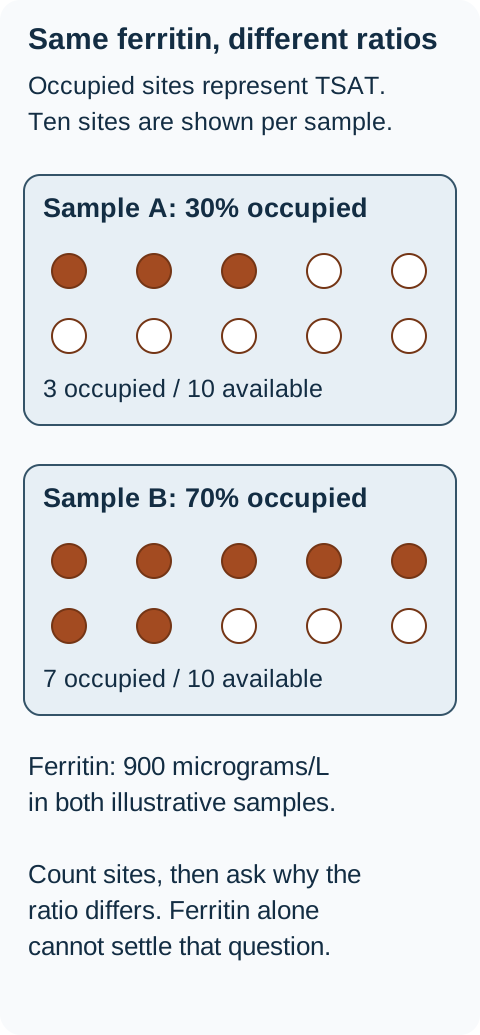
Start with a comparison: two adults have ferritin of 900 micrograms/L. One has pneumonia, a raised C-reactive protein and transferrin saturation of 30%. The other is well, has repeatedly measured saturation of 70%, and has no transfusion history. The same ferritin does not establish the same process. [1]
Ferritin answers a storage-related question, but it also responds to illness. Inflammation, infection, malignancy, alcohol-associated injury and metabolic liver disease can raise ferritin without the degree of iron excess its value might suggest. Transferrin saturation, abbreviated TSAT, describes the proportion of circulating iron-binding capacity occupied by iron. Neither result should be read in isolation. [1]
Compare before deciding. In the binding-capacity diagram, count the occupied sites. Which pattern deserves a persistent iron-loading evaluation rather than immediate blood loss based on ferritin alone?
The diagrams represent occupied binding capacity, not individual transferrin molecules or a diagnostic threshold. The ferritin is identical, but the plasma iron pattern differs. Interpret both measurements with the clinical context. [1] [1]
The repeated 70% result supports expanded circulating iron and deserves investigation. The acute illness and 30% result first require assessment of the inflammatory and liver context. They do not justify immediate phlebotomy, but a persistent unexplained abnormality still needs follow-up. [1]
Apply the distinction: a patient whose infection resolves but whose ferritin stays high and TSAT repeatedly reaches 66% now needs a different assessment. Repeated results and recovery from illness have changed the evidence. Morning sampling can help consistency; fasting is not required to improve diagnostic utility. [1]
There are three useful starting categories. HFE-related disease usually reflects inadequate restraint of absorption and iron recycling. Rare hereditary forms affect the same hormonal system or the exporter itself. Repeated transfusions deliver additional iron within donor red cells; ineffective erythropoiesis can also increase absorption even without regular transfusions. The history and blood count separate these categories before treatment is selected. [1][2]
Why does iron keep entering plasma?
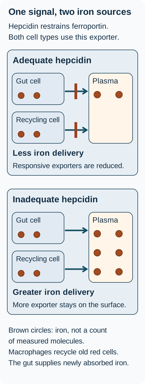
Iron absorption is only half the story. Most iron needed for new red cells is recycled from old red cells. How can one hormone regulate both routes?
Enterocytes absorb dietary iron, while macrophages recover iron from aging red cells. Both use ferroportin to export iron into plasma. Hepcidin, made by the liver, binds a responsive ferroportin protein and causes its internalization and degradation. That reduces iron export from both sources. HFE, hemojuvelin and transferrin receptor 2 help regulate hepcidin production; HAMP encodes hepcidin itself. [1][5]
Follow both routes. Less hepcidin restraint permits greater iron delivery from enterocytes and recycling macrophages. The model assumes responsive ferroportin; resistance and loss of transport function produce different outcomes in the adjacent static comparison table. [1] [5] [1][5]
With inadequately low hepcidin for the iron burden, more exporter remains active. Intestinal iron absorption and macrophage release expand the circulating iron pool. As transferrin becomes highly saturated, toxic non-transferrin-bound iron can accumulate and enter parenchymal cells. Iron-mediated oxidative injury contributes to organ dysfunction and liver fibrosis. [1][2]
Predict iron export in four conditions
For each condition below, predict whether iron remains in a cell or enters plasma. Then compare your prediction with the explanation. All four outcomes are visible together. This qualitative model holds cellular iron availability constant; it does not calculate patient laboratory values.
Hepcidin signal, exporter function and the expected result
Condition
Expected result and reason
ConditionAdequate hepcidin; responsive exporter
Expected result and reasonExport falls. Hepcidin binds ferroportin and promotes its internalization and degradation, leaving less surface exporter to deliver iron into plasma. [5]
ConditionInadequate hepcidin; responsive exporter
Expected result and reasonExport is less restrained. More ferroportin remains available, permitting greater delivery from enterocytes and recycling macrophages into plasma. [1][5]
ConditionAdequate hepcidin; resistant exporter
Expected result and reasonExport continues despite the signal when transport function is preserved. The defect is failure to respond to hepcidin, not failure to transport iron. This can support high TSAT and parenchymal loading. [1]
ConditionLoss of exporter function
Expected result and reasonIron is retained within cells because export is impaired. Classical ferroportin disease particularly affects macrophages and may include spleen loading, high ferritin and normal or low TSAT. [1]
Worked comparison: give equal hepcidin to two otherwise comparable cultures, one with responsive ferroportin and one with a hepcidin-resistant exporter that still transports iron. Export falls only in the responsive culture. Equal hormone exposure does not imply equal exporter activity. [1][5]
Transfer the prediction: now replace the resistant exporter with one that has lost transport function. Iron retention, rather than continued plasma delivery, becomes the expected result. These directions explain different patterns; they do not turn a single ferritin or TSAT into a genetic diagnosis. [1]
Try a second compartment. After predicting the enterocyte response, apply adequate hepcidin to a macrophage recovering iron from an old red cell. Export also falls because it uses the same protein. During inflammation this can restrict circulating iron despite substantial stored iron. [5]
Now separate two exporter disorders. A hepcidin-resistant SLC40A1 variant can permit continued export with high TSAT and parenchymal loading. A loss-of-function variant can instead retain iron in macrophages, producing a different distribution that often includes spleen iron and normal or low TSAT. Do not treat all SLC40A1 variants as the same defect. [1]
What confirms the diagnosis and liver risk?
Does a positive genetic result tell you how much tissue damage is present? No. Genotype, iron burden and fibrosis are separate questions. C282Y homozygosity is the most common HFE predisposition, especially in populations of northern European ancestry, but clinical penetrance varies. Normal ferritin and no organ injury do not justify intensive blood loss merely because the genotype is present. [1]
The EASL 2022 summary accepts biochemical overload in C282Y homozygotes at TSAT above 45% with ferritin above 200 micrograms/L in premenopausal women, or TSAT above 50% with ferritin above 300 micrograms/L in men and postmenopausal women. These are guideline-context thresholds, not a universal diagnosis from one isolated blood sample. Persistent unexplained high TSAT also deserves evaluation even before ferritin increases. [1]
Use the actual ratio. TSAT = serum iron divided by total iron-binding capacity, multiplied by 100, using matching units. Serum iron 120 micrograms/dL and binding capacity 300 micrograms/dL give 40%. If binding capacity falls to 150 with iron unchanged, the calculated saturation becomes 80%.
The second ratio is high because the denominator fell. Advanced liver disease, acute liver injury and low transferrin can distort interpretation. Compare the measured iron, transferrin or binding capacity, inflammatory setting and serial results before assigning hereditary overload. [1]
Choose the test for the question
Is the common hereditary pattern present? Use HFE testing with informed consent when the biochemical and family context warrants it. A C282Y homozygote with the matching iron phenotype can be diagnosed without routine biopsy. C282Y/H63D compound heterozygosity or H63D alone does not establish the cause of major iron loading. [1]
Is excess hepatic iron actually present? In a patient with high TSAT and ferritin who is not a C282Y homozygote, use validated liver MRI to document and quantify iron before attributing the pattern to a rare hereditary disorder. Quantitative R2* or other locally validated sequences answer a concentration question; an ordinary abdominal MRI description does not supply equivalent quantification. Review transfusions, hematologic disease, alcohol and metabolic liver disease. A young patient with severe unexplained tissue loading may then need specialist multigene evaluation, including HJV, HAMP, TFR2 and SLC40A1. [1]
Has fibrosis developed? Assess all diagnosed patients noninvasively at baseline, using the clinical examination, liver tests, platelets and suitable fibrosis assessment. Ferritin above 1,000 micrograms/L, abnormal transaminases, hepatomegaly or thrombocytopenia increase concern; none alone proves cirrhosis. A ferritin below 1,000 is reassuring only in the accompanying low-risk context. Noninvasive fibrosis thresholds have more limited validation in hemochromatosis than in several other liver diseases. [1]
When does biopsy help? Consider it when noninvasive findings leave clinically important fibrosis staging or a competing liver diagnosis unresolved. It is not the default test for hepatic iron concentration, and it adds little simply to reconfirm already established cirrhosis. [1]
Apply the distinction: persistent TSAT of 74%, negative common HFE testing and a normal hemoglobin do not by themselves establish a rare genetic diagnosis. MRI-confirmed hepatic iron, review of acquired causes, age and extrahepatic findings determine whether broader genetic evaluation is warranted. Hepcidin measurement is not a routine diagnostic requirement. [1]
Which organ finding changes the next assessment?
If liver iron improves, has the heart necessarily improved too? No. Assess the organ at risk, not just the serum trend. Different tissues accumulate and clear iron differently. In transfusion-dependent thalassemia, cardiac T2* predicts cardiac complications better than ferritin or liver iron alone in the cited cohort. [2][3]
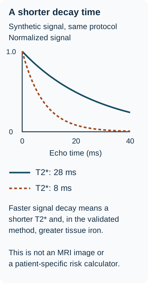
The signal-decay diagram uses synthetic curves to explain T2*: a shorter decay time corresponds to more iron in the validated cardiac method. Compare 28 ms with 8 ms, measured using the same protocol. The 8 ms curve falls faster. A fall in cardiac T2* despite a falling ferritin is not proof of global recovery. Exact thresholds depend on acquisition and validation; values from thalassemia must not be assumed to define identical risk in every HFE patient. [3]
The curves use exp(-echo time/T2*) solely to illustrate decay direction. They are not measurements or a calibration equation for iron concentration. Cardiac T2* risk evidence cited here comes from thalassemia; use validated acquisition and clinical interpretation. [3] [3]
Connect the complaint to the organ question
Finding
Relationship to test
FindingHepatomegaly or abnormal liver tests
Relationship to testQuantify iron when indicated, then separately assess fibrosis and other liver disease. [1]
FindingDiabetes
Relationship to testPancreatic injury can impair insulin secretion; metabolic factors can coexist. Assess and treat glucose abnormalities rather than assuming depletion will reverse established diabetes. [1]
FindingLow libido, amenorrhea or low testosterone
Relationship to testPituitary iron can reduce gonadotropin output. Low sex hormone with inappropriately low or normal LH and FSH supports a central rather than primary gonadal pattern. [1]
FindingPalpitations, syncope or ventricular dysfunction
Relationship to testAssess rhythm and cardiac function, and obtain cardiac iron MRI when indicated. Severe or juvenile disease warrants a low threshold for cardiac assessment. [1][3]
FindingSecond and third MCP pain, ankle disease or pigmentation
Relationship to testThese findings can prompt iron testing, but they are not a diagnosis alone. Arthropathy can resemble osteoarthritis, include chondrocalcinosis and persist after depletion. [1]
Predict before checking. A young person has severe tissue loading and new conduction disease. Would a moderately improved ferritin justify waiting for genetic sequencing before assessing the heart? No. Cardiac investigation and appropriate treatment must proceed without waiting for that result. Genetic classification and urgent organ care answer different questions. [1]
Early hereditary parenchymal liver loading tends to involve periportal hepatocytes, with little spleen iron. Transfusional overload and classical ferroportin retention often include the reticuloendothelial compartment and spleen. Distribution helps refine the differential; mixed and advanced patterns require context rather than an absolute organ rule. [1]
Can this patient safely lose red cells?
Two people have documented iron excess. One has hemoglobin of 15 g/dL and HFE-related disease; the other has hemoglobin of 8 g/dL and requires regular transfusions. Why does the same goal require a different method?
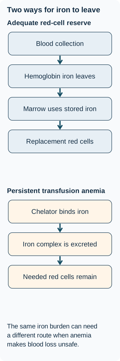
Phlebotomy exports iron in hemoglobin. The marrow then uses stored iron to replace the lost red cells. It is first-line treatment for established hereditary overload when the blood count, circulation and vascular access permit. Induction commonly uses 400 to 500 mL weekly or every two weeks, adjusted to size and tolerance. These are supervised treatment examples, not a self-treatment schedule. [1]
Trace the two exits in the diagram. For the patient with an adequate blood count, follow stored iron into new red cells after a blood collection. For the transfusion-dependent patient, identify the route that exports iron without sacrificing needed red cells.
These are conceptual routes, not equivalent regimens. Phlebotomy suits appropriate hereditary loading with adequate red-cell reserve. Persistent transfusion-dependent anemia generally requires an individualized chelation strategy. The route of iron-complex excretion depends on the drug. [1] [2] [1][2]
Chelation binds iron for excretion while preserving needed red cells. In persistent transfusion-dependent anemia, further blood loss worsens the very deficit requiring treatment. Chelator selection, dose and monitoring belong to the disease-specific specialist plan. Ferritin trends must be interpreted with liver and cardiac iron measurements where indicated. [1][2]
Use a target without overshooting it
The detailed EASL 2022 recommendation targets ferritin around 50 micrograms/L during induction, not below it to induce deficiency. Maintenance generally aims for 50 to 100 micrograms/L with individualized flexibility and follow-up. The guideline abstract uses a less-than sign, but the full recommendation explicitly warns against going below 50. [1]
Check hemoglobin before each collection. Under this EASL approach, hemoglobin below 12 g/dL prompts reduced frequency and consideration of a smaller volume; below 11 g/dL prompts a pause and reassessment. Symptoms, hemodynamic tolerance and comorbidity can require an earlier adjustment. Unexpected anemia needs investigation, not an assumption that it is simply part of hemochromatosis. [1]
During induction, check ferritin about monthly or after four collections, then every one or two sessions once it is below 200 micrograms/L to avoid overshooting. Maintenance frequency depends on reaccumulation and tolerance, not a fixed calendar for everyone. TSAT can remain high when ferritin is already at target. There is no established universal TSAT treatment target that justifies causing iron deficiency to normalize the percentage. [1]
When blood loss is unsuitable
Deferasirox, deferoxamine and deferiprone are chelators used in selected iron-loading disorders. They are not interchangeable: renal, hepatic, hematologic and other safety monitoring depends on the agent, formulation and patient. No drug-specific dosing schedule is supplied here. In HFE disease, chelation is a specialist second-line option when phlebotomy is infeasible or hazardous, including selected severe cardiac presentations; erythrocytapheresis is another option in suitable patients and centers. It selectively collects red cells and returns other blood components, potentially reducing hemodynamic changes; citrate-related effects and individual tolerance still matter. [1][2]
Apply the distinction: ferritin falls from 780 to 46 micrograms/L, but TSAT remains 72% and the patient develops fatigue. More frequent blood collections to correct the percentage can cause harm. Reassess blood count, iron status and symptoms instead. In a transfused patient, do not apply HFE phlebotomy ferritin targets to a chelation regimen. [1][2]
What remains after iron depletion?
Ferritin has reached 70 micrograms/L. Is the whole problem finished? Distinguish control of iron exposure from care of established organ damage. Some liver and cardiac changes can improve; established arthropathy, endocrine injury and cancer risk may persist. [1]
A patient with hemochromatosis and cirrhosis who remains eligible for cancer treatment or transplantation should continue hepatocellular carcinoma surveillance after iron depletion. EASL recommends a six-month interval for cirrhosis regardless of depletion. The AASLD cirrhosis pathway uses ultrasound plus alpha-fetoprotein every six months; AFP alone does not replace imaging. Suboptimal ultrasound may require an alternative imaging strategy. [1][4]
For noncirrhotic advanced bridging fibrosis, EASL's suggestion to consider six-month surveillance is weaker and depends on individual assessment. Do not describe F3 and established cirrhosis as having identical certainty of evidence. Previously advanced fibrosis or cirrhosis still matters after apparent regression; EASL advises continuing surveillance, with individualized intervals after regression. [1]
Compare two follow-up plans. One patient has uncomplicated biochemical HFE overload without advanced fibrosis; another had biopsy-confirmed cirrhosis before depletion. Both now have ferritin of 70. What history prevents their follow-up plans from being identical?
The prior cirrhosis keeps the second patient in cancer surveillance and ongoing liver care. Ferritin alone does not assign or cancel that program. Persistent MCP pain also needs its own assessment and treatment rather than an automatic increase in phlebotomy. [1][4]
Prevent the next avoidable problem
Offer adult first-degree relatives appropriate genetic counseling, targeted HFE testing and iron studies when the index diagnosis is established. A sibling's normal examination is not enough to exclude biochemical disease. Routine predictive testing for typical adult-onset HFE disease is generally an adult decision; suspected symptomatic juvenile disease is a different clinical situation. [1]
Use a balanced diet. Avoid unprescribed iron supplements and supplemental high-dose vitamin C; do not eliminate fruit and vegetables or substitute a restrictive diet for indicated iron depletion. Limit red meat and avoid heavy alcohol intake; cirrhosis calls for abstinence. Avoid raw or undercooked shellfish and protect wounds from seawater because iron overload increases susceptibility to severe Vibrio vulnificus infection. [1]
Pregnancy requires an individualized plan. In mild to moderate overload without advanced liver disease, phlebotomy can often be paused during pregnancy, while avoiding iron deficiency and considering any extrahepatic disease. This is not a blanket pause for severe organ-threatening overload. [1]
Return to the central question: determine what the iron tests actually establish, identify how iron entered, assess organ injury, choose a tolerable iron-exit strategy, and retain follow-up for risks that depletion does not erase. [1]
Apply the reasoning to a new patient
Decide what the findings establish before choosing an action. The explanations are available for every option; none is required to continue reading.
Case 1
Show answer and explanations for case 1
A. Obtain broad hereditary iron-disorder sequencing during this admission (Why this does not fit)
Genetic evaluation can identify unexplained hereditary iron-loading disorders. This patient lacks persistent biochemical or tissue evidence of hereditary loading. Establish the phenotype before broad sequencing. [1]
Reasoning steps for option A
What clinical principle makes "Obtain broad hereditary iron-disorder sequencing during this admission" a plausible option in this case?
Genetic evaluation can identify unexplained hereditary iron-loading disorders.
Which patient finding most directly decides whether "Obtain broad hereditary iron-disorder sequencing during this admission" fits this case?
This patient lacks persistent biochemical or tissue evidence of hereditary loading.
After applying that finding, why should "Obtain broad hereditary iron-disorder sequencing during this admission" be accepted or rejected here?
Establish the phenotype before broad sequencing.
B. Begin weekly therapeutic phlebotomy after her respiratory symptoms improve (Why this does not fit)
Phlebotomy is effective when excess body iron has been established and blood loss is safe. A four-digit ferritin during acute inflammation is insufficient evidence of that excess. Clinical recovery alone does not create an indication for blood loss. [1]
Reasoning steps for option B
What clinical principle makes "Begin weekly therapeutic phlebotomy after her respiratory symptoms improve" a plausible option in this case?
Phlebotomy is effective when excess body iron has been established and blood loss is safe.
Which patient finding most directly decides whether "Begin weekly therapeutic phlebotomy after her respiratory symptoms improve" fits this case?
A four-digit ferritin during acute inflammation is insufficient evidence of that excess.
After applying that finding, why should "Begin weekly therapeutic phlebotomy after her respiratory symptoms improve" be accepted or rejected here?
Clinical recovery alone does not create an indication for blood loss.
C. Repeat iron studies after the inflammatory illness has resolved (Best answer)
Ferritin can rise sharply during infection without comparable iron accumulation. The rapid change, high inflammatory marker and TSAT of 23% favor an acute response. Treat the current illness and reassess; a persistent unexplained abnormality still requires evaluation. [1]
Reasoning steps for option C
What changed when ferritin increased?
A new acute infection and inflammatory response were present.
Does TSAT of 23% demonstrate an expanded plasma iron pool?
No.
What should be established before considering depletion?
Whether an abnormal iron phenotype persists after recovery.
D. Arrange liver biopsy to quantify iron before hospital discharge (Why this does not fit)
Biopsy can clarify selected unresolved liver diagnoses or fibrosis stage. There is no persistent liver abnormality or established loading phenotype here. Use follow-up and noninvasive evaluation before an invasive test without a defined question. [1]
Reasoning steps for option D
What clinical principle makes "Arrange liver biopsy to quantify iron before hospital discharge" a plausible option in this case?
Biopsy can clarify selected unresolved liver diagnoses or fibrosis stage.
Which patient finding most directly decides whether "Arrange liver biopsy to quantify iron before hospital discharge" fits this case?
There is no persistent liver abnormality or established loading phenotype here.
After applying that finding, why should "Arrange liver biopsy to quantify iron before hospital discharge" be accepted or rejected here?
Use follow-up and noninvasive evaluation before an invasive test without a defined question.
Takeaway: A large ferritin change during inflammation requires context and follow-up, not automatic iron depletion.
A. Begin phlebotomy with hemoglobin and ferritin monitoring (Best answer)
Phlebotomy exports iron in hemoglobin and uses stored iron for replacement erythropoiesis. This patient has established biochemical loading and can tolerate blood collection. Treat before organ damage develops, with monitoring to avoid deficiency. [1]
Reasoning steps for option A
What establishes biochemical expression in this case?
Repeated high saturation and ferritin with C282Y homozygosity.
What supports safe red-cell collection?
Adequate hemoglobin, stable circulation and usable venous access.
Which treatment is first-line in that setting?
Supervised phlebotomy with monitoring.
B. Start oral chelation with renal and hepatic monitoring (Why this does not fit)
Chelation can reduce iron when phlebotomy is unsuitable. No reason that makes first-line blood collection infeasible is supplied. Do not substitute a more complex second-line strategy without a patient-specific reason. [1]
Reasoning steps for option B
What clinical principle makes "Start oral chelation with renal and hepatic monitoring" a plausible option in this case?
Chelation can reduce iron when phlebotomy is unsuitable.
Which patient finding most directly decides whether "Start oral chelation with renal and hepatic monitoring" fits this case?
No reason that makes first-line blood collection infeasible is supplied.
After applying that finding, why should "Start oral chelation with renal and hepatic monitoring" be accepted or rejected here?
Do not substitute a more complex second-line strategy without a patient-specific reason.
C. Monitor ferritin annually until clinical liver disease is apparent (Why this does not fit)
Observation is appropriate for some people with a risk genotype but no loading phenotype. Ferritin and repeated TSAT already show biochemical loading in this C282Y homozygote. Incomplete penetrance does not justify waiting for injury once overload is expressed. [1]
Reasoning steps for option C
What clinical principle makes "Monitor ferritin annually until clinical liver disease is apparent" a plausible option in this case?
Observation is appropriate for some people with a risk genotype but no loading phenotype.
Which patient finding most directly decides whether "Monitor ferritin annually until clinical liver disease is apparent" fits this case?
Ferritin and repeated TSAT already show biochemical loading in this C282Y homozygote.
After applying that finding, why should "Monitor ferritin annually until clinical liver disease is apparent" be accepted or rejected here?
Incomplete penetrance does not justify waiting for injury once overload is expressed.
D. Use dietary iron restriction with repeat studies in six months (Why this does not fit)
Dietary changes can reduce additional iron intake. They do not adequately treat the established burden described here. Counseling complements indicated depletion rather than replacing it. [1]
Reasoning steps for option D
What clinical principle makes "Use dietary iron restriction with repeat studies in six months" a plausible option in this case?
Dietary changes can reduce additional iron intake.
Which patient finding most directly decides whether "Use dietary iron restriction with repeat studies in six months" fits this case?
They do not adequately treat the established burden described here.
After applying that finding, why should "Use dietary iron restriction with repeat studies in six months" be accepted or rejected here?
Counseling complements indicated depletion rather than replacing it.
Takeaway: Treat the expressed phenotype while hemoglobin and tolerance determine whether phlebotomy is suitable.
A. Normal cells export more iron after hepcidin; variant cells retain more iron (Why this does not fit)
An exporter defect can cause cellular retention when its transport function is impaired. The supplied defect is resistance to hepcidin binding, not impaired iron transport. Do not equate hormone resistance with loss of exporter function. [1] [5]
Reasoning steps for option A
What clinical principle makes "Normal cells export more iron after hepcidin; variant cells retain more iron" a plausible option in this case?
An exporter defect can cause cellular retention when its transport function is impaired.
Which patient finding most directly decides whether "Normal cells export more iron after hepcidin; variant cells retain more iron" fits this case?
The supplied defect is resistance to hepcidin binding, not impaired iron transport.
After applying that finding, why should "Normal cells export more iron after hepcidin; variant cells retain more iron" be accepted or rejected here?
Do not equate hormone resistance with loss of exporter function.
B. Both cell cultures retain more iron after hepcidin exposure (Why this does not fit)
Responsive exporters reduce iron release after hepcidin binding. Only one culture has an exporter able to bind hepcidin. The same hormone concentration need not produce the same response. [1] [5]
Reasoning steps for option B
What clinical principle makes "Both cell cultures retain more iron after hepcidin exposure" a plausible option in this case?
Responsive exporters reduce iron release after hepcidin binding.
Which patient finding most directly decides whether "Both cell cultures retain more iron after hepcidin exposure" fits this case?
Only one culture has an exporter able to bind hepcidin.
After applying that finding, why should "Both cell cultures retain more iron after hepcidin exposure" be accepted or rejected here?
The same hormone concentration need not produce the same response.
C. Both cell cultures export more iron after hepcidin exposure (Why this does not fit)
High intracellular iron provides substrate for cellular export. Availability is held constant, while hepcidin specifically suppresses the normal exporter. Account for the altered regulatory signal rather than substrate alone. [1] [5]
Reasoning steps for option C
What clinical principle makes "Both cell cultures export more iron after hepcidin exposure" a plausible option in this case?
High intracellular iron provides substrate for cellular export.
Which patient finding most directly decides whether "Both cell cultures export more iron after hepcidin exposure" fits this case?
Availability is held constant, while hepcidin specifically suppresses the normal exporter.
After applying that finding, why should "Both cell cultures export more iron after hepcidin exposure" be accepted or rejected here?
Account for the altered regulatory signal rather than substrate alone.
D. Normal cells retain more iron; variant cells maintain iron export (Best answer)
Hepcidin reduces normal ferroportin at the cell surface. The variant cannot bind the hormone, so that restraint is lost despite equal exposure. Predict intracellular retention and plasma delivery from exporter responsiveness. [1] [5]
Reasoning steps for option D
What does hepcidin binding do to normal surface ferroportin?
It causes internalization and degradation.
What happens to export from those cells?
Export decreases and more iron stays inside.
Why does the variant culture differ?
Its exporter does not bind the hormone and continues transporting iron.
A. Obtain serum hepcidin using the local laboratory reference interval (Why this does not fit)
Hepcidin is central to iron homeostasis. Its measurement is not the recommended routine test to establish tissue overload. A mechanistically important hormone is not necessarily a clinically standardized diagnostic test. [1]
Reasoning steps for option A
What clinical principle makes "Obtain serum hepcidin using the local laboratory reference interval" a plausible option in this case?
Hepcidin is central to iron homeostasis.
Which patient finding most directly decides whether "Obtain serum hepcidin using the local laboratory reference interval" fits this case?
Its measurement is not the recommended routine test to establish tissue overload.
After applying that finding, why should "Obtain serum hepcidin using the local laboratory reference interval" be accepted or rejected here?
A mechanistically important hormone is not necessarily a clinically standardized diagnostic test.
B. Obtain validated quantitative MRI of hepatic iron concentration (Best answer)
MRI can noninvasively establish and quantify hepatic iron. Biochemical suspicion remains unconfirmed at the tissue level in this non-C282Y patient. Use the tissue phenotype and acquired-cause assessment to guide further genetic evaluation. [1]
Reasoning steps for option B
What has the repeated blood panel established?
Persistent biochemical suspicion of iron loading.
What evidence is missing in the non-C282Y patient?
Direct evidence of increased hepatic iron.
Which noninvasive study answers that question?
Validated quantitative liver iron MRI.
C. Measure liver stiffness with transient elastography and calculate FIB-4 (Why this does not fit)
Fibrosis assessment helps determine liver-risk stage. The missing evidence here is hepatic iron concentration, not a stiffness or fibrosis estimate. Use validated quantitative iron MRI to answer the tissue-loading question. [1]
Reasoning steps for option C
What clinical principle makes "Measure liver stiffness with transient elastography and calculate FIB-4" a plausible option in this case?
Which patient finding most directly decides whether "Measure liver stiffness with transient elastography and calculate FIB-4" fits this case?
The missing evidence here is hepatic iron concentration, not a stiffness or fibrosis estimate.
After applying that finding, why should "Measure liver stiffness with transient elastography and calculate FIB-4" be accepted or rejected here?
Use validated quantitative iron MRI to answer the tissue-loading question.
D. Obtain liver biopsy as the initial iron-quantification study (Why this does not fit)
Biopsy can identify iron and clarify selected liver diagnoses. Validated noninvasive MRI can answer the concentration question without routine biopsy. Reserve biopsy for unresolved staging or competing pathology when appropriate. [1]
Reasoning steps for option D
What clinical principle makes "Obtain liver biopsy as the initial iron-quantification study" a plausible option in this case?
Biopsy can identify iron and clarify selected liver diagnoses.
Which patient finding most directly decides whether "Obtain liver biopsy as the initial iron-quantification study" fits this case?
Validated noninvasive MRI can answer the concentration question without routine biopsy.
After applying that finding, why should "Obtain liver biopsy as the initial iron-quantification study" be accepted or rejected here?
Reserve biopsy for unresolved staging or competing pathology when appropriate.
Takeaway: In a non-C282Y patient, confirm hepatic iron rather than assigning a rare hereditary label from blood tests alone.
A. Saturation rises to 80% because circulating iron increases; diagnose hereditary loading (Why this does not fit)
More serum iron can raise the saturation ratio. Serum iron is unchanged here, and the ratio alone does not establish hereditary causation. Inspect the measured numerator and denominator before attributing a high percentage to excess iron input. [1]
Reasoning steps for option A
What clinical principle makes "Saturation rises to 80% because circulating iron increases; diagnose hereditary loading" a plausible option in this case?
More serum iron can raise the saturation ratio.
Which patient finding most directly decides whether "Saturation rises to 80% because circulating iron increases; diagnose hereditary loading" fits this case?
Serum iron is unchanged here, and the ratio alone does not establish hereditary causation.
After applying that finding, why should "Saturation rises to 80% because circulating iron increases; diagnose hereditary loading" be accepted or rejected here?
Inspect the measured numerator and denominator before attributing a high percentage to excess iron input.
B. Saturation falls to 20% because binding capacity decreases; exclude iron loading (Why this does not fit)
Binding capacity affects the calculated saturation. Halving the denominator doubles the ratio rather than halving it. Calculate the ratio correctly and do not use the erroneous percentage to exclude disease. [1]
Reasoning steps for option B
What clinical principle makes "Saturation falls to 20% because binding capacity decreases; exclude iron loading" a plausible option in this case?
Binding capacity affects the calculated saturation.
Which patient finding most directly decides whether "Saturation falls to 20% because binding capacity decreases; exclude iron loading" fits this case?
Halving the denominator doubles the ratio rather than halving it.
After applying that finding, why should "Saturation falls to 20% because binding capacity decreases; exclude iron loading" be accepted or rejected here?
Calculate the ratio correctly and do not use the erroneous percentage to exclude disease.
C. Saturation rises to 80% because binding capacity decreases; reassess after recovery (Best answer)
TSAT is serum iron divided by binding capacity, multiplied by 100. The result increases from 40% to 80% solely because the measured binding capacity halves. Interpret this liver-injury context and repeat the relevant assessment rather than diagnosing hereditary overload from the ratio. [1]
Reasoning steps for option C
What was the earlier saturation?
120 divided by 300, multiplied by 100, equals 40%.
What is the current saturation and which quantity changed?
120 divided by 150 gives 80%; the binding capacity, not serum iron, changed.
Does that change establish a hereditary iron disorder?
No. Liver injury and reduced binding capacity confound the interpretation, so the phenotype needs reassessment.
D. Saturation remains at 40% because serum iron is unchanged; defer further assessment (Why this does not fit)
An unchanged serum iron leaves the numerator stable. The denominator changed, so the percentage cannot be assumed to remain stable. Both the calculation and any persistent unexplained iron abnormality still need assessment. [1]
Reasoning steps for option D
What clinical principle makes "Saturation remains at 40% because serum iron is unchanged; defer further assessment" a plausible option in this case?
An unchanged serum iron leaves the numerator stable.
Which patient finding most directly decides whether "Saturation remains at 40% because serum iron is unchanged; defer further assessment" fits this case?
The denominator changed, so the percentage cannot be assumed to remain stable.
After applying that finding, why should "Saturation remains at 40% because serum iron is unchanged; defer further assessment" be accepted or rejected here?
Both the calculation and any persistent unexplained iron abnormality still need assessment.
Takeaway: A high saturation ratio can reflect low binding capacity; calculation and clinical interpretation are separate steps.
A. Start weekly blood collections until saturation is below 45% (Why this does not fit)
Blood collection reduces body iron when excess stores are present. The storage measure is normal, and TSAT is not a universal depletion target. Do not create deficiency while trying to normalize saturation. [1]
Reasoning steps for option A
What clinical principle makes "Start weekly blood collections until saturation is below 45%" a plausible option in this case?
Blood collection reduces body iron when excess stores are present.
Which patient finding most directly decides whether "Start weekly blood collections until saturation is below 45%" fits this case?
The storage measure is normal, and TSAT is not a universal depletion target.
After applying that finding, why should "Start weekly blood collections until saturation is below 45%" be accepted or rejected here?
Do not create deficiency while trying to normalize saturation.
B. Arrange periodic iron studies and counsel about future risk (Best answer)
A risk genotype can be present before appreciable storage excess develops. Ferritin is normal and there is no organ injury, so induction depletion is not established as necessary. Follow the phenotype over time instead of treating the genotype alone. [1]
Reasoning steps for option B
What does C282Y homozygosity establish?
Inherited susceptibility.
What does normal ferritin argue against now?
An established substantial storage burden requiring induction depletion.
What remains appropriate?
Counseling and periodic assessment of the iron phenotype.
C. Obtain liver biopsy before deciding whether follow-up is needed (Why this does not fit)
Biopsy can clarify important unresolved fibrosis questions. Normal liver assessment and no evidence of storage excess provide no such question here. Monitoring does not require a preliminary invasive test. [1]
Reasoning steps for option C
What clinical principle makes "Obtain liver biopsy before deciding whether follow-up is needed" a plausible option in this case?
Biopsy can clarify important unresolved fibrosis questions.
Which patient finding most directly decides whether "Obtain liver biopsy before deciding whether follow-up is needed" fits this case?
Normal liver assessment and no evidence of storage excess provide no such question here.
After applying that finding, why should "Obtain liver biopsy before deciding whether follow-up is needed" be accepted or rejected here?
Monitoring does not require a preliminary invasive test.
D. End iron-related follow-up because ferritin is currently normal (Why this does not fit)
A normal ferritin argues against current substantial storage loading. It does not eliminate the future risk associated with this genotype and family history. Distinguish present status from longitudinal risk. [1]
Reasoning steps for option D
What clinical principle makes "End iron-related follow-up because ferritin is currently normal" a plausible option in this case?
A normal ferritin argues against current substantial storage loading.
Which patient finding most directly decides whether "End iron-related follow-up because ferritin is currently normal" fits this case?
It does not eliminate the future risk associated with this genotype and family history.
After applying that finding, why should "End iron-related follow-up because ferritin is currently normal" be accepted or rejected here?
Distinguish present status from longitudinal risk.
Takeaway: Normal ferritin can change treatment today without eliminating the need for future assessment.
A. Request a specialist hereditary iron-disorder gene panel (Best answer)
Rare hereditary defects can produce severe early cardiac and endocrine disease. The age, demonstrated tissue iron and absence of a secondary loading history warrant broader evaluation. Genetic clarification should proceed alongside, not ahead of, needed organ treatment. [1]
Reasoning steps for option A
What makes this presentation atypical for common adult-onset disease?
Severe cardiac and endocrine features with marked loading at age 21.
Does a negative common HFE panel exclude all hereditary loading?
No.
What etiologic investigation fits the established phenotype?
Specialist sequencing of additional iron-regulatory genes.
B. Sequence an inherited cardiomyopathy panel before expanding iron-gene testing (Why this does not fit)
Inherited cardiomyopathies can present with early ventricular dysfunction. This patient also has demonstrated hepatic iron loading and endocrine findings without a secondary loading history. Prioritize an iron-disorder panel that can explain the multisystem phenotype while cardiac care continues. [1]
Reasoning steps for option B
What clinical principle makes "Sequence an inherited cardiomyopathy panel before expanding iron-gene testing" a plausible option in this case?
Inherited cardiomyopathies can present with early ventricular dysfunction.
Which patient finding most directly decides whether "Sequence an inherited cardiomyopathy panel before expanding iron-gene testing" fits this case?
This patient also has demonstrated hepatic iron loading and endocrine findings without a secondary loading history.
After applying that finding, why should "Sequence an inherited cardiomyopathy panel before expanding iron-gene testing" be accepted or rejected here?
Prioritize an iron-disorder panel that can explain the multisystem phenotype while cardiac care continues.
C. Investigate marrow sideroblasts as the first etiologic priority (Why this does not fit)
Ineffective erythropoiesis can cause secondary iron loading. The supplied history and blood count do not identify a chronic erythroid disorder. Use the hematologic phenotype to decide whether marrow investigation is the priority. [1]
Reasoning steps for option C
What clinical principle makes "Investigate marrow sideroblasts as the first etiologic priority" a plausible option in this case?
Ineffective erythropoiesis can cause secondary iron loading.
Which patient finding most directly decides whether "Investigate marrow sideroblasts as the first etiologic priority" fits this case?
The supplied history and blood count do not identify a chronic erythroid disorder.
After applying that finding, why should "Investigate marrow sideroblasts as the first etiologic priority" be accepted or rejected here?
Use the hematologic phenotype to decide whether marrow investigation is the priority.
D. Quantify hepcidin before expanding the genetic investigation (Why this does not fit)
Hepcidin dysregulation connects several hereditary iron disorders. The strong tissue phenotype already justifies specialist sequencing, while hepcidin measurement is not routinely recommended for this decision. Do not require an unstandardized intermediary test before warranted etiologic evaluation. [1]
Reasoning steps for option D
What physiological or clinical principle supports this choice?
Hepcidin dysregulation connects several hereditary iron disorders.
Which patient finding most directly decides whether "Quantify hepcidin before expanding the genetic investigation" fits this case?
The strong tissue phenotype already justifies specialist sequencing, while hepcidin measurement is not routinely recommended for this decision.
What follows for this decision?
Do not require an unstandardized intermediary test before warranted etiologic evaluation.
Takeaway: A negative common HFE test does not exclude a severe hereditary iron disorder.
A. Resistance to hepcidin with persistent export into the circulation (Why this does not fit)
Some SLC40A1 variants resist hepcidin and sustain iron export. That physiology more often expands plasma iron and produces high saturation with parenchymal loading. Distinguish resistance to regulation from reduced transport function. [1]
Reasoning steps for option A
What clinical principle makes "Resistance to hepcidin with persistent export into the circulation" a plausible option in this case?
Some SLC40A1 variants resist hepcidin and sustain iron export.
Which patient finding most directly decides whether "Resistance to hepcidin with persistent export into the circulation" fits this case?
That physiology more often expands plasma iron and produces high saturation with parenchymal loading.
After applying that finding, why should "Resistance to hepcidin with persistent export into the circulation" be accepted or rejected here?
Distinguish resistance to regulation from reduced transport function.
B. Reduced hepcidin synthesis with excessive intestinal iron absorption (Why this does not fit)
Insufficient hepcidin can increase absorption and macrophage iron release. Persistently low-normal saturation and substantial spleen loading favor a retention pattern instead. Use the iron distribution and saturation together to identify the physiological defect. [1]
Reasoning steps for option B
What clinical principle makes "Reduced hepcidin synthesis with excessive intestinal iron absorption" a plausible option in this case?
Insufficient hepcidin can increase absorption and macrophage iron release.
Which patient finding most directly decides whether "Reduced hepcidin synthesis with excessive intestinal iron absorption" fits this case?
Persistently low-normal saturation and substantial spleen loading favor a retention pattern instead.
After applying that finding, why should "Reduced hepcidin synthesis with excessive intestinal iron absorption" be accepted or rejected here?
Use the iron distribution and saturation together to identify the physiological defect.
C. Increased hepcidin production driven by chronic inflammatory signaling (Why this does not fit)
Inflammatory hepcidin can restrict circulating iron while ferritin rises. Normal inflammatory markers, familial organ iron loading and a pathogenic exporter variant support a hereditary retention disorder instead. A plausible laboratory pattern must also explain the family history and tissue findings. [1]
Reasoning steps for option C
What physiological or clinical principle supports this choice?
Inflammatory hepcidin can restrict circulating iron while ferritin rises.
Which patient finding most directly decides whether "Increased hepcidin production driven by chronic inflammatory signaling" fits this case?
Normal inflammatory markers, familial organ iron loading and a pathogenic exporter variant support a hereditary retention disorder instead.
What follows for this decision?
A plausible laboratory pattern must also explain the family history and tissue findings.
D. Reduced iron export with retention in reticuloendothelial cells (Best answer)
Loss of ferroportin function can retain iron in macrophages and produce high ferritin with lower saturation. The spleen involvement, TSAT of 28% and family treatment intolerance fit that pattern. Do not equate all SLC40A1 disorders with high-saturation hemochromatosis. [1]
Reasoning steps for option D
Which findings suggest intracellular retention rather than a large plasma pool?
Spleen loading with high ferritin and TSAT of 28%.
What transporter effect produces that direction?
Reduced cellular iron export.
How does hepcidin resistance differ?
It sustains export rather than preventing it.
Takeaway: Classical ferroportin retention and hepcidin-resistant ferroportin are different physiological disorders.
A. Increased intestinal absorption associated with ineffective erythropoiesis (Best answer)
Ineffective erythropoiesis can suppress the normal restraint on dietary iron absorption. Progressive loading without continuing transfusions identifies an ongoing nontransfusional source. Anemia does not exclude excess body iron. [1] [2]
Reasoning steps for option A
Are ongoing donor cells supplying the continuing new iron?
No. The last transfusion was many years earlier.
What can ineffective erythropoiesis change?
It can increase dietary iron absorption.
Does red-cell recycling itself add new iron to the body?
No. It redistributes existing iron.
B. Delayed breakdown of donor cells from the remote transfusions (Why this does not fit)
Donor red cells add iron, which can persist after the cells are cleared. Old transfusions cannot keep introducing new iron many years later. Separate a prior contribution to stores from a continuing source of net iron gain. [1] [2]
Reasoning steps for option B
What clinical principle makes "Delayed breakdown of donor cells from the remote transfusions" a plausible option in this case?
Donor red cells add iron, which can persist after the cells are cleared.
Which patient finding most directly decides whether "Delayed breakdown of donor cells from the remote transfusions" fits this case?
Old transfusions cannot keep introducing new iron many years later.
After applying that finding, why should "Delayed breakdown of donor cells from the remote transfusions" be accepted or rejected here?
Separate a prior contribution to stores from a continuing source of net iron gain.
C. Redistribution of existing iron caused by increased red-cell destruction in tissues (Why this does not fit)
Red-cell destruction returns existing iron to storage and plasma pools. Recycling alone does not create the continuing external input responsible for progressive total loading. Distinguish internal recycling from new iron entering the body. [1] [2]
Reasoning steps for option C
What clinical principle makes "Redistribution of existing iron caused by increased red-cell destruction in tissues" a plausible option in this case?
Red-cell destruction returns existing iron to storage and plasma pools.
Which patient finding most directly decides whether "Redistribution of existing iron caused by increased red-cell destruction in tissues" fits this case?
Recycling alone does not create the continuing external input responsible for progressive total loading.
After applying that finding, why should "Redistribution of existing iron caused by increased red-cell destruction in tissues" be accepted or rejected here?
Distinguish internal recycling from new iron entering the body.
D. Reduced urinary iron clearance associated with the low hemoglobin (Why this does not fit)
Reduced clearance is important for substances normally regulated through renal excretion. Body iron balance is not normally controlled by a large adjustable urinary excretion pathway. Consider increased input rather than treating iron as a routinely renally cleared solute. [1] [2]
Reasoning steps for option D
What clinical principle makes "Reduced urinary iron clearance associated with the low hemoglobin" a plausible option in this case?
Reduced clearance is important for substances normally regulated through renal excretion.
Which patient finding most directly decides whether "Reduced urinary iron clearance associated with the low hemoglobin" fits this case?
Body iron balance is not normally controlled by a large adjustable urinary excretion pathway.
After applying that finding, why should "Reduced urinary iron clearance associated with the low hemoglobin" be accepted or rejected here?
Consider increased input rather than treating iron as a routinely renally cleared solute.
Takeaway: Ineffective erythropoiesis can produce overload even without regular transfusions.
A. Begin cancer surveillance and classify the liver as cirrhotic (Why this does not fit)
Established cirrhosis would justify surveillance in an eligible patient. The supplied findings increase suspicion but do not establish cirrhosis. First resolve stage rather than relabeling a risk marker as a diagnosis. [1]
Reasoning steps for option A
What clinical principle makes "Begin cancer surveillance and classify the liver as cirrhotic" a plausible option in this case?
Established cirrhosis would justify surveillance in an eligible patient.
Which patient finding most directly decides whether "Begin cancer surveillance and classify the liver as cirrhotic" fits this case?
The supplied findings increase suspicion but do not establish cirrhosis.
After applying that finding, why should "Begin cancer surveillance and classify the liver as cirrhotic" be accepted or rejected here?
First resolve stage rather than relabeling a risk marker as a diagnosis.
B. Begin iron depletion and omit staging until symptoms develop (Why this does not fit)
Phlebotomy is indicated when biochemical HFE overload is established. Advanced fibrosis can be asymptomatic, and staging changes follow-up. Treatment of iron burden does not replace baseline assessment of structural injury. [1]
Reasoning steps for option B
What clinical principle makes "Begin iron depletion and omit staging until symptoms develop" a plausible option in this case?
Phlebotomy is indicated when biochemical HFE overload is established.
Which patient finding most directly decides whether "Begin iron depletion and omit staging until symptoms develop" fits this case?
Advanced fibrosis can be asymptomatic, and staging changes follow-up.
After applying that finding, why should "Begin iron depletion and omit staging until symptoms develop" be accepted or rejected here?
Treatment of iron burden does not replace baseline assessment of structural injury.
C. Begin iron depletion and arrange noninvasive fibrosis assessment (Best answer)
Iron treatment and fibrosis staging address related but distinct goals. His blood count permits treatment while the ferritin, ALT and platelets warrant formal staging. Do not wait for decompensation or assign a fibrosis stage from ferritin alone. [1]
Reasoning steps for option C
Do the laboratory and examination findings raise fibrosis concern?
Yes. Ferritin above 1,000, abnormal ALT and lower platelets support assessment.
Do they establish a definitive stage?
No.
Can iron depletion and noninvasive staging proceed together?
Yes, because treatment tolerance is adequate and staging answers a separate question.
D. Begin liver biopsy first, before any noninvasive staging or treatment (Why this does not fit)
Biopsy can resolve important uncertain stages or competing liver diagnoses. No completed noninvasive assessment is provided, and biopsy is not a mandatory first step at this ferritin. Use invasive staging selectively rather than automatically at a numeric threshold. [1]
Reasoning steps for option D
What clinical principle makes "Begin liver biopsy first, before any noninvasive staging or treatment" a plausible option in this case?
Biopsy can resolve important uncertain stages or competing liver diagnoses.
Which patient finding most directly decides whether "Begin liver biopsy first, before any noninvasive staging or treatment" fits this case?
No completed noninvasive assessment is provided, and biopsy is not a mandatory first step at this ferritin.
After applying that finding, why should "Begin liver biopsy first, before any noninvasive staging or treatment" be accepted or rejected here?
Use invasive staging selectively rather than automatically at a numeric threshold.
Takeaway: Ferritin above 1,000 increases concern; fibrosis assessment determines the stage and follow-up.
A. Use the ferritin concentration as the definitive fibrosis stage (Why this does not fit)
Ferritin can identify a higher-risk group for advanced fibrosis. It does not distinguish fibrosis stages reliably in this mixed clinical setting. A risk-associated marker is not a substitute for staging. [1]
Reasoning steps for option A
What clinical principle makes "Use the ferritin concentration as the definitive fibrosis stage" a plausible option in this case?
Ferritin can identify a higher-risk group for advanced fibrosis.
Which patient finding most directly decides whether "Use the ferritin concentration as the definitive fibrosis stage" fits this case?
It does not distinguish fibrosis stages reliably in this mixed clinical setting.
After applying that finding, why should "Use the ferritin concentration as the definitive fibrosis stage" be accepted or rejected here?
A risk-associated marker is not a substitute for staging.
B. Obtain liver biopsy to clarify fibrosis and competing pathology (Best answer)
Biopsy can resolve an important stage or competing disease when noninvasive findings are insufficient. The uncertainty remains after adequate assessment and will change management. Use an invasive test for a specific unresolved decision. [1]
Reasoning steps for option B
Which measurement is already established?
The hepatic iron burden.
Which consequential question remains unresolved?
The fibrosis stage and contribution of competing liver disease.
Why can biopsy now add value?
Adequate noninvasive tests have not resolved that question.
C. Use repeat iron MRI as the definitive fibrosis assessment (Why this does not fit)
Quantitative MRI is valuable for measuring hepatic iron concentration. Iron concentration and fibrosis stage are different measurements, and the former is already known. Repeating an answered measurement does not necessarily settle the unresolved structural question. [1]
Reasoning steps for option C
What clinical principle makes "Use repeat iron MRI as the definitive fibrosis assessment" a plausible option in this case?
Quantitative MRI is valuable for measuring hepatic iron concentration.
Which patient finding most directly decides whether "Use repeat iron MRI as the definitive fibrosis assessment" fits this case?
Iron concentration and fibrosis stage are different measurements, and the former is already known.
After applying that finding, why should "Use repeat iron MRI as the definitive fibrosis assessment" be accepted or rejected here?
Repeating an answered measurement does not necessarily settle the unresolved structural question.
D. Delay fibrosis assessment until ferritin reaches maintenance range (Why this does not fit)
Iron depletion can improve some liver abnormalities over time. Current stage influences follow-up and remains unresolved now. Do not postpone a consequential staging decision solely until biochemical treatment is complete. [1]
Reasoning steps for option D
What clinical principle makes "Delay fibrosis assessment until ferritin reaches maintenance range" a plausible option in this case?
Iron depletion can improve some liver abnormalities over time.
Which patient finding most directly decides whether "Delay fibrosis assessment until ferritin reaches maintenance range" fits this case?
Current stage influences follow-up and remains unresolved now.
After applying that finding, why should "Delay fibrosis assessment until ferritin reaches maintenance range" be accepted or rejected here?
Do not postpone a consequential staging decision solely until biochemical treatment is complete.
Takeaway: Biopsy is useful when it answers a consequential question that noninvasive testing has not resolved.
A. Repeat ferritin before investigating the heart (Why this does not fit)
Serial ferritin helps monitor aspects of iron burden. It does not adequately assess the new conduction abnormality or myocardial iron. An organ symptom can require action before the next routine serum measurement. [1] [3]
Reasoning steps for option A
What clinical principle makes "Repeat ferritin before investigating the heart" a plausible option in this case?
Serial ferritin helps monitor aspects of iron burden.
Which patient finding most directly decides whether "Repeat ferritin before investigating the heart" fits this case?
It does not adequately assess the new conduction abnormality or myocardial iron.
After applying that finding, why should "Repeat ferritin before investigating the heart" be accepted or rejected here?
An organ symptom can require action before the next routine serum measurement.
B. Repeat liver MRI first to infer the myocardial iron concentration (Why this does not fit)
Liver MRI measures an important storage compartment. It cannot substitute for assessment of cardiac rhythm, function or iron. Measure the organ implicated by the new findings. [1] [3]
Reasoning steps for option B
What clinical principle makes "Repeat liver MRI first to infer the myocardial iron concentration" a plausible option in this case?
Liver MRI measures an important storage compartment.
Which patient finding most directly decides whether "Repeat liver MRI first to infer the myocardial iron concentration" fits this case?
It cannot substitute for assessment of cardiac rhythm, function or iron.
After applying that finding, why should "Repeat liver MRI first to infer the myocardial iron concentration" be accepted or rejected here?
Measure the organ implicated by the new findings.
C. Await definitive genetic subtype before scheduling cardiac imaging (Why this does not fit)
Genetic subtype can inform prognosis and family testing. The acute clinical and ECG findings already justify evaluation. Etiologic classification should not delay organ-directed care. [1] [3]
Reasoning steps for option C
What clinical principle makes "Await definitive genetic subtype before scheduling cardiac imaging" a plausible option in this case?
Genetic subtype can inform prognosis and family testing.
Which patient finding most directly decides whether "Await definitive genetic subtype before scheduling cardiac imaging" fits this case?
The acute clinical and ECG findings already justify evaluation.
After applying that finding, why should "Await definitive genetic subtype before scheduling cardiac imaging" be accepted or rejected here?
Etiologic classification should not delay organ-directed care.
D. Assess cardiac rhythm, function and myocardial iron (Best answer)
Severe early iron loading can affect conduction tissue and myocardium. New symptoms and an ECG abnormality warrant organ-specific evaluation despite lower ferritin. Do not let a favorable serum trend delay assessment of the organ at risk. [1] [3]
Reasoning steps for option D
Which new findings localize the immediate concern?
Syncope, palpitations and conduction disease localize concern to the heart.
Does falling ferritin exclude myocardial iron or cardiac injury?
No.
What assessment should not wait?
Cardiac rhythm, function and indicated myocardial iron evaluation.
Takeaway: New cardiac findings take priority over reassurance from an improving ferritin.
A. Primary testicular injury with a compensatory rise in both gonadotropins (Why this does not fit)
Primary testicular failure lowers testosterone and reduces negative feedback. A responsive central system would increase LH and FSH, whereas both are low here. The observed feedback response argues against isolated primary gonadal failure. [1]
Reasoning steps for option A
What clinical principle makes "Primary testicular injury with a compensatory rise in both gonadotropins" a plausible option in this case?
Primary testicular failure lowers testosterone and reduces negative feedback.
Which patient finding most directly decides whether "Primary testicular injury with a compensatory rise in both gonadotropins" fits this case?
A responsive central system would increase LH and FSH, whereas both are low here.
After applying that finding, why should "Primary testicular injury with a compensatory rise in both gonadotropins" be accepted or rejected here?
The observed feedback response argues against isolated primary gonadal failure.
B. Androgen receptor resistance with impaired feedback and increased circulating testosterone (Why this does not fit)
Androgen resistance can impair feedback despite androgen production. Both measured testosterone fractions and the gonadotropins are low rather than increased. The full pattern fits inadequate central stimulation better than receptor resistance. [1]
Reasoning steps for option B
What clinical principle makes "Androgen receptor resistance with impaired feedback and increased circulating testosterone" a plausible option in this case?
Androgen resistance can impair feedback despite androgen production.
Which patient finding most directly decides whether "Androgen receptor resistance with impaired feedback and increased circulating testosterone" fits this case?
Both measured testosterone fractions and the gonadotropins are low rather than increased.
After applying that finding, why should "Androgen receptor resistance with impaired feedback and increased circulating testosterone" be accepted or rejected here?
The full pattern fits inadequate central stimulation better than receptor resistance.
C. Anterior pituitary injury with insufficient gonadotropin release despite low testosterone (Best answer)
Pituitary iron injury can reduce the gonadotropin signal that stimulates the testes. Low free testosterone is accompanied by an inadequate LH and FSH response. This localizes dysfunction centrally in the iron-loading context rather than to isolated testicular injury. [1]
Reasoning steps for option C
What should primary testicular failure do to LH and FSH?
Both should increase as testosterone-mediated negative feedback falls.
How does the actual response differ?
LH and FSH remain low despite repeatedly low free testosterone.
Which iron-sensitive site explains that failure of stimulation?
Anterior pituitary gonadotropin output; the clinical evaluation still considers other central causes.
D. Reduced binding-protein concentration with low total but preserved free testosterone (Why this does not fit)
Lower binding protein can reduce total testosterone while preserving free hormone. Free testosterone is also repeatedly low in this patient. An isolated binding effect cannot explain the complete low-hormone and low-gonadotropin pattern. [1]
Reasoning steps for option D
What clinical principle makes "Reduced binding-protein concentration with low total but preserved free testosterone" a plausible option in this case?
Lower binding protein can reduce total testosterone while preserving free hormone.
Which patient finding most directly decides whether "Reduced binding-protein concentration with low total but preserved free testosterone" fits this case?
Free testosterone is also repeatedly low in this patient.
After applying that finding, why should "Reduced binding-protein concentration with low total but preserved free testosterone" be accepted or rejected here?
An isolated binding effect cannot explain the complete low-hormone and low-gonadotropin pattern.
Takeaway: Low free testosterone with an inadequate gonadotropin response supports central dysfunction, not an isolated binding-protein change.
A. Measure transferrin saturation and serum ferritin together (Best answer)
Hemochromatosis can produce this joint distribution together with hepatic and endocrine abnormalities. The combined pattern warrants iron assessment rather than relying on negative arthritis serology. Confirm the biochemical phenotype before assigning genetic causation or starting depletion. [1]
Reasoning steps for option A
What makes isolated routine osteoarthritis an incomplete explanation?
The MCP and ankle distribution occurs alongside diabetes and liver-test abnormalities.
Which systemic disorder can connect those findings?
An iron-loading disorder.
What should precede attributing a genotype or starting treatment?
An initial iron panel including ferritin and TSAT.
B. Repeat rheumatoid serology and obtain additional hand ultrasound (Why this does not fit)
Seronegative rheumatoid arthritis can affect MCP joints. Brief stiffness, absent synovitis and degenerative chondrocalcinosis with endocrine and hepatic findings favor another systemic assessment first. A negative antibody test does not settle arthritis, but the full phenotype determines the next investigation. [1]
Reasoning steps for option B
What clinical principle makes "Repeat rheumatoid serology and obtain additional hand ultrasound" a plausible option in this case?
Seronegative rheumatoid arthritis can affect MCP joints.
Which patient finding most directly decides whether "Repeat rheumatoid serology and obtain additional hand ultrasound" fits this case?
Brief stiffness, absent synovitis and degenerative chondrocalcinosis with endocrine and hepatic findings favor another systemic assessment first.
After applying that finding, why should "Repeat rheumatoid serology and obtain additional hand ultrasound" be accepted or rejected here?
A negative antibody test does not settle arthritis, but the full phenotype determines the next investigation.
C. Measure serum urate and obtain dual-energy joint CT (Why this does not fit)
Urate disease can cause chronic joint symptoms. The supplied distribution and associated endocrine-liver pattern are better addressed initially by iron studies. Select the systemic test that explains the combined findings rather than joint pain alone. [1]
Reasoning steps for option C
What clinical principle makes "Measure serum urate and obtain dual-energy joint CT" a plausible option in this case?
Urate disease can cause chronic joint symptoms.
Which patient finding most directly decides whether "Measure serum urate and obtain dual-energy joint CT" fits this case?
The supplied distribution and associated endocrine-liver pattern are better addressed initially by iron studies.
After applying that finding, why should "Measure serum urate and obtain dual-energy joint CT" be accepted or rejected here?
Select the systemic test that explains the combined findings rather than joint pain alone.
D. Obtain a broad hereditary iron-disorder panel immediately (Why this does not fit)
A genetic disorder could underlie systemic iron loading. Biochemical loading has not yet been assessed in this patient. Use the iron phenotype to guide genetic testing rather than bypassing initial studies. [1]
Reasoning steps for option D
What clinical principle makes "Obtain a broad hereditary iron-disorder panel immediately" a plausible option in this case?
A genetic disorder could underlie systemic iron loading.
Which patient finding most directly decides whether "Obtain a broad hereditary iron-disorder panel immediately" fits this case?
Biochemical loading has not yet been assessed in this patient.
After applying that finding, why should "Obtain a broad hereditary iron-disorder panel immediately" be accepted or rejected here?
Use the iron phenotype to guide genetic testing rather than bypassing initial studies.
Takeaway: Joint distribution plus endocrine and liver findings can justify iron testing before genetic attribution.
A. Reduce chelation intensity because both storage markers improved (Why this does not fit)
A reduced burden can sometimes require less chelation to avoid toxicity. That decision is contradicted by the worsening cardiac measurement. Do not let two favorable measures erase an adverse result from the organ at risk. [2] [3]
Reasoning steps for option A
What clinical principle makes "Reduce chelation intensity because both storage markers improved" a plausible option in this case?
A reduced burden can sometimes require less chelation to avoid toxicity.
Which patient finding most directly decides whether "Reduce chelation intensity because both storage markers improved" fits this case?
That decision is contradicted by the worsening cardiac measurement.
After applying that finding, why should "Reduce chelation intensity because both storage markers improved" be accepted or rejected here?
Do not let two favorable measures erase an adverse result from the organ at risk.
B. Keep treatment unchanged until ejection fraction becomes abnormal (Why this does not fit)
Ventricular function is an important clinical outcome. Myocardial iron can become dangerous before systolic function declines. Prevent injury rather than requiring functional failure before reassessment. [2] [3]
Reasoning steps for option B
What clinical principle makes "Keep treatment unchanged until ejection fraction becomes abnormal" a plausible option in this case?
Ventricular function is an important clinical outcome.
Which patient finding most directly decides whether "Keep treatment unchanged until ejection fraction becomes abnormal" fits this case?
Myocardial iron can become dangerous before systolic function declines.
After applying that finding, why should "Keep treatment unchanged until ejection fraction becomes abnormal" be accepted or rejected here?
Prevent injury rather than requiring functional failure before reassessment.
C. Replace cardiac MRI follow-up with monthly serum ferritin (Why this does not fit)
Ferritin is useful for following overall trends. It has already failed to reflect this patient's worsening myocardial measurement. A convenient serum test cannot substitute for the indicated organ assessment. [2] [3]
Reasoning steps for option C
What clinical principle makes "Replace cardiac MRI follow-up with monthly serum ferritin" a plausible option in this case?
Ferritin is useful for following overall trends.
Which patient finding most directly decides whether "Replace cardiac MRI follow-up with monthly serum ferritin" fits this case?
It has already failed to reflect this patient's worsening myocardial measurement.
After applying that finding, why should "Replace cardiac MRI follow-up with monthly serum ferritin" be accepted or rejected here?
A convenient serum test cannot substitute for the indicated organ assessment.
D. Reassess adherence and intensify the specialist cardiac iron plan (Best answer)
A shorter cardiac T2* indicates greater myocardial iron in the validated method. The cardiac trend is unfavorable despite improvement in other measures and preserved ejection fraction. Use organ-specific risk to reassess chelation rather than wait for symptomatic pump failure. [2] [3]
Reasoning steps for option D
What does the fall from 18 to 9 ms mean under the same cardiac protocol?
The myocardial iron measurement has worsened.
Do lower liver iron and ferritin contradict that result?
No. Different compartments can have different trends.
Does preserved ejection fraction justify waiting for failure?
No. Cardiac loading can precede loss of systolic function.
What should the treating team reassess?
Adherence, toxicity, organ risk and the chelation strategy.
Takeaway: Shorter cardiac T2* can identify worsening myocardial loading before ejection fraction falls.
A. Use weekly phlebotomy with replacement transfusions (Why this does not fit)
Phlebotomy removes substantial iron within hemoglobin. It would worsen his anemia, while replacement transfusions would introduce additional iron. Do not solve iron excess by creating more of the deficit requiring transfusion. [1] [2]
Reasoning steps for option A
What clinical principle makes "Use weekly phlebotomy with replacement transfusions" a plausible option in this case?
Phlebotomy removes substantial iron within hemoglobin.
Which patient finding most directly decides whether "Use weekly phlebotomy with replacement transfusions" fits this case?
It would worsen his anemia, while replacement transfusions would introduce additional iron.
After applying that finding, why should "Use weekly phlebotomy with replacement transfusions" be accepted or rejected here?
Do not solve iron excess by creating more of the deficit requiring transfusion.
B. Begin chelation with organ iron and safety monitoring (Best answer)
Chelation binds iron for excretion without intentionally sacrificing needed red cells. This patient has documented loading but remains anemic and transfusion-dependent. Choose the regimen around disease-specific iron burden, organ risk, adherence and toxicity. [1] [2]
Reasoning steps for option B
What is supplying ongoing excess iron?
Necessary red-cell transfusions.
Why is routine blood collection unsuitable?
The patient remains anemic and needs the red cells.
Which route can export iron without intentional red-cell loss?
Individualized chelation.
C. Use erythrocytapheresis as the primary iron-depletion method (Why this does not fit)
Red-cell apheresis can be useful in selected people able to tolerate red-cell reduction. This patient needs those cells to manage persistent severe anemia. A more selective collection procedure does not erase the hematologic limitation. [1] [2]
Reasoning steps for option C
What clinical principle makes "Use erythrocytapheresis as the primary iron-depletion method" a plausible option in this case?
Red-cell apheresis can be useful in selected people able to tolerate red-cell reduction.
Which patient finding most directly decides whether "Use erythrocytapheresis as the primary iron-depletion method" fits this case?
This patient needs those cells to manage persistent severe anemia.
After applying that finding, why should "Use erythrocytapheresis as the primary iron-depletion method" be accepted or rejected here?
A more selective collection procedure does not erase the hematologic limitation.
D. Use dietary restriction while maintaining the transfusion schedule (Why this does not fit)
Diet changes can limit some additional absorbed iron. They cannot adequately counter the documented burden and continued donor iron input. Counseling is an adjunct to indicated chelation, not its replacement. [1] [2]
Reasoning steps for option D
What clinical principle makes "Use dietary restriction while maintaining the transfusion schedule" a plausible option in this case?
Diet changes can limit some additional absorbed iron.
Which patient finding most directly decides whether "Use dietary restriction while maintaining the transfusion schedule" fits this case?
They cannot adequately counter the documented burden and continued donor iron input.
After applying that finding, why should "Use dietary restriction while maintaining the transfusion schedule" be accepted or rejected here?
Counseling is an adjunct to indicated chelation, not its replacement.
Takeaway: Transfusion-dependent anemia changes the safe route of iron depletion.
A. Resume weekly phlebotomy through another induction course, targeting ferritin below 20 micrograms/L (Why this does not fit)
Additional blood collection reduces iron stores. The initial course already reached its endpoint, and the new rise does not justify a deficiency-range target. Reaccumulation calls for an appropriate maintenance plan rather than deliberate over-depletion. [1]
Reasoning steps for option A
What clinical principle makes "Resume weekly phlebotomy through another induction course, targeting ferritin below 20 micrograms/L" a plausible option in this case?
Additional blood collection reduces iron stores.
Which patient finding most directly decides whether "Resume weekly phlebotomy through another induction course, targeting ferritin below 20 micrograms/L" fits this case?
The initial course already reached its endpoint, and the new rise does not justify a deficiency-range target.
After applying that finding, why should "Resume weekly phlebotomy through another induction course, targeting ferritin below 20 micrograms/L" be accepted or rejected here?
Reaccumulation calls for an appropriate maintenance plan rather than deliberate over-depletion.
B. Resume maintenance phlebotomy at an individualized interval, targeting ferritin 50-100 micrograms/L (Best answer)
Maintenance addresses renewed iron accumulation after induction. The serial ferritin rise occurs without inflammation, while hemoglobin and tolerance permit further collection. Use an individualized interval to maintain controlled stores without aiming below the detailed EASL induction endpoint. [1]
Reasoning steps for option B
What does the initial ferritin of 52 establish?
The initial depletion course reached the detailed EASL endpoint near 50 micrograms/L.
What do the later serial values suggest in this setting?
Iron stores are reaccumulating without a supplied inflammatory explanation.
What determines the next phase?
Adequate hemoglobin and tolerance support individualized maintenance rather than excessive induction or observation until severe loading.
C. Monitor without collection until ferritin exceeds 1,000 micrograms/L, then repeat induction (Why this does not fit)
A high ferritin can identify greater concern for fibrosis. The fibrosis-risk threshold is not the threshold for restarting maintenance in a patient with demonstrated reaccumulation. Prevent renewed loading rather than waiting for the burden to reach a staging-risk marker. [1]
Reasoning steps for option C
What clinical principle makes "Monitor without collection until ferritin exceeds 1,000 micrograms/L, then repeat induction" a plausible option in this case?
A high ferritin can identify greater concern for fibrosis.
Which patient finding most directly decides whether "Monitor without collection until ferritin exceeds 1,000 micrograms/L, then repeat induction" fits this case?
The fibrosis-risk threshold is not the threshold for restarting maintenance in a patient with demonstrated reaccumulation.
After applying that finding, why should "Monitor without collection until ferritin exceeds 1,000 micrograms/L, then repeat induction" be accepted or rejected here?
Prevent renewed loading rather than waiting for the burden to reach a staging-risk marker.
D. Replace phlebotomy with oral chelation, targeting a transferrin saturation below 45% (Why this does not fit)
Chelation can reduce iron when blood collection is unsuitable. This patient tolerates collection, and saturation is not a universal treatment endpoint. Choose the first-line maintenance route according to storage iron and collection safety. [1]
Reasoning steps for option D
What clinical principle makes "Replace phlebotomy with oral chelation, targeting a transferrin saturation below 45%" a plausible option in this case?
Chelation can reduce iron when blood collection is unsuitable.
Which patient finding most directly decides whether "Replace phlebotomy with oral chelation, targeting a transferrin saturation below 45%" fits this case?
This patient tolerates collection, and saturation is not a universal treatment endpoint.
After applying that finding, why should "Replace phlebotomy with oral chelation, targeting a transferrin saturation below 45%" be accepted or rejected here?
Choose the first-line maintenance route according to storage iron and collection safety.
Takeaway: A successful induction endpoint does not prevent later reaccumulation; maintenance controls renewed loading without inducing deficiency.
A. Reduce the collection volume but keep the weekly schedule (Why this does not fit)
Smaller or less frequent collections can help when tolerance begins to decline. The supplied hemoglobin has crossed the threshold for pausing rather than merely reducing volume. Match the adjustment to the current safety problem. [1]
Reasoning steps for option A
What clinical principle makes "Reduce the collection volume but keep the weekly schedule" a plausible option in this case?
Smaller or less frequent collections can help when tolerance begins to decline.
Which patient finding most directly decides whether "Reduce the collection volume but keep the weekly schedule" fits this case?
The supplied hemoglobin has crossed the threshold for pausing rather than merely reducing volume.
After applying that finding, why should "Reduce the collection volume but keep the weekly schedule" be accepted or rejected here?
Match the adjustment to the current safety problem.
B. Keep the schedule until ferritin reaches 50 micrograms/L (Why this does not fit)
Ferritin is an important measure of progress toward iron depletion. The ongoing burden does not make further blood loss safe at the current hemoglobin. Treatment targets do not override physiologic tolerance. [1]
Reasoning steps for option B
What clinical principle makes "Keep the schedule until ferritin reaches 50 micrograms/L" a plausible option in this case?
Ferritin is an important measure of progress toward iron depletion.
Which patient finding most directly decides whether "Keep the schedule until ferritin reaches 50 micrograms/L" fits this case?
The ongoing burden does not make further blood loss safe at the current hemoglobin.
After applying that finding, why should "Keep the schedule until ferritin reaches 50 micrograms/L" be accepted or rejected here?
Treatment targets do not override physiologic tolerance.
C. Begin chelation to continue iron depletion while avoiding red-cell loss (Why this does not fit)
Chelation is an option when blood loss cannot safely be used. The new substantial anemia needs an explanation before an automatic treatment substitution. Specialist alternatives do not eliminate the need to identify a new adverse clinical change. [1]
Reasoning steps for option C
What clinical principle makes "Begin chelation to continue iron depletion while avoiding red-cell loss" a plausible option in this case?
Chelation is an option when blood loss cannot safely be used.
Which patient finding most directly decides whether "Begin chelation to continue iron depletion while avoiding red-cell loss" fits this case?
The new substantial anemia needs an explanation before an automatic treatment substitution.
After applying that finding, why should "Begin chelation to continue iron depletion while avoiding red-cell loss" be accepted or rejected here?
Specialist alternatives do not eliminate the need to identify a new adverse clinical change.
D. Pause phlebotomy and investigate the decline in hemoglobin (Best answer)
EASL advises pausing phlebotomy below 11 g/dL and reassessing. His hemoglobin is 10.6 with a substantial recent decline and symptoms. Investigate treatment-related depletion, bleeding or another cause rather than continuing by ferritin alone. [1]
Reasoning steps for option D
Which collection safety boundary has been crossed?
Hemoglobin is below 11 g/dL.
Does remaining excess storage iron make more blood loss safe?
No.
What is the immediate response?
Pause collection and investigate the new anemia.
Takeaway: Remaining excess iron does not authorize unsafe blood loss in a newly anemic patient.
A. Defer the collection and reassess iron status and symptoms (Best answer)
Saturation can remain high even when storage iron has reached or fallen below target. Ferritin of 34 and new fatigue argue against further collection just to correct TSAT. There is no universal saturation target that justifies inducing iron deficiency. [1]
Reasoning steps for option A
What does ferritin of 34 suggest after intensive depletion?
Stores are already below the usual target.
Can saturation remain high in that setting?
Yes.
What would further collection solely for TSAT risk?
Additional depletion and worsening symptoms.
B. Collect a smaller volume to bring saturation below 50% (Why this does not fit)
A smaller collection reduces the amount of blood lost at one visit. It still pursues the percentage despite evidence of low stores. Changing volume does not fix an inappropriate treatment endpoint. [1]
Reasoning steps for option B
What clinical principle makes "Collect a smaller volume to bring saturation below 50%" a plausible option in this case?
A smaller collection reduces the amount of blood lost at one visit.
Which patient finding most directly decides whether "Collect a smaller volume to bring saturation below 50%" fits this case?
It still pursues the percentage despite evidence of low stores.
After applying that finding, why should "Collect a smaller volume to bring saturation below 50%" be accepted or rejected here?
Changing volume does not fix an inappropriate treatment endpoint.
C. Add chelation to lower circulating saturation without further phlebotomy (Why this does not fit)
Chelation can reduce available iron in selected overload states. Low ferritin and treatment-related symptoms do not establish a new chelation indication. Do not replace excessive blood loss with another unnecessary iron-depletion method. [1]
Reasoning steps for option C
What clinical principle makes "Add chelation to lower circulating saturation without further phlebotomy" a plausible option in this case?
Chelation can reduce available iron in selected overload states.
Which patient finding most directly decides whether "Add chelation to lower circulating saturation without further phlebotomy" fits this case?
Low ferritin and treatment-related symptoms do not establish a new chelation indication.
After applying that finding, why should "Add chelation to lower circulating saturation without further phlebotomy" be accepted or rejected here?
Do not replace excessive blood loss with another unnecessary iron-depletion method.
D. Repeat the saturation fasting and collect if it remains high (Why this does not fit)
Serial sampling can help distinguish persistent from transient laboratory abnormalities. Fasting does not make high TSAT an appropriate depletion endpoint when stores are already low. Repeat testing should address a meaningful clinical uncertainty, not validate an unsafe target. [1]
Reasoning steps for option D
What clinical principle makes "Repeat the saturation fasting and collect if it remains high" a plausible option in this case?
Serial sampling can help distinguish persistent from transient laboratory abnormalities.
Which patient finding most directly decides whether "Repeat the saturation fasting and collect if it remains high" fits this case?
Fasting does not make high TSAT an appropriate depletion endpoint when stores are already low.
After applying that finding, why should "Repeat the saturation fasting and collect if it remains high" be accepted or rejected here?
Repeat testing should address a meaningful clinical uncertainty, not validate an unsafe target.
Takeaway: Do not pursue normal TSAT at the expense of depleted stores and worsening tolerance.
A. Increase phlebotomy frequency until the joint pain improves (Why this does not fit)
Iron depletion prevents further exposure and can benefit some manifestations. Joint pain is not a reliable endpoint for further blood loss when stores are controlled. Persistent symptoms can reflect structural injury rather than ongoing excess iron. [1]
Reasoning steps for option A
What clinical principle makes "Increase phlebotomy frequency until the joint pain improves" a plausible option in this case?
Iron depletion prevents further exposure and can benefit some manifestations.
Which patient finding most directly decides whether "Increase phlebotomy frequency until the joint pain improves" fits this case?
Joint pain is not a reliable endpoint for further blood loss when stores are controlled.
After applying that finding, why should "Increase phlebotomy frequency until the joint pain improves" be accepted or rejected here?
Persistent symptoms can reflect structural injury rather than ongoing excess iron.
B. Replace phlebotomy with chelation to target residual joint iron (Why this does not fit)
Chelators are used when indicated iron depletion cannot be achieved safely by blood collection. No such failure is shown, and chelation is not established as a remedy for residual degenerative arthropathy. Choose an alternative depletion route for a defined iron-management reason. [1]
Reasoning steps for option B
What clinical principle makes "Replace phlebotomy with chelation to target residual joint iron" a plausible option in this case?
Chelators are used when indicated iron depletion cannot be achieved safely by blood collection.
Which patient finding most directly decides whether "Replace phlebotomy with chelation to target residual joint iron" fits this case?
No such failure is shown, and chelation is not established as a remedy for residual degenerative arthropathy.
After applying that finding, why should "Replace phlebotomy with chelation to target residual joint iron" be accepted or rejected here?
Choose an alternative depletion route for a defined iron-management reason.
C. Treat the arthropathy while maintaining the monitored iron plan (Best answer)
Established hemochromatosis arthropathy often persists despite successful iron depletion. Her stable iron measurements do not demonstrate renewed loading as the cause of pain. Address the joint disorder directly instead of automatically intensifying depletion. [1]
Reasoning steps for option C
What do the serial ferritin values indicate?
Stable maintenance-range iron status.
Can established joint damage persist after depletion?
Yes.
What should guide care of this pain?
Direct assessment and treatment of the arthropathy, not automatic extra blood loss.
D. Suspend all follow-up because the ferritin values are normal (Why this does not fit)
Maintenance-range ferritin indicates current biochemical control. It does not erase the underlying reaccumulation risk or the need to manage joint disease. Separate successful iron treatment from completion of all clinical care. [1]
Reasoning steps for option D
What clinical principle makes "Suspend all follow-up because the ferritin values are normal" a plausible option in this case?
Maintenance-range ferritin indicates current biochemical control.
Which patient finding most directly decides whether "Suspend all follow-up because the ferritin values are normal" fits this case?
It does not erase the underlying reaccumulation risk or the need to manage joint disease.
After applying that finding, why should "Suspend all follow-up because the ferritin values are normal" be accepted or rejected here?
Separate successful iron treatment from completion of all clinical care.
Takeaway: Persistent joint disease does not necessarily mean iron depletion has failed.
A. Continue six-month surveillance with AFP testing instead of scheduled liver imaging (Why this does not fit)
AFP contributes to the recommended surveillance approach. The prior biopsy establishes a risk that requires imaging as well as a serum marker. AFP alone does not replace scheduled imaging in an eligible patient with cirrhosis. [1] [4]
Reasoning steps for option A
What clinical principle makes "Continue six-month surveillance with AFP testing instead of scheduled liver imaging" a plausible option in this case?
AFP contributes to the recommended surveillance approach.
Which patient finding most directly decides whether "Continue six-month surveillance with AFP testing instead of scheduled liver imaging" fits this case?
The prior biopsy establishes a risk that requires imaging as well as a serum marker.
After applying that finding, why should "Continue six-month surveillance with AFP testing instead of scheduled liver imaging" be accepted or rejected here?
AFP alone does not replace scheduled imaging in an eligible patient with cirrhosis.
B. Continue annual ultrasound plus AFP while ferritin remains in maintenance range (Why this does not fit)
Ultrasound plus AFP is an appropriate surveillance combination. Iron control does not justify extending the cirrhosis surveillance interval to one year. Use the six-month interval rather than deriving surveillance timing from ferritin. [1] [4]
Reasoning steps for option B
What clinical principle makes "Continue annual ultrasound plus AFP while ferritin remains in maintenance range" a plausible option in this case?
Ultrasound plus AFP is an appropriate surveillance combination.
Which patient finding most directly decides whether "Continue annual ultrasound plus AFP while ferritin remains in maintenance range" fits this case?
Iron control does not justify extending the cirrhosis surveillance interval to one year.
After applying that finding, why should "Continue annual ultrasound plus AFP while ferritin remains in maintenance range" be accepted or rejected here?
Use the six-month interval rather than deriving surveillance timing from ferritin.
C. Use symptom-triggered liver imaging with AFP testing when liver enzymes increase (Why this does not fit)
New symptoms or abnormal liver tests would warrant evaluation. The established liver architecture indicates risk before either change appears. Surveillance is scheduled to detect disease before symptomatic or biochemical deterioration. [1] [4]
Reasoning steps for option C
What clinical principle makes "Use symptom-triggered liver imaging with AFP testing when liver enzymes increase" a plausible option in this case?
New symptoms or abnormal liver tests would warrant evaluation.
Which patient finding most directly decides whether "Use symptom-triggered liver imaging with AFP testing when liver enzymes increase" fits this case?
The established liver architecture indicates risk before either change appears.
After applying that finding, why should "Use symptom-triggered liver imaging with AFP testing when liver enzymes increase" be accepted or rejected here?
Surveillance is scheduled to detect disease before symptomatic or biochemical deterioration.
D. Continue six-month ultrasound plus AFP despite normalized iron and liver tests (Best answer)
Broad fibrous septa surrounding regenerative nodules establish cirrhosis. This patient remains eligible for cancer treatment despite biochemical improvement. Continue ultrasound plus AFP every six months rather than stopping surveillance after iron depletion. [1] [4]
Reasoning steps for option D
What stage is suggested by septa surrounding regenerative nodules?
Cirrhosis, rather than isolated iron deposition or early fibrosis.
What has improved, and what risk remains?
Iron measurements have improved, but the history of cirrhosis still carries cancer risk.
What follows for this treatment-eligible patient?
Continue six-month ultrasound plus AFP; the scheduled surveillance is due now.
Takeaway: Prior cirrhotic architecture determines surveillance even when current iron and liver tests are controlled.
A. Continue imaging-based surveillance despite the lower current fibrosis estimate (Best answer)
EASL advises continued surveillance after regression of previously advanced fibrosis. This patient had documented bridging fibrosis before treatment and remains eligible for cancer treatment. Individualize the interval and explain that the F3 evidence is weaker than the cirrhosis recommendation. [1] [4]
Reasoning steps for option A
Which pretreatment fact still matters?
The biopsy had already documented advanced bridging fibrosis.
What does regression to an estimated F2 fail to establish?
It does not establish that all residual cancer risk has disappeared.
What follows for surveillance?
Continue imaging-based follow-up with an individualized interval and explain that the F3 evidence is weaker than for cirrhosis.
B. Pause liver imaging while repeat fibrosis estimates remain below F3 (Why this does not fit)
Current fibrosis estimates contribute to risk assessment. They do not erase the documented pretreatment advanced fibrosis in this follow-up setting. Do not apply a low-stage new-patient rule without the prior disease history. [1] [4]
Reasoning steps for option B
What clinical principle makes "Pause liver imaging while repeat fibrosis estimates remain below F3" a plausible option in this case?
Current fibrosis estimates contribute to risk assessment.
Which patient finding most directly decides whether "Pause liver imaging while repeat fibrosis estimates remain below F3" fits this case?
They do not erase the documented pretreatment advanced fibrosis in this follow-up setting.
After applying that finding, why should "Pause liver imaging while repeat fibrosis estimates remain below F3" be accepted or rejected here?
Do not apply a low-stage new-patient rule without the prior disease history.
C. Resume scheduled imaging only after ferritin again exceeds 1,000 micrograms/L (Why this does not fit)
A high ferritin can signal increased concern for liver fibrosis. Ferritin does not provide a validated threshold for restarting cancer surveillance after fibrosis regression. Base follow-up on liver-risk history rather than waiting for renewed severe iron loading. [1] [4]
Reasoning steps for option C
What clinical principle makes "Resume scheduled imaging only after ferritin again exceeds 1,000 micrograms/L" a plausible option in this case?
A high ferritin can signal increased concern for liver fibrosis.
Which patient finding most directly decides whether "Resume scheduled imaging only after ferritin again exceeds 1,000 micrograms/L" fits this case?
Ferritin does not provide a validated threshold for restarting cancer surveillance after fibrosis regression.
After applying that finding, why should "Resume scheduled imaging only after ferritin again exceeds 1,000 micrograms/L" be accepted or rejected here?
Base follow-up on liver-risk history rather than waiting for renewed severe iron loading.
D. Replace scheduled imaging with six-month AFP testing after fibrosis regression instead (Why this does not fit)
AFP can contribute to a surveillance strategy. Regression does not make a serum-only program an adequate substitute for indicated imaging. Preserve imaging-based assessment while discussing the appropriate interval. [1] [4]
Reasoning steps for option D
What clinical principle makes "Replace scheduled imaging with six-month AFP testing after fibrosis regression instead" a plausible option in this case?
AFP can contribute to a surveillance strategy.
Which patient finding most directly decides whether "Replace scheduled imaging with six-month AFP testing after fibrosis regression instead" fits this case?
Regression does not make a serum-only program an adequate substitute for indicated imaging.
After applying that finding, why should "Replace scheduled imaging with six-month AFP testing after fibrosis regression instead" be accepted or rejected here?
Preserve imaging-based assessment while discussing the appropriate interval.
Takeaway: Prior advanced fibrosis remains relevant after regression; continued surveillance is individualized rather than automatically replaced or stopped.
A. Repeat ferritin after recovery and end family assessment if it normalizes (Why this does not fit)
Repeat testing can establish whether the acute ferritin abnormality resolves. A normal later ferritin would not exclude the familial genotype or future loading. A temporary phenotype result cannot replace assessment of inherited susceptibility. [1]
Reasoning steps for option A
What clinical principle makes "Repeat ferritin after recovery and end family assessment if it normalizes" a plausible option in this case?
Repeat testing can establish whether the acute ferritin abnormality resolves.
Which patient finding most directly decides whether "Repeat ferritin after recovery and end family assessment if it normalizes" fits this case?
A normal later ferritin would not exclude the familial genotype or future loading.
After applying that finding, why should "Repeat ferritin after recovery and end family assessment if it normalizes" be accepted or rejected here?
A temporary phenotype result cannot replace assessment of inherited susceptibility.
B. Begin phlebotomy after recovery and perform HFE testing after iron depletion (Why this does not fit)
Phlebotomy treats demonstrated excess body iron when collection is safe. Family history and inflammatory ferritin do not establish an expressed loading phenotype in this patient. Assess susceptibility and persistent iron findings before prescribing depletion. [1]
Reasoning steps for option B
What clinical principle makes "Begin phlebotomy after recovery and perform HFE testing after iron depletion" a plausible option in this case?
Phlebotomy treats demonstrated excess body iron when collection is safe.
Which patient finding most directly decides whether "Begin phlebotomy after recovery and perform HFE testing after iron depletion" fits this case?
Family history and inflammatory ferritin do not establish an expressed loading phenotype in this patient.
After applying that finding, why should "Begin phlebotomy after recovery and perform HFE testing after iron depletion" be accepted or rejected here?
Assess susceptibility and persistent iron findings before prescribing depletion.
C. Offer targeted HFE testing with counseling and repeat iron studies after recovery (Best answer)
Adult first-degree relatives warrant appropriate genetic and biochemical assessment. The family history justifies targeted testing, while infection confounds the current ferritin. Address inherited risk with consent and reassess the iron phenotype after recovery. [1]
Reasoning steps for option C
What explains the abrupt ferritin change better than newly proven overload?
The acute inflammatory illness with TSAT of 22% and previously normal ferritin.
What remains relevant even if that change resolves?
His first-degree relationship to a person with confirmed C282Y-homozygous disease.
Which assessment addresses both questions?
Counseled targeted HFE testing plus repeat iron studies after clinical recovery.
D. Order broad iron-disorder sequencing now and reserve repeat iron studies for symptoms (Why this does not fit)
Broader sequencing can clarify selected unexplained hereditary iron disorders. The familial HFE variant is known and current biochemical results are affected by illness. Begin with targeted family assessment and timely repeat studies rather than waiting for injury. [1]
Reasoning steps for option D
What clinical principle makes "Order broad iron-disorder sequencing now and reserve repeat iron studies for symptoms" a plausible option in this case?
Broader sequencing can clarify selected unexplained hereditary iron disorders.
Which patient finding most directly decides whether "Order broad iron-disorder sequencing now and reserve repeat iron studies for symptoms" fits this case?
The familial HFE variant is known and current biochemical results are affected by illness.
After applying that finding, why should "Order broad iron-disorder sequencing now and reserve repeat iron studies for symptoms" be accepted or rejected here?
Begin with targeted family assessment and timely repeat studies rather than waiting for injury.
Takeaway: An inflammatory ferritin response does not establish overload or remove an independent indication for adult family assessment.
A. Begin phlebotomy because the two HFE variants establish overload (Why this does not fit)
Some compound heterozygotes can have increased iron under additional genetic or environmental influences. This genotype does not itself establish overload, and MRI supplies evidence against hepatic excess. Do not replace a measured phenotype with a genetic label. [1]
Reasoning steps for option A
What clinical principle makes "Begin phlebotomy because the two HFE variants establish overload" a plausible option in this case?
Some compound heterozygotes can have increased iron under additional genetic or environmental influences.
Which patient finding most directly decides whether "Begin phlebotomy because the two HFE variants establish overload" fits this case?
This genotype does not itself establish overload, and MRI supplies evidence against hepatic excess.
After applying that finding, why should "Begin phlebotomy because the two HFE variants establish overload" be accepted or rejected here?
Do not replace a measured phenotype with a genetic label.
B. Manage metabolic liver risk and follow the iron phenotype (Best answer)
Metabolic liver disease can increase ferritin independently of substantial iron overload. Normal hepatic iron and TSAT of 32% do not support phlebotomy based on the compound genotype alone. Treatment should follow demonstrated loading and the actual liver disease. [1]
Reasoning steps for option B
Does compound heterozygosity establish the cause of high ferritin?
No.
What does the quantitative MRI show here?
No excess hepatic iron.
What competing process deserves management?
Metabolic liver disease, with continued assessment of the iron phenotype.
C. Obtain liver biopsy to evaluate hepatic iron and fibrosis (Why this does not fit)
Biopsy may resolve selected competing diagnoses or uncertain fibrosis. No unresolved iron-quantification problem is supplied after validated MRI. An invasive test requires a specific question beyond disagreement with the genetic assumption. [1]
Reasoning steps for option C
What clinical principle makes "Obtain liver biopsy to evaluate hepatic iron and fibrosis" a plausible option in this case?
Biopsy may resolve selected competing diagnoses or uncertain fibrosis.
Which patient finding most directly decides whether "Obtain liver biopsy to evaluate hepatic iron and fibrosis" fits this case?
No unresolved iron-quantification problem is supplied after validated MRI.
After applying that finding, why should "Obtain liver biopsy to evaluate hepatic iron and fibrosis" be accepted or rejected here?
An invasive test requires a specific question beyond disagreement with the genetic assumption.
D. Use oral chelation to lower ferritin before repeating liver imaging (Why this does not fit)
Chelation treats established iron excess when the clinical setting warrants it. Validated MRI has not demonstrated hepatic iron excess in this patient. Do not prescribe an iron-depletion drug for ferritin elevation without an appropriate loading phenotype. [1]
Reasoning steps for option D
What clinical principle makes "Use oral chelation to lower ferritin before repeating liver imaging" a plausible option in this case?
Chelation treats established iron excess when the clinical setting warrants it.
Which patient finding most directly decides whether "Use oral chelation to lower ferritin before repeating liver imaging" fits this case?
Validated MRI has not demonstrated hepatic iron excess in this patient.
After applying that finding, why should "Use oral chelation to lower ferritin before repeating liver imaging" be accepted or rejected here?
Do not prescribe an iron-depletion drug for ferritin elevation without an appropriate loading phenotype.
Takeaway: A compound HFE genotype is not a treatment indication without an appropriate iron phenotype.
A. Investigate blood loss and other acquired causes of anemia (Best answer)
Unexpected anemia or a declining need for maintenance collections warrants evaluation. Both ferritin and hemoglobin fell despite nine months without phlebotomy. Do not assume the hereditary disorder has resolved or that the previous schedule explains all subsequent anemia. [1]
Reasoning steps for option A
Can recent collections explain the current fall in hemoglobin?
No collections occurred during the last nine months.
What has changed besides the collection requirement?
Ferritin and hemoglobin have fallen and fatigue has increased.
What deserves investigation now?
Acquired blood loss or another cause of anemia.
B. Discontinue iron follow-up after interpreting the falling ferritin as remission (Why this does not fit)
A lower collection requirement can reflect changing iron balance. New symptomatic anemia and low ferritin require an explanation beyond a label of remission. An apparently favorable iron trend can indicate another illness. [1]
Reasoning steps for option B
What clinical principle makes "Discontinue iron follow-up after interpreting the falling ferritin as remission" a plausible option in this case?
A lower collection requirement can reflect changing iron balance.
Which patient finding most directly decides whether "Discontinue iron follow-up after interpreting the falling ferritin as remission" fits this case?
New symptomatic anemia and low ferritin require an explanation beyond a label of remission.
After applying that finding, why should "Discontinue iron follow-up after interpreting the falling ferritin as remission" be accepted or rejected here?
An apparently favorable iron trend can indicate another illness.
C. Resume maintenance phlebotomy because the HFE genotype persists (Why this does not fit)
The genotype continues to confer susceptibility to iron loading. Current iron status and hemoglobin do not support more blood loss. Genetic risk does not supersede the present phenotype. [1]
Reasoning steps for option C
What clinical principle makes "Resume maintenance phlebotomy because the HFE genotype persists" a plausible option in this case?
The genotype continues to confer susceptibility to iron loading.
Which patient finding most directly decides whether "Resume maintenance phlebotomy because the HFE genotype persists" fits this case?
Current iron status and hemoglobin do not support more blood loss.
After applying that finding, why should "Resume maintenance phlebotomy because the HFE genotype persists" be accepted or rejected here?
Genetic risk does not supersede the present phenotype.
D. Attribute the anemia to marrow iron toxicity and begin chelation (Why this does not fit)
Severe iron loading can coexist with complex hematologic disease. Low ferritin and falling hemoglobin without collections do not establish residual iron toxicity as the cause. Investigate the new anemia before assigning an iron-depletion treatment. [1]
Reasoning steps for option D
What clinical principle makes "Attribute the anemia to marrow iron toxicity and begin chelation" a plausible option in this case?
Severe iron loading can coexist with complex hematologic disease.
Which patient finding most directly decides whether "Attribute the anemia to marrow iron toxicity and begin chelation" fits this case?
Low ferritin and falling hemoglobin without collections do not establish residual iron toxicity as the cause.
After applying that finding, why should "Attribute the anemia to marrow iron toxicity and begin chelation" be accepted or rejected here?
Investigate the new anemia before assigning an iron-depletion treatment.
Takeaway: An unexplained fall in maintenance requirements can signal another disease, including occult blood loss.
A. Vibrio vulnificus entering through ingestion of contaminated shellfish (Why this does not fit)
Vibrio vulnificus can cause severe infection after raw-shellfish ingestion. The supplied exposure is an open coastal-water wound, with only thoroughly cooked seafood eaten. Cooking addresses foodborne risk but not an exposed wound; lemon juice is not a substitute for cooking. [1] [6] [7]
Reasoning steps for option A
What clinical principle makes "Vibrio vulnificus entering through ingestion of contaminated shellfish" a plausible option in this case?
Vibrio vulnificus can cause severe infection after raw-shellfish ingestion.
Which patient finding most directly decides whether "Vibrio vulnificus entering through ingestion of contaminated shellfish" fits this case?
The supplied exposure is an open coastal-water wound, with only thoroughly cooked seafood eaten.
After applying that finding, why should "Vibrio vulnificus entering through ingestion of contaminated shellfish" be accepted or rejected here?
Cooking addresses foodborne risk but not an exposed wound; lemon juice is not a substitute for cooking.
B. Vibrio parahaemolyticus entering through the coastal-water skin abrasion (Why this does not fit)
Other marine Vibrio species can cause infections, including wound disease. The rapidly invasive hemorrhagic illness in a person with cirrhosis and iron overload particularly favors V. vulnificus. The route fits, but the host and syndrome are needed to select the most likely organism. [1] [6] [7]
Reasoning steps for option B
What clinical principle makes "Vibrio parahaemolyticus entering through the coastal-water skin abrasion" a plausible option in this case?
Other marine Vibrio species can cause infections, including wound disease.
Which patient finding most directly decides whether "Vibrio parahaemolyticus entering through the coastal-water skin abrasion" fits this case?
The rapidly invasive hemorrhagic illness in a person with cirrhosis and iron overload particularly favors V. vulnificus.
After applying that finding, why should "Vibrio parahaemolyticus entering through the coastal-water skin abrasion" be accepted or rejected here?
The route fits, but the host and syndrome are needed to select the most likely organism.
C. Vibrio vulnificus entering through the coastal-water skin abrasion (Best answer)
Vibrio vulnificus can cause severe wound infection and bloodstream invasion in liver disease. The skin findings begin at the exposed abrasion, with no supplied raw-food exposure. Treat urgently while obtaining confirmation; future precautions must protect wounds as well as ensure seafood is properly cooked. [1] [6] [7]
Reasoning steps for option C
Which host and illness pattern raise concern for V. vulnificus?
Cirrhosis and iron overload with rapidly progressive hemorrhagic lesions and shock after coastal exposure.
Which portal is favored by the chronology?
Direct entry at the seawater-exposed abrasion, where the lesions began.
What must not wait for definitive species identification?
Urgent treatment of the invasive wound infection; food and wound precautions address different future exposures.
D. Vibrio parahaemolyticus entering through ingestion of contaminated seafood (Why this does not fit)
Foodborne vibriosis commonly presents with gastrointestinal illness. This patient has a wound-centered invasive syndrome without diarrhea or undercooked-seafood ingestion. Use the clinical syndrome and portal of entry rather than classifying every marine infection as foodborne. [1] [6] [7]
Reasoning steps for option D
What clinical principle makes "Vibrio parahaemolyticus entering through ingestion of contaminated seafood" a plausible option in this case?
Foodborne vibriosis commonly presents with gastrointestinal illness.
Which patient finding most directly decides whether "Vibrio parahaemolyticus entering through ingestion of contaminated seafood" fits this case?
This patient has a wound-centered invasive syndrome without diarrhea or undercooked-seafood ingestion.
After applying that finding, why should "Vibrio parahaemolyticus entering through ingestion of contaminated seafood" be accepted or rejected here?
Use the clinical syndrome and portal of entry rather than classifying every marine infection as foodborne.
Takeaway: The host and syndrome identify the likely infection; the exposure history identifies its route. Food precautions do not protect an open coastal-water wound.
A. Increase collection frequency to reach ferritin below 20 micrograms/L during pregnancy (Why this does not fit)
Lower stores can reduce excess iron in an appropriate depletion phase. This endpoint risks deficiency, particularly during pregnancy, and is not justified by her current status. Do not apply intensive depletion to a mild maintenance situation. [1]
Reasoning steps for option A
What clinical principle makes "Increase collection frequency to reach ferritin below 20 micrograms/L during pregnancy" a plausible option in this case?
Lower stores can reduce excess iron in an appropriate depletion phase.
Which patient finding most directly decides whether "Increase collection frequency to reach ferritin below 20 micrograms/L during pregnancy" fits this case?
This endpoint risks deficiency, particularly during pregnancy, and is not justified by her current status.
After applying that finding, why should "Increase collection frequency to reach ferritin below 20 micrograms/L during pregnancy" be accepted or rejected here?
Do not apply intensive depletion to a mild maintenance situation.
B. Pause scheduled collections with coordinated obstetric and iron monitoring (Best answer)
For mild to moderate overload without advanced liver disease, EASL allows an individualized pause during pregnancy. Her iron burden and absence of severe organ disease fit that context. Avoid deficiency while coordinating obstetric and iron-disorder follow-up. [1]
Reasoning steps for option B
What additional risk does pregnancy introduce into a depletion plan?
Iron deficiency can harm maternal and fetal health.
Is organ-threatening overload supplied in this case?
No. The burden is mild and prior organ assessment is reassuring.
What can be considered with coordinated follow-up?
A temporary pause in scheduled phlebotomy.
C. Replace the collections with routine oral chelation until delivery (Why this does not fit)
Chelation can provide an alternative when iron depletion is necessary but blood loss is unsuitable. This mild, stable presentation does not establish a need for routine chelation during pregnancy. Do not automatically replace a paused procedure with a drug treatment. [1]
Reasoning steps for option C
What clinical principle makes "Replace the collections with routine oral chelation until delivery" a plausible option in this case?
Chelation can provide an alternative when iron depletion is necessary but blood loss is unsuitable.
Which patient finding most directly decides whether "Replace the collections with routine oral chelation until delivery" fits this case?
This mild, stable presentation does not establish a need for routine chelation during pregnancy.
After applying that finding, why should "Replace the collections with routine oral chelation until delivery" be accepted or rejected here?
Do not automatically replace a paused procedure with a drug treatment.
D. Keep the previous schedule unchanged because the genotype persists (Why this does not fit)
The underlying susceptibility does persist throughout pregnancy. Pregnancy changes the balance of iron needs and deficiency risk. Adjust the clinical plan to the current physiological context rather than genotype alone. [1]
Reasoning steps for option D
What clinical principle makes "Keep the previous schedule unchanged because the genotype persists" a plausible option in this case?
The underlying susceptibility does persist throughout pregnancy.
Which patient finding most directly decides whether "Keep the previous schedule unchanged because the genotype persists" fits this case?
Pregnancy changes the balance of iron needs and deficiency risk.
After applying that finding, why should "Keep the previous schedule unchanged because the genotype persists" be accepted or rejected here?
Adjust the clinical plan to the current physiological context rather than genotype alone.
Takeaway: Mild iron overload in pregnancy calls for individualized monitoring and avoidance of iron deficiency.
A. Plasma exchange to remove circulating transferrin-bound iron (Why this does not fit)
Plasma exchange removes plasma constituents while preserving red cells. It does not provide the intended hemoglobin-iron collection route for this patient with adequate red-cell reserve. Match the collected compartment to the effective iron-depletion mechanism. [1]
Reasoning steps for option A
What clinical principle makes "Plasma exchange to remove circulating transferrin-bound iron" a plausible option in this case?
Plasma exchange removes plasma constituents while preserving red cells.
Which patient finding most directly decides whether "Plasma exchange to remove circulating transferrin-bound iron" fits this case?
It does not provide the intended hemoglobin-iron collection route for this patient with adequate red-cell reserve.
After applying that finding, why should "Plasma exchange to remove circulating transferrin-bound iron" be accepted or rejected here?
Match the collected compartment to the effective iron-depletion mechanism.
B. Red-cell exchange to replace collected erythrocytes with equal donor mass during the procedure (Why this does not fit)
Red-cell exchange removes patient erythrocytes while supplying replacement cells. An equal donor red-cell mass returns hemoglobin iron that this patient does not need for anemia. Distinguish net red-cell collection from replacement of the same iron-bearing compartment. [1]
Reasoning steps for option B
What clinical principle makes "Red-cell exchange to replace collected erythrocytes with equal donor mass during the procedure" a plausible option in this case?
Red-cell exchange removes patient erythrocytes while supplying replacement cells.
Which patient finding most directly decides whether "Red-cell exchange to replace collected erythrocytes with equal donor mass during the procedure" fits this case?
An equal donor red-cell mass returns hemoglobin iron that this patient does not need for anemia.
After applying that finding, why should "Red-cell exchange to replace collected erythrocytes with equal donor mass during the procedure" be accepted or rejected here?
Distinguish net red-cell collection from replacement of the same iron-bearing compartment.
C. Oral chelation to promote iron excretion without collecting red cells (Why this does not fit)
Chelation can reduce iron when blood collection is not feasible or is hazardous. Here an experienced center can offer selective red-cell collection despite whole-blood volume intolerance. Consider the suitable first-line procedural alternative before defaulting to specialist second-line chelation. [1]
Reasoning steps for option C
What clinical principle makes "Oral chelation to promote iron excretion without collecting red cells" a plausible option in this case?
Chelation can reduce iron when blood collection is not feasible or is hazardous.
Which patient finding most directly decides whether "Oral chelation to promote iron excretion without collecting red cells" fits this case?
Here an experienced center can offer selective red-cell collection despite whole-blood volume intolerance.
After applying that finding, why should "Oral chelation to promote iron excretion without collecting red cells" be accepted or rejected here?
Consider the suitable first-line procedural alternative before defaulting to specialist second-line chelation.
D. Erythrocytapheresis to collect red cells while returning other blood components (Best answer)
Erythrocytapheresis selectively collects hemoglobin-bearing cells and returns other components. Adequate red-cell reserve permits collection while component return can reduce hemodynamic changes. Individual tolerance, citrate effects and center expertise still determine suitability. [1]
Reasoning steps for option D
Which finding permits collection of iron-bearing red cells?
The adequate hemoglobin and absence of chronic anemia.
Which component strategy addresses that limitation?
Selective red-cell collection with return of other blood components, subject to individual and citrate tolerance.
Takeaway: Adequate red-cell reserve and whole-blood volume intolerance pose different constraints; selective collection can address both in a suitable center.