Connect gut nitrogen, liver bypass and brain function to recognize hepatic encephalopathy, treat its triggers, and prevent recurrence without overtreatment.
A person with cirrhosis becomes confused. Is the immediate problem hepatic encephalopathy, another emergency, or both? Protect the patient first, then connect the examination to a treatable cause. A high ammonia result cannot make that decision for you. [1][10][2]
What has changed in this person?
A patient who normally manages their own medicines now repeatedly asks what day it is and cannot follow a conversation. Compare that change with a person who remains oriented but has begun making errors at work. Both deserve assessment, but they do not have the same clinical severity. Hepatic encephalopathy (HE) is brain dysfunction associated with liver insufficiency, portosystemic bypass, or both. It is a clinical explanation for a pattern, not a synonym for confusion in anyone with cirrhosis. [1][10][2]
Use function and responsiveness, not a laboratory grade
Covert HE includes minimal HE and West Haven grade I. Minimal HE requires validated cognitive or neurophysiological testing because the standard examination does not reveal overt abnormalities. Grade I can include inattention, altered sleep timing or subtle behavioral change without time disorientation. Sleep reversal alone is nonspecific. Ask about medicines, sleep disorders, mood, falls and the person's usual abilities before attributing subtle symptoms to HE. The 2026 ACG guideline favors an appropriately validated single-test strategy for covert HE rather than requiring two abnormal tests. [1][10]
West Haven grades that constitute overt HE
Grade
Observed function and care priority
GradeII
Observed function and care priorityLethargy, disorientation to time, inappropriate behavior; asterixis often present. Care priority: Assess urgently, identify causes and supervise treatment.
GradeIII
Observed function and care priorityMarked confusion and somnolence or semistupor, but some response to stimulation. Care priority: High-acuity monitoring and airway assessment.
GradeIV
Observed function and care priorityComa without meaningful response. Care priority: Emergency resuscitation and airway protection.
Use the Glasgow Coma Scale alongside West Haven grading in severe illness. Asterixis consists of brief interruptions of sustained posture, called negative myoclonus. It supports a toxic or metabolic process but also occurs with conditions such as uremia. Its absence does not exclude HE, especially when the patient cannot sustain a posture. [2]
Compare before continuing: Yesterday a patient answered questions but gave the wrong month. Today they open their eyes only after repeated stimulation and cannot sustain a conversation. Which observation changes the care setting?
The loss of sustained wakefulness matters more than whether hand flapping remains visible. This is worsening from a grade II pattern toward grade III and demands reassessment of airway safety. In a different patient who is fully awake but has new one-sided weakness, prioritize a focal neurologic emergency instead of assigning a higher HE grade. [1][10][2]
Why can a stable liver panel coexist with new confusion?
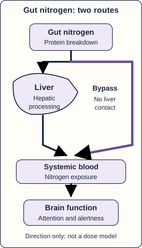
Picture blood leaving the intestine. Before it reaches the general circulation, much of its nitrogen burden normally passes through the liver. Cirrhosis can impair hepatic handling; collateral vessels or a transjugular intrahepatic portosystemic shunt (TIPS) let some blood bypass that handling. More nitrogen delivered to the gut or more bypass can therefore worsen brain exposure without a sudden rise in aminotransferases. [2][7]
Trace the path, then distinguish additional intestinal substrate from reduced hepatic contact. [2][7]
Trace the two routes in the portal-flow diagram: intestine to liver to systemic blood, and intestine to bypass to systemic blood. A bypass changes where blood travels; it does not need to create new hepatocyte injury to affect cognition. A gastrointestinal bleed adds digestible blood protein to the intestinal nitrogen load. Constipation prolongs intestinal retention. These can provoke HE in a patient whose chronic liver disease otherwise appears unchanged. [2]
From nitrogen exposure to altered brain function
Ammonia is important, but HE is not a single-toxin concentration meter. In astrocytes, conversion of ammonia and glutamate into glutamine contributes to osmotic and cellular stress. Inflammation and altered neurotransmission also influence brain function. The clinical effect depends on the patient's susceptibility and concurrent illness, not just a peripheral blood sample. [2][9]
Skeletal muscle provides an additional site of ammonia handling through glutamine formation. Loss of muscle reduces this reserve. This is why a plan that lowers intestinal nitrogen while starving the patient can work against its own long-term goal. Protein intake and muscle preservation are part of HE care, not optional concerns after the brain recovers. [6][11]
Change the load or change the route?
Make a prediction: Which situation adds intestinal nitrogen, and which reduces the fraction of portal blood that contacts the liver? Trace the diagram above, then compare the consequences below. [2][7]
Baseline: reduced hepatic reserve
The existing intestinal load reaches a liver with limited handling capacity. Some portal blood already bypasses the liver. Both routes ultimately reach systemic blood.
GI bleeding: more substrate, unchanged routing
Digestion of blood protein supplies additional intestinal nitrogen. With the same hepatic reserve and blood routes, this larger load can increase systemic exposure.
More bypass: unchanged substrate, less hepatic contact
A larger fraction of portal blood avoids hepatic processing. Systemic exposure can increase even when intestinal nitrogen production and aminotransferases are unchanged.
Explain the difference: Bleeding changes what arrives from the intestine; a shunt changes the path taken by portal blood. Neither observation supplies a numerical ammonia prediction or a clinical severity grade. [2][7]
Predict the direction: Keep intestinal nitrogen production unchanged, but direct more portal blood through a bypass. Does hepatic contact increase or decrease? Now keep the bypass unchanged and add digested blood to the intestine.
The first change decreases hepatic contact; the second increases the nitrogen delivered to a limited clearance system. Both can increase exposure of the brain. Neither predicts a particular ammonia value or West Haven grade. Transfer this distinction to post-TIPS confusion: first assess routine precipitants and competing diagnoses, then consider whether bypass is contributing. [2][7]
This lesson focuses on cirrhosis-related HE and portosystemic shunting. Encephalopathy in acute liver failure is a separate emergency with different cerebral-edema risks and uses of ammonia measurement; do not apply an outpatient lactulose plan to that situation. [2]
What must be treated before the label is settled?
Consider two patients with cirrhosis. One is awake, disoriented and constipated after melena. The other is barely breathing after an opioid dose increase. Both might have a high ammonia concentration; only the second description immediately points to ventilatory failure from a drug exposure. Stabilization and a search for alternatives happen alongside HE treatment, not after it fails. [1][10][2][8]
Protect first, investigate in parallel
Assess airway protection, ventilation, circulation and bedside glucose. Do not give oral liquid to a patient who cannot swallow safely or protect the airway. Severe somnolence or coma needs emergency high-acuity care. When opioid toxicity is suspected, support ventilation and give naloxone according to the emergency protocol; continued observation is needed because respiratory depression can recur after reversal. Correct hypoglycemia promptly and reassess cognition. Neither response excludes a second, coexisting cause. [2][8]
New focal weakness, a seizure, a significant head injury or an atypical course warrants targeted neurologic investigation, including urgent brain imaging when indicated. Routine imaging is not required for every familiar, nonfocal HE presentation. The reason to image is concern for a competing process, not the height of the ammonia result. Stroke, intracranial bleeding, seizures, intoxication, electrolyte disturbances and other metabolic disorders can mimic or accompany HE. [1][10][2]
Find the event that changed the balance
Ask about black stools or hematemesis; assess infection according to symptoms and examination, including the need to evaluate ascitic fluid in an admitted patient with ascites. Review stool frequency and consistency, actual medication use, fluid losses, diuretic changes and recent procedures. Check renal function and electrolytes. Infection, bleeding, constipation, dehydration, acute kidney injury, hypokalemia and sedating medicines may each contribute. Several often coexist. [1][10][2]
Choose two concurrent actions: An awake patient has disorientation, asterixis, fever and dysuria after missing lactulose. Should treatment focus on the bowel plan or the infection?
Treat suspected overt HE while investigating and treating the infection. Reducing intestinal nitrogen does not eradicate a urinary infection; antibiotics alone do not restore the interrupted bowel regimen. In a new patient with vomiting and excessive diuresis, the parallel priorities become volume and electrolyte correction rather than reflex antibiotic treatment. Continue reassessing instead of assuming the first precipitant found is the only one. [1][10][2]
What can an ammonia result actually answer?
A patient becomes oriented, resumes conversation and has soft stools after treatment, but the repeat ammonia concentration is still above the laboratory reference interval. Increasing lactulose solely to normalize that number can create diarrhea and kidney injury. The desired outcome is recovery of the patient, not a particular serial laboratory trajectory. [1][10][2][3]
Separate biological importance from diagnostic performance
Ammonia contributes to HE biology, but a high blood value alone does not establish the diagnosis or select a West Haven grade. Different clinical states overlap in their ammonia values, and collection and processing conditions can affect the measurement. A value cannot substitute for the examination, collateral history or investigation of alternatives. Do not delay treatment of a convincing overt syndrome while waiting for the assay. [1][10][2]
A normal value should prompt diagnostic reconsideration, not automatic dismissal of all clinical evidence. Recheck whether the presentation actually fits HE, whether another disorder explains the findings and whether more than one process is present. This matters particularly when the patient has an atypical examination or fails to improve. Current ACG guidance advises against using routine ammonia testing to direct inpatient HE management. [1][10]
Interpret the discordance: Patient A is back to their usual cognition with three soft stools and a persistently high ammonia result. Patient B remains confused despite apparently adequate treatment and has a normal ammonia result. Which patient requires a broader explanation for the current mental state?
Patient B needs renewed diagnostic evaluation; patient A does not need escalating catharsis just to change a laboratory number. For either patient, monitor wakefulness, orientation, attention, ability to eat safely, hydration, electrolytes and the precipitating illness. A recovering patient may still need medication and caregiver support even after the examination improves. [1][10][2]
Transfer the same reasoning to a person with new unilateral weakness and high ammonia: the result cannot explain away focal findings. Likewise, bilateral asterixis in someone with severe ventilatory failure is not proof of a hepatic cause. Clinical attribution requires that the entire presentation fit, not merely one familiar sign. [1][10][2]
How can a useful bowel treatment become harmful?
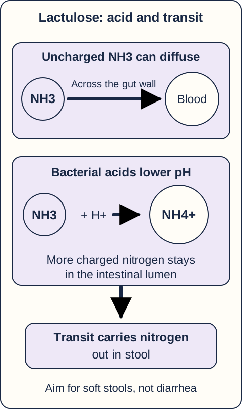
Lactulose works in the intestine, but its benefit is not proportional to the amount of diarrhea it produces. Bacteria metabolize this poorly absorbed sugar into organic acids. Lower luminal pH favors conversion of diffusible NH3 to charged NH4+, reducing absorption; laxation also increases fecal elimination of nitrogen. The diagram shows acidification and transit as related but distinct effects. [3]
Separate acid trapping from laxation, then predict why additional watery diarrhea is not an additional treatment target. [3]
Acute therapy and maintenance are different tasks
For overt HE, clinicians initiate lactulose promptly while treating precipitants. Initial supervised dosing aims to produce catharsis, then dosing is adjusted to clinical response and usually two to three soft stools each day. Count and consistency both matter. Oral or enteral delivery requires an appropriate airway and swallowing plan; a supervised rectal regimen is an option when oral administration is unsafe. This is not a home-management plan for an obtunded patient. [1][10][3]
Watery diarrhea can cause volume depletion, hypokalemia, hypernatremia and worsening renal function. These problems can themselves worsen cognition or precipitate HE. When they develop, reassess the regimen, reduce or temporarily hold excessive dosing as clinically appropriate, correct losses and retitrate. Do not add more lactulose because dehydration-associated confusion has appeared. [1][10][3]
Compare the treatment records: One patient has three soft stools, normal hydration and improving attention. Another has nine watery stools, orthostatic symptoms, potassium 2.8 mmol/L and rising creatinine. Which record indicates that more catharsis could worsen the problem?
The second record identifies treatment-associated depletion, not evidence that the bowel effect is inadequate. After correcting it, the aim returns to a tolerable soft-stool plan. In a different patient who has no stool because doses were missed, address the barrier and constipation instead of treating these opposite situations identically. [1][10][3]
Reduce recurrence without sacrificing muscle
Continue secondary prevention after recovery: lactulose is first-line after an overt episode. Add rifaximin for breakthrough overt HE on lactulose, including recurrent episodes despite a well-used regimen. The usual adult recurrence-prevention dose studied in the pivotal trial is 550 mg twice daily. It is a minimally absorbed, gut-directed antibiotic, not a replacement for systemic treatment of an infection. Acute inpatient add-on use is also supported by a conditional 2026 ACG recommendation; do not teach that it is forbidden until a second episode. [1][10][4]
Do not prescribe prolonged protein restriction. In a typical adult with cirrhosis, target about 1.2 to 1.5 g protein/kg ideal body weight/day, individualized by the nutrition team. For a clinically stable patient with sarcopenia, aim toward 1.5 g/kg ideal body weight/day rather than the lower end of that range. Spread intake across meals and include a late-evening snack. Plant and dairy protein can help meet goals. Adequate energy intake and appropriate physical activity support muscle preservation; restrictions that accelerate muscle loss can reduce extrahepatic ammonia handling. [6][11]
What does another episode tell you to reconsider?
A recurrence does not automatically mean the patient needs a larger lactulose dose. Ask what actually happened between visits: Was the medicine affordable and available? Was the stool target understood? Did bloating, work demands or diarrhea make the plan impractical? Did a caregiver notice changes before the patient did? A prevention plan must be usable by the person who will carry it out. [1][10][2]
Make the plan observable
Use a written record of stool consistency and frequency, medicine doses, early cognitive changes and contact instructions. Ask the patient or caregiver to explain the plan back in their own words. Review sedating medicines and avoid abrupt, unsupervised changes to medicines that can cause withdrawal. New confusion, inability to stay awake, bleeding or other severe illness needs urgent assessment rather than repeated extra doses at home. Discuss driving and hazardous work explicitly; recent overt HE calls for avoidance of driving and individualized reassessment of safety. Address caregiver strain as well as adherence. [1][10][2]
Distinguish an ongoing trigger from excessive bypass
Reassess immediately for new deterioration or atypical findings; 48 to 72 hours is not a required waiting period. If mental status barely improves after that interval of adequate treatment and correction of suspected precipitants, reconsider the diagnosis, drug effects and portosystemic shunts. Cross-sectional abdominal imaging can identify a large spontaneous shunt. Selected patients with refractory HE and adequate hepatic reserve may benefit from embolization after specialist assessment. This is not an indication to embolize every collateral vessel discovered on a scan. [1][10][2][7]
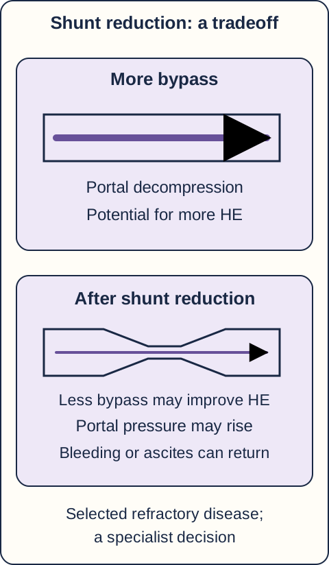
Compare the cognitive benefit with the original reason for portal decompression before considering a procedure. This is not patient anatomy or a procedural guide. [2][7]
For persistent or recurrent HE after TIPS despite appropriate medical management, a multidisciplinary team can assess shunt reduction. Reducing bypass may improve cognition but can bring back portal-hypertension complications, including ascites or variceal bleeding. Review the original reason for TIPS before narrowing it. Complete occlusion is not the automatic first answer. [2][7]
Predict the tradeoff: A patient received TIPS to control repeated variceal hemorrhage and now has refractory HE. What previous problem could return if shunt flow is reduced?
Portal pressure can rise again, increasing risk of recurrent bleeding or ascites. That is why the decision includes hepatology and interventional expertise rather than only a cognition score. A patient with a spontaneous shunt, no TIPS and preserved hepatic reserve needs a different procedural assessment, even when the brain symptoms look similar. [2][7]
Plan before TIPS and refer before the score looks dramatic
The 2026 ACG guideline recommends starting rifaximin 14 days before elective TIPS and continuing for at least six months afterward. This newer guidance differs from older recommendations against routine post-TIPS prophylaxis and is supported by a randomized prevention trial. The preprocedure interval is not a reason to delay urgently needed bleeding control. [1][10][5][2][7]
Repeated HE may profoundly impair daily life even with MELD below 15. Current guidance suggests considering living-donor liver-transplant evaluation in this setting. Evaluation is not automatic listing, allocation priority or a promise that every cognitive deficit will reverse. Recurrent or severe disease warrants early discussion of the underlying liver disease and transplant options rather than indefinite increases in catharsis. [1][10]
Apply the reasoning to a new patient
Choose the best answer using the information supplied. Compare your answer with the accompanying explanations; the lesson remains open throughout.
Case 1
Show answer and explanations for case 1
A. Start lactulose while evaluating and controlling gastrointestinal bleeding (Best answer)
Time disorientation and impaired attention in this setting support overt HE. Digested blood adds intestinal nitrogen, so bleeding can precipitate HE. Treat suspected HE and the bleeding process concurrently rather than waiting for an ammonia result. [1][2][10]
Reasoning steps for option A
What does the cognitive pattern support?
Time disorientation and impaired attention in this setting support overt HE.
Why does melena matter beyond blood loss?
Digested blood adds intestinal nitrogen, so bleeding can precipitate HE.
What should occur together?
Treat suspected HE and the bleeding process concurrently rather than waiting for an ammonia result.
B. Start lactulose and reserve endoscopy for persistent cognitive impairment (Why this does not fit)
Lactulose is first-line treatment for a convincing overt HE presentation. Lactulose does not control the source of gastrointestinal bleeding. Melena requires its own assessment; cognitive improvement does not establish hemostasis. [1][2][10]
Reasoning steps for option B
Why is lactulose reasonable?
Lactulose is first-line treatment for a convincing overt HE presentation.
What does it fail to treat?
Lactulose does not control the source of gastrointestinal bleeding.
What determines bleeding evaluation?
Melena requires its own assessment; cognitive improvement does not establish hemostasis.
C. Control the bleeding, then reassess before starting bowel therapy (Why this does not fit)
Hemostasis addresses a major reversible precipitant. An overt cognitive syndrome is already present, and constipation preceded the bleeding. Deferring HE therapy until after hemostasis leaves a treatable concurrent problem unaddressed. [1][2][10]
Reasoning steps for option C
Why might bleeding treatment seem sufficient?
Hemostasis addresses a major reversible precipitant.
What remains active now?
An overt cognitive syndrome is already present, and constipation preceded the bleeding.
Why is the sequence inadequate?
Deferring HE therapy until after hemostasis leaves a treatable concurrent problem unaddressed.
D. Obtain ammonia and use its result to select treatment (Why this does not fit)
Ammonia is involved in HE biology and may be high in cirrhosis. A blood concentration alone cannot diagnose or grade this syndrome. The clinical presentation justifies treatment without waiting for ammonia measurement. [1][2][10]
Reasoning steps for option D
What is the apparent attraction of the assay?
Ammonia is involved in HE biology and may be high in cirrhosis.
What can the result not establish?
A blood concentration alone cannot diagnose or grade this syndrome.
What should guide initial care?
The clinical presentation justifies treatment without waiting for ammonia measurement.
Takeaway: A convincing overt syndrome and its precipitant require parallel care.
A. Treat isolated HE and reassess urine after cognition improves (Why this does not fit)
The examination and interrupted regimen support HE. Fever with urinary symptoms and pyuria warrants infection-directed assessment and treatment. HE does not make a symptomatic infection incidental. [1][2][10]
Reasoning steps for option A
Why might HE be suspected?
The examination and interrupted regimen support HE.
What cannot be explained away?
Fever with urinary symptoms and pyuria warrants infection-directed assessment and treatment.
What should not be assumed?
HE does not make a symptomatic infection incidental.
B. Treat the urinary infection while restoring the HE regimen (Best answer)
New disorientation and asterixis support overt HE in this context. Infection can precipitate HE while also contributing to delirium. Treat both processes; a small ammonia change does not measure clinical severity. [1][2][10]
Reasoning steps for option B
How is the syndrome identified?
New disorientation and asterixis support overt HE in this context.
How does infection fit?
Infection can precipitate HE while also contributing to delirium.
What is the resulting plan?
Treat both processes; a small ammonia change does not measure clinical severity.
C. Treat isolated urinary delirium and postpone HE therapy (Why this does not fit)
A symptomatic infection can independently cause delirium. Missed lactulose, disorientation and asterixis also support a concurrent HE component. Finding infection does not exclude HE or justify withholding appropriate HE treatment. [1][2][10]
Reasoning steps for option C
Why is infection relevant?
A symptomatic infection can independently cause delirium.
What additional pattern is present?
Missed lactulose, disorientation and asterixis also support a concurrent HE component.
Why is postponement unsound?
Finding infection does not exclude HE or justify withholding appropriate HE treatment.
D. Repeat ammonia before deciding which condition to treat (Why this does not fit)
A prior concentration offers laboratory context. Similar ammonia values can accompany very different clinical states. The examination and symptomatic infection warrant treatment without another ammonia result. [1][2][10]
Reasoning steps for option D
Why compare with the baseline result?
A prior concentration offers laboratory context.
Why does this comparison fail to decide the diagnosis?
Similar ammonia values can accompany very different clinical states.
What should determine action?
The examination and symptomatic infection warrant treatment without another ammonia result.
Takeaway: Infection and HE can coexist; the ammonia trend does not separate them.
A. Obtain brain imaging before choosing a reversal agent (Why this does not fit)
Structural brain disease must be considered when neurologic findings suggest it. The patient has life-threatening ventilatory failure with a strong opioid exposure history. Stabilize breathing and treat the suspected opioid effect before nonresuscitative testing. [2][8]
Reasoning steps for option A
When is imaging important?
Structural brain disease must be considered when neurologic findings suggest it.
What must happen before transport?
The patient has life-threatening ventilatory failure with a strong opioid exposure history.
How should priorities be ordered?
Stabilize breathing and treat the suspected opioid effect before nonresuscitative testing.
B. Support ventilation and administer naloxone with continued monitoring (Best answer)
The dose increase, small pupils and marked bradypnea form a consistent toxidrome. Inadequate breathing causes immediate hypoxic injury regardless of the ammonia concentration. Give naloxone while supporting ventilation and monitor for recurrent respiratory depression or another coexisting cause. [2][8]
Reasoning steps for option B
What pattern points to opioid effect?
The dose increase, small pupils and marked bradypnea form a consistent toxidrome.
Why is ventilation the priority?
Inadequate breathing causes immediate hypoxic injury regardless of the ammonia concentration.
What follow-up remains necessary?
Give naloxone while supporting ventilation and monitor for recurrent respiratory depression or another coexisting cause.
C. Give rectal lactulose and reassess respiratory rate afterward (Why this does not fit)
A patient with cirrhosis can have HE and may need a nonoral regimen. Severe bradypnea and hypoxemia indicate inadequate ventilation after opioid exposure. Bowel therapy does not reverse respiratory depression and must not delay ventilatory support. [2][8]
Reasoning steps for option C
Why might this treatment be considered?
A patient with cirrhosis can have HE and may need a nonoral regimen.
What is immediately dangerous here?
Severe bradypnea and hypoxemia indicate inadequate ventilation after opioid exposure.
Why does this option fail?
Bowel therapy does not reverse respiratory depression and must not delay ventilatory support.
D. Give flumazenil while preparing further cognitive testing (Why this does not fit)
Drug-induced sedation can require targeted reversal. The timing and respiratory findings point to an opioid rather than an isolated benzodiazepine effect. Naloxone targets opioid receptors; flumazenil is not the appropriate reversal agent here. [2][8]
Reasoning steps for option D
When can an antagonist be relevant?
Drug-induced sedation can require targeted reversal.
Which exposure is actually supplied?
The timing and respiratory findings point to an opioid rather than an isolated benzodiazepine effect.
Why choose a different antidote?
Naloxone targets opioid receptors; flumazenil is not the appropriate reversal agent here.
Takeaway: A high ammonia result must not distract from a respiratory toxidrome.
A. Reassess the examination after an initial lactulose response (Why this does not fit)
Prior diffuse episodes improved with HE therapy. A new focal deficit after a fall raises concern for structural brain injury. A bowel-treatment response must not delay urgent evaluation of a focal neurologic emergency. [1][2][10]
Reasoning steps for option A
Why might a treatment trial appear useful?
Prior diffuse episodes improved with HE therapy.
What makes this episode different?
A new focal deficit after a fall raises concern for structural brain injury.
Why not wait?
A bowel-treatment response must not delay urgent evaluation of a focal neurologic emergency.
B. Measure ammonia again using a promptly processed specimen (Why this does not fit)
Processing can affect ammonia measurements. Ammonia cannot explain or localize unilateral weakness and neglect. Investigate the focal examination directly rather than refining a nonspecific assay. [1][2][10]
Reasoning steps for option B
Why is specimen handling relevant?
Processing can affect ammonia measurements.
What question remains unanswered by a better specimen?
Ammonia cannot explain or localize unilateral weakness and neglect.
What is the appropriate diagnostic focus?
Investigate the focal examination directly rather than refining a nonspecific assay.
C. Obtain urgent imaging for intracranial bleeding or stroke (Best answer)
Left-sided weakness and neglect indicate focal cerebral dysfunction. A recent fall adds concern for intracranial bleeding rather than a familiar diffuse HE episode. Urgent neuroimaging and neurologic assessment are needed even when ammonia is high. [1][2][10]
Reasoning steps for option C
What does the examination imply?
Left-sided weakness and neglect indicate focal cerebral dysfunction.
How does the history affect concern?
A recent fall adds concern for intracranial bleeding rather than a familiar diffuse HE episode.
What follows from those findings?
Urgent neuroimaging and neurologic assessment are needed even when ammonia is high.
D. Obtain formal psychometric testing for covert dysfunction (Why this does not fit)
Validated testing helps detect subtle cognitive dysfunction without overt signs. The patient has acute confusion and a focal motor-attention deficit. Acute structural disease must be assessed before elective cognitive characterization. [1][2][10]
Reasoning steps for option D
When is this testing useful?
Validated testing helps detect subtle cognitive dysfunction without overt signs.
Why is it inappropriate as the next test?
The patient has acute confusion and a focal motor-attention deficit.
What should take precedence?
Acute structural disease must be assessed before elective cognitive characterization.
Takeaway: A focal examination is not explained away by cirrhosis or hyperammonemia.
A. Give intravenous glucose and reassess mental status (Best answer)
The glucose concentration and insulin-food mismatch identify symptomatic hypoglycemia. Confusion with unsafe swallowing creates an aspiration risk. Correct glucose intravenously and reassess before attributing remaining symptoms to HE. [2]
Reasoning steps for option A
What explains the abrupt symptoms?
The glucose concentration and insulin-food mismatch identify symptomatic hypoglycemia.
Why choose a nonoral route?
Confusion with unsafe swallowing creates an aspiration risk.
What is the immediate action?
Correct glucose intravenously and reassess before attributing remaining symptoms to HE.
B. Obtain brain imaging before administering glucose therapy (Why this does not fit)
Persistent or focal neurologic abnormalities may need structural investigation. A readily reversible metabolic emergency is documented at the bedside. Give glucose now and use the reassessment to direct further investigation. [2]
Reasoning steps for option B
When would imaging be appropriate?
Persistent or focal neurologic abnormalities may need structural investigation.
What is already established here?
A readily reversible metabolic emergency is documented at the bedside.
What should not be delayed?
Give glucose now and use the reassessment to direct further investigation.
C. Give rectal lactulose and repeat glucose after catharsis (Why this does not fit)
The patient has cirrhosis and a history of overt HE. Severe symptomatic hypoglycemia requires immediate correction. Catharsis cannot promptly correct neuroglycopenia and would delay the indicated treatment. [2]
Reasoning steps for option C
Why might lactulose enter the differential plan?
The patient has cirrhosis and a history of overt HE.
What does that history fail to override?
Severe symptomatic hypoglycemia requires immediate correction.
Why is this sequence harmful?
Catharsis cannot promptly correct neuroglycopenia and would delay the indicated treatment.
D. Give oral glucose and repeat testing after swallowing (Why this does not fit)
The low glucose requires prompt correction. The patient cannot swallow safely, despite already having IV access. Use an appropriate nonoral emergency treatment instead of oral intake. [2]
Reasoning steps for option D
Why is glucose replacement relevant?
The low glucose requires prompt correction.
What makes this route unsafe?
The patient cannot swallow safely, despite already having IV access.
How should replacement be delivered?
Use an appropriate nonoral emergency treatment instead of oral intake.
Takeaway: Correct documented hypoglycemia immediately, using a route the patient can safely receive.
A. Grade IV; classify the lack of asterixis as coma (Why this does not fit)
Patients who cannot maintain posture cannot reliably demonstrate asterixis. The patient still opens the eyes and responds to stimulation. Coma is distinguished by absent meaningful responsiveness, not by absent hand flapping. [1][2][10]
Reasoning steps for option A
Why can asterixis be absent in severe illness?
Patients who cannot maintain posture cannot reliably demonstrate asterixis.
What argues against grade IV here?
The patient still opens the eyes and responds to stimulation.
What defines the distinction?
Coma is distinguished by absent meaningful responsiveness, not by absent hand flapping.
B. Grade III; increase monitoring and assess airway protection (Best answer)
The patient is markedly somnolent and cannot sustain conversation. Responses to stimulation remain present. Treat this as a grade III pattern with high-acuity monitoring and airway assessment. [1][2][10]
Reasoning steps for option B
What establishes high-grade dysfunction?
The patient is markedly somnolent and cannot sustain conversation.
Why is this not grade IV?
Responses to stimulation remain present.
What should the grade prompt?
Treat this as a grade III pattern with high-acuity monitoring and airway assessment.
C. Grade I; arrange validated testing before changing the care setting (Why this does not fit)
Subtle inattention can occur while orientation and wakefulness are largely preserved. Marked somnolence and inability to maintain interaction are overt findings. Act on the overt deterioration rather than waiting for psychometric assessment. [1][2][10]
Reasoning steps for option C
What can characterize grade I?
Subtle inattention can occur while orientation and wakefulness are largely preserved.
What exceeds that category?
Marked somnolence and inability to maintain interaction are overt findings.
What takes priority over subtle testing?
Act on the overt deterioration rather than waiting for psychometric assessment.
D. Grade II; continue care based on yesterday's cognitive assessment (Why this does not fit)
The patient was awake but disoriented to time. Marked somnolence now prevents sustained interaction. Use the current examination rather than retaining a prior grade despite deterioration. [1][2][10]
Reasoning steps for option D
What supported grade II yesterday?
The patient was awake but disoriented to time.
What has changed?
Marked somnolence now prevents sustained interaction.
How should severity be assigned?
Use the current examination rather than retaining a prior grade despite deterioration.
Takeaway: Stage the current responsiveness; an untestable asterixis examination is not a coma criterion.
A. Observe until time disorientation appears before testing cognition (Why this does not fit)
It is an overt abnormality that helps identify a grade II pattern. Covert dysfunction can impair complex tasks before that finding develops. The new functional change warrants investigation before an overt episode occurs. [1][2][10]
Reasoning steps for option A
Why is time disorientation useful?
It is an overt abnormality that helps identify a grade II pattern.
What would waiting overlook?
Covert dysfunction can impair complex tasks before that finding develops.
What should trigger assessment here?
The new functional change warrants investigation before an overt episode occurs.
B. Repeat ammonia to determine whether driving performance is impaired (Why this does not fit)
Ammonia participates in HE biology. It does not measure this patient's driving performance or diagnose covert HE. Use validated cognitive testing with a functional safety assessment rather than an ammonia threshold. [1][2][10]
Reasoning steps for option B
Why might ammonia be considered?
Ammonia participates in HE biology.
What cannot be inferred from its concentration?
It does not measure this patient's driving performance or diagnose covert HE.
What assessment fits the unresolved problem?
Use validated cognitive testing with a functional safety assessment rather than an ammonia threshold.
C. Use a validated covert-HE test and review driving safety (Best answer)
It argues against an overt episode but does not exclude subtle impairment. New errors affect a safety-sensitive occupation. Use a locally validated cognitive assessment and discuss driving safety while evaluating the cause. [1][2][10]
Reasoning steps for option C
What does the normal bedside examination establish?
It argues against an overt episode but does not exclude subtle impairment.
What makes further assessment important?
New errors affect a safety-sensitive occupation.
What is the appropriate next step?
Use a locally validated cognitive assessment and discuss driving safety while evaluating the cause.
D. Attribute the errors to grade II HE and begin inpatient treatment (Why this does not fit)
Cirrhosis and new cognitive complaints make a hepatic contribution possible. He remains oriented without the overt examination pattern described in grade II disease. Evaluate subtle dysfunction with validated methods instead of assigning an overt grade from work errors alone. [1][2][10]
Reasoning steps for option D
What makes HE a reasonable consideration?
Cirrhosis and new cognitive complaints make a hepatic contribution possible.
What does not fit grade II?
He remains oriented without the overt examination pattern described in grade II disease.
How should the label be established?
Evaluate subtle dysfunction with validated methods instead of assigning an overt grade from work errors alone.
Takeaway: Preserved orientation does not exclude clinically important covert dysfunction.
A. Obtain EEG to evaluate intermittent or nonconvulsive seizure activity (Best answer)
The normal ammonia result and limited treatment response warrant reassessment rather than certainty about HE. Repeated stereotyped behavioral arrests suggest an electrical cerebral event. A normal CT does not exclude seizures; EEG directly evaluates that possibility. [1][2][10]
Reasoning steps for option A
What changes the differential?
The normal ammonia result and limited treatment response warrant reassessment rather than certainty about HE.
What finding narrows the next investigation?
Repeated stereotyped behavioral arrests suggest an electrical cerebral event.
Why is EEG appropriate?
A normal CT does not exclude seizures; EEG directly evaluates that possibility.
B. Repeat ammonia after another day of the same observation (Why this does not fit)
A serial laboratory value offers an objective number to follow. The recurrent stereotyped pauses raise concern for intermittent seizure activity. EEG evaluates cerebral electrical activity; serial ammonia cannot assess it. [1][2][10]
Reasoning steps for option B
Why might repeated testing seem reassuring?
A serial laboratory value offers an objective number to follow.
What clinical question remains?
The recurrent stereotyped pauses raise concern for intermittent seizure activity.
Which test answers that question?
EEG evaluates cerebral electrical activity; serial ammonia cannot assess it.
C. Obtain abdominal shunt imaging as the first explanation for the spells (Why this does not fit)
Persistent HE despite appropriate therapy can prompt evaluation for portosystemic bypass. Repeated stereotyped arrests are not simply continuous diffuse inattention. Evaluate possible seizure activity rather than attributing these spells to bypass without investigation. [1][2][10]
Reasoning steps for option C
When can shunt imaging be useful?
Persistent HE despite appropriate therapy can prompt evaluation for portosystemic bypass.
Which feature demands a more immediate targeted assessment?
Repeated stereotyped arrests are not simply continuous diffuse inattention.
What should be assessed first?
Evaluate possible seizure activity rather than attributing these spells to bypass without investigation.
D. Give additional lactulose before ordering further neurologic testing (Why this does not fit)
The patient has cirrhosis and ongoing cognitive dysfunction. Constipation and dehydration are corrected, stools are adequate, and the new behavior is episodic. Investigate the specific neurologic pattern rather than create excessive catharsis. [1][2][10]
Reasoning steps for option D
Why might more HE therapy be considered?
The patient has cirrhosis and ongoing cognitive dysfunction.
What argues against reflex dose escalation?
Constipation and dehydration are corrected, stools are adequate, and the new behavior is episodic.
What is the better next action?
Investigate the specific neurologic pattern rather than create excessive catharsis.
Takeaway: Poor response and atypical episodes call for targeted reassessment, not automatic lactulose escalation.
A. Stop preventive lactulose because orientation has normalized (Why this does not fit)
The overt episode has resolved clinically. Recovery does not eliminate the risk of another episode in cirrhosis. Continue appropriate secondary prevention rather than equating remission with no need for therapy. [1][3][10]
Reasoning steps for option A
Why consider reducing treatment?
The overt episode has resolved clinically.
What risk remains?
Recovery does not eliminate the risk of another episode in cirrhosis.
What distinguishes treatment phases?
Continue appropriate secondary prevention rather than equating remission with no need for therapy.
B. Add rectal lactulose until a second normal examination is recorded (Why this does not fit)
It offers a supervised route when oral administration is unsafe or unavailable. The patient is awake, recovered and tolerating the current bowel regimen. Neither a route constraint nor inadequate clinical response justifies extra catharsis here. [1][3][10]
Reasoning steps for option B
When can rectal treatment be useful?
It offers a supervised route when oral administration is unsafe or unavailable.
Is such a route problem present?
The patient is awake, recovered and tolerating the current bowel regimen.
Why is the addition unnecessary?
Neither a route constraint nor inadequate clinical response justifies extra catharsis here.
C. Increase lactulose until ammonia enters the reference interval (Why this does not fit)
It is a persistent laboratory abnormality in a disease involving ammonia. It does not establish ongoing clinical HE or insufficient catharsis. Excess diarrhea may cause dehydration and electrolyte losses without improving cognition. [1][3][10]
Reasoning steps for option C
Why might the residual elevation attract attention?
It is a persistent laboratory abnormality in a disease involving ammonia.
What does it fail to show?
It does not establish ongoing clinical HE or insufficient catharsis.
What harm can escalation create?
Excess diarrhea may cause dehydration and electrolyte losses without improving cognition.
D. Continue the tolerated regimen and monitor clinical function (Best answer)
The examination and caregiver report both show return to baseline. Three soft stools and stable hydration meet the usual maintenance aim. Maintain a tolerable prevention regimen rather than intensify treatment to normalize ammonia. [1][3][10]
Reasoning steps for option D
What evidence indicates response?
The examination and caregiver report both show return to baseline.
Is the bowel effect inadequate?
Three soft stools and stable hydration meet the usual maintenance aim.
What should guide the plan?
Maintain a tolerable prevention regimen rather than intensify treatment to normalize ammonia.
Takeaway: Clinical recovery with a tolerable bowel regimen is not an indication to chase a laboratory number.
A. Asterixis; prioritize assessment of the missed dialysis treatments (Best answer)
Brief interruptions of sustained wrist extension represent asterixis, a form of negative myoclonus. Two missed dialysis treatments with marked azotemia support a uremic contribution to the new inattention. Assess and treat the renal problem promptly; asterixis is not liver-specific, and possible coexisting HE remains part of reassessment. [2]
Reasoning steps for option A
What is the motor sign?
Brief interruptions of sustained wrist extension represent asterixis, a form of negative myoclonus.
Which new exposure or physiological change is supplied?
Two missed dialysis treatments with marked azotemia support a uremic contribution to the new inattention.
How should that affect attribution?
Assess and treat the renal problem promptly; asterixis is not liver-specific, and possible coexisting HE remains part of reassessment.
B. Cortical myoclonus; prioritize assessment for new epileptic activity (Why this does not fit)
A sudden active muscle jerk can reflect cortical electrical activity. This patient has brief losses of a maintained posture rather than described active jerks or stereotyped seizures. The timing after missed dialysis and severe azotemia favor a toxic-metabolic assessment rather than assigning an epileptic cause from the wrist sign. [2]
Reasoning steps for option B
What can epileptic myoclonus look like?
A sudden active muscle jerk can reflect cortical electrical activity.
How does the described phenomenon differ?
This patient has brief losses of a maintained posture rather than described active jerks or stereotyped seizures.
Which associated evidence matters here?
The timing after missed dialysis and severe azotemia favor a toxic-metabolic assessment rather than assigning an epileptic cause from the wrist sign.
C. Rest tremor; prioritize assessment for chronic basal-ganglia dysfunction (Why this does not fit)
A rest tremor is a rhythmic oscillation when a body part is supported and not maintaining an active posture. The wrists briefly lose sustained extension rather than oscillate at rest. New diffuse inattention after missed dialysis is better explained by a metabolic disturbance than by newly apparent chronic basal-ganglia dysfunction. [2]
Reasoning steps for option C
When is a rest tremor observed?
A rest tremor is a rhythmic oscillation when a body part is supported and not maintaining an active posture.
What is actually described?
The wrists briefly lose sustained extension rather than oscillate at rest.
How does the time course affect the interpretation?
New diffuse inattention after missed dialysis is better explained by a metabolic disturbance than by newly apparent chronic basal-ganglia dysfunction.
D. Asterixis; prioritize intensification of the intestinal nitrogen regimen (Why this does not fit)
The brief lapses of posture are asterixis. Asterixis also occurs in uremia; cirrhosis does not make it specific for HE. The missed dialysis and marked azotemia require prompt renal assessment rather than being displaced by a familiar hepatic diagnosis. [2]
Reasoning steps for option D
Which part correctly names the sign?
The brief lapses of posture are asterixis.
Does the sign establish a hepatic source?
Asterixis also occurs in uremia; cirrhosis does not make it specific for HE.
Why is reflex intestinal treatment escalation inadequate?
The missed dialysis and marked azotemia require prompt renal assessment rather than being displaced by a familiar hepatic diagnosis.
Takeaway: Identify negative myoclonus, then use the accompanying physiology rather than the sign alone to prioritize the cause.
A. Greater renal nitrogen retention from persistent filtration failure (Why this does not fit)
Kidney dysfunction can contribute to encephalopathy and raise blood urea nitrogen. Creatinine and perfusion have returned to baseline. Digested blood can increase nitrogen delivery even without persistent renal failure. [2]
Reasoning steps for option A
Why consider renal retention?
Kidney dysfunction can contribute to encephalopathy and raise blood urea nitrogen.
What argues against it as the supplied explanation?
Creatinine and perfusion have returned to baseline.
What does the bleeding add independently?
Digested blood can increase nitrogen delivery even without persistent renal failure.
B. Greater intestinal nitrogen delivery from digestion of blood protein (Best answer)
The bleed placed blood protein inside the gastrointestinal tract. Protein breakdown increases the intestinal nitrogen burden. Limited hepatic handling makes the added load capable of provoking HE without a new rise in injury markers. [2]
Reasoning steps for option B
Where did the extra substrate enter?
The bleed placed blood protein inside the gastrointestinal tract.
What happens during digestion?
Protein breakdown increases the intestinal nitrogen burden.
Why does cirrhosis matter?
Limited hepatic handling makes the added load capable of provoking HE without a new rise in injury markers.
C. Greater hepatocyte necrosis indicated by the cognitive deterioration (Why this does not fit)
Acute hepatic deterioration can worsen brain dysfunction. No new rise in the described liver injury markers is present. A larger intestinal nitrogen load can overwhelm existing reserve without demonstrating new hepatocyte necrosis. [2]
Reasoning steps for option C
Why might new liver injury be considered?
Acute hepatic deterioration can worsen brain dysfunction.
What do the supplied laboratory trends show?
No new rise in the described liver injury markers is present.
Why is necrosis not required?
A larger intestinal nitrogen load can overwhelm existing reserve without demonstrating new hepatocyte necrosis.
D. Greater portal bypass caused by creation of a new shunt (Why this does not fit)
It reduces the fraction of portal blood exposed to hepatic handling. No shunt procedure or new bypass is described. Blood entered the intestinal lumen, adding a digestible nitrogen source. [2]
Reasoning steps for option D
How can bypass worsen HE?
It reduces the fraction of portal blood exposed to hepatic handling.
What evidence of a new shunt is supplied?
No shunt procedure or new bypass is described.
What changed directly during the bleed?
Blood entered the intestinal lumen, adding a digestible nitrogen source.
Takeaway: A gastrointestinal bleed changes intestinal substrate as well as circulating volume.
A. Reduced renal nitrogen elimination after loss of filtration (Why this does not fit)
Loss of filtration can contribute to toxic-metabolic encephalopathy. Creatinine remains near baseline, so new renal failure is not the demonstrated change. TIPS created a portal-to-systemic route that can increase exposure despite stable renal function. [2][7]
Reasoning steps for option A
How could a renal problem contribute?
Loss of filtration can contribute to toxic-metabolic encephalopathy.
What does the renal trend show?
Creatinine remains near baseline, so new renal failure is not the demonstrated change.
Which event directly changed the circulation?
TIPS created a portal-to-systemic route that can increase exposure despite stable renal function.
B. Increased intestinal nitrogen production after reduced portal pressure (Why this does not fit)
Bleeding and retained intestinal contents can increase the intestinal nitrogen burden. No recurrent bleeding or constipation is identified, and lower portal pressure does not by itself establish increased nitrogen production. The new bypass reduces contact with hepatic processing rather than demonstrating increased intestinal production. [2][7]
Reasoning steps for option B
How can intestinal nitrogen production matter?
Bleeding and retained intestinal contents can increase the intestinal nitrogen burden.
What does the supplied evaluation show?
No recurrent bleeding or constipation is identified, and lower portal pressure does not by itself establish increased nitrogen production.
What directly explains the procedure-related change?
The new bypass reduces contact with hepatic processing rather than demonstrating increased intestinal production.
C. Reduced hepatocyte metabolic capacity after acute tissue necrosis (Why this does not fit)
Acute loss of functioning hepatocytes can reduce nitrogen handling. The supplied findings show stable bilirubin and a patent newly created shunt, not evidence establishing acute hepatocyte necrosis. Altered blood routing can increase systemic exposure without a new hepatic injury process. [2][7]
Reasoning steps for option C
How can acute hepatic injury cause encephalopathy?
Acute loss of functioning hepatocytes can reduce nitrogen handling.
Is acute tissue necrosis demonstrated here?
The supplied findings show stable bilirubin and a patent newly created shunt, not evidence establishing acute hepatocyte necrosis.
What explains symptoms without that assumption?
Altered blood routing can increase systemic exposure without a new hepatic injury process.
D. Increased portal-to-systemic delivery that bypasses hepatic processing (Best answer)
TIPS created a route from the portal circulation toward systemic venous return. It bypasses part of the normal hepatic processing pathway. Altered routing can increase systemic exposure without requiring a new rise in the supplied liver tests. [2][7]
Reasoning steps for option D
What did the procedure change?
TIPS created a route from the portal circulation toward systemic venous return.
What happens to blood using that route?
It bypasses part of the normal hepatic processing pathway.
Why can cognition change with stable bilirubin?
Altered routing can increase systemic exposure without requiring a new rise in the supplied liver tests.
Takeaway: TIPS can worsen cognition through bypass even when it is patent and functioning as intended.
A. Glutamine-associated swelling; glutamine-associated connexin response (Why this does not fit)
Reducing glutamine accumulation also reduces astrocyte swelling. A connexin response mediated by the attenuated glutamine accumulation would be expected to diminish with that intervention. The connexin-43 response persists, supporting a distinct response under these experimental conditions. [9]
Reasoning steps for option A
Which observation supports the first relationship?
Reducing glutamine accumulation also reduces astrocyte swelling.
What would a similar dependence of connexin require?
A connexin response mediated by the attenuated glutamine accumulation would be expected to diminish with that intervention.
What contradicts that second relationship?
The connexin-43 response persists, supporting a distinct response under these experimental conditions.
B. Glutamine-independent swelling; glutamine-associated connexin response (Why this does not fit)
Swelling decreases when the intervention attenuates glutamine accumulation. The connexin-43 increase persists despite that attenuation. The findings support a glutamine contribution to swelling, not the proposed glutamine-dependent connexin response and independent swelling. [9]
Reasoning steps for option B
What does the swelling response show?
Swelling decreases when the intervention attenuates glutamine accumulation.
What does the connexin response show?
The connexin-43 increase persists despite that attenuation.
Why is this pairing reversed?
The findings support a glutamine contribution to swelling, not the proposed glutamine-dependent connexin response and independent swelling.
C. Glutamine-associated swelling; glutamine-independent connexin response (Best answer)
Reduced glutamine accumulation is accompanied by less astrocyte swelling. Connexin-43 remains increased despite the intervention, supporting a response independent of the attenuated glutamine accumulation in this model. These animal findings distinguish cellular responses; they do not establish a safe or effective glutamine-synthetase inhibitor treatment for human HE. [9]
Reasoning steps for option C
Which result supports a glutamine contribution?
Reduced glutamine accumulation is accompanied by less astrocyte swelling.
Which result separates the second response?
Connexin-43 remains increased despite the intervention, supporting a response independent of the attenuated glutamine accumulation in this model.
What is the evidence boundary?
These animal findings distinguish cellular responses; they do not establish a safe or effective glutamine-synthetase inhibitor treatment for human HE.
D. Glutamine-independent swelling; glutamine-independent connexin response (Why this does not fit)
Persistence despite glutamine attenuation supports independence of that response in this model. Swelling decreases when glutamine accumulation is attenuated. The two responses should be interpreted separately rather than treating both as independent of glutamine. [9]
Reasoning steps for option D
Which relationship fits the persistent connexin response?
Persistence despite glutamine attenuation supports independence of that response in this model.
Which observation contradicts independent swelling?
Swelling decreases when glutamine accumulation is attenuated.
What should be concluded instead?
The two responses should be interpreted separately rather than treating both as independent of glutamine.
Takeaway: A perturbation can separate two cellular responses without establishing a human treatment.
A. Lower NH4+ fraction; greater passive NH3 absorption (Best answer)
Buffering prevents the acidification that favors protonation of NH3 to NH4+. Preparation B therefore retains a lower NH4+ fraction and a greater NH3 fraction than preparation A. More readily diffusible NH3 is available for passive absorption; matched transit does not eliminate this pH-dependent difference. [3]
Reasoning steps for option A
What does buffering prevent?
Buffering prevents the acidification that favors protonation of NH3 to NH4+.
How does the nitrogen pool differ?
Preparation B therefore retains a lower NH4+ fraction and a greater NH3 fraction than preparation A.
What follows at the unchanged membrane?
More readily diffusible NH3 is available for passive absorption; matched transit does not eliminate this pH-dependent difference.
B. Higher NH4+ fraction; greater passive NH3 absorption (Why this does not fit)
Greater passive NH3 absorption fits reduced acid trapping. Preventing acidification lowers the NH4+ fraction rather than raising it. Reduced protonation leaves more diffusible NH3; increased NH4+ and increased NH3 availability do not describe this fixed nitrogen pool. [3]
Reasoning steps for option B
Which absorption direction fits a greater NH3 pool?
Which chemical prediction conflicts with the intervention?
Preventing acidification lowers the NH4+ fraction rather than raising it.
How should the two parts be linked?
Reduced protonation leaves more diffusible NH3; increased NH4+ and increased NH3 availability do not describe this fixed nitrogen pool.
C. Lower NH4+ fraction; lower passive NH3 absorption (Why this does not fit)
A lower NH4+ fraction fits the prevention of acidification. At fixed total nitrogen, a smaller NH4+ fraction leaves more NH3 available. The unchanged membrane is more permeable to NH3, so its passive absorption should increase rather than decrease. [3]
Reasoning steps for option C
Which chemical prediction fits buffering?
A lower NH4+ fraction fits the prevention of acidification.
What happens to the remaining nitrogen fraction?
At fixed total nitrogen, a smaller NH4+ fraction leaves more NH3 available.
Why is the predicted absorption incorrect?
The unchanged membrane is more permeable to NH3, so its passive absorption should increase rather than decrease.
D. Higher NH4+ fraction; lower passive NH3 absorption (Why this does not fit)
Successful acidification would favor a larger NH4+ fraction and less passive NH3 absorption. Preparation A acidifies, whereas the buffer prevents that change in preparation B. The question asks about B relative to A, so both directions in this option are reversed. [3]
Reasoning steps for option D
Which condition would favor both changes in this option?
Successful acidification would favor a larger NH4+ fraction and less passive NH3 absorption.
Which preparation has that condition?
Preparation A acidifies, whereas the buffer prevents that change in preparation B.
What comparison is required?
The question asks about B relative to A, so both directions in this option are reversed.
Takeaway: Separate acid trapping from transit: identical bowel transit does not establish identical nitrogen absorption.
A. Add rifaximin while leaving the intensive lactulose schedule unchanged (Why this does not fit)
It reduces recurrence risk as an adjunct in appropriate patients. The ongoing watery diarrhea and resulting depletion would continue. Address excessive catharsis and fluid-electrolyte loss before treating the situation as inadequate nitrogen suppression alone. [1][3][10]
Reasoning steps for option A
Why can rifaximin be useful?
It reduces recurrence risk as an adjunct in appropriate patients.
What active problem would remain untreated?
The ongoing watery diarrhea and resulting depletion would continue.
What must change now?
Address excessive catharsis and fluid-electrolyte loss before treating the situation as inadequate nitrogen suppression alone.
B. Correct depletion, reduce excessive lactulose, then retitrate (Best answer)
The current regimen exceeds the goal of a few soft stools. Fluid loss, hypokalemia and acute kidney dysfunction support treatment-associated depletion. Correct the losses, reassess contributing illness, and restore a tolerable soft-stool regimen rather than chase ammonia. [1][3][10]
Reasoning steps for option B
What does the stool pattern indicate?
The current regimen exceeds the goal of a few soft stools.
How do the laboratory findings fit?
Fluid loss, hypokalemia and acute kidney dysfunction support treatment-associated depletion.
What is the next treatment direction?
Correct the losses, reassess contributing illness, and restore a tolerable soft-stool regimen rather than chase ammonia.
C. Maintain dosing until ammonia normalizes and replace potassium afterward (Why this does not fit)
It uses a laboratory target as an endpoint for treatment. Ammonia normalization is not the maintenance goal, and potassium loss is already clinically important. Clinical function, stool consistency, hydration and electrolytes should guide retitration. [1][3][10]
Reasoning steps for option C
Why might this sequence seem measurable?
It uses a laboratory target as an endpoint for treatment.
Why is the target inappropriate?
Ammonia normalization is not the maintenance goal, and potassium loss is already clinically important.
What should determine dosing?
Clinical function, stool consistency, hydration and electrolytes should guide retitration.
D. Increase lactulose frequency to treat the recurrent inattention (Why this does not fit)
Inattention can be part of recurrent HE. Nine watery stools, orthostasis and electrolyte losses indicate excessive catharsis. Further fluid loss can worsen renal function and encephalopathy. [1][3][10]
Reasoning steps for option D
Why might recurrent symptoms prompt more treatment?
Inattention can be part of recurrent HE.
What additional findings change the decision?
Nine watery stools, orthostasis and electrolyte losses indicate excessive catharsis.
Why could more treatment be harmful?
Further fluid loss can worsen renal function and encephalopathy.
Takeaway: Excess catharsis can recreate the conditions that precipitate HE.
A. Validated psychometric testing to reclassify the episode as covert HE (Why this does not fit)
They help identify subtle dysfunction without overt bedside abnormalities. Persistent disorientation is an overt finding. Investigate an ongoing contributor rather than use covert testing to relabel overt illness. [1][7][10][2]
Reasoning steps for option A
When are such tests useful?
They help identify subtle dysfunction without overt bedside abnormalities.
What is the clinical state here?
Persistent disorientation is an overt finding.
What is the relevant unresolved question?
Investigate an ongoing contributor rather than use covert testing to relabel overt illness.
B. Cross-sectional abdominal imaging to assess portosystemic shunts (Best answer)
Yes; spontaneous collateral pathways can connect portal and systemic circulations. Symptoms persist despite supervised treatment and correction of common contributors. It defines potentially relevant shunt anatomy for specialist assessment rather than assuming every collateral needs treatment. [1][7][10][2]
Reasoning steps for option B
Can bypass exist without TIPS?
Yes; spontaneous collateral pathways can connect portal and systemic circulations.
Why consider that mechanism now?
Symptoms persist despite supervised treatment and correction of common contributors.
What does imaging provide?
It defines potentially relevant shunt anatomy for specialist assessment rather than assuming every collateral needs treatment.
C. Repeat endoscopy as the routine next step despite no evidence of bleeding (Why this does not fit)
Gastrointestinal bleeding can be a precipitant requiring its own investigation. No current bleeding concern is supplied after the completed precipitant assessment. Abdominal vascular imaging can identify a spontaneous shunt that medical catharsis does not correct. [1][7][10][2]
Reasoning steps for option C
Why can endoscopy matter in HE?
Gastrointestinal bleeding can be a precipitant requiring its own investigation.
What supports bleeding here?
No current bleeding concern is supplied after the completed precipitant assessment.
What alternative is now more targeted?
Abdominal vascular imaging can identify a spontaneous shunt that medical catharsis does not correct.
D. Serial ammonia measurements to identify the needed lactulose dose (Why this does not fit)
It appears to quantify the proposed toxin burden. The patient already has the intended soft-stool effect under supervision. It cannot define a persistent anatomical bypass or select a safe dose by itself. [1][7][10][2]
Reasoning steps for option D
Why might a concentration trend be tempting?
It appears to quantify the proposed toxin burden.
What does the current bowel record establish?
The patient already has the intended soft-stool effect under supervision.
What does another assay fail to address?
It cannot define a persistent anatomical bypass or select a safe dose by itself.
Takeaway: A spontaneous shunt is possible even when no shunt procedure has been performed.
A. Protect the airway; defer HE therapy until oral intake resumes (Why this does not fit)
Pooled secretions and deteriorating ventilation require emergency airway protection and ventilatory support. A supervised rectal regimen remains available when oral or tube administration is temporarily unavailable. After stabilization, HE and the bleeding precipitant can be treated together without waiting for oral intake. [2][3][10]
Reasoning steps for option A
Which immediate action is justified?
Pooled secretions and deteriorating ventilation require emergency airway protection and ventilatory support.
Does temporary loss of oral access prohibit HE treatment?
A supervised rectal regimen remains available when oral or tube administration is temporarily unavailable.
Why is deferral inappropriate?
After stabilization, HE and the bleeding precipitant can be treated together without waiting for oral intake.
B. Give rectal lactulose; reassess airway support after the bowel response (Why this does not fit)
The procedure and unsafe swallowing make a nonoral regimen useful. Respiratory deterioration can cause injury before any bowel response occurs. Stabilize the airway and ventilation before arranging the subsequent rectal regimen. [2][3][10]
Reasoning steps for option B
Why is the rectal route relevant?
The procedure and unsafe swallowing make a nonoral regimen useful.
What makes the proposed sequence unsafe?
Respiratory deterioration can cause injury before any bowel response occurs.
Which priority comes first?
Stabilize the airway and ventilation before arranging the subsequent rectal regimen.
C. Protect the airway; use supervised rectal lactulose during the procedure (Best answer)
She cannot protect the airway or sustain adequate ventilation, requiring emergency stabilization. The planned procedure temporarily prevents oral or tube delivery of medication. Supervised rectal lactulose allows HE treatment while the bleeding is addressed; restore an appropriate enteral regimen when safe. [2][3][10]
Reasoning steps for option C
What do the secretion and respiratory findings establish?
She cannot protect the airway or sustain adequate ventilation, requiring emergency stabilization.
What constraint remains after stabilization?
The planned procedure temporarily prevents oral or tube delivery of medication.
Which route addresses that constraint?
Supervised rectal lactulose allows HE treatment while the bleeding is addressed; restore an appropriate enteral regimen when safe.
D. Protect the airway; deliver oral lactulose once stimulation restores alertness (Why this does not fit)
Opening the eyes with stimulation demonstrates responsiveness rather than deep coma. Brief arousal does not establish airway protection or reliable swallowing in a patient with pooled secretions. The procedure also prevents oral or tube delivery, making a supervised rectal route preferable during that interval. [2][3][10]
Reasoning steps for option D
Why might arousal appear reassuring?
Opening the eyes with stimulation demonstrates responsiveness rather than deep coma.
Does a brief response establish safe swallowing?
Brief arousal does not establish airway protection or reliable swallowing in a patient with pooled secretions.
What additional constraint is supplied?
The procedure also prevents oral or tube delivery, making a supervised rectal route preferable during that interval.
Takeaway: Resolve the airway emergency first, then select a treatment route that fits the remaining access constraints.
A. Reduce dietary protein and postpone another preventive medicine (Why this does not fit)
Dietary nitrogen is one contributor to the intestinal nitrogen pool. It can worsen malnutrition and loss of muscle that also helps handle ammonia. Use evidence-supported pharmacologic prevention while maintaining adequate nutrition. [1][4][6][10]
Reasoning steps for option A
Why might protein restriction appear attractive?
Dietary nitrogen is one contributor to the intestinal nitrogen pool.
What longer-term effect can restriction cause?
It can worsen malnutrition and loss of muscle that also helps handle ammonia.
What is the preferred direction?
Use evidence-supported pharmacologic prevention while maintaining adequate nutrition.
B. Replace lactulose with rifaximin despite good lactulose tolerance (Why this does not fit)
It has evidence for reducing recurrent overt HE. The pivotal trial largely included patients continuing lactulose. Add rifaximin rather than automatically discontinue a tolerated first-line preventive therapy. [1][4][6][10]
Reasoning steps for option B
Why consider rifaximin?
It has evidence for reducing recurrent overt HE.
What was the main evidence context?
The pivotal trial largely included patients continuing lactulose.
What better matches this patient?
Add rifaximin rather than automatically discontinue a tolerated first-line preventive therapy.
C. Double lactulose to maintain at least six stools each day (Why this does not fit)
Inadequate use or constipation may require correction of the regimen. No; two to three soft stools are already achieved without depletion. It may cause dehydration rather than provide the supported recurrence-prevention strategy. [1][4][6][10]
Reasoning steps for option C
Why adjust lactulose in some recurrences?
Inadequate use or constipation may require correction of the regimen.
Is the supplied bowel effect inadequate?
No; two to three soft stools are already achieved without depletion.
What would higher stool output risk?
It may cause dehydration rather than provide the supported recurrence-prevention strategy.
D. Continue lactulose and add rifaximin 550 mg twice daily (Best answer)
There is breakthrough overt HE despite a well-used, tolerated lactulose regimen. Rifaximin 550 mg twice daily was studied in patients in remission from recurrent HE. The combination matches the major evidence context and avoids discarding a tolerated preventive treatment. [1][4][6][10]
Reasoning steps for option D
What type of treatment failure is present?
There is breakthrough overt HE despite a well-used, tolerated lactulose regimen.
What additional therapy has recurrence-prevention evidence?
Rifaximin 550 mg twice daily was studied in patients in remission from recurrent HE.
Why retain lactulose?
The combination matches the major evidence context and avoids discarding a tolerated preventive treatment.
Takeaway: Breakthrough HE on a tolerable lactulose regimen supports adding rifaximin, not producing more diarrhea.
A. Use teach-back and a shared bowel record with contact instructions (Best answer)
The patient expects watery diarrhea even though the effective regimen produces soft stools. Ask the patient and spouse to describe the target and planned response to problems in their own words. A simple written record and instructions for intolerance or new confusion support continued safe use. [1][2][3][10]
Reasoning steps for option A
What is the actual barrier?
The patient expects watery diarrhea even though the effective regimen produces soft stools.
How can understanding be checked?
Ask the patient and spouse to describe the target and planned response to problems in their own words.
What should accompany that explanation?
A simple written record and instructions for intolerance or new confusion support continued safe use.
B. Stop lactulose between episodes and restart it when confusion begins (Why this does not fit)
Stopping between episodes reduces daily treatment demands. Constipation and recurrent confusion have followed missed treatment. It abandons secondary prevention rather than making the existing regimen usable. [1][2][3][10]
Reasoning steps for option B
Why might intermittent use seem easier?
Stopping between episodes reduces daily treatment demands.
What has happened during interruptions?
Constipation and recurrent confusion have followed missed treatment.
Why is waiting for symptoms inappropriate?
It abandons secondary prevention rather than making the existing regimen usable.
C. Replace the written plan with routine ammonia checks at each visit (Why this does not fit)
Scheduled ammonia measurements create a regular follow-up activity. An ammonia assay does not teach the desired stool consistency or prevent intentional interruption. Medication use, a tolerable soft-stool target and early cognitive changes are the relevant home observations. [1][2][3][10]
Reasoning steps for option C
Why might monitoring seem useful?
Scheduled ammonia measurements create a regular follow-up activity.
What does it fail to explain?
An ammonia assay does not teach the desired stool consistency or prevent intentional interruption.
What should the plan make observable?
Medication use, a tolerable soft-stool target and early cognitive changes are the relevant home observations.
D. Increase the prescribed dose so missed days are compensated in advance (Why this does not fit)
A larger prescribed dose attempts to increase treatment exposure. The patient misunderstood the desired stool effect despite tolerating the supervised regimen. More adverse bowel effects may reinforce avoidance without correcting the misunderstanding. [1][2][3][10]
Reasoning steps for option D
Why might dose escalation seem preventive?
A larger prescribed dose attempts to increase treatment exposure.
What actually caused the interruptions?
The patient misunderstood the desired stool effect despite tolerating the supervised regimen.
What is the likely consequence of escalation?
More adverse bowel effects may reinforce avoidance without correcting the misunderstanding.
Takeaway: A correct prescription still needs a usable and understood home plan.
A. About 108 g, distributed across meals and a late snack (Best answer)
Documented sarcopenia supports aiming toward 1.5 g protein/kg ideal body weight/day. Use the supplied ideal body weight of 72 kg rather than the fluid-loaded current weight. Multiplying 1.5 by 72 gives 108 g/day, individualized with the nutrition team and distributed across meals and a late snack. [6][11]
Reasoning steps for option A
Which part of the clinical assessment selects the target?
Documented sarcopenia supports aiming toward 1.5 g protein/kg ideal body weight/day.
Which supplied weight should be used?
Use the supplied ideal body weight of 72 kg rather than the fluid-loaded current weight.
What daily quantity follows?
Multiplying 1.5 by 72 gives 108 g/day, individualized with the nutrition team and distributed across meals and a late snack.
B. About 138 g, distributed across meals and a late snack (Why this does not fit)
Multiplying 1.5 g/kg by the current weight of 92 kg produces 138 g/day. The supplied guidance uses ideal body weight, and current weight includes substantial ascitic fluid. Use 1.5 times 72 kg, giving 108 g/day for this sarcopenic outpatient. [6][11]
Reasoning steps for option B
Which calculation produces this quantity?
Multiplying 1.5 g/kg by the current weight of 92 kg produces 138 g/day.
Why is that weight inappropriate for the stated target?
The supplied guidance uses ideal body weight, and current weight includes substantial ascitic fluid.
Which calculation fits instead?
Use 1.5 times 72 kg, giving 108 g/day for this sarcopenic outpatient.
C. About 86 g, distributed across meals and a late snack (Why this does not fit)
Multiplying 1.2 g/kg by the ideal weight of 72 kg gives 86.4 g/day. Documented sarcopenia supports aiming toward 1.5 g/kg rather than the lower end of the general stable-cirrhosis range. Use an initial goal near 108 g/day with individualized nutritional follow-up rather than treating the lower range endpoint as the sarcopenia target. [6][11]
Reasoning steps for option C
Which calculation produces approximately 86 g?
Multiplying 1.2 g/kg by the ideal weight of 72 kg gives 86.4 g/day.
Which part of the history calls for the higher target?
Documented sarcopenia supports aiming toward 1.5 g/kg rather than the lower end of the general stable-cirrhosis range.
How should the prescription be refined?
Use an initial goal near 108 g/day with individualized nutritional follow-up rather than treating the lower range endpoint as the sarcopenia target.
D. About 72 g, distributed across meals and a late snack (Why this does not fit)
A target of 72 g/day provides 1.0 g/kg of the supplied ideal body weight. That intake is below the usual stable-cirrhosis range and the 1.5 g/kg target for sarcopenia. HE is not a reason for prolonged protein restriction; adequate nutrition supports preservation of muscle reserve. [6][11]
Reasoning steps for option D
Which dose per ideal kilogram produces this quantity?
A target of 72 g/day provides 1.0 g/kg of the supplied ideal body weight.
How does that compare with the relevant target?
That intake is below the usual stable-cirrhosis range and the 1.5 g/kg target for sarcopenia.
Does a history of HE justify the lower prescription?
HE is not a reason for prolonged protein restriction; adequate nutrition supports preservation of muscle reserve.
Takeaway: Select the sarcopenia target and the appropriate weight before calculating the daily protein goal.
A. Muscle restoration improves ammonia clearance by increasing hepatocyte mass (Why this does not fit)
The liver is a major site of nitrogen handling. Nutrition and strength restoration target depleted skeletal muscle; the stem does not show increased hepatocyte mass. Extrahepatic ammonia handling by muscle is a separate contribution from hepatic tissue mass. [6]
Reasoning steps for option A
Why does hepatic tissue matter?
The liver is a major site of nitrogen handling.
What tissue is changing in this intervention?
Nutrition and strength restoration target depleted skeletal muscle; the stem does not show increased hepatocyte mass.
Which reserve explains the proposed benefit?
Extrahepatic ammonia handling by muscle is a separate contribution from hepatic tissue mass.
B. Muscle restoration increases the reserve for ammonia incorporation into glutamine (Best answer)
Skeletal muscle can incorporate ammonia into glutamine. The capacity of this extrahepatic pathway is reduced. Preserving or rebuilding that reserve can complement gut-directed therapy and broader cirrhosis care. [6]
Reasoning steps for option B
Which organ provides additional ammonia handling?
Skeletal muscle can incorporate ammonia into glutamine.
What happens when substantial muscle is lost?
The capacity of this extrahepatic pathway is reduced.
Why support nutrition and strength?
Preserving or rebuilding that reserve can complement gut-directed therapy and broader cirrhosis care.
C. Muscle restoration increases ammonia removal by raising the renal filtration rate (Why this does not fit)
Kidney dysfunction can contribute to encephalopathy and nitrogen imbalance. The supplied intervention addresses muscle loss, not a demonstrated fall in renal filtration. Skeletal-muscle glutamine formation incorporates ammonia without requiring an increase in renal filtration. [6]
Reasoning steps for option C
Why can renal function affect cognition?
Kidney dysfunction can contribute to encephalopathy and nitrogen imbalance.
What abnormality is actually being targeted?
The supplied intervention addresses muscle loss, not a demonstrated fall in renal filtration.
What process provides the direct physiological rationale?
Skeletal-muscle glutamine formation incorporates ammonia without requiring an increase in renal filtration.
D. Muscle restoration prevents HE by suppressing bacterial nitrogen production in the intestine (Why this does not fit)
Gut-directed medicines reduce nitrogen production or absorption. Its direct ammonia-handling role is extrahepatic incorporation into glutamine, not suppression of intestinal bacteria. Nutrition and strength support complement rather than replace gut-directed treatment. [6]
Reasoning steps for option D
Why is intestinal nitrogen production relevant?
Gut-directed medicines reduce nitrogen production or absorption.
Does muscle restoration directly reproduce that action?
Its direct ammonia-handling role is extrahepatic incorporation into glutamine, not suppression of intestinal bacteria.
How should the two approaches be combined?
Nutrition and strength support complement rather than replace gut-directed treatment.
Takeaway: Muscle is part of ammonia-handling reserve, which prolonged undernutrition can diminish.
A. Elective: start on procedure day; urgent: defer for two weeks (Why this does not fit)
The 2026 ACG recommendation uses a 14-day rifaximin lead-in before elective TIPS. Starting on procedure day omits the available preprocedure interval. Uncontrolled hemorrhage requires rescue treatment; elective prophylaxis timing must not postpone bleeding control. [1][5][7][10]
Reasoning steps for option A
What preprocedure interval applies to the stable elective case?
The 2026 ACG recommendation uses a 14-day rifaximin lead-in before elective TIPS.
Does starting on procedure day supply it?
Starting on procedure day omits the available preprocedure interval.
What is wrong with the urgent plan?
Uncontrolled hemorrhage requires rescue treatment; elective prophylaxis timing must not postpone bleeding control.
B. Elective: start in seven days; urgent: defer for two weeks (Why this does not fit)
A procedure in 21 days allows treatment to start in seven days and provide the 14-day lead-in. Patient B has uncontrolled hemorrhage despite treatment and requires rescue TIPS. The elective start is appropriate, but delaying rescue TIPS to complete prophylaxis would leave the bleeding emergency untreated. [1][5][7][10]
Reasoning steps for option B
What does the elective calendar calculation give?
A procedure in 21 days allows treatment to start in seven days and provide the 14-day lead-in.
Which patient cannot follow that elective timetable?
Patient B has uncontrolled hemorrhage despite treatment and requires rescue TIPS.
Why does this pairing fail?
The elective start is appropriate, but delaying rescue TIPS to complete prophylaxis would leave the bleeding emergency untreated.
C. Elective: start in seven days; urgent: proceed without that delay (Best answer)
Subtract the recommended 14-day lead-in from the 21 days until TIPS: start in seven days. Failure of vasoactive and endoscopic bleeding control makes TIPS a rescue intervention rather than an elective appointment. Proceed with rescue TIPS without a prophylaxis delay; elective prevention continues for at least six months afterward under the planned regimen. [1][5][7][10]
Reasoning steps for option C
How is the elective start calculated?
Subtract the recommended 14-day lead-in from the 21 days until TIPS: start in seven days.
What changes the plan for Patient B?
Failure of vasoactive and endoscopic bleeding control makes TIPS a rescue intervention rather than an elective appointment.
How are prevention and urgency reconciled?
Proceed with rescue TIPS without a prophylaxis delay; elective prevention continues for at least six months afterward under the planned regimen.
D. Elective: start on procedure day; urgent: proceed without that delay (Why this does not fit)
Proceeding with rescue TIPS avoids a harmful delay in control of ongoing hemorrhage. The elective procedure is 21 days away, so the recommended 14-day lead-in can be completed. Start the elective regimen in seven days rather than omit pretreatment by waiting until procedure day. [1][5][7][10]
Reasoning steps for option D
Which part correctly prioritizes emergency care?
Proceeding with rescue TIPS avoids a harmful delay in control of ongoing hemorrhage.
Does the elective patient have time for pretreatment?
The elective procedure is 21 days away, so the recommended 14-day lead-in can be completed.
Which part needs correction?
Start the elective regimen in seven days rather than omit pretreatment by waiting until procedure day.
Takeaway: Translate an elective prevention interval into the calendar, but do not impose that interval on emergency bleeding control.
A. Expand TIPS; anticipate less bypass and less bleeding (Why this does not fit)
Increasing shunt flow can increase portal decompression and reduce pressure-related bleeding. Expansion increases the route around hepatic processing rather than decreasing bypass. The unresolved problem is refractory post-TIPS HE, so increasing bypass does not address the suspected contributor. [7][2]
Reasoning steps for option A
Why does expansion affect portal pressure?
Increasing shunt flow can increase portal decompression and reduce pressure-related bleeding.
What happens to bypass rather than hepatic contact?
Expansion increases the route around hepatic processing rather than decreasing bypass.
Why is that direction unsuitable here?
The unresolved problem is refractory post-TIPS HE, so increasing bypass does not address the suspected contributor.
B. Reduce TIPS; anticipate less bypass and less bleeding (Why this does not fit)
Reducing TIPS flow can decrease portal blood bypassing hepatic processing. Narrowing the shunt can reduce decompression and raise portal pressure. The original variceal-bleeding risk can return, rather than decrease further, so renewed bleeding-risk planning is necessary. [7][2]
Reasoning steps for option B
Which intervention addresses bypass-related exposure?
Reducing TIPS flow can decrease portal blood bypassing hepatic processing.
What happens to portal decompression?
Narrowing the shunt can reduce decompression and raise portal pressure.
Which predicted risk is reversed?
The original variceal-bleeding risk can return, rather than decrease further, so renewed bleeding-risk planning is necessary.
C. Expand TIPS; anticipate less bypass and more bleeding (Why this does not fit)
A larger functioning shunt allows more portal-to-systemic diversion rather than less. Additional portal decompression is intended to lower rather than raise portal-pressure-related bleeding risk. It predicts the wrong direction for both bypass and the portal-pressure consequence of expansion. [7][2]
Reasoning steps for option C
What is the effect of TIPS expansion on bypass?
A larger functioning shunt allows more portal-to-systemic diversion rather than less.
What is the usual pressure direction of added decompression?
Additional portal decompression is intended to lower rather than raise portal-pressure-related bleeding risk.
Why does this pairing fail twice?
It predicts the wrong direction for both bypass and the portal-pressure consequence of expansion.
D. Reduce TIPS; anticipate less bypass and more bleeding (Best answer)
Disabling HE persists despite verified dual therapy, adequate nutrition and correction of common precipitants. Less portal-to-systemic diversion can reduce systemic exposure from bypass in a selected patient. Reduced decompression can raise portal pressure and allow variceal bleeding or ascites to recur; this is a risk to plan for, not a guaranteed outcome. [7][2]
Reasoning steps for option D
Why is a shunt-directed assessment justified?
Disabling HE persists despite verified dual therapy, adequate nutrition and correction of common precipitants.
What benefit can reduction provide?
Less portal-to-systemic diversion can reduce systemic exposure from bypass in a selected patient.
What tradeoff follows?
Reduced decompression can raise portal pressure and allow variceal bleeding or ascites to recur; this is a risk to plan for, not a guaranteed outcome.
Takeaway: Assess less bypass against less portal decompression: cognitive benefit can come with renewed bleeding or ascites risk.
A. Assess a new TIPS to increase portal decompression (Why this does not fit)
TIPS provides an additional portal-to-systemic route for portal decompression. The identified contributor is an existing spontaneous bypass in medically refractory HE, not uncontrolled bleeding or refractory ascites. Creating an additional portal-to-systemic route does not occlude the demonstrated splenorenal bypass and can increase systemic exposure. [1][7][10][2]
Reasoning steps for option A
What is the purpose of a new TIPS?
TIPS provides an additional portal-to-systemic route for portal decompression.
Which current problem is supplied?
The identified contributor is an existing spontaneous bypass in medically refractory HE, not uncontrolled bleeding or refractory ascites.
Why does another bypass not target that problem?
Creating an additional portal-to-systemic route does not occlude the demonstrated splenorenal bypass and can increase systemic exposure.
B. Assess portal-vein stenting to relieve a portal obstruction (Why this does not fit)
A clinically important portal-vein obstruction could provide a target for restoring portal flow. The main portal vein is patent without stenosis; the abnormal route is a spontaneous splenorenal shunt. Target the demonstrated bypass rather than a portal obstruction that is not present. [1][7][10][2]
Reasoning steps for option B
What finding would support a portal-vein stent?
A clinically important portal-vein obstruction could provide a target for restoring portal flow.
What does this patient's imaging show?
The main portal vein is patent without stenosis; the abnormal route is a spontaneous splenorenal shunt.
What should direct the procedure assessment?
Target the demonstrated bypass rather than a portal obstruction that is not present.
C. Assess selective embolization of the splenorenal shunt (Best answer)
The splenorenal shunt diverts portal blood into systemic venous return without hepatic processing. HE persists despite supervised treatment and correction of usual contributors, with hepatic reserve judged sufficient for a shunt-directed assessment. Selective embolization can reduce this spontaneous bypass; the team must assess portal-hypertension risks and contraindications rather than assume every shunt should be closed. [1][7][10][2]
Reasoning steps for option C
What did cross-sectional imaging identify?
The splenorenal shunt diverts portal blood into systemic venous return without hepatic processing.
Why is it relevant after the medical assessment?
HE persists despite supervised treatment and correction of usual contributors, with hepatic reserve judged sufficient for a shunt-directed assessment.
What procedure directly addresses that route?
Selective embolization can reduce this spontaneous bypass; the team must assess portal-hypertension risks and contraindications rather than assume every shunt should be closed.
D. Assess transplant candidacy before pursuing a shunt-directed procedure (Why this does not fit)
Repeated disabling HE can justify transplant evaluation, particularly when it persists despite optimized treatment. A large spontaneous shunt is present, and hepatic reserve is judged sufficient for selective shunt-directed assessment. Selective embolization assessment addresses the demonstrated bypass; transplant discussion can occur in parallel but does not itself treat that route. [1][7][10][2]
Reasoning steps for option D
Why is transplant evaluation a reasonable competing consideration?
Repeated disabling HE can justify transplant evaluation, particularly when it persists despite optimized treatment.
Which potentially reversible contributor is demonstrated here?
A large spontaneous shunt is present, and hepatic reserve is judged sufficient for selective shunt-directed assessment.
Which plan most directly targets that anatomy?
Selective embolization assessment addresses the demonstrated bypass; transplant discussion can occur in parallel but does not itself treat that route.
Takeaway: Identify the actual bypass route before selecting a shunt-directed procedure.
A. A: defer transplant referral until MELD rises; B: replace effective lactulose (Why this does not fit)
Documented dual therapy, an adequate bowel effect and no treatable shunt support refractory disease rather than a refill problem. A low score does not capture the full burden of repeated disabling HE or preclude discussion of living-donor evaluation. The regimen worked after access returned; the immediate prevention need is sustained access, not replacement of an effective medicine. [1][2][10]
Reasoning steps for option A
Does Patient A have a remediable treatment interruption?
Documented dual therapy, an adequate bowel effect and no treatable shunt support refractory disease rather than a refill problem.
Does the low MELD score remove the need for referral?
A low score does not capture the full burden of repeated disabling HE or preclude discussion of living-donor evaluation.
Why is replacement of Patient B's regimen unnecessary?
The regimen worked after access returned; the immediate prevention need is sustained access, not replacement of an effective medicine.
B. A: defer transplant referral until MELD rises; B: reinforce reliable medication access (Why this does not fit)
Reinforcing access addresses the documented reason that treatment was interrupted. Patient A has recurrent disabling disease despite verified use and correction of common contributors. Repeated refractory HE can justify transplant evaluation, including living-donor options, even when MELD remains below 15. [1][2][10]
Reasoning steps for option B
Which plan fits Patient B's history?
Reinforcing access addresses the documented reason that treatment was interrupted.
Which evidence distinguishes Patient A?
Patient A has recurrent disabling disease despite verified use and correction of common contributors.
Why should referral not depend only on the score?
Repeated refractory HE can justify transplant evaluation, including living-donor options, even when MELD remains below 15.
C. A: discuss living-donor transplant evaluation; B: replace effective lactulose (Why this does not fit)
Persistent disease burden despite optimized care supports a transplant discussion despite the low score. The episodes occurred while lactulose was unavailable and resolved when the effective regimen resumed. Treatment interruption is not evidence that lactulose failed while taken, so reliable access should be addressed before replacing it. [1][2][10]
Reasoning steps for option C
Why is evaluation appropriate for Patient A?
Persistent disease burden despite optimized care supports a transplant discussion despite the low score.
What explains Patient B's episodes?
The episodes occurred while lactulose was unavailable and resolved when the effective regimen resumed.
Why does the second plan not follow?
Treatment interruption is not evidence that lactulose failed while taken, so reliable access should be addressed before replacing it.
Patient A has disabling recurrence despite documented treatment; Patient B relapsed during a remediable medication-access lapse. The score alone neither measures the entire HE burden nor rules out living-donor transplant evaluation for Patient A. Discuss multidisciplinary transplant evaluation for Patient A and maintain reliable access for Patient B; evaluation does not itself establish listing, allocation priority or donor suitability. [1][2][10]
Reasoning steps for option D
How do the recurrence mechanisms differ?
Patient A has disabling recurrence despite documented treatment; Patient B relapsed during a remediable medication-access lapse.
What does the MELD score fail to decide?
The score alone neither measures the entire HE burden nor rules out living-donor transplant evaluation for Patient A.
What paired plan follows?
Discuss multidisciplinary transplant evaluation for Patient A and maintain reliable access for Patient B; evaluation does not itself establish listing, allocation priority or donor suitability.
Takeaway: Distinguish refractory disease from treatment interruption before using the same MELD score to make different referral and prevention decisions.
A. About 52 percentage points fewer events, chiefly supporting adjunctive use (Why this does not fit)
It gives a relative risk reduction of about 52%. 45.9 minus 22.1 equals 23.8 percentage points. The adjunctive context is appropriate, but relative percent reduction is not an absolute percentage-point difference. [4]
Reasoning steps for option A
What does dividing the difference by the control risk describe?
It gives a relative risk reduction of about 52%.
What is the absolute difference?
45.9 minus 22.1 equals 23.8 percentage points.
Which part of the statement needs correction?
The adjunctive context is appropriate, but relative percent reduction is not an absolute percentage-point difference.
B. About 24 percentage points fewer events, chiefly supporting replacement use (Why this does not fit)
The absolute difference is approximately 24 percentage points. More than 90% of participants continued lactulose. The trial does not primarily test replacing tolerated lactulose with rifaximin alone. [4]
Reasoning steps for option B
Which part of this calculation is correct?
The absolute difference is approximately 24 percentage points.
What conflicts with the treatment setting?
More than 90% of participants continued lactulose.
What has not been established by these data?
The trial does not primarily test replacing tolerated lactulose with rifaximin alone.
C. About 52 percentage points fewer events, chiefly supporting replacement use (Why this does not fit)
About 52% is the relative reduction, not the absolute percentage-point reduction. Most participants received lactulose alongside the assigned study drug. About 24 percentage points fewer events in a largely adjunctive setting. [4]
Reasoning steps for option C
Which effect measure is being confused?
About 52% is the relative reduction, not the absolute percentage-point reduction.
Which treatment context is also misstated?
Most participants received lactulose alongside the assigned study drug.
What is the supported interpretation?
About 24 percentage points fewer events in a largely adjunctive setting.
D. About 24 percentage points fewer events, chiefly supporting adjunctive use (Best answer)
Subtract 22.1% from 45.9% to obtain 23.8 percentage points. The trial largely assessed rifaximin added to a regimen that included lactulose. It supports add-on prevention in a similar patient rather than proving that lactulose should be replaced. [4]
Reasoning steps for option D
How is the absolute effect calculated?
Subtract 22.1% from 45.9% to obtain 23.8 percentage points.
What does concurrent lactulose use imply?
The trial largely assessed rifaximin added to a regimen that included lactulose.
How should this inform the clinic?
It supports add-on prevention in a similar patient rather than proving that lactulose should be replaced.
Takeaway: Separate absolute from relative benefit and apply a trial within its actual background-treatment context.