Locate failed digestion or absorption, interpret celiac and pancreatic testing, and distinguish lactose intolerance, tropical sprue and Whipple disease.
Two patients have weight loss and greasy stools. One cannot digest fat in the intestinal lumen; the other cannot absorb it across an injured lining. The symptom is shared, but the test and treatment are not. Start by asking which part of nutrient handling has failed, then check whether the proposed diagnosis explains the deficiencies and findings outside the bowel.
By the end, you should be able to distinguish pancreatic maldigestion, isolated lactase deficiency and mucosal disease; interpret celiac testing without losing diagnostic information; and use systemic findings to recognize tropical sprue or Whipple disease. The examples are educational scenarios, not patient records.
Does diarrhea mean the intestinal lining is damaged?
Malabsorption describes inadequate nutrient assimilation, not one anatomic lesion. Food must be digested, products must cross the mucosa, and absorbed nutrients must reach the circulation. Failed luminal digestion is more precisely called maldigestion. These categories can overlap in a real patient. [6]
Pancreatic enzymes act before nutrients reach the absorptive surface. Lipase supports fat digestion; proteases support protein digestion; bicarbonate helps provide an appropriate luminal pH. Bile salts support fat solubilization. At the brush border, lactase splits lactose into glucose and galactose. Enterocytes absorb the resulting small nutrient molecules; substances such as monosaccharides and vitamins can also be absorbed without prior enzymatic splitting. A deficiency at one step does not require destruction of every villus. [2][6][7]
Trace two substrates through the same bowel
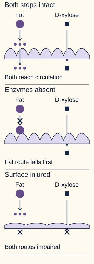
In the accompanying route diagram, trace dietary fat through digestion and then the epithelial surface. Next trace D-xylose, a simple sugar that needs no pancreatic digestion. Put a finger or an imagined mark over the pancreatic enzyme input, keeping the surface intact. Predict which route still reaches blood before checking the explanation.
Trace the large circle as dietary fat, small circles as digestion products, squares as D-xylose, and the lavender ribbon as epithelium. With absent enzymes, fat digestion fails but D-xylose still reaches circulation. With injured mucosa, both routes are impaired. Crosses mark impaired steps, not necessarily zero absorption. This is a qualitative model, not a diagnostic test result. [2][6]Check the pancreatic interruption
D-xylose can still be absorbed. Fat digestion fails before the shared absorptive surface, so steatorrhea can coexist with preserved D-xylose absorption.
Now restore enzyme delivery but shorten the absorptive surface in the lower comparison. Both prepared nutrients and D-xylose face a mucosal problem. The visible contrast is the important result: a digestion-dependent substrate and a directly absorbed substrate respond differently to the same interruption. This is a localization model, not a recommendation to order D-xylose routinely. [6]
Five causes compared by the first important failure
Cause
Failure and expected pattern
CauseCeliac disease
Failure and expected patternGluten-associated immune injury, often most prominent proximally; iron deficiency may precede obvious diarrhea.
CausePrimary lactose intolerance
Failure and expected patternInsufficient lactase with preserved villous architecture; lactose-dependent gas and watery stool.
CauseExocrine pancreatic insufficiency
Failure and expected patternInadequate digestive enzyme delivery; steatorrhea and fat-soluble vitamin deficiency despite a potentially intact lining.
CauseTropical sprue
Failure and expected patternAcquired, often diffuse small-bowel dysfunction after relevant residence or prolonged exposure; folate and B12 deficiencies.
CauseWhipple disease
Failure and expected patternInfected macrophages expand the lamina propria; intestinal symptoms belong to a systemic infection.
For a new patient with bloating only after milk and stable weight, trace lactose rather than assuming global nutrient failure. For a patient with greasy stool after pancreatic surgery, start in the lumen. Neither history alone establishes a final diagnosis. [2][4][5][6][7]
Why does gluten injure some patients but not everyone?
Celiac disease is an immune-mediated response to gluten proteins in wheat, barley and rye in a susceptible host. HLA susceptibility is important, but a positive HLA result is not a diagnosis: many unaffected people carry associated variants. This is not simply a shortage of an enzyme that digests bread. [1][9][10]
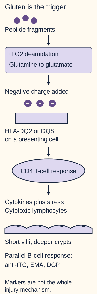
The immune diagram separates peptide processing from tissue injury. Incompletely digested, gliadin-rich peptides are sampled across the epithelium. Tissue transglutaminase 2 converts selected glutamine residues to glutamate through deamidation. The added negative charge favors binding of relevant peptides to HLA-DQ2 or HLA-DQ8 on antigen-presenting cells. These cells activate gluten-responsive CD4 T cells. [9]
Follow the negative charge added by deamidation to HLA presentation. Epithelial stress and cytotoxic lymphocytes participate in tissue injury, while B-cell responses generate useful serologic markers. Spatial arrangement is simplified and does not depict a complete signaling network. [9]
Inflammatory cytokines and epithelial stress signals, including IL-15-related signaling, support cytotoxic intraepithelial lymphocyte activity. Enterocytes are injured, villi shorten, and crypt proliferation produces crypt hyperplasia. B-cell responses generate anti-tTG, endomysial and deamidated-gliadin antibodies. These are useful markers, but the pathway should not be reduced to antibodies directly causing all epithelial destruction. [9]
Use the figure to locate deamidation before antigen presentation. If a selective experimental inhibitor prevents that chemical modification, predict the immediate effect on binding of a deamidation-dependent peptide.
Check the predicted binding effect
Binding to the susceptible HLA molecule would be less favorable. This prediction concerns the specified peptide, not guaranteed elimination of every immune response or proven clinical benefit of an experimental treatment.
The consequence downstream is less stimulation through that particular presentation pathway, not instant regrowth of villi. To transfer the idea, consider an asymptomatic person with HLA-DQ2: susceptibility supplies a presentation system, but does not demonstrate active injury. Conversely, characteristic mucosal injury needs clinical and laboratory interpretation rather than an HLA label alone. [9][10]
Can a negative celiac test be trusted?
Before interpreting a negative antibody result, ask two questions: is the patient eating gluten, and can the patient produce IgA? In most patients, tTG-IgA is the initial serology; total serum IgA helps identify an IgA-based false negative. With IgA deficiency, use IgG-based tTG or deamidated-gliadin peptide testing. EMA-IgA is highly specific and can clarify selected results, but is not a solution to IgA deficiency. [10]
A 34-year-old with iron deficiency has a negative tTG-IgA and undetectable total IgA while still eating wheat. Predict whether repeating another IgA-class antibody solves the problem.
Check which antibody class is useful
Repeating an IgA-class test leaves the same limitation in place. IgG-based testing is appropriate, with biopsy assessment if clinical suspicion remains substantial.
Starting a gluten-free diet before evaluation can reduce both antibodies and mucosal abnormalities. A negative result after restriction does not exclude untreated disease. Discuss diagnostic reassessment with the treating clinician; HLA testing can help exclude disease in selected uncertain cases, whereas a positive susceptibility result cannot confirm it. Any gluten challenge should be planned rather than improvised. [10]
For an IgA-sufficient child younger than two, ACG 2023 recommends tTG-IgA as the preferred single test. Age alone is not a reason to replace it with IgG testing. Supplemental testing can be considered in a specialist evaluation when findings and results disagree. [11]
Connect the blood result to the tissue
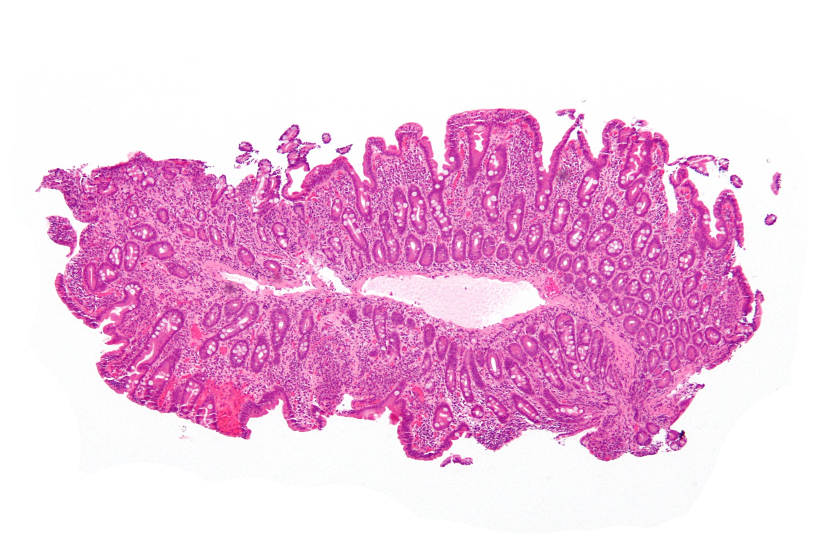
Most adults with suspected celiac disease require upper endoscopy with multiple duodenal biopsies, including bulb and distal duodenum, for confirmation. The characteristic combination is villous blunting or atrophy, crypt hyperplasia and increased intraepithelial lymphocytes. Isolated lymphocytosis is not specific. Injury can be patchy, so a single sample can mislead. [1][11]
Inspect the accompanying real H&E micrograph at low magnification. First compare the short surface projections with the depth of the gland-rich tissue beneath them. The image illustrates architectural change; it does not by itself establish the patient's antibody status or permit a reliable lymphocyte count at this displayed scale. Villous blunting also occurs in other enteropathies. [6][11]
Celiac disease, low magnification, H&E stain. Inspect the short surface projections relative to the deeper crypt-rich tissue. Architectural blunting is visible; this scale does not support a reliable intraepithelial lymphocyte count. Histology needs serologic and clinical context. Image: Nephron / Wikimedia Commons, CC BY-SA 3.0. Original image displayed in full Nephron; original source; CC BY-SA 3.0.
Iron-deficiency anemia can be an early presentation even without prominent diarrhea. An intensely pruritic vesicular eruption on extensor surfaces suggests associated dermatitis herpetiformis. Do not require the entire textbook pattern before testing. In selected children, a specialist nonbiopsy pathway uses tTG-IgA greater than ten times the assay upper limit plus positive EMA in a second sample. Do not apply that shortcut to every positive result. [1][6][9][11]
After diagnosis, treatment is a strict gluten-free diet with dietitian support, correction of deficiencies and lifelong follow-up. Persistent symptoms require reassessment of exposure and alternative or coexisting causes before labeling disease refractory. Recovered villous architecture with a changed watery-stool pattern should prompt assessment for another disorder, such as microscopic colitis, rather than immediate assignment to refractory enteropathy. A patient whose diarrhea improves but whose iron stores remain depleted still needs nutritional follow-up. [1][13]
How can an intact lining produce substantial symptoms?
Compare two meals: milk produces gas and watery stool in one patient; a mixed meal produces oily stool in a patient with chronic pancreatitis. The first suggests a selective brush-border problem, the second a luminal digestive problem. Normal villous architecture does not establish normal digestive function.[2][6][7]
Lactose: retain water, then ferment carbohydrate
When lactase activity is insufficient, undigested lactose remains in the lumen and retains osmotic water. Colonic bacteria ferment it, producing gases and organic acids. Bloating, cramps, an increase in breath hydrogen and sometimes acidic stool follow. Symptoms depend on the amount consumed, transit and colonic handling; malabsorption on a test does not necessarily mean symptomatic intolerance. A lactose hydrogen-breath test is interpreted with the patient's symptoms during the challenge. [3][6][7]
Predict the response if the same patient consumes lactose-free milk containing the same milk proteins.
Check the substrate substitution
Symptoms caused by lactose maldigestion should lessen because the problematic sugar has been digested before ingestion. This substitution would not address an immune reaction to milk proteins.
Primary lactase nonpersistence generally preserves the mucosa. Secondary deficiency can follow infectious enteritis or celiac injury to mature villus-tip enterocytes and may improve as the mucosa heals. New dairy intolerance after gastroenteritis therefore does not prove a lifelong primary defect. Conversely, weight loss and iron deficiency should prompt evaluation beyond dietary lactose alone. Individualize lactose reduction or lactase supplementation and protect nutritional intake rather than prescribing unnecessary blanket food exclusions. [7][8]
Pancreatic insufficiency: measure function carefully
Chronic pancreatitis, cystic fibrosis, pancreatic cancer and pancreatic surgery are important risk settings. Poor fat digestion produces steatorrhea, weight loss and deficiencies of vitamins A, D, E and K. A normal-looking intestinal lining is compatible with this problem. [2]
Fecal elastase is the appropriate initial test when exocrine pancreatic insufficiency is suspected. Use a semisolid or solid stool specimen: water dilution can produce a falsely low concentration. Below 100 micrograms/g provides good evidence of insufficiency; 100 to 200 micrograms/g is indeterminate. A result must be integrated with the history, and imaging investigates structural pancreatic disease rather than directly measuring exocrine function. [2]
For example, an elastase of 150 micrograms/g is not equivalent to 50 micrograms/g, and 50 in watery stool is not equivalent to 50 in a formed sample. Predict which uncertainty needs correction first when diarrhea is entirely watery.
Check the specimen limitation
Obtain an appropriate stool specimen for repeat elastase assessment when possible. A diluted sample should not be treated as definitive evidence of pancreatic failure.
Confirmed insufficiency requires pancreatic enzyme replacement during meals and snacks, plus nutritional assessment and fat-soluble vitamin monitoring. Symptom improvement during an empirical enzyme trial is not a reliable diagnostic test. In a patient already receiving replacement, fecal elastase can still be measured; stopping needed treatment is not required solely for that assay. [2]
What do the deficiencies and extraintestinal findings localize?
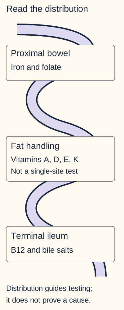
The nutrient map shows tendencies, not exclusive territories. Iron and folate deficits suggest proximal small-bowel dysfunction. Calcium and vitamin D abnormalities can accompany celiac disease and contribute to bone disease. Vitamin B12 absorption depends on several earlier digestive steps and an intact terminal ileum, so low B12 alone does not prove ileal injury. Bile-salt reabsorption also depends on the terminal ileum; substantial distal dysfunction can impair fat handling. [6]
The bowel is schematic and not drawn to scale. Nutrient associations are tendencies, not exclusive absorption territories. Fat-soluble vitamin deficiency can result from luminal, mucosal or bile-salt problems; low B12 also has upstream causes. [4][6]
Trace a proximal lesion on the map, then extend it distally. Predict which additional nutrient problem becomes more likely without assuming a fixed sequence for every patient.
Check the distal extension
B12 and bile-salt handling become more vulnerable when disease affects the terminal ileum. The same B12 deficit can also arise from gastric, pancreatic or bacterial causes, so anatomy must be interpreted with context.
Tropical sprue: exposure plus a broader enteropathy
A patient develops persistent diarrhea after months living in an endemic tropical region and later has both folate and B12 deficiency. Tropical sprue is an acquired disorder of uncertain, probably infectious etiology; no single established pathogen defines it. It often affects broad areas of small bowel, with villous blunting and chronic inflammation. Both folate and B12 deficiencies can occur; their timing does not establish a mandatory proximal-to-distal progression. [4]
Do not diagnose it from travel alone. Exclude celiac disease and parasitic infection, assess deficiencies, and obtain small-bowel biopsies in the appropriate evaluation. Celiac serology is generally negative in tropical sprue, but first establish that the celiac tests were interpretable. Treatment uses antibiotics and nutritional replacement, particularly folate and B12. Replace B12 when deficient rather than treating the anemia with folate alone. Failure to respond should trigger diagnostic reassessment. [4]
Whipple disease: the joints may speak before the bowel
Migratory arthralgia can precede weight loss and diarrhea by years. Neurologic dysfunction or endocarditis makes an isolated food intolerance particularly implausible. Tropheryma whipplei causes a systemic infection; in the bowel, foamy macrophages containing periodic acid-Schiff (PAS)-positive material fill the lamina propria and can distort villous architecture. [5]
Contrast this with celiac disease: in celiac disease the characteristic epithelial and crypt pattern matters; in Whipple disease the macrophage-rich lamina propria points toward an organism. PAS positivity is not sufficient on its own. Intestinal mycobacterial infection can resemble it, but mycobacteria are acid-fast whereas T. whipplei is not. Organism-directed PCR or immunohistochemistry helps confirm the diagnosis in context. Detection in saliva or stool may reflect carriage; a test on involved tissue better connects the organism to the lesion. Routine bacterial culture is not a reliable substitute for targeted testing. [5][14]
Apply the distinction to a patient whose joints improve on anti-inflammatory treatment but who then develops diarrhea and cognitive change. Do not assume a new unrelated intolerance. Investigate systemic infection, including cardiac or central nervous system involvement when indicated. Treatment requires specialist-directed, prolonged antibiotics with attention to central nervous system penetration and late relapse; a brief course that improves diarrhea is not proof of cure. [5]
Years after treatment, new cognitive or gait abnormalities warrant assessment for possible CNS recurrence even if diarrhea has not returned. Routine CSF indices can be normal; appropriately selected organism-specific CSF PCR addresses a different question from routine cell counts or stool testing. A negative assay does not exclude every case, and treatment planning belongs with specialists. [5][12]
Which test answers the question you are actually asking?
The historical D-xylose framework asks whether a simple sugar can cross proximal mucosa without pancreatic digestion. It does not identify a particular disease. Preserved absorption in isolated pancreatic insufficiency contrasts with reduced absorption in celiac disease or tropical sprue, provided mucosal function, renal handling, transit and specimen collection are adequate. [6]
Compare a normal-range blood concentration with a low urinary recovery in a patient with renal dysfunction. Predict whether the urine result alone localizes damage to the villi.
Check the blood and urine disagreement
No. Urinary recovery depends on renal excretion and complete collection as well as absorption. Renal dysfunction can give a misleadingly low urinary result despite nutrient entry into blood; altered clearance also means a normal-range blood result does not prove normal absorptive capacity.
Bacterial overgrowth can consume D-xylose before absorption; delayed gastric emptying and other transit or collection problems also complicate interpretation. For these reasons, D-xylose is rarely used now. Its value here is learning the difference between digestion and uptake, not choosing a universal first-line test. [6]
Match the clinical question to the modern investigation
Question
Useful direction
QuestionIs gluten-associated enteropathy likely?
Useful directionGluten-exposed serology with IgA assessment, then duodenal biopsy when indicated.
Useful directionExclude celiac disease and parasites, assess deficiencies, and evaluate small-bowel histology.
QuestionCould diarrhea be part of systemic infection?
Useful directionSmall-bowel biopsy and organism-directed testing, with extraintestinal evaluation.
Stool fat can establish steatorrhea but cannot by itself distinguish pancreatic from mucosal disease. Nutritional tests assess consequences and guide replacement: blood count, iron stores, folate, B12, albumin and targeted mineral or vitamin measurements are selected from the history. Treat dehydration and nutritional compromise while investigating, rather than waiting for a perfect disease label. [2][3][4][5][6][10]
Return to the opening patients: both lose fat, but only one needs pancreatic digestion restored. The other needs its mucosal cause identified and treated. For a new mixed presentation, allow coexistence: celiac injury can produce secondary lactose intolerance, and a single test need not explain every symptom.
Independent practice
Case 1
Show answer and explanations for case 1
A. EMA-IgA antibody (Why this does not fit)
EMA-IgA is a specific celiac-associated antibody test in patients able to produce IgA. Changing the antigen target but keeping the IgA class does not solve this patient's immunoglobulin limitation.
Reasoning steps for option A
Why does a specific EMA-IgA test still depend on antibody production?
EMA-IgA requires IgA production despite its high specificity for celiac disease.
How does total IgA below 5 undermine an EMA result?
At less than 5 mg/dL total IgA, an EMA-IgA negative could reflect inability to make IgA rather than absent celiac disease.
B. DGP-IgG antibody (Best answer)
IgG-based testing can detect a celiac-associated response when IgA production is deficient. The extremely low total IgA makes the negative tTG-IgA insufficient to exclude disease despite ongoing gluten exposure.
Reasoning steps for option B
How does DGP-IgG bypass the immunoglobulin defect?
DGP-IgG measures an IgG response and is usable despite this woman's profound IgA deficiency.
Why does eating wheat preserve a useful opportunity for IgG serology?
Because she eats wheat daily, gluten-associated antibody production remains assessable; DGP-IgG avoids the IgA limitation.
C. Repeat tTG-IgA antibody (Why this does not fit)
Repeating an assay can help with transient or technical uncertainty. Here the known deficiency of its antibody class remains, so another negative IgA result would still be difficult to interpret.
Reasoning steps for option C
When might repeating tTG-IgA clarify a negative test rather than repeating this IgA limitation?
Repeating tTG-IgA might help if a transient assay error, rather than antibody-class deficiency, were suspected.
Why would another tTG-IgA remain uninterpretable with severe IgA deficiency?
Her measured IgA deficiency persists, so another negative tTG-IgA would still not reliably exclude celiac disease.
D. HLA-DQ2/DQ8 genotype (Why this does not fit)
Absence of associated HLA variants can help exclude celiac disease in uncertain settings. A positive genotype would indicate susceptibility rather than assess the antibody response that the IgA assay cannot measure.
Reasoning steps for option D
How can absent DQ2/DQ8 clarify an uncertain diagnosis?
Absence of both associated HLA variants can make celiac disease unlikely in an uncertain case.
Why does a positive HLA result fail to demonstrate an antibody response?
Positive DQ2 or DQ8 identifies susceptibility, not whether this IgA-deficient woman has a gluten-associated antibody response.
Takeaway: Validate the antibody class before interpreting a negative celiac screen.
A. Repeat tTG-IgA after another four months of the same gluten-free diet (Why this does not fit)
Serial antibodies can be useful for monitoring known celiac disease. Continued restriction does not restore sensitivity for establishing an unconfirmed diagnosis after antibodies have become negative.
Reasoning steps for option A
Why would serial tTG-IgA be more useful after a confirmed diagnosis than in this man's unconfirmed gluten-restricted evaluation?
Serial tTG-IgA can monitor an established diagnosis, but his negative result after gluten avoidance cannot establish whether he originally had celiac disease.
Why does remaining gluten-free not restore diagnostic sensitivity?
Continuing gluten avoidance could keep tTG-IgA negative even if he had celiac disease before restriction.
B. Substitute IgG-based celiac serology while maintaining the gluten-free diet (Why this does not fit)
IgG assays can address the problem of IgA deficiency. Total IgA is normal here; switching antibody class does not resolve the reduced antigen exposure that limits current testing.
Reasoning steps for option B
Does this man's normal total IgA provide the usual reason to switch to IgG serology?
No. IgG assays address IgA deficiency, which he does not have; switching classes does not restore the missing gluten exposure.
Why does normal total IgA leave gluten restriction as the key limitation?
His IgA is normal, but four months without gluten reduces antigen stimulation for either antibody class.
C. Use duodenal biopsy on the current diet as the final exclusion test (Why this does not fit)
Biopsy is important when the intestine is exposed to gluten and a diagnosis is being established. After months of restriction, healed mucosa cannot reliably exclude prior celiac enteropathy, so this sequence can close evaluation prematurely.
Reasoning steps for option C
When can duodenal biopsy establish active celiac injury?
Duodenal biopsy can demonstrate injury while gluten exposure sustains active enteropathy.
Why can a biopsy after four months of restriction fail to exclude prior injury?
Four months of gluten restriction may heal the mucosa, so a normal biopsy now cannot rule out previous celiac injury.
D. Assess HLA susceptibility, then discuss a supervised gluten challenge if needed (Best answer)
Gluten restriction can normalize serology and mucosal findings, whereas genotype is diet independent. An incompatible HLA result can help exclude celiac disease; a compatible result may justify planned challenge and subsequent testing rather than confirm the diagnosis itself.
Reasoning steps for option D
Why is HLA typing unaffected by this man's diet?
His HLA genotype does not change when he stops eating gluten.
How should compatible HLA prompt planned challenge rather than immediate diagnosis?
A compatible genotype indicates susceptibility only; supervised gluten challenge and subsequent testing may be needed for diagnosis.
Takeaway: When treatment has already altered the test, select a reassessment strategy that accounts for exposure.
A. Obtain multiple biopsies from the duodenal bulb and distal duodenum (Best answer)
Most adults with suspected celiac disease require duodenal histologic confirmation. A modestly positive antibody result plus unexplained iron deficiency supports sampling multiple sites because injury may be patchy.
Reasoning steps for option A
Why are multiple duodenal sites sampled in a gluten-eating adult?
Celiac injury can be patchy, so sampling bulb and distal duodenum reduces the risk of missing it.
How do iron deficiency and fourfold tTG-IgA support histologic confirmation?
Her persistent iron deficiency and tTG-IgA at four times the upper limit justify histologic confirmation while she still eats gluten.
B. Repeat antibodies after three months of strict gluten avoidance (Why this does not fit)
Antibody decline can occur when the relevant dietary stimulus is absent. Starting restriction before tissue confirmation may obscure the diagnosis rather than establish the cause of the iron deficiency.
Reasoning steps for option B
How could three months of gluten avoidance change this woman's fourfold tTG-IgA elevation?
The titer could decline after gluten withdrawal even if celiac disease caused her iron deficiency, without establishing the diagnosis.
How could starting restriction before biopsy obscure the cause of anemia?
Starting the diet now could blunt tissue injury before biopsy, leaving her iron-deficiency cause unconfirmed.
C. Obtain HLA typing and confirm disease if DQ2 is present (Why this does not fit)
HLA-DQ2 is associated with celiac susceptibility. Its presence is common without disease and cannot confirm active enteropathy in this patient.
Reasoning steps for option C
Would finding DQ2 establish the cause of this woman's iron deficiency or only celiac susceptibility?
DQ2 establishes susceptibility only, not active celiac enteropathy as the cause of her iron deficiency.
Why can susceptibility alone not prove active enteropathy?
Many DQ2 carriers have no celiac enteropathy; genotype cannot prove that her anemia reflects active mucosal injury.
D. Obtain EMA once and omit biopsy if the result is positive (Why this does not fit)
EMA can improve diagnostic specificity after positive tTG-IgA. This adult has feasible endoscopy and does not meet a selected high-titer nonbiopsy pathway; confirmatory biopsy remains appropriate.
Reasoning steps for option D
What specificity can EMA add to positive tTG-IgA?
EMA can add specificity to a positive tTG-IgA result.
Why is a single positive EMA insufficient to bypass feasible adult biopsy?
At four times the tTG-IgA upper limit in an adult able to undergo endoscopy, a single EMA result does not replace biopsy.
Takeaway: Positive serology directs evaluation; it is not a universal substitute for adult biopsy.
A. More crypt proliferation and improved proximal nutrient uptake (Why this does not fit)
An expanding absorptive surface can improve uptake. However, reduced ongoing injury should lessen rather than further increase the compensatory crypt proliferative response.
Reasoning steps for option A
Why does expanding villous surface aid nutrient uptake?
Why should crypt proliferation fall rather than rise as injury resolves?
Resolution of epithelial injury should reduce, not increase, the compensatory proliferation previously seen in deep crypts.
B. Less crypt proliferation and reduced proximal nutrient uptake (Why this does not fit)
Less ongoing epithelial injury can reduce compensatory crypt proliferation. Recovery of villous height should increase functional surface rather than predict worsening proximal uptake.
Reasoning steps for option B
Why would resolving injury slow compensatory crypt growth?
Crypt hyperplasia compensates for damaged epithelium and should diminish as injury resolves.
Why should taller villi not reduce proximal uptake?
Regained villous height increases functional absorptive surface, so proximal uptake should improve rather than decline.
C. Less crypt proliferation and improved proximal nutrient uptake (Best answer)
The proliferative crypt response compensates for ongoing surface injury in celiac enteropathy. When injury subsides and the absorptive surface recovers, compensatory proliferation should decrease while proximal uptake improves.
Reasoning steps for option C
Why were deep crypts proliferating during active celiac injury?
Active celiac injury shortened villi and drove compensatory cell production in the crypts.
How do restored villi and reduced injury jointly change uptake and crypt activity?
As her villi recover, crypt proliferation falls while proximal absorption improves.
D. More crypt proliferation and reduced proximal nutrient uptake (Why this does not fit)
This pair could accompany persistent or worsening destructive enteropathy. The supplied follow-up instead describes resolving injury and restoration of the villous surface.
Reasoning steps for option D
What course could raise proliferation and lower uptake?
Persistent destructive enteropathy could produce more crypt proliferation and less absorption.
Why does regained villous height contradict worsening enteropathy?
Her follow-up explicitly shows subsiding epithelial injury and restored villous height, opposing both predicted deteriorations.
Takeaway: Predict recovery by linking the tissue response to restoration of absorptive surface.
A. Peptide-HLA binding increases and CD4 activation remains suppressed (Why this does not fit)
Pre-deamidated peptide should restore the impaired binding step. The inhibitor has no supplied effect on presentation after binding or on T cells, so persistent suppression is not the predicted downstream result.
Reasoning steps for option A
What should processed gliadin do to HLA-DQ2 binding?
Pre-deamidated peptide bypasses inhibited tTG2 and should bind HLA-DQ2 more favorably.
Why should CD4 activation not stay suppressed when only deamidation was blocked?
Because the inhibitor affects only deamidation, restored peptide presentation should also restore CD4 activation.
B. Peptide-HLA binding remains reduced and CD4 activation recovers (Why this does not fit)
A T-cell response requires adequate presentation of the relevant peptide. Bypassing deamidation should improve binding itself rather than restore activation while leaving that upstream defect unchanged.
Reasoning steps for option B
Why does CD4 activation require peptide presentation?
Gliadin-specific CD4 cells require sufficient relevant peptide displayed by HLA-DQ2.
Why should pre-deamidated peptide improve the upstream binding defect?
Pre-deamidated peptide bypasses the only blocked processing step, so its HLA binding should recover too.
C. Peptide-HLA binding increases and CD4 activation recovers (Best answer)
Deamidation adds negative charge that favors binding of relevant gliadin peptides to susceptible HLA. Supplying the processed peptide bypasses the inhibited step, restoring presentation and the downstream T-cell response under the matched conditions.
Reasoning steps for option C
How does deamidation favor gliadin binding to DQ2?
Deamidation adds negative charge to relevant gliadin peptides, improving their binding to susceptible HLA-DQ2.
Why should bypassing tTG2 inhibition restore activation under matched conditions?
Supplying peptide already deamidated restores presentation and hence CD4 activation under the matched conditions.
D. Peptide-HLA binding remains reduced and CD4 activation remains suppressed (Why this does not fit)
Both findings would be expected if an indispensable later presentation step remained blocked. Here the inhibitor acts only on deamidation, and pre-deamidated peptide bypasses that specified block.
Reasoning steps for option D
What additional defect would be needed to keep peptide-HLA binding and CD4 activation low despite pre-deamidated peptide?
A separate defect preventing effective peptide-HLA binding or presentation would be needed; the supplied inhibitor acts only on deamidation.
Why does this selective deamidation inhibitor not cause such a later block?
No such later defect is supplied: the selective inhibitor blocks tTG2 deamidation, which processed peptide bypasses.
Takeaway: A rescue experiment distinguishes an upstream processing requirement from a later signaling defect.
A. Investigate a coexisting cause of watery diarrhea, such as microscopic colitis (Best answer)
Symptoms can persist from another disorder despite healing of celiac enteropathy. The changed stool pattern and recovered mucosa argue for an additional cause rather than a refractory villous process.
Reasoning steps for option A
Why can nocturnal watery diarrhea reflect a second disorder after healing?
A separate cause such as microscopic colitis may cause watery nocturnal stools after celiac mucosa heals.
How do normalized serology and villi favor microscopic colitis evaluation?
Normal villous height, no excess lymphocytes, normalized tTG-IgA and a changed stool pattern favor another disorder.
B. Perform lymphocyte phenotyping to classify refractory celiac disease (Why this does not fit)
Lymphocyte assessment can be relevant when persistent villous atrophy raises concern for refractory disease. This biopsy shows mucosal recovery, so the key prerequisite for that explanation is absent.
Reasoning steps for option B
What tissue abnormality motivates refractory-celiac phenotyping?
Persistent villous atrophy despite appropriate gluten avoidance would raise concern for refractory celiac disease.
Why does normal villous height weaken that diagnosis?
Her repeat biopsy shows normal villous height and no excess epithelial lymphocytes, not the ongoing injury that warrants this classification.
C. Use HLA typing to reassess whether the original celiac diagnosis was valid (Why this does not fit)
HLA testing can help exclude celiac disease when the original diagnosis is uncertain. The original diagnosis was biopsy confirmed and has shown serologic and histologic response; genotype does not explain the new watery-stool pattern.
Reasoning steps for option C
When does HLA typing help exclude a doubtful original diagnosis?
An incompatible HLA result can help exclude celiac disease if the original diagnosis is doubtful.
Why cannot genotype explain new watery stools after biopsy-confirmed celiac response?
Her disease was biopsy-confirmed and improved objectively; genotype cannot explain the new watery nocturnal diarrhea.
D. Escalate gluten restriction based on persistent symptoms as the primary evidence of exposure (Why this does not fit)
Dietary exposure is an important cause of persistent symptoms and warrants careful review. Here review and objective mucosal recovery weaken active gluten injury as the explanation for a changed symptom pattern.
Reasoning steps for option D
Why review gluten exposure with persistent symptoms?
Ongoing gluten exposure is a common cause of persistent celiac symptoms and warrants dietary review.
Why do healed villi weaken persistent exposure as the primary explanation?
Review found no likely exposure, and normalized serology and villi weaken active gluten injury as the primary cause.
Takeaway: Persistent symptoms and persistent enteropathy are not interchangeable; reassess the current phenotype.
A. Reduced in A and preserved in B (Why this does not fit)
This pattern would fit a test dependent mainly on pancreatic digestion. D-xylose bypasses that step, so villous injury rather than pancreatic enzyme loss should reduce absorption.
Reasoning steps for option A
What would low D-xylose recovery in pancreatic patient A imply?
Reduced A with preserved B would incorrectly imply that D-xylose depends on pancreatic digestion.
Why should villous injury in B instead impair direct sugar uptake?
A has intact villi and should absorb this simple sugar; B has proximal villous atrophy that impairs uptake.
B. Reduced in A and reduced in B (Why this does not fit)
Both disorders can cause steatorrhea and loss of dietary nutrients. That shared stool finding does not mean both interrupt direct absorption of a simple sugar.
Reasoning steps for option B
Why might shared steatorrhea suggest both recoveries are low?
Both patients lose dietary fat, which can superficially suggest global malabsorption in both.
Why does simple D-xylose differ from digestion-dependent fat?
D-xylose needs no pancreatic enzymes, so A can absorb it despite steatorrhea while B cannot absorb it normally.
C. Preserved in A and preserved in B (Why this does not fit)
Preserved recovery is expected when proximal uptake is intact. The extensive proximal villous injury in B makes that assumption inappropriate despite normal renal handling.
Reasoning steps for option C
What permits preserved recovery in patient A?
Patient A has normal duodenal villi and can directly absorb D-xylose.
Why should extensive proximal atrophy prevent preserved recovery in B?
Patient B has extensive proximal villous atrophy, so preserved recovery is implausible despite adequate renal handling.
D. Preserved in A and reduced in B (Best answer)
D-xylose requires proximal mucosal absorption but no pancreatic enzyme digestion. A has an intact surface despite poor digestion, whereas B has the mucosal lesion expected to reduce recovery.
Reasoning steps for option D
Why does pancreatic enzyme loss spare D-xylose uptake in A?
D-xylose crosses proximal mucosa without requiring pancreatic enzyme digestion.
How does B's villous atrophy reduce recovery despite adequate collection?
A retains mucosal uptake whereas B loses absorptive surface; recovery is preserved in A and reduced in B.
Takeaway: A shared symptom does not imply an identical result for a digestion-independent substrate.
A. Use pancreatic MRI as the next functional confirmation (Why this does not fit)
Pancreatic imaging can identify structural disorders relevant to insufficiency. Changing structural imaging does not resolve a concentration measured in a dilute specimen or directly establish exocrine function.
Reasoning steps for option A
What could pancreatic MRI add after this man's normal CT, and would that information measure exocrine function?
MRI could provide additional structural information, but it would not directly measure exocrine function or resolve the liquid-stool elastase limitation.
Why cannot imaging correct dilution of fecal elastase in liquid stool?
Imaging cannot correct dilution of elastase in this man's liquid stool or directly measure exocrine output.
B. Repeat fecal elastase on semisolid or solid stool (Best answer)
Liquid stool can dilute an elastase concentration and create a falsely low result. A suitable repeat specimen addresses that unresolved analytical problem; normal CT does not settle pancreatic function.
Reasoning steps for option B
How can watery diarrhea make elastase appear below 100?
Profuse watery stool can dilute fecal elastase, yielding an artificially low concentration of 70 micrograms/g.
Why should a solid specimen resolve the central uncertainty?
Repeating on semisolid or solid stool addresses specimen dilution before assigning pancreatic insufficiency.
C. Use response to a meal-time enzyme trial as confirmation (Why this does not fit)
Enzymes can be clinically useful when insufficiency is established. Symptom response is not reliable for diagnosis and does not repair the specimen limitation in this low-risk presentation.
Reasoning steps for option C
What therapeutic role do meal-time enzymes have in confirmed pancreatic insufficiency?
Enzyme replacement can treat established pancreatic insufficiency and improve digestion.
Why does symptom improvement not confirm insufficiency here?
A symptomatic response to enzymes is not diagnostic, particularly with only three weeks of watery diarrhea and an unreliable specimen.
D. Use the low value as sufficient support for a pancreatic diagnosis (Why this does not fit)
A value below 100 can provide good evidence when obtained from an appropriate sample. The supplied value came from liquid stool, so its apparent severity must be reconciled with possible dilution first.
Reasoning steps for option D
When is elastase of 70 strong evidence of insufficiency?
Below 100 micrograms/g supports insufficiency when elastase is measured in a suitable formed specimen.
Why does this liquid specimen prevent definitive interpretation?
His 70 came from liquid stool and might be dilutional, so it cannot alone establish pancreatic failure.
Takeaway: A numerical threshold cannot compensate for an unsuitable specimen.
A. The initial sample may have been diluted; the second result leaves pancreatic insufficiency unresolved (Best answer)
A liquid sample can yield a lower concentration than a formed sample, so the rise need not represent recovered secretion. A formed value of 160 is still indeterminate; the high-risk history and ongoing nutritional abnormalities require clinical assessment rather than reassurance.
Reasoning steps for option A
Why might liquid stool yield 70 rather than 160?
Dilution of the first liquid sample could explain why elastase rose from 70 to 160 in formed stool.
Why does formed-stool 160 remain unresolved in calcific pancreatitis?
The formed result of 160 remains indeterminate, especially with calcific pancreatitis and persistent nutritional deficits.
B. Replacement may have increased measured human elastase; the second result leaves insufficiency unresolved (Why this does not fit)
The intermediate formed-stool value does leave pancreatic function unresolved. Standard fecal elastase can be measured during replacement, so assay detection of the replacement capsules is not the appropriate explanation for the rise.
Reasoning steps for option B
What does a formed-stool value of 160 leave unresolved?
A formed elastase of 160 does not settle whether pancreatic exocrine output is inadequate.
Why do replacement capsules not explain higher human elastase?
Replacement capsules do not falsely increase the standard assay's measured human pancreatic elastase.
C. The initial sample may have been diluted; the second result supports preserved pancreatic function (Why this does not fit)
Possible dilution is a valid concern for the first specimen. The formed value remains within the indeterminate interval and does not justify classifying function as preserved in this high-risk presentation.
Reasoning steps for option C
Why is dilution a plausible source of the initial low value?
Liquid stool can dilute the initial elastase concentration of 70.
Why does 160 fail to establish normal function amid steatorrhea?
Even on formed stool, 160 falls in the 100 to 200 indeterminate range, not a reassuring normal range.
D. Replacement may have increased measured human elastase; the second result supports preserved function (Why this does not fit)
This interpretation attributes the numerical rise to the treatment and then treats it as evidence of normal function. Replacement does not directly normalize this assay, and 160 remains indeterminate despite the higher number.
Reasoning steps for option D
Why is crediting the rise in elastase to replacement mistaken?
Attributing the rise to enzyme capsules mistakes therapeutic enzymes for measured human elastase.
Why are 160 and nutritional deficits incompatible with reassurance?
The formed value of 160 remains indeterminate, and ongoing deficits contradict declaring function preserved.
Takeaway: Correcting a specimen limitation can change a number without resolving the remaining diagnostic uncertainty.
A. Fecal fat decreases while fecal elastase returns to normal (Why this does not fit)
Corrected delivery can reduce the amount of undigested fat reaching stool. Replacement enzymes do not normalize the human pancreatic elastase assay when the underlying secretory defect is unchanged.
Reasoning steps for option A
How can correctly timed enzymes decrease fecal fat?
Taking replacement with meals allows enzyme action on dietary fat and lowers fecal fat.
Why should endogenous elastase not normalize without pancreatic recovery?
Because intrinsic pancreatic secretion has not changed, replacement does not normalize fecal elastase.
B. Fecal fat remains high while fecal elastase returns to normal (Why this does not fit)
Persistent fat loss could indicate an inadequate clinical response. The question specifies restored fat digestion, while unchanged pancreatic disease gives no reason for normalization of endogenous elastase.
Reasoning steps for option B
What would persistently high fecal fat suggest if meal-time enzyme replacement had not restored digestion?
It could suggest ineffective fat digestion, but this case explicitly stipulates restoration of effective digestion.
Why does stipulated restored digestion rule that out here?
The question stipulates effective fat digestion, so fecal fat should fall; unchanged pancreatic disease also keeps elastase low.
C. Fecal fat decreases while fecal elastase remains low (Best answer)
Correctly timed replacement can improve luminal fat digestion and reduce net fecal fat loss. It does not restore endogenous pancreatic secretion, and standard fecal elastase can remain low during effective replacement.
Reasoning steps for option C
Why does taking enzymes with meals improve luminal digestion?
Meal-time dosing delivers enzymes when fat is present for luminal digestion.
What happens to the elastase marker despite improved fat absorption?
Endogenous pancreatic elastase remains low because the capsules replace digestion without reversing secretory failure.
D. Fecal fat remains high while fecal elastase remains low (Why this does not fit)
Low elastase remains compatible with the unchanged pancreatic secretory defect. However, effective restoration of fat digestion should reduce fecal fat even though the secretory marker stays abnormal.
Reasoning steps for option D
Why could formed-stool elastase remain low after correcting this man's enzyme timing?
His underlying pancreatic defect remains, so fecal elastase can remain low during treatment.
Why should effective replacement nevertheless reduce fat in stool?
With effective digestion restored and adequate dietary fat, less undigested fat should pass into stool.
Takeaway: A replacement can correct nutrient digestion without correcting the organ-specific secretion marker.
A. Withhold replacement before collecting the same type of elastase sample (Why this does not fit)
Stopping a medicine is appropriate when it directly interferes with the chosen assay. Pancreatic replacement does not require interruption for standard fecal elastase testing, so a washout adds no necessary interpretive benefit.
Reasoning steps for option A
When would holding replacement before fecal elastase testing improve interpretation?
Withholding replacement would help only if capsules interfered with the human fecal elastase assay.
Why is stopping replacement unnecessary for the human elastase assay?
They do not falsely normalize this assay, so stopping helpful enzymes adds no interpretive benefit.
B. Continue replacement and collect a semisolid or solid elastase sample (Best answer)
Standard fecal elastase testing assesses human pancreatic elastase and can be performed during replacement. The capsules do not require a washout; a suitable stool consistency is still needed.
Reasoning steps for option B
What pancreatic secretion does fecal elastase reflect during therapy?
The assay measures human pancreatic elastase while replacement supplies digestive enzymes without requiring washout.
Why is formed stool still important even while capsules continue?
Semisolid or solid stool avoids water dilution even though the capsules can continue.
C. Substitute serum lipase while continuing the replacement capsules (Why this does not fit)
Serum lipase helps assess pancreatic injury in appropriate contexts. It is not a substitute for assessment of exocrine digestive function in this treated insufficiency scenario.
Reasoning steps for option C
What does serum lipase assess in pancreatic disease?
Serum lipase can indicate pancreatic injury in appropriate clinical settings.
Why does it not substitute for testing exocrine function?
It does not measure the exocrine digestive secretion being documented in this treated patient.
D. Substitute quantitative fecal fat while continuing the current treatment (Why this does not fit)
Fecal fat can assess net fat loss under the conditions of collection and treatment. Improvement on replacement can alter net fat loss, whereas the requested fecal elastase can still assess pancreatic secretion during therapy.
Reasoning steps for option D
What does fecal fat reveal during treatment?
Fecal fat reflects net fat loss under the current enzyme regimen.
Why can therapy change net fat loss without changing endogenous elastase?
Treatment can lower fecal fat without restoring endogenous elastase, which can still be measured during replacement.
Takeaway: Keep the measured biological process separate from the effect of replacement therapy.
A. Reduced hepatic protein synthesis with risk of vitamin D deficiency (Why this does not fit)
Liver disease can prolong prothrombin time and may coexist with nutritional abnormalities. The vitamin K response and strong pancreatic steatorrhea pattern favor vitamin supply failure rather than primary synthetic failure as the dominant explanation.
Reasoning steps for option A
How might hepatic synthetic failure affect prothrombin time?
Impaired hepatic synthesis can prolong prothrombin time by reducing coagulation factors.
Why does vitamin K correction favor deficiency over dominant synthetic failure?
Correction with parenteral vitamin K amid pancreatic steatorrhea favors deficient vitamin supply over primary synthetic failure.
B. Reduced proximal iron uptake with risk of folate deficiency (Why this does not fit)
Proximal mucosal injury can produce iron and folate deficiency. Those deficits do not account for a vitamin K-responsive coagulation abnormality in the supplied fat-maldigestion setting.
Reasoning steps for option B
Which nutrient deficits would fit proximal uptake failure rather than this man's pancreatic fat maldigestion?
Proximal mucosal injury could reduce iron and folate uptake, but those deficits do not explain his vitamin K-responsive prothrombin time.
Why do iron and folate not explain the coagulation response?
Neither deficiency explains the prolonged prothrombin time corrected by vitamin K in this fat-maldigestion setting.
C. Reduced intrinsic-factor production with risk of B12 deficiency (Why this does not fit)
Loss of intrinsic factor can impair B12 absorption and produce hematologic abnormalities. It does not explain vitamin K-responsive prothrombin prolongation together with pancreatic steatorrhea.
Reasoning steps for option C
Which vitamin deficit would reduced intrinsic factor cause instead of the vitamin K deficit suggested by this man's response?
Reduced intrinsic factor would impair B12 absorption, not explain his vitamin K-responsive coagulation abnormality.
Why cannot B12 malabsorption account for vitamin K-responsive bleeding?
B12 deficiency does not explain vitamin K-responsive clotting abnormality with cystic-fibrosis-associated steatorrhea.
D. Reduced luminal fat digestion with risk of vitamin D deficiency (Best answer)
Correction with vitamin K supports deficient availability of a vitamin required for coagulation-factor function. The pancreatic history and steatorrhea localize poor availability to fat maldigestion, which also threatens absorption of vitamin D.
Reasoning steps for option D
How does pancreatic steatorrhea limit vitamin K availability?
Poor pancreatic fat digestion impairs availability of fat-soluble vitamin K, and replacing it corrects the prothrombin time.
Why is vitamin D also threatened by failed fat digestion?
Vitamin D is also fat soluble and therefore at risk with the same pancreatic steatorrhea.
Takeaway: Use the replacement response to identify the deficiency, then localize its cause and predict another nutrient at risk.
A. Bile-salt delivery to the colon and stimulation of fluid secretion (Why this does not fit)
Bile acids reaching the colon can cause watery diarrhea. That process does not best explain selective tolerance of lactose-free milk with hydrogen generation after lactose.
Reasoning steps for option A
How can colonic bile acids cause watery diarrhea?
Bile acids in the colon can promote water secretion and diarrhea.
Why does lactose-triggered hydrogen challenge favor another mechanism?
They do not explain why lactose-free milk is tolerated while lactose challenge provokes hydrogen and cramps.
B. Pancreatic fat maldigestion and fecal loss of unabsorbed triglyceride (Why this does not fit)
Pancreatic insufficiency can cause meal-associated stool changes and nutritional deficits. The specific lactose challenge response and watery rather than oily stool favor carbohydrate handling over fat digestion.
Reasoning steps for option B
What stool abnormality follows pancreatic fat maldigestion?
Pancreatic fat maldigestion tends to cause oily, bulky stools and nutritional loss.
Why do lactose-free milk tolerance and watery stool argue against it?
Her watery lactose-specific symptoms and positive hydrogen challenge point instead to carbohydrate maldigestion.
C. Retention of unabsorbed carbohydrate and bacterial fermentation (Best answer)
Undigested lactose contributes osmotic particles and supplies substrate for colonic gas production. The lactose-specific symptoms and challenge-associated hydrogen rise link both findings to inadequate lactose digestion.
Reasoning steps for option C
Why does retained lactose draw water into the lumen?
Undigested lactose remains osmotically active in the lumen and retains water.
What produces hydrogen during this woman's symptomatic lactose challenge?
Colonic bacteria ferment the unabsorbed lactose, producing hydrogen and gas during the symptomatic challenge.
D. Immune recognition of milk protein and mast-cell mediator release (Why this does not fit)
Milk-protein allergy can produce symptoms after dairy exposure. Lactose-free milk retains milk proteins, so its tolerance favors the sugar-dependent process demonstrated by breath testing.
Reasoning steps for option D
What alternative dairy reaction can cause symptoms after milk?
Milk-protein allergy can cause symptoms after dairy ingestion.
Why does tolerance of lactose-free milk favor lactose rather than milk protein as this woman's trigger?
Lactose-free milk still contains milk proteins, so its tolerance and the hydrogen response favor lactose maldigestion.
A. Tolerance may return as postinfectious surface injury heals (Best answer)
Lactase at the mature surface is vulnerable to intestinal injury. Abrupt onset after infection with prior tolerance supports secondary deficiency and a potentially reversible course.
Reasoning steps for option A
Why might milk tolerance recover after this recent gastroenteritis?
Postinfectious injury can temporarily deplete mature brush-border lactase; tolerance may return as the surface heals.
How does daily milk tolerance before infection support a secondary lactase deficit?
He drank milk daily without symptoms until gastroenteritis; abrupt onset afterward fits temporary surface injury rather than longstanding low lactase.
B. Tolerance will probably remain low from newly expressed primary lactase nonpersistence (Why this does not fit)
Primary nonpersistence can become clinically apparent in adolescents or adults. The close postinfectious onset and immediately preceding daily tolerance favor a transient secondary explanation in this patient.
Reasoning steps for option B
Could primary lactase nonpersistence begin at age 19?
Yes. Primary lactase nonpersistence may emerge during adolescence or adulthood, but timing here favors secondary loss.
Why does onset ten days after gastroenteritis weaken a primary explanation?
Symptoms began just ten days after infection despite immediately preceding daily tolerance, favoring secondary deficiency over newly evident primary nonpersistence.
C. Tolerance should vary with pancreatic enzyme secretion during recovery (Why this does not fit)
Pancreatic enzyme delivery can affect digestion of fat and other substrates. The milk-specific problem after mucosal injury is more consistent with brush-border lactase than impaired pancreatic secretion.
Reasoning steps for option C
What substrate problem would pancreatic enzyme deficiency usually cause?
Pancreatic insufficiency chiefly impairs luminal digestion, especially fat, rather than brush-border lactose hydrolysis.
Why does new milk-specific diarrhea after enteritis point away from pancreatic secretion?
A postinfectious problem specifically with milk implicates surface lactase rather than generalized pancreatic enzyme failure.
D. Tolerance should return when an immune reaction to milk proteins subsides (Why this does not fit)
An immune food reaction can produce gastrointestinal symptoms. The postinfectious timing with long previous tolerance favors temporary carbohydrate digestion failure rather than new immune sensitization as the leading explanation.
Reasoning steps for option D
Can a new immune response to milk proteins cause cramps and diarrhea?
It can, but the sudden onset after infection following years of tolerance favors secondary lactase deficiency.
Why is transient postinfectious carbohydrate maldigestion favored over milk-protein sensitization?
Long prior milk tolerance and onset just after intestinal infection make temporary lactose maldigestion more likely than new immune sensitization.
Takeaway: Use the previous dietary baseline and onset after injury to predict a potentially reversible deficit.
A. A and B both have symptom-linked lactose intolerance (Why this does not fit)
Both patients have objective evidence of lactose malabsorption. Only B has symptom reproduction and a substrate-specific dietary response; A does not establish that lactose causes her complaints.
Reasoning steps for option A
What do the two positive hydrogen breath tests establish for A and B?
Both positive, interpretable breath tests establish lactose malabsorption, not necessarily symptomatic intolerance.
Why do A’s symptom-free challenge and unchanged complaints off lactose refute symptomatic intolerance in A?
A had no challenge symptoms and no improvement when she removed lactose, so her positive test alone does not attribute her complaints to lactose.
B. A has no lactose malabsorption; B has symptom-linked intolerance (Why this does not fit)
B meets the supplied biochemical and symptom pattern for intolerance. A still has an interpretable positive malabsorption test even though the finding does not explain her symptoms.
Reasoning steps for option B
Does B’s symptomatic positive challenge establish lactose intolerance?
Yes. B has a positive test with cramps and diarrhea during challenge and improvement off lactose.
Why is it incorrect to call A’s properly interpreted positive test negative for malabsorption?
A has an interpretable positive breath test and thus malabsorption, even though she has not shown lactose-linked symptoms.
C. A has demonstrated malabsorption without established intolerance; B has a milk-protein reaction (Why this does not fit)
A correctly separates test positivity from symptom attribution. B tolerates lactose-free milk with the same milk proteins, which favors lactose rather than a milk-protein reaction.
Reasoning steps for option C
What part of the proposed interpretation correctly describes A?
A has demonstrated lactose malabsorption, but her complaints are not established as lactose-related.
Why does B’s tolerance of lactose-free milk argue against a milk-protein reaction?
B tolerates lactose-free milk containing the same milk proteins, so lactose rather than those proteins explains her symptoms.
D. A has demonstrated malabsorption without established symptomatic intolerance; B has symptom-linked intolerance (Best answer)
A positive hydrogen test identifies malabsorption, but attribution of symptoms also requires concordance. A lacks both challenge symptoms and a dietary response, whereas B has reproducible symptoms tied to lactose rather than milk proteins.
Reasoning steps for option D
How should A’s positive test be interpreted without challenge symptoms or dietary benefit?
A has demonstrable malabsorption without established symptomatic intolerance because neither challenge nor dietary removal links lactose to her complaints.
What links B’s cramps and diarrhea to lactose rather than the proteins in milk?
B develops symptoms with ordinary milk, not lactose-free milk, and reproduces cramps and diarrhea with a positive lactose challenge.
Takeaway: Combine the absorption result with challenge symptoms and a substrate-specific dietary comparison.
A. The persistent symptom establishes refractory celiac disease (Why this does not fit)
Refractory disease requires more than a continuing symptom after treatment begins. The selective response to the lactose content provides a plausible coexisting explanation that should be assessed first.
Reasoning steps for option A
Does cramping six weeks into a gluten-free diet establish refractory celiac disease?
No. Six weeks of improvement followed by milk-specific cramps does not establish refractory disease.
What dietary contrast supplies an alternative explanation to treatment failure?
Ordinary milk provokes cramps while lactose-free milk does not, offering a coexisting lactose-specific explanation.
B. Residual surface lactase deficiency can persist while mucosa recovers (Best answer)
Celiac injury can reduce lactase activity and produce secondary lactose intolerance. Tolerance of lactose-free milk points to the residual sugar-digestion problem rather than proving persistent gluten exposure.
Reasoning steps for option B
How can healing celiac mucosa still produce symptoms with ordinary milk?
Celiac mucosal injury can temporarily reduce brush-border lactase even while the mucosa recovers on a gluten-free diet.
Why does tolerance of lactose-free milk implicate residual lactase deficiency?
Removing lactose while retaining milk proteins prevents her cramps, favoring persisting secondary lactase deficiency.
C. The milk response establishes an immune reaction to dairy proteins (Why this does not fit)
Milk-protein allergy can produce symptoms despite normal carbohydrate digestion. Lactose-free milk retains those proteins, making its tolerance less consistent with that explanation.
Reasoning steps for option C
Would removing lactose from milk remove its allergenic proteins?
No. Lactose-free milk retains the milk proteins that would trigger protein allergy.
How does her tolerance of lactose-free milk weaken a milk-protein allergy diagnosis?
She tolerates the same milk proteins in lactose-free milk, which argues against protein allergy.
D. The response shows that pancreatic enzymes cannot digest dietary fat (Why this does not fit)
Pancreatic insufficiency can cause ongoing symptoms in some patients with intestinal disease. The selective difference between ordinary and lactose-free milk is more consistent with lactase function than generalized fat maldigestion.
Reasoning steps for option D
Would deficient pancreatic lipase specifically distinguish ordinary from lactose-free milk?
No. Lipase-dependent fat digestion is not selectively restored by removing lactose from otherwise similar milk.
Why is this milk contrast more specific for brush-border lactase than fat maldigestion?
Only lactose content distinguishes the two tolerated or symptomatic milk exposures, favoring lactase over fat maldigestion.
Takeaway: Two digestive problems can coexist; persistent symptoms need not refute the primary diagnosis.
Celiac disease can cause villous blunting and nutrient deficiencies. It is less supported by the supplied interpretable negative serology and the prolonged exposure-associated onset, although seronegativity alone never makes it impossible.
Reasoning steps for option A
Which biopsy and deficiency findings could also occur in celiac disease?
Villous blunting, chronic diarrhea, and folate and B12 deficiency could occur with celiac disease.
Why do negative tTG-IgA with normal total IgA on gluten and the exposure history favor another cause?
Negative tTG-IgA is interpretable with normal total IgA while eating gluten; prolonged endemic exposure and the broader pattern favor tropical sprue, though celiac disease is not impossible.
B. Whipple disease (Why this does not fit)
Whipple disease can cause weight loss, diarrhea and abnormal small-bowel architecture. The biopsy lacks its characteristic macrophage-rich pattern, whereas the exposure and combined deficiencies favor tropical sprue.
Reasoning steps for option B
Why might Whipple disease initially resemble this diarrhea and weight-loss illness?
Whipple disease can cause chronic diarrhea, weight loss, and villous architectural changes.
Which absent biopsy feature argues against Whipple disease in this patient?
The biopsy lacks PAS-positive foamy macrophage accumulation characteristic of Whipple disease.
C. Exocrine pancreatic insufficiency (Why this does not fit)
Pancreatic insufficiency can cause weight loss and generalized maldigestion. It does not best explain the documented inflammatory villous lesion and exposure-associated pattern in this patient.
Reasoning steps for option C
Can exocrine pancreatic insufficiency cause weight loss and diarrhea?
Yes. Insufficient pancreatic enzymes can cause maldigestion with weight loss and diarrhea.
Why does pancreatic maldigestion not account for his inflammatory villous blunting?
His biopsy documents inflamed, blunted villi, which pancreatic enzyme failure alone does not explain.
D. Tropical sprue (Best answer)
Prolonged endemic exposure with diffuse nutrient deficiencies and mucosal injury supports tropical sprue after evaluation for important mimics. Interpretable negative celiac testing and negative parasite evaluation make this explanation more coherent than attributing the syndrome to travel alone.
Reasoning steps for option D
How do 18 months in southern India and low folate plus B12 support tropical sprue?
Long endemic residence followed by diarrhea, weight loss, villous injury, and both folate and B12 deficits supports tropical sprue.
Which negative investigations reduce support for celiac disease and parasites before selecting tropical sprue?
Negative interpretable celiac serology and repeated negative parasite tests make those alternatives less supported; endemic exposure alone is not diagnostic.
Takeaway: Tropical sprue is an exposure-associated enteropathy established through a pattern and exclusion of mimics.
A. Replace folate alone and reassess B12 after the anemia resolves (Why this does not fit)
Folate deficiency contributes to macrocytosis and may respond quickly to replacement. The measured B12 deficiency and neurologic symptoms require treatment now rather than reliance on hematologic improvement.
Reasoning steps for option A
Why might folate alone improve this patient’s macrocytic anemia?
Folate repletion can improve folate-related macrocytosis even while B12 deficiency remains untreated.
Why do B12 of 110 pg/mL and new foot numbness make delayed B12 treatment unsafe?
Her B12 is 110 pg/mL and she has new numb feet; folate-only therapy risks overlooking B12-related neurologic injury.
B. Replace both B12 and folate while treating the enteropathy (Best answer)
Tropical sprue can impair both nutrients, and B12 deficiency can cause neurologic injury. Folate alone may improve the blood count without treating the B12-related process suggested by numbness.
Reasoning steps for option B
Why are both folate and B12 replacement indicated in tropical sprue here?
Both concentrations are low in this patient, and concurrent B12 replacement addresses potential neurologic injury.
What neurologic risk could remain even if folate normalizes the blood count?
Folate can improve macrocytosis while B12 deficiency and associated numbness remain untreated.
C. Replace iron alone and defer the other vitamins until diarrhea stops (Why this does not fit)
Iron deficiency can coexist with malabsorption. The supplied macrocytosis and measured folate and B12 deficiencies identify needs that iron alone will not address.
Reasoning steps for option C
Could iron depletion coexist with her malabsorption?
Yes, iron deficiency may coexist with malabsorption, but none is documented as the principal deficit here.
Why does iron alone fail to address the documented macrocytosis and vitamin deficits?
Iron alone does not treat measured folate and B12 deficiency or the associated macrocytosis and numbness.
D. Use antibiotics alone until intestinal absorption fully recovers (Why this does not fit)
Antibiotics treat the suspected underlying process in tropical sprue. Existing neurologic symptoms and documented deficiency are reasons for concurrent replacement rather than waiting for mucosal recovery.
Reasoning steps for option D
What underlying problem are the planned antibiotics intended to treat?
Antibiotics target the underlying tropical sprue enteropathy, but do not replace immediate nutritional repletion.
Why should documented B12 deficiency with numbness be replaced before enteropathy recovery?
Documented low B12 with new foot numbness requires replacement alongside antibiotics, rather than waiting for mucosal healing.
Takeaway: Correct the documented B12 deficit even when folate replacement improves anemia.
A. Mycobacterial culture of duodenal biopsy tissue (Why this does not fit)
Mycobacterial infection can mimic the macrophage-rich intestinal lesion. The acid-fast-negative pattern and preceding joint disease with culture-negative endocarditis favor T. whipplei-directed confirmation here.
Reasoning steps for option A
What infectious mimic can produce foamy intestinal macrophages?
Intestinal mycobacterial infection can also create foamy, macrophage-rich mucosal lesions.
Why do acid-fast negativity and joint plus valve involvement favor targeted T. whipplei testing over mycobacterial culture?
Acid-fast staining is negative, while the long joint, intestinal, and culture-negative valve course supports tissue-directed T. whipplei confirmation.
B. Tropheryma whipplei-specific PCR on saliva (Why this does not fit)
Saliva testing can detect T. whipplei and can contribute to noninvasive assessment. Carriage can produce positivity without proving invasive disease in the intestinal lesion, making involved tissue the more direct confirmation.
Reasoning steps for option B
Can saliva PCR detect T. whipplei in this patient?
Yes, saliva PCR may detect T. whipplei but can also reflect carriage.
Why would a positive saliva result fail to prove invasion of his PAS-positive intestinal lesion?
A positive saliva assay can represent carriage without linking the organism to his abnormal duodenal biopsy.
C. Tropheryma whipplei-specific PCR on duodenal biopsy tissue (Best answer)
The joint-intestinal-cardiac course and PAS-positive, acid-fast-negative lesion favor T. whipplei infection. An organism-directed assay on the involved tissue connects the pathogen with the lesion rather than relying on nonspecific staining.
Reasoning steps for option C
Which clinical and staining features suggest T. whipplei in the duodenum?
Migratory arthralgia, culture-negative endocarditis, and PAS-positive, acid-fast-negative intestinal macrophages support T. whipplei.
Why does organism-specific PCR of the involved biopsy best link pathogen to lesion?
T. whipplei-specific PCR on the involved duodenal tissue directly connects the suspected organism with the lesion.
D. Routine aerobic bacterial culture of duodenal biopsy tissue (Why this does not fit)
Culture is a useful diagnostic approach for many bacterial infections. The suspected T. whipplei organism is not reliably recovered by routine culture, so targeted molecular testing is more appropriate.
Reasoning steps for option D
Why might routine aerobic culture seem attractive for suspected infection?
Routine aerobic culture diagnoses many bacterial infections, but T. whipplei is not reliably recovered by that method.
Why is routine bacterial culture less reliable than targeted tissue PCR for T. whipplei?
T. whipplei is not reliably recovered by routine aerobic culture; targeted molecular testing on duodenal tissue is preferable.
Takeaway: Select both the likely organism and a method that links it to the diseased tissue.
A. Intestinal mycobacterial infection with reduced D-xylose recovery (Best answer)
Acid-fast organisms within a macrophage-rich lesion favor mycobacterial infection over Whipple disease. The documented destructive mucosal involvement can reduce uptake of D-xylose even though that substrate bypasses pancreatic digestion.
Reasoning steps for option A
Which stain distinguishes the macrophage-rich lesion from classic Whipple disease?
Acid-fast bacilli in PAS-positive macrophages favor a mycobacterial infection rather than classic Whipple disease.
Why should marked villous distortion reduce D-xylose recovery despite normal kidneys?
Distorted duodenal villi impair mucosal uptake of D-xylose despite its independence from pancreatic digestion and the stipulated normal kidneys.
B. Intestinal mycobacterial infection with preserved D-xylose recovery (Why this does not fit)
The acid-fast stain supports a mycobacterial cause of the infiltrate. The markedly distorted absorptive surface makes preserved recovery less consistent with the stipulated mucosal injury model.
Reasoning steps for option B
Which part of a mycobacterial diagnosis is supported by acid-fast bacilli?
The acid-fast bacilli strongly support a mycobacterial component of the intestinal infiltrate.
Why is preserved D-xylose recovery inconsistent with the stipulated severe absorptive-surface injury?
Markedly distorted villi make normal D-xylose uptake and preserved recovery less consistent with the described mucosal injury.
C. Whipple disease with reduced D-xylose recovery (Why this does not fit)
Whipple disease can cause PAS-positive macrophages, mucosal dysfunction and low recovery. The positive acid-fast stain favors the mycobacterial mimic rather than T. whipplei.
Reasoning steps for option C
Why might PAS-positive macrophages and low D-xylose suggest Whipple disease?
Both Whipple disease and this mimic may cause PAS-positive macrophages and damaged mucosa with low D-xylose recovery.
Which positive stain instead identifies a mycobacterial mimic in this HIV patient?
Acid-fast-positive bacilli within the macrophages favor intestinal mycobacterial infection over T. whipplei.
D. Whipple disease with preserved D-xylose recovery (Why this does not fit)
Whipple disease is one cause of macrophage-rich intestinal pathology. Both acid-fast positivity and the disrupted absorptive architecture oppose this combined diagnosis and functional prediction.
Reasoning steps for option D
Could Whipple disease produce a foamy-macrophage intestinal lesion?
Yes, Whipple disease can create a foamy macrophage-rich small-bowel lesion, but the acid-fast bacilli argue against it here.
Why do both acid-fast bacilli and distorted villi contradict Whipple disease with preserved recovery?
Acid-fast positivity points away from Whipple disease; marked villous distortion also argues against preserved D-xylose recovery.
Takeaway: Identify the infectious mimic, then infer the functional consequence of the tissue compartment it damages.
A. T. whipplei-specific PCR on stool (Why this does not fit)
Stool testing can supply information about intestinal organism detection. It does not directly establish whether the current neurologic syndrome involves CNS infection, for which CSF is the relevant available compartment.
Reasoning steps for option A
What would a stool T. whipplei PCR sample after his prior intestinal disease?
Stool PCR samples intestinal shedding or carriage rather than direct CNS infection.
Why does stool detection not directly address progressive memory loss and ataxia?
Stool PCR addresses intestinal detection, not whether his new memory loss and gait ataxia reflect CNS infection.
B. T. whipplei-specific PCR on cerebrospinal fluid (Best answer)
Whipple disease can recur with neurologic involvement despite intestinal remission. Routine CSF indices may be normal in CNS infection, so organism-directed testing of that compartment addresses the unresolved possibility.
Reasoning steps for option B
Can CNS Whipple recurrence occur without renewed diarrhea?
Yes. Whipple disease can relapse in the CNS despite weight recovery and absent recurrent bowel symptoms.
Why is CSF-specific PCR useful despite normal CSF cell count and protein?
Routine CSF cell count and protein can be normal despite CNS infection, so T. whipplei-specific CSF PCR tests the relevant compartment.
C. Repeat quantitative fecal fat measurement (Why this does not fit)
Fecal fat can document recurrent intestinal fat malabsorption. He has recovered weight and no recurrent bowel symptoms, and this test does not evaluate the site of the new neurologic illness.
Reasoning steps for option C
What recurrent intestinal problem could quantitative fecal fat measure?
Quantitative fecal fat could measure recurrent intestinal fat malabsorption, not CNS disease.
Why does stool fat fail to evaluate his new neurologic syndrome after weight recovery?
With recovered weight and no diarrhea, fecal fat is less relevant and cannot directly evaluate the new neurologic symptoms.
D. Repeat duodenal histology to quantify villous recovery (Why this does not fit)
Duodenal tissue can help reassess intestinal Whipple disease. Bowel histology is not the most direct test for suspected CNS recurrence after routine CSF studies fail to explain the neurologic syndrome.
Reasoning steps for option D
What site would repeat duodenal histology reassess?
Repeat duodenal histology samples intestinal involvement or healing, not the CNS.
Why is bowel biopsy less direct than testing CSF for suspected neurologic recurrence?
CSF organism-directed PCR addresses potential CNS relapse more directly than repeat bowel histology even with normal routine CSF indices.
Takeaway: Past intestinal remission and normal routine CSF do not eliminate a site-specific infectious relapse.
A. Reduced proximal uptake is the dominant explanation for both measurements (Why this does not fit)
Proximal mucosal disease can reduce entry of D-xylose into blood and subsequently urine. The normal-range blood value and renal disease weaken attribution of this specific discordance primarily to uptake failure.
Reasoning steps for option A
How would true proximal D-xylose uptake failure usually affect blood and urine results?
Uptake failure would tend to reduce blood D-xylose as well as later urinary recovery.
Why do a normal-range one-hour blood level and kidney disease weaken uptake failure as the dominant cause?
Normal-range one-hour blood D-xylose indicates substrate reached circulation; CKD can reduce later urinary recovery independently of uptake.
B. Loss of part of the timed urine collection is the dominant explanation (Why this does not fit)
Incomplete collection can lower measured urinary recovery without lowering a blood concentration. The record specifically documents complete collection, leaving renal handling as the supplied explanation for the mismatch.
Reasoning steps for option B
Could an incomplete timed collection artifactually lower urinary D-xylose?
Yes. Missing urine would artifactually reduce five-hour recovery despite an unaffected blood concentration.
What detail of this five-hour collection excludes missing urine as the supplied explanation?
The record explicitly documents complete urine collection, so missing specimens do not explain low recovery.
C. Reduced renal excretion can lower urine recovery after substrate reaches blood (Best answer)
Urinary D-xylose recovery depends on renal handling after intestinal absorption. The blood-urine discrepancy and kidney disease make excretion a plausible explanation without proving quantitatively normal absorption.
Reasoning steps for option C
Which postabsorptive step governs urinary D-xylose recovery in kidney disease?
Renal filtration and excretion determine how much absorbed D-xylose appears in urine.
Why does low urine recovery with a normal-range blood concentration suggest impaired excretion without proving fully normal uptake?
Kidney disease plausibly lowers five-hour urine recovery after measurable D-xylose reaches blood; a normal-range blood result does not prove quantitatively normal absorption.
D. Consumption by luminal bacteria is the dominant explanation for both measurements (Why this does not fit)
Bacterial overgrowth can consume D-xylose before absorption. It would act before entry into blood; the supplied renal dysfunction more directly explains low urinary recovery after measurable circulating substrate.
Reasoning steps for option D
Where would bacterial consumption act in the path of ingested D-xylose?
Bacterial overgrowth consumes ingested D-xylose in the intestinal lumen before its absorption into blood.
Why is renal dysfunction more compelling than luminal consumption for this blood-urine mismatch?
Luminal consumption acts before blood entry, whereas CKD directly explains poor urinary recovery despite a normal-range blood concentration.
Takeaway: Blood and urine measurements ask related but distinct questions when renal function is impaired.
A. Bacterial consumption of substrate before mucosal uptake (Best answer)
A blind loop can support bacterial overgrowth, with B12 consumption and folate production. The same luminal organisms can consume D-xylose, explaining reduced recovery despite preserved villi and normal kidneys.
Reasoning steps for option A
Why does a surgically created blind loop favor bacterial overgrowth with low B12 and high folate?
Stasis in the blind loop favors bacterial B12 consumption and folate production, matching her low B12 and high folate.
How can luminal bacteria lower D-xylose recovery despite preserved villi and kidneys?
Blind-loop organisms can consume D-xylose before absorption, lowering recovery despite intact villi and normal kidneys.
B. Diffuse villous injury reducing the absorptive surface (Why this does not fit)
Diffuse enteropathy can cause low D-xylose recovery and multiple nutrient deficiencies. The preserved biopsy architecture and distinctive blind-loop nutrient pattern favor luminal overgrowth over this proposed structural lesion.
Reasoning steps for option B
How would diffuse villous injury ordinarily change D-xylose recovery?
Diffuse villous injury reduces absorptive surface and can lower D-xylose uptake.
Which biopsy and nutrient-pattern findings undermine diffuse enteropathy in this patient?
Preserved proximal villi and the blind-loop-associated low-B12/high-folate pattern favor luminal bacterial consumption rather than diffuse structural injury.
C. Impaired renal clearance reducing urinary substrate recovery (Why this does not fit)
Renal dysfunction can lower urinary D-xylose recovery independently of absorption. Renal function is normal here, and a renal explanation does not integrate the stagnant intestinal anatomy and B12-folate pattern.
Reasoning steps for option C
How might poor kidney function falsely lower urinary D-xylose recovery?
Renal dysfunction can reduce urinary D-xylose excretion despite intestinal absorption.
Why do normal renal function and the blind-loop B12-folate pattern rule against renal clearance as the main cause?
Her renal function is normal, and impaired renal excretion does not explain blind-loop stasis with low B12 and high folate.
D. Delayed gastric delivery reducing early substrate availability (Why this does not fit)
Slow gastric emptying can complicate a timed absorption study. It does not as coherently explain the blind loop, steatorrhea and paired low-B12/high-folate findings as bacterial overgrowth.
Reasoning steps for option D
Can slow gastric delivery affect a timed D-xylose study?
Yes. Delayed gastric emptying can postpone delivery of oral D-xylose to the small bowel during a timed test.
Why does delayed emptying fail to explain her steatorrhea and low-B12/high-folate blind-loop syndrome?
Delayed gastric delivery does not explain steatorrhea together with blind-loop stasis and bacterial low-B12/high-folate pattern.
Takeaway: Use the nutrient pattern and anatomy to distinguish real confounders of an absorption study.
A. Loss of proximal iron uptake (Why this does not fit)
Iron uptake is particularly associated with the proximal small bowel. An isolated terminal ileal resection does not directly eliminate that region, and her proximal assessment is normal.
Reasoning steps for option A
Where is dietary iron predominantly absorbed?
Iron is absorbed predominantly in the duodenum and proximal small intestine, not the terminal ileum.
Why does terminal ileal resection with normal proximal biopsies not directly abolish iron uptake?
Terminal ileal resection spares the duodenum and proximal iron-absorbing mucosa; her proximal biopsies are normal.
B. Loss of gastric intrinsic factor secretion (Why this does not fit)
Intrinsic factor production occurs in the stomach and is required for normal B12 absorption. The supplied lesion is ileal rather than gastric, so it affects uptake of the complex rather than secretion of intrinsic factor.
Reasoning steps for option B
Where is intrinsic factor made, and what is its role in B12 uptake?
Gastric parietal cells secrete intrinsic factor; its complex with B12 is absorbed in the terminal ileum.
Why does ileal removal impair uptake of the intrinsic factor-B12 complex rather than gastric secretion?
The resection removes the ileal site that absorbs intrinsic factor-bound B12, not gastric parietal cells that secrete intrinsic factor.
C. Reduced pancreatic lipase secretion (Why this does not fit)
Pancreatic insufficiency can impair fat handling and sometimes B12 processing. A terminal ileal resection directly disrupts bile-salt recycling, whereas the supplied pancreatic function test is normal.
Reasoning steps for option C
How might pancreatic enzyme deficiency impair lipid handling?
Low pancreatic lipase can impair luminal fat digestion, but her formed-stool elastase is normal.
Why do normal formed-stool elastase and an ileal operation favor bile-salt loss over low pancreatic lipase?
Normal formed-stool elastase weighs against pancreatic insufficiency; ileal removal directly interrupts bile-salt recycling.
D. Impaired bile-salt reclamation (Best answer)
The terminal ileum normally reabsorbs bile salts as well as absorbing the intrinsic factor-B12 complex. The surgical location links her B12 deficit with impaired bile-salt handling despite preserved proximal mucosa and pancreatic testing.
Reasoning steps for option D
What two absorptive functions occur in the terminal ileum?
The terminal ileum absorbs intrinsic factor-bound B12 and reclaims bile salts for enterohepatic recycling.
How do resection and low B12 support disrupted bile-salt reclamation as an additional deficit?
Her ileal resection explains low B12 and also loss of bile-salt reabsorption, despite normal proximal biopsy and pancreatic testing.
Takeaway: Use the actual lesion location to distinguish uptake failure from upstream processing defects.
A. A predominantly has impaired mucosal uptake; B predominantly has luminal maldigestion (Why this does not fit)
Either process can produce excessive fecal fat. This assignment reverses the pancreatic evidence in A and the villous evidence in B.
Reasoning steps for option A
Can both impaired pancreatic digestion and mucosal disease cause 14 g/day fecal fat?
Yes. Both pancreatic maldigestion and intestinal absorptive injury can yield fecal fat of 14 g/day.
Which findings show that assigning mucosal disease to A and pancreatic disease to B reverses their localization?
A has pancreatic calcifications and formed-stool elastase 55 micrograms/g; B has preserved elastase 340, positive tTG-IgA, and proximal villous atrophy.
B. Both predominantly have luminal maldigestion (Why this does not fit)
Both patients have steatorrhea, which can accompany pancreatic insufficiency. Only A has the supplied pancreatic evidence; B has a documented immune-associated absorptive-surface lesion.
Reasoning steps for option B
Why does steatorrhea alone not distinguish pancreatic from intestinal causes?
Fecal fat measures fat loss but does not identify whether digestion or mucosal uptake failed.
How do B’s preserved elastase and villous atrophy contradict pancreatic maldigestion in both patients?
Only A has pancreatic calcification and very low elastase; B has normal-range elastase with positive celiac serology and villous atrophy.
C. A predominantly has luminal maldigestion; B predominantly has impaired mucosal uptake (Best answer)
A has both pancreatic structural disease and a strongly supportive formed-stool elastase result. B has a celiac-compatible mucosal lesion with preserved elastase; the same degree of fecal fat loss can therefore arise at different steps.
Reasoning steps for option C
What do A’s calcifications and formed-stool elastase of 55 micrograms/g localize?
A has chronic pancreatic calcification and very low formed-stool elastase, supporting deficient luminal pancreatic digestion.
How do B’s positive tTG-IgA, villous atrophy and elastase of 340 localize the same fecal fat excess differently?
B has positive tTG-IgA with normal total IgA and proximal villous atrophy despite elastase 340, locating predominant impairment at mucosal uptake.
D. Both predominantly have impaired mucosal uptake (Why this does not fit)
A damaged intestinal lining can cause the abnormal fecal fat measured in either patient. The additional tests instead localize A to pancreatic digestion, showing why stool fat alone cannot assign a mucosal cause.
Reasoning steps for option D
Can villous injury cause the amount of steatorrhea observed in either patient?
Yes. Damaged villi can reduce fat absorption and cause steatorrhea, as in patient B.
Why does A’s pancreatic calcification and very low elastase contradict mucosal uptake failure in both?
A has pancreatic calcification and formed-stool elastase 55 on formed stool, making pancreatic maldigestion more likely than mucosal uptake failure.
Takeaway: An identical quantitative nutrient loss can arise from distinct failures; localize with independent evidence.