Separate metabolic steatosis from MASH, interpret fibrosis tests in context, and match weight, cardiovascular, liver-directed, and cirrhosis care.
A liver can contain fat without advanced injury, and a severely scarred liver can have nearly normal ALT. Keep three questions separate: what caused the fat, are hepatocytes being injured, and how much scar has accumulated? The answer that changes long-term liver care is often the last one. [1] [2] [4]
Does a fatty liver already mean MASH?
Imagine an ultrasound report that says steatosis in a 44-year-old with hypertension and high triglycerides. It identifies stored fat, not ballooning, not a fibrosis stage, and not the cause by itself. Metabolic dysfunction-associated steatotic liver disease (MASLD) requires hepatic steatosis with at least one cardiometabolic criterion, while alcohol exposure and alternative or concurrent causes are assessed. Metabolic dysfunction-associated steatohepatitis (MASH) replaces the older term NASH; older studies often retain their original terminology. [1] [2]
The qualifying metabolic categories are excess adiposity, abnormal glucose regulation or treated type 2 diabetes, hypertension or its treatment, high triglycerides or lipid treatment, and low HDL cholesterol or lipid treatment. Typical adult cutoffs include blood pressure at least 130/85 mm Hg, triglycerides at least 150 mg/dL, and fasting glucose at least 100 mg/dL. BMI and waist thresholds vary with population. One qualifying category is enough, so a normal BMI does not exclude MASLD in someone with diabetes. [1]
Quantify alcohol in grams and ask about both usual intake and episodic heavy drinking. In the consensus nomenclature, metabolic steatosis with approximately 140-350 g/week in women or 210-420 g/week in men falls in the MetALD category; above these ranges, alcohol-associated liver disease predominates. These are classification ranges, not safe-drinking targets. For example, 40 g/day in a woman is 280 g/week: metabolic and alcohol-related injury both belong in her assessment. Medication exposure, viral hepatitis, iron overload, autoimmune disease, and other causes must not disappear just because obesity is present. [1] [2]
Look at tissue in layers. Steatosis is intracellular fat. Steatohepatitis adds ballooned, injured hepatocytes and lobular inflammation. Fibrosis is extracellular collagen and is staged separately. Histologic MASH can exist without fibrosis; conversely, advanced cirrhosis may obscure the earlier steatohepatitis pattern. A biopsy is the reference for tissue grading, but routine care usually begins with noninvasive fibrosis assessment rather than a biopsy for every fatty liver. [2]
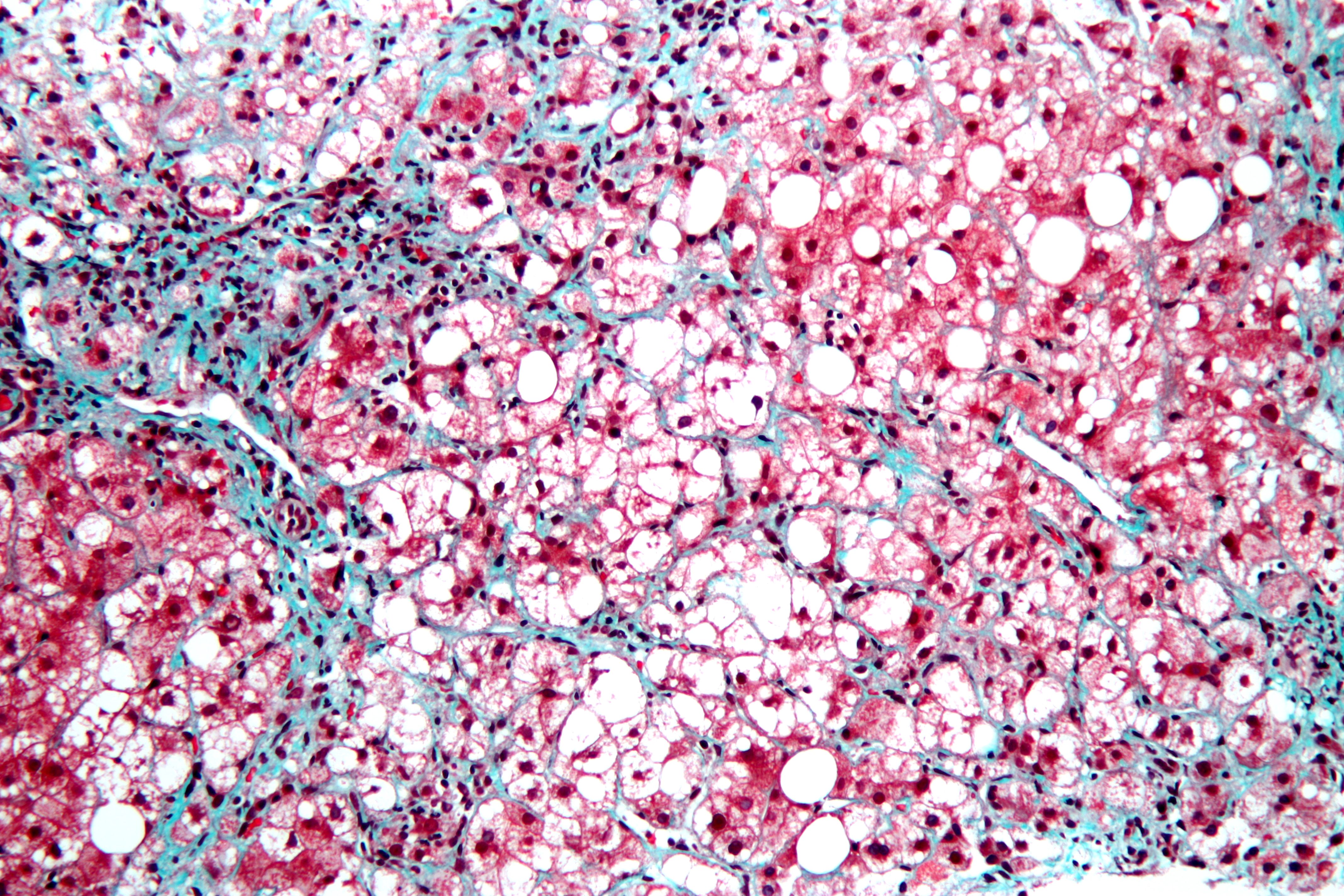
Inspect the accompanying trichrome micrograph. Locate a large clear lipid space, then a thin green collagen strand between hepatocytes. These are different structures: fat was dissolved during tissue processing, whereas the stain highlights collagen. The source documents steatohepatitis in a liver biopsy but does not establish the patient's alcohol history or metabolic phenotype. Do not infer a MASLD cause or a complete fibrosis stage from this single field. [11]
Locate a clear lipid space, then a blue-green collagen strand. The source documents steatohepatitis in a liver biopsy; neither a metabolic cause nor the complete fibrosis stage can be determined from this field alone. Image: Nephron, CC BY-SA 3.0; Nephron; original source; CC BY-SA 3.0. [11].A second report adds ballooning and lobular inflammation. What changes?
The tissue now supports steatohepatitis. In the appropriate metabolic context this is MASH, but its fibrosis stage still needs a separate assessment. Diabetes alone would not have established that tissue injury. [1] [2]
Apply it elsewhere: a lean adult with diabetes and steatosis still warrants etiologic review and fibrosis assessment. A person with obesity and positive hepatitis C testing needs evaluation of both contributors, not a choice between mutually exclusive labels. [1] [2]
How does stored fat become a scarred circulation?
The liver is not simply a container that becomes dangerous when it is full. Insulin resistance permits greater fatty acid release from adipose tissue; the liver also synthesizes new fat from available substrate. Insulin-resistant skeletal muscle also handles incoming glucose less effectively. Triglyceride droplets represent storage, while toxic lipid species and cellular stress can injure hepatocytes. Inflammation and wound-repair signaling activate hepatic stellate cells to deposit collagen. Progression is variable rather than inevitable. [2] [3]
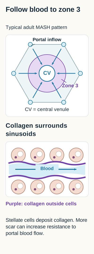
Trace the lobule diagram: start at the portal inflow, follow sinusoids toward the central venule, and locate zone 3 nearest that venule. Adult MASH often begins with perisinusoidal or pericellular fibrosis in this region. More extensive collagen can form bridges; widespread nodules enclosed by scar establish cirrhotic architecture. This is a typical adult pattern, not a mandatory finding in every patient or in pediatric disease. [2]
Trace portal blood toward the central venule, then predict how perisinusoidal collagen affects resistance. [2]
Now predict what collagen around a sinusoid does to blood flow. Increasing resistance helps produce portal hypertension. Splenic sequestration and reduced thrombopoietin can contribute to thrombocytopenia; ascites, varices, or encephalopathy indicate a different level of concern from isolated fat. Platelet loss is an indirect signal, however: immune disease, marrow disease, and medications can also lower platelets. The clinical context determines whether the signal reflects liver disease. [2]
Separate injury from reserve. ALT and AST reflect hepatocyte injury imperfectly; they do not quantify collagen. Albumin, INR, bilirubin, symptoms, and complications contribute to assessment of function and disease severity. Normal aminotransferases cannot exclude advanced fibrosis. A prospective cohort linked F3 and F4 fibrosis to worse liver outcomes, supporting attention to architecture rather than the brightest ultrasound or largest fat droplet. [2] [4]
Try the tissue-to-flow prediction: two patients have similar liver fat measurements. One has no fibrosis; the other has bridging scar and a falling platelet count. Predict which patient needs more intensive fibrosis evaluation before checking the explanation.
Compare their likely liver risk
Bridging scar is the stronger adverse liver-risk signal. Falling platelets add concern but do not independently prove portal hypertension. Equal fat content does not mean equal architecture or prognosis. [2] [4]
Apply it elsewhere: after weight loss, less hepatic fat is welcome, but it cannot certify that previously documented bridging fibrosis has resolved. Continue the follow-up appropriate to the established scar burden. [2] [4] [5]
Which result changes the next fibrosis test?
A 56-year-old with diabetes has steatosis, ALT 30 U/L, and platelets 118,000/mcL. Repeating ALT alone would leave the important question unanswered. Start with the alcohol, medication, and alternative-cause assessment, then use a fibrosis pathway. Signs suggesting cirrhosis or another active liver disease warrant evaluation even when a screening score looks reassuring. [2]
FIB-4 = age in years x AST / (platelets x square root of ALT). Use AST and ALT in U/L and platelets in 10^9/L: 160,000/mcL is entered as 160, not 160,000. For a stable 50-year-old with AST 40, ALT 25, and platelets 160, the calculation is 2,000 / (160 x 5) = 2.50. The score is a probability screen, not a histologic diagnosis. [2]
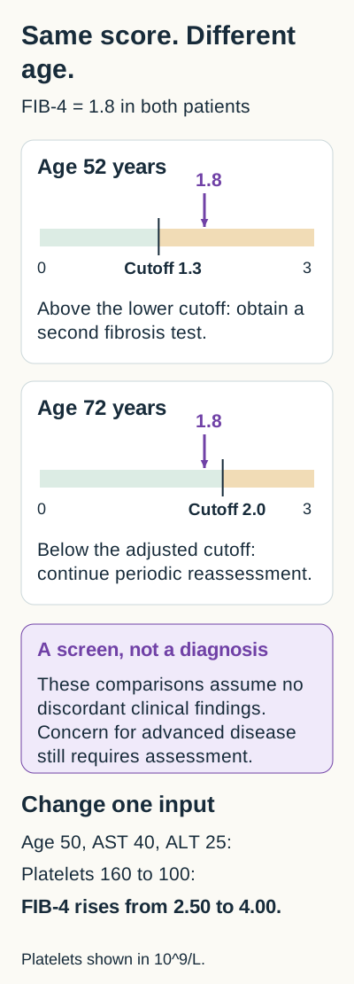
Use the age-switch figure. For adults aged 35-65, values below 1.3 generally support a low probability of advanced fibrosis in primary care; 1.3-2.67 prompts a secondary test, and above 2.67 supports specialist referral. For adults older than 65, use the higher age-adjusted lower cutoff of about 2.0, rather than 1.3, to reduce age-related false positives. Crossing 2.0 is a reason for further assessment, not a rule-in diagnosis of advanced fibrosis. Local pathways govern handling of exact boundaries. [2]
Compare the two markers on the same numerical scale before choosing the next assessment. Then hold age and enzymes constant while changing the platelet count. Screening thresholds do not diagnose a fibrosis stage. Ages are years, AST and ALT are U/L, and platelet counts are in 10^9/L. [2]
Calculate, change one input, predict the consequence. In the worked patient, holding age and enzymes constant while platelets fall from 160 to 100 changes FIB-4 from 2.50 to 4.00. The stronger signal supports referral; it does not prove F4. Ask whether the platelet decline is due to portal disease or a separate hematologic process before interpreting the score as scar progression. [2]
Two otherwise stable patients have FIB-4 1.8, but one is 52 and the other is 72. Do they cross the same lower threshold?
No. The 52-year-old crosses 1.3 and needs secondary assessment. The 72-year-old is below the age-adjusted 2.0 threshold. This comparison assumes no discordant clinical findings; a nodular liver, splenomegaly, or other concern would still justify further evaluation in either patient. [2]
FIB-4 performs poorly below age 35, so a low score should not end assessment in a young person with substantial metabolic risk or persistent liver-test abnormalities. Do not use it to stage chronic disease during acute illness: transient AST changes and thrombocytopenia can distort the result. Assess the acute problem first, then reassess chronic risk when stable. [2]
The second step is usually vibration-controlled transient elastography (VCTE) or the Enhanced Liver Fibrosis (ELF) blood test. A reliable VCTE result below about 8 kPa supports low risk of advanced fibrosis; 8-12 kPa remains uncertain, and above 12 kPa raises concern. Stiffness is not pure scar: congestion and substantial inflammation can raise it. Magnetic resonance elastography (MRE) can resolve uncertainty in specialist care, but its kPa thresholds are not interchangeable with VCTE thresholds. [2]
Refer when tests remain indeterminate, suggest advanced fibrosis, disagree with the examination or imaging, or when another liver disease is possible. Consider biopsy when resolving that uncertainty would change management. Persistent aminotransferase abnormalities for more than six months also justify specialist evaluation. Cirrhosis-based care need not wait for a biopsy when convincing clinical and noninvasive evidence already supports cirrhosis. [2]
Apply it elsewhere: low risk today is not a lifetime discharge. Reassess FIB-4 approximately every 1-2 years with prediabetes, diabetes, or multiple metabolic risks; a lower-risk person without prediabetes or diabetes may be reassessed every 2-3 years. Earlier reassessment is appropriate when symptoms, laboratory trends, or clinical circumstances change. [2]
What improves when body weight falls?
A patient loses 4 kg from an initial 100 kg and now has less fat on MRI. That is a 4% reduction, not a failed intervention. Modest weight loss, around 3-5%, can improve steatosis; larger sustained losses are associated with more improvement in inflammation and fibrosis. European guidance uses at least 5% for liver fat, 7-10% for inflammation, and at least 10% for fibrosis as practical targets. These are graded goals, not guarantees or rigid biological switches. [2] [3] [5]
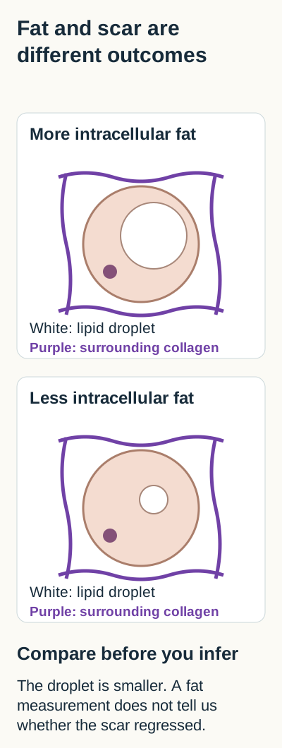
Use the separate-outcomes diagram: compare a smaller fat droplet with the collagen mesh around the cell. Reducing the droplet does not automatically erase the mesh. In a lifestyle cohort, even among people losing at least 10%, fibrosis did not regress in everyone. A normal ALT or improved fat measurement cannot substitute for reassessment of established fibrosis. [5]
Identify what changed and what the fat measurement leaves unanswered. The persistent collagen is a teaching comparison, not a claim that fibrosis never regresses after weight loss. [2][3][5]
Build a sustainable calorie deficit when weight reduction is appropriate. A Mediterranean-style eating pattern, less sugar-sweetened drink intake, and replacement of saturated with unsaturated fats can support liver and cardiovascular goals. Adapt the plan to food access, preferences, and capacity instead of prescribing a crash diet. Aerobic activity and resistance training both matter; exercise can reduce liver fat and improve metabolic health even without large weight changes. A common starting target is about 150 minutes of moderate activity weekly, individualized to ability. [2] [3]
Calculate the next target: a person starts at 120 kg and reaches 114 kg. Identify the percentage reduction and choose whether the result proves fat improvement, inflammation resolution, or fibrosis regression.
Check the percentage and its limit
The reduction is 6/120 = 5%. This is a meaningful fat-reduction target, but the scale alone does not establish any histologic outcome. Continue metabolic treatment and the fibrosis follow-up appropriate to the patient's baseline risk. [3] [5]
Apply it elsewhere: weight loss is not a universal prescription for every lean patient. In cirrhosis, loss of muscle can be harmful; nutrition, resistance activity, and weight goals require individualized care. Metabolic surgery can be considered in suitable patients meeting obesity-treatment criteria, but cirrhosis and portal hypertension require specialist risk assessment rather than routine referral for an operation. [2] [3]
Which risk matters outside the liver?
A patient with metabolic steatosis, prior myocardial infarction, and LDL cholesterol 154 mg/dL has two connected care problems. Waiting for ALT to normalize before treating established vascular disease would leave a major risk untreated. Cardiovascular disease is a leading cause of death across the MASLD population, while advanced fibrosis increases liver-related risk. Neither pathway cancels the other. [2] [4]
Assign each intervention a job: a statin lowers atherosclerotic cardiovascular risk; blood-pressure treatment reduces vascular risk; diabetes and obesity therapies address metabolic disease; fibrosis assessment determines liver follow-up. Statins are generally safe in MASLD and compensated cirrhosis when indicated. Mild stable aminotransferase abnormalities are not a reason to withhold them, although suspected drug injury, decompensated disease, and drug interactions require individualized review. Statins are not a specific treatment for steatohepatitis. [2]
Address tobacco use, lipids, blood pressure, glucose, physical activity, and sleep apnea where appropriate. Choose SGLT2 inhibitors for eligible diabetes, heart-failure, or kidney-disease indications rather than assuming a liver-fat effect establishes a MASH indication. Similarly, vitamin E or pioglitazone may have selected roles, but neither is a universal treatment for every fatty liver, and their risks and lack of established antifibrotic benefit matter. [2] [3]
Predict the treatment gap: a person with prior infarction starts a liver-directed medicine that also lowers LDL. Can that prescription automatically replace established cardiovascular secondary prevention?
Explain why the endpoints matter
No. An LDL change or improved liver histology is not itself proof of fewer cardiovascular events with that drug. Preserve evidence-based cardiovascular treatment, while checking interactions with the new liver medicine. [2] [7] [9]
Apply it elsewhere: a low FIB-4 does not mean low cardiovascular risk. Continue cardiometabolic treatment even when liver assessment supports primary-care follow-up. [2]
When does fibrosis change treatment and surveillance?
Start with the stage, not the drug name. Isolated steatosis, noncirrhotic MASH with F2-F3 fibrosis, and cirrhosis are different treatment populations. Lifestyle and cardiometabolic care remain foundational, while liver-directed treatment depends on evidence of the intended disease stage, contraindications, tolerability, and follow-up. [2] [6] [7]
Compare two approved US options. Resmetirom, an oral thyroid hormone receptor-beta agonist, and injectable Wegovy (semaglutide) have accelerated-approval indications for adults with noncirrhotic MASH and moderate to advanced fibrosis consistent with F2-F3, alongside diet and exercise. The approvals rely on histologic improvement, with confirmatory clinical benefit still required; they do not establish that either drug prevents liver death in every treated patient. Neither indication extends to isolated steatosis or MASH cirrhosis. [6] [7] [8] [9]
For MASH, the Wegovy injection label recommends gradual dose escalation toward 2.4 mg weekly; it allows reduction to 1.7 mg for intolerance with consideration of re-escalation. This is not an instruction to start at 2.4 mg. The oral product and the 7.2 mg weight-management dose should not be substituted as MASH regimens. Benefits for weight and glucose can align with metabolic goals, but diabetes, obesity, cardiovascular, and MASH indications remain product- and population-specific. Do not combine it with another semaglutide product or another GLP-1 receptor agonist. [6]
Semaglutide is contraindicated with a personal or family history of medullary thyroid carcinoma or MEN2 and with serious hypersensitivity to the product. Assess relevant pancreatic, gallbladder, gastrointestinal, pregnancy, retinal, and hypoglycemia risks. Persistent vomiting can cause volume depletion; insulin or sulfonylurea treatment may need adjustment as glycemia improves. The full current label, not a class-wide assumption, governs prescribing. [6]
Resmetirom selection requires medication reconciliation and liver and thyroid assessment. The usual daily dose is weight-based: 80 mg below 100 kg and 100 mg at or above 100 kg. Moderate CYP2C8 inhibition, such as clopidogrel, requires a lower resmetirom dose; strong inhibition, such as gemfibrozil, is not recommended. Resmetirom can increase certain statin exposures, so apply label dose limits and monitor for hepatic and muscle adverse effects. Monitor for hepatotoxicity and gallbladder events; avoid use in decompensated cirrhosis. This summary is not a substitute for the complete label. [7]
Match the stage before choosing: one patient has biopsy-supported MASH with F2 fibrosis and obesity; another has cirrhotic nodules and recurrent ascites. Identify which patient fits the noncirrhotic MASH treatment population, then identify the more urgent care priority for the other.
Check the stage-specific consequence
The F2 patient may be eligible after safety review and discussion of options. Recurrent ascites signals decompensated cirrhosis, requiring complication management and assessment for transplantation when appropriate, not extrapolation of an F2-F3 drug indication. [2] [6] [7]
Once cirrhosis is established, arrange assessment and prevention of portal-hypertensive complications and appropriate hepatocellular carcinoma (HCC) surveillance. AASLD recommends ultrasound with AFP about every six months for eligible at-risk patients. Poor ultrasound visualization may justify an alternative such as contrast-enhanced MRI. Surveillance is intended for patients who could benefit from HCC treatment; severe competing illness or non-transplant-eligible end-stage disease changes the decision. Normal ALT or less visible steatosis does not erase established cirrhosis. [2] [10]
Apply it elsewhere: interpret a treatment response by what was actually measured. In ESSENCE, semaglutide improved histologic endpoints at 72 weeks; that interim result is not a completed survival trial. Follow tolerability, metabolic outcomes, and fibrosis risk separately. Do not stop cirrhosis surveillance merely because weight or enzymes improved. [6] [8] [10]
Practice the decisions
For each patient, identify the disease context, interpret the available measurement, and choose the action it supports. Commit to an answer before comparing the explanations. These are original educational cases, not validated predictions of examination performance.
Case 1
Show answer and explanations for case 1
A. MASH is supported; inflammatory activity is established. (Why this does not fit)
Ballooning injury and lobular inflammation establish the tissue pattern, not diabetes alone. Neither is supplied; MRI steatosis does not demonstrate the required inflammatory injury.
Reasoning steps for option A
What additional finding establishes steatohepatitis?
Ballooning injury and lobular inflammation establish the tissue pattern, not diabetes alone.
Which of those findings is supplied here?
Neither is supplied; MRI steatosis does not demonstrate the required inflammatory injury.
B. Resolved liver disease is supported; normal ALT establishes recovery. (Why this does not fit)
It can accompany less hepatocyte injury in some patients. There is no prior disease assessment or fibrosis measurement, and advanced disease can occur with normal ALT.
Reasoning steps for option B
What might ALT normalization suggest during follow-up?
It can accompany less hepatocyte injury in some patients.
Why does a single normal value not establish recovery here?
There is no prior disease assessment or fibrosis measurement, and advanced disease can occur with normal ALT.
C. MASLD is supported; fibrosis risk remains unassessed. (Best answer)
Type 2 diabetes supplies a cardiometabolic criterion in a patient with documented steatosis. It does not establish the absence of MASH or advanced fibrosis; fibrosis risk assessment is still appropriate.
Reasoning steps for option C
Which metabolic finding qualifies despite his BMI?
Type 2 diabetes supplies a cardiometabolic criterion in a patient with documented steatosis.
What does the normal ALT leave unanswered?
It does not establish the absence of MASH or advanced fibrosis; fibrosis risk assessment is still appropriate.
D. Cryptogenic steatosis is supported; metabolic criteria are absent. (Why this does not fit)
It is considered when no metabolic criterion or other cause explains the steatosis. Diagnosed type 2 diabetes is a qualifying criterion even at a normal BMI.
Reasoning steps for option D
When is cryptogenic steatotic disease considered?
It is considered when no metabolic criterion or other cause explains the steatosis.
Why does this patient's glucose history matter?
Diagnosed type 2 diabetes is a qualifying criterion even at a normal BMI.
Takeaway: A normal BMI does not negate metabolic steatosis, and a normal ALT does not complete its staging.
A. MetALD; address both contributors and obtain elastography. (Best answer)
Forty grams daily totals 280 g weekly, within the consensus MetALD range for women with metabolic steatosis. It is above the usual 1.3 lower threshold and supports secondary fibrosis assessment.
Reasoning steps for option A
Where does her weekly alcohol exposure fall?
Forty grams daily totals 280 g weekly, within the consensus MetALD range for women with metabolic steatosis.
What does FIB-4 1.7 add at her age?
It is above the usual 1.3 lower threshold and supports secondary fibrosis assessment.
B. Alcohol-associated hepatitis; treat the acute inflammatory episode. (Why this does not fit)
Recent jaundice with a compatible alcohol history and laboratory pattern would support an acute clinical syndrome. No. Normal bilirubin and no jaundice do not support acute alcohol-associated hepatitis, although alcohol remains a contributor.
Reasoning steps for option B
What syndrome would suggest alcohol-associated hepatitis?
Recent jaundice with a compatible alcohol history and laboratory pattern would support an acute clinical syndrome.
Does this stable presentation establish that syndrome?
No. Normal bilirubin and no jaundice do not support acute alcohol-associated hepatitis, although alcohol remains a contributor.
C. Predominantly alcohol-associated disease; metabolic attribution is unnecessary. (Why this does not fit)
The upper MetALD boundary is approximately 350 g weekly, with higher intake classified predominantly as alcohol-associated disease. Her intake is 280 g weekly and she has clear metabolic criteria, supporting a mixed rather than alcohol-only assessment.
Reasoning steps for option C
At what exposure does the consensus classify predominantly alcohol-associated disease in women?
The upper MetALD boundary is approximately 350 g weekly, with higher intake classified predominantly as alcohol-associated disease.
Why is metabolic attribution still required in this patient?
Her intake is 280 g weekly and she has clear metabolic criteria, supporting a mixed rather than alcohol-only assessment.
D. MASLD alone; address metabolic risks and obtain elastography. (Why this does not fit)
Diabetes and obesity strongly support a metabolic contribution to her steatosis. Her sustained 280 g weekly alcohol exposure lies within the mixed MetALD category rather than the lower-alcohol MASLD range.
Reasoning steps for option D
What makes metabolic disease a credible contributor?
Diabetes and obesity strongly support a metabolic contribution to her steatosis.
Why is the word alone inaccurate?
Her sustained 280 g weekly alcohol exposure lies within the mixed MetALD category rather than the lower-alcohol MASLD range.
Takeaway: Quantified alcohol exposure changes the etiologic category, but does not eliminate the need to stage fibrosis.
A. Both have MASH and similar fibrosis-related risk. (Why this does not fit)
Both contain hepatic fat in a metabolic setting. Only B demonstrates steatohepatitis and bridging fibrosis, so fat alone cannot establish equal activity or risk.
Reasoning steps for option A
What finding is shared by the two specimens?
Both contain hepatic fat in a metabolic setting.
Why does that shared finding not make their risk equivalent?
Only B demonstrates steatohepatitis and bridging fibrosis, so fat alone cannot establish equal activity or risk.
B. A has MASH and greater fibrosis-related risk than B. (Why this does not fit)
Its larger amount of steatosis may draw attention on microscopy. A lacks ballooning, lobular inflammation, and fibrosis; B contains all three.
Reasoning steps for option B
Why might A initially appear more severe?
Its larger amount of steatosis may draw attention on microscopy.
What contradicts the proposed injury and risk assessment?
A lacks ballooning, lobular inflammation, and fibrosis; B contains all three.
C. B has cirrhosis and A has noncirrhotic MASH. (Why this does not fit)
Regenerative nodules encircled by fibrosis support F4 architecture. B has bridging scar without cirrhotic nodules, and A has no ballooning or lobular inflammation to establish MASH.
Reasoning steps for option C
What architecture would support cirrhosis?
Regenerative nodules encircled by fibrosis support F4 architecture.
How do the stated observations defeat both labels?
B has bridging scar without cirrhotic nodules, and A has no ballooning or lobular inflammation to establish MASH.
D. B has MASH and greater fibrosis-related risk than A. (Best answer)
B combines fat, ballooning injury, and lobular inflammation, while A does not show the inflammatory pattern. Bridging fibrosis indicates substantially more architectural injury despite its smaller amount of fat.
Reasoning steps for option D
Which specimen demonstrates steatohepatitis?
B combines fat, ballooning injury, and lobular inflammation, while A does not show the inflammatory pattern.
Which feature gives B greater liver-related risk?
Bridging fibrosis indicates substantially more architectural injury despite its smaller amount of fat.
Takeaway: Less visible fat can coexist with more inflammatory injury and more clinically important scar.
A. Cholangiocyte injury in periportal zone 1. (Why this does not fit)
Duct-centered injury and periportal inflammation would support a biliary process. The collagen is pericentral and the biopsy lacks duct-centered inflammation.
Reasoning steps for option A
What pattern would make cholangiocyte injury persuasive?
Duct-centered injury and periportal inflammation would support a biliary process.
Which location and negative finding oppose that explanation?
The collagen is pericentral and the biopsy lacks duct-centered inflammation.
B. Stellate-cell activation in pericentral zone 3. (Best answer)
Zone 3 is the pericentral region described by the biopsy. Activated hepatic stellate cells adopt a fibrogenic phenotype and deposit extracellular matrix.
Reasoning steps for option B
Which acinar zone lies nearest terminal hepatic venules?
Zone 3 is the pericentral region described by the biopsy.
Which response produces collagen around its sinusoids?
Activated hepatic stellate cells adopt a fibrogenic phenotype and deposit extracellular matrix.
C. Kupffer-cell iron storage in pericentral zone 3. (Why this does not fit)
They can indicate iron deposition, which requires appropriate staining and etiologic interpretation. Trichrome identifies perisinusoidal collagen, so iron storage is not the stated structural change.
Reasoning steps for option C
What can iron-laden macrophages contribute to an assessment?
They can indicate iron deposition, which requires appropriate staining and etiologic interpretation.
What process is actually demonstrated in this specimen?
Trichrome identifies perisinusoidal collagen, so iron storage is not the stated structural change.
D. Hepatocyte triglyceride storage in periportal zone 1. (Why this does not fit)
It explains intracellular fat droplets and their clear spaces after routine processing. The stain identifies collagen around zone 3 sinusoids, not stored intracellular triglyceride in zone 1.
Reasoning steps for option D
Which tissue change does triglyceride storage explain?
It explains intracellular fat droplets and their clear spaces after routine processing.
Why does it not explain this green extracellular material?
The stain identifies collagen around zone 3 sinusoids, not stored intracellular triglyceride in zone 1.
Takeaway: Typical early adult MASH scar is perisinusoidal in zone 3 and reflects activation of collagen-producing stellate cells.
A. FIB-4 2.50; initiate cirrhosis-based surveillance. (Why this does not fit)
It identifies a non-low probability of advanced fibrosis, not a cirrhosis diagnosis. No convincing clinical, imaging, or other noninvasive evidence establishes cirrhosis in this stem.
Reasoning steps for option A
What does a score of 2.50 establish?
It identifies a non-low probability of advanced fibrosis, not a cirrhosis diagnosis.
What additional evidence is missing for the proposed surveillance plan?
No convincing clinical, imaging, or other noninvasive evidence establishes cirrhosis in this stem.
B. FIB-4 2.50; obtain VCTE or ELF testing. (Best answer)
The denominator is 160 x square root of 25, or 800, so 2000/800 is 2.50. It is not a low-risk result and calls for secondary fibrosis assessment with VCTE or ELF.
Reasoning steps for option B
What is the denominator using the stated platelet units?
The denominator is 160 x square root of 25, or 800, so 2000/800 is 2.50.
What action follows from that result in this stable adult?
It is not a low-risk result and calls for secondary fibrosis assessment with VCTE or ELF.
C. FIB-4 0.50; repeat risk assessment in two years. (Why this does not fit)
Using ALT 25 itself instead of its square root would produce 2000/4000 = 0.50. The correct denominator gives 2.50, which warrants secondary testing now rather than low-risk follow-up alone.
Reasoning steps for option C
Which calculation would produce 0.50?
Using ALT 25 itself instead of its square root would produce 2000/4000 = 0.50.
Why is the proposed follow-up insufficient here?
The correct denominator gives 2.50, which warrants secondary testing now rather than low-risk follow-up alone.
D. FIB-4 4.00; refer for high-risk fibrosis evaluation. (Why this does not fit)
A platelet count of 100 x 10^9/L would give 2000/500 = 4.00. The supplied count is 160 x 10^9/L, giving 2.50 and an initial secondary-test pathway.
Reasoning steps for option D
Which platelet count would produce 4.00 with these enzymes?
A platelet count of 100 x 10^9/L would give 2000/500 = 4.00.
What count was actually supplied?
The supplied count is 160 x 10^9/L, giving 2.50 and an initial secondary-test pathway.
Takeaway: Calculate the score with the correct units, then use it to choose the next assessment rather than assign a biopsy stage.
A. Periodic reassessment for both patients; neither score exceeds 2.67. (Why this does not fit)
A result above 2.67 supports direct referral because of higher fibrosis risk. The intermediate range above 1.3 still requires a second test even without crossing the referral threshold.
Reasoning steps for option A
What is the role of the higher 2.67 threshold?
A result above 2.67 supports direct referral because of higher fibrosis risk.
Why does being below 2.67 not settle the younger patient's assessment?
The intermediate range above 1.3 still requires a second test even without crossing the referral threshold.
B. Secondary testing for both patients; their scores have identical implications. (Why this does not fit)
It is above the usual lower threshold for adults aged 35-65. Age contributes to FIB-4, so the older-adult pathway uses a higher lower threshold to reduce false positives.
Reasoning steps for option B
Why does 1.8 trigger further testing in the younger patient?
It is above the usual lower threshold for adults aged 35-65.
Why should the same numeric interpretation not automatically be applied at age 72?
Age contributes to FIB-4, so the older-adult pathway uses a higher lower threshold to reduce false positives.
C. Periodic reassessment for the younger patient; biopsy for the older. (Why this does not fit)
Age appears directly in the numerator and can raise FIB-4 independently of a change in liver architecture. The older patient's 1.8 is below the adjusted lower cutoff and there is no discordant evidence requiring immediate tissue assessment.
Reasoning steps for option C
How can older age affect the score without proving scar?
Age appears directly in the numerator and can raise FIB-4 independently of a change in liver architecture.
Why does that not justify the proposed biopsy?
The older patient's 1.8 is below the adjusted lower cutoff and there is no discordant evidence requiring immediate tissue assessment.
D. Secondary testing for the younger patient; periodic reassessment for the older. (Best answer)
At age 52, 1.8 exceeds 1.3; at age 72, it is below the age-adjusted lower cutoff near 2.0. The younger patient proceeds to secondary assessment, while the older patient can remain in periodic risk reassessment.
Reasoning steps for option D
Which lower threshold does each patient cross?
At age 52, 1.8 exceeds 1.3; at age 72, it is below the age-adjusted lower cutoff near 2.0.
What does the absence of discordant findings permit?
The younger patient proceeds to secondary assessment, while the older patient can remain in periodic risk reassessment.
Takeaway: Age adjustment reduces false-positive screening; it does not make 2.0 a stand-alone diagnosis of fibrosis.
A. Obtain elastography and specialist assessment. (Best answer)
The score has low accuracy below age 35 and can appear low partly because of age. Diabetes, obesity, and liver-test abnormalities lasting more than six months justify secondary assessment and evaluation for other causes.
Reasoning steps for option A
Why is the low FIB-4 less decisive at age 29?
The score has low accuracy below age 35 and can appear low partly because of age.
Which findings support further evaluation rather than dismissal?
Diabetes, obesity, and liver-test abnormalities lasting more than six months justify secondary assessment and evaluation for other causes.
B. Confirm MASH from the persistent aminotransferase pattern. (Why this does not fit)
It supports ongoing hepatocyte injury and the need to investigate its cause. ALT cannot establish ballooning, lobular inflammation, or a fibrosis stage.
Reasoning steps for option B
What does persistent ALT abnormality support?
It supports ongoing hepatocyte injury and the need to investigate its cause.
What tissue information cannot be supplied by ALT alone?
ALT cannot establish ballooning, lobular inflammation, or a fibrosis stage.
C. Repeat FIB-4 in three years as a low-risk result. (Why this does not fit)
It can be appropriate for adults with reliable low-risk screening and fewer metabolic risks. His young age limits FIB-4 accuracy, and persistent injury plus diabetes argues for further assessment now.
Reasoning steps for option C
When is a longer low-risk reassessment interval useful?
It can be appropriate for adults with reliable low-risk screening and fewer metabolic risks.
Why does this patient not fit that reassuring setting?
His young age limits FIB-4 accuracy, and persistent injury plus diabetes argues for further assessment now.
D. Repeat ultrasound after the liver enzymes normalize. (Why this does not fit)
It can show steatosis or gross morphologic abnormalities, but it does not reliably grade fibrosis. Normal ALT would still not exclude advanced fibrosis, and eight months of abnormalities already warrant evaluation.
Reasoning steps for option D
What can conventional ultrasound demonstrate?
It can show steatosis or gross morphologic abnormalities, but it does not reliably grade fibrosis.
Why would waiting for ALT normalization not answer the question?
Normal ALT would still not exclude advanced fibrosis, and eight months of abnormalities already warrant evaluation.
Takeaway: A low age-dependent score is less reassuring in a young adult with substantial metabolic risk and persistent liver injury.
A. The score establishes rapid progression to cirrhosis. (Why this does not fit)
It would indicate substantial concern for fibrosis and justify further assessment. The measurements were obtained during acute illness and no corroborating cirrhotic findings are supplied.
Reasoning steps for option A
What would a persistent high score suggest in a stable outpatient?
It would indicate substantial concern for fibrosis and justify further assessment.
Why does this admission score not establish rapid cirrhosis?
The measurements were obtained during acute illness and no corroborating cirrhotic findings are supplied.
B. The previous score excludes advanced fibrosis permanently. (Why this does not fit)
It supported a low probability of advanced fibrosis in a stable outpatient setting. Risk changes over time, and a prior screening result is not a permanent exclusion of fibrosis.
Reasoning steps for option B
What did the previous 1.2 support at that time?
It supported a low probability of advanced fibrosis in a stable outpatient setting.
Why can it not end future assessment?
Risk changes over time, and a prior screening result is not a permanent exclusion of fibrosis.
C. The score is confounded; reassess after recovery. (Best answer)
AST has risen and platelets have fallen during sepsis, both of which increase FIB-4. FIB-4 should not be used for chronic staging during acute illness; manage sepsis and reassess when stable.
Reasoning steps for option C
Which acute laboratory changes drive the large score?
AST has risen and platelets have fallen during sepsis, both of which increase FIB-4.
How should that affect the staging decision?
FIB-4 should not be used for chronic staging during acute illness; manage sepsis and reassess when stable.
D. The new score justifies starting an F2-F3 MASH drug. (Why this does not fit)
The relevant indications require noncirrhotic MASH with moderate to advanced fibrosis. An acutely confounded FIB-4 neither establishes MASH nor identifies a reliable F2-F3 stage.
Reasoning steps for option D
What disease state is required for the MASH drug population?
The relevant indications require noncirrhotic MASH with moderate to advanced fibrosis.
Why is that state not established by these results?
An acutely confounded FIB-4 neither establishes MASH nor identifies a reliable F2-F3 stage.
Takeaway: An acute change in FIB-4 can reflect inflammation and platelet loss rather than newly formed scar.
A. Continue metabolic care and reassess only if jaundice develops. (Why this does not fit)
He currently feels well and has reassuring noninvasive tests. Fibrosis can progress without symptoms, so diabetes supports scheduled reassessment before decompensation.
He currently feels well and has reassuring noninvasive tests.
Why is jaundice an inadequate trigger for future assessment?
Fibrosis can progress without symptoms, so diabetes supports scheduled reassessment before decompensation.
B. Continue metabolic care and begin six-monthly HCC surveillance. (Why this does not fit)
Established cirrhosis is the principal MASLD-related indication for surveillance. Both fibrosis tests are low risk and no cirrhosis evidence is supplied.
Reasoning steps for option B
Which liver population generally warrants HCC surveillance?
Established cirrhosis is the principal MASLD-related indication for surveillance.
Why do these results not establish that indication?
Both fibrosis tests are low risk and no cirrhosis evidence is supplied.
C. Continue metabolic care and obtain a biopsy every year. (Why this does not fit)
It can resolve uncertainty about diagnosis or fibrosis when the answer would change management. The noninvasive findings are concordantly low risk and there is no unresolved diagnostic concern.
Reasoning steps for option C
When might biopsy add useful information?
It can resolve uncertainty about diagnosis or fibrosis when the answer would change management.
What argues against scheduled annual biopsy here?
The noninvasive findings are concordantly low risk and there is no unresolved diagnostic concern.
D. Continue metabolic care and repeat FIB-4 in about 1-2 years. (Best answer)
FIB-4 0.9 and reliable VCTE below 8 kPa support a low probability of advanced fibrosis. Diabetes and multiple metabolic risks support repeating risk assessment about every 1-2 years.
Reasoning steps for option D
What do the concordant screening results support now?
FIB-4 0.9 and reliable VCTE below 8 kPa support a low probability of advanced fibrosis.
Why is reassessment still relatively frequent?
Diabetes and multiple metabolic risks support repeating risk assessment about every 1-2 years.
Takeaway: Low-risk fibrosis results permit longitudinal care, but diabetes warrants periodic reassessment.
A. Hematology assessment for isolated immune platelet destruction. (Why this does not fit)
Isolated thrombocytopenia without supporting liver or splenic findings would prompt a broader hematologic assessment. It does not account for the concurrent nodular liver and splenomegaly.
Reasoning steps for option A
When would a primary platelet disorder be a strong possibility?
Isolated thrombocytopenia without supporting liver or splenic findings would prompt a broader hematologic assessment.
Why is isolated immune destruction less explanatory here?
It does not account for the concurrent nodular liver and splenomegaly.
B. Repeat aminotransferases after another year of observation. (Why this does not fit)
They can describe an injury trend, but not reliably stage scar. New nodularity and splenomegaly with thrombocytopenia require assessment now.
Reasoning steps for option B
What can serial enzymes contribute to liver assessment?
They can describe an injury trend, but not reliably stage scar.
Which current findings make enzyme-only observation inadequate?
New nodularity and splenomegaly with thrombocytopenia require assessment now.
C. Hepatology review for fibrosis and portal hypertension. (Best answer)
Nodularity, splenomegaly, and falling platelets together raise concern for advanced fibrosis and portal hypertension. ALT does not quantify scar, and compensated advanced disease may preserve bilirubin and produce few symptoms.
Reasoning steps for option C
Which supplied findings point beyond isolated steatosis?
Nodularity, splenomegaly, and falling platelets together raise concern for advanced fibrosis and portal hypertension.
Why is normal ALT not reassuring enough to defer assessment?
ALT does not quantify scar, and compensated advanced disease may preserve bilirubin and produce few symptoms.
D. Repeat fat quantification to determine the fibrosis stage. (Why this does not fit)
It measures fat content rather than the architecture of fibrosis. The new concern is portal and architectural disease, which can persist even with little residual fat.
Reasoning steps for option D
What does a quantitative liver-fat measurement assess?
It measures fat content rather than the architecture of fibrosis.
Why would that miss the principal concern?
The new concern is portal and architectural disease, which can persist even with little residual fat.
Takeaway: Normal enzymes and preserved bilirubin do not negate structural evidence of advanced liver disease.
A. The technically reliable result establishes cirrhosis. (Why this does not fit)
It supports confidence in the measurement process under the conditions present. It does not exclude congestion or inflammation as causes of increased stiffness, and 17 kPa is not a stand-alone F4 diagnosis.
Reasoning steps for option A
What does technical reliability establish about VCTE?
It supports confidence in the measurement process under the conditions present.
What does technical reliability not exclude?
It does not exclude congestion or inflammation as causes of increased stiffness, and 17 kPa is not a stand-alone F4 diagnosis.
B. Reassess stiffness after treating the congestion. (Best answer)
Venous congestion from right-sided heart failure can increase liver stiffness. Treat the congestion and reassess in a stable state, with specialist evaluation if results remain discordant.
Reasoning steps for option B
Which current finding can increase stiffness without equivalent fibrosis?
Venous congestion from right-sided heart failure can increase liver stiffness.
What should follow before assigning a fixed fibrosis stage?
Treat the congestion and reassess in a stable state, with specialist evaluation if results remain discordant.
C. The stiffness result identifies increased hepatic fat content. (Why this does not fit)
Controlled attenuation parameter assesses steatosis, whereas kPa describes stiffness. It is a stiffness result whose causes include fibrosis and congestion, not a direct percentage of fat.
Reasoning steps for option C
Which VCTE-associated measure addresses fat rather than stiffness?
It is a stiffness result whose causes include fibrosis and congestion, not a direct percentage of fat.
D. The previous FIB-4 excludes meaningful liver disease. (Why this does not fit)
It makes a previously unrecognized high chronic fibrosis burden less likely in that earlier setting. Screening has imperfect sensitivity and risk can change; persistent discordance needs reassessment rather than dismissal.
Reasoning steps for option D
Why is the prior low score relevant?
It makes a previously unrecognized high chronic fibrosis burden less likely in that earlier setting.
Why can it not override the current assessment completely?
Screening has imperfect sensitivity and risk can change; persistent discordance needs reassessment rather than dismissal.
Takeaway: A reliable stiffness measurement can still be biologically confounded by congestion.
A reliable VCTE value of 6.5 is below about 8 kPa and supports low risk of advanced fibrosis. MRE uses different thresholds, and 4.1 kPa raises concern for advanced fibrosis under method-specific criteria.
Reasoning steps for option A
How does the VCTE value compare with its low-risk threshold?
A reliable VCTE value of 6.5 is below about 8 kPa and supports low risk of advanced fibrosis.
Why is the numerically smaller MRE value not necessarily reassuring?
MRE uses different thresholds, and 4.1 kPa raises concern for advanced fibrosis under method-specific criteria.
B. Both patient A and patient B. (Why this does not fit)
Their FIB-4 values of 1.8 exceeded the usual lower threshold at age 58. The secondary VCTE result supports low risk for A, whereas the MRE result does not similarly reassure for B.
Reasoning steps for option B
Why did both patients need secondary assessment?
Their FIB-4 values of 1.8 exceeded the usual lower threshold at age 58.
What new information distinguishes the next assessment?
The secondary VCTE result supports low risk for A, whereas the MRE result does not similarly reassure for B.
C. Neither patient A nor patient B. (Why this does not fit)
Comparing both with the VCTE cutoff of 8 kPa would make both appear low. MRE and VCTE thresholds are not interchangeable despite both reporting kilopascals.
Reasoning steps for option C
Which shared comparison would make both values appear reassuring?
Comparing both with the VCTE cutoff of 8 kPa would make both appear low.
Why is that comparison invalid for B?
MRE and VCTE thresholds are not interchangeable despite both reporting kilopascals.
D. Patient A, but not patient B. (Why this does not fit)
Patient A has 6.5 kPa compared with 4.1 kPa in patient B. The measurements use different techniques; VCTE 6.5 is reassuring while MRE 4.1 remains concerning.
Reasoning steps for option D
Which patient has the larger numerical stiffness result?
Patient A has 6.5 kPa compared with 4.1 kPa in patient B.
Why does that numerical ordering not establish the ordering of risk?
The measurements use different techniques; VCTE 6.5 is reassuring while MRE 4.1 remains concerning.
Takeaway: Use the threshold validated for the measurement method; MRE and VCTE share units but not cutoffs.
A. Refer for etiologic assessment and consideration of liver biopsy. (Best answer)
Persistent substantial ALT abnormality with high IgG and ANA positivity warrants evaluation beyond metabolic steatosis. Biopsy can help distinguish or identify concurrent disease when the result would change treatment.
Reasoning steps for option A
What makes an additional inflammatory liver disease possible?
Persistent substantial ALT abnormality with high IgG and ANA positivity warrants evaluation beyond metabolic steatosis.
What would tissue assessment add if uncertainty persists?
Biopsy can help distinguish or identify concurrent disease when the result would change treatment.
B. Begin corticosteroids for autoimmune hepatitis without further assessment. (Why this does not fit)
The immunoglobulin and antibody findings fit a pattern that merits specialist investigation. Autoantibodies can occur in metabolic liver disease, and diagnosis requires integration of clinical, laboratory, and often histologic evidence.
Reasoning steps for option B
Why is autoimmune hepatitis a relevant possibility?
The immunoglobulin and antibody findings fit a pattern that merits specialist investigation.
Why do they not establish an immediate treatment diagnosis by themselves?
Autoantibodies can occur in metabolic liver disease, and diagnosis requires integration of clinical, laboratory, and often histologic evidence.
C. Start resmetirom for presumed noncirrhotic F2-F3 MASH. (Why this does not fit)
The indication targets MASH with moderate to advanced noncirrhotic fibrosis. Neither the fibrosis stage nor the cause of the persistent inflammatory injury has been resolved.
Reasoning steps for option C
What evidence is needed for that treatment population?
The indication targets MASH with moderate to advanced noncirrhotic fibrosis.
Why has this patient not yet been shown to fit it?
Neither the fibrosis stage nor the cause of the persistent inflammatory injury has been resolved.
D. Continue weight treatment and repeat FIB-4 in three years. (Why this does not fit)
It estimates a low probability of advanced fibrosis in a suitable screening context. It does not explain nine months of injury with immune abnormalities or exclude a concurrent treatable disease.
Reasoning steps for option D
What does the low FIB-4 estimate?
It estimates a low probability of advanced fibrosis in a suitable screening context.
Which unresolved issue makes delayed screening alone inadequate?
It does not explain nine months of injury with immune abnormalities or exclude a concurrent treatable disease.
Takeaway: A low fibrosis score does not exclude another active liver disease, and metabolic steatosis can coexist with autoimmune disease.
A. Five percent weight reduction; histologic MASH resolution is established. (Why this does not fit)
Weight, MRI fat fraction, and ALT have improved. Resolution of ballooning and lobular inflammation was not measured, and ALT normalization is not equivalent to histologic MASH resolution.
Reasoning steps for option A
Which measurements have improved?
Weight, MRI fat fraction, and ALT have improved.
Why do these not establish the proposed histologic outcome?
Resolution of ballooning and lobular inflammation was not measured, and ALT normalization is not equivalent to histologic MASH resolution.
B. Five percent weight reduction; less liver fat, with fibrosis response uncertain. (Best answer)
Six divided by 120 is 0.05, or 5%. They support improvement in fat and an injury marker, but neither establishes regression of the known F2 fibrosis.
Reasoning steps for option B
What percentage of initial weight was lost?
Six divided by 120 is 0.05, or 5%.
What do the MRI and ALT changes leave unproven?
They support improvement in fat and an injury marker, but neither establishes regression of the known F2 fibrosis.
C. Ten percent weight reduction; the earlier fibrosis stage no longer applies. (Why this does not fit)
A loss of 12 kg would be 10%. The patient lost 6 kg, and weight change alone cannot invalidate a documented fibrosis stage.
Reasoning steps for option C
How much loss would equal 10% of 120 kg?
A loss of 12 kg would be 10%.
Why is the fibrosis conclusion unsupported even beyond the arithmetic error?
The patient lost 6 kg, and weight change alone cannot invalidate a documented fibrosis stage.
D. Six percent weight reduction; less liver fat, with fibrosis response uncertain. (Why this does not fit)
The denominator is initial weight, which was 120 kg. Six kilograms is the absolute loss; dividing it by 120 gives 5%, not 6%.
Reasoning steps for option D
Which denominator is used for percent weight change?
The denominator is initial weight, which was 120 kg.
Why is six percent not the correct calculation?
Six kilograms is the absolute loss; dividing it by 120 gives 5%, not 6%.
Takeaway: A 5% weight reduction can be meaningful even when the fibrosis response remains unknown.
A. Stop resistance training and prioritize weight loss on the scale. (Why this does not fit)
Body composition and fitness can improve without a large change in total weight. Resistance activity supports muscle function and complements aerobic activity; scale change alone misses the observed benefits.
Reasoning steps for option A
Why can preserving muscle complicate interpretation of the scale?
Body composition and fitness can improve without a large change in total weight.
Why should that not be treated as a reason to stop resistance exercise?
Resistance activity supports muscle function and complements aerobic activity; scale change alone misses the observed benefits.
B. Continue activity; the MRI change establishes that fibrosis has regressed. (Why this does not fit)
It tracked liver fat rather than collagen architecture. A lower fat measurement does not directly establish less fibrosis.
Reasoning steps for option B
Which hepatic quantity did this MRI assessment track?
It tracked liver fat rather than collagen architecture.
Why is the fibrosis claim not supported?
A lower fat measurement does not directly establish less fibrosis.
C. Continue activity; fat improved, but fibrosis is unmeasured. (Best answer)
Fitness, glycemic control, and MRI liver fat improved despite stable weight. The plan can continue while weight goals and any needed fibrosis reassessment are considered independently.
Reasoning steps for option C
Which observed changes show that the activity plan is useful?
The plan can continue while weight goals and any needed fibrosis reassessment are considered independently.
D. Replace activity with caloric restriction; hepatic benefit requires weight loss. (Why this does not fit)
It can support weight reduction and further liver and metabolic benefit. The patient already demonstrates exercise-associated benefits without weight loss; activity is not rendered ineffective by a stable scale.
Reasoning steps for option D
Why is a calorie deficit often included in a plan for excess weight?
It can support weight reduction and further liver and metabolic benefit.
Why should it not replace this effective activity plan?
The patient already demonstrates exercise-associated benefits without weight loss; activity is not rendered ineffective by a stable scale.
Takeaway: The scale is not the sole measure of exercise benefit, and lower liver fat is not proof of lower fibrosis.
A. Use vitamin E in place of lipid-lowering treatment. (Why this does not fit)
It may be considered in selected patients with steatohepatitis after assessment of benefits and risks. Vitamin E is not established cardiovascular secondary prevention and does not replace LDL-lowering treatment.
Reasoning steps for option A
What role can vitamin E have in liver care?
It may be considered in selected patients with steatohepatitis after assessment of benefits and risks.
Why does that not address this patient's immediate medication need?
Vitamin E is not established cardiovascular secondary prevention and does not replace LDL-lowering treatment.
B. Defer the statin until ALT remains normal for six months. (Why this does not fit)
It indicates some liver injury and requires clinical interpretation. Stable mild abnormalities in compensated metabolic liver disease do not justify delaying indicated secondary prevention.
Reasoning steps for option B
Why might the ALT result cause concern?
It indicates some liver injury and requires clinical interpretation.
Why is normalization not a necessary prerequisite here?
Stable mild abnormalities in compensated metabolic liver disease do not justify delaying indicated secondary prevention.
C. Use cholestyramine instead of a statin because of the fibrosis. (Why this does not fit)
Alternative or additional lipid-lowering therapies can be useful when indicated statin treatment is insufficient or not tolerated. The patient has compensated disease without a stated statin contraindication, so appropriate statin-based secondary prevention remains indicated.
Reasoning steps for option C
Why might a nonstatin LDL-lowering drug be considered?
Alternative or additional lipid-lowering therapies can be useful when indicated statin treatment is insufficient or not tolerated.
Why is fibrosis alone not a reason to make that substitution here?
The patient has compensated disease without a stated statin contraindication, so appropriate statin-based secondary prevention remains indicated.
D. Begin an indicated statin while continuing liver follow-up. (Best answer)
Prior myocardial infarction establishes secondary-prevention need, reinforced by the high LDL. Statins are generally safe when indicated in compensated MASLD, and the mild stable ALT abnormality does not by itself suggest statin-related injury.
Reasoning steps for option D
Which history establishes a cardiovascular treatment priority?
Prior myocardial infarction establishes secondary-prevention need, reinforced by the high LDL.
Why does the liver assessment not automatically prevent statin use?
Statins are generally safe when indicated in compensated MASLD, and the mild stable ALT abnormality does not by itself suggest statin-related injury.
Takeaway: Treat established cardiovascular risk while assessing liver disease; stable mild ALT abnormalities are not a categorical statin prohibition.
A. Add vitamin E solely to convert the fat response into fibrosis regression. (Why this does not fit)
Selected patients may have improvement in steatohepatitis activity after individualized assessment. It neither establishes the required disease context nor proves that vitamin E would induce fibrosis regression.
Reasoning steps for option A
What histologic benefits have been explored with vitamin E?
Selected patients may have improvement in steatohepatitis activity after individualized assessment.
Why does this response not justify the proposed addition?
It neither establishes the required disease context nor proves that vitamin E would induce fibrosis regression.
B. Stop the drug because a MASH indication has not been established. (Why this does not fit)
No. The prescription was initiated for an appropriate cardiorenal purpose. Independent approved benefits remain relevant while liver disease is assessed separately.
Reasoning steps for option B
Is its current indication dependent on a MASH diagnosis?
No. The prescription was initiated for an appropriate cardiorenal purpose.
Why would the lack of MASH confirmation not itself justify stopping it?
Independent approved benefits remain relevant while liver disease is assessed separately.
C. Continue cardiorenal treatment and stage liver fibrosis. (Best answer)
The stated diabetes and kidney-disease context provides a legitimate cardiorenal reason for therapy. Reduced fat does not establish histologic MASH, fibrosis regression, or a specific MASH indication for the drug class.
Reasoning steps for option C
Which treatment purpose is already supported?
The stated diabetes and kidney-disease context provides a legitimate cardiorenal reason for therapy.
What does the MRI response not establish?
Reduced fat does not establish histologic MASH, fibrosis regression, or a specific MASH indication for the drug class.
D. Continue as MASH-specific therapy and omit fibrosis staging. (Why this does not fit)
A decrease in liver fat is a favorable metabolic observation. Fat response does not identify the degree of scar or establish a MASH-specific treatment indication.
Reasoning steps for option D
Why might the MRI response appear encouraging for liver care?
A decrease in liver fat is a favorable metabolic observation.
Why is it insufficient to replace fibrosis assessment?
Fat response does not identify the degree of scar or establish a MASH-specific treatment indication.
Takeaway: An appropriate cardiorenal treatment can improve liver fat without becoming a specifically approved MASH treatment.
A. Resmetirom tablets, using the standard weight-based dose. (Why this does not fit)
It has an indication for noncirrhotic MASH with F2-F3 fibrosis. It is not an approved pharmacologic obesity treatment, unlike the specified semaglutide product.
Reasoning steps for option A
Which of her treatment goals does resmetirom directly match?
It has an indication for noncirrhotic MASH with F2-F3 fibrosis.
Why is it not the best match for both goals in the question?
It is not an approved pharmacologic obesity treatment, unlike the specified semaglutide product.
B. An SGLT2 inhibitor, titrated to a diabetes-treatment dose. (Why this does not fit)
Depending on the product and patient, it can address diabetes and cardiorenal indications. It is not an approved obesity treatment or a specifically approved MASH therapy.
Reasoning steps for option B
What metabolic purposes can an SGLT2 inhibitor serve?
Depending on the product and patient, it can address diabetes and cardiorenal indications.
Why does it not match both specified approvals?
It is not an approved obesity treatment or a specifically approved MASH therapy.
C. Wegovy tablets, using the oral weight-management regimen. (Why this does not fit)
The oral product has a weight-management indication. The MASH indication and regimen in the current label are for injection, not the oral product.
Reasoning steps for option C
Why are the tablets a plausible obesity option?
The oral product has a weight-management indication.
Why does its formulation matter for the second goal?
The MASH indication and regimen in the current label are for injection, not the oral product.
D. Wegovy injection, titrated toward its MASH maintenance dose. (Best answer)
Biopsy-confirmed noncirrhotic MASH with F2 fibrosis fits the stated treatment population. Wegovy injection has an obesity indication as well as the relevant MASH indication; gradual titration and continued lifestyle care are required.
Reasoning steps for option D
Which liver-disease feature matches the MASH indication?
Biopsy-confirmed noncirrhotic MASH with F2 fibrosis fits the stated treatment population.
Which additional goal does this product also address?
Wegovy injection has an obesity indication as well as the relevant MASH indication; gradual titration and continued lifestyle care are required.
Takeaway: Match treatment to the confirmed stage and the patient's goals without extrapolating approval across formulations.
A. Do not prescribe Wegovy; discuss another eligible MASH strategy. (Best answer)
A family history of medullary thyroid carcinoma is a labeled contraindication to Wegovy. The patient has noncirrhotic F3 disease, so alternatives can be evaluated according to their own indications and safety profiles.
Reasoning steps for option A
Which new history changes the prescribing decision?
A family history of medullary thyroid carcinoma is a labeled contraindication to Wegovy.
Why can a different MASH strategy still be considered?
The patient has noncirrhotic F3 disease, so alternatives can be evaluated according to their own indications and safety profiles.
B. Use oral semaglutide to avoid the injection-specific thyroid restriction. (Why this does not fit)
The contraindication concerns semaglutide treatment and the stated MTC or MEN2 history, not the needle. The oral Wegovy product does not carry the injection's MASH indication.
Reasoning steps for option B
Is the thyroid concern created by the route of injection?
The contraindication concerns semaglutide treatment and the stated MTC or MEN2 history, not the needle.
What additional limitation applies to the oral substitution?
The oral Wegovy product does not carry the injection's MASH indication.
C. Prescribe Wegovy at a lower starting dose indefinitely. (Why this does not fit)
Gradual escalation is used to improve tolerability, particularly gastrointestinal tolerability. Dose reduction does not eliminate the labeled family-history contraindication.
Reasoning steps for option C
Why is dose escalation normally gradual?
Gradual escalation is used to improve tolerability, particularly gastrointestinal tolerability.
Why does a low dose not solve this patient's safety issue?
Dose reduction does not eliminate the labeled family-history contraindication.
D. Prescribe Wegovy after confirming another normal thyroid ultrasound. (Why this does not fit)
A normal image does not negate the contraindication based on family history. The label contraindicates treatment in this family-history setting regardless of a screening ultrasound result.
Reasoning steps for option D
What is the limitation of thyroid ultrasound in this decision?
A normal image does not negate the contraindication based on family history.
Why does repeating it not provide prescribing clearance?
The label contraindicates treatment in this family-history setting regardless of a screening ultrasound result.
Takeaway: An appropriate indication does not override a labeled contraindication, and normal thyroid imaging does not negate a family history of MTC.
A. Restart at 2.4 mg after a prolonged interruption without titration. (Why this does not fit)
It aims to reduce gastrointestinal intolerance. He is already intolerant at 2.4 mg, and a prolonged interruption may require renewed lower-dose escalation rather than an immediate high-dose restart.
Reasoning steps for option A
What does gradual escalation aim to reduce?
It aims to reduce gastrointestinal intolerance.
Why would a high-dose restart be poorly suited here?
He is already intolerant at 2.4 mg, and a prolonged interruption may require renewed lower-dose escalation rather than an immediate high-dose restart.
B. Decrease to 1.7 mg weekly and consider later re-escalation. (Best answer)
He tolerated 1.7 mg weekly before developing persistent nausea at 2.4 mg. It permits a decrease to 1.7 mg for intolerance, with consideration of re-escalation to 2.4 mg.
Reasoning steps for option B
What dose did he tolerate before the current symptoms?
He tolerated 1.7 mg weekly before developing persistent nausea at 2.4 mg.
What does the MASH label permit in that situation?
It permits a decrease to 1.7 mg for intolerance, with consideration of re-escalation to 2.4 mg.
C. Switch to 25 mg oral Wegovy as an equivalent MASH regimen. (Why this does not fit)
It offers a non-injection option for its approved weight and cardiovascular indications. The MASH indication is specific to Wegovy injection; pharmacologic similarity does not establish the oral MASH regimen.
Reasoning steps for option C
Why might an oral formulation seem attractive?
It offers a non-injection option for its approved weight and cardiovascular indications.
Why is it not an equivalent MASH substitution?
The MASH indication is specific to Wegovy injection; pharmacologic similarity does not establish the oral MASH regimen.
D. Increase to 7.2 mg weekly to strengthen the MASH response. (Why this does not fit)
The 7.2 mg option is part of adult weight-reduction dosing, not the MASH maintenance regimen. His immediate problem is intolerance at 2.4 mg, not evidence that a higher dose is needed.
Reasoning steps for option D
Which treatment purpose uses the higher-dose option in the current label?
The 7.2 mg option is part of adult weight-reduction dosing, not the MASH maintenance regimen.
Why is escalation especially poorly matched to this presentation?
His immediate problem is intolerance at 2.4 mg, not evidence that a higher dose is needed.
Takeaway: MASH dosing is formulation-specific, and intolerance calls for assessment and adjustment rather than automatic escalation.
A. Resmetirom 100 mg daily while continuing clopidogrel. (Why this does not fit)
The standard dose for a patient weighing at least 100 kg is 100 mg daily. The required clopidogrel treatment triggers a lower dose because it increases resmetirom exposure.
Reasoning steps for option A
Which part of the dosing instruction supports 100 mg?
The standard dose for a patient weighing at least 100 kg is 100 mg daily.
Which additional instruction has been omitted?
The required clopidogrel treatment triggers a lower dose because it increases resmetirom exposure.
B. Resmetirom 80 mg daily while continuing clopidogrel. (Best answer)
At 112 kg, he is in the category that would otherwise receive 100 mg daily. Its moderate CYP2C8 inhibition requires reduction to 80 mg daily in this weight category.
Reasoning steps for option B
Which weight category applies?
At 112 kg, he is in the category that would otherwise receive 100 mg daily.
What does concomitant clopidogrel change?
Its moderate CYP2C8 inhibition requires reduction to 80 mg daily in this weight category.
C. Resmetirom 60 mg daily while continuing clopidogrel. (Why this does not fit)
It is the adjustment for patients below 100 kg receiving a moderate CYP2C8 inhibitor. He weighs 112 kg, so the specified adjusted dose is 80 mg rather than 60 mg.
Reasoning steps for option C
For whom is the 60 mg interaction-adjusted dose used?
It is the adjustment for patients below 100 kg receiving a moderate CYP2C8 inhibitor.
Why does this patient need the other adjusted dose?
He weighs 112 kg, so the specified adjusted dose is 80 mg rather than 60 mg.
D. Resmetirom 100 mg daily after stopping clopidogrel. (Why this does not fit)
Without the inhibitor, the standard weight-based dose could apply. Clopidogrel must continue after stenting; the available dose adjustment avoids interrupting necessary antiplatelet therapy.
Reasoning steps for option D
Would removing the interacting medicine change the dosing calculation?
Without the inhibitor, the standard weight-based dose could apply.
Why is that not the appropriate plan in this stem?
Clopidogrel must continue after stenting; the available dose adjustment avoids interrupting necessary antiplatelet therapy.
Takeaway: Weight-based dosing must be checked against interacting medications, not applied in isolation.
A. Reduce rosuvastatin to 20 mg and reassess additional LDL-lowering therapy. (Best answer)
Concomitant rosuvastatin should not exceed the label limit of 20 mg daily. Continue an appropriate compatible regimen and consider nonstatin therapy if needed to reach the individualized LDL goal.
Reasoning steps for option A
What change is required by the interaction information?
Concomitant rosuvastatin should not exceed the label limit of 20 mg daily.
How can cardiovascular prevention remain active?
Continue an appropriate compatible regimen and consider nonstatin therapy if needed to reach the individualized LDL goal.
B. Continue rosuvastatin 40 mg unless muscle pain develops. (Why this does not fit)
She has no current clinical manifestation of muscle toxicity. The label limit addresses increased exposure and potential toxicity before an adverse event occurs.
Reasoning steps for option B
Why might the absence of symptoms initially appear reassuring?
She has no current clinical manifestation of muscle toxicity.
Why should adjustment not wait for symptoms?
The label limit addresses increased exposure and potential toxicity before an adverse event occurs.
C. Substitute resmetirom for the statin as sole cardiovascular prevention. (Why this does not fit)
Resmetirom can lower LDL cholesterol. A lipid change with a liver-directed drug is not itself evidence of equivalent cardiovascular secondary-prevention benefit.
Reasoning steps for option C
Which lipid effect might make the substitution tempting?
Resmetirom can lower LDL cholesterol.
Why does that effect not make it an equivalent statin replacement?
A lipid change with a liver-directed drug is not itself evidence of equivalent cardiovascular secondary-prevention benefit.
D. Stop lipid-lowering therapy until MASH has resolved. (Why this does not fit)
The new liver medicine increases exposure to certain statins. Her prior infarction still requires secondary prevention, and a compatible lower statin dose or additional agent can be considered.
Reasoning steps for option D
What is the concern prompting medication review?
The new liver medicine increases exposure to certain statins.
Why does that not justify a pause in all LDL treatment?
Her prior infarction still requires secondary prevention, and a compatible lower statin dose or additional agent can be considered.
Takeaway: An interaction should change the regimen safely without abandoning an established cardiovascular treatment goal.
A. A 36.8-percentage-point treatment benefit, with the placebo response excluded. (Why this does not fit)
It is the treated group's observed response proportion. The placebo group also had a 22.4% response, which must be considered in the between-group comparison.
Reasoning steps for option A
What does 36.8% represent?
It is the treated group's observed response proportion.
Why is it not the treatment-versus-placebo absolute benefit?
The placebo group also had a 22.4% response, which must be considered in the between-group comparison.
B. A 14.4-percentage-point survival benefit, established at the interim analysis. (Why this does not fit)
The endpoint was fibrosis improvement without worsening steatohepatitis. The stated analysis does not report that mortality outcome, and longer-term clinical follow-up is still ongoing.
Reasoning steps for option B
Which endpoint was actually reported?
The endpoint was fibrosis improvement without worsening steatohepatitis.
Why can the same numerical difference not be assigned to survival?
The stated analysis does not report that mortality outcome, and longer-term clinical follow-up is still ongoing.
C. A 14.4% relative histologic benefit, without a completed survival conclusion. (Why this does not fit)
Relative improvement divides the difference by the placebo rate, rather than reporting the raw subtraction. Fourteen point four is the percentage-point difference; dividing 14.4 by 22.4 gives a relative increase of about 64%.
Reasoning steps for option C
How is relative improvement calculated from these response rates?
Relative improvement divides the difference by the placebo rate, rather than reporting the raw subtraction.
Why is 14.4% not the relative improvement?
Fourteen point four is the percentage-point difference; dividing 14.4 by 22.4 gives a relative increase of about 64%.
D. A 14.4-percentage-point histologic benefit, without a completed survival conclusion. (Best answer)
Subtracting 22.4 from 36.8 gives 14.4 percentage points. It describes the specified histologic endpoint at 72 weeks, not a proven reduction in liver death.
Reasoning steps for option D
What is the absolute difference in response proportions?
Subtracting 22.4 from 36.8 gives 14.4 percentage points.
What outcome does that difference describe?
It describes the specified histologic endpoint at 72 weeks, not a proven reduction in liver death.
Takeaway: Name the measured endpoint and distinguish percentage-point benefit from unmeasured clinical outcomes.
A. Replace cancer surveillance with periodic FIB-4 assessment. (Why this does not fit)
It estimates fibrosis risk, especially in initial risk stratification. It does not detect HCC, and this patient's cirrhosis is already established.
Reasoning steps for option A
What purpose does FIB-4 serve?
It estimates fibrosis risk, especially in initial risk stratification.
Why can it not replace the current surveillance program?
It does not detect HCC, and this patient's cirrhosis is already established.
B. Repeat the same ultrasound in one year because ALT is normal. (Why this does not fit)
No. The indication derives from established cirrhosis and treatment eligibility, not the ALT level. It prolongs the gap and repeats a method with persistent visualization limitations.
Reasoning steps for option B
Does normal ALT eliminate the surveillance indication?
No. The indication derives from established cirrhosis and treatment eligibility, not the ALT level.
Why does the proposed timing and method fail to address the problem?
It prolongs the gap and repeats a method with persistent visualization limitations.
C. Arrange contrast-enhanced MRI within an ongoing surveillance program. (Best answer)
Repeated ultrasound examinations have not visualized the liver sufficiently. Contrast-enhanced MRI or multiphase CT may be considered for selected patients with limited ultrasound visualization.
Reasoning steps for option C
Which part of her surveillance is currently inadequate?
Repeated ultrasound examinations have not visualized the liver sufficiently.
What alternative does the guidance allow in that setting?
Contrast-enhanced MRI or multiphase CT may be considered for selected patients with limited ultrasound visualization.
D. Use AFP alone every six months until it becomes abnormal. (Why this does not fit)
It is one reassuring observation but does not exclude HCC. It cannot compensate for the repeatedly inadequate imaging component of surveillance.
Reasoning steps for option D
What information does a normal AFP provide?
It is one reassuring observation but does not exclude HCC.
Why is AFP alone an inadequate replacement here?
It cannot compensate for the repeatedly inadequate imaging component of surveillance.
Takeaway: A normal AFP cannot turn a poorly visualized liver into an adequate surveillance examination.
A. Start half-dose resmetirom to account for decompensation. (Why this does not fit)
No. Dose reduction does not establish efficacy or safety for an indication that does not include the patient. It advises avoiding use in decompensated cirrhosis.
Reasoning steps for option A
Can a lower dose automatically validate treatment in an excluded population?
No. Dose reduction does not establish efficacy or safety for an indication that does not include the patient.
What does the resmetirom label specifically advise here?
It advises avoiding use in decompensated cirrhosis.
B. Defer specialist planning until aminotransferases rise substantially. (Why this does not fit)
Repeated ascites and encephalopathy establish clinically important decompensation. Aminotransferases may be unimpressive in advanced disease and do not determine the need for complication or transplant assessment.
Reasoning steps for option B
Which supplied findings already establish a serious liver-care need?
Repeated ascites and encephalopathy establish clinically important decompensation.
Why should planning not depend on a future enzyme rise?
Aminotransferases may be unimpressive in advanced disease and do not determine the need for complication or transplant assessment.
C. Start standard-dose resmetirom because F4 exceeds F3. (Why this does not fit)
A greater fibrosis stage does not mean that a trial or drug indication extends to that population. He has decompensated cirrhosis, not noncirrhotic F2-F3 disease.
Reasoning steps for option C
What is misleading about treating the stage number as an eligibility ladder?
A greater fibrosis stage does not mean that a trial or drug indication extends to that population.
Which key eligibility requirement does this patient fail?
He has decompensated cirrhosis, not noncirrhotic F2-F3 disease.
D. Manage cirrhosis complications and assess transplant candidacy. (Best answer)
They indicate decompensation in a patient with established cirrhosis. Its indication is noncirrhotic F2-F3 MASH, and the label advises avoiding it in decompensated cirrhosis.
Reasoning steps for option D
What do recurrent ascites and encephalopathy indicate?
They indicate decompensation in a patient with established cirrhosis.
Why is resmetirom not the next stage-based prescription?
Its indication is noncirrhotic F2-F3 MASH, and the label advises avoiding it in decompensated cirrhosis.
Takeaway: F4 with decompensation is a distinct care pathway, not a reason to intensify an F2-F3 drug regimen.
A. Replace surveillance imaging with annual aminotransferase testing. (Why this does not fit)
It can provide information about hepatocyte injury over time. It is not a cancer-detection test and can remain normal in cirrhosis.
Reasoning steps for option A
What can aminotransferase testing help monitor?
It can provide information about hepatocyte injury over time.
Why is it not a substitute for HCC surveillance?
It is not a cancer-detection test and can remain normal in cirrhosis.
B. Continue HCC surveillance and cirrhosis-related follow-up. (Best answer)
Prior biopsy-proven cirrhosis, persistent nodularity, and thrombocytopenia remain relevant. He remains an at-risk cirrhosis patient eligible for treatment, and reduced fat or normal ALT does not eliminate that indication.
Reasoning steps for option B
What findings remain despite the metabolic improvement?
Prior biopsy-proven cirrhosis, persistent nodularity, and thrombocytopenia remain relevant.
Why does the surveillance program still apply?
He remains an at-risk cirrhosis patient eligible for treatment, and reduced fat or normal ALT does not eliminate that indication.
C. Reduce surveillance to every three years because he is compensated. (Why this does not fit)
It indicates the absence of current decompensating events, which is clinically favorable. Eligible patients with compensated cirrhosis remain within the approximately six-month surveillance pathway.
Reasoning steps for option C
Does compensation lower immediate concern compared with active decompensation?
It indicates the absence of current decompensating events, which is clinically favorable.
Why does that not justify a three-year HCC interval?
Eligible patients with compensated cirrhosis remain within the approximately six-month surveillance pathway.
D. Stop surveillance because the MRI no longer shows much steatosis. (Why this does not fit)
It demonstrates a reduction in visible liver fat. It does not establish loss of the cirrhotic architecture or the associated HCC risk.
Reasoning steps for option D
What has the MRI finding demonstrated?
It demonstrates a reduction in visible liver fat.
Why is that not a sufficient reason to stop surveillance?
It does not establish loss of the cirrhotic architecture or the associated HCC risk.
Takeaway: Less fat and normal ALT do not erase an established cirrhosis-related surveillance indication.
A. Both patient A and patient B. (Why this does not fit)
Both have metabolic steatotic liver disease in the supplied clinical context. The resmetirom indication requires the narrower noncirrhotic MASH F2-F3 population, not every MASLD phenotype.
Reasoning steps for option A
Which broad disease family is shared by the two patients?
Both have metabolic steatotic liver disease in the supplied clinical context.
Why does that common category not establish equal drug eligibility?
The resmetirom indication requires the narrower noncirrhotic MASH F2-F3 population, not every MASLD phenotype.
B. Neither patient A nor patient B. (Why this does not fit)
Obesity might incorrectly be assumed necessary for MASH treatment. B has MASH with noncirrhotic F3 fibrosis, and BMI is not the missing eligibility requirement.
Reasoning steps for option B
Which absent finding might incorrectly be treated as a requirement for B?
Obesity might incorrectly be assumed necessary for MASH treatment.
Which stated findings actually satisfy the indication?
B has MASH with noncirrhotic F3 fibrosis, and BMI is not the missing eligibility requirement.
C. Patient B, but not patient A. (Best answer)
B has biopsy-confirmed MASH and noncirrhotic F3 fibrosis, which matches the indicated population. Obesity alone does not establish MASH or F2-F3 disease, and B's normal BMI does not exclude eligibility.
Reasoning steps for option C
Which patient meets the disease and fibrosis requirements?
B has biopsy-confirmed MASH and noncirrhotic F3 fibrosis, which matches the indicated population.
Why does the BMI comparison not reverse the decision?
Obesity alone does not establish MASH or F2-F3 disease, and B's normal BMI does not exclude eligibility.
D. Patient A, but not patient B. (Why this does not fit)
Severe obesity can create substantial cardiometabolic treatment needs. A lacks the specified MASH and F2-F3 findings, while B meets them despite a lower BMI.
Reasoning steps for option D
Why might patient A appear to need more metabolic treatment?
Severe obesity can create substantial cardiometabolic treatment needs.
Why is that not the same as this liver-drug indication?
A lacks the specified MASH and F2-F3 findings, while B meets them despite a lower BMI.
Takeaway: Liver-directed drug eligibility follows the disease and fibrosis population, not the highest BMI.
A. Stop resmetirom and arrange urgent evaluation for acute liver injury. (Best answer)
New jaundice and a large ALT increase indicate substantial acute injury after treatment initiation. The label advises discontinuation when hepatotoxicity is suspected, with evaluation and monitoring while alternative causes are assessed.
Reasoning steps for option A
Why is this change more concerning than mild baseline MASH activity?
New jaundice and a large ALT increase indicate substantial acute injury after treatment initiation.
How should the possible drug association guide immediate care?
The label advises discontinuation when hepatotoxicity is suspected, with evaluation and monitoring while alternative causes are assessed.
B. Continue resmetirom and repeat liver tests at the next routine visit. (Why this does not fit)
A small, clinically stable laboratory variation may be approached differently from symptomatic severe injury. Jaundice, bilirubin 5.1, and ALT 980 require prompt evaluation rather than routine delayed follow-up.
Reasoning steps for option B
What kind of change might be managed with routine trend assessment?
A small, clinically stable laboratory variation may be approached differently from symptomatic severe injury.
Why is that approach insufficient in this case?
Jaundice, bilirubin 5.1, and ALT 980 require prompt evaluation rather than routine delayed follow-up.
C. Increase resmetirom because the MASH is inadequately controlled. (Why this does not fit)
The abrupt severe injury with jaundice is not evidence that more liver-directed medication is needed. Resmetirom-associated hepatotoxicity is possible and requires discontinuation when suspected.
Reasoning steps for option C
What makes an increase in dose the wrong interpretation of these results?
The abrupt severe injury with jaundice is not evidence that more liver-directed medication is needed.
Which drug safety issue must be considered before any escalation?
Resmetirom-associated hepatotoxicity is possible and requires discontinuation when suspected.
D. Continue treatment unless another medication is proven responsible. (Why this does not fit)
No. Safety action can be appropriate before definitive attribution when significant drug-related injury is suspected. Other causes should be investigated urgently while resmetirom is stopped, rather than used as a reason to continue exposure.
Reasoning steps for option D
Must causation be proven before a suspected harmful drug is interrupted?
No. Safety action can be appropriate before definitive attribution when significant drug-related injury is suspected.
Why should the incomplete alternative-cause evaluation not delay action?
Other causes should be investigated urgently while resmetirom is stopped, rather than used as a reason to continue exposure.
Takeaway: Significant new injury and jaundice during therapy require prompt safety action and etiologic evaluation, not automatic attribution to the underlying MASH.