Connect watery diarrhea to colonic histology, choose representative biopsies, and distinguish recurrent responsive disease from true treatment nonresponse.
A patient has watery diarrhea every night, but the colonoscopy photographs look unremarkable. Has disease been excluded, or has the examination answered only the question it can see? Microscopic colitis requires us to connect symptoms, sampling and tissue rather than stop at the surface. By the end, you should be able to choose useful biopsies, interpret their measurements and distinguish recurrent disease from an ineffective treatment course.
Microscopic colitis is an inflammatory cause of chronic watery, usually nonbloody diarrhea. Urgency, nighttime stools and fecal incontinence can disrupt daily life even without striking endoscopic lesions. Abdominal discomfort, bloating or weight loss may accompany the diarrhea. Older adults, particularly women, are commonly affected, but age and sex are not diagnostic requirements. [1][2]
Imagine two examinations of the same colon: a camera surveys its surface, while a biopsy examines cells and the supporting tissue beneath them. A reassuring photograph cannot supply a missing microscopic measurement. Conversely, a biopsy finding must fit the clinical syndrome; neither a symptom nor a single number establishes the entire diagnosis.
Ask about duration, overnight symptoms, diet, travel, infectious exposures, weight change, medication dates and smoking. Celiac disease and autoimmune thyroid disease occur in the associated clinical cluster. Check for celiac disease rather than assuming that a colonic diagnosis explains every symptom. Smoking is associated with microscopic colitis; recommend cessation for health, without promising that cessation alone will induce remission. [1][2]
Proton pump inhibitors, nonsteroidal anti-inflammatory drugs and selective serotonin reuptake inhibitors have observational associations with microscopic colitis. Association is not proof that a particular prescription caused this patient’s illness. A new drug preceding symptoms makes a supervised withdrawal trial more reasonable than a drug first prescribed after diarrhea began. The reason for each prescription and the safety of discontinuation still matter. [1]
Compare two timelines: Patient A started naproxen six weeks before diarrhea; Patient B started acid suppression after diarrhea was already present. Which history better supports a medication as an initiating exposure?
Check the chronology
A has the more plausible sequence. In B, the prescription followed the illness and cannot explain its initial onset. Neither sequence, by itself, proves a drug effect.
Apply the same reasoning to a patient taking essential long-term therapy: review indications and alternatives with the prescriber instead of discontinuing several medications because their names appear on an association list.
Where is the abnormal material or cell population?
The two principal subtypes often produce indistinguishable symptoms. The location of the tissue abnormality assigns the subtype. Surface epithelial cells face the lumen. The lamina propria is the connective tissue beneath and between epithelial structures. Intraepithelial lymphocytes, abbreviated IELs, are lymphocytes within the epithelial layer, not every lymphocyte in the specimen. [1]
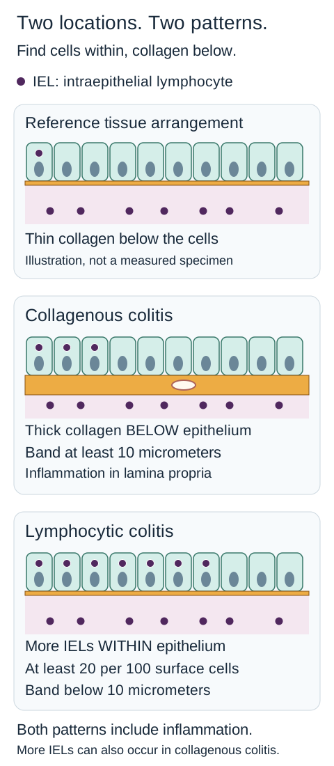
Use the tissue-layer diagram to separate a deposit below the surface from cells within it. In collagenous colitis, an abnormally thick subepithelial collagen band accompanies lamina propria inflammation. The deeper edge can be irregular and can entrap capillaries or inflammatory cells. Increased IELs can also occur, so their presence does not automatically make the disease lymphocytic colitis. In lymphocytic colitis, increased surface IELs accompany inflammation without a collagen band reaching the collagenous threshold. [1]
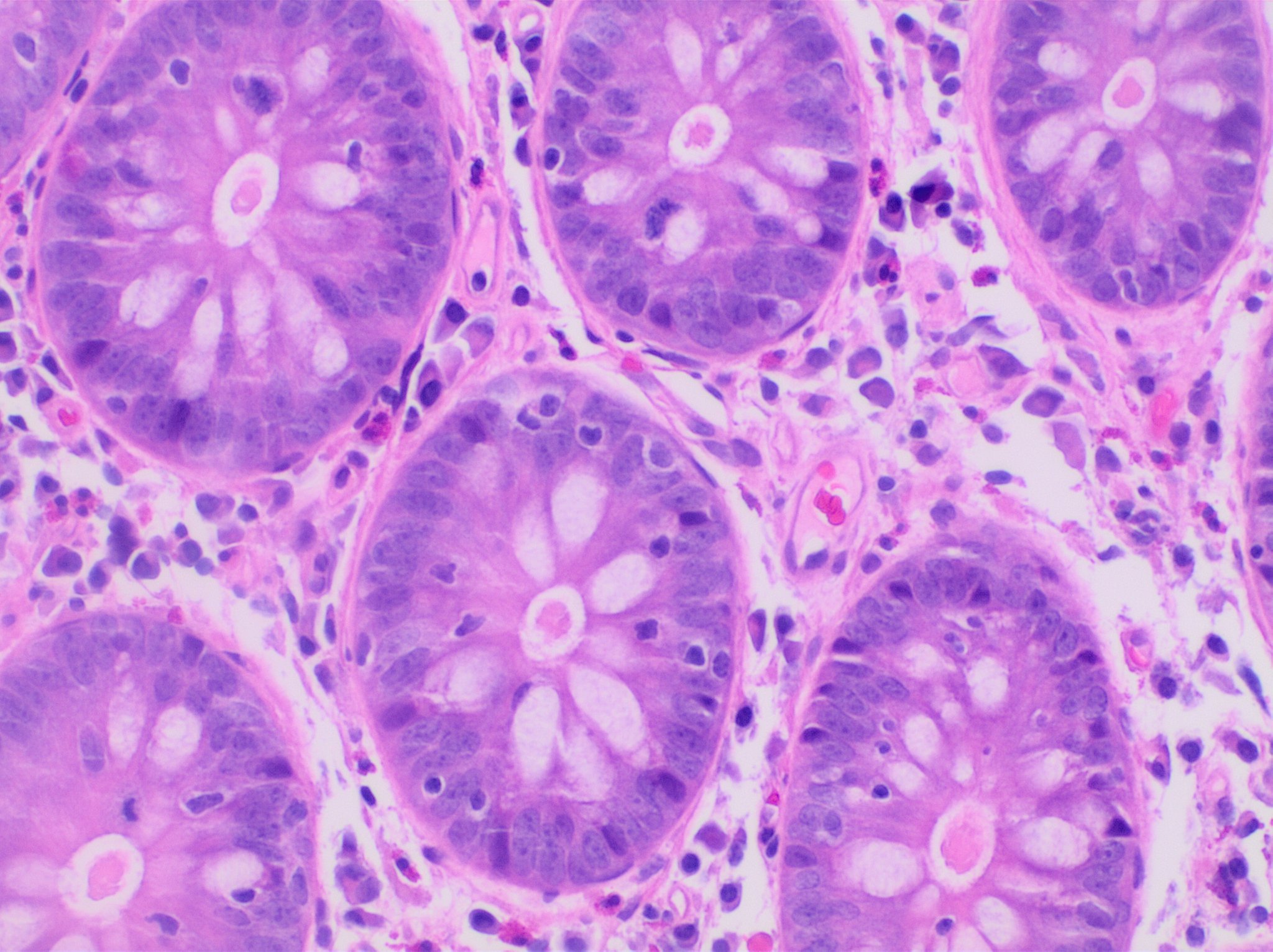
The accompanying original H&E micrograph shows lymphocytes within colonic epithelium and in the surrounding tissue. First locate a gland lining, then distinguish a small dark round lymphocyte from a larger epithelial nucleus. This field illustrates cellular location; it is not a calibrated surface-cell count and cannot independently establish the numerical diagnostic threshold. The clinical image is preserved without cropping or added markings. [6]
Locate the epithelial lining of a gland and distinguish small dark lymphocytes from the larger epithelial nuclei. This is an authentic clinical image illustrating location, not a field from which the diagnostic surface-IEL threshold should be calculated. Image: Mikael Häggström, M.D., CC0 1.0. [6] Mikael Häggström, M.D.; original source; CC0 1.0 Universal.
Prominent persistent architectural distortion, a characteristic distribution of gross inflammation or granulomas can redirect assessment toward ulcerative colitis, Crohn disease or another process. Do not teach the converse as an absolute: subtle nonspecific endoscopic abnormalities, occasional cryptitis and surface injury can occur in microscopic colitis. Endoscopy, histology and the clinical course must agree. [1][2]
A biopsy has numerous surface IELs and a clearly thickened collagen band. Predict whether the lymphocyte finding cancels the collagen finding.
Check the tissue relationship
It does not. Collagenous colitis may include increased IELs. Assess the collagen measurement and supporting inflammation before assigning the subtype.
For a second biopsy with increased surface IELs but a thin collagen band, the relevant question changes: does the surface count meet the lymphocytic threshold? Symptoms alone cannot distinguish these two patients.
Count the right cells and measure the right plane
Histologic thresholds are useful only after the specimen and measurement are trustworthy. The following criteria refer to appropriately oriented H&E sections, compatible symptoms and accompanying lamina propria inflammation. Measurements are in micrometers, not millimeters. [1]
Follow the same epithelial layer through all three panels. The orange band is below it; the small dark lymphocytes can lie within it. Use the labeled diagnostic thresholds rather than counting drawn dots. Collagenous disease can also have increased IELs. [1][1]
Two measurements answer different questions
Pattern
What supports it?
PatternCollagenous colitis
What supports it?Subepithelial collagen band at least 10 micrometers, with inflammation.
PatternLymphocytic colitis
What supports it?At least 20 IELs per 100 surface epithelial cells, with inflammation and a collagen band below 10 micrometers.
PatternIncomplete collagenous pattern
What supports it?Collagen band greater than 5 but below 10 micrometers, with compatible inflammation.
PatternIncomplete lymphocytic pattern
What supports it?More than 10 but fewer than 20 IELs per 100 surface cells, with a normal collagen band and compatible inflammation.
A tangential cut travels obliquely through a thin layer and can make the collagen band appear thicker. Proper orientation and additional levels can therefore change the interpretation without any biological change in the patient. A collagen stain can help in a borderline specimen, but stronger staining does not correct the wrong cutting plane. Count surface IELs away from adjacent lymphoid aggregates; a lymphoid focus is not a representative substitute for the surface epithelium. [1]
A well-oriented specimen contains 45 IELs among 150 surface epithelial cells and a 4-micrometer band. Convert the count to a denominator of 100 before naming the pattern.
Check the normalized count
45 divided by 150, multiplied by 100, gives 30 IELs per 100 surface cells. With the thin band and compatible inflammation, that supports lymphocytic colitis.
The important calculation is the proportion, not the raw count. In a different specimen, 18 IELs among 150 surface cells is 12 per 100, not 18 or 27. A borderline result deserves pathological correlation rather than rounding upward to a preferred diagnosis.
Change the sampling plan, not the photograph
Obtain biopsies from both right and left colon when evaluating suspected microscopic colitis. A rectal specimen alone is insufficient. Changes can vary along the colon, and sampling only the most convenient distal area can leave the important finding unobserved. Ileocolonoscopy also assesses other explanations for chronic diarrhea; an ileal biopsy, when indicated, does not replace colonic sampling. [1][2]
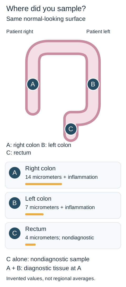
The sampling diagram represents a patient whose surface looks normal at all three sites. Site A is right colon, B is left colon and C is rectum. Its specimen cards show: A, a 14-micrometer collagen band with inflammation; B, a 7-micrometer band with inflammation; C, a 4-micrometer band without diagnostic inflammation. These values are an example, not typical regional averages.
Compare C alone with the A-plus-B plan before reading the conclusion. The right-colon sample supplies the diagnostic evidence missed by rectal-only sampling. The photographs would still look the same. The measurements are not regional norms or a sensitivity estimate. [1][2][1][2]
Start with C alone. What finding has the laboratory actually excluded? Then include A and B and predict whether the interpretation changes.
Compare the two sampling plans
C alone supplies a nondiagnostic rectal sample, not a negative examination of every colonic segment. Adding A and B reveals a collagenous pattern in the right colon. The sampling plan changes the evidence available, while the endoscopic photograph remains the same.
The complete interpretation is visible in the specimen cards and this rule: a nondiagnostic sample cannot describe an unsampled site. To repeat the exercise, cover the A and B specimen cards and explain what information disappears. This is a sampling demonstration, not a validated sensitivity calculator.
Sending right- and left-sided samples in separately labeled containers preserves location for the pathologist. Normal cellularity and collagen thickness vary by region, which can matter when findings are borderline. The recommendation to sample both sides has stronger evidence than the expert preference for separate containers; do not present the two as equally proven. [1]
Transfer the principle to a patient with only mildly red mucosa: inspect and sample appropriately rather than treating redness as proof of a specific subtype. Both an unremarkable surface and nonspecific erythema can coexist with diagnostic microscopic disease.
Let each test answer a defined competing explanation
Chronic watery diarrhea is a syndrome, not a diagnosis. Infection, celiac disease, bile acid diarrhea, medication effects, thyroid dysfunction, dietary carbohydrate malabsorption and functional disorders can resemble microscopic colitis. Some can coexist with it. Assess hydration and electrolytes when stool losses are substantial. New bleeding, significant pain, fever or circulatory compromise requires assessment for another or additional illness, rather than automatic attribution to an established diagnosis. [1][2]
A normal fecal calprotectin does not reliably exclude microscopic colitis. This differs from the useful role that inflammatory stool markers can have in selected evaluations for other inflammatory bowel disease. Likewise, an irritable-bowel label should be reconsidered when new nocturnal diarrhea, weight loss, risk factors or failure of appropriate therapy changes the clinical picture. A diagnostic test should be interpreted in the population and disease for which it is informative. [1][2]
Screen patients with microscopic colitis for celiac disease. Tissue transglutaminase IgA with assessment of total IgA is a common starting approach while the patient is eating gluten. An IgA-based negative result is unreliable in IgA deficiency; use an appropriate IgG-based test. Positive testing or persistent strong suspicion may require duodenal biopsy. A colonic lymphocyte count is not evidence of duodenal villous injury. [1][2]
Testing for bile acid diarrhea is not required in every newly diagnosed microscopic-colitis patient. It becomes especially useful when an adequate budesonide course fails or the broader history supports that possibility. Excess bile acids reaching the colon can sustain watery stools even when another diagnosis is already established. Locally available testing can include SeHCAT retention, serum C4 or fecal bile acids; availability and reference intervals differ. [1][2]
A patient takes the prescribed budesonide consistently but remains symptomatic. Another has repeated remission on treatment and prompt recurrence after stopping it. Which patient first needs the nonresponse workup?
Separate the two trajectories
The first patient needs verification of diagnosis, preparation, dose, adherence and coexisting causes such as bile acid diarrhea. The second has demonstrated response and may need a plan to sustain it. A new infection or changed symptom pattern can still require investigation in either patient.
For a new situation, consider diarrhea that occurs reproducibly after dairy, stops when dairy is avoided and lacks overnight symptoms, with adequate negative colonic biopsies. The evidence now points toward a dietary explanation, not repeated corticosteroid courses. A prior diagnostic suspicion should not outweigh the subsequently obtained results. [2]
Treat the clinical burden, then follow the response
For clinically active microscopic colitis, oral budesonide is the preferred induction treatment. A common adult regimen is 9 mg daily for 6 to 8 weeks, followed by assessment and an individualized plan. Treating significant watery diarrhea and incontinence should not depend on whether the subtype is collagenous or lymphocytic. The lymphocytic-colitis randomized trial directly supports budesonide induction; mesalazine did not establish a comparable role. [1][3]
Budesonide acts as a glucocorticoid and undergoes substantial first-pass metabolism. Lower systemic exposure than conventional systemic corticosteroids does not mean no systemic effects. Review interactions, diabetes, blood pressure, eye and bone risks, infection and liver function as appropriate. Strong CYP3A4 inhibitors such as itraconazole or ritonavir can increase systemic exposure; the product labeling advises avoiding such combinations. Adrenal suppression can occur, particularly with prolonged exposure. A formulation's Crohn-disease label is not the source of the microscopic-colitis maintenance recommendation. [5]
Loperamide can help selected mild or residual symptoms, although microscopic-colitis-specific trial evidence is limited. A bile acid sequestrant is relevant when bile acid diarrhea is documented or strongly supported. Neither should distract from effective induction in a patient with substantial active disease. Routine mesalazine, conventional systemic prednisone or prednisolone, antibiotics and probiotics are not preferred substitutes for budesonide under the European recommendations. Bismuth evidence is limited and guideline approaches differ; do not present it as an equally established first choice. [1]
After treatment, assess stool consistency as well as frequency, urgency, overnight symptoms and continence. The guideline's practical remission criterion uses a seven-day diary: mean fewer than 3 total stools per day and mean fewer than 1 watery stool per day. Both conditions are required. These thresholds support assessment; they do not replace discussion of ongoing impairment or constitute a fully validated universal activity scale. [1]
Over seven days, one patient records 18 total stools, including 8 watery stools. Which part of the remission definition is still unmet?
Check both averages
The means are 18/7 = 2.57 total stools per day and 8/7 = 1.14 watery stools per day. Total frequency meets the first condition, but watery frequency does not meet the second. Improvement is not identical to remission.
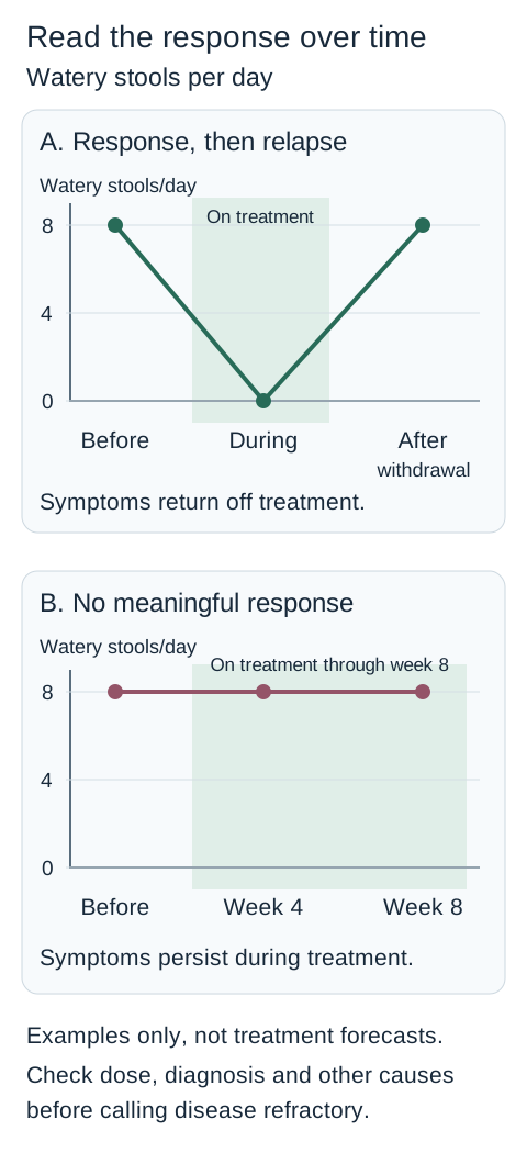
The response diagram compares two response trajectories. A fall from eight watery stools per day to one formed stool, followed by recurrence off therapy, demonstrates response followed by relapse. Persistently frequent watery stools throughout an adequate course suggest nonresponse. The diagram is qualitative: its dates and counts are not promises about treatment speed.
Read the treatment period before interpreting the current stool count. The first pattern demonstrates benefit that was lost after withdrawal. The second requires verification of an adequate course and competing causes before escalation. Zero watery stools can coexist with formed stools; these plots are not total stool counts. [1][4][1][4]
Relapse after withdrawal is common. After recurrent disease is controlled, use the lowest effective budesonide maintenance dose when clinically appropriate, with periodic reassessment. Trials support maintenance in collagenous colitis; direct randomized maintenance evidence is weaker for lymphocytic colitis. The collagenous-colitis trial studied patients who had first responded to induction, so its results do not prove that maintenance dosing will rescue an initial nonresponder. [1][4]
For prolonged treatment, individualize bone-health assessment and calcium or vitamin D needs, particularly with existing fracture risk. Do not increase treatment solely to normalize a follow-up biopsy in a clinically well patient: histology can persist despite symptom resolution, and routine histologic monitoring is not recommended. Fecal calprotectin is also not a reliable microscopic-colitis monitoring target. [1][5]
Before labeling disease refractory, recheck the pathology, formulation, actual dose taken, adherence, medication exposures and coexisting infection, celiac disease or bile acid diarrhea. Selected persistent cases need specialist consideration of steroid-sparing therapies, including thiopurines or biologics such as anti-TNF agents or vedolizumab. Evidence is limited; no single escalation fits every patient. Surgery is an exceptional last option after medical approaches have failed. [1]
Microscopic colitis alone does not require a special colorectal-cancer surveillance program. Continue ordinary screening or surveillance determined by age, prior polyps, family history and other independent indications. New symptoms still warrant investigation. [1]
Carry three questions forward: Was the relevant tissue sampled? Does the measurement fit the clinical syndrome? Did treatment never work, or did its benefit disappear after withdrawal?
Apply the lesson
Case 1
Show answer and explanations for case 1
A. Fecal elastase concentration (Why this does not fit)
It helps evaluate pancreatic exocrine insufficiency, a possible cause of chronic diarrhea. Watery nocturnal stools with an unbiopsied normal colon make a mucosal diagnosis a more direct target than pancreatic testing. Choose the unresolved explanation best supported by the full phenotype.
Reasoning steps for option A
What process would fecal elastase evaluate in this patient?
Low fecal elastase suggests pancreatic exocrine insufficiency, which can produce chronic diarrhea.
Why is elastase less targeted than colonic tissue here?
Nocturnal watery stools and a normal-looking but unbiopsied colon leave microscopic colitis untested.
Which unresolved diagnostic gap should take priority over fecal elastase?
The normal-looking colon has not been biopsied; right- and left-colon tissue directly tests for a microscopic cause of the nocturnal watery diarrhea.
B. CT enterography of the small bowel (Why this does not fit)
It can demonstrate structural and some inflammatory small-intestinal abnormalities. It would not supply the colonic microscopic evidence missing from the prior colonoscopy. Match the investigation to the tissue compartment in question.
Reasoning steps for option B
Which compartment does CT enterography principally investigate?
It evaluates small-bowel wall and structural or inflammatory abnormalities, not microscopic colonic mucosa.
What was missing from the normal colonoscopy?
No mucosal biopsies were taken, so normal photographs cannot exclude microscopic colitis.
Why would CT not establish the most likely unresolved cause?
Imaging cannot measure colonic epithelial lymphocytes or the subepithelial collagen band needed for diagnosis.
C. Repeat tissue transglutaminase IgA (Why this does not fit)
Celiac disease can coexist with microscopic colitis and cause chronic diarrhea. The recent negative result was obtained with normal total IgA during gluten intake; colonic tissue has not been assessed. A valid completed test should not displace a missing diagnostic test.
Reasoning steps for option C
Why is celiac disease a reasonable initial consideration?
It can coexist with microscopic colitis and cause chronic diarrhea.
Why was the negative tissue transglutaminase IgA interpretable?
She was eating gluten and had normal total IgA, avoiding the main exposure and IgA-deficiency limitations.
What should be investigated instead of repeating that valid screen?
The unbiopsied colon remains the diagnostic gap despite completed celiac serology.
D. Repeat fecal calprotectin measurement (Why this does not fit)
A marker can help follow some intestinal inflammatory conditions. Calprotectin is not a reliable exclusion test for microscopic colitis, so repetition does not correct the absent biopsies. A poorly suited exclusion test does not become definitive by repetition.
Reasoning steps for option D
What does fecal calprotectin measure rather than diagnose?
It reflects intestinal inflammatory activity but is not a reliable exclusion test for microscopic colitis.
Does the value of 28 micrograms/g rule out microscopic colitis?
No; microscopic colitis can occur below the laboratory cutoff of 50.
Why would repeating calprotectin not repair the workup?
A second stool marker cannot replace right- and left-colon histology absent from the prior examination.
E. Right- and left-colon mucosal biopsies (Best answer)
It assesses the visible surface, not the cellular pattern. Both-sided mucosal sampling can establish microscopic colitis despite normal calprotectin and normal photographs. Normal appearance cannot replace histologic sampling.
Reasoning steps for option E
What does a visually normal colonoscopy leave unresolved?
It documents surface appearance but not lymphocytic infiltration or collagen-band thickness.
Where should tissue be sampled in this case?
Right- and left-colon mucosal biopsies provide representative assessment of potentially patchy microscopic colitis.
Why choose biopsy despite the normal calprotectin?
Nocturnal watery diarrhea remains compatible with microscopic colitis, which a normal stool marker cannot exclude.
Takeaway: Normal appearance cannot replace histologic sampling.
A. Collagenous colitis; oral budesonide (Why this does not fit)
Collagenous colitis requires a subepithelial band reaching at least 10 micrometers with compatible inflammation. The band is 4 micrometers, so the treatment choice is reasonable for microscopic colitis but the subtype is wrong. A correct treatment does not rescue an incorrect histologic interpretation.
Reasoning steps for option A
What band thickness is required for collagenous colitis?
A subepithelial collagen band of at least 10 micrometers with compatible inflammation is required.
How does the measured 4-micrometer band affect this proposed subtype?
It is too thin for collagenous colitis despite 45 lymphocytes among 150 epithelial cells.
Does appropriate budesonide salvage this pairing?
No; budesonide may be appropriate, but the collagenous diagnosis in the pairing is incorrect.
B. Lymphocytic colitis; oral budesonide (Best answer)
45/150 multiplied by 100 equals 30 IELs per 100 surface cells. The findings support lymphocytic colitis, for which oral budesonide is the preferred induction treatment. Normalize the count, assign the pattern, then address clinical activity.
Reasoning steps for option B
What is 45 IELs among 150 surface cells normalized to 100?
45 divided by 150 times 100 equals 30 IELs per 100 surface epithelial cells.
How do 30 IELs and a 4-micrometer band classify the biopsy?
The elevated surface IEL count with a thin collagen band and lamina propria inflammation supports lymphocytic colitis.
Which induction therapy fits six watery stools daily and nocturnal urgency?
Oral budesonide is preferred for clinically active lymphocytic colitis.
C. Lymphocytic colitis; oral mesalazine (Why this does not fit)
Thirty surface IELs per 100 cells with a thin band supports lymphocytic colitis. Budesonide has stronger induction evidence than mesalazine for the substantial active symptoms described. Match the treatment evidence to the established subtype and clinical burden.
Reasoning steps for option C
Is the lymphocytic diagnosis in this pairing supported?
Yes; 45/150 equals 30 IELs per 100 cells, with a nonthickened collagen band.
Which half of the pairing is inferior?
Oral mesalazine has weaker induction evidence than oral budesonide for active microscopic colitis.
What should direct initial therapy for these substantial symptoms?
Choose budesonide induction rather than treating the correct histologic label with a less-supported drug.
D. Ulcerative colitis; oral mesalazine (Why this does not fit)
Mesalazine has an established role in ulcerative colitis. Surface lymphocytosis with a thin collagen band and compatible lamina propria inflammation supports lymphocytic colitis in this watery-diarrhea presentation. Do not substitute a familiar colitis regimen for the demonstrated microscopic pattern.
Reasoning steps for option D
What histology would support choosing an ulcerative-colitis regimen?
Chronic architectural distortion and an ulcerative-colitis inflammatory pattern would support that diagnosis.
What do the measured epithelial count and band instead show?
Thirty IELs per 100 surface cells and a 4-micrometer band support lymphocytic colitis.
Why is mesalazine for ulcerative colitis the wrong pairing here?
Its role in ulcerative colitis does not override the demonstrated microscopic-colitis pattern, for which budesonide is preferred.
Takeaway: Normalize the count, assign the pattern, then address clinical activity.
A. Incomplete collagenous colitis with mild inflammation (Why this does not fit)
An incomplete collagenous pattern has a band greater than 5 but below 10 micrometers with compatible inflammation. Sixteen micrometers is above the full collagenous threshold, not within the incomplete interval. Do not call a clearly qualifying measurement borderline.
Reasoning steps for option A
What band range defines incomplete collagenous colitis?
A band greater than 5 but less than 10 micrometers with compatible inflammation supports an incomplete pattern.
Does the well-oriented 16-micrometer band fall in that range?
No; it exceeds the 10-micrometer threshold for established collagenous colitis.
Do irregular edges and entrapped capillaries make an incomplete label more plausible?
No; those characteristic features reinforce a genuine collagenous deposit rather than a borderline band.
B. Lymphocytic colitis without a collagenous component (Why this does not fit)
Lymphocytic colitis requires a collagen band below the collagenous threshold. The 16-micrometer band supplies a definite collagenous component. A qualifying IEL count alone does not specify the subtype.
Reasoning steps for option B
Why might 31 IELs per 100 cells suggest lymphocytic colitis?
That count exceeds the usual 20-per-100 surface-cell lymphocytic threshold.
What finding defeats the phrase “without a collagenous component”?
The 16-micrometer irregular subepithelial band with entrapped capillaries is a definite collagenous component.
How should simultaneous high IELs and thick collagen be integrated?
Increased IELs can coexist with collagenous colitis; do not discard the defining collagen-band finding.
C. Collagenous colitis with intraepithelial lymphocytosis (Best answer)
A 16-micrometer subepithelial band with inflammation exceeds the collagenous threshold. No. Increased IELs can accompany collagenous colitis and do not cancel the characteristic band. Integrate both measurements instead of choosing the first abnormal number.
Reasoning steps for option C
Which feature establishes the collagenous pattern?
A well-oriented 16-micrometer subepithelial band with irregular deep edge, entrapped capillaries and inflammation exceeds the diagnostic threshold.
Do 31 surface IELs per 100 cells contradict collagenous colitis?
No; surface intraepithelial lymphocytosis may accompany collagenous colitis.
How should this biopsy be reported as an integrated finding?
Collagenous colitis with intraepithelial lymphocytosis accounts for both measured abnormalities.
D. Ulcerative colitis with secondary surface injury (Why this does not fit)
A compatible clinical distribution and characteristic chronic architectural injury would strengthen it. The irregular thick collagen band with entrapped capillaries and largely preserved architecture favors collagenous colitis. Use the characteristic positive tissue pattern rather than inflammation alone.
Reasoning steps for option D
What chronic tissue feature would favor ulcerative colitis instead?
Characteristic chronic crypt architectural distortion would lend support to ulcerative colitis.
Which observed morphology favors collagenous colitis?
The thick irregular collagen band with entrapped capillaries and largely preserved crypt architecture favors collagenous colitis.
Why is inflammation alone insufficient to call this ulcerative colitis?
Lamina propria inflammation is compatible with microscopic colitis and does not negate its specific collagenous band.
Takeaway: Integrate both measurements instead of choosing the first abnormal number.
A. Reorient the tissue and examine additional levels (Best answer)
A tangential plane can make a thin collagen layer appear abnormally thick. Reorientation and additional properly cut levels test whether the thick band persists in an interpretable plane. Resolve a technical measurement problem before assigning a biological diagnosis.
Reasoning steps for option A
How can oblique cutting create an apparent 12-micrometer band?
A tangential section traverses the collagen layer diagonally and exaggerates apparent thickness.
What comparison makes this artifact likely?
Adjacent perpendicular sections measure only 4 micrometers, with minimal inflammation and no increased IELs.
Why reorient and examine additional levels?
Properly perpendicular levels can determine whether a genuinely thick band persists before assigning a subtype.
B. Apply a collagen stain to the same oblique section (Why this does not fit)
It can make collagen easier to recognize in a borderline specimen. No. A stain can highlight the layer but cannot establish its true perpendicular thickness in the same tangential section. Correct the measurement geometry rather than merely increasing its visual contrast.
Reasoning steps for option B
What would a collagen stain add to the oblique fragment?
It would highlight collagen and help identify its boundaries.
Would staining the same oblique plane yield a valid perpendicular thickness?
No; contrast cannot correct geometric exaggeration from tangential sectioning.
What must occur before calling the 12-micrometer focus collagenous colitis?
Reorientation and additional well-oriented levels should verify the measurement and accompanying pattern.
C. Count lymphocytes beside the largest lymphoid aggregate (Why this does not fit)
Nearby lymphoid tissue can increase local lymphocyte numbers. It would neither correct the cutting plane nor provide a representative surface IEL count. Count representative surface epithelium away from lymphoid aggregates.
Reasoning steps for option C
What bias arises when counting IELs near a lymphoid aggregate?
Local enrichment can inflate the count relative to representative surface epithelium.
Does such a count explain the 12-versus-4-micrometer collagen discrepancy?
No; epithelial cell counting does not correct an oblique collagen measurement.
Where would a valid IEL assessment be performed if needed?
Count representative surface epithelial cells away from lymphoid aggregates on interpretable tissue.
D. Assign the subtype from the maximum apparent thickness (Why this does not fit)
A true focal thick band can be diagnostically useful in heterogeneous disease. The only high measurement is from the plane identified as oblique, while perpendicular tissue lacks the supporting pattern. A maximum value is useful only when the measurement itself is valid.
Reasoning steps for option D
When could a focal maximum collagen thickness matter?
A genuinely thick well-oriented focal band with supporting inflammation could establish heterogeneous collagenous disease.
Why is the reported 12-micrometer maximum unreliable?
It occurs only in a section identified as oblique; perpendicular sections show a 4-micrometer layer.
What decision should precede subtype assignment?
Verify true perpendicular band thickness on reoriented additional levels rather than accepting the artifact-prone maximum.
Takeaway: Resolve a technical measurement problem before assigning a biological diagnosis.
A. The descending sample establishes a functional bowel disorder (Why this does not fit)
It is based on an appropriate clinical pattern and evaluation, not on a single low collagen measurement. Another colonic site contains a diagnostic inflammatory collagenous pattern. A subthreshold measurement at one site is not proof of a functional cause.
Reasoning steps for option A
Does a 7-micrometer descending-colon band establish a functional disorder?
No; a subthreshold collagen measurement does not diagnose a functional bowel disorder.
Which other site contradicts a functional-only explanation?
The ascending colon has an inflamed 15-micrometer collagen band diagnostic of collagenous colitis.
Why did the earlier rectal-only examination miss that explanation?
The rectum measured 4 micrometers and did not sample the diagnostic proximal segment.
B. The proximal band represents normal regional collagen variation (Why this does not fit)
Normal cellularity and collagen thickness can vary by colonic region. A 15-micrometer band with compatible inflammation meets the collagenous threshold in the symptomatic patient rather than supplying a normal regional measurement. Use regional context without dismissing a clearly diagnostic inflammatory pattern.
Reasoning steps for option B
How might regional variation complicate collagen-band interpretation?
Band thickness can differ between colonic segments, so location matters when interpreting samples.
Why is the ascending 15-micrometer band not dismissed as normal variation?
It exceeds the 10-micrometer collagenous threshold and has compatible inflammation.
How does this finding explain the prior negative rectal biopsy?
Disease is regionally heterogeneous; the rectum was nondiagnostic while the proximal colon was involved.
C. The rectal measurement invalidates the proximal tissue finding (Why this does not fit)
Regional sampling tests whether the tissue findings are consistent or heterogeneous. No. A nondiagnostic rectum does not negate a well-oriented diagnostic proximal specimen with compatible inflammation. Heterogeneous sampling results require integration, not cancellation.
Reasoning steps for option C
What did the rectal biopsy actually measure?
The rectal band was 4 micrometers, below the collagenous threshold at that sampled site.
Does this invalidate the ascending-colon 15-micrometer finding?
No; a nondiagnostic distal sample does not negate diagnostic inflamed proximal tissue.
How should differing sites be synthesized?
Recognize a heterogeneous collagenous-colitis pattern rather than require every segment to cross the threshold.
D. Earlier sampling omitted the region with diagnostic changes (Best answer)
The ascending-colon specimen has a 15-micrometer band with inflammation. No. The prior rectal sample did not represent the subsequently diagnostic proximal tissue. A nondiagnostic distal specimen cannot exclude a proximal microscopic pattern.
Reasoning steps for option D
Which later sample establishes collagenous colitis?
The ascending-colon biopsy shows a 15-micrometer collagen band with inflammation.
Was that diagnostic region sampled by the initial sigmoidoscopy?
No; the initial examination obtained rectal biopsies only.
What reconciles normal endoscopic views with differing biopsy results?
Microscopic changes can be invisible and patchy; the first sampling omitted the diagnostic proximal region.
Takeaway: A nondiagnostic distal specimen cannot exclude a proximal microscopic pattern.
A. Normal colonic mucosa without inflammatory abnormalities (Why this does not fit)
A representative specimen without qualifying cellular or stromal abnormalities could be nondiagnostic. The supplied mild inflammation and intermediate IEL increase support a recognized incomplete pattern. Not meeting full subtype criteria does not make every lesser abnormality normal.
Reasoning steps for option A
Are the adequate biopsies wholly devoid of inflammatory abnormality?
No; they show mild lamina propria inflammation and 16 surface IELs per 100 cells.
What category does an IEL count of 16 support?
It exceeds 10 but is below 20, fitting an incomplete lymphocytic microscopic-colitis pattern with compatible inflammation.
Why not report normal mucosa simply because full criteria are unmet?
A meaningful intermediate epithelial lymphocytosis remains present despite a normal collagen band.
B. Fully developed collagenous-colitis pattern (Why this does not fit)
A collagen band at least 10 micrometers with compatible inflammation supports collagenous colitis. The band is normal rather than thickened. Distinguish an increased cell population from a collagen deposit.
Reasoning steps for option B
What collagen measurement would support fully developed collagenous colitis?
A band of at least 10 micrometers with compatible inflammation.
What band is actually reported here?
The subepithelial collagen band is normal and thin, not thickened.
Which abnormal compartment accounts for these biopsies instead?
Surface epithelial lymphocytes are increased to 16 per 100 cells, supporting an incomplete lymphocytic pattern.
C. Incomplete lymphocytic microscopic-colitis pattern (Best answer)
It lies above 10 but below the full lymphocytic threshold of 20. No. The band is normal, supporting an incomplete lymphocytic pattern with the stated inflammation. Report a supported incomplete pattern rather than rounding toward a full diagnosis.
Reasoning steps for option C
How does 16 IELs per 100 compare with lymphocytic thresholds?
It is above the incomplete threshold of 10 but below the full threshold of 20 per 100 surface cells.
What accompanying features support the incomplete lymphocytic category?
Mild lamina propria inflammation, a normal thin collagen band and representative counts away from lymphoid aggregates.
Why retain the word “incomplete” in the report?
Calling 16 IELs fully developed would incorrectly round a subthreshold count up to 20.
D. Fully developed lymphocytic-colitis pattern (Why this does not fit)
At least 20 IELs per 100 surface epithelial cells is required with the other criteria. Sixteen does not reach 20 even though the symptoms are compatible. Symptoms cannot substitute for a missing histologic threshold.
Reasoning steps for option D
How many IELs per 100 surface cells are required for full lymphocytic colitis?
At least 20 are required with the compatible tissue pattern.
Can chronic watery diarrhea raise this measured count of 16 to that threshold?
No; compatible symptoms cannot change the histologic measurement.
What diagnosis preserves the observed inflammation without overcalling it?
Incomplete lymphocytic microscopic-colitis pattern, rather than fully developed lymphocytic colitis.
Takeaway: Report a supported incomplete pattern rather than rounding toward a full diagnosis.
A. Fecal calprotectin testing for small-bowel villous injury (Why this does not fit)
It is a stool inflammatory marker rather than a direct measure of celiac villous damage. It does not provide the appropriate celiac evaluation in this IgA-deficient patient. Use disease-directed testing rather than substituting a nonspecific marker.
Reasoning steps for option A
What does fecal calprotectin reflect?
It is a nonspecific stool marker of intestinal inflammation, not a direct assay for celiac villous injury.
Why is celiac evaluation still unresolved despite negative tTG-IgA?
Total IgA is undetectable, so an IgA-based negative result cannot exclude celiac disease.
What test answers that unresolved question better?
IgG-based tissue transglutaminase testing can assess celiac disease without relying on IgA.
B. Repeat rectal biopsy for colonic IEL counting (Why this does not fit)
It samples colonic mucosa and can contribute to a colonic histologic assessment. Celiac disease concerns the small bowel; another rectal count cannot replace its serologic or duodenal evaluation. Localize the unanswered diagnostic question before repeating tissue sampling.
Reasoning steps for option B
What tissue would another rectal IEL count evaluate?
Colonic surface epithelium, relevant to the already confirmed microscopic colitis.
What condition is suggested by persistent loose stools and iron deficiency?
Coexisting celiac disease involving the small bowel remains possible.
Why not repeat the rectal biopsy to investigate it?
A colonic IEL count cannot replace IgA-independent celiac serology or, when indicated, duodenal assessment.
C. Serum TSH testing for excess thyroid replacement (Why this does not fit)
Thyrotoxicosis can cause frequent stools and belongs in the broader differential. The absent IgA invalidates the negative IgA celiac screen, a problem that TSH measurement cannot resolve. Address a demonstrated limitation directly rather than ordering an unrelated plausible test.
Reasoning steps for option C
How could TSH relate to frequent stools?
Excess thyroid hormone or replacement can cause diarrhea.
What documented assay limitation would TSH leave untouched?
Undetectable total IgA makes the negative tTG-IgA uninformative about celiac disease.
Which choice directly addresses that limitation in this gluten-eating patient?
IgG-based tTG evaluates celiac serology without depending on the missing IgA class.
D. Repeat tissue transglutaminase IgA testing (Why this does not fit)
Repetition can help when timing, exposure or a transient technical problem made the original result unreliable. The patient lacks measurable IgA, so simply repeating an IgA-based assay does not repair the biological limitation. Choose a test that addresses the reason the first result was uninformative.
Reasoning steps for option D
Why is a second tTG-IgA unlikely to change interpretation?
The first result is biologically limited by undetectable total IgA, not simply a one-time timing error.
Does ongoing gluten intake solve the IgA deficiency?
No; appropriate gluten exposure cannot make an IgA-based assay reliable in absent IgA.
What modification to serologic testing is needed?
Switch to an IgG-based celiac assay such as tTG-IgG instead of repeating tTG-IgA.
E. IgG-based tissue transglutaminase testing (Best answer)
It makes a negative IgA result unreliable for excluding celiac disease. IgG-based transglutaminase testing addresses celiac disease without depending on the missing IgA class. Check immunoglobulin adequacy before interpreting a negative IgA screen.
Reasoning steps for option E
What makes the prior negative tTG-IgA unreliable?
Total IgA is below the assay detection limit, so the patient may not produce detectable IgA antibody despite celiac disease.
Why is tTG-IgG appropriate now?
It uses IgG rather than the deficient IgA class while the patient continues eating gluten.
What clinical findings warrant pursuing this rather than attributing all stools to colitis?
Persistent loose stools and iron deficiency despite improved nocturnal urgency suggest an additional cause such as celiac disease.
Takeaway: Check immunoglobulin adequacy before interpreting a negative IgA screen.
A. Tissue transglutaminase IgA with total IgA (Best answer)
No. The conditions can coexist, and celiac screening is recommended in microscopic colitis. An appropriate celiac screen, interpreted with total IgA, remains missing despite a normal thyroid result. An established diagnosis does not eliminate associated causes of persistent symptoms.
Reasoning steps for option A
Why screen for celiac disease after collagenous colitis is confirmed?
Celiac disease can coexist with microscopic colitis and contribute to persistent loose stools or fatigue.
Which celiac evaluation is missing?
Tissue transglutaminase IgA together with total IgA has not been obtained.
Why include total IgA with the tTG-IgA?
It identifies IgA deficiency that could make a negative IgA-based celiac result falsely reassuring.
B. Routine serial fecal calprotectin measurements (Why this does not fit)
They can assist monitoring in selected inflammatory bowel diseases. Calprotectin neither reliably monitors microscopic colitis nor provides the recommended celiac screen. Do not transfer a monitoring strategy indiscriminately between diseases.
Reasoning steps for option B
What might serial fecal calprotectin monitor in other conditions?
It can help track inflammation in some inflammatory bowel diseases.
Why is it a poor routine marker for this patient’s collagenous colitis?
Calprotectin does not reliably reflect microscopic-colitis activity, which may be present despite low values.
Does it complete the missing associated-disease workup?
No; it does not screen for celiac disease, for which tTG-IgA with total IgA is indicated.
C. Repeat TSH before ordering other studies (Why this does not fit)
It helps assess thyrotoxicosis or excessive thyroid replacement. A normal result on an unchanged dose supplies no current evidence of excess replacement, while celiac screening is incomplete. Let a completed informative test redirect the evaluation.
Reasoning steps for option C
What cause of diarrhea could TSH testing assess?
Biochemical thyrotoxicosis or excessive levothyroxine replacement.
What does this patient’s current TSH and dosing history show?
TSH is within reference range on an unchanged dose, so overreplacement lacks current biochemical support.
Which unperformed test should take priority over immediate repeat TSH?
Celiac serology with total IgA addresses an associated enteropathy not yet screened for.
D. Screening small-bowel follow-through for structural obstruction (Why this does not fit)
Distention, vomiting and an obstructive clinical pattern could direct structural imaging. The supplied question concerns a recognized associated enteropathy without an obstructive syndrome; serologic screening is the targeted next assessment. A broad imaging study is not interchangeable with an indicated associated-disease screen.
Reasoning steps for option D
Which findings would support small-bowel obstruction imaging?
Vomiting, distention and an obstructive symptom pattern would favor structural assessment.
Are those findings present in this patient?
No; fatigue and loose stools with confirmed collagenous colitis do not suggest obstruction.
What narrower small-bowel-associated evaluation is warranted?
Screen for coexisting celiac disease using tTG-IgA and total IgA rather than follow-through imaging.
E. Repeat colonoscopy to document gross mucosal healing (Why this does not fit)
It can assess visible inflammatory lesions in diseases where those lesions track activity. Microscopic colitis can have an unremarkable surface, and routine healing colonoscopy does not complete the missing associated-disease evaluation. Choose a test for the unresolved question rather than repeating an already established diagnosis.
Reasoning steps for option E
What would gross mucosal healing on repeat colonoscopy measure?
Visible lesion resolution, useful for disorders with endoscopically apparent activity.
Why is that endpoint unsuitable for microscopic colitis?
The colonic surface may appear normal even while collagenous histology and symptoms are present.
What remains unanswered by repeating the colonoscopy?
It would not supply the missing celiac screen despite normal TSH and persistent symptoms.
Takeaway: An established diagnosis does not eliminate associated causes of persistent symptoms.
A. Taper stable sertraline before considering the newer medication (Why this does not fit)
SSRIs have been associated with microscopic colitis in observational studies. Five years of unchanged use gives a less compelling sequence than the newly introduced naproxen, and withdrawal carries its own clinical considerations. Prioritize the individual exposure history over class membership.
Reasoning steps for option A
What evidence makes sertraline worth reviewing?
SSRIs are observationally associated with microscopic colitis, so its indication and timing merit review.
Why is tapering it first less compelling than stopping naproxen?
Sertraline has been unchanged for five years; naproxen began seven weeks before diarrhea and is no longer needed for the healed sprain.
What risk does a reflex sertraline taper introduce?
It can disrupt needed psychiatric treatment without first testing the more plausible, dispensable exposure; any taper requires its prescriber.
B. Discontinue naproxen with the prescriber and reassess symptoms (Best answer)
Naproxen preceded diarrhea and no longer has a stated ongoing indication. Prescriber-directed discontinuation tests a plausible contribution while preserving necessary therapy and continuing appropriate colitis care. Use medication timing and clinical necessity together.
Reasoning steps for option B
Which drug was newly introduced before symptoms?
Naproxen for a sprain preceded diarrhea by seven weeks, establishing plausible timing but not proof of causation.
Why is discontinuation feasible here?
The sprain has healed, so there is no stated continuing need for this NSAID; the prescriber can direct cessation.
What should follow the withdrawal trial?
Reassess diarrhea while continuing appropriate collagenous-colitis care and preserving indicated long-term medications.
C. Reduce thyroid replacement while keeping the other prescriptions (Why this does not fit)
Thyrotoxicosis can increase stool frequency. TSH is normal on a stable dose, whereas the new NSAID precedes the illness. Do not change a replacement dose without evidence that it is excessive.
Reasoning steps for option C
How could excessive levothyroxine affect stools?
Excess thyroid hormone can accelerate bowel activity and cause frequent stools.
Does this patient's thyroid evidence support a dose reduction?
No: TSH is normal on stable replacement, unlike the newly added naproxen's temporal association.
What would reducing replacement without evidence risk?
Undertreating hypothyroidism while leaving the plausible, unnecessary NSAID exposure in place.
D. Retain naproxen and add loperamide as the medication response (Why this does not fit)
It can reduce selected mild or residual diarrheal symptoms. It would retain a temporally plausible, unnecessary exposure rather than reassessing it with the prescriber. Symptom suppression should not replace a safe review of a plausible contributing drug.
Reasoning steps for option D
What does loperamide actually address?
It slows intestinal transit and can relieve selected mild or residual loose stools, not remove a suspected medication trigger.
What would retaining naproxen overlook?
The NSAID preceded the onset and its original sprain indication has resolved.
How should symptom control relate to medication review?
Loperamide, if appropriate, can be adjunctive; it should not substitute for prescriber-guided withdrawal and reassessment of naproxen.
E. Discontinue acid suppression while continuing the pain regimen (Why this does not fit)
Proton pump inhibitors have an observational association with microscopic colitis. Its important bleeding-related indication and longstanding use contrast with a recent, no-longer-needed NSAID. An association does not override a medication indication or stronger temporal evidence.
Reasoning steps for option E
Why is the proton pump inhibitor a candidate for review?
PPIs are associated observationally with microscopic colitis, though that association does not prove this patient's drug caused it.
What argues against stopping it before naproxen?
Acid suppression protects after a severe ulcer bleed and is longstanding; naproxen is recent and no longer indicated.
What harm could this proposed swap create?
It sacrifices important bleeding-related therapy while continuing a more temporally plausible NSAID exposure; coordinate any PPI change with its prescriber.
Takeaway: Use medication timing and clinical necessity together.
A. Define exposure before the diagnostic colonic biopsy (Why this does not fit)
It establishes that the medication preceded diagnostic confirmation. It is the existing definition and still includes prescriptions first issued after diarrhea had already begun. Pre-diagnosis exposure is not necessarily pre-disease exposure.
Reasoning steps for option A
What date does the proposed biopsy cutoff establish?
It establishes only that the PPI prescription preceded diagnostic confirmation.
Which patients does that cutoff still misclassify as possible initiating exposures?
Those first prescribed a PPI after diarrhea started but before biopsy, explicitly present in this study.
Why does repeating the biopsy cutoff fail to test initiation?
The design already uses that cutoff; a drug cannot initiate symptoms that began before its prescription.
B. Restrict cases to those with thicker collagen bands (Why this does not fit)
It would change the histologic case spectrum and likely emphasize a particular subtype. No. The medication could still have been prescribed after the symptoms began, regardless of band thickness. Stricter case morphology does not repair a misordered exposure timeline.
Reasoning steps for option B
What would selecting thicker collagen bands improve?
It could restrict cases to a more specific collagenous histologic pattern.
Would band thickness order PPI use relative to symptom onset?
No; a patient with a thick band might still have started the PPI only after diarrhea began.
What design error therefore persists?
Histologic case restriction cannot fix exposure classification anchored to a later biopsy rather than disease onset.
C. Match participants more closely by age and sex (Why this does not fit)
It can reduce some confounding by age and sex. The exposure would still be timed to biopsy, so medication begun after symptom onset could still be mislabeled as an initiating cause. Control the demonstrated temporal error rather than assuming demographic adjustment solves it.
Reasoning steps for option C
What concern does closer age-and-sex matching address?
It can reduce confounding from demographic differences between cases and controls.
Why does the observed timing error survive that adjustment?
Within matched strata, affected patients may still receive PPIs after symptoms but before biopsy.
What additional change is needed to assess initiation?
Ascertain symptom onset and classify exposure before that date; matching alone cannot establish temporal precedence.
D. Define exposure before the first diarrheal symptoms (Best answer)
The first diarrheal symptoms identify clinical onset more directly than the later diagnostic biopsy. Requiring exposure before symptoms prevents post-onset prescriptions from being classified as initiating exposures; other confounding would still need assessment. Define exposure relative to the outcome onset, not simply its later confirmation.
Reasoning steps for option D
Which event should anchor an initiating exposure?
First diarrheal symptoms, the clinical onset of the illness proposed to be caused.
How does this redefine the misclassified prescriptions?
PPIs begun after diarrhea but before biopsy become post-onset exposures, not candidates for initiating that episode.
Does correct temporal ordering prove PPI causation?
No; it directly fixes this sequence error, while indication bias and other confounding still require evaluation.
Takeaway: Define exposure relative to the outcome onset, not simply its later confirmation.
A. Use a lactose-directed dietary trial with nutritional guidance (Best answer)
Symptoms disappear without dairy and recur with the same milk servings. A lactose-directed dietary approach, with testing when uncertainty remains, fits better than treating an unsupported microscopic colitis diagnosis. Update the working diagnosis when exposure-response evidence and adequate testing point elsewhere.
Reasoning steps for option A
What reproducible exposure-response pattern points toward lactose?
Loose stools and bloating stop during two dairy-free weeks and return with the same milk servings.
What evidence weighs against microscopic colitis as the current explanation?
Adequate right- and left-colon biopsies are nondiagnostic, with no nocturnal diarrhea or weight loss.
Why include nutritional guidance in a lactose-directed trial?
Adjust lactose-containing foods while maintaining adequate nutrition; consider testing if the dietary relationship remains uncertain.
B. Begin mesalazine for unclassified inflammatory colitis (Why this does not fit)
It is used in some inflammatory colitides. No inflammatory-colitis pattern has been established, while the milk relationship is reproducible. Establish an inflammatory target before prescribing an anti-inflammatory regimen.
Reasoning steps for option B
What condition normally gives mesalazine an inflammatory target?
It is used for established inflammatory colitides, especially ulcerative colitis.
What inflammatory evidence is missing here?
Bilateral biopsies do not support microscopic colitis, and symptoms follow milk exposure rather than an established inflammatory pattern.
Why is empiric mesalazine less fitting than dietary management?
It does not address the reproducible milk-associated mechanism and commits the patient to anti-inflammatory treatment without a supported target.
C. Begin budesonide induction for occult collagenous colitis (Why this does not fit)
It is preferred for clinically active microscopic colitis established by appropriate evaluation. Representative biopsies are nondiagnostic and symptoms reproducibly depend on a dietary exposure. Do not replace an evidenced dietary explanation with empiric corticosteroids.
Reasoning steps for option C
When is budesonide induction justified?
It is preferred for clinically active, appropriately established microscopic colitis.
What makes 'occult collagenous colitis' an unsupported premise?
Representative right- and left-colon biopsies are nondiagnostic, while dairy withdrawal and rechallenge track symptoms.
What would empiric corticosteroids miss?
The plausible lactose-related dietary explanation can be tested and managed without treating an unproven collagenous subtype.
D. Schedule serial rectal biopsies during symptom-free intervals (Why this does not fit)
It can address a specific unresolved tissue question when clinically indicated. Adequate bilateral sampling has already been obtained, and the subsequent dietary pattern provides a more direct explanation. Do not repeat a narrower test instead of addressing the newly supported cause.
Reasoning steps for option D
Why might repeat tissue sampling ever be useful?
New alarm findings or inadequate earlier sampling could create a specific unresolved colonic question.
Why are serial rectal biopsies especially weak here?
Both right and left colon were already adequately sampled; rectal-only sampling is narrower and may miss microscopic colitis.
What makes symptom-free-interval biopsies less useful than the proposed alternative?
The reproducible milk withdrawal/rechallenge already offers a direct actionable explanation, absent a new reason to revisit tissue.
Takeaway: Update the working diagnosis when exposure-response evidence and adequate testing point elsewhere.
A. A higher routine dose of systemic prednisone (Why this does not fit)
They would intensify anti-inflammatory treatment. The abnormal retention result identifies bile acid diarrhea, for which binding luminal bile acids is the directed approach. Persistent diarrhea is not automatically proof that stronger systemic immunosuppression is needed.
Reasoning steps for option A
What would higher-dose systemic prednisone chiefly treat?
It increases glucocorticoid anti-inflammatory exposure rather than binding bile acids.
What does 4% SeHCAT retention versus normal above 15% suggest?
Markedly low retention supports bile acid diarrhea as an additional mechanism despite adequate budesonide.
Why not reflexively intensify systemic steroids?
Persistent watery stools can reflect colonic bile acids, and conventional systemic steroids add exposure without targeting that measured abnormality.
B. Mesalazine as the preferred replacement induction agent (Why this does not fit)
It is an intestinal anti-inflammatory treatment used in other colitides. It does not bind the bile acids implicated by this test and is not the preferred microscopic-colitis induction substitute. Choose therapy for the mechanism demonstrated by the new investigation.
Reasoning steps for option B
What is mesalazine intended to affect?
It is an anti-inflammatory used for other colitides, not a luminal bile-acid binder.
Why is replacing budesonide with it poorly targeted?
An adequately taken eight-week budesonide course left symptoms, but 4% SeHCAT retention points to a coexisting bile acid mechanism.
Which treatment property is needed instead?
Binding excess bile acids in the intestinal lumen directly addresses the abnormal retention finding.
C. An empiric antibiotic course for presumed bacterial infection (Why this does not fit)
They could be appropriate for a demonstrated or strongly suspected treatable infection. Infection has been excluded and the new abnormality is bile acid retention, not a pathogen result. Do not relabel a positive mechanistic test as infection without supporting evidence.
Reasoning steps for option C
When would an antibiotic course be relevant?
A demonstrated or compellingly suspected treatable bacterial infection would warrant organism-appropriate treatment.
What does the supplied workup show instead?
Infection has been excluded, and SeHCAT retention of 4% against normal above 15% implicates bile acid loss.
Why is empiric antibiotic treatment a mismatch?
It neither follows a pathogen result nor binds the excess bile acids reaching the colon.
D. A probiotic regimen as the primary disease-directed treatment (Why this does not fit)
They are often sought for gastrointestinal symptoms. They do not directly treat documented bile acid loss and are not recommended as an established microscopic-colitis treatment. Popularity of an intervention is not evidence that it addresses the measured abnormality.
Reasoning steps for option D
What is the proposed role of probiotics here?
They would be used as a primary attempt to modify gastrointestinal symptoms or microbiota.
Does a probiotic correct the measured SeHCAT abnormality?
No; it does not sequester bile acids, and probiotics lack an established disease-directed role in microscopic colitis.
Which evidence should take priority for this persistent diarrhea?
The markedly reduced retention supplies a specific, treatable bile acid mechanism rather than a rationale for nonspecific supplementation.
E. A bile acid sequestrant such as cholestyramine (Best answer)
It supports excessive loss of bile acids from enterohepatic recycling. A bile acid sequestrant binds luminal bile acids and targets a coexisting cause of watery diarrhea. A second demonstrated mechanism deserves its own targeted treatment.
Reasoning steps for option E
How abnormal is seven-day SeHCAT retention?
Four percent is well below the laboratory normal threshold of greater than 15%, supporting bile acid diarrhea.
How does cholestyramine address that finding?
It binds bile acids in the intestinal lumen, reducing the colonic bile acid exposure that drives watery secretion.
Why can this treatment be appropriate despite confirmed lymphocytic colitis?
Coexisting bile acid diarrhea can sustain six watery stools daily after correctly administered budesonide; treat the demonstrated additional cause.
Takeaway: A second demonstrated mechanism deserves its own targeted treatment.
A. Reduce the capsule frequency to establish a maintenance schedule (Why this does not fit)
It aims to preserve a remission already achieved. She remains highly symptomatic after an inadvertently low dose and has not completed effective induction. Maintenance dosing is not the response to an untested induction regimen.
Reasoning steps for option A
What does maintenance preserve?
Remission after effective induction, not ongoing seven-watery-stool disease.
What dose was actually taken?
One 3-mg capsule daily rather than three capsules totaling 9 mg.
Why not reduce frequency further?
Intended induction was never delivered; correct dosing and assess response first.
B. Begin biologic therapy for confirmed budesonide-refractory disease (Why this does not fit)
It can be appropriate in selected patients with persistent disease despite adequate budesonide and reassessment. This patient has not received the intended induction dose, so refractory disease is not established. An implementation error must be addressed before escalation for pharmacologic failure.
Reasoning steps for option B
When might biologics be considered?
Selected persistent cases after adequate budesonide and reassessment may warrant specialist escalation.
Is budesonide refractoriness established?
No; she took 3 mg rather than the prescribed 9 mg daily.
What must precede escalation?
Supervise the correct induction regimen and assess its effect before declaring pharmacologic failure.
C. Clarify dosing and reassess after an appropriately supervised induction course (Best answer)
One 3-mg capsule supplies 3 mg, not the intended 9 mg. The intended induction has not been tested; correct the misunderstanding and reassess an appropriately delivered course. Verify the treatment received before declaring the treatment ineffective.
Reasoning steps for option C
How do prescribed and received doses compare?
Three 3-mg capsules give 9 mg each morning, whereas one gave only 3 mg.
Why is eight weeks of continued diarrhea not proof of drug failure?
The intended induction dose has never been tested because of a bottle-instruction misunderstanding.
What next action tests the actual regimen?
Clarify and verify administration, complete appropriately supervised induction, then reassess symptoms and safety.
D. Replace budesonide with mesalazine for presumed steroid resistance (Why this does not fit)
A true adequate-course nonresponse can justify reconsidering the strategy. The prescribed course was not delivered, and mesalazine has weaker evidence than budesonide for this disease. Correct a dosing misunderstanding before inferring drug-class resistance.
Reasoning steps for option D
What would genuine steroid nonresponse require?
Persistent symptoms despite an adequately delivered budesonide induction course.
Why is steroid resistance premature here?
The patient received only one instead of three 3-mg capsules each morning.
Why not substitute mesalazine now?
Its microscopic-colitis evidence is weaker and switching would abandon an untested effective-dose budesonide course.
Takeaway: Verify the treatment received before declaring the treatment ineffective.
A. Oral mesalazine 3 g daily as the preferred initial course (Why this does not fit)
It is an established anti-inflammatory option in ulcerative colitis. In lymphocytic colitis, budesonide has stronger randomized induction support than mesalazine. Use disease-specific evidence rather than transferring another colitis regimen.
Reasoning steps for option A
Why might mesalazine 3 g seem familiar?
It treats ulcerative colitis, a different inflammatory condition.
Which pattern is present here?
Thirty-two surface IELs per 100 cells and a 3-micrometer band support lymphocytic colitis.
Why is mesalazine not preferred induction?
Budesonide has stronger direct induction evidence for active lymphocytic colitis despite mesalazine's role elsewhere.
B. Oral budesonide 9 mg daily for about 6 to 8 weeks (Best answer)
The high surface IEL count and thin band support lymphocytic colitis. Oral budesonide 9 mg daily for a defined induction course is preferred for this clinically active disease. Both major microscopic-colitis subtypes can require budesonide induction.
Reasoning steps for option B
How do the two biopsy measures classify disease?
Thirty-two IELs exceed 20 per 100 surface cells while the 3-micrometer band is below 10, supporting lymphocytic colitis.
Why induce treatment now?
Eight watery stools daily, nocturnal urgency and incontinence persist after NSAID withdrawal and exclusion of infection and celiac disease.
What regimen has strongest support?
Oral budesonide 9 mg daily for about 6 to 8 weeks, followed by clinical response assessment.
C. Oral prednisone as the routine first corticosteroid course (Why this does not fit)
It provides systemic glucocorticoid activity. Budesonide has the better disease-specific role with lower systemic exposure, and European recommendations advise against routine conventional corticosteroids for microscopic colitis. Do not confuse greater systemic exposure with better first-line evidence.
Budesonide has better disease-specific induction support and lower systemic exposure due to first-pass metabolism.
Does severe lymphocytic colitis require prednisone instead?
No; substantial symptoms warrant evidence-based budesonide induction, not automatically a conventional systemic steroid.
D. Oral methotrexate as the initial steroid-sparing course (Why this does not fit)
It can reduce dependence on corticosteroids in selected chronic inflammatory diseases. Budesonide induction has not yet been attempted, and methotrexate is not recommended for microscopic colitis by the European guideline. A general therapeutic category does not establish efficacy of every drug in it.
Reasoning steps for option D
Why does methotrexate sound attractive?
It can be steroid-sparing in some other inflammatory disorders.
Which treatment has not been tried?
Budesonide induction for active lymphocytic colitis, despite removal of the recent NSAID.
Why reject methotrexate first?
European guidance does not recommend it for microscopic colitis; a broad steroid-sparing label does not establish efficacy.
E. Oral azathioprine before any locally active corticosteroid course (Why this does not fit)
They can be considered by specialists in selected difficult cases. The patient has not yet received the preferred induction therapy, so initial azathioprine bypasses the better-supported first step. Reserve less-established escalation for a properly evaluated need.
Reasoning steps for option E
When might azathioprine be considered?
Specialists may consider thiopurines in selected difficult cases.
Why is its proposed timing wrong?
The patient has not yet received the preferred budesonide induction course.
What distinguishes first-line from escalation here?
High-burden symptoms call for established induction before less-certain immunosuppression reserved for evaluated refractory disease.
Takeaway: Both major microscopic-colitis subtypes can require budesonide induction.
A. A new full induction course of oral budesonide (Why this does not fit)
It is preferred for clinically active microscopic colitis with a meaningful sustained symptom burden. The patient describes limited intermittent symptoms and seeks occasional relief without evidence of the prior active pattern. Do not equate an occasional loose stool with a need for full reinduction.
Reasoning steps for option A
What pattern usually warrants full induction?
Sustained clinically active watery diarrhea or meaningful relapse.
Does this patient show that pattern?
No; one or two stools daily are usually formed and occasional urgency occurs on long trips without nocturnal symptoms.
Why avoid reinduction now?
A full anti-inflammatory course is disproportionate to infrequent symptoms and the patient's preference for occasional relief.
B. Cholestyramine for suspected bile acid diarrhea (Why this does not fit)
It binds luminal bile acids and is useful when bile acid diarrhea is documented or strongly supported. No bile acid evaluation or supporting history is supplied, while the stated aim is occasional relief of mild residual symptoms. Select mechanism-directed therapy when the mechanism is supported, not merely possible.
Reasoning steps for option B
What does cholestyramine bind?
Luminal bile acids when bile acid diarrhea is documented or strongly suspected.
What evidence supports that diagnosis here?
None is given; the patient is generally well with only intermittent loose urgency.
Why not select it empirically as first choice?
Mechanism-specific therapy is less justified than intermittent symptomatic relief without evidence of bile acid loss.
C. Daily low-dose budesonide as ongoing maintenance (Why this does not fit)
It can preserve remission in a patient whose disease repeatedly returns when treatment is withdrawn. The patient is already doing well off therapy apart from occasional symptoms, without a demonstrated need for daily maintenance. Base maintenance on the actual recurrence pattern rather than the diagnosis alone.
Reasoning steps for option C
What is maintenance budesonide for?
Preventing substantial recurrent symptoms in patients who relapse after stopping effective therapy.
Is that recurrence pattern documented?
No; off budesonide, daily stools remain usually formed apart from rare urgency.
Why not use daily low-dose treatment solely for the diagnosis?
Ongoing glucocorticoid exposure needs a demonstrated sustained benefit or recurring clinical burden.
D. Daily mesalazine as an anti-inflammatory alternative (Why this does not fit)
It is a familiar anti-inflammatory treatment for some other colonic inflammatory diseases. Its microscopic-colitis evidence does not support replacing proportionate symptomatic care with a daily anti-inflammatory regimen. A familiar treatment for another colitis is not automatically a suitable maintenance alternative.
Reasoning steps for option D
Why could daily mesalazine seem plausible?
It has anti-inflammatory use in other colitides.
What is the current target?
Occasional travel-related urgency rather than sustained high-burden inflammation.
Why is mesalazine a mismatch?
It has no established superior microscopic-colitis maintenance role and imposes daily therapy for limited symptoms.
E. Intermittent loperamide with clinical reassessment (Best answer)
The remaining problem is occasional loose stool and urgency rather than sustained high-burden diarrhea. Loperamide can help selected mild symptoms while response and any change in the pattern are reassessed. Use a limited symptomatic treatment for limited symptoms without overstating its disease-modifying evidence.
Reasoning steps for option E
What residual problem needs treatment?
An occasional loose stool with urgency during long trips amid otherwise formed daily stools.
How does intermittent loperamide help?
It slows transit for symptom relief without claiming to modify microscopic inflammation.
When should this strategy be reassessed?
If diarrhea becomes frequent or nocturnal, warning symptoms develop or occasional dosing no longer suffices.
Takeaway: Use a limited symptomatic treatment for limited symptoms without overstating its disease-modifying evidence.
A. Switch to mesalazine maintenance to treat presumed budesonide resistance (Why this does not fit)
It would involve persistent disease despite an adequately delivered course rather than remission on each course. The treatment repeatedly restores remission, and mesalazine has no established superior maintenance role. Do not call a treatment ineffective when its benefit is repeatedly demonstrated.
Reasoning steps for option A
What would budesonide resistance mean?
Failure to achieve remission during adequately taken therapy.
What does repeated remission show instead?
Three courses restore formed stools; watery diarrhea returns after withdrawal, not during treatment.
Why not switch to mesalazine for resistance?
Resistance is unsupported and mesalazine has no established superior maintenance role.
B. Schedule repeated antibiotics whenever symptoms return after withdrawal (Why this does not fit)
A new or recurrent treatable infection would justify pathogen-directed therapy. Infection was reassessed, and recurrence consistently follows withdrawal of an effective anti-inflammatory treatment. A relapse pattern does not itself establish infection.
Reasoning steps for option B
When would antibiotics be indicated?
A newly demonstrated or strongly suspected treatable bacterial infection.
What explains these recurrences instead?
Infection was reassessed, and symptoms recur reproducibly within two weeks of stopping effective budesonide.
Why avoid scheduled antibiotics?
Withdrawal-associated inflammatory relapse is not evidence of a bacterial pathogen.
C. Replace clinical follow-up with serial calprotectin-guided dose changes (Why this does not fit)
It can provide an objective treatment target when the biomarker is validated for that condition. Calprotectin does not reliably monitor microscopic colitis, whereas this patient’s clinical response and recurrence are clear. Choose monitoring targets that are informative for the disease being treated.
Reasoning steps for option C
When is biomarker-directed dosing defensible?
When the biomarker reliably tracks the specific disease's activity.
Why not use calprotectin as the main target?
It does not reliably monitor microscopic colitis while this patient's stool response and relapses are clear.
What should clinical review follow?
Stool frequency, consistency, urgency and continence plus prolonged-treatment risks.
D. Use the lowest effective budesonide maintenance dose with monitoring (Best answer)
Yes. Remission has repeatedly occurred during treatment, including the current course. The patient needs consideration of sustained low-dose maintenance rather than classification as a primary nonresponder. Repeated response followed by off-treatment relapse supports a maintenance discussion.
Reasoning steps for option D
How does this trajectory differ from nonresponse?
Remission occurs during each budesonide course and diarrhea recurs only after stopping.
What ongoing treatment addresses that pattern?
The lowest effective budesonide maintenance dose can sustain the remission restored by the third induction.
What monitoring should accompany it?
Periodic symptom and adverse-effect review, with individualized bone-health and glucocorticoid-risk assessment.
E. Continue induction-dose prednisone to prevent any future recurrence (Why this does not fit)
It could suppress inflammatory symptoms, but systemic exposure adds risk. An effective lower-systemic-exposure treatment already exists, and maintenance should minimize rather than maximize glucocorticoid exposure. Sustain benefit with the lowest effective exposure, not a more toxic default.
Reasoning steps for option E
Could prednisone suppress diarrhea?
Systemic glucocorticoids may suppress inflammation but increase systemic exposure.
Why is induction-dose prednisone unsuitable for ongoing prevention?
Budesonide repeatedly works with lower systemic exposure, and indefinite high-dose prednisone adds avoidable risk.
What is the safer maintenance principle?
Sustain the demonstrated response using the lowest effective budesonide dose with periodic reassessment.
Takeaway: Repeated response followed by off-treatment relapse supports a maintenance discussion.
A. Repeat the same course without reassessing the therapeutic plan (Why this does not fit)
It can be useful for a patient who previously responded and later relapsed. There was no response to an adequately delivered course, so automatic repetition adds little without a revised specialist plan. Prior response is important when deciding whether repetition is a rational strategy.
Reasoning steps for option A
What history would make repeating budesonide induction rational?
A previous clear response followed by relapse off treatment would support another induction course.
Did this patient ever improve during the verified eight-week 9-mg course?
It repeats a demonstrated ineffective strategy without changing the specialist plan for verified nonresponse.
B. Assess specialist-directed steroid-sparing treatment options (Best answer)
The diagnosis, preparation, received dose, adherence and important coexisting causes have been reassessed. Selected refractory cases warrant individualized assessment of options such as thiopurines or biologic therapy, acknowledging limited evidence. Escalation follows verification of genuine nonresponse, not merely the passage of time.
Reasoning steps for option B
Which checks distinguish genuine failure from apparent budesonide failure?
Bilateral histology, formulation, 9-mg dose, eight-week adherence, exposures, infection, celiac disease and bile acid diarrhea have been reviewed.
What does continued severe diarrhea after those checks imply?
This is persistent active collagenous colitis despite adequate induction, not simply relapse after withdrawal.
What decision does that evidence support?
A specialist can assess individualized steroid-sparing options such as thiopurines or biologics, recognizing limited evidence and treatment risks.
C. Treat the symptoms as functional solely because colonoscopy looked normal (Why this does not fit)
Both may have an unremarkable gross colonic surface. Reviewed bilateral biopsies establish an inflammatory collagenous pattern in a patient with substantial symptoms. A normal endoscopic appearance does not negate diagnostic histology.
Reasoning steps for option C
Can a normal colonoscopy surface exclude collagenous colitis?
No; its defining inflammatory collagen band is a microscopic biopsy finding.
What directly contradicts a purely functional explanation here?
Confirmed right- and left-colon collagenous histology accompanies ongoing eight watery stools each day.
Why would labeling this functional solely from the photograph be unsafe?
It would discard established tissue evidence and bypass management of verified treatment-resistant inflammatory disease.
D. Reduce budesonide to routine low-dose maintenance (Why this does not fit)
Maintenance is intended to preserve a response or remission. Substantial watery diarrhea persists despite induction, so lowering to maintenance does not address the unresolved active disease. Do not apply a maintenance solution to initial nonresponse.
Reasoning steps for option D
What is low-dose budesonide maintenance designed to do?
It preserves clinical benefit after induction response or remission, often when symptoms recur off treatment.
Has this patient reached the state maintenance preserves?
No; watery diarrhea remains severe throughout adequate full-dose induction.
Why would lowering the dose miss the present decision?
A lower maintenance dose is not evidence-based rescue for primary nonresponse; specialist reassessment of therapy is needed.
Takeaway: Escalation follows verification of genuine nonresponse, not merely the passage of time.
A. In remission; watery stools are fewer than half of all stools (Why this does not fit)
Eight of eighteen is less than one half. No. The endpoint uses daily averages, and the watery-stool average is above one. Do not replace the specified denominator with a more favorable one.
Reasoning steps for option A
Is eight watery stools out of eighteen fewer than half?
Yes, 8/18 is about 44%, but the supplied endpoint never uses the watery proportion of total stools.
What denominator does the watery criterion actually use?
Seven days: 8/7 is about 1.14 watery stools per day, not below one.
Does the favorable fraction establish remission?
No; the daily watery average fails the required criterion regardless of its fraction of all stools.
B. In remission; the total-stool average is below three (Why this does not fit)
Yes. Eighteen divided by seven is approximately 2.57 total stools per day. No. The watery-stool average also has to be below one and is approximately 1.14. One satisfied condition does not satisfy a two-part definition.
Reasoning steps for option B
Does 18 total stools over seven days meet the total-frequency limit?
Yes; 18/7 is about 2.57 per day, below three.
What other simultaneous condition must be checked?
The watery frequency must also average below one daily; 8/7 is about 1.14.
Why is remission based only on total stools incorrect?
The endpoint requires both strict inequalities, and the watery condition is unmet.
C. Not in remission; the watery-stool average remains too high (Best answer)
Eight divided by seven is approximately 1.14 watery stools per day. The total average is 2.57, but remission requires both conditions; the watery average remains above the stated limit. Check every component of a combined endpoint.
Reasoning steps for option C
What mean daily watery-stool frequency follows from eight watery stools in seven days?
Eight watery stools divided by seven days equals about 1.14 per day.
How does the separate total-stool mean compare with the limit of three per day?
Eighteen divided by seven equals about 2.57 total stools daily, below three.
Which component determines the final classification?
The watery mean is not below one, so the combined definition says not in remission despite the acceptable total mean.
D. Not in remission; the total-stool average remains too high (Why this does not fit)
Eighteen over seven days averages approximately 2.57 stools daily. The total is below three; it is the watery average, not the total average, that fails. A correct overall label still requires the correct numerical explanation.
Reasoning steps for option D
Does the total count of eighteen exceed the stated daily threshold?
No; the relevant value is 18/7, about 2.57 total stools daily, below three.
Which numerical threshold is actually missed?
Eight watery stools over seven days average 1.14 daily, above the required below-one limit.
Why is this option still wrong despite saying not in remission?
Its attribution to excessive total frequency is false; only the watery-stool component fails.
Takeaway: Check every component of a combined endpoint.
A. Continue clinically guided follow-up and review the current dose periodically (Best answer)
Yes. Histologic features may persist despite clinical remission. No. In this clinically well patient, the biopsy alone does not justify escalation; ongoing need and safety should be reviewed clinically. Treat the patient’s disease burden rather than chasing routine histologic normalization.
Reasoning steps for option A
What clinical evidence supports continued control on the current dose?
One formed stool daily, no urgency and normal activities indicate clinical remission.
How should the incidentally measured 13-micrometer band be interpreted?
It remains a collagenous histologic feature but can persist despite resolution of symptoms.
What follow-up decision follows from that discordance?
Continue symptom-guided follow-up and periodically reassess need, lowest effective dose and safety, rather than escalate for biopsy thickness alone.
B. Increase budesonide to induction dosing until the band is below threshold (Why this does not fit)
It shows that a characteristic microscopic feature remains present. Histologic persistence correlates poorly with clinical activity and is not the recommended routine monitoring target. Persistent morphology is not synonymous with uncontrolled clinical disease.
Reasoning steps for option B
Does a 13-micrometer band still meet the collagenous histologic threshold?
Yes; it exceeds the 10-micrometer threshold, but a persistent band is not proof of active symptomatic disease.
What would raising to induction dosing seek to change?
It would chase histologic normalization despite one formed stool daily and restored function.
Is that a suitable treatment target here?
No; routine follow-up biopsies and dose escalation solely to clear the band are not recommended in clinical remission.
C. Switch to a biologic because the tissue demonstrates steroid resistance (Why this does not fit)
Persistent clinically active disease despite adequate therapy and appropriate reassessment would support it. The patient is in clinical remission with restored function on a low dose. Do not infer clinical resistance from an isolated persistent microscopic feature.
Reasoning steps for option C
What clinical situation might warrant specialist biologic consideration?
Persistent burdensome diarrhea despite adequate therapy and reassessment of the diagnosis and coexisting causes.
Does this patient exhibit that refractory trajectory?
No; the patient is well on stable low-dose budesonide, despite a residual collagen band.
Why does the biopsy not demonstrate steroid resistance?
D. Use monthly fecal calprotectin values to decide whether the collagen has cleared (Why this does not fit)
It would need to reliably represent the histologic or clinical target being monitored. It is not a reliable exclusion or monitoring test for this disease and cannot determine collagen clearance. Do not replace an unneeded tissue target with an unreliable surrogate.
Reasoning steps for option D
What would monthly calprotectin need to predict to guide this option?
It would need a reliable relationship to collagen-band clearance or clinically important activity.
Does fecal calprotectin reliably provide that information in microscopic colitis?
No; it cannot determine whether a 13-micrometer band has cleared and is not a reliable monitoring target.
What should direct follow-up instead?
Stool consistency and frequency, urgency, function and treatment safety, not monthly biomarker-driven pursuit of histologic normalization.
Takeaway: Treat the patient’s disease burden rather than chasing routine histologic normalization.
A. Stop effective therapy immediately and omit a recurrence plan (Why this does not fit)
It can be discussed through individualized review of sustained benefit, risks and the patient’s preference. Repeated relapse and prolonged exposure require a supervised plan that considers both recurrence and steroid-related risks. Balance withdrawal decisions with a documented disease course and safe follow-up.
Reasoning steps for option A
Why is stopping budesonide an understandable consideration?
Osteopenia and a fragility fracture make prolonged glucocorticoid exposure worth reassessing.
What history argues against abruptly stopping without a plan?
Her colitis has repeatedly relapsed off therapy and is controlled on the lowest effective maintenance dose.
What is missing from immediate cessation?
A shared supervised recurrence and bone-safety plan; stopping without follow-up trades established benefit for unplanned relapse risk.
B. Increase to induction dosing to prevent future dose adjustments (Why this does not fit)
It addresses active disease rather than the goal of minimizing exposure during remission. The current minimum dose is already sustaining remission, so a higher dose would add exposure without a supplied need. Avoid increasing treatment intensity to solve an administrative inconvenience.
Reasoning steps for option B
What is the purpose of induction rather than maintenance dosing?
Induction dosing treats active disease, whereas maintenance aims to preserve a response with less exposure.
Is a higher dose needed to control current symptoms?
No; the existing minimum dose already sustains remission.
How would increasing the dose affect her fracture-risk problem?
It adds glucocorticoid exposure without clinical benefit and cannot replace bone-health assessment.
C. Substitute chronic prednisone to reduce glucocorticoid bone effects (Why this does not fit)
Conventional prednisone produces substantial systemic glucocorticoid exposure. It would not offer the proposed reduction in steroid bone risk and is not the preferred microscopic-colitis maintenance strategy. Do not replace a lower-exposure therapy with a more systemic one for a false safety advantage.
Reasoning steps for option C
Would chronic prednisone reduce systemic steroid exposure relative to budesonide?
No; prednisone is a conventional systemic glucocorticoid, whereas budesonide undergoes substantial first-pass metabolism.
Why is this difference important in a patient with a prior low-trauma fracture?
More systemic glucocorticoid exposure can worsen skeletal risk rather than solve it.
Does prednisone offer the proposed safer maintenance substitution?
No; it is not preferred maintenance for microscopic colitis and provides no bone-sparing rationale here.
D. Continue the same dose without further skeletal assessment (Why this does not fit)
Substantial first-pass metabolism reduces systemic exposure. Her pre-existing fracture risk remains relevant, and prolonged treatment can affect bone health. A favorable pharmacokinetic property does not erase patient-specific risk.
Reasoning steps for option D
Does first-pass metabolism make long-term budesonide free of systemic effects?
No; it lowers systemic exposure but does not eliminate glucocorticoid adverse effects.
Which facts make omission of skeletal review especially problematic?
She has osteopenia, a prior low-trauma wrist fracture and ongoing maintenance exposure.
Why is unchanged dosing alone an incomplete plan?
The effective dose may be continued, but individualized bone-health and steroid-risk assessment remain necessary.
E. Continue the effective minimum dose with bone-health and steroid-risk review (Best answer)
No. Budesonide can still produce systemic glucocorticoid effects during prolonged treatment. It strengthens the need for individualized bone-health assessment, nutritional measures and monitoring while reviewing the minimum effective maintenance dose. Lower systemic exposure warrants proportionate monitoring, not dismissal of risk.
Reasoning steps for option E
Why preserve the current minimum effective dose for now?
It has prevented repeated collagenous-colitis relapses without need for induction dosing.
What does her osteopenia plus fragility fracture demand despite first-pass metabolism?
Individualized fracture-risk and bone-health assessment, with appropriate nutrition and monitoring while reviewing steroid exposure.
How does the chosen plan balance both concerns?
Continue effective low-dose maintenance with periodic benefit and safety review rather than assume zero systemic risk or abandon disease control.
Takeaway: Lower systemic exposure warrants proportionate monitoring, not dismissal of risk.
A. Increased endogenous cortisol production compensates for steroid clearance (Why this does not fit)
They can suppress endogenous cortisol production through negative feedback. It increases exposure to the administered steroid rather than providing a reason for compensatory endogenous cortisol excess. Exogenous glucocorticoid excess can coexist with suppressed endogenous adrenal function.
Reasoning steps for option A
Would exogenous glucocorticoid excess normally stimulate endogenous cortisol?
No; negative feedback may suppress ACTH and endogenous adrenal cortisol production.
What does itraconazole alter instead of increasing endogenous secretion?
It inhibits CYP3A4-mediated budesonide metabolism, raising exposure to the administered steroid.
Why does the proposed cortisol-compensation mechanism fail?
Bruising, proximal weakness and hyperglycemia can reflect exogenous steroid excess while endogenous cortisol is suppressed.
B. Reduced hepatic steroid exposure prevents systemic adverse effects (Why this does not fit)
Metabolism of absorbed drug reduces the amount reaching the systemic circulation. The protection is reduced, so systemic adverse effects become more rather than less plausible. First-pass metabolism is an interaction-sensitive process, not an absolute safety barrier.
Reasoning steps for option B
How does hepatic first-pass metabolism normally affect oral budesonide?
It limits the fraction of absorbed drug reaching systemic circulation.
Does itraconazole enhance that protective metabolism?
No; as a strong CYP3A4 inhibitor it reduces metabolism and may increase systemic budesonide exposure.
Why do the new adverse effects oppose this option?
Bruising, weakness and hyperglycemia after itraconazole fit increased, not prevented, systemic glucocorticoid action.
C. Reduced budesonide metabolism increases systemic glucocorticoid exposure (Best answer)
Its clearance can decrease and systemic exposure can increase. Itraconazole can raise budesonide exposure, producing glucocorticoid effects despite the same prescribed dose. An unchanged dose does not guarantee unchanged exposure when interacting medications are added.
Reasoning steps for option C
Which metabolic enzyme does the new drug inhibit?
Itraconazole strongly inhibits CYP3A4, a major pathway for budesonide metabolism.
What happens to exposure at an unchanged prescribed budesonide dose?
Reduced metabolism can raise systemic budesonide concentrations even when the dose stays constant.
How do the observed findings support that direction?
New bruising, proximal weakness and hyperglycemia with controlled diarrhea are consistent with systemic glucocorticoid excess from the interaction.
D. Increased budesonide metabolism produces inflammatory treatment withdrawal (Why this does not fit)
It would tend to reduce systemic drug exposure. No. The supplied inhibitor favors increased exposure, and the observed findings fit glucocorticoid excess rather than loss of intestinal control. Predict the direction of an interaction before assigning its clinical effect.
Reasoning steps for option D
Would increased budesonide metabolism increase or decrease exposure?
Faster metabolism would tend to decrease budesonide exposure and could impair treatment.
Does itraconazole drive metabolism in that direction?
No; CYP3A4 inhibition decreases metabolism, so exposure tends to rise.
Does the clinical course resemble inflammatory withdrawal?
No; diarrhea remains controlled while systemic steroid-type adverse effects appear.
E. Reduced intestinal steroid action causes renewed mucosal blood loss (Why this does not fit)
It could produce recurrence of the patient’s diarrheal syndrome. Diarrhea remains controlled, while bruising, weakness and hyperglycemia point to systemic glucocorticoid effects. Separate a drug adverse effect from recurrence of the disease it treats.
Reasoning steps for option E
What symptom would renewed microscopic-colitis activity most likely cause?
A return of watery diarrhea and urgency, not new steroid-type bruising, weakness and hyperglycemia alone.
What does the stem say about intestinal control?
Diarrhea remains controlled after itraconazole is added.
Why is mucosal blood loss an especially poor explanation?
Microscopic colitis usually causes nonbloody watery diarrhea, and this pattern instead points to increased systemic budesonide exposure.
Takeaway: An unchanged dose does not guarantee unchanged exposure when interacting medications are added.
A. Repeat colonic biopsies to measure the collagen band (Why this does not fit)
It helps classify a colonic microscopic pattern. The positive serology and iron deficiency require evaluation of possible small-bowel disease, not simply remeasurement of the colon. Repeat the right test for the new anatomic question.
Reasoning steps for option A
What does a colonic collagen-band measurement classify?
It helps characterize a colonic microscopic-colitis subtype, not duodenal villous disease.
What evidence points to a different anatomic site?
Positive tTG-IgA with normal total IgA during gluten intake plus iron deficiency and weight loss suggests coexisting celiac enteropathy.
Why not start by repeating colonic biopsies?
They cannot establish the suspected small-bowel cause of residual illness; duodenal evaluation answers that question.
B. Fecal calprotectin to distinguish celiac disease from microscopic colitis (Why this does not fit)
It reflects intestinal inflammation nonspecifically rather than identifying this small-bowel histology. No. It neither establishes celiac villous disease nor reliably separates it from microscopic colitis. A nonspecific marker cannot replace a targeted tissue question.
Reasoning steps for option B
Can fecal calprotectin specifically establish celiac disease?
No; it is a nonspecific inflammatory stool marker, not a test for gluten-related villous injury.
Would a calprotectin result reliably distinguish microscopic colitis from celiac disease here?
No; it is not reliable for excluding or monitoring microscopic colitis and does not resolve positive celiac serology.
What examination directly addresses the new evidence?
Upper endoscopy with duodenal biopsies while the patient continues eating gluten can evaluate the suspected enteropathy.
C. CT enterography for suspected small-bowel Crohn disease (Why this does not fit)
It can assess structural and inflammatory abnormalities of the small bowel, including Crohn disease. Valid positive celiac serology, iron deficiency and weight loss support evaluating a coexisting enteropathy with duodenal tissue rather than beginning with cross-sectional Crohn imaging. Choose the investigation that answers the strongest new disease-specific evidence.
Reasoning steps for option C
What condition can CT enterography help evaluate?
It can show structural or inflammatory small-bowel disease, including Crohn disease.
What makes celiac disease the more directed target in this stem?
tTG-IgA is fourfold elevated with normal total IgA on gluten, alongside iron deficiency and weight loss.
Why choose duodenal biopsies ahead of Crohn imaging?
Tissue from the duodenum tests the serology-supported villous disease that imaging cannot establish.
D. Upper endoscopy with appropriate duodenal biopsies (Best answer)
It raises concern for a coexisting gluten-related small-bowel enteropathy. Duodenal biopsies while the patient is still eating gluten can help establish the associated diagnosis. Partial response to treatment for one condition should not conceal evidence of a second condition.
Reasoning steps for option D
Is the elevated tTG-IgA interpretable in this patient?
Yes; total IgA is normal and the patient is still eating gluten.
Why might persistent symptoms despite partial budesonide response have another source?
Iron deficiency and weight loss with positive celiac serology support coexisting small-bowel disease despite confirmed colitis.
What should the upper endoscopy accomplish?
Obtain appropriate duodenal biopsies before gluten withdrawal to assess celiac enteropathy rather than label colitis refractory by default.
E. Repeat IgA testing after beginning a strict gluten-free diet (Why this does not fit)
Continuing gluten intake helps preserve the diagnostic signal before testing is complete. A strict gluten-free diet may alter subsequent findings, while the current positive result with normal IgA already justifies further evaluation. Complete a supported diagnostic evaluation before changing the exposure on which it depends.
Reasoning steps for option E
Why does current gluten exposure matter to celiac evaluation?
Continued gluten intake helps preserve interpretable serologic and duodenal histologic findings.
Is repeating IgA after diet change needed to correct IgA deficiency?
No; total IgA is normal and tTG-IgA is already fourfold elevated.
What risk does starting a strict gluten-free diet before biopsy pose?
It can reduce diagnostic yield while delaying evaluation of the existing positive screen and malabsorptive features.
Takeaway: Partial response to treatment for one condition should not conceal evidence of a second condition.
A. Treat the demonstrated infection and assess severity before colitis escalation (Best answer)
Acute febrile diarrhea after antibiotics raises concern for superimposed infection. With compatible symptoms, they support C. difficile infection that needs appropriate assessment and treatment rather than automatic steroid escalation. A previous diagnosis must not override new evidence for an acute second illness.
Reasoning steps for option A
What change separates this episode from established microscopic-colitis remission?
Ten acute watery stools daily with fever arose after clindamycin, suggesting a new infectious illness.
How should antigen and toxin positivity be read with these symptoms?
The compatible syndrome plus toxin supports active C. difficile infection rather than an assumed chronic-colitis flare.
What immediate action follows?
Assess infection severity and treat appropriately before considering escalation of microscopic-colitis therapy.
B. Restart budesonide induction as the sole response to recurrent watery stools (Why this does not fit)
Budesonide previously treated the patient’s microscopic-colitis symptoms. The new febrile post-antibiotic syndrome with detectable toxin supplies evidence of an infection needing targeted care. Treatment that worked for an earlier episode may not address a different current cause.
Prior microscopic colitis can cause watery stools, and budesonide previously controlled it.
What new facts outweigh that familiar pattern?
Fever shortly after clindamycin and a stool positive for C. difficile toxin support a superimposed infection.
Why is budesonide induction alone insufficient?
It does not treat the demonstrated infection or assess its severity, and automatic steroid escalation could delay appropriate care.
C. Wait for histologic confirmation of microscopic relapse before treating infection (Why this does not fit)
It may clarify an uncertain colonic diagnosis or a changed unexplained course. The compatible acute syndrome and toxin-positive stool already support infection; repeat microscopic-colitis biopsies are not the prerequisite for treating it. Do not delay a supported acute diagnosis to re-prove a chronic one.
Reasoning steps for option C
What question could repeat colonic histology answer later?
It could revisit an uncertain microscopic-colitis diagnosis if symptoms persist after another cause is addressed.
Does toxin-positive febrile diarrhea require repeat microscopic biopsies first?
What is the harm in waiting for histologic relapse confirmation?
It delays treatment and severity assessment of an acute infection for which evidence is already present.
D. Interpret the toxin result as colonization because colitis is already known (Why this does not fit)
Some testing can detect carriage rather than symptomatic infection. The patient has a new compatible antibiotic-associated febrile diarrheal syndrome and detectable toxin. Interpret microbiology with the current syndrome rather than dismissing it because another diagnosis exists.
Reasoning steps for option D
When can a positive C. difficile assay reflect carriage rather than disease?
Organism detection without compatible diarrhea may represent colonization, particularly with tests that do not demonstrate toxin.
What facts argue for active infection rather than incidental carriage here?
New profuse diarrhea and fever after clindamycin occur with both antigen and toxin positivity and no laxative use.
Why is preexisting microscopic colitis not a reason to dismiss toxin?
A chronic diagnosis can coexist with a new toxin-associated acute diarrheal infection.
E. Use loperamide alone while continuing to observe the fever (Why this does not fit)
It may reduce stool frequency without addressing its cause. It would omit treatment and severity assessment of the demonstrated febrile infection. Symptom control alone is inadequate when a clinically important infection has been established.
Reasoning steps for option E
What can loperamide alone accomplish?
It may reduce stool output but does not eradicate C. difficile or evaluate dehydration and severity.
Which danger sign remains unaddressed by observing fever?
Fever with ten watery stools daily and toxin positivity signals a potentially significant acute infection.
What is the appropriate priority instead of isolated symptomatic suppression?
Assess severity and provide infection-directed treatment; do not leave the established febrile infection untreated.
Takeaway: A previous diagnosis must not override new evidence for an acute second illness.
A. Repeat colonoscopy until the collagen band returns to normal (Why this does not fit)
They would measure persistence or change in microscopic features. Routine histologic normalization is neither the recommended monitoring target nor the determinant of a special cancer program in microscopic colitis. Histologic activity and cancer-surveillance indications are separate questions.
Reasoning steps for option A
What would repeating colonoscopy until collagen clears measure?
It would follow histologic collagen thickness rather than a validated cancer-surveillance indication.
Does persistent microscopic morphology require serial scopes in an otherwise well patient?
No; routine histologic normalization is not a recommended follow-up goal.
Why does this option not answer the dysplasia question?
Collagen-band clearance does not determine colorectal-cancer screening intervals in microscopic colitis.
B. Follow the ordinary age- and risk-based screening program (Best answer)
No. A special program is not recommended for microscopic colitis alone. Usual screening or surveillance continues according to age and independent risk factors. Absence of a disease-specific surveillance program is not absence of ordinary screening.
Reasoning steps for option B
Does six years of microscopic colitis itself trigger dedicated dysplasia surveillance?
No; microscopic colitis alone does not require a disease-specific colorectal-cancer surveillance schedule.
What independent risk modifiers are present in this stem?
No personal neoplasia, relevant family history or other inflammatory bowel disease is supplied.
Which program should the patient therefore follow?
Ordinary age- and risk-based colorectal screening, with any later independent indications assessed separately.
C. Discontinue screening because this colitis does not increase cancer risk (Why this does not fit)
It means microscopic colitis alone does not justify a special surveillance program. No. Usual screening obligations remain even without an additional risk from microscopic colitis. No added disease-specific surveillance does not mean no routine screening.
Reasoning steps for option C
What does absence of a special microscopic-colitis surveillance indication mean?
The colitis diagnosis itself does not add a dedicated dysplasia schedule.
Does that remove ordinary colorectal-cancer risk with aging?
No; routine screening remains appropriate according to age and other independent risks.
Why is discontinuing screening a mistaken inference?
It confuses no extra disease-specific surveillance with no need for standard preventive screening.
D. Adopt the duration-based dysplasia schedule used for extensive ulcerative colitis (Why this does not fit)
Longstanding extensive ulcerative colitis can require a dedicated dysplasia-surveillance approach. Microscopic colitis does not carry the same indication for a special surveillance program. Do not assume all disorders labeled colitis share the same neoplasia risk.
Reasoning steps for option D
For which colitis may duration and extent alter dysplasia surveillance?
Longstanding extensive ulcerative colitis can warrant a dedicated surveillance schedule.
Does this patient have that condition or another independent indication?
No; the only stated chronic colonic condition is microscopic colitis, without additional risk factors.
Why not transfer the ulcerative-colitis schedule?
Microscopic colitis does not carry the same disease-specific surveillance indication merely because both diagnoses contain 'colitis'.
Takeaway: Absence of a disease-specific surveillance program is not absence of ordinary screening.
A. Rescue benefit versus mesalazine after budesonide nonresponse (Why this does not fit)
The comparator was placebo, not mesalazine. The randomized participants were induction responders with collagenous colitis, unlike the current primary nonresponder. Do not infer a comparator or population that the trial did not include.
Reasoning steps for option A
What was the trial's randomized comparator?
Placebo, not mesalazine; maintenance remission occurred in 27/44 versus 8/48.
Were primary budesonide nonresponders enrolled in the randomized phase?
No; only collagenous-colitis patients who remitted during open-label induction were randomized.
Why cannot the trial establish rescue versus mesalazine for this patient?
It neither compared mesalazine nor tested rescue of a lymphocytic-colitis induction nonresponder.
B. Durable prevention of recurrence after treatment withdrawal (Why this does not fit)
It was measured while participants received randomized maintenance treatment. No. Benefit during maintenance does not establish prevention after stopping; recurrence after withdrawal was common in follow-up. Distinguish suppression during therapy from permanent elimination of relapse risk.
Reasoning steps for option B
When were the 27/44 and 8/48 remissions measured?
During the randomized year on low-dose budesonide or placebo maintenance.
Was sustained protection after stopping treatment the randomized comparison?
No; the reported comparison concerns maintenance while treatment was assigned, not a drug-free cure.
Why reject durable prevention after withdrawal?
On-treatment preservation of remission does not prove recurrence will remain prevented once maintenance is withdrawn; recurrence after withdrawal was common in follow-up.
C. Induction benefit from a low dose in both microscopic subtypes (Why this does not fit)
The randomized comparison was maintenance after induction had already achieved remission. Initial induction efficacy of a low maintenance dose in either subtype was not the randomized question. Maintenance evidence cannot be relabeled as a dose-comparison induction trial.
Reasoning steps for option C
What had happened before patients received randomized low-dose therapy?
They had already achieved remission during open-label budesonide induction.
Did the randomized comparison test low-dose initial induction?
No; low-dose budesonide was studied as maintenance, not as the initial rescue or induction regimen.
Can its result be extended to both histologic subtypes?
No; randomized participants had collagenous colitis, so the trial does not directly establish low-dose induction in lymphocytic colitis.
D. Maintenance benefit in collagenous-colitis induction responders (Best answer)
Patients with collagenous colitis who had already achieved remission during induction. The trial supports maintenance in that responder population but does not demonstrate rescue of primary lymphocytic-colitis nonresponse. Match the study population and treatment phase to the clinical question.
Reasoning steps for option D
Which population actually entered randomization?
Collagenous-colitis patients who had responded to open-label budesonide induction.
What do 27/44 versus 8/48 demonstrate?
More of these responders maintained remission during a year of low-dose budesonide than with placebo.
Does this answer the lymphocytic-colitis nonresponder's rescue question?
No; differing subtype and absence of any induction response prevent claiming maintenance will now control primary nonresponse.
Takeaway: Match the study population and treatment phase to the clinical question.